Embed Size (px)
Citation preview
SUN1 and SUN2 play critical but partially redundantroles in anchoring nuclei in skeletal muscle cellsin miceKai Leia,1, Xiaochang Zhanga,1, Xu Dinga, Xue Guoa, Muyun Chena, Binggen Zhua,2, Tian Xua,b, Yuan Zhuanga,c,Rener Xua,3, and Min Hana,d,3
aInstitute of Developmental Biology and Molecular Medicine, School of Life Science, Fudan University, Shanghai 200433, China; bHoward Hughes MedicalInstitute and Department of Genetics, Yale University School of Medicine, New Haven, CT 06520; cDepartment of Immunology, Duke University MedicalCenter, Durham, NC 27710; and dHoward Hughes Medical Institute and Department of Molecular, Cellular, and Developmental Biology, University ofColorado, Boulder, CO 80309
Edited by Eric N. Olson, University of Texas Southwestern Medical Center, Dallas, TX, and approved May 1, 2009 (received for review November 28, 2008)
How the nuclei in mammalian skeletal muscle fibers properlyposition themselves relative to the cell body is an interesting andimportant cell biology question. In the syncytial skeletal musclecells, more than 100 nuclei are evenly distributed at the peripheryof each cell, with 3–8 nuclei anchored beneath the neuromuscularjunction (NMJ). Our previous studies revealed that the KASHdomain–containing Syne-1/Nesprin-1 protein plays an essentialrole in anchoring both synaptic and nonsynaptic myonuclei in mice.SUN domain–containing proteins (SUN proteins) have been shownto interact with KASH domain–containing proteins (KASH pro-teins) at the nuclear envelope (NE), but their roles in nuclearpositioning in mice are unknown. Here we show that the synapticnuclear anchorage is partially perturbed in Sun1, but not in Sun2,knockout mice. Disruption of 3 or all 4 Sun1/2 wild-type allelesrevealed a gene dosage effect on synaptic nuclear anchorage. Theorganization of nonsynaptic nuclei is disrupted in Sun1/2 double-knockout (DKO) mice as well. We further show that the localizationof Syne-1 to the NE of muscle cells is disrupted in Sun1/2 DKO mice.These results clearly indicate that SUN1 and SUN2 function criticallyin skeletal muscle cells for Syne-1 localization at the NE, which isessential for proper myonuclear positioning.
KASH domain � Nesprin � neuromuscular junction �nuclear envelope protein � Syne-1
Proper nuclear positioning relative to the cell body is impor-tant for many cellular processes during animal development.
Mammalian skeletal muscle fibers are giant syncytial cells, eachcontaining more than 100 nuclei. Each muscle fiber contains aneuromuscular junction (NMJ) that starts to form at approxi-mately embryonic day 14 (E14) in mice (1). Following a multi-step process involving the function of the agrin-MuSK-rapsyn-AChR pathway, the motor nerve terminal and the postsynapticmembrane become highly differentiated, ensuring reliable ter-minal transmission. The nuclei in the muscle fibers are posi-tioned in a nonrandom manner, with 3–8 nuclei (synaptic nuclei)clustered beneath each NMJ and other nuclei (nonsynapticnuclei) distributed evenly along the cell membrane (1, 2). Howsynaptic and nonsynaptic nuclei properly position themselveswithin each muscle fiber and the consequences of disrupting thenuclear positioning are attractive problems to investigate. Re-cent studies have indicated that KASH (Klarsicht/ANC-1/Synehomologue) domain–containing and SUN domain–containingproteins (KASH/SUN proteins) form a complex at the nuclearenvelope (NE) for various cellular functions (3–6). Whereas amammalian KASH protein has been found to have a criticalfunction in myonuclear anchorage (7–9), the roles of SUNproteins in this process remain unclear.
The SUN domain was first defined as a domain of sharedhomology between Sad1 in Schizosaccharomyces pombe andUNC-84 in Caenorhabditis elegans (10). SUN proteins are con-
served inner nuclear membrane proteins containing at least 1transmembrane domain and a C-terminal SUN domain that islocalized inside the lumen of the NE (4, 11). The functions ofSUN proteins in nuclear positioning were first revealed bygenetic analysis of the unc-84 gene in C. elegans. In C. elegans,UNC-84 has been shown to recruit KASH proteins ANC-1 andUNC-83 to the outer NE for critical functions in both nuclearanchorage and nuclear migration (10, 12, 13). The conservedKASH domain of UNC-83 physically interacts with the SUNdomain of UNC-84 in the lumen of NE (14). Similarly, in flies,the SUN protein Klaroid is required for the localization ofKASH protein Klarsicht to the NE and for nuclear migration inthe eye (15). Genetic studies in the worm and yeast also haveindicated roles of SUN proteins in mediating the interactionsbetween the NE and centrosome, centromeres, and telomeres atvarious stages of mitotic and meiotic cells (16–22).
Mammalian SUN1 and SUN2 proteins were first identified ashomologues of C. elegans UNC-84 (10). SUN1 and SUN2 arebroadly distributed in mouse tissues, whereas 2 other, morerecently identified SUN proteins, SUN3 and SPAG4, appear tobe expressed in a limited amount of tissues (4, 23–25). In tissueculture cells, SUN1 and SUN2 have been shown to be inner NEproteins with their N-terminal domains localized in the nucle-oplasm and the C-terminal SUN domains in the lumen of the NE(24, 26–28). We have previously shown that SUN1 mediates thetelomere–NE interactions during meiosis; loss of Sun1 functiondisrupts the telomere–NE attachment, homologue pairing,and synapsis formation, leading to the abolishment of game-togenesis (29). Analysis of a recently generated Sun1�/� mousestrain has linked the SUN1 function to piRNA expression ingerm cells (30).
The KASH domain is a conserved protein motif of about 60residues located at the C terminus of KASH proteins (13).Mammalian Syne-1/Nesprin-1 and Syne-2/Nesprin-2 belong to afamily of giant KASH proteins (�6,800 residues) that includesANC-1 in C. elegans and MSP-300 in Drosophila, each of whichalso contains actin-binding domains at the N terminus and a
Author contributions: K.L., X.Z., R.X., and M.H. designed research; K.L., X.Z., X.D., X.G., B.Z.,and R.X. performed research; K.L., X.Z., X.D., M.C., T.X., and Y.Z. contributed new reagents/analytic tools; K.L., X.Z., and X.G. analyzed data; and K.L., X.Z., R.X., and M.H. wrote thepaper.
The authors declare no conflict of interest.
This article is a PNAS Direct Submission.
1K.L. and X.Z. contributed equally to this work.
2Present address: Department of Physiology and Psychiatry, Tongji University School ofMedicine, Shanghai 200092, China.
3To whom correspondence should be addressed. E-mail: rener�[email protected] [email protected].
This article contains supporting information online at www.pnas.org/cgi/content/full/0812037106/DCSupplemental.
www.pnas.org�cgi�doi�10.1073�pnas.0812037106 PNAS � June 23, 2009 � vol. 106 � no. 25 � 10207–10212
DEV
ELO
PMEN
TAL
BIO
LOG
Y
large central region with mostly spectrin-like repeats (3, 5).Consistent with the studies in C. elegans, SUN1 and SUN2proteins have been shown to be necessary for the localization ofthe mammalian KASH protein Syne-2/Nesprin-2 to the NE intissue culture cells (24, 27). A role for Syne-1/Nesprin-1 innuclear positioning in skeletal muscle cells was revealed in ourprevious genetic analysis (7, 8). Overexpression of the KASHdomain–containing C-terminal fragment of either Syne-1 orSyne-2 partially damages the anchorage of synaptic nuclei;however, deletion of the KASH domain of Syne-1, but not ofSyne-2, severely disrupts both synaptic and nonsynaptic nuclearanchorage. A deletion of the N-terminal actin binding domain ofSyne-2/Nesprin-2 also has been generated in mice and shown tocause defects in NE architecture and skin development (31).
In this study, we explored the roles of SUN proteins inmyonuclear anchorage in mice carrying single or double muta-tions in the Sun1 and Sun2 genes. Our results indicate that the2 SUN proteins play critical roles in recruiting Syne-1/Nesprin-1to the NE for both synaptic and nonsynaptic nuclear anchorage.These results provide important insights into the mechanismunderlying the functions of the KASH–SUN complexes in ani-mal development.
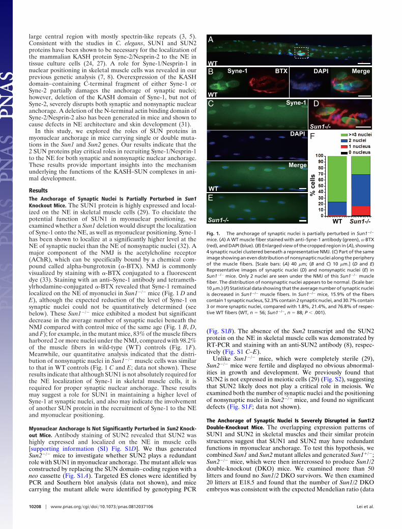
ResultsThe Anchorage of Synaptic Nuclei Is Partially Perturbed in Sun1Knockout Mice. The SUN1 protein is highly expressed and local-ized on the NE in skeletal muscle cells (29). To elucidate thepotential function of SUN1 in myonuclear positioning, weexamined whether a Sun1 deletion would disrupt the localizationof Syne-1 onto the NE, as well as myonuclear positioning. Syne-1has been shown to localize at a significantly higher level at theNE of synaptic nuclei than the NE of nonsynaptic nuclei (32). Amajor component of the NMJ is the acetylcholine receptor(AChR), which can be specifically bound by a chemical com-pound called alpha-bungrotoxin (�-BTX). NMJ is commonlyvisualized by staining with �-BTX conjugated to a fluorescentdye (33). Staining with an anti–Syne-1 antibody and tetrameth-ylrhodamine-conjugated �-BTX revealed that Syne-1 remainedlocalized on the NE of myonuclei in Sun1�/� mice (Fig. 1 D andE), although the expected reduction of the level of Syne-1 onsynaptic nuclei could not be quantitatively determined (seebelow). These Sun1�/� mice exhibited a modest but significantdecrease in the average number of synaptic nuclei beneath theNMJ compared with control mice of the same age (Fig. 1 B, D,and F); for example, in the mutant mice, 83% of the muscle fibersharbored 2 or more nuclei under the NMJ, compared with 98.2%of the muscle fibers in wild-type (WT) controls (Fig. 1F).Meanwhile, our quantitative analysis indicated that the distri-bution of nonsynaptic nuclei in Sun1�/� muscle cells was similarto that in WT controls (Fig. 1 C and E; data not shown). Theseresults indicate that although SUN1 is not absolutely required forthe NE localization of Syne-1 in skeletal muscle cells, it isrequired for proper synaptic nuclear anchorage. These resultsmay suggest a role for SUN1 in maintaining a higher level ofSyne-1 at synaptic nuclei, and also may indicate the involvementof another SUN protein in the recruitment of Syne-1 to the NEand myonuclear positioning.
Myonuclear Anchorage Is Not Significantly Perturbed in Sun2 Knock-out Mice. Antibody staining of SUN2 revealed that SUN2 washighly expressed and localized on the NE in muscle cells[supporting information (SI) Fig. S1D]. We thus generatedSun2�/� mice to investigate whether SUN2 plays a redundantrole with SUN1 in myonuclear anchorage. The mutant allele wasconstructed by replacing the SUN domain–coding region with aneo cassette (Fig. S1 A). Targeted ES clones were identified byPCR and Southern blot analysis (data not shown), and micecarrying the mutant allele were identified by genotyping PCR
(Fig. S1B). The absence of the Sun2 transcript and the SUN2protein on the NE in skeletal muscle cells was demonstrated byRT-PCR and staining with an anti-SUN2 antibody (8), respec-tively (Fig. S1 C–E).
Unlike Sun1�/� mice, which were completely sterile (29),Sun2�/� mice were fertile and displayed no obvious abnormal-ities in growth and development. We previously found thatSUN2 is not expressed in meiotic cells (29) (Fig. S2), suggestingthat SUN2 likely does not play a critical role in meiosis. Weexamined both the number of synaptic nuclei and the positioningof nonsynaptic nuclei in Sun2�/� mice, and found no significantdefects (Fig. S1F; data not shown).
The Anchorage of Synaptic Nuclei Is Severely Disrupted in Sun1/2Double-Knockout Mice. The overlapping expression patterns ofSUN1 and SUN2 in skeletal muscles and their similar proteinstructures suggest that SUN1 and SUN2 may have redundantfunctions in myonuclear anchorage. To test this hypothesis, wecombined Sun1 and Sun2 mutant alleles and generated Sun1�/�;Sun2�/� mice, which were then intercrossed to produce Sun1/2double-knockout (DKO) mice. We examined more than 50litters and found no Sun1/2 DKO survivors. We then examined20 litters at E18.5 and found that the number of Sun1/2 DKOembryos was consistent with the expected Mendelian ratio (data
A
B
C D
E
F
BTX DAPI Merge
% c
ells
Syne-1
DAPI
Merge
Sun1-/-
Sun1-/-
Sun1-/-WT
WT
WT
WT
Syne-1
Fig. 1. The anchorage of synaptic nuclei is partially perturbed in Sun1�/�
mice. (A) A WT muscle fiber stained with anti–Syne-1 antibody (green), �-BTX(red), and DAPI (blue). (B) Enlarged view of the cropped region in (A), showing4 synaptic nuclei clustered beneath a representative NMJ. (C) Part of the sameimage showing an even distribution of nonsynaptic nuclei along the peripheryof the muscle fibers. [Scale bars: (A) 40 �m; (B and C) 10 �m.] (D and E)Representative images of synaptic nuclei (D) and nonsynaptic nuclei (E) inSun1�/� mice. Only 2 nuclei are seen under the NMJ of this Sun1�/� musclefiber. The distribution of nonsynaptic nuclei appears to be normal. (Scale bar:10 �m.) (F) Statistical data showing that the average number of synaptic nucleiis decreased in Sun1�/� muscle fibers. In Sun1�/� mice, 15.9% of the fiberscontain 1 synaptic nucleus, 52.3% contain 2 synaptic nuclei, and 30.7% contain3 or more synaptic nuclei, compared with 1.8%, 21.4%, and 76.8% of respec-tive WT fibers (WT, n � 56; Sun1�/�, n � 88; P � .001).
10208 � www.pnas.org�cgi�doi�10.1073�pnas.0812037106 Lei et al.
not shown). Further analysis found that the Sun1/2 DKO pupsreached full term but died soon after birth (Fig. S3).
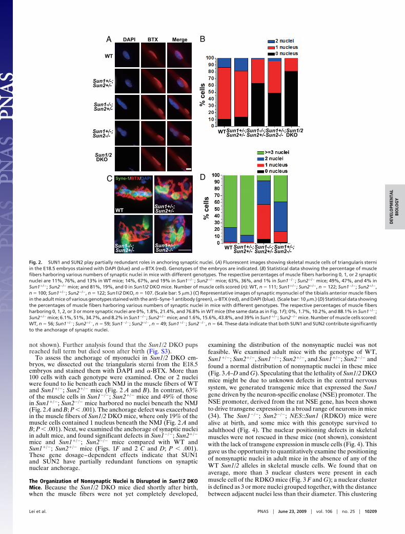
To assess the anchorage of myonuclei in Sun1/2 DKO em-bryos, we dissected out the triangularis sterni from the E18.5embryos and stained them with DAPI and �-BTX. More than100 cells with each genotype were examined. One or 2 nucleiwere found to lie beneath each NMJ in the muscle fibers of WTand Sun1�/�; Sun2�/� mice (Fig. 2 A and B). In contrast, 63%of the muscle cells in Sun1�/�; Sun2�/� mice and 49% of thosein Sun1�/�; Sun2�/� mice harbored no nuclei beneath the NMJ(Fig. 2 A and B; P � .001). The anchorage defect was exacerbatedin the muscle fibers of Sun1/2 DKO mice, where only 19% of themuscle cells contained 1 nucleus beneath the NMJ (Fig. 2 A andB; P � .001). Next, we examined the anchorage of synaptic nucleiin adult mice, and found significant defects in Sun1�/�; Sun2�/�
mice and Sun1�/�; Sun2�/� mice compared with WT andSun1�/�; Sun2�/� mice (Figs. 1F and 2 C and D; P � .001).These gene dosage–dependent effects indicate that SUN1and SUN2 have partially redundant functions on synapticnuclear anchorage.
The Organization of Nonsynaptic Nuclei Is Disrupted in Sun1/2 DKOMice. Because the Sun1/2 DKO mice died shortly after birth,when the muscle fibers were not yet completely developed,
examining the distribution of the nonsynaptic nuclei was notfeasible. We examined adult mice with the genotype of WT,Sun1�/�; Sun2�/�, Sun1�/�; Sun2�/�, and Sun1�/�; Sun2�/� andfound a normal distribution of nonsynaptic nuclei in these mice(Fig. 3 A–D and G). Speculating that the lethality of Sun1/2 DKOmice might be due to unknown defects in the central nervoussystem, we generated transgenic mice that expressed the Sun1gene driven by the neuron-specific enolase (NSE) promoter. TheNSE promoter, derived from the rat NSE gene, has been shownto drive transgene expression in a broad range of neurons in mice(34). The Sun1�/�; Sun2�/�; NES::Sun1 (RDKO) mice werealive at birth, and some mice with this genotype survived toadulthood (Fig. 4). The nuclear positioning defects in skeletalmuscles were not rescued in these mice (not shown), consistentwith the lack of transgene expression in muscle cells (Fig. 4). Thisgave us the opportunity to quantitatively examine the positioningof nonsynaptic nuclei in adult mice in the absence of any of theWT Sun1/2 alleles in skeletal muscle cells. We found that onaverage, more than 3 nuclear clusters were present in eachmuscle cell of the RDKO mice (Fig. 3 F and G); a nuclear clusteris defined as 3 or more nuclei grouped together, with the distancebetween adjacent nuclei less than their diameter. This clustering
% c
ells
WTSun1+/-; Sun2+/-
Sun1-/-; Sun2+/-
Sun1+/-; Sun2-/-
B
D
% c
ells
WT Sun1+/-; Sun2+/-
Sun1-/-; Sun2+/-
Sun1+/-; Sun2-/-
Sun1/2 DKO
WT
Sun1+/-; Sun2+/-
Sun1-/-; Sun2+/-
Sun1+/-; Sun2-/-
Sun1/2 DKO
A
C
WTSun1+/-;Sun2+/-
Sun1-/-;Sun2+/-
Sun1+/-;Sun2-/-
DAPI BTX Merge
Syne-1/BTX/DAPI
Fig. 2. SUN1 and SUN2 play partially redundant roles in anchoring synaptic nuclei. (A) Fluorescent images showing skeletal muscle cells of triangularis sterniin the E18.5 embryos stained with DAPI (blue) and �-BTX (red). Genotypes of the embryos are indicated. (B) Statistical data showing the percentage of musclefibers harboring various numbers of synaptic nuclei in mice with different genotypes. The respective percentages of muscle fibers harboring 0, 1, or 2 synapticnuclei are 11%, 76%, and 13% in WT mice; 14%, 67%, and 19% in Sun1�/�; Sun2�/� mice; 63%, 36%, and 1% in Sun1�/�; Sun2�/� mice; 49%, 47%, and 4% inSun1�/�; Sun2�/� mice; and 81%, 19%, and 0 in Sun1/2 DKO mice. Number of muscle cells scored (n): WT, n � 111; Sun1�/�; Sun2�/�, n � 122; Sun1�/�; Sun2�/�,n � 100; Sun1�/�; Sun2�/�, n � 122; Sun1/2 DKO, n � 107. (Scale bar: 5 �m.) (C) Representative images of synaptic myonuclei of the tibialis anterior muscle fibersin the adult mice of various genotypes stained with the anti–Syne-1 antibody (green), �-BTX (red), and DAPI (blue). (Scale bar: 10 �m.) (D) Statistical data showingthe percentages of muscle fibers harboring various numbers of synaptic nuclei in mice with different genotypes. The respective percentages of muscle fibersharboring 0, 1, 2, or 3 or more synaptic nuclei are 0%, 1.8%, 21.4%, and 76.8% in WT mice (the same data as in Fig. 1F); 0%, 1.7%, 10.2%, and 88.1% in Sun1�/�;Sun2�/� mice; 6.1%, 51%, 34.7%, and 8.2% in Sun1�/�; Sun2�/� mice; and 1.6%, 15.6%, 43.8%, and 39% in Sun1�/�; Sun2�/� mice. Number of muscle cells scored:WT, n � 56; Sun1�/�; Sun2�/�, n � 59; Sun1�/�; Sun2�/�, n � 49; Sun1�/�; Sun2�/�, n � 64. These data indicate that both SUN1 and SUN2 contribute significantlyto the anchorage of synaptic nuclei.
Lei et al. PNAS � June 23, 2009 � vol. 106 � no. 25 � 10209
DEV
ELO
PMEN
TAL
BIO
LOG
Y
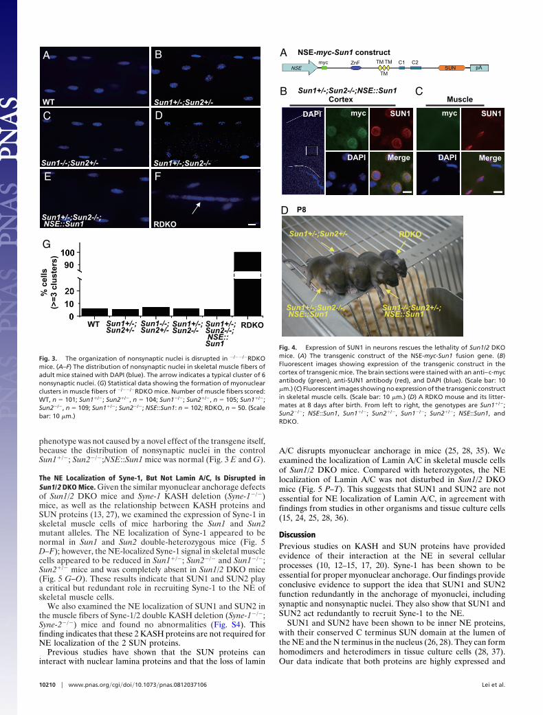
phenotype was not caused by a novel effect of the transgene itself,because the distribution of nonsynaptic nuclei in the controlSun1�/�; Sun2�/�;NSE::Sun1 mice was normal (Fig. 3 E and G).
The NE Localization of Syne-1, But Not Lamin A/C, Is Disrupted inSun1/2 DKO Mice. Given the similar myonuclear anchorage defectsof Sun1/2 DKO mice and Syne-1 KASH deletion (Syne-1�/�)mice, as well as the relationship between KASH proteins andSUN proteins (13, 27), we examined the expression of Syne-1 inskeletal muscle cells of mice harboring the Sun1 and Sun2mutant alleles. The NE localization of Syne-1 appeared to benormal in Sun1 and Sun2 double-heterozygous mice (Fig. 5D–F); however, the NE-localized Syne-1 signal in skeletal musclecells appeared to be reduced in Sun1�/�; Sun2�/� and Sun1�/�;Sun2�/� mice and was completely absent in Sun1/2 DKO mice(Fig. 5 G–O). These results indicate that SUN1 and SUN2 playa critical but redundant role in recruiting Syne-1 to the NE ofskeletal muscle cells.
We also examined the NE localization of SUN1 and SUN2 inthe muscle fibers of Syne-1/2 double KASH deletion (Syne-1�/�;Syne-2�/�) mice and found no abnormalities (Fig. S4). Thisfinding indicates that these 2 KASH proteins are not required forNE localization of the 2 SUN proteins.
Previous studies have shown that the SUN proteins caninteract with nuclear lamina proteins and that the loss of lamin
A/C disrupts myonuclear anchorage in mice (25, 28, 35). Weexamined the localization of Lamin A/C in skeletal muscle cellsof Sun1/2 DKO mice. Compared with heterozygotes, the NElocalization of Lamin A/C was not disturbed in Sun1/2 DKOmice (Fig. 5 P–T). This suggests that SUN1 and SUN2 are notessential for NE localization of Lamin A/C, in agreement withfindings from studies in other organisms and tissue culture cells(15, 24, 25, 28, 36).
DiscussionPrevious studies on KASH and SUN proteins have providedevidence of their interaction at the NE in several cellularprocesses (10, 12–15, 17, 20). Syne-1 has been shown to beessential for proper myonuclear anchorage. Our findings provideconclusive evidence to support the idea that SUN1 and SUN2function redundantly in the anchorage of myonuclei, includingsynaptic and nonsynaptic nuclei. They also show that SUN1 andSUN2 act redundantly to recruit Syne-1 to the NE.
SUN1 and SUN2 have been shown to be inner NE proteins,with their conserved C terminus SUN domain at the lumen ofthe NE and the N terminus in the nucleus (26, 28). They can formhomodimers and heterodimers in tissue culture cells (28, 37).Our data indicate that both proteins are highly expressed and
G
WT Sun1+/-;Sun2+/-
Sun1-/-;Sun2+/- Sun1+/-;Sun2-/-
Sun1+/-;Sun2-/-; NSE::Sun1 RDKO
B
C D
E F
A
10
90
Sun1+/-; Sun2+/-
WT Sun1-/-;Sun2+/-
Sun1+/-; Sun2-/-
Sun1+/-; Sun2-/-; NSE::Sun1
RDKO
% c
ells
(>
=3
clu
ster
s)
Fig. 3. The organization of nonsynaptic nuclei is disrupted in �/��/�RDKOmice. (A–F) The distribution of nonsynaptic nuclei in skeletal muscle fibers ofadult mice stained with DAPI (blue). The arrow indicates a typical cluster of 6nonsynaptic nuclei. (G) Statistical data showing the formation of myonuclearclusters in muscle fibers of �/��/�RDKO mice. Number of muscle fibers scored:WT, n � 101; Sun1�/�; Sun2�/�, n � 104; Sun1�/�; Sun2�/�, n � 105; Sun1�/�;Sun2�/�, n � 109; Sun1�/�; Sun2�/�; NSE::Sun1: n � 102; RDKO, n � 50. (Scalebar: 10 �m.)
A NSE-myc-Sun1 construct
NSEmyc ZnF TM
TM
TM C1 C2SUN pA
DAPI
DAPI DAPI
myc SUN1
Merge
myc SUN1
Merge
BCortex Muscle
CSun1+/-;Sun2-/-;NSE::Sun1
P8D
Sun1+/-;Sun2-/-; NSE::Sun1
Sun1+/-;Sun2+/-
Sun1-/-;Sun2+/-; NSE::Sun1
RDKO
Fig. 4. Expression of SUN1 in neurons rescues the lethality of Sun1/2 DKOmice. (A) The transgenic construct of the NSE-myc-Sun1 fusion gene. (B)Fluorescent images showing expression of the transgenic construct in thecortex of transgenic mice. The brain sections were stained with an anti–c-mycantibody (green), anti-SUN1 antibody (red), and DAPI (blue). (Scale bar: 10�m.) (C) Fluorescent images showing no expression of the transgenic constructin skeletal muscle cells. (Scale bar: 10 �m.) (D) A RDKO mouse and its litter-mates at 8 days after birth. From left to right, the genotypes are Sun1�/�;Sun2�/�; NSE::Sun1, Sun1�/�; Sun2�/�, Sun1�/�; Sun2�/�; NSE::Sun1, andRDKO.
10210 � www.pnas.org�cgi�doi�10.1073�pnas.0812037106 Lei et al.
localized on the NE of muscle cells. These features underlie theredundant roles of SUN1 and SUN2 in skeletal muscle cells.
Investigation of the physiological function of myonuclearanchorage has been an interesting and difficult problem. Previ-ously, it was speculated that synaptic nuclei have specialized rolesin maintaining the high expression levels of postsynaptic com-ponents and thus the proper formation of the NMJ (1, 38);however, our previous study of the Syne-1/Nesprin-1 proteinindicated that neither the formation of the NMJ nor the expres-sion of several synaptic components (synaptophysin, AchR,rapsyn, MuSK, and utrophin) was obviously disrupted (7, 8). Inthe present study, we also examined the expression of rapsyn,synaptophysin, and utrophin in E18.5 skeletal muscles and foundno obvious decrease in these protein levels at the NMJ in theSun1/2 DKO mice (Fig. S5). Moreover, the lack of prominentdevelopmental defects in the Syne-1/Nesprin-1 KASH deletionmice also suggests that myonuclear anchorage is not essential formouse development. We did find disturbed formation of propermuscle–nerve connections due to the altered positions of theNMJ within individual mutant myofibers, however (ref. 8 anddata not shown). The effects of this abnormal myonuclearpositioning on animal development and behavior, as well asother important physiological effects of disrupted myonuclearanchorage, remain to be determined. It is important to note thatSyne-1/Nesprin-1 does play essential roles (although redundantlywith Syne-2/Nesprin-2) in developmental aspects other thanmyonuclear anchorage, as clearly indicated by the dramatic
lethal phenotype of Syne-1 and Syne-2 double KASH deletionmutants (8).
KASH–SUN protein complexes also have been implicated inroles related to muscular dystrophies (9, 39). The degenerativephenotype may be related to the defects in myonuclear anchor-age, including nonsynaptic anchorage. It is interesting that in arecent report, independently generated mice with deletions inthe C-terminal KASH domain exhibited more severe develop-mental and behavior defects than the mice that we generatedpreviously (refs. 8 and 9; L.K. and X.Z., unpublished data). Sucha difference might be linked to the different genetic backgroundsof the mice or to mutations induced in ES cells during thetargeting process (40), possibly suggesting that mice becomehighly dependent on the Syne-1/Nesprin-1 function when certainother cellular functions are compromised. Alternatively, thevariation in phenotypes could be due to the small difference inthe deletion alleles generated in the 2 Syne-1/Nesprin-1 strains.For example, 1 of the alleles might disrupt a KASH domain–independent function. In humans, mutations in Syne-1/Nesprin-1 have been associated with a certain type of recessivecerebellar ataxia (41). Although the precise mechanism of thisdisease is not clear, it is unlikely to be caused primarily by thedefect in nuclear anchorage in muscle cells, based on our analysisof the Syne-1/Nesprin-1 KASH deletion mice (data not shown).
Our observations that the neonatal lethality of Sun1/2 DKOmice was rescued by expressing the Sun1 gene driven by aneuron-specific promoter (RDKO mice) indicates that SUN1and SUN2 likely play important but redundant roles in thedevelopment of the central nervous system. However, theRDKO mice had a smaller body size (Fig. 4) and exhibiteddefects on a neuronal behavior assay (e.g., feet clasping; data notshown), suggesting that the NSE::Sun1 transgene may not haverescued all neuronal defects in the Sun1/2 DKO mice. The rolesof SUN1 and SUN2 in brain development are currently underinvestigation using Sun1/2 DKO as well as conditional KO mice.
Materials and MethodsGeneration of Sun2 Knockout Mice and Sun1/2 DKO Mice. To construct the Sun2gene targeting vector, the 11.5-kb BamH I DNA fragment containing exons5–10 of Sun2 and the 1.4-kb EcoR I fragment containing part of exon 17 wereobtained from a BAC clone of the Sv129 strain (no. 49709; Invitrogen). These2 fragments were cloned into a pPNT vector, flanking the pgk-Neo cassette asthe targeting arms. The resulting targeting vector was linearized and elec-troporated into W4/129S6 ES cells (Taconic Transgenics). After 2 rounds ofselection, the ES clones were screened for successful targeting by PCR analysisusing primers prDX016 and prDX081. The positive ones were further identi-fied by Southern blot analysis using a 778-bp genomic fragment as the probe(PCR-amplified with primers prDX086 and prDX087). The targeted ES cloneswere injected into C57BL/6J blastocysts. The genotype of the targeted Sun2allele in mice was determined by PCR analysis using primers prDX016,prDX081, and prDX115. Sun1/2 DKO mice were generated by intercrossingSun1�/� and Sun2�/� mice. The primer sequences used are listed in SI Materialsand Methods.
Mouse breeding and experimental manipulations were carried out follow-ing the general guidelines published by the Association for Assessment andAccreditation of Laboratory Animal Care. All animal-related procedures werereviewed and approved by the Institute of Developmental Biology and Mo-lecular Medicine Institutional Animal Care and Use Committee.
Immunofluorescent Staining and Microscopic Analysis. Immunofluorescentstaining of frozen sections was carried out following standard protocols (42).Whole-mount staining of skeletal muscle cells was carried out as describedpreviously (7, 8). To isolate individual myofibers from E18.5 embryos, thethoraxes were fixed and triangularis sterni were extracted. After staining withtetramethylrhodamine-conjugated �-BTX (Molecular Probes) and DAPI, indi-vidual fibers were teased out and mounted in the mounting medium (Sigma).At the E18.5 stage, the motor neuron has not yet invaginated into the musclefiber. When triangularis stermi were dissected out to obtain the single musclefiber, a clean separation between the fiber and surrounding cell/nuclei wasusually achieved. Thus, the distraction of nonmuscle nuclei in these experi-ments was minimal and did not present a significant problem for quantitative
S
R
T
DAPISyne-1
Sun1+/-;Sun2-/-
Sun1-/-;Sun2+/-
Sun1+/-;Sun2+/-
WT
Sun1/2 DKO
G
Q
A B CMerge LaminA/C
/DAPI
L
P
H
E
I
D
J K
M N O
F
Fig. 5. The NE localization of Syne-1 but not Lamin A/C is disrupted in Sun1/2DKO mice. (A–O) Representative images of intercostal muscle sections of theE18.5 embryos stained with the anti–Syne-1 antibody (green) and DAPI (blue).Genotypes of the embryos are indicated. Syne-1 displays the NE localizationpattern in muscle cells in WT (A–C), Sun1�/�; Sun2�/� (D–F), Sun1�/�; Sun2�/�
(G–I), and Sun1�/�; Sun2�/� (J–L) mice, but not in the Sun1/2 DKO cells (M–O).(P–T) Representative merged images of intercostal muscle sections of theE18.5 embryos stained with the anti-Lamin A/C antibody (green) and DAPI(blue). Lamin A/C displays NE localization in muscle cells of all genotypes.(Scale bar: 10 �m.)
Lei et al. PNAS � June 23, 2009 � vol. 106 � no. 25 � 10211
DEV
ELO
PMEN
TAL
BIO
LOG
Y
scoring of the mutant phenotype. For scoring the nonsynaptic nuclei clustersin adult mice, we chose only fibers with minimal contamination of nonmusclecells. The reliability of the phenotype scoring is supported by the fact that wedid not detect a single prominent nuclear cluster in the WT fibers that weexamined, and that the clustering phenotype was obvious and highly pene-trant in the RDKO mice. The antibodies used in this study were mouseanti-Lamin A/C (Chemicon), goat anti-mouse IgG-FITC (Chemicon), goat anti-rabbit IgG-FITC (Sigma), mouse anti-rapsyn (Sigma), rabbit anti-synaptophysin(Zymed), and mouse anti-utrophin (Novocastra). The production of antibodiesagainst SUN1, SUN2, and Syne-1 has been described previously (8, 29).
Photographs were taken with a Leica DMRXA2 system. Images were ma-nipulated with Leica FW4000, Adobe Photoshop software, and Adobe Illus-trator software.
ACKNOWLEDGMENTS. We thank Xiaohui Wu, Yanling Yang, and YanfengTan for their help in producing the chimeras and transgenic mice and BeibeiYing, Xiaohui Wu, Kejing Deng, Lin Sun, Wufan Tao, Aileen Sewell, and othermembers of the IDM for their assistance and discussions. This work wassupported by grants from the National Natural Science Foundation of China,the Chinese Ministry of Science and Technology (973 Program) and Ministry ofEducation (211 Program), and the Shanghai Municipal Government.
1. Sanes JR, Lichtman JW (2001) Induction, assembly, maturation and maintenance of apostsynaptic apparatus. Nat Rev Neurosci 2:791–805.
2. Bruusgaard JC, Liestol K, Ekmark M, Kollstad K, Gundersen K (2003) Number and spatialdistribution of nuclei in the muscle fibres of normal mice studied in vivo. J Physiol551(Pt 2):467–478.
3. Starr DA, Fischer JA (2005) KASH ’n karry: The KASH domain family of cargo-specificcytoskeletal adaptor proteins. Bioessays 27:1136–1146.
4. Tzur YB, Wilson KL, Gruenbaum Y (2006) SUN-domain proteins: ‘‘Velcro’’ that links thenucleoskeleton to the cytoskeleton. Nat Rev Mol Cell Biol 7:782–788.
5. Wilhelmsen K, Ketema M, Truong H, Sonnenberg A (2006) KASH-domain proteins innuclear migration, anchorage and other processes. J Cell Sci 119(Pt 24):5021–5029.
6. Starr DA (2009) A nuclear-envelope bridge positions nuclei and moves chromosomes.J Cell Sci 122(Pt 5):577–586.
7. Grady RM, Starr DA, Ackerman GL, Sanes JR, Han M (2005) Syne proteins anchor musclenuclei at the neuromuscular junction. Proc Natl Acad Sci USA 102:4359–4364.
8. Zhang X, et al. (2007) Syne-1 and Syne-2 play crucial roles in myonuclear anchorage andmotor neuron innervation. Development 134:901–908.
9. Puckelwartz MJ, et al. (2009) Disruption of nesprin-1 produces an Emery Dreifussmuscular dystrophy–like phenotype in mice. Hum Mol Genet 18(4):607–620.
10. Malone CJ, Fixsen WD, Horvitz HR, Han M (1999) UNC-84 localizes to the nuclearenvelope and is required for nuclear migration and anchoring during C. elegansdevelopment. Development 126:3171–3181.
11. Worman HJ, Gundersen GG (2006) Here come the SUNs: A nucleocytoskeletal missinglink. Trends Cell Biol 16(2):67–69.
12. Starr DA, et al. (2001) UNC-83 encodes a novel component of the nuclear envelope andis essential for proper nuclear migration. Development 128:5039–5050.
13. Starr DA, Han M (2002) Role of ANC-1 in tethering nuclei to the actin cytoskeleton.Science 298:406–409.
14. McGee MD, Rillo R, Anderson AS, Starr DA (2006) UNC-83 is a KASH protein requiredfor nuclear migration and is recruited to the outer nuclear membrane by a physicalinteraction with the SUN protein UNC-84. Mol Biol Cell 17:1790–1801.
15. Kracklauer MP, Banks SM, Xie X, Wu Y, Fischer JA (2007) Drosophila klaroid encodes aSUN domain protein required for Klarsicht localization to the nuclear envelope andnuclear migration in the eye. Fly (Austin) 1:75–85.
16. Hagan I, Yanagida M (1995) The product of the spindle formation gene sad1�
associates with the fission yeast spindle pole body and is essential for viability. J Cell Biol129:1033–1047.
17. Malone CJ, et al. (2003) The C. elegans hook protein, ZYG-12, mediates the essentialattachment between the centrosome and nucleus. Cell 115:825–836.
18. Fridkin A, et al. (2004) Matefin, a Caenorhabditis elegans germ line–specific SUN-domain nuclear membrane protein, is essential for early embryonic and germ celldevelopment. Proc Natl Acad Sci USA 101:6987–6992.
19. Chikashige Y, et al. (2006) Meiotic proteins bqt1 and bqt2 tether telomeres to form thebouquet arrangement of chromosomes. Cell 125:59–69.
20. Jaspersen SL, et al. (2006) The Sad1–UNC-84 homology domain in Mps3 interacts withMps2 to connect the spindle pole body with the nuclear envelope. J Cell Biol 174:665–675.
21. Penkner A, et al. (2007) The nuclear envelope protein Matefin/SUN-1 is required forhomologous pairing in C. elegans meiosis. Dev Cell 12:873–885.
22. King MC, Drivas TG, Blobel G (2008) A network of nuclear envelope membrane proteinslinking centromeres to microtubules. Cell 134:427–438.
23. Shao X, Tarnasky HA, Lee JP, Oko R, van der Hoorn FA (1999) Spag4, a novel spermprotein, binds outer dense-fiber protein Odf1 and localizes to microtubules ofmanchette and axoneme. Dev Biol 211:109–123.
24. Crisp M, et al. (2006) Coupling of the nucleus and cytoplasm: Role of the LINC complex.J Cell Biol 172:41–53.
25. Hasan S, et al. (2006) Nuclear envelope localization of human UNC84A does not requirenuclear lamins. FEBS Lett 580:1263–1268.
26. Hodzic DM, Yeater DB, Bengtsson L, Otto H, Stahl PD (2004) Sun2 is a novel mammalianinner nuclear membrane protein. J Biol Chem 279:25805–25812.
27. Padmakumar VC, et al. (2005) The inner nuclear membrane protein Sun1 mediates theanchorage of Nesprin-2 to the nuclear envelope. J Cell Sci 118(Pt 15):3419–3430.
28. Haque F, et al. (2006) SUN1 interacts with nuclear lamin A and cytoplasmic nesprins toprovide a physical connection between the nuclear lamina and the cytoskeleton. MolCell Biol 26:3738–3751.
29. Ding X, et al. (2007) SUN1 is required for telomere attachment to nuclear envelope andgametogenesis in mice. Dev Cell 12:863–872.
30. Chi YH, et al. (2009) Requirement for Sun1 in the expression of meiotic reproductivegenes and piRNA. Development 136:965–973.
31. Luke Y, et al. (2008) Nesprin-2 giant (NUANCE) maintains nuclear envelope architec-ture and composition in skin. J Cell Sci 121(Pt 11):1887–1898.
32. Apel ED, Lewis RM, Grady RM, Sanes JR (2000) Syne-1, a dystrophin- and Klarsicht-related protein associated with synaptic nuclei at the neuromuscular junction. J BiolChem 275:31986–31995.
33. Wigston DJ (1990) Repeated in vivo visualization of neuromuscular junctions in adultmouse lateral gastrocnemius. J Neurosci 10:1753–1761.
34. Forss-Petter S, et al. (1990) Transgenic mice expressing beta-galactosidase in matureneurons under neuron-specific enolase promoter control. Neuron 5:187–197.
35. Mejat A, et al. (2009) Lamin A/C–mediated neuromuscular junction defects in Emery-Dreifuss muscular dystrophy. J Cell Biol 184:31–44.
36. Lee KK, et al. (2002) Lamin-dependent localization of UNC-84, a protein required fornuclear migration in Caenorhabditis elegans. Mol Biol Cell 13:892–901.
37. Wang Q, Du X, Cai Z, Greene MI (2006) Characterization of the structures involved inlocalization of the SUN proteins to the nuclear envelope and the centrosome. DNA CellBiol 25:554–562.
38. Schaeffer L, de Kerchove d’Exaerde A, Changeux JP (2001) Targeting transcription tothe neuromuscular synapse. Neuron 31:15–22.
39. Zhang Q, et al. (2007) Nesprin-1 and -2 are involved in the pathogenesis of EmeryDreifuss muscular dystrophy and are critical for nuclear envelope integrity. Hum MolGenet 16:2816–2833.
40. Liang Q, Conte N, Skarnes WC, Bradley A (2008) Extensive genomic copy numbervariation in embryonic stem cells. Proc Natl Acad Sci USA 105:17453–17456.
41. Dupre N, Bouchard JP, Gros-Louis F, Rouleau GA (2007) Mutations in SYNE-1 lead to anewly discovered form of autosomal recessive cerebellar ataxia. Med Sci (Paris) 23:261–262 (in French).
42. Harlow DL, ed (1999) Using Antibodies: A Lab Manual (Cold Spring Harbor Lab Press,Cold Spring Harbor, NY).
10212 � www.pnas.org�cgi�doi�10.1073�pnas.0812037106 Lei et al.
![Benchmarking von Java Cards - LMU · 2005. 3. 7. · Card 2.0 Language Subset Specification [Sun2] und Java Card 2.0 API [Sun1]. Ab 1999 wird Java Card als Standard in GSM genormt](https://img.pdfslide.us/doc/110x75/612a29c8ae0270635f23bcc6/benchmarking-von-java-cards-2005-3-7-card-20-language-subset-specification.jpg)