Embed Size (px)
Citation preview

I. exp. Biol. 148, 449-459 (1990) 4 4 9Primed in Great Britain © The Company of Biologists Limited 1990
BACKBONE MECHANICS OF THE BLUE MARLIN MAKAIRANIGRICANS (PISCES, ISTIOPHORIDAE)
BY JOHN H. HEBRANK1, MARY R. HEBRANK2, JOHN H. LONG, JR2,BARBARA A. BLOCK3 AND STEPHEN A. WAIN WRIGHT2
Pacific Gamefish Research Foundation, 74-425 Kealakehe Pkwy 15,Kailua-Kona, HI 96740, USA, 1 Department of Mechanical and Aerospace
Engineering, PO Box 7910, North Carolina State University, Raleigh, NC 27695,USA, 2Zoology Department, Duke University, Durham, NC 27706, USA and
3 Department of Organismal Biology and Anatomy, University of Chicago,Chicago, IL 60637, USA
Accepted 9 October 1989
Summary
Over most of its length, the backbone of the blue marlin, like that of all otheristiophorids, contains enlarged and flattened neural and hemal spines andzygapophyses, all of which span the intervertebral joints. These plates of bonerestrict dorso-ventral bending of the backbone but their arrangement permits ahigh degree of lateral flexion. The spines and zygapophyses also appear to beimportant in stabilizing the relatively large intervertebral joints against axialcompression and lateral shearing during bending. Although bone is an elasticmaterial, these overlapping structures are not arranged so as to contribute toelastic recoil of the backbone during normal swimming movements.
Introduction
The blue marlin {Makaira nigricans Lace"pede) is among the world's mostspectacular high-performance fishes. It is commonly known as a billfish, anassemblage that also includes the black marlin (Makaira indica Cuvier), thewhite and striped marlins and the spearfishes (Tetrapturus spp.), and the sailfishes(Istiophorus spp.). The blue marlin is one of the largest marine teleosts, attainingmasses of 900 kg. Although marlin have been characterized for years as sprintersand are popular among sport fishermen for their acrobatic leaps out of the water,known as 'tail-walking', it has become apparent only recently that these fish arecapable of remarkable transoceanic migrations. The long-distance travel recordfor marlin is a 9200-km tag return from a 59-kg black marlin (Pacific GamefishResearch Foundation, personal communication). Such performance indicates thatthese fish are capable of prolonged periods of cruising, and contrasts with the viewin the literature of these fish entirely as sprinters. The recent tag returnsdemonstrating long-distance migrations suggest that these fish may be using
Key words: backbone, mechanics, blue marlin, Makaira nigricans.

450 J. H . HEBRANK AND OTHERS
energy-conserving strategies while cruising across the oceans. Telemetry of free-swimming blue marlin off the coast of Kona, Hawaii, indicates these fish havecruising swimming speeds ranging from 1 to Skmh"1 (Yuen et al. 1974).
The large size of the blue marlin and their pelagic mode of life have made itdifficult for biologists to study these fish. The billfish in general have receivedmuch less study than their close relatives and competitors, the tunas (Scombri-dae). Recent evidence has shown that, like the tunas, the billfish have physiologi-cal specializations for protecting the nervous system from temperature fluctu-ations (Carey, 1982; Block, 1986). However, unlike the tunas, the temperature ofthe body musculature of marlin remains close to that of the ambient water. Thehuge mass of axial musculature in marlin is composed primarily of white anaerobicmuscle fibers, which is the main reason they have been characterized as sprinters.
All istiophorids have a backbone morphology that is unique among teleosts.The osteology of the marlin backbone was described by Cuvier and Valenciennes(1831), and then received some attention early this century (Gregory and Conrad,1937). Rockwell et al. (1938) further speculated that the istiophorid backbone mayhave a 'spring-like' role in locomotion. Although several workers have noted itsunusual morphology (Fierstine and Walters, 1968; Nakamura, 1983), the func-tional significance of the istiophorid backbone has yet to be determined, and itsrole as a spring has remained conjectural.
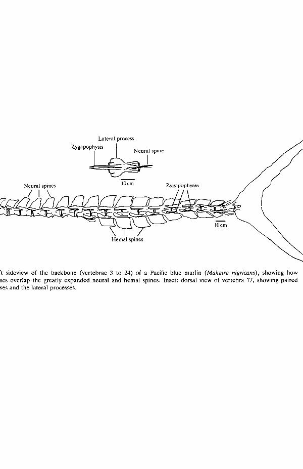
Especially notable is the large quantity of bone in the backbone (Fig. 1). In themarlin the neural spines of precaudal and caudal vertebrae and the hemal spines ofcaudal vertebrae are elaborated into large flat plates that lie in the plane of themedian septum and extend caudad across the intervertebral joints. At the base ofeach neural spine are a pair of laterally compressed zygapophyses that projectforward, flanking the base of the neural spine of the preceding vertebra. Thesezygapophyses are tightly bound to the neural spine by short collagen fibers that runbetween the outer surface of the spine and the inner face of the zygapophysis. Inaddition, in the caudal vertebrae, a much smaller but analogous pair of hemalzygapophyses flanks the base of each preceding hemal spine, again projectingforward across the intervertebral joint. The result of the zygopophyses overlap-ping the spines - across the intervertebral joint - is an interlocking backbone.
The object of this study was to investigate the function of this elaborate bonystructure. In particular we focused attention on the mechanical roles of the neuraland hemal zygapophyses and spines. We executed a variety of bending tests onwhole backbones and segments of backbones to assess the component mechanicalroles, as well as to determine the backbone's ability to contribute to locomotorefficiency through elastic recoil.
Materials and methods
Experimental animals
Vertebral columns of Pacific blue marlin (Makaira nigricans) ranging in sizefrom about 90 to 320 kg were obtained from fish wholesalers in Kailua-Kona
Lat
eral
pro
cess
Neu
ral
spin
es
Fig.
1.
Lef
t si
devi
ew o
f th
e ba
ckbo
ne (
vert
ebra
e 3
to 2
4) o
f a
Paci
fic
blue
mar
lin
(Mak
nira
nig
rica
ns),
sho
win
g ho
w
? z
zyga
poph
yses
ove
rlap
the
gre
atly
exp
ande
d ne
ural
and
hem
al s
pine
s. I
nset
: do
rsal
vie
w o
f ve
rteb
ra 1
7, s
how
ing
pair
ed
3
zyga
poph
yses
and
the
lat
eral
pro
cess
es.

452 J. H . HEBRANK AND OTHERS
Hawaii. The marlin were captured on hook and line by sport fishermen during theHawaiian International Billfish Tournament. Fish were stored for variable lengthsof time up to 40h at 4.4°C before the backbone was removed. Just beforemechanical testing any remaining muscle or viscera were removed from thebackbone, while keeping the underlying connective tissue moist and intact. Thecleaned backbone was bathed in marine teleost saline (Pantin, 1964) before andduring testing.
Backbone stiffness and elastic energy storage tests
The stiffness and storage of elastic energy in whole backbones and in the caudalsegments of backbones flexed laterally were measured by potting the entirebackbone or a segment in polyester resin to secure the most anterior vertebra, andthen loading the backbone or segment as a cantilever (Fig. 2).
Loads applied to the backbones were measured with an Interface load cell(model SSM-50), having a full-scale force of 222 N, repeatability of 0.02%, andhysteresis of 0.03 % of full load. The load cell was energized with 6 V, amplified bya factor of 100 or 1000 as appropriate, and sampled by a 12-bit analog-to-digitalconverter, resulting in a resolution of 0.18 and O.OlSNbit"1, respectively. Datawere sampled and recorded at a rate of 200 samples s"1.
Backbone deflections were measured using a Celesco position transducer(model PT-101). This device has a thin stainless-steel cable that we attached to oneend (the non-backbone end) of the load cell and that exerted a nearly constanttension of 2.22 N. Resolution of this device is rated at 0.008 % of its full-scale valueof 1.83m, and accuracy is rated at 0.10% (1.8mm) of full scale. The device wasexcited by 5 V and sampled by a 12-bit analog-to-digital converter, giving asampling resolution of 0.5 mm.
Force was sampled approximately 100^s before displacement; in the analysisthe measured forces and deflections were therefore considered to occur simul-taneously. All data were recorded on microcomputer disks for later analysis.
In practice, a string attached to the load cell was pulled by hand as smoothly aspossible, so that loading and unloading were each accomplished in about 4 s. The
•pp
111111111 /IT 7 / T i n 1111111111PP —
/ f / /TTTI f I / / / / / / /
Fig. 2. Apparatus and two methods for bending backbones as cantilevers. (A) Methodfor small bends; (B) for extreme bends. BB, backbone; PP, polyester pot that holdsthe backbone; LC, load cell; PT, position transducer.

Backbone mechanics of the blue marlin 453
string was pulled so as to remain parallel with the cable from the displacementtransducer, since work is the dot product of the force and displacement vectors.
In the data analysis, work was calculated as the area under the force-deflectioncurve. The efficiency of elastic energy storage, or resilience, was calculated as thework recovered from an elastic recoil expressed as a percentage of the work put in(Wainwright et al. 1976). Graphically, resilience is the percentage of the areaunder the unloading force-deflection relative to the area under the loadingforce-deflection curve.
Stiffness of individual joints in lateral and dorso-ventral bending
Joint stiffness was measured by clamping a segment of backbone containingeither six caudal or six precaudal vertebrae, and applying a moment to the jointthrough a cantilever load at the free end of the segment. In caudal segments of twofish, the joint between vertebrae 14 and 15 was tested; in a third fish the jointbetween vertebrae 16 and 17 was tested. In precaudal segments of three fish thejoint between vertebrae 4 and 5 was tested in each. Applied moment wascalculated as the product of the applied force, measured by the same load celldescribed above, and the perpendicular distance from the point of application ofthe force to the joint being tested.
The joint angle resulting from the applied moment was measured by glueing orscrewing thin, straight 30 cm rods (bicycle spokes) into the vertebral centra locatedon each side of the joint and measuring with calipers the distances between themfollowing each increment of applied load (Fig. 3). Prior to loading, two marksspaced approximately 20 cm apart were placed on each rod. Joint angle wascalculated as the arcsine of the difference in mark spacing divided by the distancebetween the marks on the rod. A measurement error of lmm corresponded to a0.5° error in joint angle. Repeated measurements indicated actual accuracy near0.1°. During placement of the rods, care was taken to avoid damaging theintervertebral joint capsule or the centra, and to avoid restricting motion of thejoint or its parts through the addition of the measurement rods.
//
•''' A A
B\ \B
t
1/ /" / / / / / 7 7/// / / / 7 / / / / / / / / / / / / / / / / / / I
Fig. 3. Apparatus for measuring deflection of a single intervertebral joint. A,A andB,B are tabs on bicycle spokes; distances AA and BB were measured before and afterbending. F, direction of applied force normal to backbone.

454 J. H . HEBRANK AND OTHERS
To assess the contributions of neural and hemal spines and zygapophyses to jointstiffness, we first measured the stiffness of the intact joint in the lateral and dorsaldirections and then removed the neural zygapophyses (by cutting with a diamondabrasive wheel driven by a flexible shaft drill) and measured the joint stiffness ineach direction again. Joint stiffness in each direction was then measured a thirdtime after removing the neural and hemal spines, and hemal zygapophyses ifpresent. This last pair of measurements thus reflected the behavior of a singleisolated intervertebral joint, with no overlapping bone.
Results
Backbone stiffness and elastic energy storage
Force-deflection curves for a precaudal backbone segment bent in the lateraland ventral directions are shown in Fig. 4. Backbone stiffness is indicated by theslopes of these curves and, as shown here, the backbone segment was found to beabout 10 times stiffer in dorso-ventral bending than in lateral bending. Thisobservation was consistent in three lateral and three dorso-ventral bending tests ofprecaudal segments, and four lateral and three dorsoventral bending tests ofcaudal segments.
For curves such as that shown in Fig. 4, resilience was in the range 60-80 % forboth dorso-ventral and lateral cantilever loading of backbone segments.
In a bending test of an entire backbone taken from a 318-kg marlin, wemeasured a resilience of only 50%. Due to its large size, this backbone was nottested in either cantilever manner described earlier, but was instead mountedvertically and bent laterally into a C-shape by tension applied to the ends of the
0.2Deflection (m)
Fig. 4. Graph of cantilever bending of a precaudal backbone segment from anapproximately 90-kg marlin. Length of the segment was 58 cm (length of the entirebackbone was 141cm); loading was as shown in Fig. 2A. Solid symbols, ventralbending; open symbols, lateral bending.

Backbone mechanics of the blue marlin
10
455
10 oJoint angle (degrees)
10 12
Fig. 5. Graph of bending experiments of intact backbone segments, those withzygapophyses removed, and those with neural and hemal spines removed. (A) Precau-dal joint 4-5 of a 204-kg fish. (B) Caudal joint 14-15 of a 159-kg fish. Solid symbols,dorso-ventral bending; open symbols, lateral bending. Squares, joint intact; triangles,joint without zygapophyses; circles, joint only.
backbone. Subsequent testing resulted in failure, which occurred near the mid-point of the backbone, and for which 34 J of work was required.
Stiffness of individual joints in lateral and dorso-ventral bending
Measurements of joint stiffnesses with and without their surrounding bonesproduced the graphs shown in Fig. 5 (for one 159-kg fish). To compare the effectsof removing joint parts, an average stiffness for each graph was calculated bydividing the maximum applied moment by the deflection angle occurring at thatapplied moment. Fig. 6 shows bar graphs comparing these stiffnesses for allbackbone segments tested. (A logarithmic scale is necessary to show the range ofvalues obtained.) The results may be summarized as follows. (1) Larger animalsare proportionally stiffer in posterior backbone regions, but precaudal stiffnessdoes not vary with size in the sizes tested here. (2) Caudal dorso-ventral stiffnessfor an intact backbone is about 20-30 times lateral stiffness. Precaudal dorso-ventral stiffness is about three times lateral stiffness. (3) Removal of zygapophysesand spines affects lateral stiffness only slightly. (4) Removal of zygapophyses andspines reduces dorso-ventral stiffness to a value approximately the same as lateralstiffness. (5) For caudal segments, removal of the zygapophyses approximatelyhalves dorso-ventral stiffness, suggesting that zygapophyses and spines makesimilar contributions to dorso-ventral stiffness. In precaudal segments, removal ofzygapophyses has only a small effect on dorso-ventral stiffness, suggesting that inthese segments the spines are largely responsible for dorso-ventral stiffness.
The occasional small increase in lateral stiffness observed after removal of partsis probably attributable to the difficulty of realigning an increasingly compliantbackbone following removal of parts.

456 J. H. HEBRANK AND OTHERS
100
10 r
-aEZ
£ l.O
0.1
''•'•''MN ''.uk \
n '
] . \1 '
;
132 204 272Precaudal
90 159 272Caudal
Total body mass (kg)
Fig. 6. Histogram showing flexural stiffnesses of either precaudal or caudal backbonesegments of intact intervertebral joints (/), joints without zygapophyses (N) and jointswith neither zygapophyses nor spines (/). Cross-hatch, dorso-ventral bending; black,lateral bending. Each mass signifies a single fish. (The 272-kg fish appears twice.)
Discussion
These results indicate that the enlarged and flattened neural and hemal spines,along with their overlapping zygapophyses, significantly affect the mechanicalcharacteristics of the marlin backbone. Our observations demonstrate twoimportant roles of these bony structures and eliminate a third possible role.
First, the zygapophyses, spines and connective tissue between them act torestrict bending in the dorso-ventral direction while allowing lateral bending. Thisanisotropy is largely accomplished by the placement of bone and its connectionsabove and below the intervertebral joint. By placing these connections at adistance from the neutral plane of the joint, dorso-ventral flexural stiffness isincreased. This situation is analogous to flexural stiffness in beams, where flexuralstiffness is proportional to the second moment of area (Wainwright et al. 1976). Inthe marlin backbone, the connective tissue of primary importance is locatedbetween the zygapophyses and the spines, and is loaded in shear by lateral anddorso-ventral flexion, and by axial compression. The area resisting this shear isquite large, virtually the entire area of each zygapophysis.
In contrast, lateral bending is allowed by the position of the bones and theirassociated connective tissues near the sagittal plane of the fish, which correspondswith the neutral plane of lateral bending. Both the zygapophyses and the spinesare laterally compressed: for blue marlin weighing 90kg or more, the zygapo-physes range from about 1 mm in thickness at the anterior tip to about 5 mm at theorigin, compared with lengths of about 10cm or more; spines range from about Lto 2mm in thickness and are also about 10cm or more in length. In addition, the?

Backbone mechanics of the blue marlin 457
zygapophyses are recessed into the bases of the neural spines, so that theirpositions close to the neutral axis do not restrict lateral bending. Phrased anotherway, by placing the connections between the zygapophyses and spines close to theneutral plane in lateral bending, large lateral joint angles produce only small shearstrains in the connective tissue between the zygapophyses and spines.
Because of their rigid, high aspect-ratio caudal fins, marlin have been assumedto be thunniform swimmers, i.e. having a propulsive wave of low amplitude andhigh frequency (Nursall, 1956; Lindsey, 1978). However, in an underwater videoof a blue marlin approaching a live bait (Sharkbait Productions, Kailua-Kona,HI), the marlin swam with a sinuous motion, somewhat like that of a shark, withlateral bending occurring throughout the length of the fish. To swim in thismanner, clearly not thunniform, the backbone must have low lateral stiffness.
The second role of the overlapping zygapophyses and spines is in stabilizing theintervertebral joint. The marlin has only 24 vertebrae, hence the degree of angularflexure per joint is probably relatively high due to its sinuous locomotion. (Sharkbackbones, in contrast, have about 100 precaudal vertebrae; trout have about 60.)Large angles between adjacent vertebrae mean that axial compressive loadssupported by the bent backbone [some of the loads were the result of tendonattachments directly on the centra (S. A. Wainwright and S. M. Weeks, personalcommunication)] will have significant components of lateral shear in the interver-tebral joints (Fig. 7).
This lateral shear could potentially cause dislocation, especially since theintervertebral disk appears to be relatively long. We obtained joint length tocentrum length ratios of about 0.28 for precaudal vertebrae and 0.13 for caudalvertebrae in blue marlin over 90 kg. For comparison, in the American eel(Anguilla rostrata), which has over 100 vertebrae, this ratio is 0.17; in the Norfolkspot (Leisostomus xanthurus), having 24 vertebrae, it is 0.13; and in the skipjack
Fig. 7. Diagram of forces applied in the horizontal plane to a vertebra through aposterior oblique tendon (POT) seen in dorsal view. Anterior is to the left.M, direction of muscle pull; C, compression component; 5, shearing component.

458 J. H . HEBRANK AND OTHERS
tuna (Katsuwonus pelamis), having about 40 body vertebrae, it is 0.07 (M. R.Hebrank, unpublished data). Since the intervertebral disks probably allowrelatively large joint angles, the connections between the zygapophyses and thespines appear to act as brackets, allowing bending but not displacement.
The axial compressive forces and their corresponding components of lateralshear acting on the marlin backbone must be quite large. These fish capture theirprey by 'sprinting', and cross-sections through the blue marlin indicate the bodymusculature is primarily composed of white, fast-twitch glycolytic fibers. Scombridfish such as the skipjack tuna are often the preferred prey (Brock, 1984). Theskipjack is another high-performance fish capable of swimming speeds over10 m s"1 (Dizon et al. 1978). Furthermore, the acceleration required to hurl severalhundred kilograms of marlin out of the water requires very high muscular forces,which will compress, shear and bend an intervertebral joint. Stabilizing the jointshould be an important role of the bony processes surrounding the marlinbackbone.
Finally, although bone is an elastic material, these bony structures do not appearto be arranged to contribute to elastic recoil by the backbone. Based onmeasurements of muscle power output of individual fibers and small bundles offibers, Johnston and Salamonski (1984) estimate a marlin mean muscle poweroutput of 37 Wkg"1 body mass during high-speed swimming. For us to bend thebackbone of a 318-kg marlin to breaking, 34 J was required. Assuming a 50%efficiency of elastic energy storage by the backbone and a tail beat frequency of2Hz (based on unpublished data of B. A. Block and F. G. Carey) for fastswimming, this backbone would be capable of contributing 68 W of power. Thisrepresents only 0.6% of the 11772W of muscle power required, as determinedfrom the 37Wkg - 1 figure of Johnston and Salamonski (1984). Similar powerrelationships can be estimated from the data shown in Fig. 4. It is thereforeunlikely that elastic energy storage by the backbone can make any significantcontribution to marlin swimming energetics. Instead, the roles of the elaboratedmarlin backbone appear to be mechanical - restricting dorso-ventral bendingwhile permitting lateral bending, and stabilizing the intervertebral joints againstaxial compression and lateral shearing dislocation during locomotion.
We are grateful to David Grobecker, Scientific Director of the Pacific GamefishResearch Foundation, who provided laboratory space, and, along with thefishermen of the Hawaiian International Billfish Tournament, fresh fish to test. Wealso thank the Suisan and Hawaiian Fish companies for graciously supplying uswith additional cleaned blue marlin backbones. JHL was funded by a grant fromthe Explorers Club. Rosemary Calvert prepared all line drawings.
ReferencesBLOCK, B. A. (1986). Structure of the brain and eye heater tissue in marlins, sailfish, andj
spearfishes. / Morph. 190, 169-189. *

Backbone mechanics of the blue marlin 459
BROCK, R. E. (1984). A contribution to the trophic biology of the blue marlin (Makaira nigricansLacepede, 1802), in Hawaii. Pac. Sci. 38, 141-149.
CAREY, F. G. (1982). A brain heater in the swordfish. Science 216, 1327-1329.CUVIER, G. AND VALENCIENNES, A. (1831). Histoire Naturelle des Poissons, vol. 8, Paris.
(Reprinted by A. Asher, Amsterdam, 1969).DIZON, A. E., BRILL, R. W. AND YUEN, H. S. H. (1978). Correlations between environment,
physiology and activity and the effects on thermoregulation in skipjack tuna. In ThePhysiological Ecology of Tunas (ed. G. D. Sharp and A. E. Dizon), pp. 233-259. New York,London: Academic Press.
FEERSTINE, H. L. AND WALTERS, V. (1968). Studies in locomotion and anatomy of scombroidfishes. Mem. South. Calif. Acad. Sci. 6,1-67.
GREGORY, W. K. AND CONRAD, G. M. (1937). The comparative osteology of the swordfish(Xiphias) and the sailfish (Istiophorus). Am. Mus. Novitates 952, 7-25.
JOHNSTON, I. A. AND SALAMONSKI, J. (1984). Power output and force-velocity relationship ofred and white muscle fibres from the pacific blue marlin (Makaira nigricans). J. exp. Biol. I l l ,171-177.
LINDSEY, C. C. (1978). Form, function, and locomotory habits in fish. In Fish Physiology, vol. 7(ed. W. S. Hoar and D. J. Randall), pp. 1-100. New York: Academic Press.
NAKAMURA, I. (1983). Systematics of the billfishes (Xiphiidae and Istiophoridae). Publ. Setomar. biol. Lab. 28, 255-396.
NURSALL, J. R. (1956). The lateral musculature and the swimming of fish. Proc. zool. Soc,Lond. 126, 127-143.
PANTIN, C. F. A. (1964). Notes on Microscopical Technique for Zoologists. Cambridge:Cambridge University Press.
ROCKWELL, H., EVANS, F. G. AND PHEASANT, H. C. (1938). The comparative morphology of thevertebrate spinal column: Its form as related to function. /. Morph. 63, 87-117.
WAINWRIGHT, S. A., BIGGS, W. D., CURREY, J. D. AND GOSLINE, J. M. (1976). MechanicalDesign in Organisms. London: Edward Arnold.
YUEN, H. S. H., DIZON, A. E. AND UCIMYAMA, J. H. (1974). Notes on the tracking of the Pacificblue marlin, Makaira nigricans. In Proceedings of the International Billfish Symposium (ed. R.Shomura and F. Williams), pp. 265-268. Kailua-Kona, Hawaii: Part 2, NOAA Tech. Rep.NMFS-SSRF675.