Embed Size (px)
Citation preview
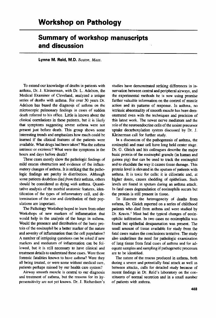
Workshop on Pathology
Summary of workshop manuscripts and discussion
Lynne M. Reid, M.D. Boston, Mass.
To extend our knowledge of deaths in patients with asthma, Dr. J. Kleinerman, with Dr. L. Adelson, the Medical Examiner of Cleveland, analyzed a unique series of deaths with asthma. For over 30 years Dr. Adelson has based the diagnosis of asthma on the microscopic pulmonary findings in cases of sudden death referred to his office. Little is known about the clinical correlations in these patients, but it is likely that symptoms suggesting severe asthma were not present just before death. This group shows some interesting trends and emphasizes how much could be learned if the clinical features of the patients were available. What drugs had been taken? Was the asthma intrinsic or extrinsic? What were the symptoms in the hours and days before death?
These cases mostly show the pathologic findings of mild mucus obstruction and evidence of the inflam- matory changes of asthma. It is striking that the patho- logic findings are patchy in distribution. Although some patients doubtless died from their asthma, others should be considered as dying with asthma. Quanti- tative analysis of the morbid anatomic features, iden- tification of the types of inflammatory cell, and de- termination of the size and distribution of their pop- ulations are important.
The Pathology Workshop hoped to learn from other Workshops of new markers of inflammation that would help in the analysis of the lungs in asthma. Would the presence and distribution of the basic pro- tein of the eosinophil be a better marker of the nature and severity of inflammation than the cell population? A number of intriguing questions can be asked if new markers and mediators of inflammation can be fol- lowed, but it is still necessary to have clinical and treatment details to understand these cases. Were those forensic fatalities known to have asthma? Were they all being treated, or were some without medical care, patients perhaps missed by our health care system?
Airway smooth muscle is central to our diagnosis and treatment of asthma, but the reasons for its hy- persensitivity are not yet known. Dr. J. Richardson’s
studies have demonstrated striking differences in in- nervation between central and peripheral airways, and the experimental methods he is now using promise further valuable information on the control of muscle action and its patterns of response. In asthma, no intrinsic abnormality of smooth muscle has been dem- onstrated even with the techniques and precision of this latest work. The newer nerve mediators and the role of the neuroendocrine cells of the amine precursor uptake decarboxylation system discussed by Dr. J. Kleinerman call for further study.
In a discussion of the pathogenesis of asthma, the eosinophil and mast cell have long held center stage. Dr. G. Gleich and his colleagues describe the major basic protein of the eosinophil granule (in human and guinea pig) that can be used to track the eosinophil and to elucidate the way it causes tissue damage. This protein level is elevated in the sputum of patients with asthma. It is toxic for cells: it is ciliostatic and, at higher doses, causes shedding of epithelium. Such levels are found in sputum during an asthma attack. In fatal cases degranulation of eosinophils occurs but the protein is still present in tissue.
To illustrate the heterogeneity of deaths from asthma, Dr. Gleich reported on a series of childhood patients who died from asthma and were studied by Dr. Kravis.’ Most had the typical changes of eosin- ophilic infiltration. In two cases no eosinophilia was found but epithelial desquamation was present. The small amount of tissue available for study from the fatal cases makes the conclusions tentative. The study also underlines the need for pathologic examination of lung tissue from fatal cases of asthma and for ad- equate samples and sampling if pathogenetic processes are to be identified.
The nature of the mucus produced in asthma, both during a severe and potentially fatal attack as well as between attacks, calls for detailed study because of recent findings in Dr. Reid’s laboratory on the con- stituents of normal secretion and in a small number of patients with asthma.
403
404 Reid J. ALLERGY CLIN. IMMUNOL.
SEPTEMBER 1987
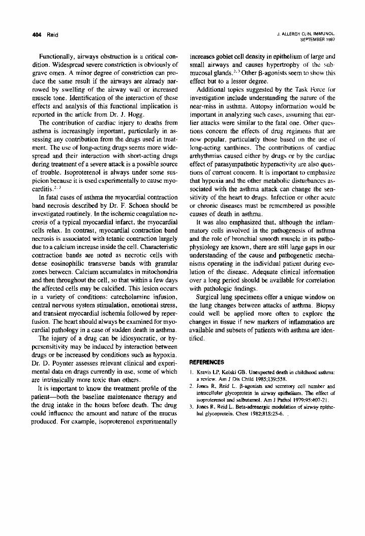
Functionally, airways obstruction is a critical con- dition. Widespread severe constriction is obviously of grave omen. A minor degree of constriction can pro- duce the same result if the airways are already nar- rowed by swelling of the airway wall or increased muscle tone. Identification of the interaction of these effects and analysis of this functional implication is reported in the article from Dr. J. Hogg.
The contribution of cardiac injury to deaths from asthma is increasingly important, particularly in as- sessing any contribution from the drugs used in treat- ment. The use of long-acting drugs seems more wide- spread and their interaction with short-acting drugs during treatment of a severe attack is a possible source of trouble. Isoproterenol is always under some sus- picion because it is used experimentally to cause myo- carditis2. 3
In fatal cases of asthma the myocardial contraction band necrosis described by Dr. F. Schoen should be investigated routinely. In the ischemic coagulation ne- crosis of a typical myocardial infarct. the myocardial cells relax. In contrast, myocardial contraction band necrosis is associated with tetanic contraction largely due to a calcium increase inside the cell. Characteristic contraction bands are noted as necrotic cells with dense eosinophilic transverse bands with granular zones between. Calcium accumulates in mitochondria and then throughout the cell, so that within a few days the affected cells may be calcified. This lesion occurs in a variety of conditions: catecholamine infusion, central nervous system stimulation, emotional stress: and transient myocardial ischemia followed by reper- fusion. The heart should always be examined for myo- cardial pathology in a case of sudden death in asthma.
The injury of a drug can be idiosyncratic, or hy- persensitivity may be induced by interaction between drugs or be increased by conditions such as hypoxia. Dr. D. Poynter assesses relevant clinical and experi- mental data on drugs currently in use, some of which are intrinsically more toxic than others.
It is important to know the treatment profile of the patient-both the baseline maintenance therapy and the drug intake in the hours before death. The drug could influence the amount and nature of the mucus produced. For example, isoproterenol experimentally
increases goblet cell density in epithelium of large and small airways and causes hypertrophy of the sub- mucosal glands.‘. 3 Other p-agonists seem to show this effect but to a lesser degree.
Additional topics suggested by the Task Force l’cu investigation include understanding the nature of the near-miss in asthma. Autopsy information would be important in analyzing such cases, assuming that ear- lier attacks were similar to the fatal one. Other qucs- tions concern the effects of drug regimens that are now popular, particularly those based on the use ot long-acting xanthines. The contributions of cardiac arrhythmias caused either by drugs or by the cardiac effect of parasympathetic hyperactivity are also ques- tions of current concern. It is important to emphasize that hypoxia and the other metabolic disturbances as- sociated with the asthma attack can change the sen- sitivity of the heart to drugs. Infection or other acute or chronic diseases must be remembered as possible causes of death in asthma.
It was also emphasized that, although the inflam- matory cells involved in the pathogenesis of asthma and the role of bronchial smooth muscle in its patho- physiology are known, there are still large gaps in our understanding of the cause and pathogenetic mecha- nisms operating in the individual patient during evo- lution of the disease. Adequate clinical information over a long period should be available for correlation with pathologic findings.
Surgical lung specimens offer a unique window on the lung changes between attacks of asthma. Biopsy could well be applied more often to explore the changes in tissue if new markers of inflammation are available and subsets of patients with asthma are iden- tified.
REFERENCES
I. Kravis LP, Kolski GB. Unexpected death in childhood asthma: a review. Am J Dis Child 1985;139:558.
2. Jones R, Reid L. P-agonists and secretory cell number and intracellular glycoprotein in airway epithehelium. The effect of isoproterenol and salbutamol. Am J Path01 1979:95:407-21.
3. Jones R, Reid L. Beta-adrenergic modulation of airway epithe- lial glycoprotein. Chest 1982:813:25-6.