Embed Size (px)
Citation preview
SUMMARY OF ASSAY PROCEDURE
Pipette 25 µl standard, control or sample
Pipette 200 µl Assay buffer
Incubate 30 min at RT
Wash 4x (300 µl)
Pipette 100 µl Anti-Ferritin HRP
Incubate 30 min. at RT
Wash 4x (300 µl)
Pipette 100 µl TMB
Incubate 15 min. at RT
Pipette 100 µl Stop Solution
Read at 450/630 nm
VII. Calculation of results
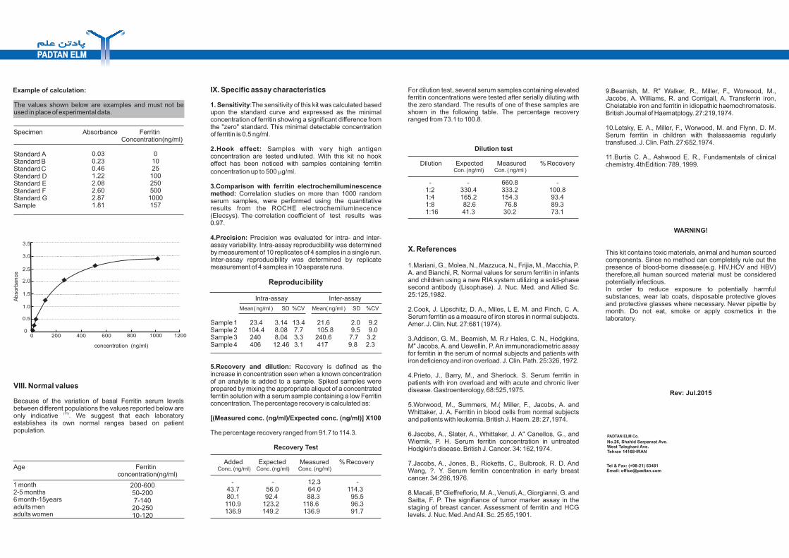
The results can be calculated by either microtiter plate, spectrophotometer reader or manual evaluation. If a microtiter plate spectrophotometer reader with data calculation program is used, refer to the plate reader and create a program using the concentration of each of the Ferritin standards (in ng/ml).For manual evaluation, a standard curve is constructed by plotting the absorbance (A) values obtained for each Ferritin standard against the corresponding Ferritin concentrations (in ng/ml).The unknown Ferritin concentration, in ng/ml, can then be read from the standard curve using the absorbance value of each patient specimen.
THE PADTAN ELM EIA KIT FOR 96 TESTS
Ferritin EIA KitIntended use:Quantitative determination of Ferritin levels in human serum and plasma.
For in vitro diagnostic use.
I.Introduction
Ferritin is the major iron storage protein found in nature. This iron-containing protein, with a molecular weight of 450 kDa, may contain as many as 4,000 iron atoms. Normally approximately 1% of the plasma iron is contained in Ferritin. Ferritin levels are high at birth but fall during the first few months and remain low in childhood, generally rising again
(1)after puberty . In normal adults Ferritin levels are higher in (2)men than in women . The plasma Ferritin is in equilibrium
with body stores, and variations in the quantity of iron in the storage compartment are reflected in plasma Ferritin
(3)concentration . Thus, the levels of serum Ferritin are determined to evaluate iron stores in normal patients, patients with iron deficiency and iron overload. Measurements of serum iron concentration and total iron binding capacity (TIBC) have been widely used as aids in the diagnosis of iron deficiency. However, assay of serum Ferritin concentration is a much more sensitive and reliable means for demonstration of this disorder. On the other hand, a large number of hemolytic or chronic diseases result in increased serum Ferritin concentration. These diseases include Thalassemias, chronic infections, chronic
(4)inflammatory disorders, acute or chronic liver disease and (5)numerous malignancies, especially acute leukemia ,
(6) (7-8)Hodgkin's disease and breast cancer . In cases of iron overload (e. g. hemochromatosis and Thalassemia) the determination of Ferritin concentration is used to monitor the
(9-10)response to therapy .
II.Principle of the test
The Ferritin quantitative test kit is based on solid phase enzyme immunoassay (EIA). This assay system uses two mouse monoclonal antibodies directed against distinct antigenic determinants on the Ferritin molecule. The polystyrene wells are coated with captured mouse monoclonal antibodies against Ferritin. Standards, controls and patient samples are added to the wells (solide phase) and incubate. The Ferritin present in the wells is bound to the anti Ferritin antibodies. The unbound material is removed by aspiration and washing. After washing, the HRP labeled anti-Ferritin Mab is added to the wells. After second incubation and washing, a solution of TMB (3,3',5,5' tetra-methylbenzidine) is added to each well, resulting in the development of a blue color. The intensity of the color is proportional to the amount of Ferritin present in the sample. The color development is stopped by addition of Stop solution, causing the blue color to change to
yellow. The color intensity is determined in a microtiter plate spectrophotometer at 450 nm. Standard curves are constructed for each assay by plotting absorbance value against the concentration of each standard. The Ferritin concentrations of patient samples are then read from the standard curve.
III. Kit contents
The reagents provided with the Ferritin Kits(Cat.No.P-FRI) are sufficient for 96 wells. The expiry date of each reagent is shown on the vial label.
Store the Kit at 2-8° C.
1.Coated microtiter wells: 96wells,coated with mouse monoclonal anti Ferritin antibodies.
2.Zero standard: 1 vial (4 ml). The zero standard should also be used as sample diluent.
3.Standards: 6 vials (1.0 ml) of human Ferritin in serum matrix with thimerosal as a preservative. The exact Ferritin concentration for each standard is specified on the label of each vial.The standards supplied with the kit were calibrated against the international standard . IRP 80/602.
4.Serum controls: 2 vials (1.0 ml) of human Ferritin in processed human serum containing thimerosal. The nominal Ferritin concentrations in the controls are specified on the label of each vial.
5.Enzyme tracer: 1 vial (12 ml) of monoclonal anti-Ferritin antibody conjugated to horseradish peroxidase (HRP) in phosphate buffered saline (PBS) with proteins and thimerosal.
6.Assay buffer: 1 vial (25 ml) of PBS with proteins and thimerosal.
7.Wash solution (concentrate 20X): 1 vial (25 ml) of PBS- Tween 20 and thimerosal.
8.TMB HRP-Substrate: 1 vial (12 ml) of buffered H 0 and 2 2
3,3',5,5' tetra methylbenzidine.
9.Stop solution: 1 vial (12ml) of 2N H S0 .2 4
IV.Materials required (but not supplied with the kit)
1.Microtiter plate spectrophotometer reader with a wavelength of 450 nm(with reference wavelength at 630 nm) and an absorbance range of 0 to 3.0.
2.Precision micropipettes with disposable plastic tips to deliver 25- 200 µl.
3.Distilled or deionized water for preparation of diluted Wash Solution.
V. Specimen collection and preparation
The assay can be performed on serum or heparinized plasma samples. Keep samples at 2-8°C for 1-2 weeks; for longer periods it is recommended to store the sample in aliquot form at -20°C. Avoid repeated freezing and thawing of samples. Prior to assay, frozen specimens should be slowly brought to room temperature and gently mixed by hand. Do not vortex patient samples.Serum sample with Ferritin greater than the last standards should be diluted with "zero" standard and reassayed to give a quantitative result. The value obtained must be multiplied by the dilution factor to give the correct Ferritin concentration.
VI. Assay procedures
All reagents should be brought to room temperature prior to use.
Concentrated wash solution must be diluted with distilled or deionizer water ( Dilute one part concentrated wash solution with 19 parts of water). Diluted wash solution is stable for 7 days at 2 to 8°C. In presence of undissolved crystals, resuspend the solution by placing the vial at 37° C for a few minutes.
1. Dispense 25 µl of Ferritin standards, control serums and patient samples into appropriate wells. Pipette 200 µl of Assay Buffer to each well. Thoroughly mix the plate for 15 seconds.
2. Incubate the strips for 30 minutes at room temperature.
3.Aspirate and wash each well 4 times with 300 µl of diluted Wash solution.
4. Add 100 µl of HRP anti-Ferritin conjugate to each well. Then incubate for 30 minutes at room temperature.
5. Aspirate and wash each well 4 times with 300 µl of diluted Wash solution.
6. Dispense 100 µl of TMB HRP-Substrate into each well.
7. Incubate at room temperature in the dark for 15 minutes.
8. Add 100 µl of Stop Solution and mix for 10 seconds. Read absorbance at 450 nm (with reference wavelength at 630 nm) in a microtiter plate reader within 15 minutes after addition of Stop solution.
For dilution test, several serum samples containing elevated ferritin concentrations were tested after serially diluting with the zero standard. The results of one of these samples are shown in the following table. The percentage recovery ranged from 73.1 to 100.8.
Dilution test
Dilution Expected Measured % Recovery Con. (ng/ml) Con. ( ng/ml )
- - 660.8 - 1:2 330.4 333.2 100.8 1:4 165.2 154.3 93.4 1:8 82.6 76.8 89.3 1:16 41.3 30.2 73.1
X. References
1.Mariani, G., Molea, N., Mazzuca, N., Frijia, M., Macchia, P. A. and Bianchi, R. Normal values for serum ferritin in infants and children using a new RIA system utilizing a solid-phase second antibody (Lisophase). J. Nuc. Med. and Allied Sc. 25:125,1982.
2.Cook, J. Lipschitz, D. A., Miles, L E. M. and Finch, C. A. Serum ferritin as a measure of iron stores in normal subjects. Amer. J. Clin. Nut. 27:681 (1974).
3.Addison, G. M., Beamish, M. R.r Hales, C. N., Hodgkins, M" Jacobs, A. and Uewellin, P. An immunoradiometric assay for ferritin in the serum of normal subjects and patients with iron deficiency and iron overload. J. Clin. Path. 25:326, 1972.
4.Prieto, J., Barry, M., and Sherlock. S. Serum ferritin in patients with iron overload and with acute and chronic liver disease. Gastroenterology, 68:525,1975.
5.Worwood, M., Summers, M.( Miller, F., Jacobs, A. and Whittaker, J. A. Ferritin in blood cells from normal subjects and patients with leukemia. British J. Haem. 28: 27,1974.
6.Jacobs, A., Slater, A., Whittaker, J. A" Canellos, G., and Wiernik, P. H. Serum ferritin concentration in untreated Hodgkin's disease. British J. Cancer. 34: 162,1974.
7.Jacobs, A., Jones, B., Ricketts, C., Bulbrook, R. D. And Wang, ?. Y. Serum ferritin concentration in early breast cancer. 34:286,1976.
8.Macali, B" Gieffreflorio, M. A., Venuti, A., Giorgianni, G. and Saitta, F. P. The signifiance of tumor marker assay in the staging of breast cancer. Assessment of ferritin and HCG levels. J. Nuc. Med. And All. Sc. 25:65,1901.
IX. Specific assay characteristics
1. Sensitivity:The sensitivity of this kit was calculated based upon the standard curve and expressed as the minimal concentration of ferritin showing a significant difference from the "zero" standard. This minimal detectable concentration of ferritin is 0.5 ng/ml.
2.Hook effect: Samples with very high antigen concentration are tested undiluted. With this kit no hook effect has been noticed with samples containing ferritin concentration up to 500 mg/ml.
3.Comparison with ferritin electrochemiluminescence method: Correlation studies on more than 1000 random serum samples, were performed using the quantitative results from the ROCHE electrochemiluminecence (Elecsys). The correlation coefficient of test results was 0.97.
4.Precision: Precision was evaluated for intra- and inter-assay variability. Intra-assay reproducibility was determined by measurement of 10 replicates of 4 samples in a single run. Inter-assay reproducibility was determined by replicate measurement of 4 samples in 10 separate runs.
Reproducibility
Intra-assay Inter-assay
Mean( ng/ml ) SD %CV Mean( ng/ml ) SD %CV
Sample 1 23.4 3.14 13.4 21.6 2.0 9.2Sample 2 104.4 8.08 7.7 105.8 9.5 9.0Sample 3 240 8.04 3.3 240.6 7.7 3.2Sample 4 406 12.46 3.1 417 9.8 2.3
5.Recovery and dilution: Recovery is defined as the increase in concentration seen when a known concentration of an analyte is added to a sample. Spiked samples were prepared by mixing the appropriate aliquot of a concentrated ferritin solution with a serum sample containing a low Ferritin concentration. The percentage recovery is calculated as:
[(Measured conc. (ng/ml)/Expected conc. (ng/ml)] X100
The percentage recovery ranged from 91.7 to 114.3.
Recovery Test
Added Expected Measured % Recovery Conc. (ng/ml) Conc. (ng/ml) Conc. (ng/ml)
- - 12.3 - 43.7 56.0 64.0 114.3 80.1 92.4 88.3 95.5 110.9 123.2 118.6 96.3 136.9 149.2 136.9 91.7
9.Beamish, M. R" Walker, R., Miller, F., Worwood, M., Jacobs, A. Williams, R. and Corrigall, A. Transferrin iron, Chelatable iron and ferritin in idiopathic haemochromatosis. British Journal of Haematplogy. 27:219,1974.
10.Letsky, E. A., Miller, F., Worwood, M. and Flynn, D. M. Serum ferritin in children with thalassaemia regularly transfused. J. Clin. Path. 27:652,1974.
11.Burtis C. A., Ashwood E. R., Fundamentals of clinical chemistry. 4thEdition: 789, 1999.
WARNING!
This kit contains toxic materials, animal and human sourced components. Since no method can completely rule out the presence of blood-borne disease(e.g. HIV,HCV and HBV) therefore,all human sourced material must be considered potentially infectious.In order to reduce exposure to potentially harmful substances, wear lab coats, disposable protective gloves and protective glasses where necessary. Never pipette by month. Do not eat, smoke or apply cosmetics in the laboratory.
Rev: Jul.2015
Specimen Absorbance Ferritin Concentration(ng/ml)
Standard A Standard B Standard C Standard D Standard E Standard F Standard G Sample
Example of calculation:
VIII. Normal values
Because of the variation of basal Ferritin serum levels between different populations the values reported below are
(11)only indicative . We suggest that each laboratory establishes its own normal ranges based on patient population.
Age Ferritin concentration(ng/ml)
1 month 2-5 months 6 month-15years adults men adults women
No.26, Shahid Sarparast Ave.West Taleghani Ave.Tehran 14168-IRAN
Tel & Fax: (+98-21) 63481Email: [email protected]
PADTAN ELM Co.
0 200 400 600 800 10000
0.5
1.5
1.0
2.0
2.5
3.0
concentration (ng/ml)
Abso
rbance
1200
3.5
0.030.230.461.222.082.602.871.81
01025
100250500
1000157
200-60050-2007-140
20-25010-120
The values shown below are examples and must not be used in place of experimental data.