Embed Size (px)
Citation preview
NANO EXPRESS
Sulfonated Styrene-(ethylene-co-butylene)-styrene/Montmorillonite Clay Nanocomposites: Synthesis, Morphology,and Properties
Anirban Ganguly Æ Anil K. Bhowmick
Received: 20 August 2007 / Accepted: 28 November 2007 / Published online: 18 December 2007
� to the authors 2007
Abstract Sulfonated styrene-(ethylene-butylene)-styrene
triblock copolymer (SSEBS) was synthesized by reaction
of acetyl sulfate with SEBS. SSESB-clay nanocomposites
were then prepared from hydrophilic Na-montmorillonite
(MT) and organically (quaternary amine) modified hydro-
phobic nanoclay (OMT) at very low loading. SEBS did not
show improvement in properties with MT-based nano-
composites. On sulfonation (3 and 6 weight%) of SEBS,
hydrophilic MT clay-based nanocomposites exhibited
better mechanical, dynamic mechanical, and thermal
properties, and also controlled water–methanol mixture
uptake and permeation and AC resistance. Microstructure
determined by X-ray diffraction, atomic force microscopy,
and transmission electron microscopy due to better dis-
persion of MT nanoclay particles and interaction of MT
with SSEBS matrix was responsible for this effect. The
resulting nanocomposites have potential as proton transfer
membranes for Fuel Cell applications.
Key words SEBS � Sulfonation � Montmorillonite clay �Nanocomposites
Introduction
In recent years, there has been considerable interest in
special composite materials that consist of a matrix, usually
a polymer, filled with plate-like or flake-like inorganic
fillers having at least one dimension in nanometer length
scale and high aspect ratio. Such fillers can be extremely
effective in modifying the properties of polymers. Several
orders of change in mechanical, transport, rheological,
electrical, or thermal properties have been demonstrated in
these composites containing only a few volume percent of
nano-filler [1–9].
SEBS, (styrene-ethylene-butylene-styrene) triblock co-
polymer, chosen as the base material in this work, is exten-
sively used as a thermoplastic elastomer [10]. This is a
nonpolar polymer and not compatible with a polar substance.
Hence, polar modification of SEBS has gained recent
attention. However, till now most researchers have concen-
trated on the maleation of SEBS with maleic anhydride in
organic solution or in the melt or graft copolymerization of
SEBS with methacrylic acid in organic solution. Though
sulfonation of SEBS has been reported in literature [11–17],
no report is available on the montmorillonite clay (MT)-
based nanocomposites of sulfonated SEBS. The authors have
reported earlier preparation and properties of SEBS–MT-
based nanocomposites [18, 19] and numerous other rubber–
clay nanocomposites from this laboratory [20–25]. A few
reports on polymer composites acting as proton conducting
membranes [26, 27] and a few on block copolymer–clay
nanocomposites [28–33] are available. Sulfonated SEBS-
montmorillonite clay-based nanocomposite as a strong
member for controlling the proton transfer is a novel
approach in this new field of renewable energy source in a
world of crisis of energy.
This is a new approach as all the earlier studies on
SEBS-MT clay nanocomposites have concentrated on
intercalating the clay after organically modifying it by
long-chain amines. Here, in this present work, unmodified
clay (MT) has been successfully intercalated and exfoliated
by sulfonated SEBS systems. The work reported here is
concerned with the synthesis of sulfonated SEBS, and
preparation and characteristics of the unmodified
A. Ganguly � A. K. Bhowmick (&)
Rubber Technology Centre, Indian Institute of Technology,
Kharagpur, Kharagpur 721302, India
e-mail: [email protected]
123
Nanoscale Res Lett (2008) 3:36–44
DOI 10.1007/s11671-007-9111-3
montmorillonite clay (MT)-based nanocomposites. Low-
cost montmorillonite clay can be used in place of organi-
cally modified nanoclays in making nanocomposites with
sulfonated SEBS.
Experimental
Starting Materials
(styrene-ethylene-butylene-styrene) triblock copolymer
(SEBS) with molecular weight Mn = 50,000 and styrene/
ethylene-butylene (w/w) = 30/70 was supplied by Shell
Chemical Co, USA. Acetic anhydride (Analytical grade)
was procured from Aldrich, Milwaukee, WI. 1,2-dichloro
ethane (DCE), sulfuric acid (assay content, [99%),
methanol, and tetra-hydrofuran (THF, analytical grade)
were obtained from Merck Ltd., Mumbai, India. Unmodi-
fied sodium montmorillonite clay (MT, having cation
exchange capacity = 92.6 meq./100 gm with 2:1 tetrahe-
dral:octahedral layer structure) and long-chain quaternary
ammonium ion-modified nanoclay (OMT, Cloisite�20A)
were generously supplied by Southern Clay Products,
Gonzales, TX, USA. Double deionized water was prepared
in this laboratory.
Sulfonation Reaction
Sulfonation was carried out onto SEBS backbone in an
analogous method to that described by Weiss et al. [11]
Acetyl sulfate was synthesized at temperature near to
-20 �C as per Scheme 1 in dry oxygen free N2
atmosphere.
A solution of SEBS (10% w/v in DCE) was prepared in
a three necked round bottom flask equipped with condenser
and the solution was heated to 60 �C and stirred for 4 h for
full solubilization of SEBS. O2 free dry N2 gas was passed
through the polymer solution in order to drive out the
dissolved oxygen present in the solvent and also in the
reaction flask. The required amount of freshly prepared
acetyl sulfate was then added drop-wise to the reaction
mixture. The reaction mixture (Scheme 2) was maintained
at 60 �C under stirring in nitrogen atmosphere. After 2 h of
optimized reaction time at this condition, the reaction was
stopped by gradually adding an excess of isopropanol for
10 min and cooling to room temperature. Finally, the sul-
fonated SEBS was isolated, steam stripped in excess of
double de-ionized (dd) boiling water, followed by washing
several times with boiling and cold dd water (to eliminate
the solvent, free acids, and hydrolyze the acetyl sulfate).
The product was filtered and dried under vacuum at 70 �Cup to a constant weight and was stored in a desiccator to
avoid moisture.
Sulfonated SEBS (SSEBS) was dissolved in a THF/
methanol mixture (9/1 v/v) and the homogeneous solution
was left under stirring for 2 h after which the solvents were
evaporated under reduced pressure (about 1 mmHg) at
50 �C for 7 days.
Measurement of Percentage Sulfonation
The dried sulfonated SEBS samples were weighted (Wg)
and the extent of grafting was calculated from the weight
gain by the samples using the following equation:
% Grafting ¼ Wg � W0
W0
� �� 100 ð1Þ
where, W0 = weight of neat SEBS and Wg = weight of
the sulfonic acid-grafted SEBS. Infrared Spectroscopy
(Perkin Elmer FTIR–spectrophotometer) and elemental
analysis (CHNSO Analyzer, Perkin Elmer) were also per-
formed to quantify the graft percentage. Both the results
revealed *3 and *6 wt.% of sulfonation onto SEBS
backbone.
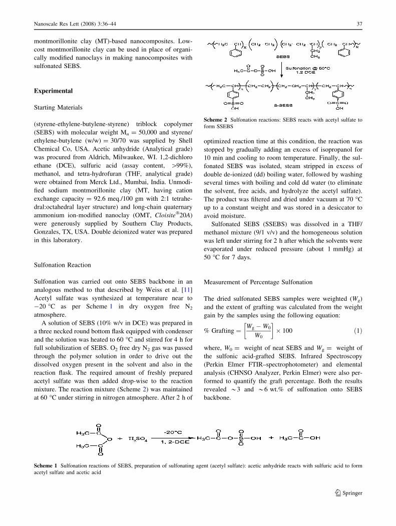
Scheme 1 Sulfonation reactions of SEBS, preparation of sulfonating agent (acetyl sulfate): acetic anhydride reacts with sulfuric acid to form
acetyl sulfate and acetic acid
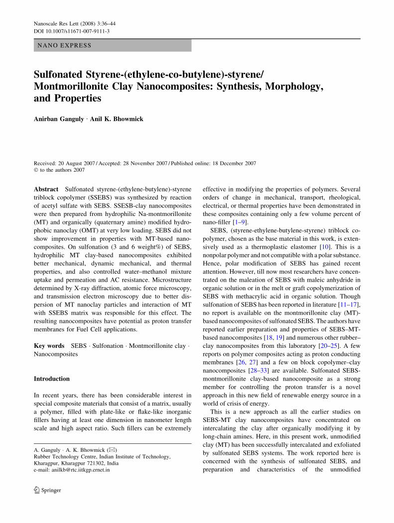
Scheme 2 Sulfonation reactions: SEBS reacts with acetyl sulfate to
form SSEBS
Nanoscale Res Lett (2008) 3:36–44 37
123
SSEBS Clay Hybrid Nanocomposite Film Preparation
S3SEBS (with 3 wt.% sulfonation to SEBS) and S6SEBS
(with 6 wt.% sulfonation to SEBS)/ MT4 and OMT4 clay
nanocomposites (with 4 wt.% of clay) were prepared using
a THF solvent-casting method. Initially, SSEBS was
dissolved in THF overnight and MT or OMT clay at opti-
mized 4 wt.% were suspended in THF for 6 h and stirred for
2 h using a magnetic stirrer. The polymer solution and clay
particle suspension were then mixed together at 25 �C and
stirred for 1 day in order to complete the mixing. Next, the
samples were dried in a hood by evaporating the solvent to
get a film thick in the range of 50–60 lm.
Fourier Transform Infrared (FT-IR) Spectroscopic
Studies
FT-IRstudies were carried out in dispersive mode on thin
film samples using Perkin Elmer FTIR–spectrophotometer
(model Spectrum RXI,UK), within a range of 400–
4,400 cm-1 using a resolution of 4 cm-1. An average of 32
scans have been reported for each sample.
Microstructure by Wide Angle X-ray Diffraction
(WAXD)
Wide angle X-ray diffraction analysis of the nanocom-
posites was carried out in a PANalytical XPert Pro (3040/
60 the Netherlands) X-ray diffractometer (operated at
30 kV and 40 mA) at room temperature, equipped with
Cu–Ka radiation.
The scanning rate was 1�/min and the range of Goni-
ometer angle (2h) was from 2�–10�. Subsequently, the
d-spacing of the clay layers was calculated using the
Bragg’s equation,
nk ¼ 2d Sinh ð2Þ
where k = wavelength of the X-ray with Cu–Ka tar-
get = 0.154 nm, d = interplanar distance of the clay
platelets, h = angle of the incident radiation.
Morphological Investigation
Transmission Electron Microscopy (TEM)
The samples for transmission electron microscopy analysis
were prepared by ultra cryo-microtomy using a Leica Ul-
tracut UCT (Wien, Austria). Freshly sharpened glass knives
with cutting edge of 45� were used to get the cryosections of
50–70 nm thickness at a sub-ambient temperature of
-80 �C using a JEOL 2010, Japan TEM, operating at an
accelerating voltage of 200 kV. Selective staining of aro-
matic moieties in the samples was done with vapor of OsO4.
Phase Imaging by Atomic Force Microscopy (AFM)
The effects of sulfonation on SEBS and of inclusion of
inorganic silicate clay layers on the morphology of SEBS
and its nanocomposite were investigated by using atomic
force microscopy (MultiMode AFMTM from Digital
Instruments, Santa Barbara, CA, USA) in air at ambient
conditions (25 �C, 60% RH) in the tapping mode using
etched silicon probe tips (TESP), with a spring constant in
the range of 40 N/m. For each sample, minimum three
images were analyzed.
Dynamic Mechanical Thermal Analysis (DMTA)
The dynamic mechanical spectra of the samples were
obtained by using Rheometric Scientific DMTA IV, NJ,
USA analyzed in tension-compression mode at a constant
frequency of 1 Hz, a strain of 0.01%, and a temperature
range from -100 to 130 �C at a heating rate of 2 �C/min.
The temperature corresponding to the peak in tand versus
temperature plot was taken as the glass–rubber transition
temperature (Tg).
Studies of Mechanical Properties
Tensile properties were measured on dumb-bell specimens
at room temperature using a ZWICK Z010 tensile test
machine (Zwick Inc., Ulm, Germany). The gauge length
and cross-head speed were 25 mm and 500 mm/min,
respectively. At least five samples were tested and the
average was used.
Thermogravimetric Analysis (TGA)
The thermal degradation analysis of SEBS, grafted SEBS,
and their nanocomposites was performed with TGA Q50 of
TA Instruments- Waters LLC, USA operated at a heating
rate of 20 �C/min in N2 atmosphere at a flow rate of
60 mL/min in the temperature range of 25–700 �C.
Water–Methanol Uptake and Permeability
The films of sulfonated SEBS and their MT-based
nanocomposites were soaked in deionized water–Methanol
(80–20) mix for 3 weeks to determine the uptake content
by the following Eq. 3:
38 Nanoscale Res Lett (2008) 3:36–44
123
Uptake content =ðwwet � wdryÞ
wdry
� 100 ð3Þ
where wwet = weight of wet samples after blotting the
surface water–MeOH and wdry = weight of dry sample
before wetting. Permeability of water–MeOH (80–20) mix
into free air through the films of SSEBS and MT4-based
nanocomposites was measured by diffusion process with an
airtight glass diffusion cell.
AC Resistance and Proton Conductivity
AC electrical resistance was measured at room temperature
for film samples in the transverse direction with a two
probe INSTEK LCR meter (LCR 819, Taiwan) operating
in AC frequency range from 0.4 to 10 kHz. The proton
conductivity was measured in an indirect process for the
water–methanol-immersed samples after wiping out the
surface water and measuring the resistance employing the
same set up.
Results and Discussion
Sulfonation of SEBS
Scheme 2 portrays the synthetic route to graft -SO3H ions
onto SEBS backbone. Elemental analysis by CHNSO
analyzer reveals 3 and 6 wt.% of sulfonation in the SEBS
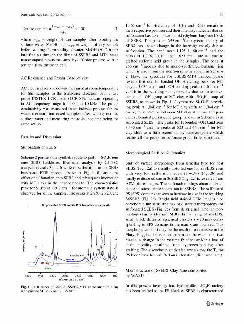
backbone. FTIR spectra, shown in Fig. 1, illustrate the
effect of sulfonation onto SEBS and subsequent interaction
with MT clays in the nanocomposite. The characteristics
peak for SEBS at 1,602 cm-1 for aromatic system stays is
observed for all the samples. The peaks at 2,850, 2,920, and
1,465 cm-1 for stretching of –CH3 and –CH2 remain in
their respective position and their intensity indicates that no
sulfonation has taken place in mid ethylene–butylene block
of SEBS. The peak at 699 cm-1for styrenic moiety of
SEBS has shown change in the intensity mostly due to
sulfonation. The band near 1,125–1,160 cm-1 and the
peaks at 1,376, 1,010, and 1,035 cm-1 are all due to
grafted sulfonic acid group in the samples. The peak at
756 cm-1 appears due to mono-substituted benzene ring
which is clear from the reaction scheme shown in Scheme
2. Now, the spectrum for SSEBS-MT4 nanocomposite
reveals that non-H- bonded OH stretching peak for MT
clay at 3,634 cm-1 and –OH bending peak at 1,641 cm-1
vanish in the resulting nanocomposite due to ionic inter-
action of –OH group of MT clay with –SO3H group of
SSEBS, as shown in Fig. 1. Asymmetric Si–O–Si stretch-
ing peak at 1,040 cm-1 for MT clay shifts to 1,044 cm-1
owing to interaction between MT clay structure and pen-
dant sulfonated polystyrene group (shown in Scheme 2) in
sulfonated SEBS . The peaks for H bonded –OH band near
3,430 cm-1 and the peaks at 523 and 466 cm-1 for MT
clay shift to a little extent in the nanocomposite which
retains all the peaks for sulfonate group in its spectrum.
Morphological Shift on Sulfonation
Shift of surface morphology from lamellar type for neat
SEBS (Fig. 2a) to slightly distorted one for S3SEBS even
with very low sulfonation levels (3 wt.%) (Fig. 2b) and
finally to distorted one in S6SEBS (Fig. 2c) is revealed from
AFM phase images. The sulfonation brings about a distur-
bance in micro-phase separation in SSEBS. The sulfonated
PS (SPS) domains are seen to increase in size in the resulting
S6SEBS (Fig. 2c). Bright field-stained TEM images also
corroborate the same findings of distorted morphology for
sulfonated SEBS (Fig. 2e) from its original lamellar mor-
phology (Fig. 2d) for neat SEBS. In the image of S6SEBS,
small black distorted spherical clusters (*20 nm) corre-
sponding to SPS domains in the matrix are obtained. This
morphological shift may be the result of an increase in the
Flory–Huggins interaction parameter between the two
blocks, a change in the volume fraction, and/or a loss of
chain mobility resulting from hydrogen-bonding after
grafting. The viscoelastic study also reveals that the Tg for
PS block have been shifted on sulfonation (discussed later).
Microstructure of SSEBS–Clay Nanocomposites
by WAXD
In this present investigation, hydrophilic –SO3H moiety
has been grafted to the PS block of SEBS as characterizedFig. 1 FTIR traces of SSEBS, SSEBS-MT4 nanocomposite along
with pristine MT clay and SEBS film
Nanoscale Res Lett (2008) 3:36–44 39
123
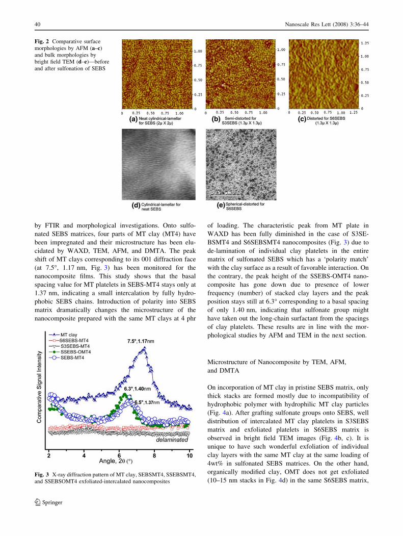
by FTIR and morphological investigations. Onto sulfo-
nated SEBS matrices, four parts of MT clay (MT4) have
been impregnated and their microstructure has been elu-
cidated by WAXD, TEM, AFM, and DMTA. The peak
shift of MT clays corresponding to its 001 diffraction face
(at 7.5�, 1.17 nm, Fig. 3) has been monitored for the
nanocomposite films. This study shows that the basal
spacing value for MT platelets in SEBS-MT4 stays only at
1.37 nm, indicating a small intercalation by fully hydro-
phobic SEBS chains. Introduction of polarity into SEBS
matrix dramatically changes the microstructure of the
nanocomposite prepared with the same MT clays at 4 phr
of loading. The characteristic peak from MT plate in
WAXD has been fully diminished in the case of S3SE-
BSMT4 and S6SEBSMT4 nanocomposites (Fig. 3) due to
de-lamination of individual clay platelets in the entire
matrix of sulfonated SEBS which has a ‘polarity match’
with the clay surface as a result of favorable interaction. On
the contrary, the peak height of the SSEBS-OMT4 nano-
composite has gone down due to presence of lower
frequency (number) of stacked clay layers and the peak
position stays still at 6.3� corresponding to a basal spacing
of only 1.40 nm, indicating that sulfonate group might
have taken out the long-chain surfactant from the spacings
of clay platelets. These results are in line with the mor-
phological studies by AFM and TEM in the next section.
Microstructure of Nanocomposite by TEM, AFM,
and DMTA
On incorporation of MT clay in pristine SEBS matrix, only
thick stacks are formed mostly due to incompatibility of
hydrophobic polymer with hydrophilic MT clay particles
(Fig. 4a). After grafting sulfonate groups onto SEBS, well
distribution of intercalated MT clay platelets in S3SEBS
matrix and exfoliated platelets in S6SEBS matrix is
observed in bright field TEM images (Fig. 4b, c). It is
unique to have such wonderful exfoliation of individual
clay layers with the same MT clay at the same loading of
4wt% in sulfonated SEBS matrices. On the other hand,
organically modified clay, OMT does not get exfoliated
(10–15 nm stacks in Fig. 4d) in the same S6SEBS matrix,
Fig. 2 Comparative surface
morphologies by AFM (a–c)
and bulk morphologies by
bright field TEM (d–e)—before
and after sulfonation of SEBS
Fig. 3 X-ray diffraction pattern of MT clay, SEBSMT4, SSEBSMT4,
and SSEBSOMT4 exfoliated-intercalated nanocomposites
40 Nanoscale Res Lett (2008) 3:36–44
123
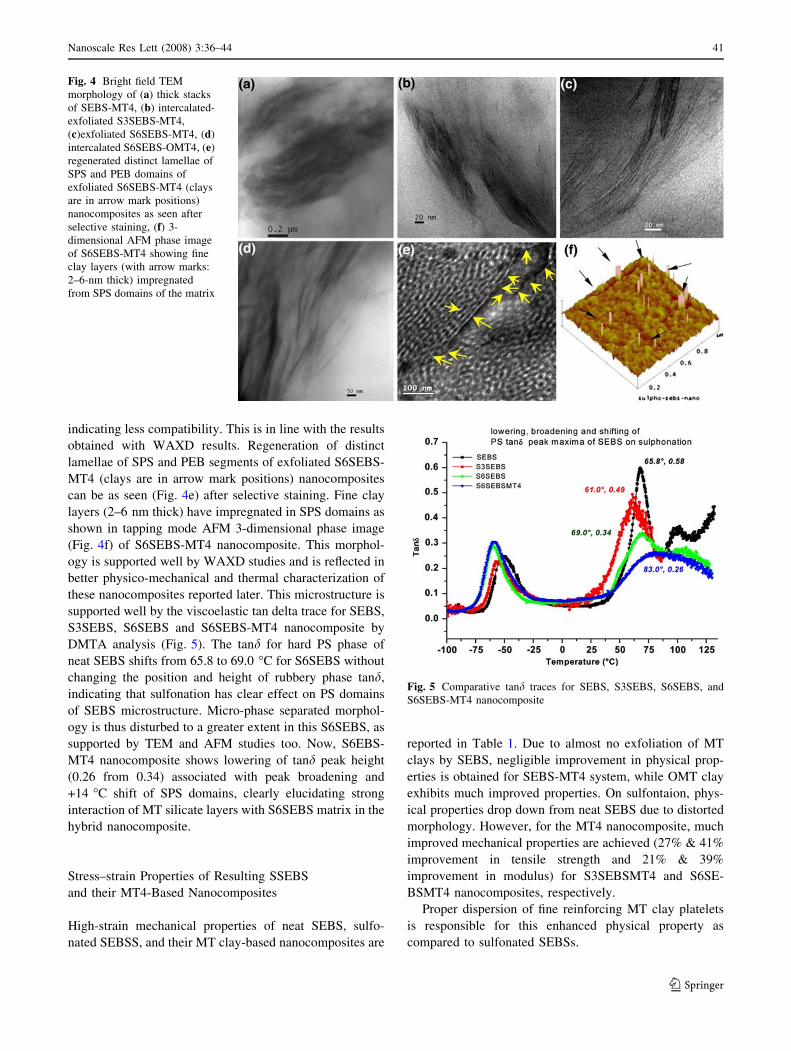
indicating less compatibility. This is in line with the results
obtained with WAXD results. Regeneration of distinct
lamellae of SPS and PEB segments of exfoliated S6SEBS-
MT4 (clays are in arrow mark positions) nanocomposites
can be as seen (Fig. 4e) after selective staining. Fine clay
layers (2–6 nm thick) have impregnated in SPS domains as
shown in tapping mode AFM 3-dimensional phase image
(Fig. 4f) of S6SEBS-MT4 nanocomposite. This morphol-
ogy is supported well by WAXD studies and is reflected in
better physico-mechanical and thermal characterization of
these nanocomposites reported later. This microstructure is
supported well by the viscoelastic tan delta trace for SEBS,
S3SEBS, S6SEBS and S6SEBS-MT4 nanocomposite by
DMTA analysis (Fig. 5). The tand for hard PS phase of
neat SEBS shifts from 65.8 to 69.0 �C for S6SEBS without
changing the position and height of rubbery phase tand,
indicating that sulfonation has clear effect on PS domains
of SEBS microstructure. Micro-phase separated morphol-
ogy is thus disturbed to a greater extent in this S6SEBS, as
supported by TEM and AFM studies too. Now, S6EBS-
MT4 nanocomposite shows lowering of tand peak height
(0.26 from 0.34) associated with peak broadening and
+14 �C shift of SPS domains, clearly elucidating strong
interaction of MT silicate layers with S6SEBS matrix in the
hybrid nanocomposite.
Stress–strain Properties of Resulting SSEBS
and their MT4-Based Nanocomposites
High-strain mechanical properties of neat SEBS, sulfo-
nated SEBSS, and their MT clay-based nanocomposites are
reported in Table 1. Due to almost no exfoliation of MT
clays by SEBS, negligible improvement in physical prop-
erties is obtained for SEBS-MT4 system, while OMT clay
exhibits much improved properties. On sulfontaion, phys-
ical properties drop down from neat SEBS due to distorted
morphology. However, for the MT4 nanocomposite, much
improved mechanical properties are achieved (27% & 41%
improvement in tensile strength and 21% & 39%
improvement in modulus) for S3SEBSMT4 and S6SE-
BSMT4 nanocomposites, respectively.
Proper dispersion of fine reinforcing MT clay platelets
is responsible for this enhanced physical property as
compared to sulfonated SEBSs.
Fig. 4 Bright field TEM
morphology of (a) thick stacks
of SEBS-MT4, (b) intercalated-
exfoliated S3SEBS-MT4,
(c)exfoliated S6SEBS-MT4, (d)
intercalated S6SEBS-OMT4, (e)
regenerated distinct lamellae of
SPS and PEB domains of
exfoliated S6SEBS-MT4 (clays
are in arrow mark positions)
nanocomposites as seen after
selective staining, (f) 3-
dimensional AFM phase image
of S6SEBS-MT4 showing fine
clay layers (with arrow marks:
2–6-nm thick) impregnated
from SPS domains of the matrix
Fig. 5 Comparative tand traces for SEBS, S3SEBS, S6SEBS, and
S6SEBS-MT4 nanocomposite
Nanoscale Res Lett (2008) 3:36–44 41
123
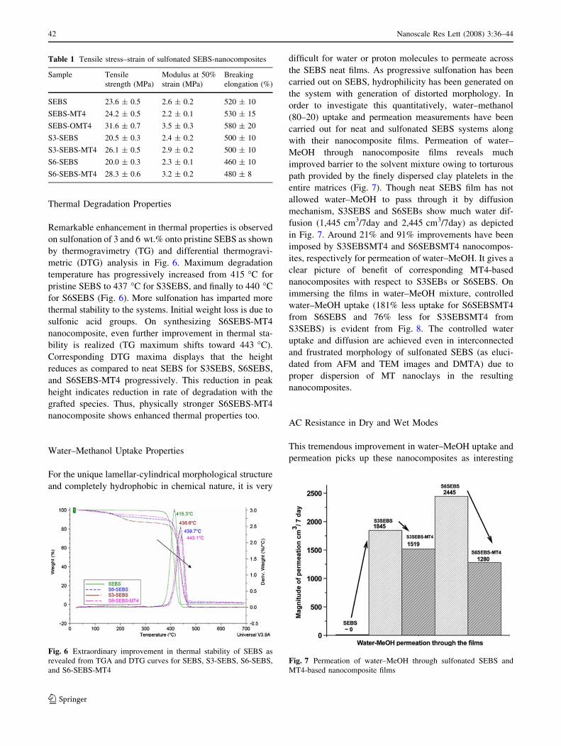
Thermal Degradation Properties
Remarkable enhancement in thermal properties is observed
on sulfonation of 3 and 6 wt.% onto pristine SEBS as shown
by thermogravimetry (TG) and differential thermogravi-
metric (DTG) analysis in Fig. 6. Maximum degradation
temperature has progressively increased from 415 �C for
pristine SEBS to 437 �C for S3SEBS, and finally to 440 �Cfor S6SEBS (Fig. 6). More sulfonation has imparted more
thermal stability to the systems. Initial weight loss is due to
sulfonic acid groups. On synthesizing S6SEBS-MT4
nanocomposite, even further improvement in thermal sta-
bility is realized (TG maximum shifts toward 443 �C).
Corresponding DTG maxima displays that the height
reduces as compared to neat SEBS for S3SEBS, S6SEBS,
and S6SEBS-MT4 progressively. This reduction in peak
height indicates reduction in rate of degradation with the
grafted species. Thus, physically stronger S6SEBS-MT4
nanocomposite shows enhanced thermal properties too.
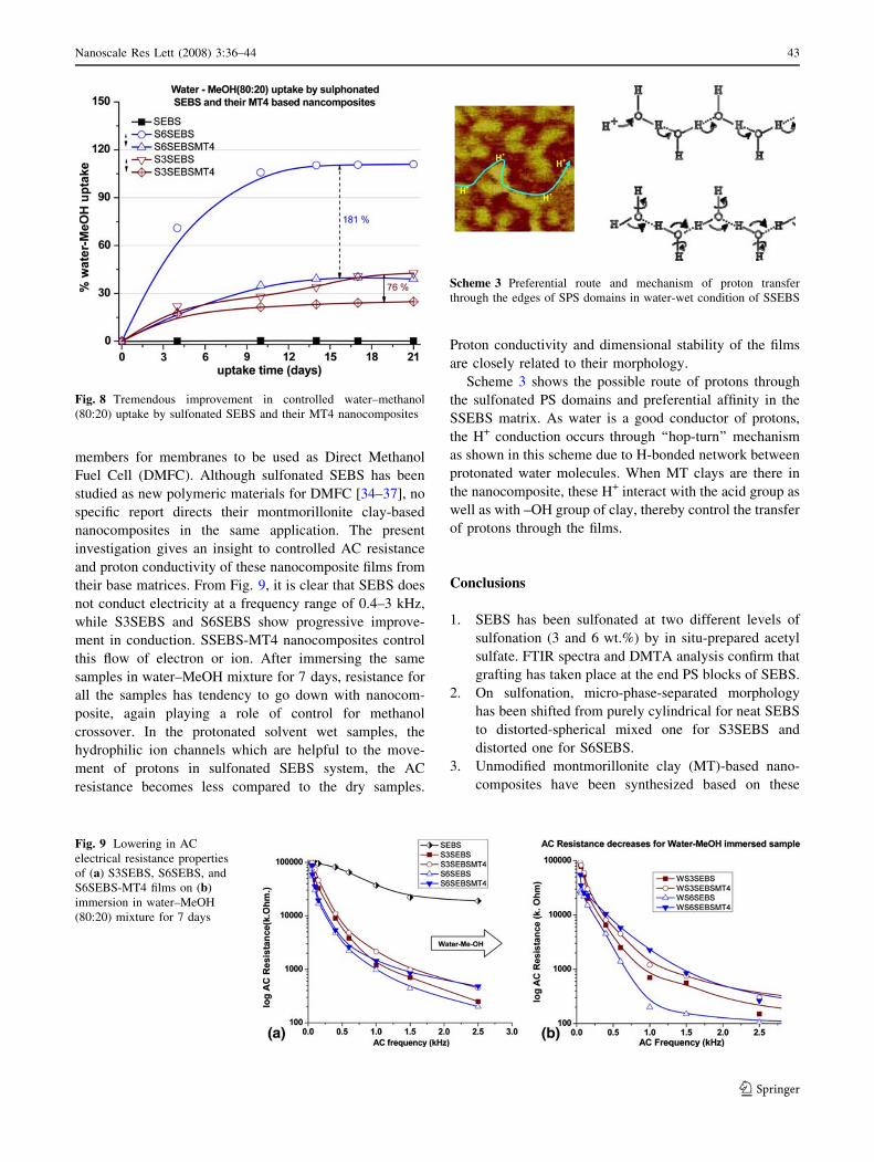
Water–Methanol Uptake Properties
For the unique lamellar-cylindrical morphological structure
and completely hydrophobic in chemical nature, it is very
difficult for water or proton molecules to permeate across
the SEBS neat films. As progressive sulfonation has been
carried out on SEBS, hydrophilicity has been generated on
the system with generation of distorted morphology. In
order to investigate this quantitatively, water–methanol
(80–20) uptake and permeation measurements have been
carried out for neat and sulfonated SEBS systems along
with their nanocomposite films. Permeation of water–
MeOH through nanocomposite films reveals much
improved barrier to the solvent mixture owing to torturous
path provided by the finely dispersed clay platelets in the
entire matrices (Fig. 7). Though neat SEBS film has not
allowed water–MeOH to pass through it by diffusion
mechanism, S3SEBS and S6SEBs show much water dif-
fusion (1,445 cm3/7day and 2,445 cm3/7day) as depicted
in Fig. 7. Around 21% and 91% improvements have been
imposed by S3SEBSMT4 and S6SEBSMT4 nanocompos-
ites, respectively for permeation of water–MeOH. It gives a
clear picture of benefit of corresponding MT4-based
nanocomposites with respect to S3SEBs or S6SEBS. On
immersing the films in water–MeOH mixture, controlled
water–MeOH uptake (181% less uptake for S6SEBSMT4
from S6SEBS and 76% less for S3SEBSMT4 from
S3SEBS) is evident from Fig. 8. The controlled water
uptake and diffusion are achieved even in interconnected
and frustrated morphology of sulfonated SEBS (as eluci-
dated from AFM and TEM images and DMTA) due to
proper dispersion of MT nanoclays in the resulting
nanocomposites.
AC Resistance in Dry and Wet Modes
This tremendous improvement in water–MeOH uptake and
permeation picks up these nanocomposites as interesting
Table 1 Tensile stress–strain of sulfonated SEBS-nanocomposites
Sample Tensile
strength (MPa)
Modulus at 50%
strain (MPa)
Breaking
elongation (%)
SEBS 23.6 ± 0.5 2.6 ± 0.2 520 ± 10
SEBS-MT4 24.2 ± 0.5 2.2 ± 0.1 530 ± 15
SEBS-OMT4 31.6 ± 0.7 3.5 ± 0.3 580 ± 20
S3-SEBS 20.5 ± 0.3 2.4 ± 0.2 500 ± 10
S3-SEBS-MT4 26.1 ± 0.5 2.9 ± 0.2 500 ± 10
S6-SEBS 20.0 ± 0.3 2.3 ± 0.1 460 ± 10
S6-SEBS-MT4 28.3 ± 0.6 3.2 ± 0.2 480 ± 8
Fig. 6 Extraordinary improvement in thermal stability of SEBS as
revealed from TGA and DTG curves for SEBS, S3-SEBS, S6-SEBS,
and S6-SEBS-MT4
Fig. 7 Permeation of water–MeOH through sulfonated SEBS and
MT4-based nanocomposite films
42 Nanoscale Res Lett (2008) 3:36–44
123
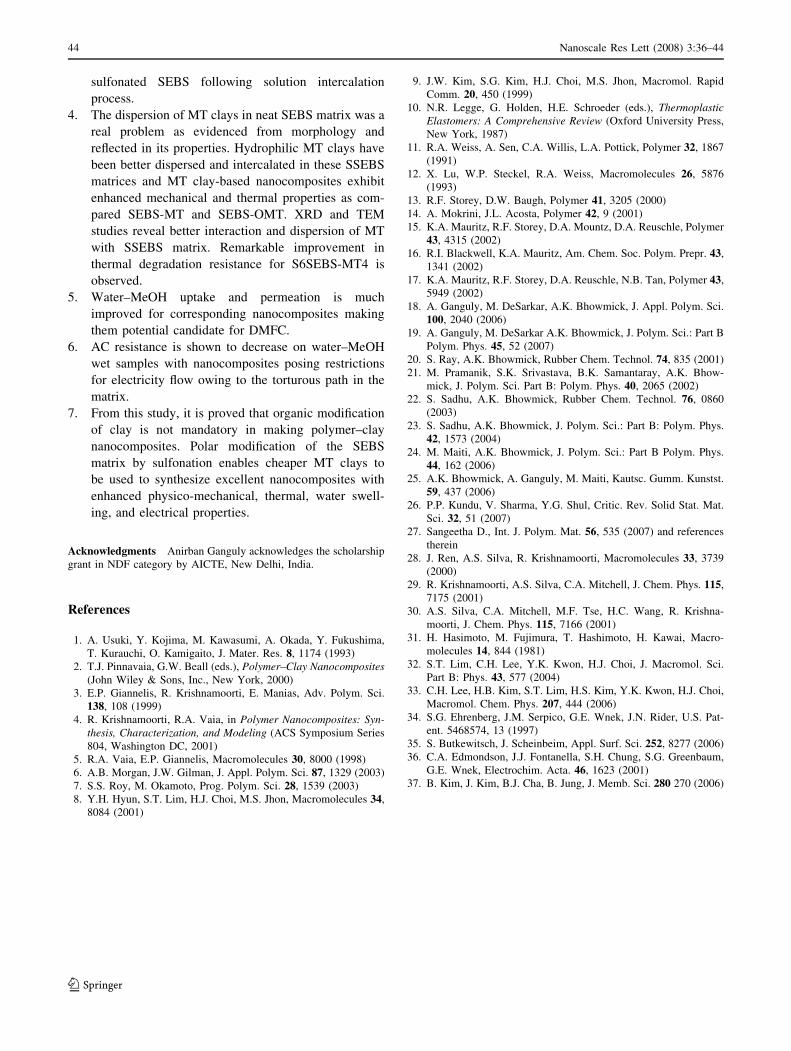
members for membranes to be used as Direct Methanol
Fuel Cell (DMFC). Although sulfonated SEBS has been
studied as new polymeric materials for DMFC [34–37], no
specific report directs their montmorillonite clay-based
nanocomposites in the same application. The present
investigation gives an insight to controlled AC resistance
and proton conductivity of these nanocomposite films from
their base matrices. From Fig. 9, it is clear that SEBS does
not conduct electricity at a frequency range of 0.4–3 kHz,
while S3SEBS and S6SEBS show progressive improve-
ment in conduction. SSEBS-MT4 nanocomposites control
this flow of electron or ion. After immersing the same
samples in water–MeOH mixture for 7 days, resistance for
all the samples has tendency to go down with nanocom-
posite, again playing a role of control for methanol
crossover. In the protonated solvent wet samples, the
hydrophilic ion channels which are helpful to the move-
ment of protons in sulfonated SEBS system, the AC
resistance becomes less compared to the dry samples.
Proton conductivity and dimensional stability of the films
are closely related to their morphology.
Scheme 3 shows the possible route of protons through
the sulfonated PS domains and preferential affinity in the
SSEBS matrix. As water is a good conductor of protons,
the H+ conduction occurs through ‘‘hop-turn’’ mechanism
as shown in this scheme due to H-bonded network between
protonated water molecules. When MT clays are there in
the nanocomposite, these H+ interact with the acid group as
well as with –OH group of clay, thereby control the transfer
of protons through the films.
Conclusions
1. SEBS has been sulfonated at two different levels of
sulfonation (3 and 6 wt.%) by in situ-prepared acetyl
sulfate. FTIR spectra and DMTA analysis confirm that
grafting has taken place at the end PS blocks of SEBS.
2. On sulfonation, micro-phase-separated morphology
has been shifted from purely cylindrical for neat SEBS
to distorted-spherical mixed one for S3SEBS and
distorted one for S6SEBS.
3. Unmodified montmorillonite clay (MT)-based nano-
composites have been synthesized based on these
Fig. 8 Tremendous improvement in controlled water–methanol
(80:20) uptake by sulfonated SEBS and their MT4 nanocomposites
Fig. 9 Lowering in AC
electrical resistance properties
of (a) S3SEBS, S6SEBS, and
S6SEBS-MT4 films on (b)
immersion in water–MeOH
(80:20) mixture for 7 days
Scheme 3 Preferential route and mechanism of proton transfer
through the edges of SPS domains in water-wet condition of SSEBS
Nanoscale Res Lett (2008) 3:36–44 43
123
sulfonated SEBS following solution intercalation
process.
4. The dispersion of MT clays in neat SEBS matrix was a
real problem as evidenced from morphology and
reflected in its properties. Hydrophilic MT clays have
been better dispersed and intercalated in these SSEBS
matrices and MT clay-based nanocomposites exhibit
enhanced mechanical and thermal properties as com-
pared SEBS-MT and SEBS-OMT. XRD and TEM
studies reveal better interaction and dispersion of MT
with SSEBS matrix. Remarkable improvement in
thermal degradation resistance for S6SEBS-MT4 is
observed.
5. Water–MeOH uptake and permeation is much
improved for corresponding nanocomposites making
them potential candidate for DMFC.
6. AC resistance is shown to decrease on water–MeOH
wet samples with nanocomposites posing restrictions
for electricity flow owing to the torturous path in the
matrix.
7. From this study, it is proved that organic modification
of clay is not mandatory in making polymer–clay
nanocomposites. Polar modification of the SEBS
matrix by sulfonation enables cheaper MT clays to
be used to synthesize excellent nanocomposites with
enhanced physico-mechanical, thermal, water swell-
ing, and electrical properties.
Acknowledgments Anirban Ganguly acknowledges the scholarship
grant in NDF category by AICTE, New Delhi, India.
References
1. A. Usuki, Y. Kojima, M. Kawasumi, A. Okada, Y. Fukushima,
T. Kurauchi, O. Kamigaito, J. Mater. Res. 8, 1174 (1993)
2. T.J. Pinnavaia, G.W. Beall (eds.), Polymer–Clay Nanocomposites(John Wiley & Sons, Inc., New York, 2000)
3. E.P. Giannelis, R. Krishnamoorti, E. Manias, Adv. Polym. Sci.
138, 108 (1999)
4. R. Krishnamoorti, R.A. Vaia, in Polymer Nanocomposites: Syn-thesis, Characterization, and Modeling (ACS Symposium Series
804, Washington DC, 2001)
5. R.A. Vaia, E.P. Giannelis, Macromolecules 30, 8000 (1998)
6. A.B. Morgan, J.W. Gilman, J. Appl. Polym. Sci. 87, 1329 (2003)
7. S.S. Roy, M. Okamoto, Prog. Polym. Sci. 28, 1539 (2003)
8. Y.H. Hyun, S.T. Lim, H.J. Choi, M.S. Jhon, Macromolecules 34,
8084 (2001)
9. J.W. Kim, S.G. Kim, H.J. Choi, M.S. Jhon, Macromol. Rapid
Comm. 20, 450 (1999)
10. N.R. Legge, G. Holden, H.E. Schroeder (eds.), ThermoplasticElastomers: A Comprehensive Review (Oxford University Press,
New York, 1987)
11. R.A. Weiss, A. Sen, C.A. Willis, L.A. Pottick, Polymer 32, 1867
(1991)
12. X. Lu, W.P. Steckel, R.A. Weiss, Macromolecules 26, 5876
(1993)
13. R.F. Storey, D.W. Baugh, Polymer 41, 3205 (2000)
14. A. Mokrini, J.L. Acosta, Polymer 42, 9 (2001)
15. K.A. Mauritz, R.F. Storey, D.A. Mountz, D.A. Reuschle, Polymer
43, 4315 (2002)
16. R.I. Blackwell, K.A. Mauritz, Am. Chem. Soc. Polym. Prepr. 43,
1341 (2002)
17. K.A. Mauritz, R.F. Storey, D.A. Reuschle, N.B. Tan, Polymer 43,
5949 (2002)
18. A. Ganguly, M. DeSarkar, A.K. Bhowmick, J. Appl. Polym. Sci.
100, 2040 (2006)
19. A. Ganguly, M. DeSarkar A.K. Bhowmick, J. Polym. Sci.: Part B
Polym. Phys. 45, 52 (2007)
20. S. Ray, A.K. Bhowmick, Rubber Chem. Technol. 74, 835 (2001)
21. M. Pramanik, S.K. Srivastava, B.K. Samantaray, A.K. Bhow-
mick, J. Polym. Sci. Part B: Polym. Phys. 40, 2065 (2002)
22. S. Sadhu, A.K. Bhowmick, Rubber Chem. Technol. 76, 0860
(2003)
23. S. Sadhu, A.K. Bhowmick, J. Polym. Sci.: Part B: Polym. Phys.
42, 1573 (2004)
24. M. Maiti, A.K. Bhowmick, J. Polym. Sci.: Part B Polym. Phys.
44, 162 (2006)
25. A.K. Bhowmick, A. Ganguly, M. Maiti, Kautsc. Gumm. Kunstst.
59, 437 (2006)
26. P.P. Kundu, V. Sharma, Y.G. Shul, Critic. Rev. Solid Stat. Mat.
Sci. 32, 51 (2007)
27. Sangeetha D., Int. J. Polym. Mat. 56, 535 (2007) and references
therein
28. J. Ren, A.S. Silva, R. Krishnamoorti, Macromolecules 33, 3739
(2000)29. R. Krishnamoorti, A.S. Silva, C.A. Mitchell, J. Chem. Phys. 115,
7175 (2001)
30. A.S. Silva, C.A. Mitchell, M.F. Tse, H.C. Wang, R. Krishna-
moorti, J. Chem. Phys. 115, 7166 (2001)
31. H. Hasimoto, M. Fujimura, T. Hashimoto, H. Kawai, Macro-
molecules 14, 844 (1981)
32. S.T. Lim, C.H. Lee, Y.K. Kwon, H.J. Choi, J. Macromol. Sci.
Part B: Phys. 43, 577 (2004)
33. C.H. Lee, H.B. Kim, S.T. Lim, H.S. Kim, Y.K. Kwon, H.J. Choi,
Macromol. Chem. Phys. 207, 444 (2006)
34. S.G. Ehrenberg, J.M. Serpico, G.E. Wnek, J.N. Rider, U.S. Pat-
ent. 5468574, 13 (1997)
35. S. Butkewitsch, J. Scheinbeim, Appl. Surf. Sci. 252, 8277 (2006)
36. C.A. Edmondson, J.J. Fontanella, S.H. Chung, S.G. Greenbaum,
G.E. Wnek, Electrochim. Acta. 46, 1623 (2001)
37. B. Kim, J. Kim, B.J. Cha, B. Jung, J. Memb. Sci. 280 270 (2006)
44 Nanoscale Res Lett (2008) 3:36–44
123


![Reinforced sulfonated poly(phenylene sulfone) membranes · sulfonated polysulfones and hydrophobic polymers •Hydrophilic-hydrophobic Multiblock Copolymers[3] Previous study utilizing](https://img.pdfslide.us/doc/110x75/60f8ec38147b7a3a2e50e030/reinforced-sulfonated-polyphenylene-sulfone-membranes-sulfonated-polysulfones.jpg)
![MEMBRANES FOR FLUE GAS TREATMENT DISSERTATION · Poly styrene PS 970 388 [3, 6] Sulfonated poly ether sulfone ... Sulfonated poly ether ether ketone (S-PEEK) can be obtained by sulfonation](https://img.pdfslide.us/doc/110x75/6121e88d85512935481dfaa9/membranes-for-flue-gas-treatment-dissertation-poly-styrene-ps-970-388-3-6-sulfonated.jpg)