Embed Size (px)
Citation preview
Harmful Algae 31 (2014) 177–187
Suitability of a cytotoxicity assay for detection of potentially harmfulcompounds produced by freshwater bloom-forming algae
Ryan J. Sorichetti a, Jace T. McLaughlin a, Irena F. Creed a,*, Charles G. Trick a,b
a Department of Biology, Western University, London, Ontario, Canada N6A 5B7b Schulich School of Medicine and Dentistry, Western University, London, Ontario, Canada N6A 5B7
A R T I C L E I N F O
Article history:
Received 1 May 2013
Received in revised form 1 November 2013
Accepted 2 November 2013
Keywords:
Cyanobacteria
Freshwater
Harmful algal bloom
Rainbow trout Gill cytotoxicity assay
Taste and odor compounds
Toxins
A B S T R A C T
Detecting harmful bioactive compounds produced by bloom-forming pelagic algae is important to assess
potential risks to public health. We investigated the application of a cell-based bioassay: the rainbow
trout gill-w1 cytotoxicity assay (RCA) that detects changes in cell metabolism. The RCA was used to
evaluate the cytotoxic effects of (1) six natural freshwater lake samples from cyanobacteria-rich lakes in
central Ontario, Canada; (2) analytical standards of toxins and noxious compounds likely to be produced
by the algal communities in these lakes; and (3) complex mixtures of compounds produced by
cyanobacterial and chrysophyte cultures. RCA provided a measure of lake water toxicity that could not
be reproduced using toxin or noxious compound standards. RCA was not sensitive to toxins and only
sensitive to noxious compounds at concentrations higher than reported environmental averages
(EC50 � 103 nM). Cultured algae produced bioactive compounds that had recognizable dose dependent
and toxic effects as indicated by RCA. Toxicity of these bioactive compounds depended on taxa
(cyanobacteria, not chrysophytes), growth stage (stationary phase more toxic than exponential phase),
location (intracellular more toxic than extracellular) and iron status (cells in high-iron treatment more
toxic than cells in low-iron treatment). The RCA provides a new avenue of exploration and potential for
the detection of natural lake algal toxic and noxious compounds.
� 2013 Elsevier B.V. All rights reserved.
Contents lists available at ScienceDirect
Harmful Algae
jo u rn al h om epag e: ww w.els evier .c o m/lo cat e/ha l
1. Introduction
Algal blooms are marked by significant increases in thepopulation of pelagic algae resulting in the esthetic, odorousand/or biochemical fouling of surface waters (Reynolds andWalsby, 1975). Harmful algal blooms (HABs) are a sub-categoryof these events with most being distinguished by the presence ofparticular taxa at high levels of biomass that have the ability toproduce toxins, irritants and/or noxious secondary metabolites inaddition to other harmful effects including fish gill clogging,shading light access at depths and creating anoxic waters(Carmichael, 1992, 2001; Watson, 2003). Freshwater HABs canhave serious ecological, toxicological and physiological effects onaquatic and terrestrial biota and increased occurrence of theseevents in North American over the past three decades poses causefor concern (Skulberg et al., 1984; Falconer, 1999; Carmichael,2001, 2008; Paerl et al., 2001; Winter et al., 2011; Huber et al.,2012).
* Corresponding author at: Department of Biology, Western University, London,
Ontario, Canada. Tel.: +1 519 661 4265.
E-mail addresses: [email protected] (R.J. Sorichetti), [email protected]
(J.T. McLaughlin), [email protected] (I.F. Creed), [email protected] (C.G. Trick).
1568-9883/$ – see front matter � 2013 Elsevier B.V. All rights reserved.
http://dx.doi.org/10.1016/j.hal.2013.11.001
Blooms of high biomass are commonly attributed to excessnutrients entering water bodies (Heisler et al., 2008; Paerl andHuisman, 2008). HABs are formed when these nutrients arepreferentially assimilated by harmful algal species that accumu-late in biomass (Reynolds and Walsby, 1975; Paerl, 1988). Despitethe multiple factors that promote HAB formation, acceleratedeutrophication of surface waters by human inputs is the leadingcause of HABs in freshwater systems (Schindler, 1987; Smith,2003). Loading of macronutrients such as nitrogen (N) andphosphorus (P) from atmospheric and terrestrial sources is ofparticular concern and has been implicated as key nutrients forbloom development (Schindler, 1977; Guildford and Hecky, 2000).
Additional work has illustrated the importance of micronu-trients in algal growth (Klausmeier et al., 2004; Molot et al., 2010;Fujii et al., 2011; c.f. Sorichetti et al., in press). Iron (Fe) has beenidentified as a critical micronutrient for bloom development andmaintenance because it is required for essential cellular processessuch as photosynthesis, pigment biosynthesis and (in somecyanobacteria) nitrogen fixation (Guikema and Sherman, 1983;Raven et al., 1999; Sterner et al., 2004). Fe has also been identifiedas a regulator of microcystin production, a potent cyanobacterialhepatotoxin (Kaplan et al., 2012). However, unlike most cellularfunctions, the production of microcystin increases when cyano-bacteria are Fe-limited (Utkilen and Gjølme, 1995; Alexova et al.,

R.J. Sorichetti et al. / Harmful Algae 31 (2014) 177–187178
2011; Kaplan et al., 2012). The role of Fe in chrysophyte noxiouscompound production is unknown.
Cyanobacteria are the most common group of algae associatedwith freshwater HABs. Common genera in lakes include: Micro-
cystis, Anabaena, Aphanizomenon and Gloeotrichia; all of which havethe potential to produce toxins, noxious and/or bioactivecompounds (Carmichael, 2001; Paerl et al., 2001; Watson et al.,2008; Molot et al., 2010; Winter et al., 2011). The incidence ofblooms dominated by chrysophyte algae may also be increasing inNorth America (Winter et al., 2011). Chrysophyte genera that arecommonly observed dominating bloom events in lakes include:Dinobryon, Synura, Uroglena and Mallomonas (Nicholls, 1995;Watson, 2003; Paterson et al., 2004, 2008). While chrysophytealgae common to freshwaters do not produce toxins, they areinfamous for their ability to produce a variety of noxiouscompounds that act as irritants or offensive taste and odor causingsubstances (Juttner et al., 1986; Nicholls, 1995; Watson et al.,2008; Paterson et al., 2004).
This study addresses the need for quick, reliable and costeffective tools to detect and evaluate the cytotoxicity of freshwatersamples that may be contaminated with toxic, noxious and/orbioactive algal metabolites. In 2008, Environment Canada releaseda report addressing the increase in HAB occurrence (Charlton et al.,2008). Three important research needs were outlined in thisreport, including: improving detection, characterization andmodeling of toxic and noxious algal metabolites produced duringHABs (Charlton et al., 2008). Recent research has investigated theability of mammalian cell lines to detect the cytotoxic effects ofcompounds produced by marine algal isolates (Dorantes-Arandaet al., 2011) and this work has illustrated the potential for theadaptation of these assays to detect compounds produced byfreshwater algae (Burkholder et al., 2005; Dorantes-Aranda et al.,2011). Adapting existing cell-based assays to quantify the toxicityof natural lake water samples is one way to improve detection andmay offer a solution to the first step in the line of research needs.The low volume of sample required for analysis, the large numberof samples that can be tested, the rapid exposure time, ease ofsample preparation and avoiding the sacrifice of whole organismsare the main advantages of cell-based assays (Dayeh et al., 2005).
A cell-based assay of particular interest is the rainbow trout gill(RTgill-W1) cytotoxicity assay (RCA) that has been used inconjunction with various in vitro and in vivo methods of analysisto serve as a proxy and assess the effects of a wide range ofenvironmental pollutants and toxicants on aquatic organisms suchas fish and invertebrates (Lee et al., 2009). The RCA considersviability of cell metabolism using the reducing environment ofcells to measure changes in cellular metabolic activity. Reductionin the rezasurin-containing fluorescent dye PrestoBlueTM ismeasured spectrofluorometrically as recovery from suspectedcellular metabolic impairment can be evaluated by applying thisdye to living cell lines. A decline in PrestoBlueTM reduction is thusindicative of cellular metabolic impairment. The adaptation of theRCA for investigations into polycyclic aromatic hydrocarbons,industrial effluents, petrochemicals, jellyfish venom and manyother compounds occurring in both freshwater and marinesystems has made it a prominent candidate for further use infreshwater research (Schirmer et al., 1998, 2001; Dayeh et al.,2005; Helmholz et al., 2010).
In this study, the effectiveness of the RCA is assessed byapplying it to natural lake water samples, analytical standards oftoxins and noxious compounds produced by freshwater algae andanalysis of complex mixtures of metabolites produced by culturedfreshwater algae, including both toxic and/or noxious compound-producing cyanobacteria and chrysophyte algae. To the best of ourknowledge, this study represents the first application of the RCAusing algal toxins and noxious compounds with related studies
focusing primarily on the cellular effects of microcystins on fishliver cell lines (Boaru et al., 2006). A fish cell based bioassay isappropriate for this investigation because the toxic, noxious andbioactive compounds produced by cyanobacteria and chryso-phytes are known to affect growth rate, modify behavior and exerthistopathological effects in the liver, intestine, kidneys, heart,spleen and gills of fish and other aquatic organisms (Malbrouckand Kestemont, 2006).
The following hypotheses are tested:
H1. The RCA is effective in assessing cytotoxicity in natural lakewater samples collected as a part of a routine water qualitysampling strategy.
H2. The application of analytical standards of toxins and/or nox-ious compounds produced by freshwater algae will result in aconcentration dependent decrease in the viability of cells in theRCA.
H3. The application of complex mixtures of algal metabolitesextracted from cultures of toxic and/or noxious compound-pro-ducing cyanobacteria and chrysophyte algae will result in a con-centration dependent decrease in viability as measured by the RCAwhere: (a) exposure solutions derived from exponential growthphases will result in significant reductions in cell viability relativeto those of stationary growth phase; (b) exposure to solutionsderived from lysed cultures (intracellular + extracellular com-pounds) will result in significant reductions in cell viability relativeto solutions derived from non-lysed samples (extracellular com-pounds only); and (c) exposure solutions extracted from low Fetreatments (0.1 mM) will result in significant reductions in cellviability in relative to the high Fe treatments (10 mM).
Adapting the RCA to detect toxins, noxious and/or bioactivecompounds produced by cyanobacteria and chrysophytes willprovide insight into the additive, synergistic and/or antagonisticbiological effects that these compounds may exhibit whileproduced in complex mixtures by various cyanobacteria andchrysophyte algae (Dayeh et al., 2005).
2. Materials and methods
2.1. Development of cytotoxicity assays
RTgill-W1 cells were obtained from the American Type CultureCollection (ATCC CRL-2523) (Bols et al., 1994). The cell line culturewas maintained in the dark at 20 8C in sterile, plug sealed, tissueculture treated flasks containing a sterile hydrophilic surface thatpromotes cell attachment on the inside walls of the flask.
Cells were cryopreserved by suspending cell solutions in L-15Complete-5% (v/v) dimethylsulfoxide (DMSO) medium at a densityof 106 cells mL�1. Aliquots (1 mL) were pipetted into 2 mLpolypropylene Cryule1 vials (985,746, Wheaton) and immediatelysealed. A �1 8C minute�1 ‘Mr. Frosty’ freezing container (5100–0001, Thermo Fischer Scientific Inc.) filled with isopropyl alcoholwas used to hold the vials and was immediately placed into �80 8Cfreezer. Cells remained viable in the freezer for a minimum of oneyear following initial freezing, as evidenced by successful culturingof cryopreserved cells. The absence of cell culture contaminationby mycoplasma was confirmed through the use of a MycoAlertTM
assay kit (LT07-118, Lonza) every two to three weeks. A 1 mLaliquot taken from the supernatant after the cell suspension wascentrifuged at 200 � g for 5 min. The sample was combined withbuffer solution and pipetted into a 96-micro well plate (MWP) intriplicate. Positive and negative control solutions were loaded into

R.J. Sorichetti et al. / Harmful Algae 31 (2014) 177–187 179
the plate and all wells were analyzed with the cary eclipsefluorescence spectrophotometer (Agilent Technologies, SantaClara, CA) set to read chemi/bio-luminescence from 540 nm to700 nm.
All solutions and equipment were sterilized via autoclave, 70%ethanol, or 0.2 mm filtration prior to use and handling occurred in alaminar flow hood with the appropriate aseptic techniquesemployed to maintain the sterility of the culture. Small cultureflasks (25 cm2) were used for initial sub-culturing from cryopre-served samples and large culture flasks (75 cm2) were used for thesubsequent sub-cultures (353,109/353,110, BD Biosciences). Thissub-culturing sequence ensured that an ample volume of cells atthe appropriate density was available for use in multiple assays.Cells were grown in 0.2 mm filter-sterilized L-15 Complete(Leibovitz’s L-15 Complete) medium (MT10-045-CV Mediatech),10% (v/v) fetal bovine serum (FBS, A15-204, PAA Laboratories) and2% (v/v) antibiotic/antimycotic solution (17-745E, Lonza), whichwas renewed twice weekly (old medium removed and replacedwith fresh medium). Sub-culturing and/or cell cultivation wascarried out when a confluent monolayer of cells was observed overthe majority of growth surface via inverted microscopy (Axiovert100 TV, Zeiss). Cell detachment was achieved by applying aphosphate buffered saline (PBS)-0.53 mM EDTA solution rinse (oneminute) followed by incubating (5 min) with 0.25% Trypsin-2.21 mM EDTA in Hank’s balanced salt solution (CA45000-664,VWR Canada). Cells were collected by pipet and concentrated bycentrifugation at 200 � g for 5 min. The sub-culturing ratio wascommonly 1:4, although this varied according to the degree ofconfluence observed prior to detachment.
The RCAs were conducted in 96-MWPs. Cell concentrationswere adjusted to a density of 1.5 � 105 cells mL�1 in L-15 Completemedium based on haemocytometer counts. Aliquots (200 mL) ofthe cell solution were transferred into the wells of a sterile, tissueculture treated 96-MWP (353,075, BD Biosciences). Wells on theperimeter of the 96-MWP were not seeded with the cellsuspension to avoid edge effects. Plates were immediately coveredwith a lid, wrapped with Parafilm M1 and incubated in the dark at20 8C until a confluent monolayer was observed in each well viamicroscopy (�three days). Plates were moved into the laminarflow hood, inverted over a waste basin to drain the L-15 Completemedium overlying the monolayer at the bottom of each seededwell, then kept inverted and placed onto a stack of paper towels todrain any remaining medium. Once the L-15 Complete mediumwas drained, wells were rinsed with 200 mL of PBS followed byanother inversion over a waste basin and drying on paper towel.
A 200 mL aliquot of the exposure solutions, controls (L-15Complete and BG-11 medium) and blanks (Barnstead NanopureInfinity Ultrapure water) were loaded into the appropriate wells ofthe 96-MWP. Silicone sealing-mats (521-01-151, Axygen) wereused to prevent cross contamination among wells. Plates werewrapped in Parafilm M1 to limit evaporative loss. Invertedmicroscopy was used to minimize cell detachment during therinse and application steps and then incubated in the dark at 20 8C.Following removal of exposure solutions (via inversion asdescribed previously) and a rinse with PBS (200 mL), 100 mL
Table 1Lake name, coordinates (lat/long) and chemical characteristics (surface water temperatur
central Ontario, Canada.
Lake name Sample date Latitude (N) Longitude (W)
Woodrow 09/10/2011 46824031.7000 83819057.1400
Bright 09/10/2011 46815016.0000 83816047.0000
Dean 09/10/2011 46813039.1700 83810043.8700
Constance 09/10/2011 46825053.2100 83813030.3500
Hilton beach 10/10/2011 46815054.4900 83853030.3700
Desbarats 10/10/2011 46823011.8300 83855055.6600
aliquots of exposure medium (5% (v/v) PrestoBlueTM L-15Complete solution) were applied to all wells. PrestoBlueTM (A-13,262, Life Technologies), a resazurin-based compound that usesthe reducing environment of cells to measure metabolic activity,was used to quantitatively assess cell viability. The plate waswrapped in Parafilm M1 and left to incubate in the dark at 20 8C for2 h. The plate was read using a fluorescence spectrophotometer(Agilent Technologies, Santa Clara, CA) with multi-well plateattachment set to read excitation/emission of 540 nm/590 nm.Raw fluorescence (RFU) was converted into cell viability as apercentage of the control.
2.2. Preparation of algal metabolites for RCA
2.2.1. Natural lake water assessment
Six oligotrophic lakes (Hilton Beach, Desbarats Lake, BrightLake, Dean Lake, Woodrow Lake and Constance Lake) locatedwithin a 150 km radius around Sault Ste. Marie, Ontario wereselected based on minimal direct anthropogenic influence andpublic concern about the potential of cyanobacterial HABs (cHABs)in the lakes (c.f. Sorichetti et al., in press; Table 1). The lakescovered a wide range of algal biomass and relative cyanobacterialcontributions to the biomass. The lakes were sampled during peakbiomass in October 2011. Surface water temperature wasmeasured at 1 m depth below the lake surface using the YSI 600QS multi-parameter sonde with the YSI 650 MDS display (YSIIncorporated, Yellow Springs, OH). Lake surface water sampleswere collected with a custom built 1 m long PVC tube water grabsampler near the center of the lake, outside of an algal bloom ifpresent and stored in 1 L pre-rinsed polyethylene bottles in a darkcooler on ice until returning to the field laboratory. Best effortswere made to sample outside of an algal bloom and not directly inthe highest algal biomass to avoid sampling senescent cells and tocapture those actively growing.
All lake water samples were processed in the field within 12 h ofsample collection and analyzed immediately upon returning to thelaboratory or frozen at �80 8C before analysis. A 90 mL subsampleof lake water was preserved with 10% H2SO4 (v/v) in screw-topborosilicate tubes for total P and measured with the TechniconAutoAnalyzer (AAII) System (SEAL Analytical, Mequon, WI). A300 mL subsample of lake water was filtered through 0.45 mm PallLife Sciences polysulfonate membrane disk filters and analyzed fordissolved Fe with inductively coupled plasma spectrometry. A300 mL subsample of lake water was filtered through 0.7 mmWhatman GF/F filters and analyzed for chlorophyll-a (chl-a) usingthe Turner 10-AU field fluorometer (Turner Designs, Sunnyvale,CA) according to EPA Method 445.0 (Arar & Collins, 1997).
Subsamples of lake water (15 mL) were centrifuged at 5000 � g
for 10 min, re-suspended in 5 mL of L-15 Complete buffer solutionand sonicated using a Virsonic 100 ultrasonic cell disruptor (SPScientific) at a continuous power output of 10 for 5 min per 10 mLof sample to lyse cells. This resulted in a 3� concentration of thesample prior to application to the RCA in best efforts to elicit ameasurable signal in a limited timeframe while lakes wereblooming during peak biomass. Two exposure periods (36 h and
e, chl-a, Fe and TP concentration) of the six natural lakes in the Algoma Highlands of
Surface temp. (8C) Chl-a (mg L�1) Fe (mg L�1) TP (mg L�1)
15.84 1.65 2.80 3.50
16.40 6.19 3.20 16.60
16.77 9.84 35.80 13.80
17.60 2.18 3.20 5.40
15.11 1.27 1.50 6.20
16.80 20.90 50.90 37.40
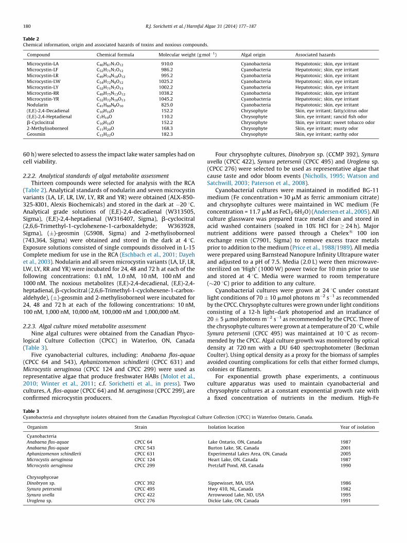
Table 2Chemical information, origin and associated hazards of toxins and noxious compounds.
Compound Chemical formula Molecular weight (g mol�1) Algal origin Associated hazards
Microcystin-LA C46H67N7O12 910.0 Cyanobacteria Hepatotoxic; skin, eye irritant
Microcystin-LF C52H71N7O12 986.2 Cyanobacteria Hepatotoxic; skin, eye irritant
Microcystin-LR C49H74N10O12 995.2 Cyanobacteria Hepatotoxic; skin, eye irritant
Microcystin-LW C54H72N8O12 1025.2 Cyanobacteria Hepatotoxic; skin, eye irritant
Microcystin-LY C52H71N7O13 1002.2 Cyanobacteria Hepatotoxic; skin, eye irritant
Microcystin-RR C49H75N13O12 1038.2 Cyanobacteria Hepatotoxic; skin, eye irritant
Microcystin-YR C52H72N10O13 1045.2 Cyanobacteria Hepatotoxic; skin, eye irritant
Nodularin C41H60N8O10 825.0 Cyanobacteria Hepatotoxic; skin, eye irritant
(E,E)-2,4-Decadienal C10H16O 152.2 Chrysophyte Skin, eye irritant; fatty/citrus odor
(E,E)-2,4-Heptadienal C7H10O 110.2 Chrysophyte Skin, eye irritant; rancid fish odor
b-Cyclocitral C10H16O 152.2 Chrysophyte Skin, eye irritant; sweet tobacco odor
2-Methylisoborneol C11H20O 168.3 Chrysophyte Skin, eye irritant; musty odor
Geosmin C12H22O 182.3 Chrysophyte Skin, eye irritant; earthy odor
R.J. Sorichetti et al. / Harmful Algae 31 (2014) 177–187180
60 h) were selected to assess the impact lake water samples had oncell viability.
2.2.2. Analytical standards of algal metabolite assessment
Thirteen compounds were selected for analysis with the RCA(Table 2). Analytical standards of nodularin and seven microcystinvariants (LA, LF, LR, LW, LY, RR and YR) were obtained (ALX-850-325-KI01, Alexis Biochemicals) and stored in the dark at �20 8C.Analytical grade solutions of (E,E)-2,4-decadienal (W313505,Sigma), (E,E)-2,4-heptadienal (W316407, Sigma), b-cyclocitral(2,6,6-Trimethyl-1-cyclohexene-1-carboxaldehyde; W363928,Sigma), (�)-geosmin (G5908, Sigma) and 2-methylisoborneol(743,364, Sigma) were obtained and stored in the dark at 4 8C.Exposure solutions consisted of single compounds dissolved in L-15Complete medium for use in the RCA (Eschbach et al., 2001; Dayehet al., 2003). Nodularin and all seven microcystin variants (LA, LF, LR,LW, LY, RR and YR) were incubated for 24, 48 and 72 h at each of thefollowing concentrations: 0.1 nM, 1.0 nM, 10 nM, 100 nM and1000 nM. The noxious metabolites (E,E)-2,4-decadienal, (E,E)-2,4-heptadienal, b-cyclocitral (2,6,6-Trimethyl-1-cyclohexene-1-carbox-aldehyde), (�)-geosmin and 2-methylisoborneol were incubated for24, 48 and 72 h at each of the following concentrations: 10 nM,100 nM, 1,000 nM, 10,000 nM, 100,000 nM and 1,000,000 nM.
2.2.3. Algal culture mixed metabolite assessment
Nine algal cultures were obtained from the Canadian Phyco-logical Culture Collection (CPCC) in Waterloo, ON, Canada(Table 3).
Five cyanobacterial cultures, including: Anabaena flos-aquae
(CPCC 64 and 543), Aphanizomenon schindlerii (CPCC 631) andMicrocystis aeruginosa (CPCC 124 and CPCC 299) were used asrepresentative algae that produce freshwater HABs (Molot et al.,2010; Winter et al., 2011; c.f. Sorichetti et al., in press). Twocultures, A. flos-aquae (CPCC 64) and M. aeruginosa (CPCC 299), areconfirmed microcystin producers.
Table 3Cyanobacteria and chrysophyte isolates obtained from the Canadian Phycological Cultu
Organism Strain
Cyanobacteria
Anabaena flos-aquae CPCC 64
Anabaena flos-aquae CPCC 543
Aphanizomenon schindlerii CPCC 631
Microcystis aeruginosa CPCC 124
Microcystis aeruginosa CPCC 299
Chrysophyceae
Dinobryon sp. CPCC 392
Synura petersenii CPCC 495
Synura uvella CPCC 422
Uroglena sp. CPCC 276
Four chrysophyte cultures, Dinobryon sp. (CCMP 392), Synura
uvella (CPCC 422), Synura petersenii (CPCC 495) and Uroglena sp.(CPCC 276) were selected to be used as representative algae thatcause taste and odor bloom events (Nicholls, 1995; Watson andSatchwill, 2003; Paterson et al., 2008).
Cyanobacterial cultures were maintained in modified BG-11medium (Fe concentration = 30 mM as ferric ammonium citrate)and chrysophyte cultures were maintained in WC medium (Feconcentration = 11.7 mM as FeCl3�6H2O) (Andersen et al., 2005). Allculture glassware was prepared trace metal clean and stored inacid washed containers (soaked in 10% HCl for � 24 h). Majornutrient additions were passed through a Chelex1 100 ionexchange resin (C7901, Sigma) to remove excess trace metalsprior to addition to the medium (Price et al., 1988/1989). All mediawere prepared using Barnstead Nanopure Infinity Ultrapure waterand adjusted to a pH of 7.5. Media (2.0 L) were then microwave-sterilized on ‘High’ (1000 W) power twice for 10 min prior to useand stored at 4 8C. Media were warmed to room temperature(�20 8C) prior to addition to any culture.
Cyanobacterial cultures were grown at 24 8C under constantlight conditions of 70 � 10 mmol photons m�2 s�1 as recommendedby the CPCC. Chrysophyte cultures were grown under light conditionsconsisting of a 12-h light–dark photoperiod and an irradiance of20 � 5 mmol photons m�2 s�1 as recommended by the CPCC. Three ofthe chrysophyte cultures were grown at a temperature of 20 8C, whileSynura petersenii (CPCC 495) was maintained at 10 8C as recom-mended by the CPCC. Algal culture growth was monitored by opticaldensity at 720 nm with a DU 640 spectrophotometer (BeckmanCoulter). Using optical density as a proxy for the biomass of samplesavoided counting complications for cells that either formed clumps,colonies or filaments.
For exponential growth phase experiments, a continuousculture apparatus was used to maintain cyanobacterial andchrysophyte cultures at a constant exponential growth rate witha fixed concentration of nutrients in the medium. High-Fe
re Collection (CPCC) in Waterloo Ontario, Canada.
Isolation location Year of isolation
Lake Ontario, ON, Canada 1987
Burton Lake, SK, Canada 2001
Experimental Lakes Area, ON, Canada 2005
Heart Lake, ON, Canada 1987
Pretzlaff Pond, AB, Canada 1990
Sippewisset, MA, USA 1986
Hwy 410, NL, Canada 1982
Arrowwood Lake, ND, USA 1995
Dickie Lake, ON, Canada 1991
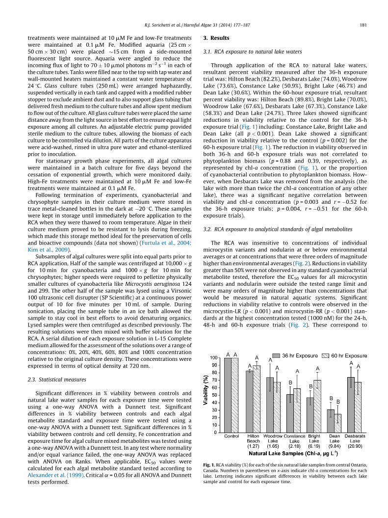
Fig. 1. RCA viability (%) for each of the six natural lake samples from central Ontario,
Canada. Numbers in parentheses on x-axis indicate chl-a concentrations for each
lake. Lettering indicates significant differences in viability between each lake
sample and control for each exposure time.
R.J. Sorichetti et al. / Harmful Algae 31 (2014) 177–187 181
treatments were maintained at 10 mM Fe and low-Fe treatmentswere maintained at 0.1 mM Fe. Modified aquaria (25 cm �50 cm � 30 cm) were placed �15 cm from a side-mountedfluorescent light source. Aquaria were angled to reduce theincoming flux of light to 70 � 10 mmol photons m�2 s�1 in each ofthe culture tubes. Tanks were filled near to the top with tap water andwall-mounted heaters maintained a constant water temperature of24 8C. Glass culture tubes (250 mL) were arranged haphazardly,suspended vertically in each tank and capped with a modified rubberstopper to exclude ambient dust and to also support glass tubing thatdelivered fresh medium to the culture tubes and allow spent mediumto flow out of the culture. All glass culture tubes were placed the samedistance away from the light source in best effort to ensure equal lightexposure among all cultures. An adjustable electric pump providedsterile medium to the culture tubes, allowing the biomass of eachculture to be controlled via dilution. All parts of the culture apparatuswere acid-washed, rinsed in ultra pure water and ethanol-sterilizedprior to inoculation.
For stationary growth phase experiments, all algal cultureswere maintained in a batch culture for five days beyond thecessation of exponential growth, which were monitored daily.High-Fe treatments were maintained at 10 mM Fe and low-Fetreatments were maintained at 0.1 mM Fe.
Following termination of experiments, cyanobacterial andchrysophyte samples in their culture medium were stored intrace metal-cleaned bottles in the dark at �20 8C. These sampleswere kept in storage until immediately before application to theRCA when they were thawed to room temperature. Algae in theirculture medium proved to be resistant to lysis during freezing,which made this storage method ideal for the preservation of cellsand bioactive compounds (data not shown) (Furtula et al., 2004;Kim et al., 2009).
Subsamples of algal cultures were split into equal parts prior toRCA application. Half of the sample was centrifuged at 10,000 � g
for 10 min for cyanobacteria and 1000 � g for 10 min forchrysophytes; higher speeds were required to pelletize physicallysmaller cultures of cyanobacteria like Microcystis aeruginosa 124and 299. The other half of the sample was lysed using a Virsonic100 ultrasonic cell disrupter (SP Scientific) at a continuous poweroutput of 10 for five minutes per 10 mL of sample. Duringsonication, placing the sample tube in an ice bath allowed thesample to stay cool in best efforts to avoid denaturing organics.Lysed samples were then centrifuged as described previously. Theresulting solutions were then mixed with buffer solution for theRCA. A serial dilution of each exposure solution in L-15 Completemedium allowed for the assessment of the solutions over a range ofconcentrations: 0%, 20%, 40%, 60%, 80% and 100% concentrationrelative to the original culture density. These concentrations wereexpressed in terms of optical density at 720 nm.
2.3. Statistical measures
Significant differences in % viability between controls andnatural lake water samples for each exposure time were testedusing a one-way ANOVA with a Dunnett test. Significantdifferences in % viability between controls and each algalmetabolite standard and exposure time were tested using aone-way ANOVA with a Dunnett test. Significant differences in %viability between controls and cell density, Fe concentration andexposure time for algal culture mixed metabolites was tested usinga one-way ANOVA with a Dunnett test. In any test where normalityand/or equal variance failed, the one-way ANOVA was replacedwith ANOVA on Ranks. When applicable, EC50 values werecalculated for each algal metabolite standard tested according toAlexander et al. (1999). Critical a = 0.05 for all ANOVA and Dunnetttests performed.
3. Results
3.1. RCA exposure to natural lake waters
Through application of the RCA to natural lake waters,resultant percent viability measured after the 36-h exposuretrial was: Hilton Beach (82.2%), Desbarats Lake (74.0%), WoodrowLake (73.6%), Constance Lake (50.9%), Bright Lake (46.7%) andDean Lake (30.6%). Within the 60-hour exposure trial, resultantpercent viability was: Hilton Beach (89.8%), Bright Lake (70.0%),Woodrow Lake (67.6%), Desbarats Lake (67.3%), Constance Lake(58.3%) and Dean Lake (24.7%). Three lakes showed significantreductions in viability relative to the control for the 36-hexposure trial (Fig. 1) including: Constance Lake, Bright Lake andDean Lake (all p < 0.001). Dean Lake showed a significantreduction in viability relative to the control (p = 0.002) for the60-h exposure trial (Fig. 1). The reduction in viability observed inboth 36-h and 60-h exposure trials was not correlated tophytoplankton biomass (p = 0.88 and 0.39, respectively), asrepresented by chl-a concentration (Fig. 1), or the proportionof cyanobacterial contribution to phytoplankton biomass. How-ever, when Desbarats Lake was removed from the analysis (thelake with more than twice the chl-a concentration of any otherlake), there was a significant negative correlation betweenviability and chl-a concentration (p = 0.003 and r = �0.52 forthe 36-h exposure trials; p = 0.004, r = �0.51 for the 60-hexposure trials).
3.2. RCA exposure to analytical standards of algal metabolites
The RCA was insensitive to concentrations of individualmicrocystin variants and nodularin at or below environmentalaverages or at concentrations that were three orders of magnitudehigher than environmental averages (Fig. 2). Reductions in viabilitygreater than 50% were not observed in any standard cyanobacterialmetabolite tested, therefore the EC50 values for all microcystinvariants and nodularin were outside the tested range limit andwere many orders of magnitude higher than concentrations thatwould be measured in natural aquatic systems. Significantreductions in viability relative to controls were observed in themicrocystin-LR (p < 0.001) and microcystin-RR (p < 0.001) stan-dards at the highest concentration tested (1000 nM) for the 24-h,48-h and 60-h exposure trials (Fig. 2). These correspond to
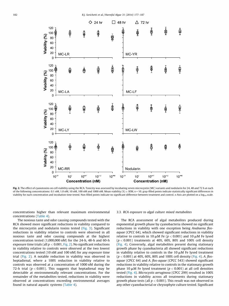
Fig. 2. The effect of cyanotoxins on cell viability using the RCA. Toxicity was assessed by incubating seven microcystin (MC) variants and nodularin for 24, 48 and 72 h at each
of the following concentrations: 0.1 nM, 1.0 nM, 10 nM, 100 nM and 1000 nM. Mean viability (%) � SEM, n = 10; gray-filled points indicate statistically significant differences in
viability for each concentration and incubation time tested. Non-filled points indicate no significant difference between treatment and control. x-Axis are plotted on a log10 scale.
R.J. Sorichetti et al. / Harmful Algae 31 (2014) 177–187182
concentrations higher than relevant maximum environmentalconcentrations (Table 4).
The noxious taste and odor causing compounds tested with theRCA showed more significant reductions in viability compared tothe microcystin and nodularin toxins tested (Fig. 3). Significantreductions in viability relative to controls were observed in allnoxious taste and odor causing compounds at the highestconcentration tested (1,000,000 nM) for the 24-h, 48-h and 60-hexposure time trials (all p < 0.001, Fig. 2). No significant reductionsin viability relative to controls were observed at the two lowestconcentrations tested (10 nM and 100 nM) for any exposure timetrial (Fig. 2). A notable reduction in viability was observed inheptadienal, where a 100% reduction in viability relative tocontrols was observed at a concentration of 1000 nM during the72-h trial (p < 0.001). This suggests that heptadienal may bedetectable at environmentally relevant concentrations. For theremainder of the metabolites tested, reductions in viability wereobserved at concentrations exceeding environmental averagesfound in natural aquatic systems (Table 4).
3.3. RCA exposure to algal culture mixed metabolites
The RCA assessment of algal metabolites produced duringexponential growth phase by cyanobacteria showed no significantreductions in viability with one exception being Anabaena flos-
aquae (CPCC 64), which showed significant reductions in viabilityrelative to controls in 10 mM Fe (p < 0.001) and 10 mM Fe lysed(p < 0.001) treatments at 40%, 60%, 80% and 100% cell density(Fig. 4). Conversely, algal metabolites present during stationarygrowth phase by cyanobacteria all showed significant reductionsin viability relative to controls in the 10 mM Fe lysed treatment(p < 0.001) at 40%, 60%, 80% and 100% cell density (Fig. 4). A. flos-
aquae (CPCC 64) and A. flos-aquae (CPCC 543) showed significantreductions in viability relative to controls in the stationary growthphase 10 mM Fe lysed treatment (p < 0.001) at all cell densitiestested (Fig. 4). Microcystis aeruginosa (CPCC 299) resulted in 100%reductions in viability across all treatments during stationarygrowth phase tests (all p < 0.001). This result was not observed inany other cyanobacterial or chrysophyte culture tested. Significant
Table 4Algal metabolites, RCA EC50 values calculated from 72 hour exposure trials [using the formula in Alexander et al. (1999)] and mean and maximum environmental
concentrations for toxins and noxious compounds (from Watson, 2003).
Compound Mean environmental concentration (nM) Maximum environmental concentration (nM) RCA EC50 values (nM)
Microcystin-LA <100 – >103
Microcystin-LF <100 – >103
Microcystin-LR <100 <102 >103
Microcystin-LW <100 – >103
Microcystin-LY <100 – >103
Microcystin-RR <100 – >103
Microcystin-YR <100 – >103
Nodularin <100 – >103
(E,E)-2,4-decadienal <101 <102 2 � 103
(E,E)-2,4-heptadienal <101 <102 5 � 103
b-Cyclocitral <101 <102 3 � 106
Geosmin <10�1 <101 7 � 102
2-Methylisoborneol <100 <100 6 � 103
R.J. Sorichetti et al. / Harmful Algae 31 (2014) 177–187 183
reductions in viability in the stationary growth phase tests wereadditionally observed in 0.1 mM Fe, 10 mM Fe and 0.1 mM Fe lysedtreatments with significant differences relative to controlsindicated in Fig. 4 for each cyanobacterial culture tested.
Four trends were observed across all stationary growth phasetests of cyanobacterial cultures: (1) in treatments that resulted insignificant reductions in viability at low Fe treatments (0.1 mM Fe),equal or greater reductions in viability were observed at high Fetreatments (10 mM Fe); (2) 10 mM Fe treatments showedreductions in viability more frequently than the 0.1 mM Fetreatments; (3) exposure solutions composed of intracellularand extracellular (lysed) material resulted in greater reductions in
Fig. 3. The effect of noxious taste and odor compounds on cell viability using the RCA. To
following concentrations: 10 nM, 100 nM, 1000 nM, 10,000 nM, 100,000 nM and 1,00
significant differences in viability for each concentration and incubation time tested. Non-fi
plotted on a log10 scale.
viability than their counterparts containing only extracellular(non-lysed) material; and (4) optical densities of cultures thatresulted in mild reductions (�50%) in viability ranged from �0.1a.u. to 0.6 a.u. (720 nm), with large variation among algal culturesand treatments.
The RCA assessment of algal metabolites produced duringexponential and stationary growth phase by chrysophytesshowed no reductions in viability, with one exception. Synura
petersenii (CPCC 495) resulted in significant reductions in viabilityrelative to controls in stationary growth phase 10 mM Fe lysedtreatment (p < 0.001) at 40%, 60%, 80% and 100% cell density(Fig. 5).
xicity was assessed by incubating five compounds for 24, 48 and 72 h at each of the
0,000 nM. Mean viability (%) � SEM, n = 10; gray-filled points indicate statistically
lled points indicate no significant difference between treatment and control. x-Axis are
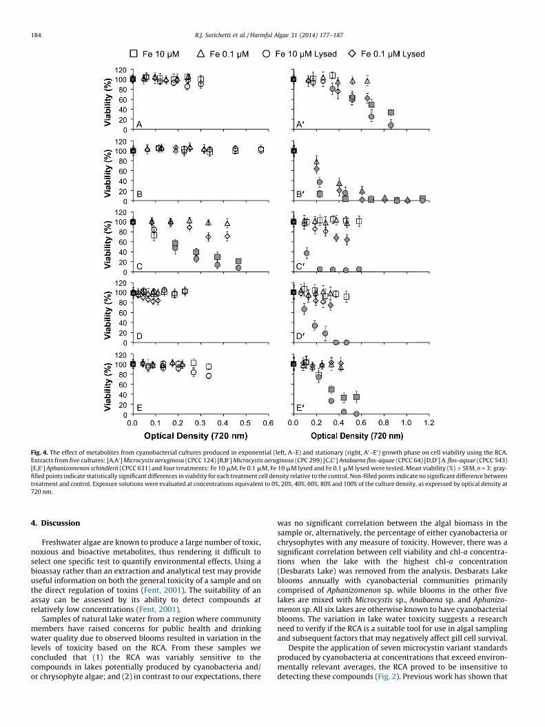
Fig. 4. The effect of metabolites from cyanobacterial cultures produced in exponential (left, A–E) and stationary (right, A0–E0) growth phase on cell viability using the RCA.
Extracts from five cultures: [A,A0] Microcystis aeruginosa (CPCC 124) [B,B0] Microcystis aeruginosa (CPC 299) [C,C0] Anabaena flos-aquae (CPCC 64) [D,D0] A. flos-aquae (CPCC 543)
[E,E0] Aphanizomenon schindlerii (CPCC 631) and four treatments: Fe 10 mM, Fe 0.1 mM, Fe 10 mM lysed and Fe 0.1 mM lysed were tested. Mean viability (%) � SEM, n = 3; gray-
filled points indicate statistically significant differences in viability for each treatment cell density relative to the control. Non-filled points indicate no significant difference between
treatment and control. Exposure solutions were evaluated at concentrations equivalent to 0%, 20%, 40%, 60%, 80% and 100% of the culture density, as expressed by optical density at
720 nm.
R.J. Sorichetti et al. / Harmful Algae 31 (2014) 177–187184
4. Discussion
Freshwater algae are known to produce a large number of toxic,noxious and bioactive metabolites, thus rendering it difficult toselect one specific test to quantify environmental effects. Using abioassay rather than an extraction and analytical test may provideuseful information on both the general toxicity of a sample and onthe direct regulation of toxins (Fent, 2001). The suitability of anassay can be assessed by its ability to detect compounds atrelatively low concentrations (Fent, 2001).
Samples of natural lake water from a region where communitymembers have raised concerns for public health and drinkingwater quality due to observed blooms resulted in variation in thelevels of toxicity based on the RCA. From these samples weconcluded that (1) the RCA was variably sensitive to thecompounds in lakes potentially produced by cyanobacteria and/or chrysophyte algae; and (2) in contrast to our expectations, there
was no significant correlation between the algal biomass in thesample or, alternatively, the percentage of either cyanobacteria orchrysophytes with any measure of toxicity. However, there was asignificant correlation between cell viability and chl-a concentra-tions when the lake with the highest chl-a concentration(Desbarats Lake) was removed from the analysis. Desbarats Lakeblooms annually with cyanobacterial communities primarilycomprised of Aphanizomenon sp. while blooms in the other fivelakes are mixed with Microcystis sp., Anabaena sp. and Aphanizo-
menon sp. All six lakes are otherwise known to have cyanobacterialblooms. The variation in lake water toxicity suggests a researchneed to verify if the RCA is a suitable tool for use in algal samplingand subsequent factors that may negatively affect gill cell survival.
Despite the application of seven microcystin variant standardsproduced by cyanobacteria at concentrations that exceed environ-mentally relevant averages, the RCA proved to be insensitive todetecting these compounds (Fig. 2). Previous work has shown that
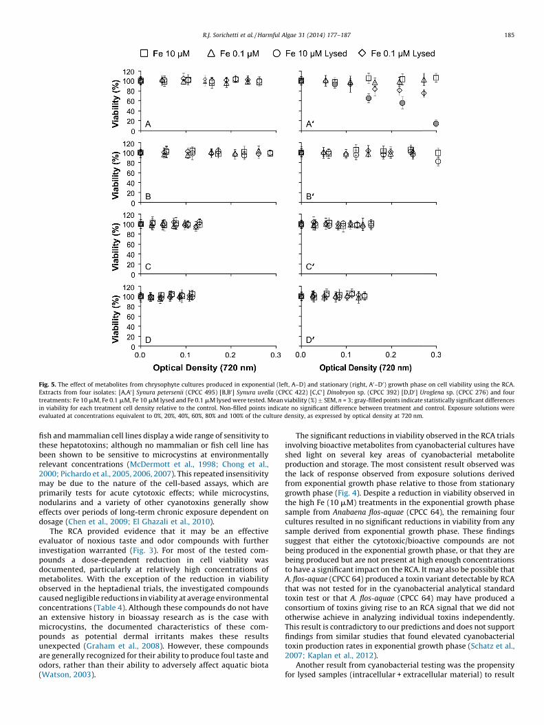
Fig. 5. The effect of metabolites from chrysophyte cultures produced in exponential (left, A–D) and stationary (right, A0–D0) growth phase on cell viability using the RCA.
Extracts from four isolates: [A,A0] Synura petersenii (CPCC 495) [B,B0] Synura uvella (CPCC 422) [C,C0] Dinobryon sp. (CPCC 392) [D,D0] Uroglena sp. (CPCC 276) and four
treatments: Fe 10 mM, Fe 0.1 mM, Fe 10 mM lysed and Fe 0.1 mM lysed were tested. Mean viability (%) � SEM, n = 3; gray-filled points indicate statistically significant differences
in viability for each treatment cell density relative to the control. Non-filled points indicate no significant difference between treatment and control. Exposure solutions were
evaluated at concentrations equivalent to 0%, 20%, 40%, 60%, 80% and 100% of the culture density, as expressed by optical density at 720 nm.
R.J. Sorichetti et al. / Harmful Algae 31 (2014) 177–187 185
fish and mammalian cell lines display a wide range of sensitivity tothese hepatotoxins; although no mammalian or fish cell line hasbeen shown to be sensitive to microcystins at environmentallyrelevant concentrations (McDermott et al., 1998; Chong et al.,2000; Pichardo et al., 2005, 2006, 2007). This repeated insensitivitymay be due to the nature of the cell-based assays, which areprimarily tests for acute cytotoxic effects; while microcystins,nodularins and a variety of other cyanotoxins generally showeffects over periods of long-term chronic exposure dependent ondosage (Chen et al., 2009; El Ghazali et al., 2010).
The RCA provided evidence that it may be an effectiveevaluator of noxious taste and odor compounds with furtherinvestigation warranted (Fig. 3). For most of the tested com-pounds a dose-dependent reduction in cell viability wasdocumented, particularly at relatively high concentrations ofmetabolites. With the exception of the reduction in viabilityobserved in the heptadienal trials, the investigated compoundscaused negligible reductions in viability at average environmentalconcentrations (Table 4). Although these compounds do not havean extensive history in bioassay research as is the case withmicrocystins, the documented characteristics of these com-pounds as potential dermal irritants makes these resultsunexpected (Graham et al., 2008). However, these compoundsare generally recognized for their ability to produce foul taste andodors, rather than their ability to adversely affect aquatic biota(Watson, 2003).
The significant reductions in viability observed in the RCA trialsinvolving bioactive metabolites from cyanobacterial cultures haveshed light on several key areas of cyanobacterial metaboliteproduction and storage. The most consistent result observed wasthe lack of response observed from exposure solutions derivedfrom exponential growth phase relative to those from stationarygrowth phase (Fig. 4). Despite a reduction in viability observed inthe high Fe (10 mM) treatments in the exponential growth phasesample from Anabaena flos-aquae (CPCC 64), the remaining fourcultures resulted in no significant reductions in viability from anysample derived from exponential growth phase. These findingssuggest that either the cytotoxic/bioactive compounds are notbeing produced in the exponential growth phase, or that they arebeing produced but are not present at high enough concentrationsto have a significant impact on the RCA. It may also be possible thatA. flos-aquae (CPCC 64) produced a toxin variant detectable by RCAthat was not tested for in the cyanobacterial analytical standardtoxin test or that A. flos-aquae (CPCC 64) may have produced aconsortium of toxins giving rise to an RCA signal that we did nototherwise achieve in analyzing individual toxins independently.This result is contradictory to our predictions and does not supportfindings from similar studies that found elevated cyanobacterialtoxin production rates in exponential growth phase (Schatz et al.,2007; Kaplan et al., 2012).
Another result from cyanobacterial testing was the propensityfor lysed samples (intracellular + extracellular material) to result
R.J. Sorichetti et al. / Harmful Algae 31 (2014) 177–187186
in greater reductions in viability relative to the non-lysed samples(extracellular material only) of the same Fe treatment (Fig. 4) in theRCA. This observation is supported by previous research doc-umenting that the proportion of bioactive material retained insidethe cell over the course of population growth is greater than theproportion that is excreted to the extracellular environment priorto senescence (Park et al., 1998). This may explain the differencesobserved between cyanobacterial exponential and stationarygrowth phase tests, as the accumulation of intracellular compoundwould increase proportionally with the age of the cells prior toentering senescence.
The role of Fe was observed to be a contributing factor in thecytotoxicity of algal metabolites investigated in cyanobacterialcultures. While Fe is commonly limiting in freshwater lakes, theexperimental levels chosen here reflect natural levels of Fe(0.1 mM) and a treatment in which Fe was in excess (10 mM).The latter treatment resulted in the greatest reductions in viabilityobserved with the RCA (Fig. 4). Cells grown under the high Fe(10 mM) treatment repeatedly resulted in the most cytotoxicsamples, providing support for the claim that it is under optimumgrowth conditions, rather than nutrient limitation, that bioactivecompound synthesis peaks in cyanobacteria (Kaebernick andNeilan, 2001). This result suggests that the Fe-limited conditionsthat have been reported to favor microcystin production are notnecessarily ideal for the production of a wider variety of bioactivecompounds as has been suggested by previous work (Alexova et al.,2011; Kaplan et al., 2012). One possible explanation is that thebioactive products responsible for reductions in viability observedwith the RCA are derivatives of molecules produced by photosyn-thetic processes (Kaebernick and Neilan, 2001), though this wasnot specifically measured. Fe-limited treatments may decrease thecell’s photosynthetic efficiency and, therefore, limit the productionof these metabolites.
Aside from the reductions in viability observed in the stationarygrowth phase, high Fe (10 mM) and lysed treatment of S. petersenii
(CPCC 495), no significant reduction in viability was observed withthe RCA in any other chrysophyte-derived treatment or culture(Fig. 5). The reduction in viability observed in CPCC 495 suggestssimilar patterns to that of the cyanobacterial tests: high Fe (10 mM)conditions and intracellular storage may be important factors inthe production of bioactive compounds. However, these findingsare not supported by any of the other three chrysophyte culturemixed metabolites investigated. The overall lack of responseobserved in the RCA when exposed to metabolite mixtures fromchrysophyte cultures was likely a result of the inability to growcultures to sufficient cell densities. An additional step toconcentrate chrysophyte cell cultures and obtain higher celldensity for the experiment was considered but this additionalmethodology would be contradictory to the rapid assessmentcharacteristic and intent for this bioassay.
5. Conclusions
It is possible that the RCA measured toxicity of bioactivemetabolites in lake samples and in cyanobacterial cultures thatwere not tested as isolated analytical standards. For example, b-methyl amino alanine is a cyanotoxin with the potential forproduction by all cyanobacterial genera, but isolated standards forthis cyanotoxin could not be acquired for experimentation. Othermetabolites not tested as isolated standards, though could havebeen present and detected in the lake samples and algal culturesinclude: cylindrospermopsins, anatoxins, saxitoxins, aplysiatoxinsand lyngbyatoxins. Further research into the applicability of theRCA to measure cytotoxicity in lake samples should focus ontesting a wider range of known cyanotoxins that are most likely tooccur, or those previously identified, in specific study lakes of
interest. An additional next step in the methodological aspect ofthe RCA is to test a gradient in concentration factors of lake watersamples. This study concentrated all lake water samples 3� andachieved variable responses to cell viability as measured by theRCA. Testing a gradient in concentration factors would identifywhether the RCA is suitable for use in lakes with low or extremelyhigh biomass accumulation.
In spite of the induction of cytotoxic responses to natural lakesamples, the RCA was relatively insensitive to known or putativetoxins and/or noxious compounds. The selected test did respond tothe analytical standards of metabolites but only at excessively highlevels compared to average environmental concentrations. Analy-sis of noxious compounds produced by chrysophyte algae revealedsimilar insensitivity with the RCA, although reductions in viabilitywere observed at concentrations greater than two orders ofmagnitude higher than average environmental concentrations.Cyanobacterial cultures grown in relatively high Fe (10 mM)conditions were responsible for greater reductions in cell viabilitybased on the RCA, suggesting that the production of cytotoxins andbioactive compounds responsible for observed reductions isgreater in these conditions compared to low Fe (0.1 mM)conditions. Comparing this finding to the established link betweenlow Fe conditions and the stimulation of microcystin productionsuggests that the monitoring of microcystin levels in naturalsystems is likely not a good indicator of the overall levels ofcytotoxic and/or bioactive compounds present in a sample,although it is still an important parameter to monitor withregards to drinking water quality.
Acknowledgements
This work was supported by Discovery Grants to IFC and to CGT.We gratefully thank our dedicated assistants J. Miller and D.Aldred. Anonymous reviewers provided wise insight into thepresentation of this work.
References
Alexander, B., Browse, D.J., Reading, S.J., Benjamin, I.S., 1999. A simple and accuratemathematical method for calculation of the EC50. J. Pharmacol. Toxicol. Meth-ods 41 (2) 55–58.
Alexova, R., Fujii, M., Birch, D., Cheng, J., Waite, T.D., Ferrari, B.C., Neilan, B., 2011.Iron uptake and toxin synthesis in the bloom-forming Microcystis aeruginosaunder iron limitation. Environ. Microbiol. 13 (4) 1064–1077.
Andersen, R.A., Berges, J.A., Harrison, P.J., Watanabe, M.M., 2005. Appendix A:recipes for freshwater and seawater media. In: Andersen, R.A. (Ed.), AlgalCulturing Techniques. Elsevier Academic Press, Burlington, MA, pp. 431–475.
Arar, E.J., Collins, G.B., 1997. Method 445.0, in vitro determination of chlorophyll aand pheophytin a in marine and freshwater algae by fluorescence. In: Methodsfor the Determination of Chemical Substances in Marine and Estuarine Envi-ronmental Matrices. 2nd ed. National Exposure Research Laboratory, Office ofResearch and Development, USEPA, Cincinnati, OH.
Bols, N.C., Barlian, A., Chirino-Trejo, M., Caldwell, S.J., Goegan, P., Lee, L.E.J., 1994.Development of a cell line from primary cultures of rainbow trout, Oncor-hynchus mykiss (Walbaum), gills. J. Fish Dis. 17 (6) 601–611.
Boaru, D.A., Dragos, N., Schirmer, K., 2006. Microcystin-LR induced cellular effects inmammalian and fish primary hepatocyte cultures and cell lines: a comparativestudy. Toxicology 218 (2–3) 134–148.
Burkholder, J.M., Gordon, A.S., Moeller, P.D., Law, J.M., Coyne, J., Lewitus, A.J.,Ramsdell, J.S., Marshall, H.G., Deamer, N.J., Cary, S.C., Kempton, J.W., Morton,S.L., Rublee, P.A., 2005. Demonstration of toxicity by fish and mammalian cellsby Pfiesteria species: comparison of assay methods and strains. Proc. Natl. Acad.Sci. U.S.A. 102 (9) 3471–3476.
Carmichael, W.W., 1992. Cyanobacteria secondary metabolites – the cyanotoxins. J.Appl. Bacteriol. 72 (6) 445–459.
Carmichael, W.W., 2001. Health effects of toxin-producing cyanobacteria, thecyanoHABs. Hum. Ecol. Risk Assess. 7 (5) 1393–1407.
Carmichael, W.W., 2008. A world overview – one-hundred-twenty-seven years ofresearch on toxic cyanobacteria – where do we go from here? In: Hudnell, H.(Ed.), Cyanobacterial Harmful Algal Blooms: State of the Science and ResearchNeeds. Springer, Berlin, pp. 105–125.
Charlton, M., Giddings, M., Holmes, C., Carmichael, W., Ridal, J., 2008. Threats toSources of Drinking Water and Aquatic Ecosystem Health in Canada: algal
R.J. Sorichetti et al. / Harmful Algae 31 (2014) 177–187 187
toxins and taste and odour. National Water Research Institute, EnvironmentCanada, , pp. 5–8.
Chen, J., Xie, P., Li, L., Xu, J., 2009. First identification of the hepatotoxic microcystinsin the serum of a chronically exposed human population together with indica-tion of hepatocellular damage. Toxicol. Sci. 108 (1) 81–89.
Chong, M.W., Gu, K.D., Lam, P.K., Yang, M., Fong, W.F., 2000. Study on the cytotoxic-ity of microcystin-LR on cultured cells. Chemosphere 41 (1–2) 143–147.
Dayeh, V.R., Schirmer, K., Lee, L.E.J., Bols, N.C., 2003. The use of fish-derived cell linesfor investigation of environmental contaminants. Curr. Protoc. Toxicol. 1 (5) 1–17.
Dayeh, V.R., Schirmer, K., Lee, L.E.J., Bols, N.C., 2005. Rainbow trout gill cell linemicroplate cytotoxicity test. In: Blaise, C., Ferard, J.F. (Eds.), Small-ScaleFreshwater Toxicity Investigations, vol. 1. Springer, Netherlands, pp. 473–503.
Dorantes-Aranda, J.J., Waite, T.D., Godrant, A., Rose, A.L., Tovar, C.D., Woods, G.M.,Hallegraeff, G.M., 2011. Novel application of a fish gill cell line assay to assessichthyotoxicity of harmful marine microalgae. Harmful Algae 10 (4) 366–373.
El Ghazali, I., Saqrane, S., Carvalho, A.P., Ouahid, Y., del Campo, F.F., Vasconcelos, V.,Oudra, B., 2010. Effects of the microcystin profile of a cyanobacterial bloom ongrowth and toxin accumulation in common carp Cyprinus carpio larvae. J. FishBiol. 76 (6) 1415–1430.
Eschbach, E., Scharsack, P., John, U., Medlin, L.K., 2001. Improved erythrocyte lysisassay in microtiter plates for sensitive detection and efficient measurement ofhaemolytic compounds from ichthyotoxic algae. J. Appl. Toxicol. 21, 513–519.
Falconer, I.R., 1999. An overview of problems caused by toxic blue-green algae(cyanobacteria) in drinking and recreational water. Environ. Toxicol. 14, 5–12.
Fent, K., 2001. Fish cell lines as versatile tools in ecotoxicology: assessment ofcytotoxicity, cytochrome P4501A induction potential and estrogenic activity ofchemicals and environmental samples. Toxicol. In Vitro 15, 477–488.
Fujii, M., Rose, A.L., Waite, T.D., 2011. Iron uptake by toxic and nontoxic strains ofMicrocystis aeruginosa. Appl. Environ. Microbiol. 77 (19) 7068–7071.
Furtula, V., Davies, J., Mazumder, A., 2004. An automated headspace SPME-GC-ITMStechnique for taste and odour compound identification. W. Qual. Res. J. Can. 39(3) 213–222.
Graham, J.L., Loftin, K.A., Ziegler, A.C., Meyer, M.T., 2008. Cyanobacteria in Lakes andReservoirs: Toxin and Taste-and-Odor Sampling Guidelines. National FieldManual for Collection of Water-Quality Data. USGS, , pp. 1–65.
Guikema, J.A., Sherman, L.A., 1983. Organization and function of chlorophyll inmembranes of cyanobacteria during iron starvation. Plant Physiol. 73 (2) 250–256.
Guildford, J., Hecky, R., 2000. Total phosphorus, and nitrogen limitation in lakes andoceans: is there a common relationship? Limnol. Oceanogr. 45 (6) 1213–1223.
Heisler, J., Glibert, P.M., Burkholder, J.M., Anderson, D.M., Cochlan, W., Dennison,W.C., Dortch, Q., Gobler, C.J., Heil, C.A., Humphries, E., Lewitus, A., Magnien, R.,Marshall, H.G., Sellner, K., Stockwell, D.A., Stoecker, D.K., Suddleson, M., 2008.Eutrophication and harmful algal blooms: a scientific consensus. Harmful Algae8 (1) 3–13.
Helmholz, H., Johnston, B.D., Ruhnau, C., Prange, A., 2010. Gill cell toxicity ofnorthern boreal scyphomedusae Cyanea capillata and Aurelia aurita measuredby an in vitro cell assay. Hydrobiologia 645 (1) 223–234.
Huber, V., Wagner, C., Gerten, D., Adrian, R., 2012. To bloom or not to bloom:contrasting responses of cyanobacteria to recent heat waves explained bycritical thresholds of abiotic drivers. Oecologia 169 (1) 245–256.
Juttner, F., Hhjacher, B., Wurster, K., 1986. Seasonal analysis of volatile organicbiogenic substances (vobs) in freshwater phytoplankton populations dominat-ed by Dinobryon, Microcystis and Aphanizomenon. J. Phycol. 22, 169–175.
Kaebernick, M., Neilan, B.A., 2001. Ecological and molecular investigations ofcyanotoxin production. FEMS Microbiol. Ecol. 35 (1) 1–9.
Kaplan, A., Harel, M., Kaplan-Levy, R.N., Hadas, O., Sukenik, A., Dittmann, E., 2012.The languages spoken in the water body (or the biological role of cyanobacterialtoxins). Front. Microbiol. 3 (138) 1–11.
Kim, I.S., Nguyen, G.H., Kim, S.Y., Lee, J.W., Yu, H.W., 2009. Evaluation of methods forcyanobacterial cell lysis and toxin (microcystin-LR) extraction using chro-matographic and mass spectrometric analyses. Environ. Eng. Res. 14 (4)250–254.
Klausmeier, C.A., Litchman, E., Levin, S.A., 2004. Phytoplankton growth and stoi-chiometry under multiple nutrient limitation. Limnol. Oceanogr. 49 (4) 1463–1470.
Lee, L.E.J., Dayeh, V.R., Schirmer, K., Bols, N.C., 2009. Applications and potential usesof fish gill cell lines: examples with RTgill-W1. In Vitro Cell. Dev. Biol. Anim. 45,127–134.
Malbrouck, C., Kestemont, P., 2006. Effects of microcystins on fish. Environ. Toxicol.25, 72–86.
McDermott, C.M., Nho, C.W., Howard, W., Holton, B., 1998. The cyanobacterial toxin,microcystin-LR, can induce apoptosis in a variety of cell types. Toxicon 36 (12)1981–1996.
Molot, L.A., Li, G., Findlay, D.L., Watson, S.B., 2010. Iron-mediated suppression ofbloom-forming cyanobacteria by oxine in a eutrophic lake. Freshw. Biol. 55 (5)1102–1117.
Nicholls, K.H., 1995. Chrysophyte blooms in the plankton and neuston of marineand freshwater systems. In: Sandgren, C.D., Smol, J.P., Kristiansen, J. (Eds.),Chrysophyte Algae: Ecology, Phylogeny, and Development. Cambridge Univer-sity Press, New York, pp. 181–213.
Paerl, H.W., 1988. Nuisance phytoplankton blooms in coastal, estuarine, and inlandwaters. Limnol. Oceanogr. 33 (4) 823–847.
Paerl, H.W., Fulton, R.S., Moisander, P.H., Dyble, J., 2001. Harmful freshwater algalblooms, with an emphasis on cyanobacteria. Sci. World J. 1, 76–113.
Paerl, H.W., Huisman, J., 2008. Blooms like it hot. Science 320 (5872) 57–58.Park, H., Iwami, C., Watanabe, M.F., Harada, K., Okino, T., 1998. Temporal variabil-
ities of the concentrations of intra- and extracellular microcystin and toxicmicrocystis species in a hypertrophic lake, Lake Suwa, Japan (1991–1994).Environ. Toxicol. W. 13 (1) 61–72.
Paterson, A.M., Cumming, B.F., Smol, J.P., Hall, R.I., 2004. Marked recent increases ofcolonial scaled chrysophytes in boreal lakes: implications for the managementof taste and odour events. Freshw. Biol. 49 (2) 199–207.
Paterson, A.M., Winter, J.G., Nicholls, K.H., Clark, B.J., Ramcharan, C.W., Yan, N.D.,Somers, K.M., 2008. Long-term changes in phytoplankton composition in sevenCanadian Shield lakes in response to multiple anthropogenic stressors. Can. J.Fish. Aquat. Sci. 65, 846–861.
Pichardo, S., Jos, A., Zurita, J.L., Salguero, M., Camean, A.M., Repetto, G., 2005. The useof the fish cell lines RTG-2 and PLHC-1 to compare the toxic effects produced bymicrocystins LR and RR. Toxicol. In Vitro 19, 865–873.
Pichardo, S., Jos, A., Zurita, J., Salguero, M., Camean, A.M., Repetto, G., 2006. Toxiceffects produced by microcystins from a natural cyanobacterial bloom and aMicrocystis aeruginosa isolated strain on the fish cell lines RTG-2 and PLHC-1.Arch. Environ. Contam. Toxicol. 51 (1) 86–96.
Pichardo, S., Jos, A., Zurita, J.L., Salguero, M., Camean, A.M., Repetto, G., 2007. Acuteand subacute toxic effects produced by microcystin-YR on the fish cell linesRTG-2 and PLHC-1. Toxicol. In Vitro 21, 1460–1467.
Price, N.M., Harrison, G.I., Hering, J.G., Hudson, R.J., Nirel, P.M.V., Palenik, B., Morel,F.M.M., 1989. Preparation and chemistry of artificial algal culture medium.Aquil. Biol. Oceanogr. 6, 443–461.
Raven, J.A., Evans, M.C.W., Korb, R.E., 1999. The role of trace metals in photosyntheticelectron transport in O2 - evolving organisms. Photosyn. Res. 60, 111–149.
Reynolds, C., Walsby, A., 1975. Water-blooms. Biol. Rev. Camb. Philos. 50 (4) 437–456.
Schatz, D., Keren, Y., Vardi, A., Sukenik, A., Carmeli, S., Borner, T., Dittmann, E.,Kaplan, A., 2007. Towards clarification of the biological role of microcystins, afamily of cyanobacterial toxins. Environ. Microbiol. 9 (4) 965–970.
Schindler, D.W., 1977. Evolution of phosphorus limitation in lakes. Science 195(4275) 260–262.
Schindler, D.W., 1987. Detecting Ecosystem Responses to Anthropogenic Stress.Dept. Fish. Oceans Freshwater Inst. vol. 44 (Suppl. 1) 6–25.
Schirmer, K., Chan, A.G., Greenberg, B.M., Dixon, D.G., Bols, N.C., 1998. Ability of 16priority PAHs to be photocytotoxic to a cell line from the rainbow trout gill.Toxicology 127, 143–155.
Schirmer, K., Tom, D.J., Bols, N.C., Sherry, J.P., 2001. Ability of fractionated petroleumrefinery effluent to elicit cyto- and photocytotoxic responses and to induce 7-ethoxyresorufin-O-deethylase activity in fish cell lines. Sci. Total Environ. 271,61–78.
Skulberg, O.M., Codd, G.A., Carmichael, W., 1984. Toxic blue–green algal blooms inEurope – a growing problem. Ambio 13 (4) 244–247.
Smith, V.H., 2003. Eutrophication of freshwater and coastal marine ecosystems: aglobal problem. Environ. Sci. Pollut. Rev. Int. 10 (2) 126–139.
Sorichetti, R.J., Creed, I.F., Trick, C.G., in press. Evidence for iron regulated cyano-bacterial predominance in oligotrophic lakes. Freshw. Biol., in press.
Sterner, R.W., Smutka, T.M., Mckay, R.M.L., Xiaoming, Q., Erik, T., Sherrell, R.M.,Brown, E.T., 2004. Phosphorus and trace metal limitation of algae and bacteriain Lake Superior. Limnol. Oceanogr. 49 (2) 495–507.
Utkilen, H., Gjølme, N., 1995. Iron-stimulated toxin production in Microcystisaeruginosa. Appl. Environ. Microbiol. 61 (2) 797–800.
Watson, S.B., 2003. Cyanobacterial and eukaryotic algal odour compounds: signals orby-products? A review of their biological activity. Phycologia 42 (4) 332–350.
Watson, S.B., Satchwill, T., 2003. Chrysophyte odour production: resource-mediatedchanges at the cell and population levels. Phycologia 42 (4) 393–405.
Watson, S.B., Ridal, J., Boyer, G.L., 2008. Taste and odour and cyanobacterial toxins:impairment, prediction, and management in the Great Lakes. Can. J. Fish. Aquat.Sci. 65 (8) 1779–1796.
Winter, J.G., DeSellas, A.M., Fletcher, R., Heintsch, L., Morley, A., Nakamoto, L.,Utsumi, K., 2011. Algal blooms in Ontario, Canada: increases in reports since1994. Lake Reserv. Manage. 27 (2) 107–114.
![LDH Cytotoxicity Detection Kit User Manual Manual/PT3947-… · Cr] release assay (Decker & Lohmann-Matthes, 1988; Korzeniewski & Callewaert, 1983). High sensitivity: Low numbers](https://img.pdfslide.us/doc/110x75/5e9cd3af3ffbcb545926c569/ldh-cytotoxicity-detection-kit-user-manual-manualpt3947-cr-release-assay-decker.jpg)