Embed Size (px)
Citation preview
Case ReportSuccessful Treatment of Streptococcal Toxic Shock Syndrome withBoth Diffuse Peritonitis and Necrotizing Fasciitis
Shuichi Sato , Masahiro Ito, Tsuyoshi Sakai, Anri Kaneta, and Fumie Sato
Department of Surgery, Kensei Hospital, Hirosaki-shi, Aomori, Japan
Correspondence should be addressed to Shuichi Sato; [email protected]
Received 6 July 2018; Revised 25 August 2018; Accepted 9 October 2018; Published 28 October 2018
Academic Editor: Hajime Imura
Copyright © 2018 Shuichi Sato et al. This is an open access article distributed under the Creative Commons Attribution License,which permits unrestricted use, distribution, and reproduction in any medium, provided the original work is properly cited.
Streptococcal toxic shock syndrome (STSS) is a life-threatening disease caused by infection of beta-hemolytic streptococci.Here, we report an uncommon case of STSS with both diffuse peritonitis and necrotizing fasciitis and summarizeprevious cases. The patient was diagnosed with STSS due to an infection of the soft tissue of the lower extremity aftersurgery for diffuse peritonitis. The general condition had rapidly deteriorated with multiple organ dysfunction. Immediateintensive care, including mechanical ventilation, hemodiafiltration, and repeated debridement, is indispensable for afavorable outcome.
1. Introduction
Management of streptococcal toxic shock syndrome (STSS),a rare and life-threatening disease, requires immediatesurgical intervention in addition to administration ofappropriate antibiotics and intravenous immunoglobulin(IVIG) in an intensive care setting to achieve a favorableoutcome. Here, we report our experience with a rare andextremely severe case of STSS combined with both diffuseperitonitis and necrotizing fasciitis (NF).
2. Case Presentation
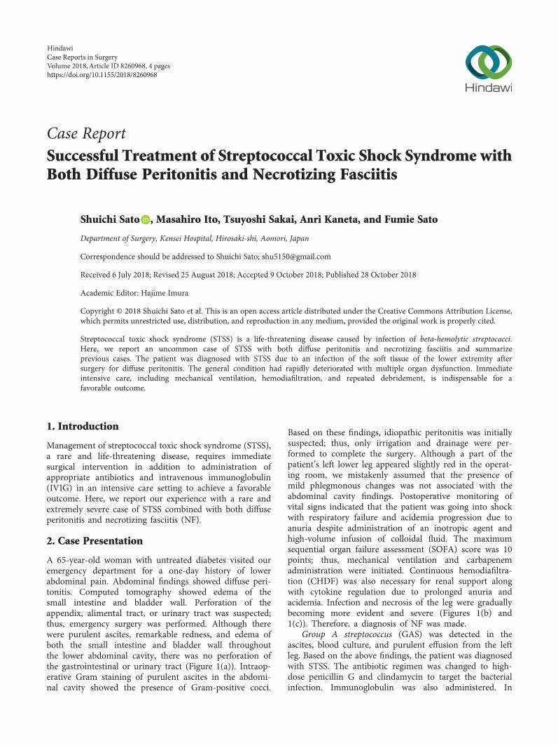
A 65-year-old woman with untreated diabetes visited ouremergency department for a one-day history of lowerabdominal pain. Abdominal findings showed diffuse peri-tonitis. Computed tomography showed edema of thesmall intestine and bladder wall. Perforation of theappendix, alimental tract, or urinary tract was suspected;thus, emergency surgery was performed. Although therewere purulent ascites, remarkable redness, and edema ofboth the small intestine and bladder wall throughoutthe lower abdominal cavity, there was no perforation ofthe gastrointestinal or urinary tract (Figure 1(a)). Intraop-erative Gram staining of purulent ascites in the abdomi-nal cavity showed the presence of Gram-positive cocci.
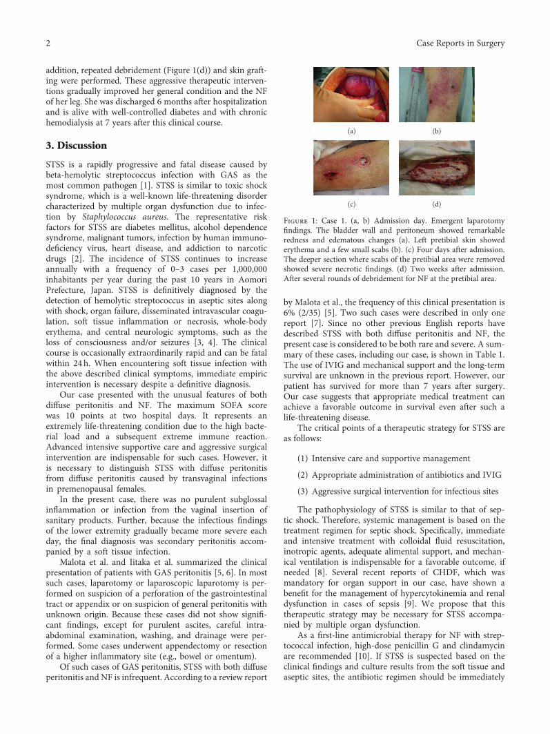
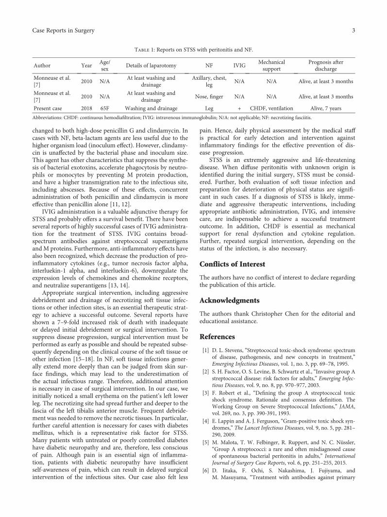
Based on these findings, idiopathic peritonitis was initiallysuspected; thus, only irrigation and drainage were per-formed to complete the surgery. Although a part of thepatient’s left lower leg appeared slightly red in the operat-ing room, we mistakenly assumed that the presence ofmild phlegmonous changes was not associated with theabdominal cavity findings. Postoperative monitoring ofvital signs indicated that the patient was going into shockwith respiratory failure and acidemia progression due toanuria despite administration of an inotropic agent andhigh-volume infusion of colloidal fluid. The maximumsequential organ failure assessment (SOFA) score was 10points; thus, mechanical ventilation and carbapenemadministration were initiated. Continuous hemodiafiltra-tion (CHDF) was also necessary for renal support alongwith cytokine regulation due to prolonged anuria andacidemia. Infection and necrosis of the leg were graduallybecoming more evident and severe (Figures 1(b) and1(c)). Therefore, a diagnosis of NF was made.
Group A streptococcus (GAS) was detected in theascites, blood culture, and purulent effusion from the leftleg. Based on the above findings, the patient was diagnosedwith STSS. The antibiotic regimen was changed to high-dose penicillin G and clindamycin to target the bacterialinfection. Immunoglobulin was also administered. In
HindawiCase Reports in SurgeryVolume 2018, Article ID 8260968, 4 pageshttps://doi.org/10.1155/2018/8260968
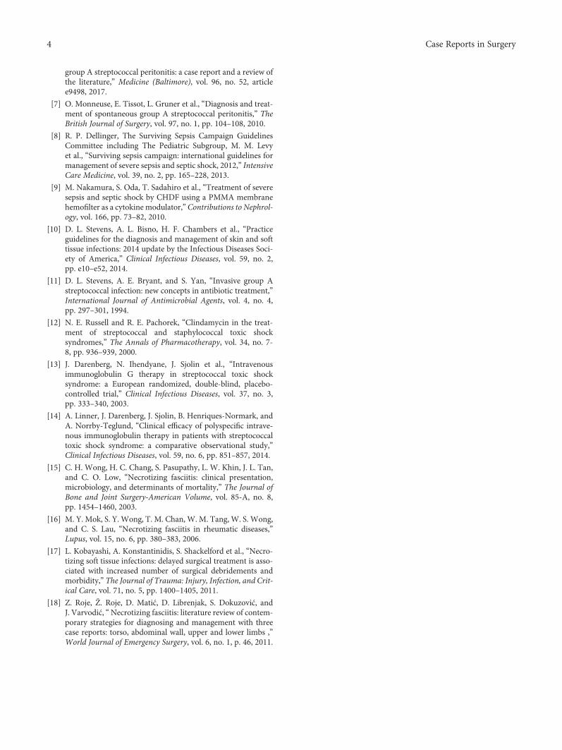
addition, repeated debridement (Figure 1(d)) and skin graft-ing were performed. These aggressive therapeutic interven-tions gradually improved her general condition and the NFof her leg. She was discharged 6 months after hospitalizationand is alive with well-controlled diabetes and with chronichemodialysis at 7 years after this clinical course.
3. Discussion
STSS is a rapidly progressive and fatal disease caused bybeta-hemolytic streptococcus infection with GAS as themost common pathogen [1]. STSS is similar to toxic shocksyndrome, which is a well-known life-threatening disordercharacterized by multiple organ dysfunction due to infec-tion by Staphylococcus aureus. The representative riskfactors for STSS are diabetes mellitus, alcohol dependencesyndrome, malignant tumors, infection by human immuno-deficiency virus, heart disease, and addiction to narcoticdrugs [2]. The incidence of STSS continues to increaseannually with a frequency of 0–3 cases per 1,000,000inhabitants per year during the past 10 years in AomoriPrefecture, Japan. STSS is definitively diagnosed by thedetection of hemolytic streptococcus in aseptic sites alongwith shock, organ failure, disseminated intravascular coagu-lation, soft tissue inflammation or necrosis, whole-bodyerythema, and central neurologic symptoms, such as theloss of consciousness and/or seizures [3, 4]. The clinicalcourse is occasionally extraordinarily rapid and can be fatalwithin 24h. When encountering soft tissue infection withthe above described clinical symptoms, immediate empiricintervention is necessary despite a definitive diagnosis.
Our case presented with the unusual features of bothdiffuse peritonitis and NF. The maximum SOFA scorewas 10 points at two hospital days. It represents anextremely life-threatening condition due to the high bacte-rial load and a subsequent extreme immune reaction.Advanced intensive supportive care and aggressive surgicalintervention are indispensable for such cases. However, itis necessary to distinguish STSS with diffuse peritonitisfrom diffuse peritonitis caused by transvaginal infectionsin premenopausal females.
In the present case, there was no purulent subglossalinflammation or infection from the vaginal insertion ofsanitary products. Further, because the infectious findingsof the lower extremity gradually became more severe eachday, the final diagnosis was secondary peritonitis accom-panied by a soft tissue infection.
Malota et al. and Iitaka et al. summarized the clinicalpresentation of patients with GAS peritonitis [5, 6]. In mostsuch cases, laparotomy or laparoscopic laparotomy is per-formed on suspicion of a perforation of the gastrointestinaltract or appendix or on suspicion of general peritonitis withunknown origin. Because these cases did not show signifi-cant findings, except for purulent ascites, careful intra-abdominal examination, washing, and drainage were per-formed. Some cases underwent appendectomy or resectionof a higher inflammatory site (e.g., bowel or omentum).
Of such cases of GAS peritonitis, STSS with both diffuseperitonitis and NF is infrequent. According to a review report
by Malota et al., the frequency of this clinical presentation is6% (2/35) [5]. Two such cases were described in only onereport [7]. Since no other previous English reports havedescribed STSS with both diffuse peritonitis and NF, thepresent case is considered to be both rare and severe. A sum-mary of these cases, including our case, is shown in Table 1.The use of IVIG and mechanical support and the long-termsurvival are unknown in the previous report. However, ourpatient has survived for more than 7 years after surgery.Our case suggests that appropriate medical treatment canachieve a favorable outcome in survival even after such alife-threatening disease.
The critical points of a therapeutic strategy for STSS areas follows:
(1) Intensive care and supportive management
(2) Appropriate administration of antibiotics and IVIG
(3) Aggressive surgical intervention for infectious sites
The pathophysiology of STSS is similar to that of sep-tic shock. Therefore, systemic management is based on thetreatment regimen for septic shock. Specifically, immediateand intensive treatment with colloidal fluid resuscitation,inotropic agents, adequate alimental support, and mechan-ical ventilation is indispensable for a favorable outcome, ifneeded [8]. Several recent reports of CHDF, which wasmandatory for organ support in our case, have shown abenefit for the management of hypercytokinemia and renaldysfunction in cases of sepsis [9]. We propose that thistherapeutic strategy may be necessary for STSS accompa-nied by multiple organ dysfunction.
As a first-line antimicrobial therapy for NF with strep-tococcal infection, high-dose penicillin G and clindamycinare recommended [10]. If STSS is suspected based on theclinical findings and culture results from the soft tissue andaseptic sites, the antibiotic regimen should be immediately
(a) (b)
(c) (d)
Figure 1: Case 1. (a, b) Admission day. Emergent laparotomyfindings. The bladder wall and peritoneum showed remarkableredness and edematous changes (a). Left pretibial skin showederythema and a few small scabs (b). (c) Four days after admission.The deeper section where scabs of the pretibial area were removedshowed severe necrotic findings. (d) Two weeks after admission.After several rounds of debridement for NF at the pretibial area.
2 Case Reports in Surgery
changed to both high-dose penicillin G and clindamycin. Incases with NF, beta-lactam agents are less useful due to thehigher organism load (inoculum effect). However, clindamy-cin is unaffected by the bacterial phase and inoculum size.This agent has other characteristics that suppress the synthe-sis of bacterial exotoxins, accelerate phagocytosis by neutro-phils or monocytes by preventing M protein production,and have a higher transmigration rate to the infectious site,including abscesses. Because of these effects, concurrentadministration of both penicillin and clindamycin is moreeffective than penicillin alone [11, 12].
IVIG administration is a valuable adjunctive therapy forSTSS and probably offers a survival benefit. There have beenseveral reports of highly successful cases of IVIG administra-tion for the treatment of STSS. IVIG contains broad-spectrum antibodies against streptococcal superantigensandM proteins. Furthermore, anti-inflammatory effects havealso been recognized, which decrease the production of pro-inflammatory cytokines (e.g., tumor necrosis factor alpha,interluekin-1 alpha, and interluekin-6), downregulate theexpression levels of chemokines and chemokine receptors,and neutralize superantigens [13, 14].
Appropriate surgical intervention, including aggressivedebridement and drainage of necrotizing soft tissue infec-tions or other infection sites, is an essential therapeutic strat-egy to achieve a successful outcome. Several reports haveshown a 7–9-fold increased risk of death with inadequateor delayed initial debridement or surgical intervention. Tosuppress disease progression, surgical intervention must beperformed as early as possible and should be repeated subse-quently depending on the clinical course of the soft tissue orother infection [15–18]. In NF, soft tissue infections gener-ally extend more deeply than can be judged from skin sur-face findings, which may lead to the underestimation ofthe actual infectious range. Therefore, additional attentionis necessary in case of surgical intervention. In our case, weinitially noticed a small erythema on the patient’s left lowerleg. The necrotizing site had spread further and deeper to thefascia of the left tibialis anterior muscle. Frequent debride-ment was needed to remove the necrotic tissues. In particular,further careful attention is necessary for cases with diabetesmellitus, which is a representative risk factor for STSS.Many patients with untreated or poorly controlled diabeteshave diabetic neuropathy and are, therefore, less consciousof pain. Although pain is an essential sign of inflamma-tion, patients with diabetic neuropathy have insufficientself-awareness of pain, which can result in delayed surgicalintervention of the infectious sites. Our case also felt less
pain. Hence, daily physical assessment by the medical staffis practical for early detection and intervention againstinflammatory findings for the effective prevention of dis-ease progression.
STSS is an extremely aggressive and life-threateningdisease. When diffuse peritonitis with unknown origin isidentified during the initial surgery, STSS must be consid-ered. Further, both evaluation of soft tissue infection andpreparation for deterioration of physical status are signifi-cant in such cases. If a diagnosis of STSS is likely, imme-diate and aggressive therapeutic interventions, includingappropriate antibiotic administration, IVIG, and intensivecare, are indispensable to achieve a successful treatmentoutcome. In addition, CHDF is essential as mechanicalsupport for renal dysfunction and cytokine regulation.Further, repeated surgical intervention, depending on thestatus of the infection, is also necessary.
Conflicts of Interest
The authors have no conflict of interest to declare regardingthe publication of this article.
Acknowledgments
The authors thank Christopher Chen for the editorial andeducational assistance.
References
[1] D. L. Stevens, “Streptococcal toxic-shock syndrome: spectrumof disease, pathogenesis, and new concepts in treatment,”Emerging Infectious Diseases, vol. 1, no. 3, pp. 69–78, 1995.
[2] S. H. Factor, O. S. Levine, B. Schwartz et al., “Invasive group Astreptococcal disease: risk factors for adults,” Emerging Infec-tious Diseases, vol. 9, no. 8, pp. 970–977, 2003.
[3] F. Robert et al., “Defining the group A streptococcal toxicshock syndrome. Rationale and consensus definition. TheWorking Group on Severe Streptococcal Infections,” JAMA,vol. 269, no. 3, pp. 390-391, 1993.
[4] E. Lappin and A. J. Ferguson, “Gram-positive toxic shock syn-dromes,” The Lancet Infectious Diseases, vol. 9, no. 5, pp. 281–290, 2009.
[5] M. Malota, T. W. Felbinger, R. Ruppert, and N. C. Nüssler,“Group A streptococci: a rare and often misdiagnosed causeof spontaneous bacterial peritonitis in adults,” InternationalJournal of Surgery Case Reports, vol. 6, pp. 251–255, 2015.
[6] D. Iitaka, F. Ochi, S. Nakashima, J. Fujiyama, andM. Masuyama, “Treatment with antibodies against primary
Table 1: Reports on STSS with peritonitis and NF.
Author YearAge/sex
Details of laparotomy NF IVIGMechanicalsupport
Prognosis afterdischarge
Monneuse et al.[7]
2010 N/AAt least washing and
drainageAxillary, chest,
legN/A N/A Alive, at least 3 months
Monneuse et al.[7]
2010 N/AAt least washing and
drainageNose, finger N/A N/A Alive, at least 3 months
Present case 2018 65F Washing and drainage Leg + CHDF, ventilation Alive, 7 years
Abbreviations: CHDF: continuous hemodiafiltration; IVIG: intravenous immunoglobulin; N/A: not applicable; NF: necrotizing fasciitis.
3Case Reports in Surgery
group A streptococcal peritonitis: a case report and a review ofthe literature,” Medicine (Baltimore), vol. 96, no. 52, articlee9498, 2017.
[7] O. Monneuse, E. Tissot, L. Gruner et al., “Diagnosis and treat-ment of spontaneous group A streptococcal peritonitis,” TheBritish Journal of Surgery, vol. 97, no. 1, pp. 104–108, 2010.
[8] R. P. Dellinger, The Surviving Sepsis Campaign GuidelinesCommittee including The Pediatric Subgroup, M. M. Levyet al., “Surviving sepsis campaign: international guidelines formanagement of severe sepsis and septic shock, 2012,” IntensiveCare Medicine, vol. 39, no. 2, pp. 165–228, 2013.
[9] M. Nakamura, S. Oda, T. Sadahiro et al., “Treatment of severesepsis and septic shock by CHDF using a PMMA membranehemofilter as a cytokine modulator,” Contributions to Nephrol-ogy, vol. 166, pp. 73–82, 2010.
[10] D. L. Stevens, A. L. Bisno, H. F. Chambers et al., “Practiceguidelines for the diagnosis and management of skin and softtissue infections: 2014 update by the Infectious Diseases Soci-ety of America,” Clinical Infectious Diseases, vol. 59, no. 2,pp. e10–e52, 2014.
[11] D. L. Stevens, A. E. Bryant, and S. Yan, “Invasive group Astreptococcal infection: new concepts in antibiotic treatment,”International Journal of Antimicrobial Agents, vol. 4, no. 4,pp. 297–301, 1994.
[12] N. E. Russell and R. E. Pachorek, “Clindamycin in the treat-ment of streptococcal and staphylococcal toxic shocksyndromes,” The Annals of Pharmacotherapy, vol. 34, no. 7-8, pp. 936–939, 2000.
[13] J. Darenberg, N. Ihendyane, J. Sjolin et al., “Intravenousimmunoglobulin G therapy in streptococcal toxic shocksyndrome: a European randomized, double-blind, placebo-controlled trial,” Clinical Infectious Diseases, vol. 37, no. 3,pp. 333–340, 2003.
[14] A. Linner, J. Darenberg, J. Sjolin, B. Henriques-Normark, andA. Norrby-Teglund, “Clinical efficacy of polyspecific intrave-nous immunoglobulin therapy in patients with streptococcaltoxic shock syndrome: a comparative observational study,”Clinical Infectious Diseases, vol. 59, no. 6, pp. 851–857, 2014.
[15] C. H. Wong, H. C. Chang, S. Pasupathy, L. W. Khin, J. L. Tan,and C. O. Low, “Necrotizing fasciitis: clinical presentation,microbiology, and determinants of mortality,” The Journal ofBone and Joint Surgery-American Volume, vol. 85-A, no. 8,pp. 1454–1460, 2003.
[16] M. Y. Mok, S. Y. Wong, T. M. Chan, W. M. Tang, W. S. Wong,and C. S. Lau, “Necrotizing fasciitis in rheumatic diseases,”Lupus, vol. 15, no. 6, pp. 380–383, 2006.
[17] L. Kobayashi, A. Konstantinidis, S. Shackelford et al., “Necro-tizing soft tissue infections: delayed surgical treatment is asso-ciated with increased number of surgical debridements andmorbidity,” The Journal of Trauma: Injury, Infection, and Crit-ical Care, vol. 71, no. 5, pp. 1400–1405, 2011.
[18] Z. Roje, Ž. Roje, D. Matić, D. Librenjak, S. Dokuzović, andJ. Varvodić, “Necrotizing fasciitis: literature review of contem-porary strategies for diagnosing and management with threecase reports: torso, abdominal wall, upper and lower limbs ,”World Journal of Emergency Surgery, vol. 6, no. 1, p. 46, 2011.
4 Case Reports in Surgery

Stem Cells International
Hindawiwww.hindawi.com Volume 2018
Hindawiwww.hindawi.com Volume 2018
MEDIATORSINFLAMMATION
of
EndocrinologyInternational Journal of
Hindawiwww.hindawi.com Volume 2018
Hindawiwww.hindawi.com Volume 2018
Disease Markers
Hindawiwww.hindawi.com Volume 2018
BioMed Research International
OncologyJournal of
Hindawiwww.hindawi.com Volume 2013
Hindawiwww.hindawi.com Volume 2018
Oxidative Medicine and Cellular Longevity
Hindawiwww.hindawi.com Volume 2018
PPAR Research
Hindawi Publishing Corporation http://www.hindawi.com Volume 2013Hindawiwww.hindawi.com
The Scientific World Journal
Volume 2018
Immunology ResearchHindawiwww.hindawi.com Volume 2018
Journal of
ObesityJournal of
Hindawiwww.hindawi.com Volume 2018
Hindawiwww.hindawi.com Volume 2018
Computational and Mathematical Methods in Medicine
Hindawiwww.hindawi.com Volume 2018
Behavioural Neurology
OphthalmologyJournal of
Hindawiwww.hindawi.com Volume 2018
Diabetes ResearchJournal of
Hindawiwww.hindawi.com Volume 2018
Hindawiwww.hindawi.com Volume 2018
Research and TreatmentAIDS
Hindawiwww.hindawi.com Volume 2018
Gastroenterology Research and Practice
Hindawiwww.hindawi.com Volume 2018
Parkinson’s Disease
Evidence-Based Complementary andAlternative Medicine
Volume 2018Hindawiwww.hindawi.com
Submit your manuscripts atwww.hindawi.com


![VirtualSurgicalPlanningandPiezoelectricSurgeryinTumor ...downloads.hindawi.com/journals/cris/2017/4397178.pdf · [7,8].Despitetheirnormalslowgrowthprocess,signicant growthpotentialcanbeobservediftheselesionsareleft](https://img.pdfslide.us/doc/110x75/5f86515c45015f14175f83d1/virtualsurgicalplanningandpiezoelectricsurgeryintumor-78despitetheirnormalslowgrowthprocesssignicant.jpg)