Embed Size (px)
Citation preview

CASE REPORT
Successful Use of Squeezed-Fat Grafts to Correct a BreastAffected by Poland Syndrome
Hyunjin Yang • Heeyoung Lee
Received: 28 February 2010 / Accepted: 19 August 2010 / Published online: 17 October 2010
� The Author(s) 2010. This article is published with open access at Springerlink.com
Abstract This study attempted to reconstruct deformities
of a Poland syndrome patient using autologous fat tissues.
All injected fat tissues were condensed by squeezing cen-
trifugation. Operations were performed four times with
intervals over 6 months. The total injection volume was
972 ml, and the maintained volume of 628 ml was mea-
sured by means of a magnetic resonance image (MRI). The
entire follow-up period was 4.5 years. After surgery, sev-
eral small cysts and minimal calcifications were present but
no significant complications. The cosmetic outcomes and
volume maintenance rates were excellent despite the
overlapped large-volume injections. In conclusion, higher
condensation of fat tissues through squeezing centrifuga-
tion would help to achieve better results in volume main-
tenance and reduce complications. It is necessary, however,
to perform more comparative studies with many clinical
cases for a more scientific analysis. The study experiments
with squeezed fat simply suggest a hypothesis that
squeezing centrifugation could select healthier cells
through pressure disruption of relatively thinner mem-
branes of larger, more vulnerable and more mature fat
cells.
Keywords Breast fat augmentation � Condensed fat � Fat
gel � Poland syndrome � Squeezed fat
Poland syndrome is a birth defect with a variety of asso-
ciated anomalies occurring around the upper limbs and
torso. It may include ipsilateral deformities such as
underdevelopment or absence of the main chest muscle
(pectoralis major) and secondary muscles of the chest and
axillary region; missing sternal head of the pectoralis
muscle; underdevelopment or absence of the nipple, areola,
and (in women) underlying breast tissues; hypoplastic skin
and subcutaneous layers; missing portions of the ribs or
costal cartilages; missing axillary hair; and short, webbed
fingers (cutaneous syndactyly) and a shortened forearm [4].
The main concerns for women with Poland syndrome
usually are aesthetic corrections of the breast rather than its
functional restoration [4].
For this procedure, we used a large-volume injection of
fat, which generally is not recommended for this type of
procedure. The cosmetic result exceeded our expectations.
A magnetic resonance image (MRI) showed an increase in
the maintenance of fat graft volume after performance of a
breast augmentation for the patient with Poland syndrome.
The formation of cysts was minimal.
For several decades, many reports and articles have
noted the significant problems associated with autologous
fat injections into the breasts [5]. On the other hand,
significant developments of the methods for autologous
fat injections into the breasts have provided improved
results [6].
Although the mechanism contributing to the increased
graft survival rate in large-volume fat (or tissues) injection
into the breast of the patient with Poland syndrome calls for
further research, various possibilities are discussed.
Assuming that the exact mechanism is found, this proce-
dure would be a viable alternative for the treatment of
congenital breast deformity and postmastectomy conditions
in patients with breast cancer.
H. Yang � H. Lee (&)
Kangnam Plastic Surgery, 135-891, Gujung Building 3rd Floor,
Shinsa-Dong 577-7, Kangnam-Gu, Seoul, South Korea
e-mail: [email protected]
123
Aesth Plast Surg (2011) 35:418–425
DOI 10.1007/s00266-010-9601-z

Materials and Methods
Materials
We used an aseptic squeezing centrifugation lipotransfer
(SCL) system (Lipokit; Medikan Co. Ltd., Seoul, Korea)
that increases the density of adipose-derived stem cells
(ASCs) and interstitial structures through the removal of
older fat cells and liquid triglycerides. This procedure
provides an all-in-one, closed-circuit process using a sin-
gle-use, 50-ml syringe container with a weight-mesh-fil-
tering piston plunger. During the entire procedure, fat
always stays within the same single-use syringe. During the
centrifugation, the weight-mesh-filtering piston squeezes
the liposuction aspirates within the same syringe, disrupt-
ing the larger and older vulnerable fat cells and condensing
the fat tissues while simultaneously removing liquid tri-
glycerides and fluid impurities.
In the other preparations, this technique was not signif-
icantly different from the conventional syringe techniques.
Tumescent fluid was made using 1 l of Hartmann’s solution
with 2 ml of epinephrine (1 mg/1 ml). The tumescent fluid
injection was made with compressed air using the same
syringe.
Centrifugation was performed at 3,000 g for 5 min with
the same syringes used in harvesting. Injection of pro-
cessed fat also was performed with the same syringes using
compressed air and a manual spiral plunger. During fat
injection, we tried to disperse the fat evenly. We injected
fat into the soft tissues so that we would not invade
intercostal spaces.
We instructed the patient not to use the right arm vig-
orously for 2 weeks. We did not use a massage technique,
which usually is applied with silicon implantation tech-
niques. We injected first-generation cephalosporin as a
prophylactic antibiotic 1 h before a surgery. Oral antibi-
otics were used for 5 days after the operations.
Patients and Methods
A 23-year-old woman showed several signs of Poland
syndrome on her right side including absence of the right
breast, defects of chest muscles such as the pectoralis
major, defects in the ribs and costal cartilages, and webbing
of the axillary fold, hypoplastic skin, and subcutaneous
tissues. The patient’s rib cage also was unusually small.
Her height was 160 cm, and her body weight was 57 kg.
The latter gradually increased by 6 kg and was at 63 kg at
the final surgery.
The patient did feel some discomfort, but her contra-
lateral side, including the breast, was well developed. She
underwent several plastic surgery consultations that offered
the possibility of free flaps but required the sacrifice of
other body parts for breast reconstruction. Flap surgery was
not an option because the patient did not want any type of
‘‘bodily sacrifice’’ or scarring. Therefore, we at first rec-
ommended using a skin expander and a silicon implant.
The patient also refused this option due to the size limi-
tation of the implant and the possible asymmetry of the
depressed chest wall. Finally, we recommended serial fat
injections.
We performed the first surgery on 12 February 2005.
During the surgery, we injected 220 ml of autologous fat
into the right breast. The second surgery was performed
nearly 1 year later, on 3 January 2006. For the second
surgery, we injected 212 ml of fat.
We performed mammography at 6-month intervals. A
breast MRI was performed on 24 July 2006. Abnormalities
such as masses, cysts, and calcifications were not present at
the final mammography. As a result, we decided to perform
a third operation. We recommended using a cohesive gel
implant, but the patient also refused this option due to
possible complications arising from a foreign material
inside her body. As a result, we performed the third surgery
on 5 January 2007, injecting 295 ml of fat.
Results
We performed a breast MRI 6 months after the third surgery
on 5 January 2007, approximately 2 years after the first fat
injection. The MRI indicated no significant problems,
including no instance of capsule formation. We planned to
perform the final fat injection of 240 ml on 3 March 2008.
We then performed the final mammography, sonography,
and size-calculable MRI with a dye contrast. We found
several small benign cysts. Based on our 269 cases dealing
with fat injections into the breast, we considered these small
cysts to be a minimal problem. The cysts could be treated
easily by sono-guided needle aspiration. However, the
remaining capsule still carried the possibility of microcal-
cification development, which in turn could have caused
confusion in the diagnosis by radiologists.
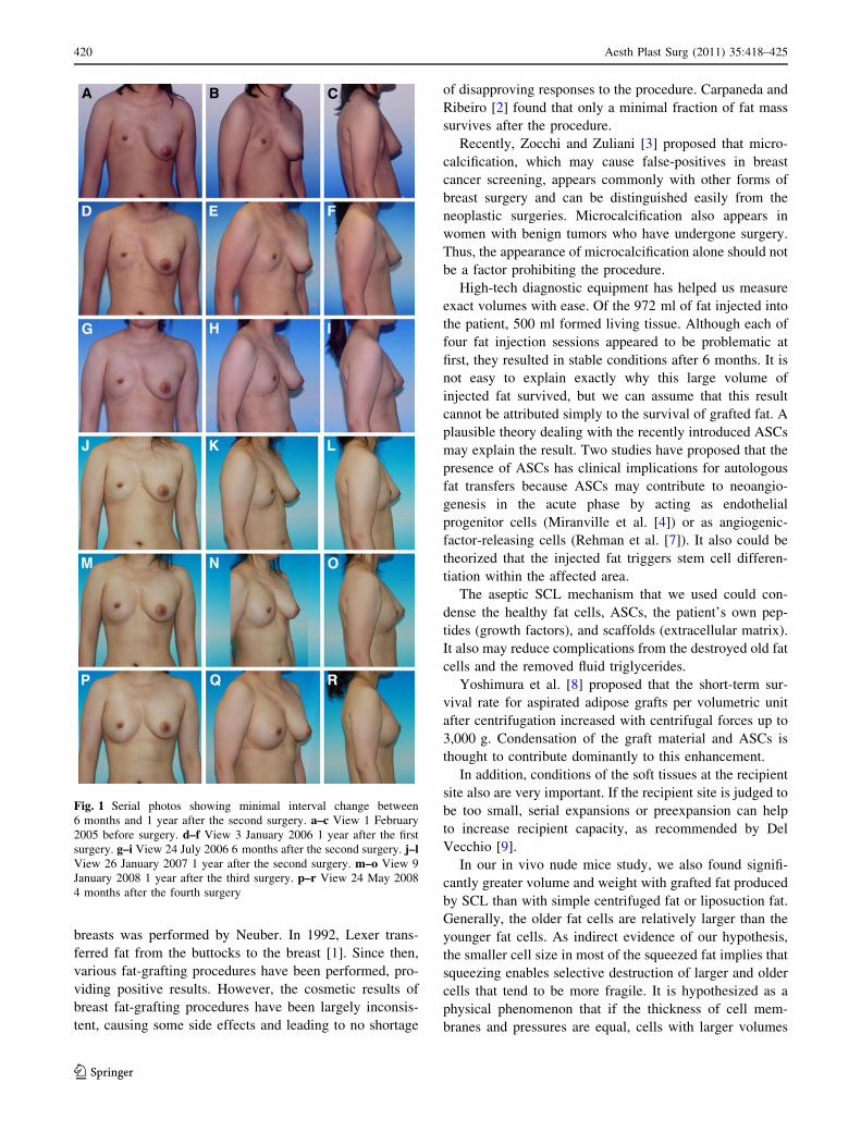
There was no lump or hardness after surgery, and the
patient’s satisfaction was very high. Serial photos showed
little interval change and good skin expansion (Fig. 1). All
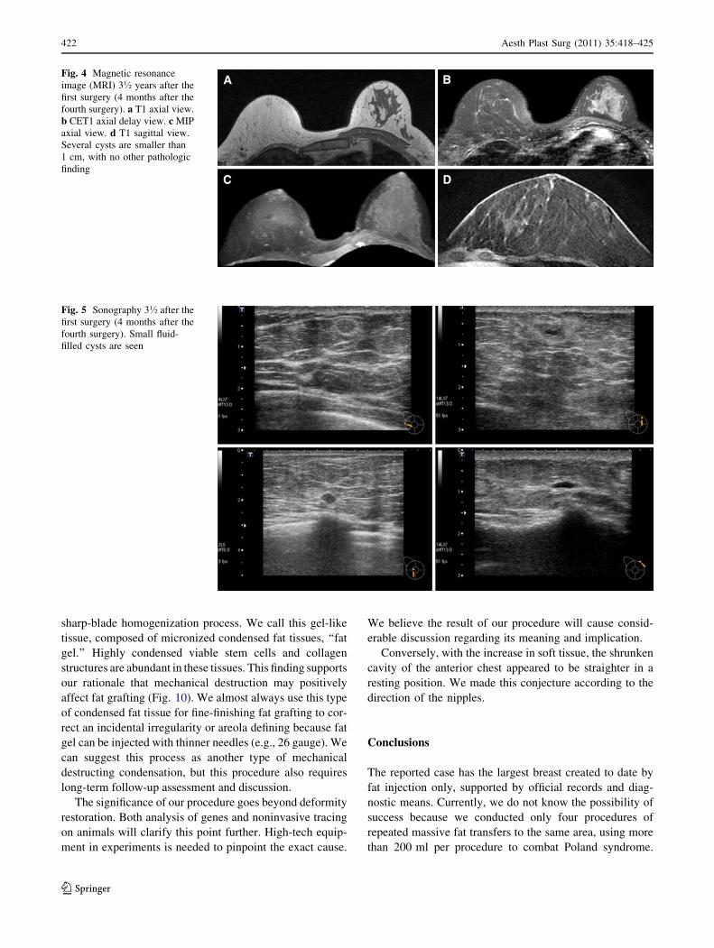
radiologic findings showed only small cysts and no other
abnormal findings (Figs. 2, 3, 4, 5). The MRI digital vol-
ume calculation showed 627 ml of volume in the right
breast (Fig. 6).
Discussion
The transfer of autologous fat dates back to the 1890s. In
1890, fat transplantation to body parts other than the
Aesth Plast Surg (2011) 35:418–425 419
123
breasts was performed by Neuber. In 1992, Lexer trans-
ferred fat from the buttocks to the breast [1]. Since then,
various fat-grafting procedures have been performed, pro-
viding positive results. However, the cosmetic results of
breast fat-grafting procedures have been largely inconsis-
tent, causing some side effects and leading to no shortage
of disapproving responses to the procedure. Carpaneda and
Ribeiro [2] found that only a minimal fraction of fat mass
survives after the procedure.
Recently, Zocchi and Zuliani [3] proposed that micro-
calcification, which may cause false-positives in breast
cancer screening, appears commonly with other forms of
breast surgery and can be distinguished easily from the
neoplastic surgeries. Microcalcification also appears in
women with benign tumors who have undergone surgery.
Thus, the appearance of microcalcification alone should not
be a factor prohibiting the procedure.
High-tech diagnostic equipment has helped us measure
exact volumes with ease. Of the 972 ml of fat injected into
the patient, 500 ml formed living tissue. Although each of
four fat injection sessions appeared to be problematic at
first, they resulted in stable conditions after 6 months. It is
not easy to explain exactly why this large volume of
injected fat survived, but we can assume that this result
cannot be attributed simply to the survival of grafted fat. A
plausible theory dealing with the recently introduced ASCs
may explain the result. Two studies have proposed that the
presence of ASCs has clinical implications for autologous
fat transfers because ASCs may contribute to neoangio-
genesis in the acute phase by acting as endothelial
progenitor cells (Miranville et al. [4]) or as angiogenic-
factor-releasing cells (Rehman et al. [7]). It also could be
theorized that the injected fat triggers stem cell differen-
tiation within the affected area.
The aseptic SCL mechanism that we used could con-
dense the healthy fat cells, ASCs, the patient’s own pep-
tides (growth factors), and scaffolds (extracellular matrix).
It also may reduce complications from the destroyed old fat
cells and the removed fluid triglycerides.
Yoshimura et al. [8] proposed that the short-term sur-
vival rate for aspirated adipose grafts per volumetric unit
after centrifugation increased with centrifugal forces up to
3,000 g. Condensation of the graft material and ASCs is
thought to contribute dominantly to this enhancement.
In addition, conditions of the soft tissues at the recipient
site also are very important. If the recipient site is judged to
be too small, serial expansions or preexpansion can help
to increase recipient capacity, as recommended by Del
Vecchio [9].
In our in vivo nude mice study, we also found signifi-
cantly greater volume and weight with grafted fat produced
by SCL than with simple centrifuged fat or liposuction fat.
Generally, the older fat cells are relatively larger than the
younger fat cells. As indirect evidence of our hypothesis,
the smaller cell size in most of the squeezed fat implies that
squeezing enables selective destruction of larger and older
cells that tend to be more fragile. It is hypothesized as a
physical phenomenon that if the thickness of cell mem-
branes and pressures are equal, cells with larger volumes
Fig. 1 Serial photos showing minimal interval change between
6 months and 1 year after the second surgery. a–c View 1 February
2005 before surgery. d–f View 3 January 2006 1 year after the first
surgery. g–i View 24 July 2006 6 months after the second surgery. j–lView 26 January 2007 1 year after the second surgery. m–o View 9
January 2008 1 year after the third surgery. p–r View 24 May 2008
4 months after the fourth surgery
420 Aesth Plast Surg (2011) 35:418–425
123

should endure mechanical stress, such as squeezing pres-
sure, with a relatively smaller surface area than cells with
smaller volumes. We regarded this squeezing process as a
kind of preliminary pressure stage because all injecting fat
must pass through high-pressure environments during
injection in syringes, needles, and recipient tissues.
We used a 22-g piston plunger 2.6 cm in diameter and
centrifuged at 3,000 g for 5 min. We were able to calculate
the pressure within fat tissues during centrifugation by
assessing the actual weight (0.022 kg 9 3,000) and cut
surface area (3.14 9 1.3 9 1.3 cm). The pressure within
the fat tissue was 12.5 kg/cm2. We also tried to evaluate
the harmful influences of this squeezing process by com-
paring the viabilities of 1 ml of crude fat and 1 ml of
squeezed fat. The total number of ASCs increased in unit
volume, but the viability comparison did not show signif-
icant differences (Fig. 7a–c). This finding implies that
actual condensation occurred and that the squeezing pro-
cess did not have significant harmful effects on ASC.
In our other experimental trials, we injected squeezed
fat, normal fat, and simple centrifuged fat into nude mice
(n = 10) and killed the mice at 8 weeks. We compared the
volume, weight, and histologic findings of each fat graft.
The average size of a fat cell in each sample was compared
in microscopic fields. We counted the number of pixels
within each cell membrane and compared the average
number of pixels of fat cell size before injection.
As a result, the survival rate for injected squeezed fat
was significantly higher than for the fats of other groups
(Fig. 8a–c). The average cell size in the squeezed fat was
significantly smaller than in the centrifuged fat (Fig. 9a–c).
It may be thought that the findings emerged by coinci-
dence, but it is important to note the involvement of
scaffolds and cell signals in the growth of undifferentiated
stem cells and body tissues. Therefore, we suggest that the
more destructed forms of fat tissues can be regarded as the
more exaggerated experimental models of destructed fat.
Gross tissue destruction usually indicates a reduction in
tissue viability, but our destruction method of squeezing fat
did not reduce tissue viability.
Fat tissues are composed not only of adipocytes but also
of stem cells, scaffolds, and growth signals such as growth
factors. We therefore have hypothesized that targets of
condensation may not be adipocytes. In small-volume
fat grafts, we often use gel-like fat tissues made by the
Fig. 2 Magnetic resonance image (MRI) 1 year after second surgery. a T2 axial view. b T1 axial view. c T1 sagittal view. Several cysts are
smaller than 1 cm, with no other pathologic findings
Fig. 3 Mammography 3� years after the first surgery (4 months
after the fourth surgery). a CC view. b MLO view. No significant
calcification is seen
Aesth Plast Surg (2011) 35:418–425 421
123
sharp-blade homogenization process. We call this gel-like
tissue, composed of micronized condensed fat tissues, ‘‘fat
gel.’’ Highly condensed viable stem cells and collagen
structures are abundant in these tissues. This finding supports
our rationale that mechanical destruction may positively
affect fat grafting (Fig. 10). We almost always use this type
of condensed fat tissue for fine-finishing fat grafting to cor-
rect an incidental irregularity or areola defining because fat
gel can be injected with thinner needles (e.g., 26 gauge). We
can suggest this process as another type of mechanical
destructing condensation, but this procedure also requires
long-term follow-up assessment and discussion.
The significance of our procedure goes beyond deformity
restoration. Both analysis of genes and noninvasive tracing
on animals will clarify this point further. High-tech equip-
ment in experiments is needed to pinpoint the exact cause.
We believe the result of our procedure will cause consid-
erable discussion regarding its meaning and implication.
Conversely, with the increase in soft tissue, the shrunken
cavity of the anterior chest appeared to be straighter in a
resting position. We made this conjecture according to the
direction of the nipples.
Conclusions
The reported case has the largest breast created to date by
fat injection only, supported by official records and diag-
nostic means. Currently, we do not know the possibility of
success because we conducted only four procedures of
repeated massive fat transfers to the same area, using more
than 200 ml per procedure to combat Poland syndrome.
Fig. 5 Sonography 3� after the
first surgery (4 months after the
fourth surgery). Small fluid-
filled cysts are seen
Fig. 4 Magnetic resonance
image (MRI) 3� years after the
first surgery (4 months after the
fourth surgery). a T1 axial view.
b CET1 axial delay view. c MIP
axial view. d T1 sagittal view.
Several cysts are smaller than
1 cm, with no other pathologic
finding
422 Aesth Plast Surg (2011) 35:418–425
123

Fig. 6 Magnetic resonance
image (MRI) volume
calculation 3� years after the
first surgery (4 months after the
fourth surgery). a–c Left breast,
normal side with no operation
(412.876 cm3). d–f Right
breast, affected side with four
operations (627.701 cm3)
Fig. 7 Cell counts and viabilities in simple centrifuged fat and
squeezed fat. a Numbers of viable cells in simple centrifuged fat and
squeezed fat, respectively. b Numbers of cells in simple centrifuged
fat and squeezed fat, respectively. c Cell viability in simple
centrifuged fat and squeezed fat, respectively
Aesth Plast Surg (2011) 35:418–425 423
123

Fig. 8 Morphologic analysis comparing simple centrifuged fat and
squeezed fat. A (a,c,d) Left: Simple centrifuged fat. Right: Squeezed
fat. b Upper: Squeezed fat. Lower: Simple centrifuged fat. B Com-
parative analysis of the remaining fat volumes of simple centrifuged
fat versus squeezed fat. C Graph showing the percentage of the
remaining fat volume after 4 and 8 weeks in simple centrifuged fat
and squeezed fat, respectively
Fig. 9 A (a,b) Simple centrifuged fat. a: X50. b: X100.
(c,d) Squeezed fat. c: X50. d: X100. Violet (nucleus). Pink red(cytoplasm). B Comparison of the fat cell size in the simple centrifuged
fat and squeezed fat by measuring the number of pixels using the
Adobe Photoshop CS2 program (Adobe, CA, USA). C Number of
pixels used by the Adobe Photoshop CS2 program. To assess the size
of the Coleman fat and the squeezed fat quantitatively, we randomly
selected 30 single fat cells from five fat photos (9100) in each group
and then measured the pixel numbers of each fat image used for the
polygonal Lasso Tool in the Adobe Photoshop CS2 program
424 Aesth Plast Surg (2011) 35:418–425
123

For additional analysis of this mechanism, more investi-
gations and cases are needed.
In conclusion, a higher condensation of fat tissues
through squeezing centrifugation would improve results,
increase volume maintenance, and reduce complications.
Again, however, it is necessary to perform further com-
parative studies using several clinical cases to achieve a
more scientific analysis.
Conflict of interest The author has no conflict of interest to declare
with regard to this article.
Open Access This article is distributed under the terms of the
Creative Commons Attribution Noncommercial License which per-
mits any noncommercial use, distribution, and reproduction in any
medium, provided the original author(s) and source are credited.
References
1. Hinderer UT, Del Rio JL (1992) Erich Lexer’s mammaplasty.
Aesthetic Plast Surg 16:101–107
2. Carpaneda CA, Ribeiro MT (1993) Study of the histological
alterations and viability of the adipose graft in humans. Aesthetic
Plast Surg 17:43–47
3. Zocchi ML, Zuliani F (2008) Bicompartmental breast lipostruc-
turing. Aesthetic Plast Surg 32:313–328
4. Miranville A, Heeschen C, Sengenes C et al (2004) Improvement
of postnatal neovascularization by human adipose tissue-derived
stem cells. Circulation 110:349
5. Pohl P, Uebel O (1985) Complications with homologous fat grafts
in breast augmentation surgery. Aesthetic Plast Surg 9:87–89
6. Coleman S, Saboeiro A (2007) Fat grafting to the breast revisited:
safety and efficacy. Plast Reconstr Surg 119:775–785
7. Rehman J, Traktuev D, Li J et al (2004) Secretion of angiogenic
and antiapoptotic factors by human adipose stromal cells. Circu-
lation 109:1292
8. Yoshimura K, Harii K et al (2008) Influences of centrifugation on
cells and tissues in liposuction aspirates: optimized centrifugation
for lipotransfer and cell isolation. Plast Reconstr Surg 121:1033
9. Del Vecchio D (2009) Breast reconstruction for breast asymmetry
using recipient-site preexpansion and autologous fat grafting: a
case report. Ann Plast Surg 62:523–527
Fig. 10 Highly condensed fat tissues (‘‘fat gel’’) using sharp-blade
medical homogenizer. a Morphology of fat gel made by the medical
homogenizer (Filler-Geller; Medikan Co. Ltd., Seoul, Korea). b Gross
morphology of fat gel. c Histologic analysis of fat gel. Hematoxylin
and eosin (H&E) staining of fresh fat gel. Violet (nucleus). Pink
(cytoplasm). White rectangle (location at which the stem cell is highly
condensed). d Comparison of volume maintenance between squeezed
fat and fat gel in nude mice after 4 weeks. e Cell-counting analysis
comparing squeezed fat and fat gel. f Comparison of adipose-derived
stem cell (ASC) viability between squeezed fat and fat gel
Aesth Plast Surg (2011) 35:418–425 425
123