Embed Size (px)
Citation preview
The Korean Journal of Internal Medicine: 21:283-286, 2006
∙Received : February 27, 2006
∙Accepted : April 11, 2006
∙Correspondence to : Dong-Gu Shin, M.D., Division of Cardiology, Department of Internal Medicine, Yeung-nam University Hospital, #317-1
Daemyung-Dong, Nam-Gu, Daegu 705-717, Korea Tel : 82-53-620-3843, Fax : 82-53-654-8386, E-mail : [email protected]
Successful Treatment of Ischemic Dysfunction of the
Sinus Node with Thrombolytic Therapy:
A Case Report
Jong-Seon Park, M.D., Dong-Gu Shin, M.D., Young-Jo Kim, M.D., Gu-Ru Hong, M.D., Hyung-Jun Kim, M.D. and Bong-Sup Shim, M.D.
Division of Cardiology, Department of Internal Medicine, Yeung-nam University Hospital, Daegu, Korea
We report on a case of ischemic dysfunction of the sinus node as a complication after percutaneous transluminal
coronary angioplasty of the distal left circumflex artery. After local thrombolytic therapy in the sinus node artery, sinus
node arterial flow was re-established and sinus node function normalized over the period of a week. Our experience
suggests that immediate reperfusion of a totally occluded nodal artery can be re-established. Ischemic dysfunction of
the sinus node, as a complication of angioplasty, is generally transient and requires a prolonged period for recovery.
Therefore the decision to implant a permanent pacemaker should be delayed for at least one week after the ischemic
insult.
Key Words : Angioplasty, Complication, Sinus node dysfunction
INTRODUCTION
Recent advances in new devices and skills have broadened
the indications of percutaneous coronary intervention (PCI).
However, complications such as dissection, perforation, side
branch jail and distal embolization of plaque materials and
thrombi remain problems that can result in catastrophic
outcomes1-3). There may be thrombi and plaque materials
present related to the acute coronary syndrome, which have a
high risk for embolization of neighboring arterial branches as
well as distal small artery branches. The artery supplying the
sinus node (SN) is very small and difficult to protect from jailing
or plaque embolization during PCI. Acute occlusion of the SN
artery can occur during PCI of proximal lesions of the right
coronary artery (RCA) or the left circumflex artery (LCX)4).
Therefore understanding of the prognosis of ischemic sinus
node dysfunction is important for deciding on the need for
permanent pacemaker implantation. We describe here the first
reported case of transient sinus node dysfunction as a
complication of balloon angioplasty in the distal LCX.
CASE REPORT
A 56-year-old male was admitted to the hospital with resting
chest pain that developed three days before admission. He had
effort-induced chest pain for three years, but did not take any
medications and had no prior cardiac evaluations. Physical
examination findings, blood pressure and heart rate were all
normal. An echocardiography showed normal left ventricular wall
motion and a normal ejection fraction. The electrocardiogram
showed T wave inversions in lead aVL. The coronary
angiography revealed a 50% discrete stenosis at the ostium of
the left anterior descending artery (LAD), an 80% tubular
stenosis at the intermedius branch, and a 90% tubular stenosis
at the distal LCX (Figure 1A). A ramus intermedius angioplasty
was successfully performed with placement of a sirolimus-
eluting stent (Cypher, Johnson & Johnson). We performed a
balloon dilatation with a balloon, 2.25 mm in diameter (Aqua,
Johnson & Johnson). A coronary angiography immediately after
balloon removal from the LCX demonstrated a patent distal LCX
but total occlusion of the sinus node branch (Figure 1B). About
1 minute later, the patient complained of dyspnea and chest
The Korean Journal of Internal Medicine: Vol. 21, No. 4, December, 2006284
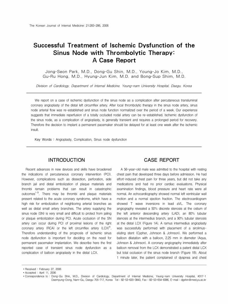
Figure 1. Left coronary angiograms on shallow right anterior oblique view with a 30 degree caudal angulation. (A) First coronary
angiogram showing critical ramus intermedius and distal left circumflex artery stenosis. Arrow: sinus node artery. (B) Angiogram showing
total occlusion of the sinus node artery after balloon dilatation at the distal left circumflex artery.
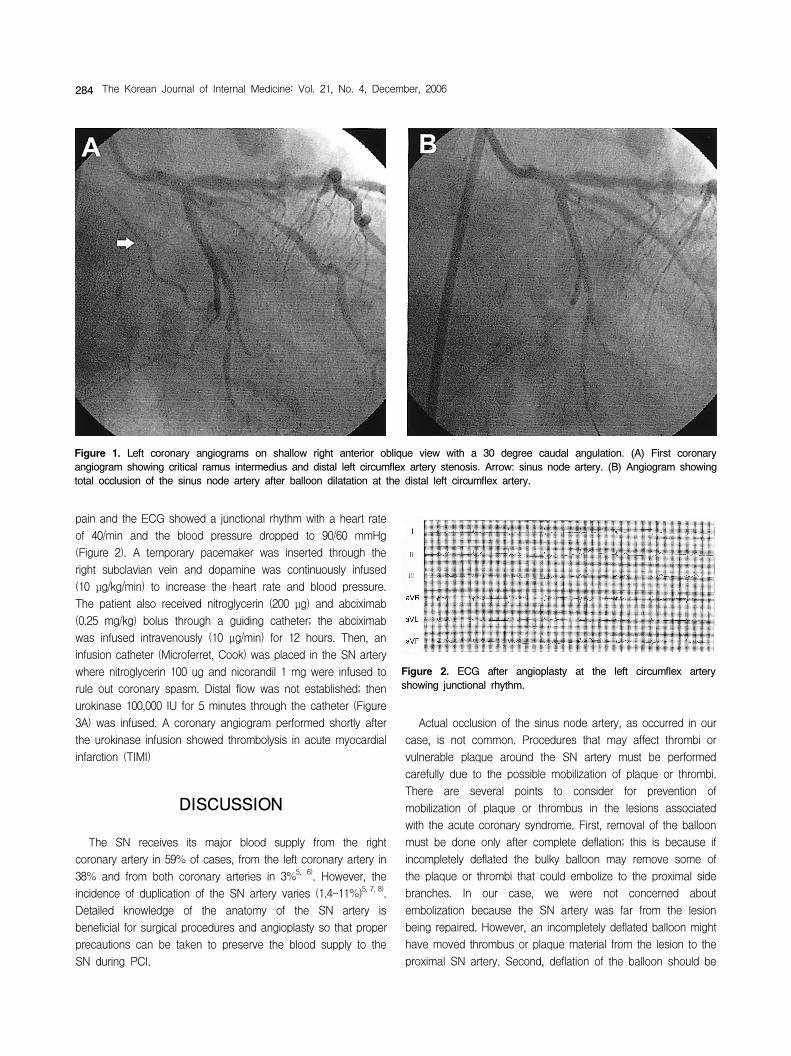
Figure 2. ECG after angioplasty at the left circumflex artery
showing junctional rhythm.
pain and the ECG showed a junctional rhythm with a heart rate
of 40/min and the blood pressure dropped to 90/60 mmHg
(Figure 2). A temporary pacemaker was inserted through the
right subclavian vein and dopamine was continuously infused
(10 μg/kg/min) to increase the heart rate and blood pressure.
The patient also received nitroglycerin (200 μg) and abciximab
(0.25 mg/kg) bolus through a guiding catheter; the abciximab
was infused intravenously (10 μg/min) for 12 hours. Then, an
infusion catheter (Microferret, Cook) was placed in the SN artery
where nitroglycerin 100 ug and nicorandil 1 mg were infused to
rule out coronary spasm. Distal flow was not established; then
urokinase 100,000 IU for 5 minutes through the catheter (Figure
3A) was infused. A coronary angiogram performed shortly after
the urokinase infusion showed thrombolysis in acute myocardial
infarction (TIMI)
DISCUSSION
The SN receives its major blood supply from the right
coronary artery in 59% of cases, from the left coronary artery in
38% and from both coronary arteries in 3%5, 6). However, the
incidence of duplication of the SN artery varies (1.4-11%)5, 7, 8).
Detailed knowledge of the anatomy of the SN artery is
beneficial for surgical procedures and angioplasty so that proper
precautions can be taken to preserve the blood supply to the
SN during PCI.
Actual occlusion of the sinus node artery, as occurred in our
case, is not common. Procedures that may affect thrombi or
vulnerable plaque around the SN artery must be performed
carefully due to the possible mobilization of plaque or thrombi.
There are several points to consider for prevention of
mobilization of plaque or thrombus in the lesions associated
with the acute coronary syndrome. First, removal of the balloon
must be done only after complete deflation; this is because if
incompletely deflated the bulky balloon may remove some of
the plaque or thrombi that could embolize to the proximal side
branches. In our case, we were not concerned about
embolization because the SN artery was far from the lesion
being repaired. However, an incompletely deflated balloon might
have moved thrombus or plaque material from the lesion to the
proximal SN artery. Second, deflation of the balloon should be
Jong-Seon Park, et al : Sinus Node Dysfunction after Angioplasty 285
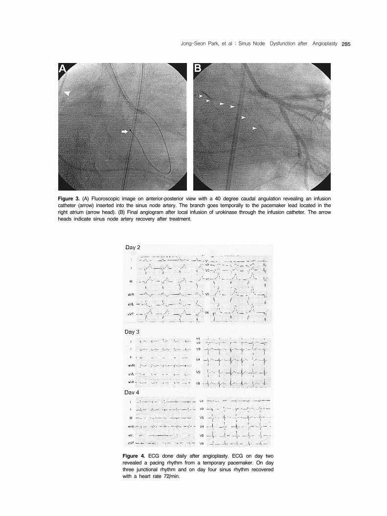
Figure 3. (A) Fluoroscopic image on anterior-posterior view with a 40 degree caudal angulation revealing an infusion
catheter (arrow) inserted into the sinus node artery. The branch goes temporally to the pacemaker lead located in the
right atrium (arrow head). (B) Final angiogram after local infusion of urokinase through the infusion catheter. The arrow
heads indicate sinus node artery recovery after treatment.
Figure 4. ECG done daily after angioplasty. ECG on day two
revealed a pacing rhythm from a temporary pacemaker. On day
three junctional rhythm and on day four sinus rhythm recovered
with a heart rate 72/min.
The Korean Journal of Internal Medicine: Vol. 21, No. 4, December, 2006286
done slowly. Rapid deflation of the balloon may cause
movement of mural thrombi or plaque materials into the vessel
lumen. Careful angioplasty, especially with an unstable lesion, is
the most important measure for preventing mobilization or distal
embolization of the thrombi or plaque.
The SN is a mass of pace-making cells with higher
metabolic requirements than other cells surrounding the atrial
myocardium9). The SN has an abundant arterial supply and is
generally resistant to ischemia10). The prognosis of acute
ischemic injury to the SN is not completely understood in
human patients. Chronic SN ischemia caused by a coronary
artery anomaly or occlusion of the coronary artery can lead to
a compromised blood supply to the sinus node, and hence
cause the sick sinus syndrome11, 12). There are few reports on
the prognosis in patients with acute ischemic SN dysfunction
secondary to SN artery occlusion during PCI. Previously a
report of one case of SN occlusion, following thrombosis of the
SN artery during stent implantation, normalized spontaneously
after one week in a patient with acute myocardial infarction.4
Prolonged SN ischemia can lead to exercise intolerance in
young patients12, 13). In our patient, we suggest that thrombi in
the LCX lesion might have embolized to the SN artery during
balloon removal. The SN artery was successfully recanalized by
local infusion with urokinase. However, we are not sure if this
was helpful in the recovery of SN function. Although SN function
in our patient worsened for two days after the procedure, the
function was nearly completely recovered after seven days.
More importantly, the patient an active young man did not
complain of any symptoms. We have treated five other acute,
transient flow disturbances of the SN artery and nodal
dysfunction complicated by balloon angioplasty or stent
implantation. In all of these cases the sinus rhythm recovered
within an hour without any further interventions.
In conclusion, acute ischemic SN dysfunction may occur as
a result of compromise of the SN artery during PCI. In addition,
after establishing coronary flow, the decision to implant a
permanent pacemaker should be delayed for one week after
the insult to allow for spontaneous recovery.
REFERENCES
1) Cavallini C, Rugolotto M, Savonitto S. Prognostic significance of
creatine kinase release after percutaneous coronary intervention. Ital
Heart J 6:522-529, 2005
2) Corcos T, Guerin Y, Garcia-Cantu E, Zimarino M, Tamburino C,
Toussaint M, Favereau X. Bail-out of stent jail: stent delivery through
stent struts. J Invasive Cardiol 8:113-116, 1996
3) Iijima R, Tsunoda T, Yamamoto M, Shiba M, Wada M, Tsuji T,
Yamamoto M, Nakajima R, Yoshitama T, Hara H, Hara H, Nakamura
M. Fate of unprotected side branches as related to embolic
complications during stent implantation for acute coronary syndromes
using a distal protection procedure. Am J Cardiol 95:636-639, 2005
4) Ahrensfield D, Balke CW, Benitez RM, Peters RW. Transient sinus
node dysfunction in acute myocardial infarction associated with the use
of a coronary stent. Catheter Cardiovasc Interv 50:349-351, 2000
5) Kyriakidis M, Vyssoulis G, Barbetseas J, Toutouzas P. A clinical
angiographic study of the arterial blood supply to the sinus node. Chest
94:1054-1057, 1988
6) Kyriakidis MK, Kourouklis CB, Papaioannou JT, Christakos SG,
Spanos GP, Avgoustakis DG. Sinus node coronary arteries studied
with angiography. Am J Cardiol 51:749-750, 1983
7) Bokeriya LA, Mikhailin SI, Revishvili AS. Anatomical variants of
sinoatrial and atrioventricular node arteries. Cor Vasa 26:220-228, 1984
8) Vieweg WV, Alpert JS, Hagan AD. Origin of the sinoatrial node and
atrioventricular node arteries in right, mixed, and left inferior emphasis
systems. Cathet Cardiovasc Diagn 1:361-373, 1975
9) Verhaeghe L, van der Hauwaert L. Arterial blood supply of the human
sinus node. Br Heart J 29:801-806, 1967
10) Yaln B KY, Ozan H. The sinus node artery: anatomic investigations
based on injection-corrosion of 60 sheep hearts. Interactive Cardiovasc
Thorac Surg 3:249-253, 2004
11) Liu PY, Chao TH, Tsai WC, Li YH, Tsai LM, Chen JH. Sick sinus
syndrome in a patient with single coronary artery anomaly. J Formos
Med Assoc 99:785-788, 2000
12) Osborn LA, Icenogle M. Resolution of exercise intolerance secondary
to ischemic sinus node dysfunction following percutaneous transluminal
angioplasty. Cathet Cardiovasc Diagn 42:44-47, 1997
13) Bashour TT, Chen F, Feeney J. Ischemic sinus node hibernation:
resolution following angioplasty. Am Heart J 122:1156-1158, 1991