Embed Size (px)
Citation preview
Success of secondary alveolar bone grafting and
canine eruption in cleft subjects
Nahul Patel
BDS, MFDS (RCPSG)
A thesis submitted to the University of Birmingham for the degree of
Masters of Philosophy
Department of Orthodontics
School of Dentistry
St Chad's Queensway
Birmingham
B4 6NN

University of Birmingham Research Archive
e-theses repository This unpublished thesis/dissertation is copyright of the author and/or third parties. The intellectual property rights of the author or third parties in respect of this work are as defined by The Copyright Designs and Patents Act 1988 or as modified by any successor legislation. Any use made of information contained in this thesis/dissertation must be in accordance with that legislation and must be properly acknowledged. Further distribution or reproduction in any format is prohibited without the permission of the copyright holder.
Abstract Aims A retrospective observational cohort study to:
1. Determine the success of secondary alveolar bone grafting (SABG) defined by
spontaneous canine eruption.
2. Investigate the influence of the following factors on canine eruption;
Post-Operative Bone Fill Index
Age at SABG
Presence of lateral incisor
Horizontal position and angulation of the canine
Expansion prior to SABG
Unilateral or bilateral cleft
Method Records of 66 subjects amounting to 86 clefts were examined to determine the
height of bone post-SABG using the Post-Operative Bone Fill Index as well as the
eruptive status of the canine. Study models, radiographs and clinical entries were
used to determine the presence and morphology of the lateral incisor, position of the
unerupted canine and presence of expansion.
Results
74.4% of 86 canines erupted spontaneously. Using the Post-Operative Bone Fill
Index, 54.4% scored 1, 26.7% scored 2, 10.5% scored 3 and 8.1% scored 4. 52.3%
of clefts were associated with missing lateral incisors and 30.2% were expanded pre-
SABG. There was no statistical significant correlation (P>0.05) between
spontaneous eruption and any of the variables.
Conclusion There was a good success rate of SABG, however, the height of bone post-SABG
was not statistically correlated with spontaneous canine eruption.
Acknowledgements
I would like to thank the following people:
Mr Lars Enocson and Professor Thomas Dietrich for their continuous guidance and
support.
Mr Lars Enocson and Mr Ian Sharp for supporting the study with clinical data.
Dr Jay Kindelan and Dr Gunvor Semb for their help on using the Post-Operative
Bone Fill Index.
Dr Paul Davies and Professor Thomas Dietrich for statistical support.
Mr David Spary, Mr John Turner, Mr Rognvald Linklater, Mr Lars Enocson and Mr
James Dickson for allowing me to visit their departments in order to collect data.
I would like to dedicate this thesis to my uncle, aunt, parents, Hitendra, Ruth,
Jayendra and Meena for their support and inspiration.
Chapter Page 1 Literature review 1 2 Participants and methods 60 3 Results 70 4 Discussion 80 5 Conclusion 94
Contents
Chapter 1 Literature review Page
1.1.1 Prenatal development of the lip and palate 4 1.1.2 Post natal development of the maxilla 5 1.1.3 Tooth formation 5 1.1.4 Theories of tooth eruption 6 1.2 Incidence of cleft lip and palate 10 1.3 Aetiology- genetic 11 1.3.1 Palatal shelf elevation 13 1.3.2 Palatal shelf fusion 14 1.4 Aetiology- environmental 16 1.4.1 Teratogents 16 1.4.2 Seasonal 17 1.4.3 Maternal and paternal age 17 1.4.5 Stress 17 1.4.6 Multivitamins and folic acid 18 1.5 Classification 19 1.6 Anomalies 21 1.6.1 Growth 21 1.6.2 Associated anomalies 22 1.6.3 Cognitive function 23 1.6.4 Hypodontia 23 1.6.5 Size and form of teeth 24 1.6.6 Caries 25 1.6.7 Tooth formation and eruption 26 1.6.8 Speech and hearing (velopharyngeal insufficiency) 27 1.6.9 Psychological 27 1.7 Management 28 1.7.1 Parental management 29 1.7.2 Respiratory management 30 1.7.3 Feeding 31 1.7.4 Presurgical orthopaedics 32 1.7.5 Lip repair 34 1.7.6 Alveolar repair in infancy 35
1.7.8 Palate repair 37 1.7.9 Speech therapy 39 1.7.10 Ear, nose and throat 40 1.7.11 Paediatric and orthodontic considerations 40 1.8 Secondary alveolar bone grafting 41 1.8.1 Timing of Secondary alveolar bone grafting 43 1.8.2 Donor sites 45 1.9 Assessment of bone fill 47 1.9.1 Bergland Score 47 1.9.2 Modified Bergland score 48 1.9.3 Enmark scale 48 1.9.4 Long et al scale 48 1.9.5 Post-Operative Bone Fill Index 49 1.9.6 Chelsea scale 49 1.9.8 Cone Beam Computer Topography (CBCT) 50 1.10 Outcome of SABG 50 1.11 Factors influencing outcome of SABG 51 1.12 Canine eruption 53 1.12.1 Prevalence of canine impaction in cleft subjects 53 1.12.2 Eruption path of canines in cleft subjects 55 1.13 Factors influencing canine eruption 56 1.13.1 Timing of SABG 56 1.13.2 Genetic causes 56 1.13.4 Presence of lateral incisor and guidance theory 57 1.13.5 Canine position in Non-cleft subjects 58 1.13.6 Canine position in cleft subjects 58 1.13.7 Space creation 59
Chapter 2 Participants and methods Page 2.1 Aims 62 2.2 Study design 62 2.3 Null Hypothesis 63 2.4 Ethical approval 63 2.5 Sample size calculation 63 2.6 Subject selection 64 2.7 Method 64 2.8 Statistical analysis 69
Chapter 3 Results Page
3.1 Characteristic of the sample 72 Table 3.1.1 Spontaneous eruption of canine 72 Table 3.1.2 Spontaneous eruption of the canine on the 73 contralateral side in subjects with unilateral clefts Figure 3.1.3 Post-Operative Bone Fill Index Score for all clefts 73 Figure 3.1.4 Post-Operative Bone Fill Index Score of clefts 74 associated with absence of canine eruption Table 3.1.5 Subjects with expansion 74 Table 3.1.6 First and repeat bone grafts 75 Table 3.1.7 Presence of lateral incisor 75 Table 3.1.8 Morphology of lateral incisor 75 Table 3.1.9 Angulation of canine 76 Table 3.1.10 Horizontal position of canine 76 Figure 3.1.11 A box and whisker plot for the age at SABG 76 Figure 3.1.12 Box and Whisker plot for the time between 77 between SABG and post-SABG occlusal radiograph Table 3.1.13 Binary logistic regression analysis of independent 78 variables Table 3.1.14 Initial and repeat Post-Operative Bone Fill Index 79 Scores used to test intra-examiner reliability.
Chapter 4 Discussion Page 4.1 Discussion 82 4.2 Limitations of the study 91 Chapter 5 Conclusion 5.1 Conclusions 96 5.2 Null Hypothesis 96 5.3 Clinical significance 97 5.4 Suggestions for further study 97
List of figures
Figure 1.5.1 The LAHSAL code 20 Figure 1.13.3 Association of dental anomalies 56
References 98
Raw data 112

1
Chapter 1
Literature review
2
Chapter 1 Literature review Page
1.1.1 Prenatal development of the lip and palate 4 1.1.2 Post natal development of the maxilla 5 1.1.3 Tooth formation 5 1.1.4 Theories of tooth eruption 6 1.2 Incidence of cleft lip and palate 10 1.3 Aetiology- genetic 11 1.3.1 Palatal shelf elevation 13 1.3.2 Palatal shelf fusion 14 1.4 Aetiology- environmental 16 1.4.1 Teratogents 16 1.4.2 Seasonal 17 1.4.3 Maternal and paternal age 17 1.4.5 Stress 17 1.4.6 Multivitamins and folic acid 18 1.5 Classification 19 1.6 Anomalies 21 1.6.1 Growth 21 1.6.2 Associated anomalies 22 1.6.3 Cognitive function 23 1.6.4 Hypodontia 23 1.6.5 Size and form of teeth 24 1.6.6 Caries 25 1.6.7 Tooth formation and eruption 26 1.6.8 Speech and hearing (velopharyngeal insufficiency) 27 1.6.9 Psychological 27 1.7 Management 28 1.7.1 Parental management 29 1.7.2 Respiratory management 30 1.7.3 Feeding 31 1.7.4 Presurgical orthopaedics 32 1.7.5 Lip repair 34 1.7.6 Alveolar repair in infancy 35 1.7.8 Palate repair 37 1.7.9 Speech therapy 39 1.7.10 Ear, nose and throat 40 1.7.11 Paediatric and orthodontic considerations 40
3
1.8 Secondary alveolar bone grafting 41 1.8.1 Timing of Secondary alveolar bone grafting 43 1.8.2 Donor sites 45 1.9 Assessment of bone fill 47 1.9.1 Bergland Score 47 1.9.2 Modified Bergland score 48 1.9.3 Enmark scale 48 1.9.4 Long et al scale 48 1.9.5 Post-Operative Bone Fill Index 49 1.9.6 Chelsea scale 49 1.9.8 Cone Beam Computer Topography (CBCT) 50 1.10 Outcome of SABG 50 1.11 Factors influencing outcome of SABG 51 1.12 Canine eruption 53 1.12.1 Prevalence of canine impaction in cleft subjects 53 1.12.2 Eruption path of canines in cleft subjects 55 1.13 Factors influencing canine eruption 56 1.13.1 Timing of SABG 56 1.13.2 Genetic causes 56 1.13.4 Presence of lateral incisor and guidance theory 57 1.13.5 Canine position in Non-cleft subjects 58 1.13.6 Canine position in cleft subjects 58 1.13.7 Space creation 59
4
1.1.1 Prenatal development of the lip and palate
The embryological development of the maxilla is well described in books written by
Mitchell, (2007) and Cobourne and Dibiase, (2010). The maxilla, palate and the
lateral portion of the upper lip is formed from the maxillary process of the first
pharyngeal arch. The development of the face is a complicated process involving
neural crest cell migration, proliferation and fusion of the swelling surrounding the
stomodeum. At approximately four weeks in utero, five processes appear; the
frontonasal process, two maxillary processes and two mandibular processes. At
approximately five weeks in utero, the medial and lateral nasal processes form within
the frontonasal process where olfactory cells are developed. The two nasal passages
are formed by the enlargement of the frontonasal and maxillary processes resulting
in the formation of the nasal septum. Maxillary swellings enlarge by growing medially
and ventrally and fuse to create the philtrum and the growth pushes the nasal
process inferiorly to form the primary palate. The upper lip is formed from the lateral
aspects of the maxillary process and the medial nasal process. The palate is formed
by the medial extensions of maxillary swellings which are formed approximately
seven weeks in utero and grow inferiorly lateral to the tongue. At approximately eight
weeks in utero, the tongue moves in a downward direction and the palatine shelves
rotate upwards and continues to enlarge horizontally. The palatine shelves fuse at
approximately 9 weeks in utero in a direction from the ventral aspect to the dorsal
aspect and following fusion, an epithelial seam is formed. A fibroblastic morphology
is developed by apoptosis of the epithelial cells as well as epithelial- mesenchymal
transformation which again differentiates into keratinocytes.
5
1.1.2 Post-natal development of the maxilla
This is well described by Cobourne and Dibiase, (2010). Maxillary growth occurs
though surface remodelling and growth at the circum maxillary sutures including the
frontomaxillary, zygomaticomaxillary, pterygomaxillary, zygomaticofrontal and
zygomaticotemporal sutures. The height of the maxilla increases by deposition at the
infrazygomatic crest, hard palate and alveolar process with resorption at the orbital
and nasal floor. The maxilla widens by growth at the mid palatal suture, deposition
posteriorly and laterally with resorption below the zygomatic buttress.
1.1.3 Tooth formation
Tooth formation can be classified into three basic stages (Cobourne and Dibiase,
2010).
Bud stage
There is localised proliferation of the oral epithelium which invaginates into the
underlying bone. This is regulated by Sonic Hedgehog and is antagonised by WNT7.
MSX1 and PAX9 also play a key role at this stage.
Cap stage
The tooth bud begins to resemble the early morphology of the crown. The signalling
originates from the enamel knot by the release of various mediators such as FGF4
which mediate proliferation of the epithelium. FGF10 mediates cell division the
epithelium and BMP induces MSX1 and PAX9.
6
Bell stage
At this stage, there is formation of the enamel, dentine and cement in the crown and
root. The inner enamel epithelium activates the cells in the dental papilla to
differentiate into odontobalsts which lay predentine. The predentine induces the cells
of the inner enamel epithelium to differentiate into ameloblasts which lays down
enamel. The root morphology is induced by growth of cells at the cervical loop in an
apical direction with differentiation of root odontoblasts. The dental follicle is then
exposed to the root dentine inducing the differentiation of cementoblasts which lay
cementum. The dental follicle cells also produce the alveolar bone and collagen
fibres of the periodontium
1.1.4 Theories of tooth eruption
Sandy, (1992) summarises the various theories of tooth eruption which are described
below.
Pulp theory
Extrusion of the pulp creates propulsive forces by;
Growth of dentine
Interstitial pulp growth
Vasculature hydraulic effect
7
Vascular Theory
The pressure in the blood vessels creates the propulsive force, however, there are
many authors that have found that eruption was unaffected by the use of hypotensive
drugs.
Root elongation Theory
The root of the tooth by elongation is pushed against the alveolar bone.
PDL Theory
The fibroblasts vibrate and contract creating a force that lifts the root against gravity.
Administration of Lathyritic compounds which inhibit cross-linking of collagen should
prevent the fibroblasts from vibrating and contracting have not been shown to inhibit
eruption.
Alveolar Bone growth
Bone is laid down beneath the crypts of erupting teeth. Kurihara et al, (1980)
examined dried skull of humans from birth to fourteen years of age in order to
examine the pattern of resorption and deposition in relation to developing teeth. The
authors reported that at the perinatal stage, there was no remodelling of the maxilla
of the mandible. During the early deciduous dentition stage, there was remodelling of
the premaxilla which involved little cortical bone. In the late deciduous dentition
stage, the resorption had spread superiorly, laterally and inferiorly whilst in the
mandible, the resorptive field were positioned irregularly. There were localised
8
remodelling of cortical bone in relation to tooth movements occurring during eruption
and drift. They reported that bone grew more rapidly when teeth erupted and there
were small thin deposits of periosteal lamellar bone covering localised bulges of
crowns and roots of erupting teeth. The growth in bone from erupting teeth was not
differentiated between cause and effect.
Follicular Theory
Factors such as cytokines, eicosanoids and growth factors are released by the dental
follicle which are responsible for bone remodelling and hence eruption.
9
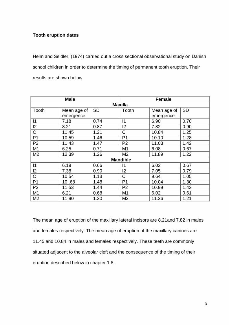
Tooth eruption dates
Helm and Seidler, (1974) carried out a cross sectional observational study on Danish
school children in order to determine the timing of permanent tooth eruption. Their
results are shown below
Male Female
Maxilla
Tooth Mean age of emergence
SD Tooth Mean age of emergence
SD
I1 7.18 0.74 I1 6.90 0.70
I2 8.21 0.87 I2 7.82 0.90
C 11.45 1.21 C 10.84 1.25
P1 10.59 1.46 P1 10.10 1.28
P2 11.43 1.47 P2 11.03 1.42
M1 6.25 0.71 M1 6.08 0.67
M2 12.39 1.26 M2 11.89 1.22
Mandible
I1 6.19 0.66 I1 6.02 0.67
I2 7.38 0.90 I2 7.05 0.79
C 10.54 1.13 C 9.64 1.05
P1 10..68 1.48 P1 10.04 1.30
P2 11.53 1.44 P2 10.99 1.43
M1 6.21 0.68 M1 6.02 0.61
M2 11.90 1.30 M2 11.36 1.21
The mean age of eruption of the maxillary lateral incisors are 8.21and 7.82 in males
and females respectively. The mean age of eruption of the maxillary canines are
11.45 and 10.84 in males and females respectively. These teeth are commonly
situated adjacent to the alveolar cleft and the consequence of the timing of their
eruption described below in chapter 1.8.
10
1.2 Incidence of cleft lip and palate
Cleft lip and palate is the most common craniofacial abnormality. The incidence of it
varies among racial groups, however, the non-syndromic form is approximately 1:700
live births in the UK (Gorlin et al, 1971). Coupland and Coupland, (1988) examined
930 children born in the Trent region between 1973 and 1982. The incidence of cleft
palate varied by a small degree each year with an incidence of 1.47 per 1000 live
births in 1974 to 0.91 per 1000 live births in 1982. The lowest incidence was found in
1977 with an incidence of 0.91/1000 live births. They found a distribution of types of
clefts amongst genders. 39% had isolated cleft palates and 61% had a cleft lip with
or without a cleft palate. In the isolated cleft palate group, females accounted for 55%
and males 45%. In the group of cleft lips, a higher proportion was seen in males
which accounted for 62%. Jensen et al, (1988) studied the incidence of clefts in a
Danish population between 1986 and 1981. They found that 678 live births were
registered with cleft lip, palate or both resulting in a incidence of 1.89 per 1000 live
births, however, they excluded isolated submucous clefts. The unilateral cleft group
accounted for 55.8% of males and 33.5% of females. The bilateral cleft group
accounted for 6.3% of males and 1.4 % of females. The unilateral cleft group
consisted of cleft lip in (33.5%) almost twice as prevalent in males. The cleft lip and
palate (39.1%) was again almost twice as prevalent in males, however, the cleft
palate only (27.4%) was 1.5 times common in females. The authors also found that a
small proportion of the cleft palate group were associated with Pierre Robin
syndrome. Seventy percent of the cleft cases were non-syndromic occurring in
isolation whereas the remaining 30% were syndromic. Cobourne, (2004), Carmichael
11
et al, (2007) and Botto et al, (2002) found that the greatest incident of orofacial clefts
were amongst Caucasian or Hispanic patients with the least incidence in afro-
Caribbean and Asian ethnicity.
Nagese et al, (2010) carried out a cross sectional study in Japan to investigate the
gender differences in cleft pattern. The authors analysed 782 subjects and the
following results were documented. They reported that the most frequent type was
the cleft lip and palate which was more common in males whereas the cleft palate
was more common in females. A possible explanation for the gender difference was
mentioned including a relationship between female sex hormones and the palatine
process with the slight differences in the timing of embryological development. The
cleft lip/cleft lip and palate was also more common on the left and they theorised that
a possible reason for this was that the facial artery development is slower on the left.
1.3.1 Aetiology- Genetic
Losee and Kirschner, (2009) discuss the embryology of cleft palate. The clefting of
the palate is due to lack of fusion of the 3 elements that form the palate which are
median frontonasal prominence, the primary palate and the two lateral palatal
shelves. The V-shaped clefts are a result of lack of tissue in the shelves to complete
closure whereas the U-shaped clefts are a result of micrognathia and glossoptosis.
The greater the posterior involvement of the palate, the greater the severity of the
cleft. A cleft of the secondary palate is due to deficient fusion posterior to the incisive
foramen whereas a cleft of the primary palate is due to deficient fusion anterior to the
12
incisive foramen of the medial, lateral nasal processes and the maxillary process.
The palatal shelves elevation and fusion is discussed in greater detail below.
It is believed that the aetiology of cleft lip and palate may be due to a genetic
predisposition, an environmental cause or combination of both. Syndromic disorders
are caused by single gene mutations with varied levels of penetrance and
expressivity. There are a broad range of variations in structure and number that can
cause gene malformations and therefore altered gene function. Deletions and
duplications can both have an aetiological impact in the development of cleft lip and
palate (Cobourne, 2004).
Developmental disturbances can broadly be categorised into one of the following
Alteration in the force to elevate the palatal shelves
Failure of tongue to drop
Non-fusion of shelves
Failure of mesodermal migration
Rupture of a cyst formed at the site of fusion
Sonic Hedgehog is a protein that is secreted in the oral epithelium that induces
growth of the palatine shelves. Rice et al, (2004) found that signalling from Sonic
Hedgehog protein induced palatal shelves proliferation and growth with lack of this
protein resulting in a reduced development of the mesenchymal cells. The authors
also showed that mice lacking fibroblast growth factor 10 or its receptor, fibroblast
growth factor 2b developed a cleft palate due to the lack of outward growth of the
palatine shelves. Bush and Jiang, (2012) suggest that Sonic Hedgehog and
13
Fibroblast Growth Factor function as a positive feedback loop. Bone morphogenic
proteins are also required for palatogenesis. Liu et al, (2005) knocked out Bone
Morphogenic Protein 4 in mice and found that they developed a cleft lip but not a cleft
palate as it affected the function of the maxillary mesenchyme. Bone morphogenic
proteins are cytokines which play a significant role in embryonic development by
stimulating gene expression via phosphorylation. In particular BMPs play a specific
role in the epithelial- mesenchymal interaction during the formation of organs. Li et al,
(2011) investigated the effects of Bmprla and Bmprlb which are encoders for BMP
receptors and BMP signalling. The authors found that inactivation of Bmprla led to
the development of a cleft palate as well as lack of tooth development and a
hypoplastic mandible.
1.3.1 Palatal shelves elevation
Several theories have been proposed for the elevation and rotation of the palatal
shelves. The following were discussed by Bush and Jiang, (2012).
There could be regression of the distal portion of the shelves and outgrowth in
the horizontal direction.
A mechanism exists whereby the anterior portion of the shelves swing
upwards and the posterior flow down.
Stanier and Moore, (2004) describes the following theories
Elevation could be due to an intrinsic force due to increased turgidity by
recruitment of water resulting in an increased level of glycosaminoglycans
such as hyaluronan.
14
The author discusses the involvement of Pax9 and a mutation in the gene
resulting in an abnormal morphology of the palatal shelves in which they are
shorter and broader. This experiment was carried out in mice and the palatal
shelves had a mechanical inhibition of elevation.
1.3.2 Palatal shelves fusion
As discussed above the fusion and elevation of the palatal shelves can be influenced
a number of specific gene mutation. Fusion of the palate occurs by interaction of
desmosomes and cell adhesion molecules. They contact in a midposition horizontally
and close in a direction from the primary palate to the uvula. Experiments on mice
have shown that halpoinsufficiency of Interferon Regulatory Factor 6 has lead to
failure of palatal shelves fusion (Coubourne, 2004). Polovirus receptor 1 gene
encodes a cell adhesion molecule called nectin 1 and a nonsense mutation of this
gene has resulted in the failure of fusion of the palatal shelves. Suzuki et al, (2000)
and Stanier and Moore, (2004) discussed a number of genes in which the mutations
have contributed to the development of cleft palate by inhibiting mesenchymal
proliferation and therefore fusion in the horizontal plane including MSX1 and Lhr8.
Linde, (2007) presents a table in the review article which lists the gene involved with
failure of palatine shelves proliferation, elevation or fusion.
Bmprl1a- the positioning of the shelves are altered and positioned more
anteriorly
Fgf10- apoptosis is increased of the shelves and there is adhesion of the
shelves to other epithelia
Tgfbr2- Proliferation defects of the palatal mesenchyme
15
Alpha v integrins- the shelves elevate but fail to make contact
Ephb2- results in hypoplastic shelves
Pdgfc- There is delayed elevation and well as hypolasia and failure of the
fusion
Lateif et al, (2012) carried out a study to compare the widths and angulation of the
palatal shelves in unilateral and bilateral cleft patients using study casts. They
examined the casts of unoperated patients aged 13 consisting of 68 unilateral, 13
bilateral cleft lip and palate and 24 in the non-cleft control group. In the unilateral cleft
group, the palatine shelves width was significantly smaller on the cleft and non cleft
side compared to control group. They were also positioned more vertically and
rotated towards the cranium. In the bilateral group, the shelves were rotated by 10°
compared to the control group. This study confirms the work of others mentioned
above. The fact that the shelves were smaller suggests hypoplasia or failure of
proliferation.
Bush and Jiang, (2012) present a table in which all the gene mutations have been
identified to cause syndromic and non syndromic cleft lip+/- palate. They have
presented the following:
Non-syndromic cleft lip and palate
BMP4, FGF8, FGFR2, FOXE1, IRF6, MSX1, PDGFC and SUM01
Non-syndromic cleft palate only
FOXE, IRF6, MSX1, SATB2 and TBX22
16
There are a host of syndromic genes and an example of a few are demonstrated
below.
TCOF1- Treacher Collins syndrome
TBX1- DiGeorge
GLI3 – Oro- facial- digital syndrome
FGFR2- Crouzon’s and Apert’s syndrome
IRF6- Van de Woude
1.4 Aetiology- Environmental
1.4.1 Teratogens
A teratogen is any agent that can cause a birth defect by disturbing the development
of the embryo or foetus. Teratogens linked to cleft lip and palate include Phenytoin,
Thalidomide, Valproic acid, maternal alcohol, tobacco smoking, altitude and
herbicides such as Digoxin. Murray, (2002) and Little et al, (2004) carried out a meta-
analysis to determine a correlation between maternal smoking and cleft lip and
palate. They included 22 case control publications and 10 cohort studies and
reported that there was a significant association between maternal smoking and cleft
lip and palate.
17
1.4.2 Seasonal
Coupland and Coupland, (1988) found a seasonal variation between the years of
1973 and 1982. In the group consisting of cleft palate only, the greatest incidence
occurred in August with approximately 36 births and the lowest incidence was in April
with approximately 26. This was different to the incidence of cleft lip with or without
palate. In this group, the greatest incidence was found in December with 54 births
and the lowest in May with 41 births. The authors acknowledge that that season
variability is a well recognised phenomenon and has been reported in neural tube
defects.
1.4.3 Maternal and paternal age
Jensen et al, (1988) in their retrospective study found that maternal age group was
increased in the bilateral complete cleft lip and palate group and increased paternal
age was associated with left side complete cleft lip. Paternal age was however
normal for the cleft palate group. Botto et al, (2002), however, found that the greatest
incidence was in a maternal age between 25 and 29.
1.4.5 Stress
There are many studies that have reported an increased in maternal stress and
neural tube defects. Carmichael et al, (2000) and Suarez et al, (2003) have
suggested that stress may lead to an increase in the levels of corticosteroids which
act as teratogens as well as elevated corticotrophin releasing hormones. Carmichael
18
et al, (2007) carried out a telephone based interview study with 1335 mothers with
affected children and 700 control mothers. The mothers were asked about stressful
events such as a lost or new job, financial or legal problems, serious illnesses, drugs,
alcohol, violence or crime, losing a loved one or relationship troubles. The results
showed that an increase in the number of stressful life events was associated with
birth defects especially cleft lip with or without a cleft palate.
1.4.6 Multivitamins and Folic acid
Folic acid is also known as foliate, vitamin B9, vitamin Bc or vitamin M is a vitamin of
the B complex which is found naturally in leafy green vegetables, kidney or liver. It is
necessary for DNA synthesis, cell production and prevention of gene mutations.
There are many studies which have reported an association between deficiency in
folic acid and a greater incidence of cleft lip and palate. Badovinac et al, (2007) found
in the meta-analysis of 5 prospective and 12 case control studies that there was a
positive correlation between the two, but also acknowledge the potential for bias and
uncontrolled confounding factors. Matok et al, (2009) examined an association
between folic acid antagonists and congenital malformations during the first trimester
of pregnancy in a retrospective cohort study. The folic acid antagonists are
commonly found in medication such as Phenytion, Valproic acid, Carbamazepine,
Lamotrigine, Primidone and Cholesyramine. The workers found that folic acid
antagonists were significantly associated with neural tube and cardiovascular
defects. Johnson and little, (2008) found in their meta-analysis study that there was
weak evidence for associations between orofacial clefts and folic acid intake. The
19
researchers did, however, conclude that multivitamin use may protect against
orofacial clefts but there are many confounding factors not accounted for. Mosey et
al, (2009) discusses the role of nutrition and mentioned that a deficiency in zinc leads
to an increase risk of cleft lip and palate development and vitamin B6 reduces the
serum concentration of homocysteine which is thought to be a risk factor. Botto et al,
(2002) found in their case control study that febrile risk with no multivitamin intake
around time of conception was associated with increased risk with neural tube
defects and cleft lip and palate. An explanation for these results is that multivitamins
and febrile illnesses affect vascular disruption and apoptosis antagonistically. The
authors mention the possibility of recall bias in their discussion and also that the
results could have occurred by chance, however, that could be unlikely as it is
agreement with other workers.
Little et al, (2004) in their meta-analysis mention that the smoking could interact with
a number of genes involved in the aetiology of cleft lip and palate as mentioned
below.
Smoking or alcohol or vitamins alter TGFA expression
Smoking and alcohol can also alter MSX1 and TGFB gene
1.5 Classification
Veau, (1931) classified his 4 categories by morphology.
1. Cleft of the soft palate only
2. Cleft of the hard and soft palate up to the incisive foramen
20
3. Complete unilateral cleft of the soft and hard palate as well as the alveolar ridge
and the lip on that side
4. Complete bilateral cleft of the soft and hard palate as well as the alveolar ridge
and the lip on both sides
Kernahan and Stark, (1958) based the categories according to the embryology and
designates the incisive foramen as the dividing structure. Group 1 involves clefts of
the primary palate up to the incisive foramen. This is subgrouped as unilateral,
bilateral or median. Group 2 involves clefts of the soft and hard palate up to the
incisive foramen which can be subgrouped as unilateral or bilateral. Group 3 involves
the soft or hard palate but not up to the incisive foramen and therefore can be
complete or incomplete as well as unilateral or bilateral.
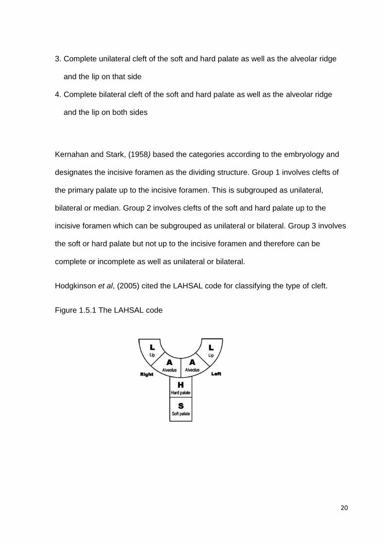
Hodgkinson et al, (2005) cited the LAHSAL code for classifying the type of cleft.
Figure 1.5.1 The LAHSAL code
21
The LAHSAL code is a modification of the classification of the Kernahan and Stark
index. The Y diagram is divided first into halves which are the left and right. It is then
divided into 6 parts which consist of the right lip, right alveolus, hard palate, soft
palate, left alveolus and left lip. The code is written as if the clinician facing the
patient with the first character being the right lip and the last character being the left
lip.
1.6 Anomalies
1.6.1 Growth
Jensen et al, (1988) examined the general condition of cleft children. They found that
body length in females with cleft palate was significantly reduced at age 9 weeks.
The height at 22 weeks of boys and girls with cleft lip+/- palate was significantly less
than children with cleft lip only and the weight for both was significantly reduced
compared non-cleft controls. Head circumference was not greatly impacted except
for the boys with cleft palate only at 22 months. Semb, (1991.a) carried out
cephalometric analysis of 257 patients in Oslo with unilateral cleft lip and palate. The
researcher found that patients had increased anterior vertical facial proportions,
reduced posterior face height, short retrusive maxilla, retrusive mandible and a flatter
nose. The nose showed a more downward and backward growth. These subjects
had their hard palate repaired with Von Lagnenbeck procedure and the lip was
closed with Le Mesurier’s procedure before 1961 and Millards procedure after 1961.
They also had their secondary alveolar bone grafts between ages 9 and 11. Semb,
22
(1991b) examined the growth and facial characteristics of 90 patients with bilateral
cleft lip and palate using cephalometric measurements. The pattern was similar to
that of the unilateral cleft lip and palate group with increased anterior facial height,
shorted posterior face height and reduced length of the maxilla as well as the
mandible when compared to controls. The maxilla was prominent until aged 5,
however, receded from aged 7 to 18. It was also reported that the influence of the
vomer flap during palate closure on facial growth was insignificant. The facial
characteristics are attributed to the primary surgery resulting in scar tissue hindering
maxillary and concomitant mandibular growth. Heidbuchel, (1997) reports that in
unoperated adults of bilateral cleft lip and palate, the columella and prolabium is
underdeveloped, however, there is no difference in the sagittal dimension with the
exception of the cleft when compared to non-cleft controls. The mandibular plane is
steep in unoperated cleft subjects. Heidbuchel et al, (1998) examined study casts to
investigate the development of maxillary arch dimensions of operated cleft subjects
compared to non-cleft subjects. The results showed that following lip closure, the
anterior arch width and depth reduced and following palatoplasty, the posterior arch
width reduced. Semb and Shaw, (1990), reported that subjects with pharyngeal flaps
had reduced maxillary length, reduced upper and lower face height and a more
retrusive mandible.
1.6.2 Associated anomalies
Andersson et al, (2010) studied 994 patients born with a cleft palate to describe
associated anomalies. They found miscellaneous facial anomalies as the most
common anomaly in 132 subjects, skeletal system anomalies in 51 subjects, cardio-
23
vascular system anomalies in 47 subjects, mental disabilities in 43 subjects and
central nervous system anomalies in 40 subjects. Other organ anomalies included
the ear, eye, urogenital, skin, gastro-intestinal, endocrine, respiratory system and the
brain
1.6.3 Cognitive Functioning
Lack of cognitive functioning could be due to smaller frontal lobe, smaller brain
volumes, psychological effects, speech deficits and the effect of living with a facial
disfigurement. Roberts et al, (2012) studied the effects of cognitive function of cleft
patients in a meta-analysis study. They reported that cleft patients can present with
deficits of immediate memory, attention and language abilities, processing speed,
sensory-motor function and academic ability.
1.6.4 Hypodontia
Andersson et al, (2009) found in their sample of 994 cleft subjects that 88 individuals
had hypodontia. One hundred and fifty one teeth were missing with the second
premolars in the maxilla or mandible being the most frequently missing. 39 subjects
showed hypodontia of two or more teeth. There was an increased trend for
hypodontia with a greater severity of cleft. Many other authors have reported a
similar prevalence of hypodontia between 35 and 45 percent. Ranta et al, (1983)
examined orthopantograms of children aged between 6 and 12 and found that there
was an increased risk of hypodontia with an increasing extension of cleft with a
24
prevalence ranging from 45% to 56% with 3rd molars being excluded. Larson et al,
(1998) examined orthopantograms and found a prevalence of 30% with the most
common missing tooth being the mandibular second premolar followed by maxillary
lateral incisor and maxillary second premolar. Tortora et al, (2008) evaluated
panoramic radiographs of 29 subjects with bilateral cleft lip and palate and 87
subjects with unilateral cleft lip and palate. They reported a 48.8% prevalence of
hypodontia involving the upper lateral incisors and 7.3% with supernumerary lateral
incisors. The second most common missing tooth was the maxillary second premolar
on the cleft side in 4.9% of the subjects.
1.6.5 Size and form of teeth
McCance et al, (1993) examined the study models of 23 cleft subjects and 100
control subjects to determine any associations in tooth size, chord length and arch
width. The author reported significantly smaller tooth size in cleft patients, especially
with incisors and canines. The cleft group also had smaller incisor to canine chord
length but not from canines to first molars. The mean arch widths were also smaller
in the cleft group indicating a narrow arch and the possibility of crossbite
development. Ranta, (1986) reports that the lateral incisor on the cleft side is almost
always smaller in size and different in shape. There is an extremely high incidence of
hypoplasia in the incisor region, which may be due to the lip repair.
25
1.6.6 Caries
Bokhout et al, (1997) investigated the prevalence of caries in the primary dentition in
subject with cleft palate compared to controls. The study included 158 subjects, 81
subjects with oral clefts and 77 control subjects. The subjects were examined at
months three, six, nine and twelve followed by six monthly intervals until aged 4. The
parents were given a questionnaire asking about the subject’s dietary habits, fluoride
exposure and socio-economic status of the parents. The results showed that the
subjects with orofacial clefts had significantly poorer oral hygiene and greater gingival
inflammation. 101 carious teeth were registered with 91% belonging to the group with
orofacial clefts. The teeth most affected were the maxillary incisors followed by
maxillary molars, mandibular molars and maxillary canines. 62% of the cleft subjects
came from a low socio-economic class compared to 29% in the control group. The
authors discuss the possibility that parents from a higher socio-economic class are
more likely to comply with nutritional recommendations and restrict cariogenic foods.
The incidence of caries was 3.5 times greater in subject with oral clefts. This is in
agreement with Britton and Welbury, (2010) who reported a greater prevalence of
caries in children with oral clefts aged between 0.5 years and 6 years of age
compared to non cleft subjects. Both anterior and posterior teeth were involved with
more carious anterior teeth in the younger age group. Hasslöf and Twetman, (2007)
in their systematic review of case studies did not find that children with cleft lip and
palate had a greater prevalence of caries.
26
1.6.7 Tooth formation and eruption
Ranta, (1986) discusses that tooth formation and eruption is delayed by 0.3 to 0.7
years with an increase in delay which is proportional to the severity of the cleft
extension. This delay occurs throughout the permanent dentition and in both upper
and lower arches. The delay in tooth formation increases with an increase in the
number of missing units and the delay can be significantly longer with increasing age
(1.1 years for subjects aged between 9 and 12, and 0.6 years for subjects aged 6 to
9). The canines and incisors on the cleft side can erupt later than the corresponding
teeth on the non-cleft side. Bjerklin et al, (1993) examined the prevalence of maxillary
first permanent molar ectopia in 225 children using radiographs. They reported a
significantly greater prevalence in the cleft patients of 21.8% compared to 4.3% in the
control group with 94 ectopic molars in the cleft lip and palate group, 65 in the cleft lip
and 66 in the cleft palate group. The authors believe that the surgical treatment is a
major local factor as well as a genetic tendency. Borodkin et al, (2008) found a delay
of 0.52 years with boys accounting for all the delay and no significant difference
between unilateral and bilateral clefts. This is in agreement with Pioto et al, (2005),
who found delayed root development of the lateral incisor on the cleft side compared
to the non-cleft side. Heidbuchel et al, (2002) discussed that delay in tooth formation
could be due to factors that are associated with the aetiology of the oral cleft such as
risk factors during gestation, unfavourable postnatal environments such as recurrent
chest and ear infections, operations, feeding problems, inadequate bone support or
scar tissue created from surgical procedures.

27
1.6.8 Speech and hearing (velopharyngeal insufficiency)
The aetiology of speech defects are complex and multifactorial. This can be due to
velopharyngeal insufficiency, hearing problems, oronasal fistulas, occlusal and dental
problems (Habel et al, 1996). Velopharyngeal insufficiency occurs when the lateral
pharyngeal walls and the soft palate ineffectively separate the nasal cavity from the
oral cavity resulting in hypernasality or an abnormal tone. The aetiology can be due
to structural deficits, faulty learning, neurological deficits or a combination of them.
Treatment can include surgery, speech therapy or prosthetics (Sell and Ma, 1996).
There is an increased risk of middle ear infections due to abnormal attachments of
the Levator Palati muscle to the eustacian tube which in turn leads to poor drainage
(Habel et al, 1996).
1.6.9 Psychological
Turner et al, (1998) highlight particular important psychological issues that cleft
subjects can encounter. The environment that the family creates is an important
factor as it can impact on the attitude and self esteem of the child. Facially disfigured
children can have a negative reaction from the parents at an early age. Surgery for
the disfigurement can result in an increase in self confidence, self esteem and
satisfaction with appearance. Cleft patients may experience an increase frequency
with learning problems and speech in addition to verbal and language deficiency.
Snyder et al, (2005) examined medical records of 64 adolescents with craniofacial
anomalies that contained the self-report psychosocial adjustment scales and the
parent-report psychosocial adjustment scales. The craniofacial syndromes included
28
Crouzons, Treacher Collins syndrome, hemifacial microsomia, cleft lip and many
others involving a facial asymmetry. They showed that parents reported a social
deficit and a withdrawn feeling. Common psychological disturbances by the cleft
subjects included activity and social incompetence, anxiety, depression, identity
problems and aggressive behaviour.
1.7 Management
The clinical standards advisory group is an independent source of expert advice to
the U.K. Health Ministers on standards of clinical care and to advise on access to
and availability of selected NHS specialised services in order to prevent the standard
of care from deteriorating to an unacceptable level (Sandy et al, 2001).
Sandy et al, (1998) carried out a questionnaire survey which was sent to each cleft
team in the United Kingdom in order to ascertain the outcome of care. They identified
57 active cleft teams in the UK of which 48 replied with the questionnaire. The results
from their study showed that there were 75 cleft surgeons with few cases each year
with poor outcomes in bone grafting demonstrated by a failure or defective rate of
42%. A high need of treatment for caries was required for 40% of 5 year olds and
20% of 12 year olds. 19% of the parents of 5 year olds and 28% of the parents of 12
year olds reported that their child’s self confidence was affected mainly by teasing
with 9% dissatisfied with overall outcome of care.
Bearn et al, (2001) listed many recommendations that were made by the CSAG to
the UK government in 1998 of which a few are listed below.
29
Concentration of the 57 cleft units to 18-5 centres in the UK.
The services should be reviewed by each trust ensuring that the range of
clinical skill are available.
A database should be created detailing information of when and what
information should be available for audit and research purposes.
The cleft centres should provide the cleft clinicians with training programmes
where a high volume of clinical experience is available.
As discussed in the previous section, there are many anomalies associated with cleft
lip and palate and therefore the management involves a multidisciplinary approach.
The multidisciplinary team would involve the plastic surgeons, orthodontists, speech
therapists, oral and maxillofacial surgeons, psychologists, specialist nurses, ear,
nose and throat surgeons, audiologists, paediatricians and geneticists. Diagnosis can
be made upon an ultrasound scan and an immediate referral should be made to the
regional cleft lip and palate team (Habel et al, 1996).
1.7.1 Parental management
Hodgkinson et al, (2005) describes their experience in Newcastle. Parents are often
shocked and therefore need time, reassurance and support. All parents would need
help understanding their emotions with counselling. During pregnancy, the families
are allowed to meet the cleft team as well as other parents who have children with
oral clefts.

30
1.7.2 Respiratory management
Children born with a cleft may have respiratory problems, for example, in Pierre
Robin syndrome, the mandible is micrognathic and therefore, the tongue may
occlude the airway because it is posteriorly attached. This may be managed by
inserting a nasopharyngeal airway tube above the epiglottis until the surrounding
structures have grown sufficiently enough. The worst case scenario would involve a
tracheostomy (Habel et al, 1996).
Kirschner et al, (2003) advocated the use of lip adhesion to manage the airway
obstruction. The authors report that in all their cases that had a tongue- lip adhesion,
a flap was sutured from the ventral surface of the tongue to a superiorly based
mucosal flap from the posterior lower lip and this was successful in 25 out of the 29
cases. The main causal factor for failure was due to the mucosal dehiscence. Hong
and Bezuhyl, (2013) report that the tongue can tether leading to swallowing
difficulties requiring a nasogastric tube. They also describe the use of mandibular
distraction osteogenesis in order to relieve upper airway obstruction by lengthening
the mandible resulting in a more forward position of the genioglossus and other
muscular attachments. Following the osteotomy, the distractor is usually activated by
1-2mm per day which causes the soft tissues to gradually stretch. The authors report
good outcomes with distraction osteogenesis overall but poorer outcomes in patients
with Pierre Robin Syndrome due to other medical problems such as neurological
dysfunction.

31
1.7.3 Feeding
Feeding difficulties are common and children often have poor weight gain. Feeding
can also be prolonged due to ulceration of the nasal mucosa and a high metabolic
rate due to other associated conditions such as congenital heart disease. Negative
intraoral pressure is required for successful breast feeding, however, this would not
be possible with a cleft palate. Habel et al, (1996) and Hodgkinson et al, (2005)
describe the mechanism for successful feeding. The baby has to lift the soft palate to
separate the nasal cavity from the oral cavity and the tongue has to move forward
over the lower gum in order to cup the nipple or teat. The negative intra-oral pressure
with a rhythmic jaw and tongue movement enables the nipple to be help in position.
Martin and Bannister, (2004) describe various feeding techniques and variety of
bottles and teats available. Most mothers are able to breastfeed if they have been
taught to exaggerate the attachment technique. Other methods include the use of
various bottles which should be soft and easy to squeeze with a simple design. The
Haberman feeding system is a bottle with a one way valve, however, the teat can
result in ulceration of the nasal turbulence. The Mead Johnston bottle is useful for
parents who have reduced manual dexterity, however, it can sometimes be too soft.
They mention that a dummy at feeding times will enhance the development of the
oral skills and hence the cognitive link between sucking and feeling of hunger.
Counselling, support and advice is usually available from a specialist nurse to help
reduce anxiety about weight gain and promote adequate intake. They highlight the
importance of specialist nurses for successful feeding immediately after birth. If the
mother chooses a formula feed, then the specialist nurse would demonstrate feeding
techniques and provide the soft bottles and if the mother prefers to breastfeed,

32
equipment is used to help the expression of breast milk (Habel et al, 1996;
Hodgkinson et al, 2005)
1.7.4 Presurgical orthopaedics
Presurgical orthopaedics was first advocated by a McNiel who was a prosthodontist
from Scotland. The process would reduce the size of the alveolar gap and the
distance between the medial and lateral aspects of the alar cartilages as well as
increasing nasal tip projection (Losee and Kirschner, 2009).The aim was to
approximate and align the alveolar segments whilst to some degree correct the nasal
cartilage and soft tissue deformity which would allow symmetric growth of the
mandible (Grayson and Cutting, 2001). Advocates claim that the benefits include
facilitation with lip and palate repair, help with feeding as well as enhance speech
and facial growth, however, the benefits and the use remain a controversy.
There are broadly two types of appliances.
Passive appliances
A passive appliance as suggested by its name is an appliance without an active
component. An example of such appliance is the Hotz plate (Hotz, 1969). The author
describes an appliance made from hard and soft acrylic that obturates the hard
palate and as much of the soft palate that the child tolerates. Normal growth cause
the maxillary segment to grow, however, the desired direction of growth can be
controlled by selectively grinding the plate. The author also mentions that the plate
allows the tongue to adapt to a more normal position resulting in a more normal

33
swallowing pattern. The plate is normally worn after the lip closure in order to prevent
collapse of the maxillary segments.
Active appliances
Active appliances are secured intraorally and apply a mechanical force with screws,
plates and elastic chains. An example of an active appliance is the Latham appliance
(Latham, 1980). The author described an intraoral appliance made from acrylic that
was used in subjects with a unilateral cleft palate. The appliance was retained by the
non-cleft segment with pins and had a split down the midline with the two halves held
together by a stainless steel bar posteriorly. A screw measuring 25mm in length was
secured onto the appliance in an antero-posterior direction. Activation of the screw
once a day moved the cleft segment anteriorly resulting in a reduced discrepancy
between the two maxillary segments and better alignment of the dento-alveolar ridge.
Naso-alveolar moulding (NAM)
NAM was a term that followed the term presurgical orthopaedics as is consists of an
appliance to mould the alveolus such as the Hotz plate with the addition nasal
moulding which occurs independently of alveolar moulding (Loose and Kirschner,
2009)
These appliances also involve the use of an external force which is applied to
position the segments posteriorly. The external force may be a head cap with elastic
straps, external taping, or surgical adhesion for the lip.
34
Grabowski et al, (2006) followed up 43 subjects with a cleft lip and palate that were
managed with pre-surgical orthopaedics until the primary dentition was established.
Study models were then examined. Their findings included normal development
between ages 3 and 4 compared to non-cleft subjects and advocate the use to guide
the growth of the alveolus. Mishima et al, (1996) used a Hotz plate which is an active
appliance that postures the tongue into its normal position and guides the growth of
the maxillary segments. They found that the plate prevents tongue intrusion into the
cleft, leading to a larger palate and smaller cleft. Maull et al, (1999) reported that
nasal moulding with alveolar moulding increased symmetry of the nose, however, the
children were not fully grown and the control group was not matched for age. Konst
et al, (2003) studied the effects of presurgical orthopaedics on speech. They
performed a prospective trial in which 27 subjects wore a passive appliance and 27
subjects acted as controls (cleft patients who were not managed with presurgical
orthopaedics). The authors reported that between the ages of 2.5 and 3, the group
with the intervention did produce longer sentences but this result was diminished by
age 6.Other authors have reported no beneficial effects of naso-alveolar moulding
(Lee et al, 2004). Bongaarts et al (2006) and Prahl et al, (2003) found no indication in
the use of passive plates.
1.7.5 Lip repair
This is normally undertaken at approximately 3 months in the UK. The lip is closed for
restoration of a normal appearance, functional restoration to allow drinking and
eating as well as speech development. It also allows facial growth, preventing the
35
deformity from worsening (Hodgkinson et al, 2005). The two most commonly used
techniques are the modification of Tennison and the Millards procedure. The
Tennison procedure was described by Tennsion, (1952) which produces a zig zag
repair results in a fuller appearance of the lip. The Millard technique devised by
Millard, (1957) allows advancement of the lateral aspect of the lip by lengthening and
rotation inferiorly. The Tennison procedure is easier to perform and lengthens the
upper lip, preserving Cupids Bow, however a prominent horizontal scar is normally
visible giving an anaesthetic appearance, whilst with the Millard technique, the
horizontal scar is hidden (Cobourne, 2004).
Lip adhesion is a procedure used to approximate the alveolar segments at an early
stage approximately at the time of primary bone grafting and was first described by
Gustav Simon in 1964. It converted a complete cleft into an incomplete cleft,
released the tension in the lip by elongation of the prolabium, thus facilitating the lip
repair at a later date (Hamilton et al, 1969).
1.7.6 Alveolar repair in infancy
The primary grafting involves grafting of bone in the neonatal cleft at the time of the
lip repair thus prior to the eruption of the deciduous incisors. Secondary alveolar
bone grafting is carried after the lip repair and can be further classified according to
the age (Loose and Kirschner 2009). Primary bone grafting is carried out before the
age of 2, early secondary bone grafting is carried out between ages 2 and 5,
intermediate secondary bone grafting between ages 6 and 15 and late secondary
alveolar bone grafting is performed after the cessation of growth. Rosenstein et al
36
(1982) discuss that poor dental arch relationships following primary bone grafting due
to many factors such as tissue hypoplasia, surgical interferences on overlying tissue,
undermining of the alveolar ridge in order to accommodate the graft and increased lip
pressure. Friede and Johanson, (1974) suggested that advocates of primary bone
grafting believed that growth of the maxilla was aided by growth of the nasal septum
and there was reduced tendency for crossbite development due to the bone graft
bridging the cleft. The researchers found that their sample of patients who had
undergone primary bone grafting developed significant anterior and lateral
crossbites. The alveolus was repaired via a vomerine flap which is freed from the
vomer and sutured to the oral tissue to form the nasal floor resulting in the formation
of a mucosal layer. Boyne and Sands, (1972) discuss that early bone grafting would
be too time consuming and would result in constriction of the palate causing failure of
the maxilla to grow to a normal width. On the other hand, the bone segment could be
brought under the control of growth stimulus of the nasal septal cartilage.
Gingivoperioplasty first described by Skoog in 1967 is often used in conjunction with
naso-alveolar moulding, involves repair of the gingivoperiosteum at the site of the
alveolus to form a bony union and hence eliminate the intention of secondary
alveolar bone grafting during the mixed dentition (Santiago et al, 1998). They
reported that that 12 of the 20 sites that had gingivoperioplasty in their study did not
require secondary alveolar bone grafting. The adverse effects include iatrogenic
restriction of facial growth resulting in deterioration of facial projection thereby
increasing the likelihood for orthognathic treatment. It therefore play a controversial
role (Loose and Kirschner, 2009).
37
1.7.8 Palate repair
The palate consists of the hard and soft palate and the timing and order in which the
palate is closed remains a controversy. Some of the combinations include closure of
the hard palate followed by the soft palate, closure of the soft palate followed by the
hard palate or delayed hard palate closure. The consequences of the combination
and timing include effects on growth of the maxilla and speech.
Hard palate repair
The two most common procedures for repair of the hard palate are the Von
Langenbeck technique and the modified Veau-Wardill- Kilner pushback technique
(Martin and Bannister, 2004). The Von Langenbeck procedure involves the use of
mucoperiostal flaps which are moved medially in order to close in the midline. The
incisions begin from the alveolus anteriorly and extend posteriorly along the alveolus
laterally and then anteriorly along the medial aspect of the cleft segment. The
incisions can be extended onto the soft palate, however, the aim of this technique is
not to lengthen the soft palate which in turn results in less scar tissue compared to
the Veau-Wardill- Kilner pushback technique (Martin and Bannister, 2004; Loose and
Kirschner, 2009). The Veau-Wardill- Kilner pushback technique described by Veau,
1931 is named pushback because it aims to lengthen the soft palate using the
mucoperiosteum from the hard palate in order to improve speech outcome by
facilitating velopharyngeal closure. This technique resulted in formation of excess
scar tissue and therefore increased the risk of adverse growth effects. (Howard and
Lohmander, 2011; Loose and Kirschner, 2009).
38
Soft palate repair
One of the major techniques in order to repair the soft palate includes the Furlow
double-opposing Z-plasty. Incisons are made asymmetrically in order to create a Z-
shaped flap allowing alignment of the levator sling (consists of the tensor and levator
palate to form a sling) and lengthening of the soft palate. Other techniques involve
the intravelar veloplasty which again involves dissection of the muscles of the soft
palate and reconstruction of the levator sling (Howard and Lohmander, 2011; Loose
and Kirschner, 2009).
Delayed hard palate closure
In 1944 Herman Schweckendiek suggested a delayed hard palate closure in which
the soft palate was closed at approximately 8 months of age and the hard palate was
closed between ages 12 and 15. Advocates of the philosophy report that facial
growth was less affected because the clefted palate grows at normal rate due to
reduced scarring and interruption to the blood supply (Loose and Kirschner, 2009).
Friede et al, (2012) performed a retrospective study to investigate the long term
effects of the delayed hard palate closure on maxillary growth. The authors reported
satisfactory outcomes with only 10% of the 50 subject requiring orthognathic surgery
and concluded that a scar as closer to the posterior border of the hard palate would
interfere less with maxillary growth than a more anteriorly positioned scar.
39
1.7.9 Speech therapy
As described in the previous section, many cleft patients have speech deficiencies
due to a multifactorial aetiology. Hypernasality is produced when the sound waves
enter the oral and nasal cavity of which the main cause is velopharyngeal
insufficiency. The oro-nasal fistula may also play a role as air enters the nasal cavity
through the fistula in the soft or hard palate. When velopharyngeal insufficiency is
suspected, the following investigations may be performed (Habel et al, 1996):
Multiview videofluoroscopy involves the child drinking a small amount of Barium and
also the Barium is also placed into the nasal cavity via a syringe. A moving x-ray is
then taken to which gives a 3 dimensional view of the velopharyngeal port.
Nasendoscopy which involves a fibre optic examination allowing visualisation and
evaluation of the velopharyngeal structure. Other speech problems may include
difficulty in consonant articulation and this needs to be distinguished between other
aetiological causes. The speech and language therapists aim to find the aetiology,
enhance development, monitor as well as advise other members of the team.
Another instrument for investigation is with the use of an electropalatography plate
which has a high diagnostic role as it provides visual feedback (Howard and
Lohmander, 2011). A custom fitted device with electronic sensors records the
position of the tongue and speech sounds to provide a real time visual feedback.
40
1.7.10 Ear, Nose and Throat input
Middle ear infections and hearing loss is a very common complication of cleft palates
with a prevalence of 90% in Caucasians and 69% in Japanese children requiring
ENT input at some stage (Sharma and Nanda, 2009). Hearing can be impaired due
to the following reasons. Hodgkinson et al, (2005) highlights three potential causes
1. The eustacian tube inadequate in length
2. The lumen collapses because the lateral lamina of the tube is deficient
3. The insertion of the tensor veli palatine is inserted into the tube in an
abnormal position.
As described in the previous section, cleft subjects are at an increased risk of middle
ear infections due to poor drainage. Audiologist monitor hearing and grommets may
need to be inserted by an ENT surgeon. A grommet is a Tympanostomy tube which
is a small tube inserted into ear drum in order to prevent accumulation of mucus due
to velopharyngeal incompetence (Habel et al, 1996).
1.7.11 Paediatric and orthodontic considerations
Paediatric dentists would monitor the developing dentition, provide preventative
advice such as dietary advice, oral hygiene instruction as well as restoration and
extraction of carious teeth. Orthodontists role include the following as described by
Hodgkinson et al, (2005)
1. Fabrication of intra-oral appliances for the speech and language therapist
in order to reduce hypernasality.
41
2. Presurgical orthopaedics.
3. Preparation for secondary alveolar bone grafting.
4. Alignment of the maxillary dentition if the appearance causes distress
or if there is soft tissue trauma.
5. Preparation for orthognatic treatment.
1.8 Secondary Alveolar bone grafting (SABG)
The primary grafting involves grafting of bone in the neonatal cleft at the time of the
lip repair thus prior to the eruption of the deciduous incisors Bone grafting in the
mixed dentition has been referred to secondary alveolar bone grafting.
Advantages
Secondary alveolar bone grafting is carried out by restoring the osseous defect in the
alveolar cleft. The Cochrane review by Gou et al (2011) lists the following reasons of
performing a SABG. These are
It gives bony support to the teeth adjacent to the cleft site
It provides a bone for eruption of the teeth above the cleft and prevents
drifting of the teeth adjacent to the cleft into the cleft resulting in their
premature loss
It reunites the maxillary arch and establishes a contour
It prevents maxillary arch collapse by maintaining the arch width
It provides stability to the maxillary arch required during mastication
It reduces the notching in the alveolar ridge
42
It eliminates the oro-nasal fistula.
It provides support to the alar base thereby improving naso labial contour and
improving facial appearance
Boyne and Sands, (1972) felt that following the bone graft, the canine eruption could
be encouraged maintaining osseous support around the tooth. They describe
particular advantages such as complete restoration of the osseous dental arch,
improved facial appearance and closure of oro-nasal fistulas. In addition to the
advantages mentioned above, Enmark et al, (1985) reported absence of
hypernasality following elimination of the oro-nasal fistula and better periodontal
conditions. They also reported complications involving external root resorption at the
cement-enamel junction. This is in agreement with Andlin- Sobocki et al, (1995) who
found that canine erupting though grafted bone showed periodontal conditions similar
to those on the contralateral non-cleft side. Waite and Waite, (1996) report that
failure to reconstruct the alveolus may results in reduced maxillary growth, dental
crowding and facial asymmetry. Eldeeb et al, (1986) compared canines that erupted
though grafted bone with canine that erupted in non-cleft patients. The authors
reported greater plaque indices in cleft subjects and more gingival inflammation on
the palatal surfaces of the canine through the grafted bone and significantly more
attachment loss on the mesiolabial, labial and mesiopalatal surface of the canine on
the cleft side when compared to the canine on the non-cleft side. Canines that
erupted through the grafted cleft defect also had a smaller width of attached labial
gingivae compared to non-cleft control group and non-cleft contralateral side.
43
1.8.1 Timing of secondary alveolar bone grafting
Boyne and Sands, (1972) suggested that the procedure can be undertaken anytime
after 8 years of age but the preferred time is between age 9 and 11 just before full
eruption of the canine teeth. Bergland et al, (1986) advocates the timing of the bone
graft between the ages of 9 and 11 when the canine root is a half to two thirds
formed. When the graft was performed before the eruption of the canine, 64% of the
cleft had greater than 75% bone height compared to 37% after the canine eruption.
The authors also discuss that when the bone is grafted before the full eruption of the
canine, sufficient bone would be present to allow forward migration allowing erupting
tooth to generate further bone. If the lateral incisor is missing, the canine would drift
into its space and allow some spontaneous space closure. The researchers also
advocate the timing because growth of the maxilla has peaked by age 9 and any
further significant retardation of maxillary growth, particularly in the transverse
direction is unlikely. Brattstrom and McWilliam, (1989) investigated the effects of
timing of SABG with the height of the bone graft and dental anomalies. There
described 3 groups
Group 1- had the bone graft before 1 year of age
Group 2- had the bone graft after the eruption of the incisor but before eruption
of the canine
Group 3- had the bone graft after the eruption of the canine
Dental anomalies were investigated by a panoramic radiograph and the height of the
bone was measured using the Bergland scale. The study showed that group 1 had
the fewest supernumerary teeth with the least amount of malformed incisors. Group 2
had the greatest bone height and the least number of abnormal lateral incisors.
44
Group 3 had the greatest frequency of hypodontia. The authors advocate the timing
of SABG after incisor eruption and before canine eruption. Helms et al, (1987) found
that bone grafting less than a year of age resulted in increased bone height and
greater number of tooth retention adjacent to the cleft. The bone graft performed
when the canine was a quarter to a half formed or after canine eruption resulted in a
reduced bone height and greater prevalence of anterior and posterior crossbites. The
researchers did use rib grafts for the early surgery and iliac crest grafts for the latter
two which could be a confounding factor. Freihofer et al, (1993) found that the
greatest success of the bone graft was achieved before the eruption of the canine,
however, they defined success as closure of the oro-nasal fistula with at least 50% of
the bone graft in situ. Shashua and Omnell, (2000) suggested that an earlier SAGB
can be performed before the eruption of the lateral incisor if it is in the cleft in order
to provide an osseous support and prevent the risk of its loss. Enmark et al, (1987)
reported significantly shorter lengths of the maxilla when the SABG was performed
after the eruption of the canine and suggest better results are obtained prior to the
eruption of the canine. Lilja, (2009) reports that the success of bone grafting is lower
following the eruption of the canine and complete space close is more difficult. This is
in agreement with Lilja et al, (2000) who found that when the bone was grafted to
facilitate eruption of the lateral incisors, the cleft space could be closed in all patients.
They suggest that the bone graft should take place when the lateral incisor or the
canine adjacent to the cleft is covered by a thin shell of bone. Tertiary alveolar bone
grafting or late secondary alveolar bone grafting has also been performed in
conjunction with a the orthognatic surgery. Browns and Egyegi, (1980) reported
having carried out an osteotomy of the premaxilla in conjunction with a maxillary
45
osteotomy in bilateral cleft palate cases. In a few of the cases, a bone graft was
required as tilting of the premaxilla was necessary. Satisfactory results were reported
with regards to occlusal relationships and stabilisation, however, they mentioned
technical difficulties resulting in irregular and scarred gaps adjacent to the premaxilla.
In summary, most authors advocate bone grafting prior to the eruption of the teeth
adjacent to the cleft to allow their eruption and make complete space closure
possible.
1.8.2 Donor sites
Donor sites can include
1. The iliac crest
2. Rib grafts
3. Mandibular symphysis
4. Calvarium
5. Tibial
6. Synthetic bone
Cancellous bone is preferred to cortical bone as it has the ability to revascularise
quickly and remodel to alveolar bone. The survival of cortical bone depends heavily
on blood flow though the canaliculi and if this is blocked, the bone will die. The iliac
crest provides a large reservoir of cancellous bone, however, there are complications
of scarring, pain, joint injury and risk to the nerve (Sivarajasingam et al, 2001).
Newlands, (2000) reported one case involving neuopraxia of the lateral femoral
cutaneous nerve as a possible complication of grafting from the iliac crest. This is in
46
agreement with Lilja, (2009), who reports that cancellous bone is superior to cortical
bone due to the increased vascularity and greater ability for bone regeneration due to
primary healing by osteogeneis. Cortical bone dies after grafting and regenerate
following invasion of bone cells from the recipient site resulting in a delay of tooth
bearing function.
The most common technique used is the one described by Boyne and Sands,
(1972). They describe a procedure where the periosteum from the walls of the cleft is
raised and sutured to establish the nasal boundary. The donor site is the cancellous
bone taken from the lateral aspect of the iliac crest which is packed into the cleft
defect. The labial mucosa is then extended over the grafted area and the arch is
stabilised with orthodontic banding and an archwire. Complications of the operation
include a shortening of the vestibular sulcus, which may result in tension on the
philtrum and a dehiscence of the palatal incision with loss of some of the bone
fragments grafted from the iliac crest. Borstlap et al, (1990) discuss their technique
used for rib and chin grafts. With regards to the rib graft, a 4 cm infra-mammary
incision is made, the muscles and tissue of the rib chosen is divided and
approximately 8cm of the rib is removed. The chin graft is performed by a marginal
incision of the gingivae of the lower incisors with two vertical relieving incisions to
expose the anterior surface of the mandibular symphysis. The bone is drilled avoid in
the developing mandibular canines. The authors advocated chin bone as opposed to
rib graft as it was reported that over 50% of the rib graft was resorbed in 15.7% of the
patients. Complications involving the chin graft included a wound dehiscence and
dilacerations of the developing mandibular canine. Freihofer et al, (1993) reported
that when the bone graft was performed before the eruption of the canine, the
47
greatest success was achieved with grafted chin bone followed by the rib graft.
Sivarajasingam et al, (2001) compared the optical densities of bone grafts from the
iliac crest with tibial bone grafts and found no significant difference. Lilja, (2009)
reports that the source of the bone graft does not seem to influence the outcome.
Losee and Kirschner (2009) mentioned that bone morphogenic protein which is a
type of synthetic human protein have been reported in the literature as having good
preliminary results, however, currently there is lack of indication for grafting this bone
as quoted in the FDA standards.
1.9 Assessment of bone fill
Assessment of the bone fill should ideally be carried out at least 3 months after the
bone grafting. This is because the grafted bone transforms into normal trabecular
bone by 3 months (Bergland et al, 1986).
1.9.1 Bergland Score
Bergland et al, (1986) assessed the amount of bone fill using a four point scale
measuring the inderdental adjacent to the erupted canine.
Grade 1- Height approximately normal
Grade 2- Height at least three quarters of normal height
Grade 3- Less than three quarters of normal height
Grade 4- No continuous bony bridge across the cleft
48
1.9.2 Modified Bergland score
The modified Bergland scale was devised by Hynes and Earley, (2003). They
highlighted that the original Bergland scale does not take into account the basal level
of the graft but only the height of the interdental bone. The authors propose using the
same scoring system but including the full height extending from the root apices to
interdental height. They suggest that scores of 3 may have enough bone for
prosthodontic and periodontal support as well as arch stabilisation in the short term
but may however be inadequate in subjects who require orthognathic surgery.
1.9.3 Enmark scale
Enmark et al (1987) assessed the marginal bone level of teeth adjacent to the cleft
using intra oral films on a 4 point scale.
1- Marginal bone level between 100% and 75%
2- Marginal bone level between 75% and 50%
3- Marginal bone level between 50% and 25%
4- Marginal bone level between 25% and 0%
1.9.4 Long et al scale Long et al (1995) used periapical radiographs taken 6 months postoperatively to
evaluate the contour and bone height relative to root length of the tooth in the
proximal segment and root length in the distal segment.
49
1.9.5 Post-Operative Bone Fill Index
Kindelan et al, (1997) produced the Post-Operative Bone Fill Index which assesses
the height of the grafted bone in the cleft on an oblique occlusal 6 months post-
operatively using a four point scale. The author highlights that this scale can be used
prior to eruption of the canine
Grade 1- Greater than 75% bony fill of the cleft
Grade 2- Between 50 and 75% bony fill of the cleft
Grade 3- Less than 50% bony fill of the cleft
Grade 4- No complete bony bridge of the cleft
Dobynn et al, (2012) assessed the reliability of the Post-Operative Bone Fill Index.
Two examiners examined 84 radiographs independently two weeks apart. The films
were scanned and viewed on a computer. The authors reported a very good intra-
examiner agreement, moderate to good inter-examiner agreement and both were
within acceptable limits. They suggest that more formalised training may result in
higher level of agreement.
1.9.6 Chelsea Scale
Witherow et al, (2002) described the Chelsea scale using intra-oral radiographs and
the teeth mesial and distal to the cleft which can be used prior to canine eruption.
The scale involves bisecting the cleft vertically and the teeth on either side is divided
50
into 4 equal parts along the root. The bone is measured in relation to the midline of
the cleft using the two teeth. Nightingale et al, (2003) compared the reproducibility of
the Bergland scale, Kindelan score and the Chelsea scale and reported that no scale
was more reproducible than the other. Neither occlusal nor periapical radiograph was
more reproducible, however, greater reproducibility was found in the mixed dentition
as opposed to the permanent dentition.
1.9.8 Cone Beam Computer Topography (CBCT)
Traditionally the bone fill of the cleft has been measured on two dimensional
radiographs. Oberoi et al, (2009) reports that a two dimensional image of a three
dimensional defect allow measurements of the bone height only and not the
volumetric infill of the cleft defect. They advocate the use of CBCT as its estimations
are more precise and does not overestimate the measurement as with previous
studies.
1.10 Outcome of Secondary Alveolar Bone Grafting (SABG)
Numerous studies have been published in which authors have evaluated the success
of bone grafts by predominantly measuring the infill of bone using the Bergland scale
or the Post-Operative Bone Fill Index. Amanat. and Langdon, (1991) evaluated 47
bone grafts using the Bergland scale and reported 63.3% of subject with score 1 and
19.9% with score 2 with only 2 grafts with total failures. Bayerlein et al, (2006)
measured success using the Bergland scale and reported that 76% of subject scored

51
1 or 2 with 13% requiring a repeat bone graft. Collins et al, (1998) reported a higher
success rate as defined by score 1 or 2 on the Bergland scale with 86.86%. Kalaaji et
al, (1996) reported that 81% of the subjects in their study had a bone height of 75%
or greater, 13% had bone heights between 50 and 75% and 6% had less than 50%.
LIlja et al, (1998) reported similar results with 94% of subjects achieving more than
75% of bone height, 4% of subjects achieving between 50% and 75% and 2%
achieving less than 50%. It appears from most of the reports that the success varies
with a range from 70% to 100% of subjects achieving a 75% or greater bony infill of
the cleft following bone grafting.
1.11 Factors that may influence outcome of SABG
Factors that may influence the outcome of SABG include age at SABG, root
development of teeth in line or adjacent to the cleft, size of cleft, surgical experience
and technique, pre-graft orthodontics with emphasis on expansion, type of cleft
(unilateral or bilateral) and oral hygiene post-operatively. As described previously
numerous studies have reported greater heights of the grafted bone just before
eruption of the canine. Van der Meij et al, (2003) performed a study to evaluate an
association between cleft width and bone height 1 year post-operatively. They
reported an average cleft width of 6.4mm in 54 unilateral cleft subjects. The clefts
with a width less than 4mm had 67.6% of residual bone left, whereas the clefts wider
than 9mm had on average 38.2% bone present. They discuss that revascularisation
at the centre of the graft is more unlikely the wider the cleft as well as the amount of
bone harvested may be insufficient. Surgical factors may play a role such as good
separation of the nasal and oral cavity. Unfavourable loading may result in the
52
collapse of the flaps, exposure of the graft and reduced stability. All these factors
would suggest that bilateral clefts would be more likely to fail than unilateral clefts,
however, Bergland et al, (1986) reported that the bone heights were similar in
subjects with unilateral and bilateral clefts following proper immobilisation of the
premaxilla postoperatively. Van der Meij et al, (2001) reported 70% of grafted bone
retention after 1 year in unilateral cleft subjects and 45% in the bilateral cleft subjects.
Feichtinger et al, (2006) reported the absence of correlation between the size of the
cleft and success. Revington et al, (2010) assessed 235 bone grafts using the Post-
Operative Bone Fill Index at 8 different units in England and Wales. They reported
71.7% subjects with grade 1 in unilateral cleft group and 54.8% subjects with grade 1
in the bilateral group. Although the unilateral clefts had more successful bone grafts,
the difference was not significantly different. They also reported no significant
difference between centres or surgeons. Newlands, (2000) also found no difference
in success between unilateral and bilateral cleft however, the success was greater
when orthodontic expansion was carried out prior to the SABG. The author suggests
that grafting a wide cleft is technically easier and expanding narrow clefts may
improve the success of grafting. Kindelan and Roberts-Harry, (1999) reported 63.2%
of clefts attaining a grade 1 Post-Operative Bone Fill Index score in expanded
subjects and 40% with grade 1 in the non-expanded subjects thus concluding that a
greater success when the maxillary arch was expanded.
Losee and Kirschner, (2010) discuss that presurgical arch expansion is not always
favourable because more bone is required to restore the defect. They suggest that a
greater success of bone grafts is achieved when the bone is stimulated by teeth
53
erupting through the bone graft or by orthodontic movement of adjacent teeth in to
the bone graft.
Feichtinger et al, (2008) examined CT scans of 20 subjects with unilateral cleft lip
and palate. They reported more that more than half of the bone volume was lost in 11
out of 20 subjects and in the 2 subjects who had missing lateral incisors, the bone
graft completely failed. The amount of bone resorbed after 1 year correlated
positively with the volume and width of cleft. Kalaaji et al, (1996) found in their
retrospective study that significantly better bone heights were found following SABG
when the operation was carried out by the more experienced surgeons. They
reported that a surgeon that is most familiar with one procedure is likely to obtain
better results and is therefore the most important variable thus recommending that a
restricted number of experienced surgeons should perform the alveolar bone
grafting. Williams et al, (2003) carried out a study to examine factors influencing
success of secondary alveolar bone grafting. Their logistic regression showed a
significant positive relationship between age at bone grafting and success. They
however reported that post-operative complications, grade of surgeon and specialty
of surgeon had no effect. Bergland et al (1986) reported 2 bone graft failures due to
gingivitis.
1.12 Canine eruption
1.12.1 Prevalence of canine impaction in cleft subjects
Canine impaction has been reported as a risk following secondary alveolar bone
grafting. Eldeeb et al, (1982) examined the behaviour of the canine following SABG.
54
They reported that at no stage was the canine on the cleft side ahead of the canine
on the non-cleft side with regards to root development, however, in bilateral cleft
subjects, the canines were at the same stage in root development. Complete root
formation occurred for all subjects and in the unilateral cleft subjects, the cleft canine
erupted 1.1 years after eruption of the contralateral canine with a mean time of 3.4
years to erupt through the graft. 17% of cleft canines required surgical exposure,
56% required exposure and orthodontic traction and 27% erupted spontaneously.
Bergland et al, (1986) reported that 95% of canines erupted spontaneously. Turvey
et al, (1984) reported that 95% of the 33 canines erupted spontaneously with only 1
canine requiring surgical exposure and orthodontic traction. Oberio et al, (2005)
reported that 12% of the canines required surgical exposure. Matsui et al, (2005)
reported 78.9% of the 150 canines erupted spontaneously with the remaining
requiring surgical exposure. The incidence of canine impaction has been reported to
be significant lower in non cleft subjects. Ericson and Kurol, (1987) diagnosed 2.8%
potentially ectopic erupting maxillary canines in 3000 non-cleft subjects. Bishara,
(1992) discussed the aetiology and sequelae of canine impaction. General causes
include febrile diseases, endocrine disorders and irradiation. Local factors include
crowding, ectopic position of the developing bud, ankylosis, neoplastic or cystic
formation, dilaceration, missing lateral incisor or the presence of an alveolar cleft.
The sequelae include ectopic position eruption, migration of adjacent teeth resulting
in a reduced arch length, internal resorption, external root resorption of the canine or
adjacent tooth, dentigerous cyst formation, referred pain or infection.
55
1.12.2 Eruption path of canines in cleft subjects
Oberio et al, (2005) examined the eruption pathway of 25 canines for up to 1 year
following secondary alveolar bone grafting using CBCT. They reported that most of
the canines moved mesially, labially and incisally, 5 canines moved palatally and 3
moved distally. Geretzul et al, (2005) examined the eruption path of canines in bone
grafted clefts, non-bone grafted clefts and non-cleft subjects. They defined the canine
angle as the internal angle between the long axis of the canine and a horizontal
reference line at the level of the most inferior point of the orbital cavity. The
researchers reported that the initial canine angle was not significantly different
between the bone grafted and non-bone grafted group in cleft subjects, however,
was significantly greater i.e. more vertical in the non-cleft group. The angle of
canines in the bone grafted group changed by approximately 0.6°, in the non-bone
grafted group changed by 9.3° and in the non-cleft group changed by 9.6°. This
study demonstrated that canines erupt without a significant change in angle through
the bone graft. They postulate the reason for the angular change in the non-cleft
group could be due to the lateral incisor and in the non-bone grafted group due to
guidance from the cortical bone.
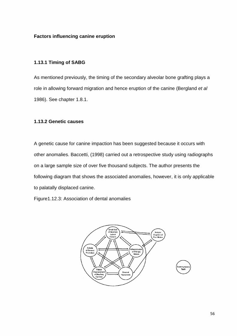
56
Factors influencing canine eruption
1.13.1 Timing of SABG
As mentioned previously, the timing of the secondary alveolar bone grafting plays a
role in allowing forward migration and hence eruption of the canine (Bergland et al
1986). See chapter 1.8.1.
1.13.2 Genetic causes
A genetic cause for canine impaction has been suggested because it occurs with
other anomalies. Baccetti, (1998) carried out a retrospective study using radiographs
on a large sample size of over five thousand subjects. The author presents the
following diagram that shows the associated anomalies, however, it is only applicable
to palatally displaced canine.
Figure1.12.3: Association of dental anomalies
57
The diagram shows that there is a statistical reciprocal (represented by two arrows)
association between palatally displaced canines and small size of maxillary lateral
incisors, infraocclusion of primary molars, enamel hypoplasia and hypodontia of
second premolars but no association with supernumerary teeth. Chung et al, (2011)
also reported that palatal canine impaction was significantly greater in females which
again leads to believe that a genetic cause is present.
1.13.4 Presence of lateral incisors and Guidance theory
Enemark et al, (2001) reported an increase risk in canine impaction when the lateral
incisor was missing. In the subjects who had grafts from the iliac crest, 35.1% of the
canines were impacted when the lateral incisor was present as opposed to 50.1% of
canine impactions when the lateral incisor was missing. They mention that the
reason for this could be that the root of the lateral incisor guides the canine down
towards the occlusal plane. This is in agreement with Becker, (1995) who theorised
that the root of the lateral incisor guides the eruption the canine. Brin et al, (1986)
found that 42.6% of palatally impacted canines were associated with peg shaped,
small or missing lateral incisors in non-cleft subjects. Oberio et al, (2005) and Tortora
et al, (2008), however, reported a lack of correlation between missing lateral incisor
and spontaneous canine eruption in cleft subjects
58
1.13.5 Canine position in Non-cleft subjects
There are also numerous other factors that influence canine eruption. Erikson and
Kurol, (1988) found a success rate of 78% spontaneous canine eruption following
extraction of the deciduous canine in non-cleft subjects between 10 and 13 years of
age with no crowding. The authors reported that 19 of 24 canines which were distal
to the midline of the lateral improved in position whereas 4 of the 22 canines mesial
to the midline of the lateral improved. This was similar to the findings reported by
Power and Short, (1993). They reported that the prognosis for spontaneous canine
eruption was good if the overlap of the adjacent lateral incisor was less than a half
and if the angulation to the vertical plane was less than 31°. Vertical height and the
eruptive distance was not found to have a significant effect on eruption.
1.13.6 Canine position in cleft subjects
The factors mentioned above in non-cleft subjects are also applicable to the cleft
population. Russell and McLeod, (2008) carried out a study examining the vertical
and lateral position of the canine following SABG. With respect to the vertical
position, the canine was at risk if the long axis of the canine root was greater than 45
degrees to the midsagittal vertical reference line. Laterally the canine was at risk if
the canine tip was mesial to the midplane of the lateral incisor root or the midplane to
the space distal to the central incisor, if the lateral incisor was missing. The author
found that there was a significant reduction in proportion of canines with an abnormal
59
vertical position after SABG, indicating a reduce risk of impactions following SABG.
They also found that 61% of cleft sites had abnormal lateral incisor anomaly with
more cases of abnormally positioned canines and a lateral incisor anomaly on the
cleft side compared to the non-cleft side. There was also a significant greater
percentage of canines post-SABG that had abnormal lateral positions when the
lateral incisors were missing. Tortora et al, (2008) reported that inclination of the
canine to the vertical plane had a significant effect on their spontaneous eruption with
no canines greater than 45° erupting spontaneously.
1.13.7 Space creation
The provision of space creation appears to be an important factor as shown by
Baccetti et al, (2008) who found that 87.5% of palatally displaced canines erupted
spontaneously when the C was extracted and space was created with the use of
headgear compared to 50% success in the control group and 62.5% success in the
extraction of the C group. Expansion of the arch in order to create space in non-cleft
subject has also been associated with greater likelihood of spontaneous canine
eruption Baccetti et al, (2009). Enemark et al, (2001) found that expansion of the
arch had no statistically significant effect on spontaneous canine eruption in cleft
subjects.

60
Chapter 2
Participants and Methods
61
Chapter 2 Participants and methods Page 2.1 Aims 62 2.2 Study design 62 2.3 Null Hypothesis 63 2.4 Ethical approval 63 2.5 Sample size calculation 63 2.6 Subject selection 64 2.7 Method 64 2.8 Statistical analysis 69

62
2.1 Aims
1. To determine the success of secondary alveolar bone grafting (SABG) as
determined by spontaneous canine eruption.
2. To investigate the influence of the following factors on canine eruption
Post-Operative Bone Fill Index (Kindelan et al 1997)
Age at SABG
Presence of a lateral incisor
Horizontal position of canine relative to the midplane of the lateral incisor
Angulation of canine relative to a 45° vertical reference line
Presence of expansion prior to SABG
Type of cleft (unilateral or bilateral)
2.2 Study Design
This was a retrospective observational cohort study using records primarily from the
Birmingham Children’s Hospital with supplementation from the Birmingham Dental
Hospital and several district hospitals in the West Midlands. All subjects had their
SABG carried out at the Birmingham Children’s Hospital and their orthodontic
treatment carried out at the Birmingham Dental Hospital or a district hospital in the
West Midlands, hence the reason for the multicentre approach.
63
2.3 Null Hypothesis
There is no correlation between spontaneous canine eruption and Post-Operative
Bone Fill Index , age at SABG, presence of the lateral incisor, horizontal position
relative to midplane of lateral incisor, angulation of canine relative to 45° in the
vertical plane, presence of expansion prior to SABG and type of cleft.
2.4 Ethical approval
Ethical approval was gained from the University of Birmingham Research and Ethics
Committee (ERN_12-1244). Site specific approval at each trust was also gained from
the Research and Development or the audit department. An honorary contract was
also issued by the Birmingham Children’s Hospital.
2.5 Sample size calculation
A sample size calculation based upon an alpha significance level of 0.05 and a
power of 80% estimated that subjects with a total of 59 cleft canines would be
required to detect a correlation between the Post-Operative Bone Fill Index score
and spontaneous canine eruption. This was based upon an assumption that
following SABG, 80% of the clefts would have a Post-Operative Bone Fill Index score
of 1 and 80% of canines with a Post-Operative Bone Fill Index score of 1 would erupt
spontaneously. This was in comparison to 20% of clefts having a Post-Operative
Bone Fill Index score of 2, 3 or 4 and 80% of canines with a Post-Operative Bone Fill
Index score 2, 3 or 4 not erupting spontaneously.
64
2.6 Subject selection
Inclusion criteria
Subjects born with a cleft in the alveolus who have undergone an alveolar
bone grafting procedure between October 2004 and December 2006 at the
Birmingham Children’s Hospital.
Exclusion Criteria
Subjects with congenitally missing or extracted permanent canines
Subjects with erupted canines prior to SABG
Subjects with unusable or missing occlusal radiographs
2.7 Method
Data was collected from five centres which included:
1. Birmingham Children’s Hospital
2. Birmingham Dental Hospital
3. Warwick Hospital
4. Burton-Upon-Trent Hospital
5. Gloucester Royal Hospital
65
Subject records including clinical entries, radiographs which included occlusal
radiographs and orthopantograms as well as study models were examined.
The following data was collected;
1. Gender of the subject
2. Type of cleft- unilateral or bilateral
3. The post operative Bone Fill Index score
4. Presence of expansion prior to SABG
5. Age at SABG
6. First or repeat bone graft
7. Presence of lateral incisor
8. Morphology of lateral incisor
9. Time between SABG and post-operative occlusal radiographs
10. Eruption of canine on cleft side- spontaneous or with surgical intervention
11. Eruption of canine on contra-lateral side in subjects with unilateral clefts
12. Angulation of canine relative to a 45° vertical reference line
13. Horizontal position of canine relative to the midplane of the lateral incisor
The gender of the subject and the type of cleft was determined from the subject
notes.
The Post-Operative Bone Fill Index score devised by Kindelan et al, (1997) assesses
the height of the grafted bone in the cleft using an oblique occlusal radiograph 6
months post-operatively on a four point scale.
66
Grade 1- Greater than 75% bony fill of the cleft
Grade 2- Between 50 and 75% bony fill of the cleft
Grade 3- Less than 50% bony fill of the cleft
Grade 4- No complete bony bridge of the cleft
Mr Nahul Patel, specialist registrar in Orthodontics contacted the author of the index,
Dr Jay Kindelan by telephone and was informed that a calibration course was not
available. Upon learning this, the researcher attended the orthodontic specialist
interest group meeting on the 20th January 2012 hosted by the Manchester
Children’s Hospital where Dr Jay Kindelan and Dr Gunvor Semb demonstrated the
use of the Post-Operative Bone Fill Index. The session also included a practical
session involving scoring and marking of 30 cases followed by answers. The
researcher learned the following with regards to accurate application in the use of the
index;
1. The pre-operative and the post-operative occlusal radiographs are both
required which should be taken at the same angle and same exposure in
order to accurately compare the percentage of the cleft filled by bone.
2. The radiographs should be viewed in a dark room over a light background.
3. The assessment is subjective.
Most of the occlusal radiographs were viewed on the AGFA PACS system 6.5.2.657
at the Birmingham Children’s Hospital on which the researcher received training on.
The pre-operative and post-operative occlusal radiographs on the PACS system
were viewed simultaneously and the contrast was adjusted to optimise similarity. The
67
very few remaining occlusal radiographs on a hard copy were viewed on light box
and the difference in contrast was accepted.
In order to test the intra-examiner reliability, occlusal radiographs of 20 randomly
chosen subjects were assessed for the Post-Operative Bone Fill Index score one
month following initial assessment.
The age at secondary alveolar bone grafting was determined to the nearest whole
year calculated by using the date of birth and the date of SABG. If the SABG was
repeated which was determined from the examination of the operation histories, the
age was calculated using the date of the repeated SABG.
The presence of the lateral incisor was determined from the pre-SABG
orthopantogram as well as clinical entries. If the lateral incisor was extracted at the
time of the SABG, it was classified as missing. The morphology was determined
using clinical entries, orthopantograms and study models which was classified as one
of the following
1. Normal
2. Peg shaped or small
3. Unerupted or impacted
4. Not known
Unerupted or impacted was also included in this group as it would affect the
guidance of the canine. The following criteria was used to assess morphology of the
lateral incisor
Normal - The mesio-distal width was greater than its mandibular counterpart
68
Small - The mesio-distal width was smaller than its mandibular counterpart
Peg – The mesio-distal with was the greatest at the cervical margin
On the study models, mesio-distal widths were measured using a steel ruler with
callipers and on the digital orthopantograms, they were measured using a digital
ruler.
The lateral incisor was deemed impacted if there was evidence of a physical
obstruction preventing it from erupting such as a supernumerary.
The time between SABG and post-operative occlusal radiographs was recorded in
months. If the SABG was repeated, the time was recorded following the repeat
operation.
Using the method adopted by Russell and McLeod, (2008), the horizontal position of
the canine was defined as at ‘risk’ or ‘no risk’ depending on its position relative to the
lateral incisor. It was defined as at ‘risk’ if the tip of the canine was mesial to the
midplane of the lateral incisor root or mesial to the space that the canine would
occupy if the lateral incisor was missing. The width of the canine was measured
using a digital ruler and halved to ascertain the midplane of the lateral incisor if it was
missing. If the canine was distal to the midplane, it was defined as ‘no risk’. The
angulation of the canine was again subcategorised as being ‘at risk’ if the long axis of
the canine root was greater than 45° to the midsagittal vertical reference line and at
‘no risk’ if was less than 45°. The vertical reference line was guided by the use the
anterior nasal spine. The measurements were made on orthopantograms, which
were either taken prior to or after SABG depending on the availability. If both were
available, it was measured on the post-SABG orthopantogram.
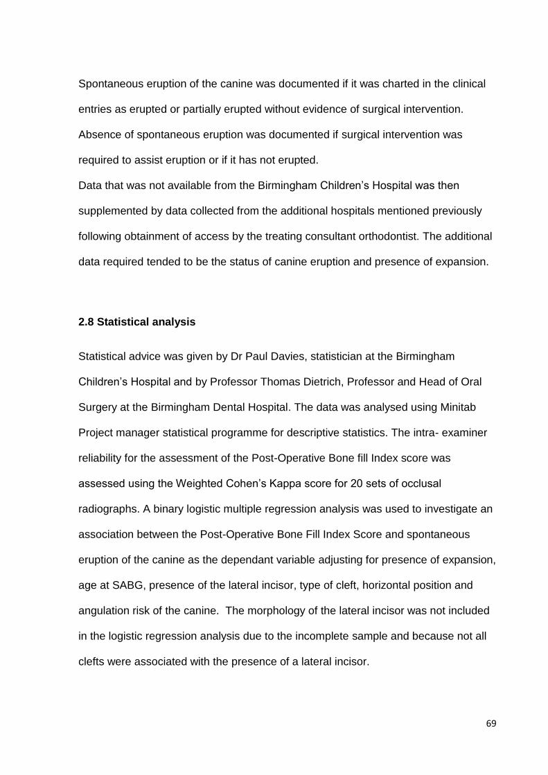
69
Spontaneous eruption of the canine was documented if it was charted in the clinical
entries as erupted or partially erupted without evidence of surgical intervention.
Absence of spontaneous eruption was documented if surgical intervention was
required to assist eruption or if it has not erupted.
Data that was not available from the Birmingham Children’s Hospital was then
supplemented by data collected from the additional hospitals mentioned previously
following obtainment of access by the treating consultant orthodontist. The additional
data required tended to be the status of canine eruption and presence of expansion.
2.8 Statistical analysis
Statistical advice was given by Dr Paul Davies, statistician at the Birmingham
Children’s Hospital and by Professor Thomas Dietrich, Professor and Head of Oral
Surgery at the Birmingham Dental Hospital. The data was analysed using Minitab
Project manager statistical programme for descriptive statistics. The intra- examiner
reliability for the assessment of the Post-Operative Bone fill Index score was
assessed using the Weighted Cohen’s Kappa score for 20 sets of occlusal
radiographs. A binary logistic multiple regression analysis was used to investigate an
association between the Post-Operative Bone Fill Index Score and spontaneous
eruption of the canine as the dependant variable adjusting for presence of expansion,
age at SABG, presence of the lateral incisor, type of cleft, horizontal position and
angulation risk of the canine. The morphology of the lateral incisor was not included
in the logistic regression analysis due to the incomplete sample and because not all
clefts were associated with the presence of a lateral incisor.

70
Chapter 3
Results
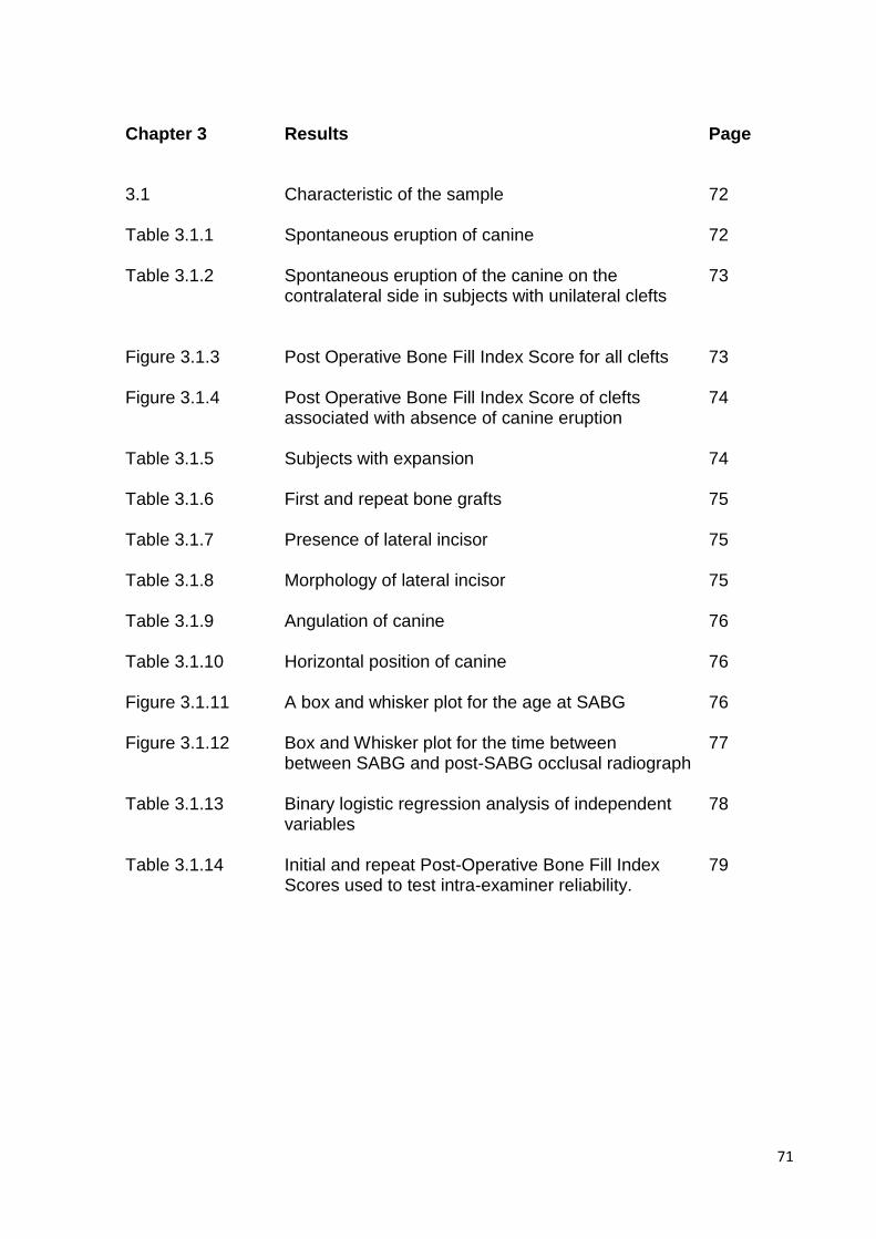
71
Chapter 3 Results Page
3.1 Characteristic of the sample 72 Table 3.1.1 Spontaneous eruption of canine 72 Table 3.1.2 Spontaneous eruption of the canine on the 73 contralateral side in subjects with unilateral clefts Figure 3.1.3 Post Operative Bone Fill Index Score for all clefts 73 Figure 3.1.4 Post Operative Bone Fill Index Score of clefts 74 associated with absence of canine eruption Table 3.1.5 Subjects with expansion 74 Table 3.1.6 First and repeat bone grafts 75 Table 3.1.7 Presence of lateral incisor 75 Table 3.1.8 Morphology of lateral incisor 75 Table 3.1.9 Angulation of canine 76 Table 3.1.10 Horizontal position of canine 76 Figure 3.1.11 A box and whisker plot for the age at SABG 76 Figure 3.1.12 Box and Whisker plot for the time between 77 between SABG and post-SABG occlusal radiograph Table 3.1.13 Binary logistic regression analysis of independent 78 variables Table 3.1.14 Initial and repeat Post-Operative Bone Fill Index 79 Scores used to test intra-examiner reliability.
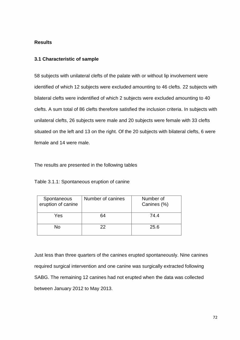
72
Results 3.1 Characteristic of sample 58 subjects with unilateral clefts of the palate with or without lip involvement were
identified of which 12 subjects were excluded amounting to 46 clefts. 22 subjects with
bilateral clefts were indentified of which 2 subjects were excluded amounting to 40
clefts. A sum total of 86 clefts therefore satisfied the inclusion criteria. In subjects with
unilateral clefts, 26 subjects were male and 20 subjects were female with 33 clefts
situated on the left and 13 on the right. Of the 20 subjects with bilateral clefts, 6 were
female and 14 were male.
The results are presented in the following tables
Table 3.1.1: Spontaneous eruption of canine
Spontaneous eruption of canine
Number of canines Number of Canines (%)
Yes
64 74.4
No
22 25.6
Just less than three quarters of the canines erupted spontaneously. Nine canines
required surgical intervention and one canine was surgically extracted following
SABG. The remaining 12 canines had not erupted when the data was collected
between January 2012 to May 2013.
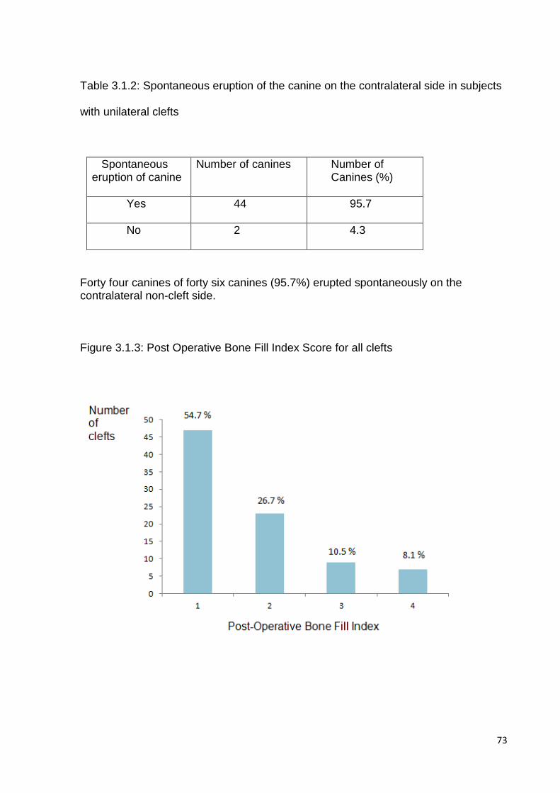
73
Table 3.1.2: Spontaneous eruption of the canine on the contralateral side in subjects
with unilateral clefts
Spontaneous eruption of canine
Number of canines Number of Canines (%)
Yes
44 95.7
No
2 4.3
Forty four canines of forty six canines (95.7%) erupted spontaneously on the contralateral non-cleft side. Figure 3.1.3: Post Operative Bone Fill Index Score for all clefts
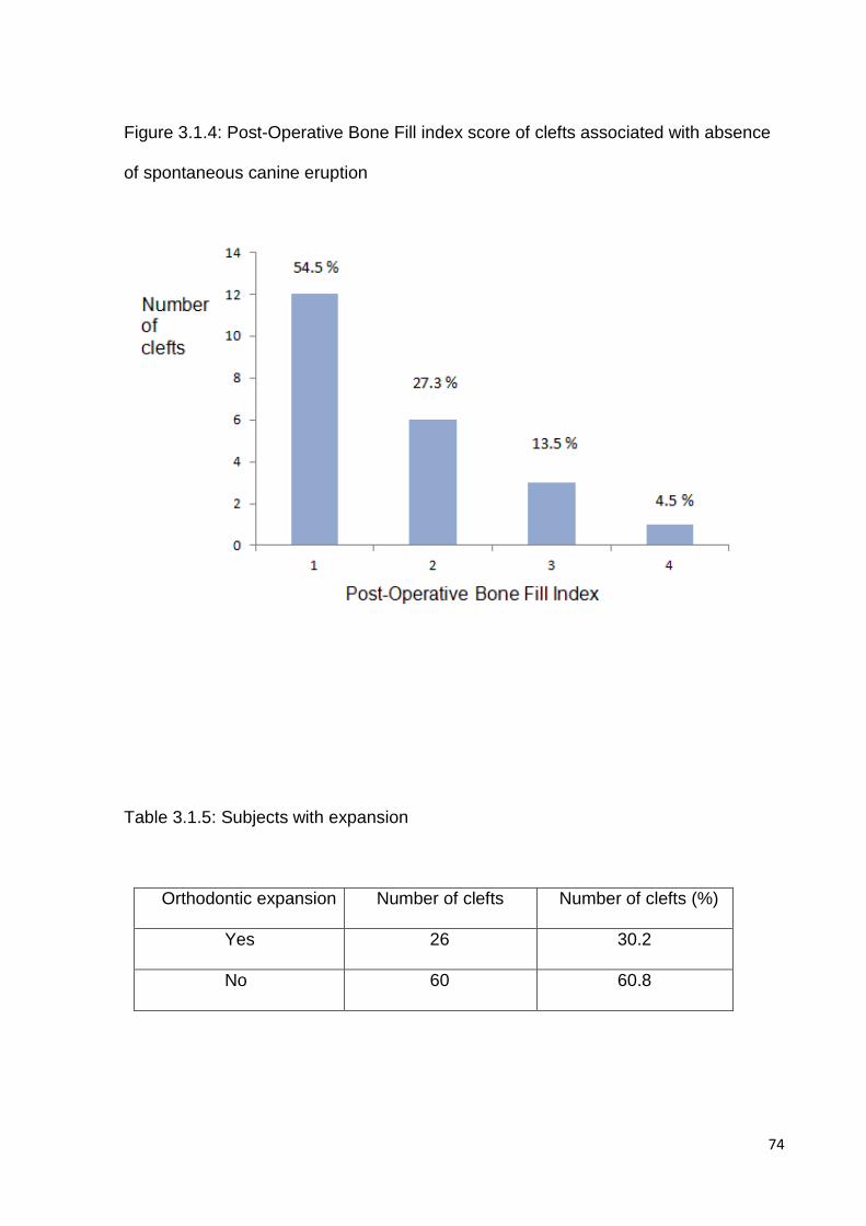
74
Figure 3.1.4: Post-Operative Bone Fill index score of clefts associated with absence
of spontaneous canine eruption
Table 3.1.5: Subjects with expansion
Orthodontic expansion
Number of clefts Number of clefts (%)
Yes
26 30.2
No
60 60.8
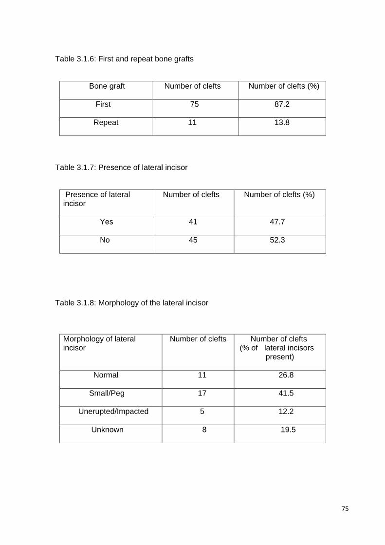
75
Table 3.1.6: First and repeat bone grafts
Bone graft
Number of clefts Number of clefts (%)
First
75 87.2
Repeat
11 13.8
Table 3.1.7: Presence of lateral incisor
Presence of lateral incisor
Number of clefts Number of clefts (%)
Yes
41 47.7
No
45 52.3
Table 3.1.8: Morphology of the lateral incisor
Morphology of lateral incisor
Number of clefts Number of clefts (% of lateral incisors present)
Normal
11 26.8
Small/Peg
17 41.5
Unerupted/Impacted
5 12.2
Unknown
8 19.5

76
Table 3.1.9: Angulation of canine
Risk
Number of clefts Number of clefts (%)
Yes
24 27.9
No
62 72.1
Table 3.1.10: Horizontal position of canine
Risk
Number of clefts Number of clefts (%)
Yes
12 14.0
No
74 86.0
Figure 3.1.11: A box and whisker plot for the age at SABG
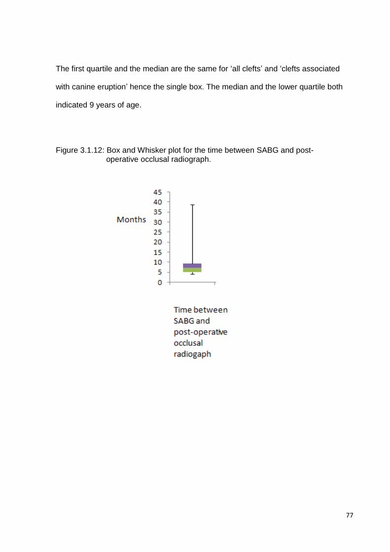
77
The first quartile and the median are the same for ‘all clefts’ and ‘clefts associated
with canine eruption’ hence the single box. The median and the lower quartile both
indicated 9 years of age.
Figure 3.1.12: Box and Whisker plot for the time between SABG and post- operative occlusal radiograph.
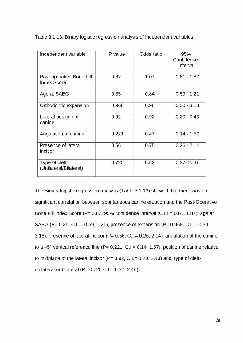
78
Table 3.1.13: Binary logistic regression analysis of independent variables
Independent variable P value
Odds ratio 95% Confidence Interval
Post-operative Bone Fill Index Score
0.82 1.07 0.61 - 1.87
Age at SABG
0.35 0.84 0.59 - 1.21
Orthodontic expansion
0.968 0.98 0.30 - 3.18
Lateral position of canine
0.92 0.92 0.20 - 0.43
Angulation of canine
0.221 0.47 0.14 - 1.57
Presence of lateral incisor
0.56 0.75 0.26 - 2.14
Type of cleft (Unilateral/Bilateral)
0.725 0.82 0.27- 2.46
The Binary logistic regression analysis (Table 3.1.13) showed that there was no
significant correlation between spontaneous canine eruption and the Post-Operative
Bone Fill Index Score (P= 0.82, 95% confidence interval (C.I.) = 0.61, 1.87), age at
SABG (P= 0.35, C.I. = 0.59, 1.21), presence of expansion (P= 0.968, C.I. = 0.30,
3.18), presence of lateral incisor (P= 0.56, C.I.= 0.26, 2.14), angulation of the canine
to a 45° vertical reference line (P= 0.221, C.I.= 0.14, 1.57), position of canine relative
to midplane of the lateral incisor (P= 0.92, C.I.= 0.20, 2.43) and type of cleft-
unilateral or bilateral (P= 0.725 C.I.= 0.27, 2.46).
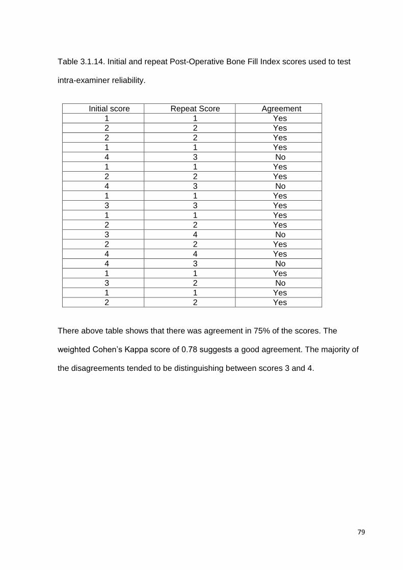
79
Table 3.1.14. Initial and repeat Post-Operative Bone Fill Index scores used to test
intra-examiner reliability.
Initial score Repeat Score Agreement
1 1 Yes
2 2 Yes
2 2 Yes
1 1 Yes
4 3 No
1 1 Yes
2 2 Yes
4 3 No
1 1 Yes
3 3 Yes
1 1 Yes
2 2 Yes
3 4 No
2 2 Yes
4 4 Yes
4 3 No
1 1 Yes
3 2 No
1 1 Yes
2 2 Yes
There above table shows that there was agreement in 75% of the scores. The
weighted Cohen’s Kappa score of 0.78 suggests a good agreement. The majority of
the disagreements tended to be distinguishing between scores 3 and 4.

80
Chapter 4
Discussion
81
Chapter 4 Discussion Page 4.1 Discussion 82 4.2 Limitations of the study 91
82
4.1 Discussion
The inclusion criteria of subjects receiving SABG between October 2004 and 2006
would allow ample time for canines to erupt given that the average age of upper
permanent canine emergence is 11.45+/- 1.21 years in non-cleft patients (Helm and
Seidler, 1974) which is delayed in cleft patients by 0.3 to 0.7 years (Ranta, 1986).
The operating surgeon began performing alveolar bone grafts at the Birmingham
Children’s Hospital in October 2004 and hence confounding factors such as operator
specialty and technique would be reduced. Operator experience would however not
be eliminated.
Sixty six of the eighty eight canines erupted spontaneously (Table 1), 10 canines
required surgical intervention and the remaining 12 had not erupted when the data
was collected. Of the 10 canines requiring surgical intervention, 7 canines were
exposed and bonded, 1 canine had an apically repositioned flap, 1 canine was
extracted and the 1 remaining canine was associated with extraction of the lateral
incisor into order to encourage its eruption. The success of spontaneous canine
eruption was therefore 74.4%. This is a reasonable level of success as defined by
spontaneous canine eruption when compared to other researchers e.g. Eldeeb et al,
(1982) reported a 27% success rate, Oberio et al, (2005) reported a 78.9% success
rate, Bergland et al, (1986) and Turvey et al, (1984) reported that 95% success rate.
In subjects with unilateral clefts, spontaneous canine eruption on the contralateral
side occurred in 95.7%. (Table 2). The absence of spontaneous eruption on the
contralateral side in these subjects of 4.3% is comparable to the general population
83
with a prevalence of 3.58% (Aydin et al, 2004). This study found that the failure of
spontaneous eruption was 6 times greater when the canine was situated adjacent to
a cleft in patients with unilateral oral clefts. Subject with clefts in this study compared
to the general population have a 9 fold increase in canine impaction which is much
less than the 20 fold increase reported by Russel and Mcleod, (2008).
The Post-Operative Bone Fill Index scores (Figure 3.1.3) demonstrate that over half
of all clefts (54.7%) had a score of one, 26.7% clefts had a score of two, 10.5% had a
score of three and 8.1% had a score of 4. The success rate in this study according
the bone fill would translate to 81.4% assuming that scores one and two are defined
as a success. This is again comparable to other reports in which the Post-Operative
Bone Fill Index has been used. Kindelan et al, (1997) reported 50% of grafts
achieving grade one, 23% with grade two, 22% with grade three and 5% with grade
four. Rawashdeh and Nimri, (2007) reported 90% with grade one, 5% with grade two,
5% with grade three and none with grade four in subjects with unilateral clefts. They
however defined success as score one only. Although there are a multitudinous
number of studies reporting eruption of canines through unoperated alveolar clefts, to
the best of my knowledge, no study in the published literature has reported on the
amount of bone in the clefts associated with absence of spontaneous canine eruption
(Figure 3.1.4). This study found that out of 22 clefts associated with absence of
spontaneous canines eruption, twelve clefts (54.5%) achieved a score of one, six
clefts (27.3%) scored two, four clefts (13.5%) scored three and one cleft (4.5%)
scored four. The results of the overall scores and scores associated with no canine
eruption are very similar with both sets scoring one in just over half the sample of
clefts and approximately a quarter scoring two.
84
Twenty six of eighty six clefts (30.2%) were expanded and sixty of eighty six clefts
(60.8%) were not expanded (Table 3.1.5). The low rate of expansion was because a
full time orthodontist was not present at the Birmingham Children’s Hospital during
the period involved in which the SABG was carried out. Examination of the records
revealed that the expansion was carried out with a quadhelix or a trihelix.
Eleven of the eighty six clefts (13.8%) had a repeat bone graft (Table 3.1.6). The
main reason documented in the records was the inadequate level of bone in the
grafted alveolar cleft.
The lateral incisor was present in forty one of the eighty six clefts (47.7%) and
therefore missing in greater than half the clefts (52.3%). Of the forty one lateral
incisors present, eleven had a normal morphology (26.8%), seventeen were small or
peg shaped (41.5%), five were unerupted or impacted (12.2%) and it was not
possible to determine the morphology of the remaining due to missing or inadequate
records. The prevalence of a peg or diminutive lateral incisor in this study was 19.7%
which is approximately three times greater than that reported by Tortora et al, (2008)
of 6.8%. Agenisis of lateral incisor in this study was 52.3% which is similar to the
prevalence reported by Bartzela et al, (2013) of 39.1%, Hermus et al, (2013) of
49.1% and Dentino et al, (2012) of 37.6%. The prevalence of missing maxillary
lateral incisors in the non-cleft population is reported as 1.58% to 1.78% (Poulder et
al, 2004) which translates to approximately a twenty nine fold increase of missing
maxillary lateral incisors in cleft subjects.
Hypodontia or malformations of the lateral incisor as mentioned previously is a
common feature in subjects with oral clefts. Ranta, (1986) discusses many theories
85
including an increase in length of the oral epithelium due to the cleft resulting in an
increase in length of the dental lamina thus creating a supernumerary tooth. The
tooth germ develops after the development of the cleft, therefore, the cleft may divide
the tooth bud as it lies across the nasopalatal sulcus. There may be inadequate
support for the bud due to deficiency in mesenchyme or previous surgery may
interfere with the bud formation. This is in agreement with Dentino et al, (2012) who
suggest that the lack of fusion between the medial nasal process and maxillary
process results in lack of blood supply and mesenchyme. Loose and Kirschner,
(2009) report that a gingivoperioplasty performed at three months at the time of the
lip repair can result in damage to the developing central and lateral incisor. As the
calcification of the lateral incisor begins at approximately six to seven months, at the
time of the lip repair, the lateral incisor would be in the bud or cap stage and
therefore any dissection of the follicle would disturb its formation. A common
genetical aetiology may also be involved.
Table 3.1.9 shows that 24 of 68 canines (27.9%) were at greater than 45° to the
vertical plane and sixty two of the eighty six canines and (72.1%) were less than 45°
to the vertical plane.
Table 3.1.10 shows that twelve of the eighty six canines(14%) were mesial to the
midplane of the lateral incisor and seventy four canines were distal to the midplane of
the lateral incisor (86%). Oberio et al found that twice as many canines moved when
compared to the non-cleft side with a minimum of 0.6mm and a maximum of 9.8mm.
The reason for the mesial drift could be due to the large number of missing lateral
incisors or absence of guidance from the lateral incisor root.
86
Table 3.1.11 shows that all alveolar bone grafting was carried out at the immediate
stage i.e. between the ages of 6 and 15. The minimum age of SABG was 8 and the
maximum was 14 for all clefts and therefore that includes clefts associated with
spontaneous canine eruption as well as clefts associated with absence of
spontaneous canine eruption. The median and the lower interquartile range of SABG
for all clefts and clefts associated with spontaneous canine eruption was 9 years of
age whereas the median for the clefts associated with no spontaneous eruption was
8.75 years, which was deemed clinically insignificant.
Figure 3.1.12 shows the box and whisker plot for the time between SABG and the
post-SABG occlusal radiograph. The minimum time post-SABG was 2.9 months,
median was 7.2 months and the maximum was 29.2 months. The interquartile range
was 4.4 months. The time at which the post-operative occlusal radiograph used for
measurement of bone levels is an important factor. Ozawa et al, (2007) showed that
the volume of bone varies immediately post-SABG and at 6 months post-SABG.
Using Cone Beam CTs, the authors reported a mean volume of 1.37mL immediately
post- SABG, which reduced by 46.7% to 0.73mL at 6 months post-SABG due to the
volume occupied by the eruption of the teeth, however, they reported a range of
13.11% to 63.79% of bone resorption at 6 months. They also stated that the eruption
of the canine and formation of the bony bridge were proportional hence eruption of
the canine in itself was associated with increased levels of bone in the cleft, however
they did not distinguish between cause and effect. Feichtinger et al, (2008) reported
an average of 64% bone loss at one year following secondary alveolar bone grafting
and stated that the clefts with greater bone levels were associated with eruption of
teeth. In this study, the level of eruption of the unerupted canine was not assessed
87
and therefore the amount of bone formed by the eruption would make the
assessment of bone fill less accurate. The fact that the occlusal radiographs were all
taken at different times would again reduce the accuracy of the Post-Operative Bone
Fill Index score as the bone levels may be subject to change.
The Binary logistic regression analysis (Table 3.1.13) showed that there was no
significant correlation between spontaneous canine eruption and the Post-Operative
Bone Fill Index Score, age at SABG, presence of expansion, presence of lateral
incisor, angulation of canine to a 45° vertical reference line, horizontal position of
canine relative to midplane of the lateral incisor and type of cleft- unilateral or
bilateral.
The odds ratios have not been commented upon with regards to trends as the p
values are significantly above 0.05.
It has been reported in the literature that teeth adjacent to the cleft erupt in the
absence of bone but with poor long term prognosis and poor periodontal health.
Collins et al, (1998) in their sample of 115 subjects, aged between eight and eighteen
reported that 58.4% of canines erupted prior to bone grafting. Gerutzel et al, (2005)
reported that in non-bone graft subjects, the canine still erupted, however it was
guided by the available cortical bone. No subjects in study were excluded on the
basis that the canines had erupted prior to SABG. This study has shown that there is
no statistical correlation between the amount of bone the cleft and spontaneous
canine eruption. Bergland et al, (1986) states that it is not the percentage of bone in
the graft that is of significance but the percentage of graft cells that survive. It
therefore seems that surgical factors may play a more significant role in canine
eruption such as the surgical technique and type of bone grafted which can improve
88
the quality of the bone. Loose and Kirschner, (2009) mention that the bone margins
of the cleft are hypoplastic and bone should be grafted over the margins thus there
should be adequate bone grafted that is compressed sufficiently with tight secure
closure of the mucoperiosteal flaps. Bergland et al (1986) and Lilja, (2009) both
discuss the impact of the choice of bone grafted. The ischaemic cancellous bone
revascularises quickly in comparison to the ischaemic cortical bone because it is
more vascular whereas cortical bone requires invasion of bone cells from the
recipient site therefore delaying turnover, remodelling and transformation. Bergland
et al (1986) describes a case where spontaneous migration of teeth adjacent to the
cleft was prevented due to the slow transformation of the cortical graft. All the
subjects included in this study had cancellous bone grafted from the Iliac crest.
When the cleft is grafted prior to eruption of the canine, alveolar bone is generated as
it erupts, therefore, perhaps it is not the amount of bone but the quality of the bone
which is related to the surgical technique that plays an influence on canine eruption
hence operator experience cannot be eliminated as a confounding factor. This is in
agreement with Kalaaji et al, (1996) who found significantly better bone height
following SABG when the operation was carried out by the more experienced
surgeons. Boyarski et al, (2006), however, found that there was no statistical
difference in bone height amongst surgeons.
The dental age which is related to the root length and hence the eruptive potential
would also affect whether the canine erupts spontaneously. Bergland et al, (1986)
reported that the SABG should be carried out when the canine is a half to two-thirds
formed in order to maximise canine eruptive potential however Boyarski et al, (2006)
found that differences in the canine root development had no statistical effect.
89
Enemark et al, (2001) theorised that a greater prevalence of canine impaction in cleft
patients is due to the primary surgery. They found that after changing the type of
primary surgery to close the anterior hard palate from a double to a single vomerine
flap, less lateral crossbites were observed and speculated that the greater the tilt of
the maxillary segment in the medial direction, the greater the likelihood of canine
retention.
Matsui, et al (2005) reported that the cleft width at the piriform aperture was
significantly greater in subjects who had surgical exposure of the canines.
In non-cleft subjects, there is a trend in evidence to suggest that expansion, hence
creation of space and presence of lateral incisor is associated with canine eruption
(Brin et al, 1986: Baccetti et al, 2009) There is conflict within the literature as to
whether expansion and presence of the lateral incisor has an effect on alveolar bone
heights and canine eruption in cleft subjects. This study also found that angulation of
the canine greater than 45° relative to the vertical plane had no significant effect on
the eruption of the canine whereas Tortora et al, (2008) found that no canines greater
than 45° erupted. This is because 34 of the 36 canines in their study were less than
15° to the vertical plane and only 2 canines were between 15° and 45° which leads to
the question of whether an adequate sample size was utilised. The findings from this
study also revealed that spontaneous canine eruption was not associated with the
horizontal position of the canine relative to the midplane of the lateral incisor. This is
because a large proportion of the lateral incisors were missing and if so, it is likely
that the canines erupted into the lateral incisor space.
Studies that have reported a correlation between the age of SABG and dental
eruption have compared early, intermediate and late SABG (Brattstrom and
90
McWilliam, 1989: Helms et al, 1987: Bergland et al, 1986) As all the SABG in this
study was carried out prior to canine eruption and in the intermediate stage, a
significant correlation would not be expected.
Other factors for canine impaction include a genetic predisposition associated with
both cleft formation and dental anomalies, interference with neural crest cell
migration which may alter formation, development and differentiation into dental
lamina, delayed root development and scar tissue from previous surgery (Solis et al
1998). It therefore seems that no one factor has a significant effect on the eruption of
canine in cleft patients. There are more variables that effect the eruption of canine in
cleft subjects in comparison to non-cleft subjects which explains why there was no
statistical correlation between the angulation the canine, lateral position the canine,
presence of the lateral incisor and type of cleft (unilateral or bilateral) with canine
eruption. All studies mentioned in this thesis that have found a statistical correlation
between any of the factors have been retrospective in nature without a power
calculation which therefore reduces the external validity.
It appears that spontaneous eruption of the canine is likely to be due to a small
additive effect of the factors listed below
Cleft
Genetic association between presence of cleft and canine impaction
Width and morphology of cleft Surgical factors
Type and timing of primary surgery
91
Operator experience of primary surgery
Insults to developing tooth germs
Timing of SABG
Quality and height of bone in cleft
Operator specialty, operator experience of SABG
Surgical technique
Type of bone grafted Canine factors
Stage at root development
Horizontal position of the canine
Angulation of the canine Other factors
Presence and morphology of lateral incisor
Presence of expansion prior to SABG
4.2 Limitations of the study
This is a retrospective observational cohort study and the data was not designed in a
manner to be used for such study. Although the ideal study would be a randomised
control trail, it would be unethical not to bone graft subjects and observe the
behaviour of the canine due to the multitudinous purpose of alveolar bone grafting.
As a consequence, searching for historical data introduces bias resulting in
92
undermining of the external validity. Selection bias cannot be extrapolated to this
study as all participants who had alveolar bone grafting in the defined period were
allocated to either the inclusion or exclusion criteria. There was however
measurement bias as radiographs were not standardised in their exposure and
position, analysis bias as some data was missing and the presence of numerous
confounding factors as they were not included in the inclusion and exclusion criteria
such as oral hygiene post-operatively.
Errors of measuring the amount of bone on occlusal radiographs are not without their
disadvantages. Limitations of occlusal radiographs include two dimensional
representation of three dimensional images and therefore the anterior-posterior depth
of the cleft is unable to be examined. In the horizontal plane, there may be
superimpositions of the adjacent structures along the curvature of the arch and in the
vertical plane, there is often distortion due to elongation. Other factors include
difference is contrast making it difficult to assess the bone margin as well as lack of
reliability of landmark identification. As the occlusal radiographs were not taken by
the same operator, the exposure and angle also varied. Ozawa et al, (2007) in their
CT study found that the bone is not evenly distributed with one side being thinner and
therefore volume thickness and architecture cannot be detected with two dimensional
radiography. Rosenstein et al (2007) compared the amount of bone using CT images
to periapical and occlusal radiographs in subjects following alveolar bone grafting.
They found that there was good agreement between 3-dimensional and 2-
dimensional radiography when the bone levels were greater than 90% or less than
50% but up to 25% variability when in between. The researchers concluded that
caution must be taken when assessing bone heights from 2-dimensional radiographs
93
and that 3-dimensional radiographs are superior in determining bone heights.
Accuracy of bone fill would have been significantly more accurate had CBCTs been
used, however, due to the increased exposure and cost, they are not routinely being
taken by cleft team post- alveolar bone grafting.
Following a practical session with Dr Jay Kindelan and Dr Gunvor Semb on the use
of the Post-Operative Bone Fill Index, it was observed by the author, Mr Nahul Patel
that the scores for each cleft varied amongst different raters and was therefore
subjective.
The bone heights were scored from occlusal radiographs at varying times post-SABG
and therefore different stages of eruption of the canine would be observed amongst
the radiographs. This would alter the amount of bone observed in the alveolar cleft as
eruption of the canine would in itself bring down bone into the cleft. The most
accurate method of measuring the bone would have been with the use of a 3D
technique at a defined period following SABG.
The power calculation was only carried out to determine a significant correlation
between spontaneous canine eruption and the Post-Operative Bone Fill Index score,
therefore, the validity of the binary logistic regression analysis in determining a
correlation between the other variables and canine eruption is reduced.
94
Chapter 5
Conclusion
95
Chapter 5 Conclusion 96 5.1 Conclusions 96 5.2 Null Hypothesis 97 5.3 Clinical significance 5.4 Suggestions for further study 97
96
5.1 Conclusions
1. Success of secondary alveolar bone grafting as defined by spontaneous
canine eruption was 74.4%
2. There was no significant association between the height of bone in the
alveolar cleft and spontaneous canine.
3. There was no significant association between spontaneous canine eruption
and the following variables:
Presence of lateral incisor
Presence of expansion
Angulation of canine relative to a 45° in the vertical reference line
Horizontal position of canine relative to the midplane of the lateral incisor
Type of cleft (unilateral or bilateral) 5.2 Null Hypothesis In the present study, there was no statistical correlation between spontaneous canine
eruption and Post-Operative Bone Fill Index, age at SABG, presence of the lateral
incisor, horizontal position relative to midplane of lateral incisor, angulation of canine
relative to 45° in the vertical plane, presence of expansion prior to SABG and type of
cleft.
Accepted
97
5.3 Clinical significance
This study does not add any new information, however, it does show that if alveolar
bone grafts are performed according to recommendations made by credible authors,
satisfactory outcomes can be achieved. It also confirms that canine eruption in cleft
subjects is not influenced by the amount of bone present in the cleft but due to the
high biological variability of factors that influence canine eruption such as in non-cleft
subjects. See chapter 4.1 for list of factors influencing canine eruption in cleft
subjects.
5.4 Suggestions for further study
Most of the studies involving cleft subject have been retrospective in nature and there
is conflicting evidence of whether expansion prior to SABG would influence the
height of bone and facilitate spontaneous canine eruption. Consideration could be
given to a prospective randomised control trial in order to ascertain whether
expansion (if required) prior to or post SABG would influence the height of bone in
the alveolus and influence spontaneous canine eruption.
98
References
Amanat, N., Langdon, JD. (1991) Secondary alveolar bone grafting in clefts of the lip and palate. J Craniomaxillofac Surg. 19(1):7-14.
Andersson, EM., Sandvik, L., Abyholm, F., Semb, G. (2010) Clefts of the secondary palate referred to the Oslo Cleft Team: epidemiology and cleft severity in 994 individuals. Cleft Palate Craniofac J 47(4):335-42. Andlin-Sobocki, A., Eliasson, LA., Paulin, G. (1995) Periodontal evaluation of teeth in bone grafted regions in patients with unilateral cleft lip and cleft palate. Am J Orthod Dentofacial Orthop. 107(2):144-52. Aydin, U., Yilmaz, HH., Yildirim, D. (2004) Incidence of canine impaction and transmigration in a patient population. Dentomaxillofac Radiol. 33(3):164-9. Baccetti, T. (1998) A controlled study of associated dental anomalies. Angle Orthod. 68(3):267-74. Baccetti, T., Leonardi, M,. Armi, P. (2008) A randomized clinical study of two interceptive approaches to palatally displaced canines. Eur J Orthod. 30(4):381-5. Baccetti, T., Mucedero, M., Leonardi, M., Cozza, P. (2009) Interceptive treatment of palatal impaction of maxillary canines with rapid maxillary expansion: a randomized clinical trial. Am J Orthod Dentofacial Orthop. 136(5):657-61. Badovinac, RL., Werler, MM., Williams, PL., Kelsey, KT., Hayes, C. (2007) Folic acid-containing supplement consumption during pregnancy and risk for oral clefts: a meta-analysis. Birth Defects Res A Clin Mol Teratol. 79(1):8-15 Bartzela, TN., Carels, CE., Bronkhorst, EM., Jagtman, AM. (2013) Tooth agenesis patterns in unilateral cleft lip and palate in humans. Arch Oral Biol.(6):596-602 Bayerlein, T., Proff. P., Heinrich, A., Kaduk, W., Hosten, N., Gedrange, T. (2006) Evaluation of bone availability in the cleft area following secondary osteoplasty.J Craniomaxillofac Surg. 34(2):57-61.
99
Bearn, D., Mildinhall, S., Murphy, T., Murray, JJ., Sell, D., Shaw, WC., Williams, AC., Sandy, JR. (2001) Cleft lip and palate care in the United Kingdom--the Clinical Standards Advisory Group (CSAG) Study. Part 4: outcome comparisons, training, and conclusions. Cleft Palate Craniofac J. 38(1):38-43. Becker, A. (1995) In defense of the guidance theory of palatal canine displacement. Angle Orthod. 65(2):95-8 Bergland, O., Semb, G., Abyholm, FE. (1986) Elimination of the residual alveolar cleft by secondary bone grafting and subsequent orthodontic treatment. Cleft Palate J. 23(3):175-205. Bishara ,SE. (1992) Impacted maxillary canines: a review. Am J Orthod Dentofacial Orthop. 101(2):159-71. Bjerklin, K., Kurol, J., Paulin, G. (1993) Ectopic eruption of the maxillary first permanent molars in children with cleft lip and/or palate. Eur J Orthod. 15(6):535-40. Bokhout, B., Hofman, FX., van Limbeek, J., Kramer, GJ., Prahl-Andersen, B. (1997) Incidence of dental caries in the primary dentition in children with a cleft lip and/or palate.Caries Res. 31(1):8-12 Bongaarts, CA., Van 't Hof, MA., Prahl-Andersen, B., Dirks, IV., Kuijpers-Jagtman, AM. (2006) Infant orthopedics has no effect on maxillary arch dimensions in the deciduous dentition of children with complete unilateral cleft lip and palate (Dutchcleft). Cleft Palate Craniofac J. 43(6):665-72. Borodkin, AF., Feigal, R.J., Beiraghi, S., Moller, K.T., Hodges, JS. (2008) Permanent tooth development in children with cleft lip and palate. Pediatr Dent. 30(5):408-13. Borstlap, WA., Heidbuchel, KL., Freihofer, HP., Kuijpers-Jagtman, AM. (1990) Early secondary bone grafting of alveolar cleft defects. A comparison between chin and rib grafts. J Craniomaxillofac Surg. 18(5):201-5. Botto, LD., Erickson, JD., Mulinare, J., Lynberg, MC., Liu, Y. (2002) Maternal fever, multivitamin use, and selected birth defects: evidence of interaction? Epidemiology. 13(4):485-8.
100
Boyarskiy, S., Choi, HJ., Park K. (2006) Evaluation of alveolar bone support of the permanent canine in cleft and noncleft patients. Cleft Palate Craniofac J. 43(6):678-82. Boyne, PJ., Sands, NR. (1972) Secondary bone grafting of residual alveolar and palatal clefts. J Oral Surg. 30(2):87-92. Brattström, V., McWilliam, J. (1989) The influence of bone grafting age on dental abnormalities and alveolar bone height in patients with unilateral cleft lip and palate. Eur J Orthod . 11(4):351-8. Brin, I., Becker, A., Shalhav, M. (1986) Position of the maxillary permanent canine in relation to anomalous or missing lateral incisors: a population study. Eur J Orthod. 8(1):12-6 Britton, KF., Welbury, RR. (2010) Dental caries prevalence in children with cleft lip/palate aged between 6 months and 6 years in the West of Scotland. Eur Arch Paediatr Dent. 11(5):236-41. Brouns ,J., Egyedi P. (1980) Osteotomy of the premaxilla. J Maxillofac Surg. 8(3):182-6. Bush, J., Jiang, R (2012) Palatogenesis: morphogenetic and molecular mechanisms of secondary palate development. Development. 139(2):231-43. Carmichael, SL., Shaw, GM. (2000) Maternal life event stress and congenital anomalies. Epidemiology. 11(1):30-5. Carmichael, SL., Shaw, GM., Yang, W., Abrams, B., Lammer, EJ. (2007) Maternal stressful life events and risks of birth defects. Epidemiology. 18(3):356-61. Chung, DD., Weisberg, M., Pagala, M. (2011) Incidence and effects of genetic factors on canine impaction in an isolated Jewish population. Am J Orthod Dentofacial Orthop. 139(4):e331-5 Cobourne, M. (2004) The complex genetics of cleft lip and palate. Eur J Orthod. 26(1):7-16. Cobourne, M., Diabiase. T. (2010) Handbook of Orthodontics. First edition, Elsevier Ltd Collins, M., James, DR., Mars, M. (1998) Alveolar bone grafting: a review of 115 patients. Eur J Orthod. 20(2):115-20.
101
Coupland, MA., Coupland, AL. (1988) Seasonality, incidence, and sex distribution of cleft lip and palate births in Trent Region, 1973-1982. Cleft Palate J. 25(1):33-7 Dentino, KM., Peck, S., Garib, DG. (2012) Is missing maxillary lateral incisor in complete cleft lip and palate a product of genetics or local environment? Angle Orthod. 82(6):959-63. Dobbyn, LM., Gillgrass, TJ., Devlin, MF. (2012) Reliability of the Kindelan scoring system for alveolar bone grafting with and without a pre-graft occlusal radiograph in patients with cleft lip and palate. Br J Oral Maxillofac Surg. 50(7):617-20 Eldeeb, M., Messer, LB., Lehnert, MW., Hebda, TW., Waite, DE. (1982) Canine eruption into grafted bone in maxillary alveolar cleft defects. Cleft Palate J. 19(1):9-16. Eldeeb, ME., Hinrichs, JE., Waite, DE., Bandt, CL., Bevis, R. (1986) Repair of alveolar cleft defects with autogenous bone grafting: periodontal evaluation. Cleft Palate J. 23(2):126-36. Enemark, H., Krantz-Simonsen, E., Schramm, JE. (1985) Secondary bonegrafting in unilateral cleft lip palate patients: indications and treatment procedure. Int J Oral Surg. 14(1):2-10. Enemark, H., Sindet-Pedersen, S., Bundgaard, M. (1987) Long-term results after secondary bone grafting of alveolar clefts. J Oral Maxillofac Surg. 45(11):913-9. Enemark, H., Jensen, J., Bosch, C. (2001) Mandibular bone graft material for reconstruction of alveolar cleft defects: long-term results. Cleft Palate Craniofac J. 38(2):155-63.
Ericson, S., Kurol, J. (1987) Radiographic examination of ectopically erupting maxillary canines. Am J Orthod Dentofacial Orthop. 91(6):483-92 Ericson, S., Kurol, J. (1988) Early treatment of palatally erupting maxillary canines by extraction of the primary canines. Eur J Orthod 10(4): 283-295. Feichtinger, M., Mossböck, R., Kärcher, H. (2006) Evaluation of bone volume following bone grafting in patients with unilateral clefts of lip, alveolus and palate using a CT-guided three-dimensional navigation system. J Craniomaxillofac Surg. 34(3):144-9
102
Feichtinger, M., Zemann, W., Mossböck, R., Kärcher, H. (2008) Three-dimensional evaluation of secondary alveolar bone grafting using a 3D- navigation system based on computed tomography: a two-year follow-up. Br J Oral Maxillofac Surg. 46(4):278-82 Friede, H., Johanson, B. (1974) A follow-up study of cleft children treated with primary bone grafting. 1. Orthodontic aspects. Scand J Plast Reconstr Surg. 8(1-2):88-103. Friede, H., Lilja, J., Lohmander, A. (2012) Long-Term, Longitudinal Follow-Up of Individuals With UCLP After the Gothenburg Primary Early Veloplasty and Delayed Hard Palate Closure Protocol: Maxillofacial Growth Outcome. Cleft Palate Craniofac J. 49(6):649-56 Freihofer, HP., Borstlap, WA., Kuijpers-Jagtman, AM., Voorsmit, RA., Van Damme, PA., Heidbüchel, KL., Borstlap-Engels, VM. (1993) Timing and transplant materials for closure of alveolar clefts. A clinical comparison of 296 cases. J Craniomaxillofac Surg. 21(4):143-8.
Gereltzul, E., Baba, Y., Ohyama, K. (2005) Attitude of the canine in secondary bone-grafted and nongrafted patients with cleft lip and palate. Cleft Palate Craniofac J. 42(6):679-86. Gorlin, RJ., Cervenka, J., Pruzansky, S. (1971) Facial clefting and its syndromes. Birth effects, 7: 3-49 Grayson, B.H., Cutting, C.B. (2001) Presurgical nasoalveolar orthopedic molding in primary correction of the nose, lip, and alveolus of infants born with unilateral and bilateral clefts. Cleft Palate Craniofac J. 38(3):193-8. Grabowski, R., Kopp, H., Stahl. F., Gundlach, KK. (2006) Presurgical orthopaedic treatment of newborns with clefts--functional treatment with long-term effects. J Craniomaxillofac Surg. (2):34-44 Guo, J., Li. C., Zhang, Q., Wu, G., Deacon, SA., Chen, J., Hu, H., Zou, S., Ye, Q. (2011) Secondary bone grafting for alveolar cleft in children with cleft lip or cleft lip and palate. Cochrane Database Syst Rev. Jun 15;(6):CD008050 Habel, A., Sell, D., Mars, M. (1996) Management of cleft lip and palate. Arch Dis Child. 74(4):360-6. Hamilton, R., Graham, W., Randall, P (1969) The role of lip adhesion procedure in cleft repair. Presentation of paper at the 38th annual meeting of the American Society of Plastic and Reconstructive Surgery.
103
Hasslöf, P., Twetman, S. (2007) Caries prevalence in children with cleft lip and palate--a systematic review of case-control studies. Int J Paediatr Dent. 17(5):313-9. Heidbuchel, KL. (1997) Blateral cleft lip and palate: Maxillofacial growth and dental development. 1st edition, Sweden. Heidbuchel, KL., Kuijpers-Jagtman, AM., Kramer, GJ., Prahl-Andersen, B. (1998) Maxillary arch dimensions in bilateral cleft lip and palate from birth until four years of age in boys. Cleft Palate Craniofac J. 35(3):233-9. Heidbüchel, KL., Kuijpers-Jagtman, AM., Ophof, R., Van Hooft, RJ. (2002) Dental maturity in children with a complete bilateral cleft lip and palate. Cleft Palate Craniofac J. 39(5):509-12. Helm, S., Seidler, B. (1974) Timing of permanent tooth emergence in Danish children. Community Dent Oral Epidemiol. 2(3):122-9.
Helms, JA., Speidel, TM., Denis, KL. (1987) Effect of timing on long-term clinical success of alveolar cleft bone grafts. Am J Orthod Dentofacial Orthop. 92(3):232-40. Hermus, RR., van Wijk, AJ., Tan, SP., Kramer, GJ., Ongkosuwito, EM. (2013) Patterns of tooth agenesis in patients with orofacial clefts. Eur J Oral Sci. 121(4):328-32. Hodgkinson, P., Brown, S., Duncan, D., Grant, C., Mc Naughten, A., Thomas, P., Mattick, CR. (2005) Management of children with cleft lip and palate: A review describing the application of multidisciplinary team working in this condition based upon the experiences of a regional cleft lip and palate centre in the United Kingdom. Fetal and maternal medicine review. 16:1-27 Hong, P., Bezuhly, M. (2013) Mandibular distraction osteogenesis in the micrognathic neonate: a review for neonatologists and pediatricians. Pediatr Neonatol. 54(3):153-60 Hotz, MM. (1969) Pre- and early postoperative growth-guidance in cleft lip and palate cases by maxillary orthopedics (an alternative procedure to primary bone-grafting). Cleft Palate J. 6:368-72. Howard, s., Lohmander, A. (2011) Cleft Palate Speech: Assessment And Intervention. 1st edition, John Wiley & Sons, Sussex
104
Hynes, PJ., Earley, MJ. (2003) Assessment of secondary alveolar bone grafting using a modification of the Bergland grading system. Br J Plast Surg. 56(7):630-6. Jensen, BL., Kreiborg, S., Dahl, E., Fogh-Andersen, P. (1988) Cleft lip and palate in Denmark, 1976-1981: epidemiology, variability, and early somatic development. Cleft Palate J. 25(3):258-69. Johnson, CY., Little, J. (2008) Folate intake, markers of folate status and oral clefts: is the evidence converging? Int J Epidemiol. 37(5):1041-58. Kalaaji, A., Lilja, J., Friede, H., Elander, A. (1996) Bone grafting in the mixed and permanent dentition in cleft lip and palatepatients: long-term results and the role of the surgeon's experience. J Craniomaxillofac Surg. 24(1):29-35. Kernahan, DA., Stark, RB. (1958) A new classification of cleft lip and palate. Plast. & Reconstr. Surg. 22: 435-441 Kindelan, JD., Nashed, RR., Bromige, MR. (1997) Radiographic assessment of secondary autogenous alveolar bone grafting in cleft lip and palate patients. Cleft Palate Craniofac J. 34(3):195-8. Kindelan, J., Roberts-Harry, D. (1999) A 5-year post-operative review of secondary alveolar bone grafting in the Yorkshire region. Br J Orthod. 26(3):211-7. Kirschner, RE., Low, DW., Randall, P., Bartlett, SP., McDonald-McGinn, DM., Schultz, PJ., Zackai, EH., LaRossa D. (2003) Surgical airway management in Pierre Robin sequence: is there a role for tongue-lip adhesion? Cleft Palate Craniofac J. 40(1):13-8. Konst, EM., Rietveld, T., Peters, HF., Kuijpers-Jagtman, AM. (2003) Language skills of young children with unilateral cleft lip and palate following infant orthopedics: a randomized clinical trial. Cleft Palate Craniofac J. 40(4):356-62.
Kurihara, S., Enlow, DH., Rangel, RD. (1980)
Remodeling reversals in anterior parts of the human mandible and maxilla. Angle Orthod. 50(2):98-106. Larson, M., Hellquist, R., Jakobsson, OP. (1998) Dental abnormalities and ectopic eruption in patients with isolated cleft palate. Scand J Plast Reconstr Surg Hand Surg.32(2):203-12.
105
Latham, RA. (1980) Orthopedic advancement of the cleft maxillary segment: a preliminary report. Cleft Palate J. 17(3):227-33. Latief, BS., Lekkas, KC., Schols, JG., Fudalej, PS., Kuijpers, MA. (2012) Width and elevation of the palatal shelves in unoperated unilateral and bilateral cleft lip and palate patients in the permanent dentition. J Anat. 220(3):263-70 Lee, CT., Grayson, BH., Cutting, CB., Brecht, LE., Lin, WY. (2004) Prepubertal midface growth in unilateral cleft lip and palate following alveolar molding and gingivoperiosteoplasty. Cleft Palate Craniofac J. 41(4):375-80 Li, L., Lin, M., Wang, Y., Cserjesi, P., Chen, Z. and Chen, Y. (2011). BmprIa isrequired in mesenchymal tissue and has limited redundant function with BmprIb in tooth and palate development. Dev Biol. 349(2):451-61 Lilja, J. (2009) Alveolar bone grafting. Indian J Plast Surg. 42(3)110–S115. Lilja, J., Kalaaji, A., Friede, H., Elander, A. (2000) Combined bone grafting and delayed closure of the hard palate in patients with unilateral cleft lip and palate: facilitation of lateral incisor eruption and evaluation of indicators for timing of the procedure. Cleft Palate Craniofac J. 37(1):98-105. Linde, A (2007) Molecular control of secondary palate development. Dev Biol. 301(2):309-26 Little, J., Cardy, A., Munger, RG. (2004) Tobacco smoking and oral clefts: a meta-analysis. Bull World Health Organ. 82(3):213-8 Liu, W., Sun, X., Braut, A., Mishina, Y., Behringer, R. R., Mina, M, Martin, JF. (2005). Distinct functions for Bmp signaling in lip and palate fusion in mice. Development. 132(6):1453-61 Long, RE Jr., Spangler, BE., Yow, M. (1995) Cleft width and secondary alveolar bone graft success. Cleft Palate Craniofac J. 32(5):420-7. Losee, J., Kirschner, R. (2009) Comprehensive cleft care. 1st edition, McGraw-Hill company, China. Martin, V., Bannister, P. (2004) Cleft care: A practical guide for health professionals on cleft lip and/or palate. 1st Edition, APS, Wiltshire.

106
Matok, I., Gorodischer, R., Koren, G., Landau, D., Wiznitzer, A., Levy, A. (2009) Exposure to folic acid antagonists during the first trimester of pregnancy and the risk of major malformations. Br J Clin Pharmacol. 68(6):956-62. Matsui, K., Echigo, S., Kimizuka, S., Takahashi, M., Chiba. M. (2005) Clinical study on eruption of permanent canines after secondary alveolar bone grafting. Cleft Palate Craniofac J. 42(3):309-13. Maull, D.J., Grayson, BH., Cutting, CB., Brecht, LL., Bookstein, FL., Khorrambadi, D., Webb. JA., Hurwitz. DJ. (1999) Long-term effects of nasoalveolar molding on three-dimensional nasal shape in unilateral clefts. Cleft Palate Craniofac J. 36(5):391-7. McCance, A., Roberts-Harry, D., Sherriff, M., Mars, M., Houston, WJ. (1993) Sri Lankan cleft lip and palate study model analysis: clefts of the secondary palate. Cleft Palate Craniofac J. 30(2):227-30. Millard, DR. (1957) A primary camouflage in the unilateral harelip. Transactions of the First International Congress of Plastic Surgery, Stockholm, Sweden; 160 – 72 Mishima, K., Sugahara, T., Mori. Y., Sakuda, M. (1996) Three-dimensional comparison between the palatal forms in complete unilateral cleft lip and palate with and without Hotz plate from cheiloplasty to palatoplasty. Cleft Palate Craniofac J. 33(4):312-7. Mitchell, L. (2007) An introduction to Orthodontics. 3rd edition, Oxford university press, Oxford. Mossey, PA., Little, J., Munger, RG., Dixon, MJ., Shaw, WC. (2009) Cleft lip and palate. Lancet. 374(9703):1773-85 Murray, JC. (2002) Gene/environment causes of cleft lip and/or palate. Clin Genet (61):248–256 Nagase, Y., Natsume, N., Kato, K., Hayakawa, T. (2010) Epidemiological Analysis of Cleft Lip and/or Palate by Cleft Pattern. J Maxillofac Oral Surg. 9(4): 389–395. Newlands, LC. (2000) Secondary alveolar bone grafting in cleft lip and palate patients. Br J Oral Maxillofac Surg. 38(5):488-91. Nightingale, C., Witherow, H., Reid, FD., Edler, R. (2003) Comparative reproducibility of three methods of radiographic assessment of alveolar bone grafting. Eur J Orthod. 25(1):35-41.
107
Oberoi, S., Chigurupati, R., Gill, P., Hoffman, WY., Vargervik, K. (2009) Volumetric assessment of secondary alveolar bone grafting using cone beam computed tomography. Cleft Palate Craniofac J. 46(5):503-11. Oberoi, S., Gill, P., Chigurupati, R., Hoffman, WY., Hatcher, DC., Vargervik, K. (2010) Three-dimensional assessment of the eruption path of the canine in individuals with bone-grafted alveolar clefts using cone beam computed tomography. Cleft Palate Craniofac J. Sep;47(5):507-12. Ozawa T, Omura S, Fukuyama E, Matsui Y, Torikai K, Fujita K. (2007) Factors influencing secondary alveolar bone grafting in cleft lip and palate patients: prospective analysis using CT image analyzer. Cleft Palate Craniofac J. 44(3):286-91. Pioto, N.R., Costa, B., Gomide, M.R. (2005) Dental development of the permanent lateral incisor in patients with incomplete and complete unilateral cleft lip. Cleft Palate Craniofac J. 42(5):517-20. Polder, BJ., Van't Hof, MA., Van der Linden, FP., Kuijpers-Jagtman, AM. (2004) A meta-analysis of the prevalence of dental agenesis of permanent teeth. Community Dent Oral Epidemiol. 32(3):217-26. Power, S.M., Short, M.B. (1993) An investigation into the response of palatally displaced canines to the removal of deciduous canines and an assessment of factors contributing to favourable eruption. Br J Orthod. 20(3):215-23. Prahl, C., Kuijpers-Jagtman, AM., Van 't Hof, MA., Prahl-Andersen, B. (2003) A randomized prospective clinical trial of the effect of infant orthopedics in unilateral cleft lip and palate: prevention of collapse of the alveolar segments (Dutchcleft). Cleft Palate Craniofac J. 40(4):337-42. Ranta, R. (1986) A review of tooth formation in children with cleft lip/palate. Am J Orthod Dentofacial Orthop. 90(1):11-8. Ranta, R., Stegars, T., Rintala, AE. (1983) Correlations of hypodontia in children with isolated cleft palate. Cleft Palate J. 20(2):163-5 Rawashdeh, MA., Al Nimri, KS. (2007) Outcome of secondary alveolar bone grafting before and after eruption of the canine in Jordanian patients with cleft lip and palate. J Craniofac Surg.18(6):1331-7.
108
Revington, PJ., McNamara, C., Mukarram, S., Perera, E., Shah, HV., Deacon, SA. (2010) Alveolar bone grafting: results of a national outcome study. Ann R Coll Surg Engl. 92(8):643-6. Rice, R., Spencer-Dene, B., Connor, E. C., Gritli-Linde, A., McMahon, A. P., Dickson, C., Thesleff, I. and Rice, DP. (2004) Disruption of Fgf10/Fgfr2b coordinated epithelial-mesenchymal interactions causes cleft palate. J Clin Invest. 113(12):1692-700 Roberts, RM., Mathias, J.L., Wheaton, P. (2012) Cognitive functioning in children and adults with nonsyndromal cleft lip and/or palate: a meta-analysis. J Pediatr Psychol. 37(7):786-97 Rosenstein, SW., Monroe, CW., Kernahan, DA., Jacobson, BN., Griffith, BH., Bauer, BS. (1982) The case of early bone grafting in cleft lip and cleft palate. Plast Reconstr Surg. 70(3):297-309 Rosenstein, SW., Long, RE Jr., Dado, DV., Vinson B., Alder ME. (1997) Comparison of 2-D calculations from periapical and occlusal radiographs versus 3-D calculations from CAT scans in determining bone support for cleft-adjacent teeth following early alveolar bone grafts. Cleft Palate Craniofac J. 34(3):199-205. Russell, KA., McLeod, CE. (2008) Canine eruption in patients with complete cleft lip and palate. Cleft Palate Craniofac J. 45(1):73-80. Sandy, J. (1992) Tooth eruption and Orthodontic movement. Br Dent J. 172(4):141-9. Sandy, J., Williams, A., Mildinhall, S., Murphy, T., Bearn, D., Shaw, B., Sell, D., Devlin, B., Murray, J. (1998) The Clinical Standards Advisory Group (CSAG) Cleft Lip and Palate Study. Br J Orthod. 25(1):21-30. Sandy, JR., Williams, AC., Bearn, D., Mildinhall, S., Murphy, T., Sell, D., Murray, JJ., Shaw, WC. (2001) Cleft lip and palate care in the United Kingdom--the Clinical Standards Advisory Group (CSAG) Study. Part 1: background and methodology. Cleft Palate Craniofac J. 38(1):20-3. Santiago, PE., Grayson, BH., Cutting, CB., Gianoutsos, MP., Brecht, LE., Kwon, SM. (1998) Reduced need for alveolar bone grafting by presurgical orthopedics and primary gingivoperiosteoplasty. Cleft Palate Craniofac J. 35(1):77-80.
109
Sell, D., Ma, L. (1996) A model of practice for the management of velopharyngeal dysfunction. Br J Oral Maxillofac Surg. 34(5):357-63. Semb, G. (1991.a) A study of facial growth in patients with unilateral cleft lip and palate treated by the Oslo CLP Team. Cleft Palate Craniofac J. 28(1):1-21 Semb, G. (1991b) A study of facial growth in patients with bilateral cleft lip and palate treated by the Oslo CLP Team. Cleft Palate Craniofac J. 28(1):22-39 Semb, G., Shaw, WC. (1990) Pharyngeal flap and facial growth. Cleft Palate J. 217-24. Sharma, R., Nanda, V. (2009) Problems of middle ear and hearing in cleft children. Indian J Plast Surg. (42) S144–S148. Shashua, D., Omnell, ML. (2000) Radiographic determination of the position of the maxillary lateral incisor in the cleft alveolus and parameters for assessing its habilitation prospects. Cleft Palate Craniofac J. 37(1):21-5. Sivarajasingam, V., Pell, G., Morse, M., Shepherd, JP. (2001) Secondary bone grafting of alveolar clefts: a densitometric comparison of iliac crest and tibial bone grafts. Cleft Palate Craniofac J. 38(1):11-4. Snyder, H.T., Bilboul, M.J., Pope, A.W. (2005) Psychosocial adjustment in adolescents with craniofacial anomalies: a comparison of parent and self-reports. Cleft Palate Craniofac J. 42(5):548-55. Solis, A., Figueroa, AA., Cohen, M., Polley, JW., Evans, CA. (1998) Maxillary dental development in complete unilateral alveolar clefts. Cleft Palate Craniofac J. 35(4):320-8. Stanier, P., Moore, GE. (2004) Genetics of cleft lip and palate: syndromic genes contribute to the incidence of non-syndromic clefts. Hum Mol Genet. 13 (1):R73-81 Suarez, L., Cardarelli, K., Hendricks, K., (2003) Maternal stress, social support, and risk of neural tube defects among Mexican Americans Epidemiology. 14(5):612-6.
110
Suzuki, K., Hu, D., Bustos, T., Zlotogora, J., Richieri-Costa, A., Helms, JA., Spritz, RA. (2000) Mutations of PVRL1, encoding a cell-cell adhesion molecule/herpesvirus receptor, in cleft lip/palate-ectodermal dysplasia. Nat Genet. 25(4):427-3
Tennison, CW. (1952) The repair of the unilateral cleft lip by the stencil method. Plast Reconstr Surg. 9(2):115-20. Tessier, P. (1976) Anatomical Classification of Facial, Cranio-Facial and Latero-Facia Clefts. Journal of Maxillofacial Surgery. 4:69-92 Tortora, C., Meazzini, M.C., Garattini, G., Brusati, R. (2008) Prevalence of abnormalities in dental structure, position, and eruption pattern in a population of unilateral and bilateral cleft lip and palate patients. Cleft Palate Craniofac J. 45(2):154-62. Turner, S.R., Rumsey, N., Sandy, JR. (1998) Psychological aspects of cleft lip and palate. Eur J Orthod. 20(4):407-15. Turvey, TA., Vig, K., Moriarty, J., Hoke, J. (1984) Delayed bone grafting in the cleft maxilla and palate: a retrospective multidisciplinary analysis. Am J Orthod. 86(3):244-56 Van der Meij, AW., Baart., JA., Prahl-Andersen, B., Kostense, PJ., van der Sijp, JR., Tuinzing, DB. (2003) Outcome of bone grafting in relation to cleft width in unilateral cleft lip and palate patients. Oral Surg Oral Med Oral Pathol Oral Radiol Endod. (1):19-25. Van der Meij, AJ., Baart, JA., Prahl-Andersen, B., Valk, J., Kostense, PJ., Tuinzing, DB. (2001) Bone volume after secondary bone grafting in unilateral and bilateral clefts determined by computed tomography scans. Oral Surg Oral Med Oral Pathol Oral Radiol Endod. 92(2):136-41. Veau, V. (1931) Treatment of the unilateral harelip Birt. J. Surg . 46:311-321 Waite, PD., Waite, DE. (1996) Bone grafting for the alveolar cleft defect. Semin Orthod. 2(3):192-6. Williams, A., Semb, G., Bearn, D., Shaw, W., Sandy, J. (2003) Prediction of outcomes of secondary alveolar bone grafting in children born with unilateral cleft lip and palate. Eur J Orthod. 25(2):205-11.
111
Witherow, H., Cox, S., Jones, E., Carr, R., Waterhouse, N. (2002) A new scale to assess radiographic success of secondary alveolar bone grafts. Cleft Palate Craniofac J. 39(3):255-60.
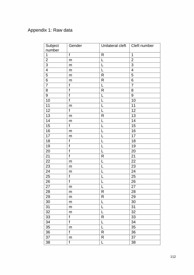
112
Appendix 1: Raw data
Subject number
Gender Unilateral cleft Cleft number
1 f R 1
2 m L 2
3 m L 3
4 m L 4
5 m R 5
6 m R 6
7 f L 7
8 f R 8
9 f L 9
10 f L 10
11 m L 11
12 f L 12
13 m R 13
14 m L 14
15 f L 15
16 m L 16
17 m L 17
18 f L 18
19 f L 19
20 f L 20
21 f R 21
22 m L 22
23 m L 23
24 m L 24
25 f L 25
26 f L 26
27 m L 27
28 m R 28
29 m R 29
30 m L 30
31 m L 31
32 m L 32
33 f R 33
34 f L 34
35 m L 35
36 f R 36
37 m R 37
38 f L 38
113
39 m L 39
40 m R 40
41 m L 41
42 f R 42
43 m L 43
44 f L 44
45 f L 45
46 m L 46
47 m bilateral 47
47 m bilateral 48
48 f bilateral 49
48 f bilateral 50
49 m bilateral 51
49 m bilateral 52
50 m bilateral 53
50 m bilateral 54
51 m bilateral 55
51 m bilateral 56
52 m bilateral 57
52 m bilateral 58
53 m bilateral 59
53 m bilateral 60
54 m bilateral 61
54 m bilateral 62
55 m bilateral 63
55 m bilateral 64
56 m bilateral 65
56 m bilateral 66
57 m bilateral 67
57 m bilateral 68
58 f bilateral 69
58 f bilateral 70
59 f bilateral 71
59 f bilateral 72
60 m bilateral 73
60 m bilateral 74
61 m bilateral 75
61 m bilateral 76
62 f bilateral 77
62 f bilateral 78
63 f bilateral 79
63 f bilateral 80
64 m bilateral 81
114
64 m bilateral 82
65 m bilateral 83
65 m bilateral 84
66 f bilateral 85
67 f bilateral 86
Cleft number
Spontaneous eruption of canine
Age at SABG Orthodontic expansion
1 y 8 n
2 y 14 y
3 n 11 y
4 y 8 n
5 y 9 y
6 n 9 n
7 y 8 n
8 y 11 n
9 y 10 n
10 y 10 n
11 n 8 n
12 n 8 y
13 n 11 y
14 n 8 n
15 y 8 n
16 y 8 n
17 y 8 n
18 y 10 n
19 n 9 n
20 n 9 y
21 n 9 y
22 y 9 y
23 y 10 y
24 y 11 n
25 n 9 n
26 y 9 n
27 y 9 y
28 y 12 y
29 y 9 n
30 y 8 n
31 y 10 n
32 y 8 n
115
33 y 9 n
34 y 11 y
35 n 9 n
36 y 8 y
37 y 9 n
38 y 10 n
39 y 9 n
40 y 10 n
41 y 9 y
42 y 9 n
43 n 8 n
44 y 9 n
45 y 10 n
46 y 9 n
47 y 9 n
48 y 9 n
49 n 14 n
50 n 14 n
51 y 13 y
52 n 13 y
53 y 9 n
54 y 9 n
55 n 10 n
56 n 10 n
57 n 10 y
58 y 10 y
59 y 9 y
60 y 9 y
61 y 10 n
62 y 10 n
63 n 9 y
64 y 9 y
65 y 8 n
66 y 8 n
67 n 8 n
68 y 8 n
69 y 9 n
70 y 9 n
71 y 8 n
72 y 8 n
73 y 10 y
74 y 10 y
75 y 9 n
116
76 y 9 n
77 y 10 y
78 y 10 y
79 n 11 n
80 n 11 n
81 y 11 n
82 y 11 n
83 y 12 n
84 y 12 n
85 y 10 n
86 y 10 n
Cleft number
First/repeat bone graft
Presence of lateral incisor
Morphology of lateral incisor normal(1)/small/peg(2))/ impacted/UE(3) missing (4),don't know (6)
1 1 n 4
2 1 y 2
3 1 n 4
4 1 y 2
5 1 n 4
6 1 n 4
7 1 y 1
8 1 n 4
9 1 n 4
10 1 y 2
11 1 y 2
12 1 n 4
13 1 y 6
14 1 n 4
15 1 n 4
16 1 n 4
17 1 n 4
18 2 y 1
19 1 n 4
20 1 n 4
21 1 n 4
22 2 y 2
23 2 y 1
24 1 n 4
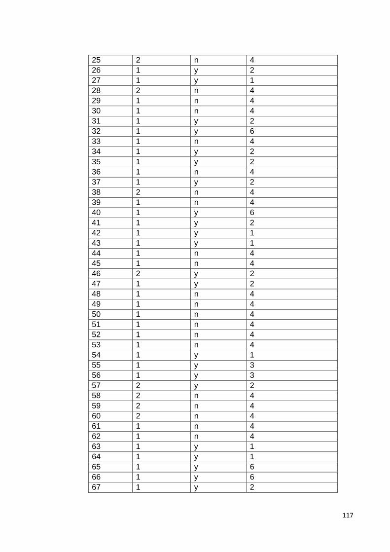
117
25 2 n 4
26 1 y 2
27 1 y 1
28 2 n 4
29 1 n 4
30 1 n 4
31 1 y 2
32 1 y 6
33 1 n 4
34 1 y 2
35 1 y 2
36 1 n 4
37 1 y 2
38 2 n 4
39 1 n 4
40 1 y 6
41 1 y 2
42 1 y 1
43 1 y 1
44 1 n 4
45 1 n 4
46 2 y 2
47 1 y 2
48 1 n 4
49 1 n 4
50 1 n 4
51 1 n 4
52 1 n 4
53 1 n 4
54 1 y 1
55 1 y 3
56 1 y 3
57 2 y 2
58 2 n 4
59 2 n 4
60 2 n 4
61 1 n 4
62 1 n 4
63 1 y 1
64 1 y 1
65 1 y 6
66 1 y 6
67 1 y 2
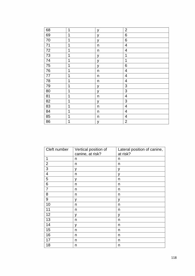
118
68 1 y 2
69 1 y 6
70 1 y 6
71 1 n 4
72 1 n 4
73 1 y 1
74 1 y 1
75 1 y 6
76 1 n 4
77 1 n 4
78 1 n 4
79 1 y 3
80 1 y 3
81 1 n 4
82 1 y 3
83 1 n 4
84 1 n 4
85 1 n 4
86 1 y 2
Cleft number Vertical position of canine, at risk?
Lateral position of canine, at risk?
1 n n
2 n n
3 y y
4 n y
5 y n
6 n n
7 n n
8 n n
9 y y
10 n n
11 n n
12 y y
13 n n
14 y n
15 n n
16 n n
17 n n
18 n n
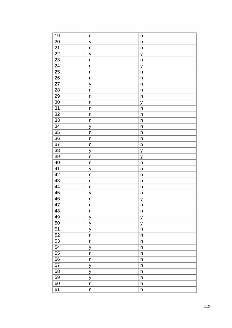
119
19 n n
20 y n
21 n n
22 y y
23 n n
24 n y
25 n n
26 n n
27 y n
28 n n
29 n n
30 n y
31 n n
32 n n
33 n n
34 y n
35 n n
36 n n
37 n n
38 y y
39 n y
40 n n
41 y n
42 n n
43 n n
44 n n
45 y n
46 n y
47 n n
48 n n
49 y y
50 y y
51 y n
52 n n
53 n n
54 y n
55 n n
56 n n
57 y n
58 y n
59 y n
60 n n
61 n n

120
62 n n
63 n n
64 n n
65 n n
66 n n
67 y n
68 n n
69 n n
70 n n
71 n n
72 n n
73 n n
74 n n
75 n n
76 n n
77 y n
78 y n
79 y n
80 n n
81 n n
82 y n
83 n n
84 n n
85 n n
86 n n
Cleft number Post Op bone fill index score
Repeat post Op bone fill index score
Difference Date of SABG and Date of rad
1 2 11.4
2 2 7.5
3 1 3.2
4 1 1 9.4
5 2 6.6
6 2 2 9.4
7 1 7.8
8 2 2 8.4
9 1 1 24.6
10 4 5.0
11 1 3.8
12 4 3 5.7
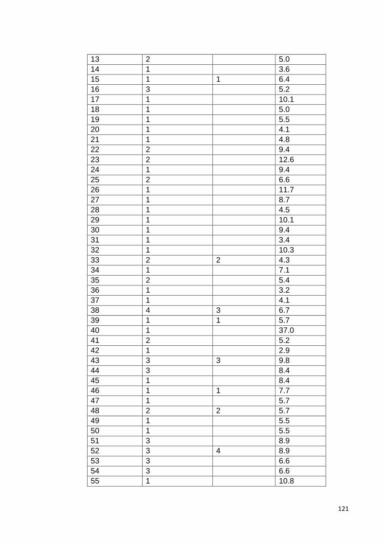
121
13 2 5.0
14 1 3.6
15 1 1 6.4
16 3 5.2
17 1 10.1
18 1 5.0
19 1 5.5
20 1 4.1
21 1 4.8
22 2 9.4
23 2 12.6
24 1 9.4
25 2 6.6
26 1 11.7
27 1 8.7
28 1 4.5
29 1 10.1
30 1 9.4
31 1 3.4
32 1 10.3
33 2 2 4.3
34 1 7.1
35 2 5.4
36 1 3.2
37 1 4.1
38 4 3 6.7
39 1 1 5.7
40 1 37.0
41 2 5.2
42 1 2.9
43 3 3 9.8
44 3 8.4
45 1 8.4
46 1 1 7.7
47 1 5.7
48 2 2 5.7
49 1 5.5
50 1 5.5
51 3 8.9
52 3 4 8.9
53 3 6.6
54 3 6.6
55 1 10.8
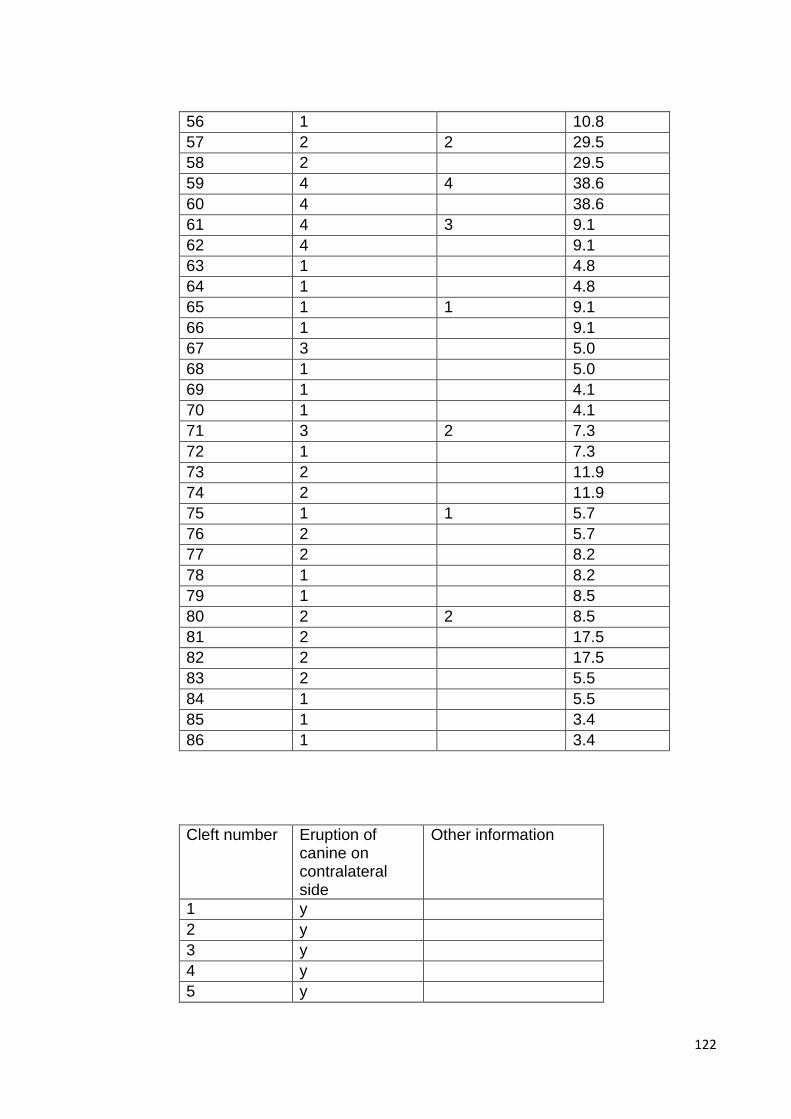
122
56 1 10.8
57 2 2 29.5
58 2 29.5
59 4 4 38.6
60 4 38.6
61 4 3 9.1
62 4 9.1
63 1 4.8
64 1 4.8
65 1 1 9.1
66 1 9.1
67 3 5.0
68 1 5.0
69 1 4.1
70 1 4.1
71 3 2 7.3
72 1 7.3
73 2 11.9
74 2 11.9
75 1 1 5.7
76 2 5.7
77 2 8.2
78 1 8.2
79 1 8.5
80 2 2 8.5
81 2 17.5
82 2 17.5
83 2 5.5
84 1 5.5
85 1 3.4
86 1 3.4
Cleft number Eruption of canine on contralateral side
Other information
1 y
2 y
3 y
4 y
5 y
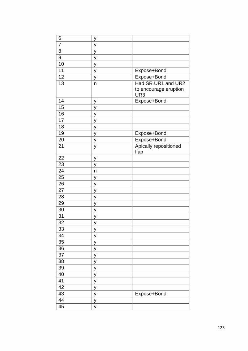
123
6 y
7 y
8 y
9 y
10 y
11 y Expose+Bond
12 y Expose+Bond
13 n Had SR UR1 and UR2 to encourage eruption UR3
14 y Expose+Bond
15 y
16 y
17 y
18 y
19 y Expose+Bond
20 y Expose+Bond
21 y Apically repositioned flap
22 y
23 y
24 n
25 y
26 y
27 y
28 y
29 y
30 y
31 y
32 y
33 y
34 y
35 y
36 y
37 y
38 y
39 y
40 y
41 y
42 y
43 y Expose+Bond
44 y
45 y
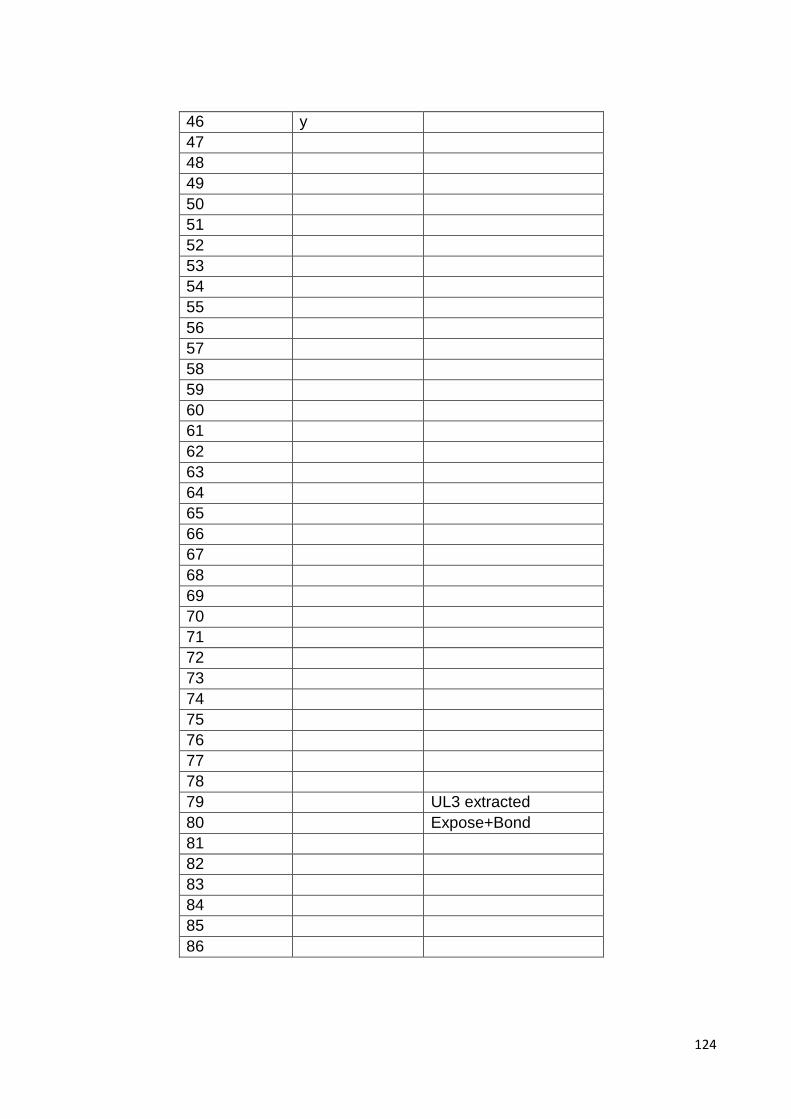
124
46 y
47
48
49
50
51
52
53
54
55
56
57
58
59
60
61
62
63
64
65
66
67
68
69
70
71
72
73
74
75
76
77
78
79 UL3 extracted
80 Expose+Bond
81
82
83
84
85
86

125