Embed Size (px)
Citation preview
Biophys. Struct. Mechanism 2, 43--59 (1976) Biophysics ~
" Mechanism �9 Springer-Verlag 1976
Subunit Symmetry of Tetrameric Phosphorylase a
K. Barrels* and P. M. Colman**
Max-Planck-Institut fiir Biochemie and Physikalisch-Chemisches Institut der Teehnisehen Univcrsit~t, D-8033 Martinsried, Fed. l~ep. Germany
Abstract. Native crystallographic data of tetrameric phosphorylase a crystals, space group P21, have been collected photographically to 3 • resolu- tion. These data have been used in Patterson search methods in reciprocal and real space.
The tetramers were found to exhibit molecular 222 symmetry. The cross vector between the centres of the two symmet ry related tetramers in the unit cell was determined by two different translation function methods.
On the basis of these rotation and translation function results a model for the arrangement of monomers within the tc t ramer and of tetramers in the unit cell is proposed.
The 222 symmet ry of the tetrameric molecule is found only when high resolution diffraction data are included (i.e. higher than 6 A). At lower resolu- tion other symmetries dominate.
Calculations with the proposed model have shown that these spurious symmetries result from the nonspeeifie overlap of protein-protein and solvent- solvent cross vectors.
These results emphasize the importance of high resolution data when non- crystallographic symmetry of globular proteins is studied.
Key words: Glycogen Phosphorylase -- Subunit Symmetry --Non- crystallographic Symmetry -- Rotation Function -- Translation Function.
Introduction
Phosphorylase (E.C.2.4.t.i.) is the key enzyme for the breakdown of glycogen. I t exists in two interconvertible forms. The b form, which under physiological conditions is a dimer, is inactive without effectors, such as AMP, IMP and/or Glucose-t-P. Phosphorylase kinase converts phosphorylase b into the a form which
* Extract from Dissertation, Technische Universit~t Miinchen. ** :Present address: Dept. of Inorganic Chemistry, University of Sydney, Australia.
44 K. Barrels and P. M. Colman
in solutions at concentrations around 0.2 to l0 mg exists ~s a tetramer and is active without effectors, but its activity is enhanced by AMP.
In 1972 we reported crystallization of both glycogen phosphorylase a and b. Both enzymes derived from rabbit muscle were crystallized in a tetrameric form (MW = 400000) in the presence of AMP. The two crystal forms were isomorphous in space group 1)21, with a = l i9 .4A, b = i88.6A, c = 88.2/~, f l= 108.6~ V = t .88.106/~a (Fasold et al., 1972). Apparently identical crystals have been reported by F. S. ~athews (i967) and Madsen et al. (1972). Four chemically identical subunits of MW 100000 are contained in the asymmetric unit.
From electron micrographs it was concluded that these four subunits are arranged on the vertices of a tetrahedron according to point symmetry 222 (Kiselev et al., t971; 1974). These optically filtered micrographs show resolved monomers of elongated bent shape. Johnson et al. (1974) reported the crystal structure of dimeric phosphorylase b with IMP at 6 A resolution, showing a rather compact molecule, but they have not yet Been able to resolve the monomer- monomer-contact within the dimer.
Phosphorylase b with IMP and phosphorylase b and a with AMP have been shown by ESR measurements to be in different conformational states (Campbell et al., 1972 ; Dwek et al., 1972 ; Griffiths et al., i974). A structure analysis of the tetramerie form and its comparison with the dimer might show the structural differences responsible for the different functional properties. This communication describes crystallographic data collection and the analysis of the subunit symmetry which leads to a preliminary model of the crystal structure of tetramerie phos- phorylase a.
Methods, Results and Discussion
I. Crystallographic Intensity Data Collection
Crystallographic data were collected by the screenless precession technique, using a focus to crystal and crystal to film distance of 300 mm and 100 mm respectively. The source was graphite monochromatized Cu K~ radiation from a sealed tube operated at 1.2 kW. The cameras were equipped with long collimators (200 ram). Pinholes of 0.3 to 0.5 mm were used depending on the crystal size.
Each crystal 1 tolerated about 250 hrs irradiation time, thus providing be- tween i and 5 intensity data photographs, depending on the crystal size. A precession angle of 1.5 ~ was used, yielding theoretically about 8500 reflexions (up to 3 f~ resolution) per film.
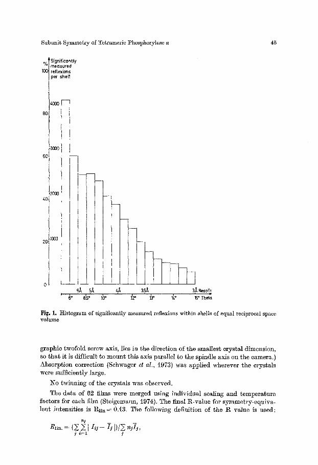
24000 out of 73000 erystallographically independent refiexions out to 3/~ resolution (60000 observations) were measured significantly above background ( > i.5 a), using the film evaluation programs by Schwager and Barrels (1975); cf. Fig. I.
l~efinement of the crystal orientation parameters (Schwager et al., 1975) proved to be critical: the Rs~m-value for individual films cannot be used as a monitor of satisfactory evaluation because of the ~lmost complete absence of symmetry- equivalent reftexions on the films. (The long crystal ~xis b, which is the crystallo-
1 The crystals used in this work were kindly provided by It. Fasold and coworkers, Institut ffir Biochemie der UniversR~t Frankfurt.
Subunit Symmetry of Tetramerie Phosphorylase a 45
%
lOO
8C
6Z
z~C
2(;
Significantly measured refIexions per shelf
-300C
-2000
.I000
6 o 8.5" 10 ~ 1'2 ~ 1~ ~~ 15" Theta
Fig. 1. Histogram of significantly measured reflexions within shells of equal reciprocal space ~rolume
graphic twofold screw axis, lies in the direction of the smallest crystal dimension, so that it is difficult to mount this axis parallel to the spindle axis on the camera.) Absorption correction (Sehwager et al., 1973) was applied wherever the crystals were sufficiently large.
No twinning of the crystals was observed.
The data of 62 films were merged using individual sealing and temperature factors for each film (Steigemann, 1974). The final R-value for symmetry-equiva- lent intensities is Rli~L= 0A3. The following definition of the R value is used:
~j
Rl,n. = (5 511~j-- ij I)/Y -jiS, i ' i=1 i
46 K. Barrels and P. M. Colman
:':l((((~//: i t i:'(:~ :~::'-".
" i , ',,' " - " " i I J ~ "
, , ; ;_ z--,~,..... _ . .
,,,,_., > ,, ,.., ...... ,. ~ "~::::-- ..,,,, :_ ,'! \\\\',"" ":, , , "-- " . - " . , ' .-k ~ Z L " ,"7~, . . . . \\\\", "- ~ , ' . ~ -41~.
l
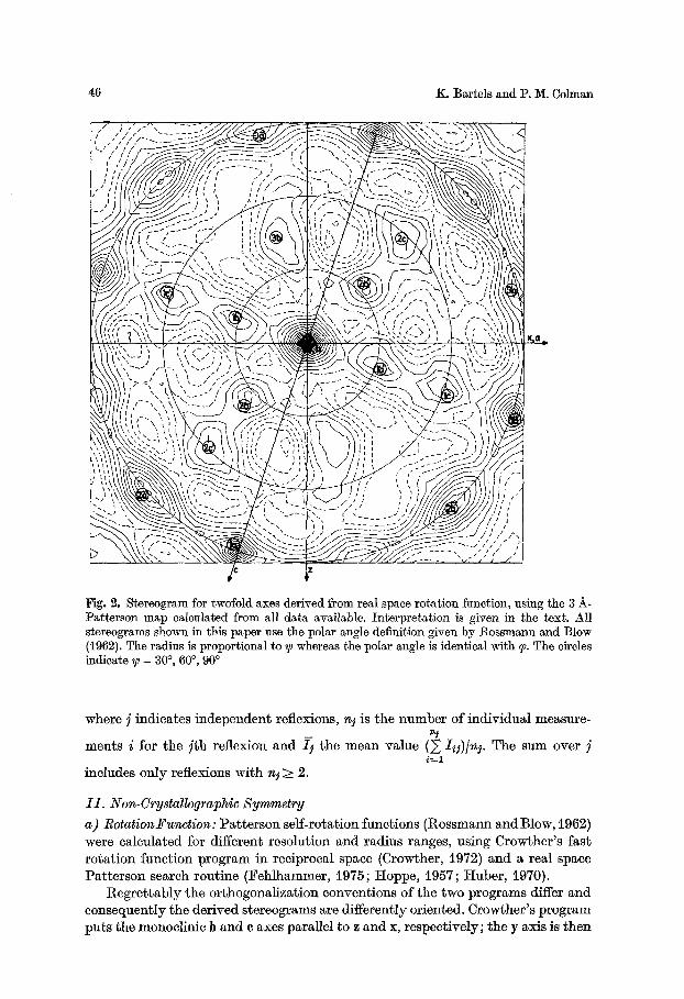
Fig. 2. Stereogram for twofold axes derived from real space rotation function, using the 3 h- Patterson map calculated from all data available. Interpretation is given in the text. ~ll stereograms shown in this paper use the polar angle definition given by Rossmann and Blow (i962). The radius is proportional to ~ whereas the polar angle is identical with ~. The circles indicate ~ = 30 ~ 60 ~ 90 ~
where j indicates independent reflexions, n t is the number of individual measure- n~
merits i for the : t h reflexion and ~ the mean value (~, I~,j)[nj. The sum over ]
includes only reflexions with nj > 2.
I I . Non-Crystallographic Symmetry a) Rotation2'unction: Pat te rson serf-rotation functions (Rossmann and Blow, 1962) were calculated for different resolution and radius ranges, using Crowther 's fast ro ta t ion funct ion program in reciprocal space (Crowther, 1972) and a real space Pa t te rson search routine (Fehlhammer, 1975; Hoppe, i957; Huber , i970).
Regre t t ab ly the orthogonalizat ion conventions of the two programs differ and consequently the derived stereograms are differently oriented. Crowbher's program puts the monoclinic b and c axes parallel to z and x, respectively; the y axis is then
Subunit Symmetry of Tetrameric Phosphorylase a 47
/ . . . . <
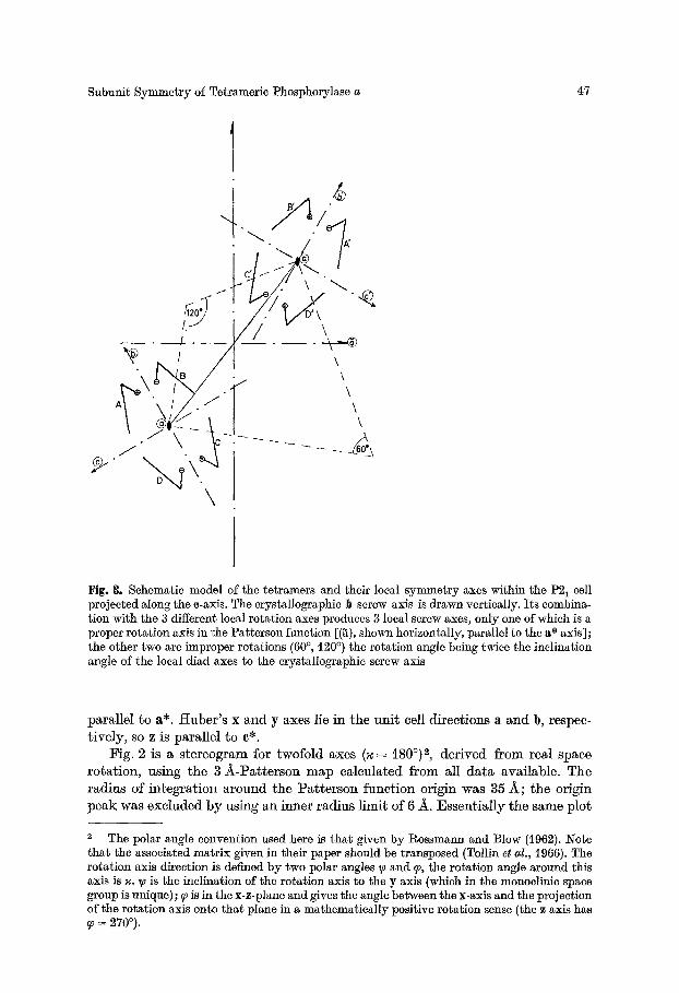
Fig. 8. Schematic model of the tetramers and their local symmetry axes within the P2~ cell projected along the e-axis. The crystallographic b screw axis is drawn vertically. Its combina- tion with the 3 different local rotation axes produces 3 local screw axes, only one of which is a proper rotation axis in the l~atterson function [($), shown horizontally, parallel to the a* axis]; the other two are improper rotations (60 ~ , 120 ~ ) the rotation angle being twice the inclination angle of the local diad axes to the crystallographic screw axis
parallel to a*. Huber ' s x and y axes lie in the un i t cell directions a and b, respec- t ively, so z is parallel to e*.
Fig. 2 is a stereogram for twofold axes (~ = i80 ~ 2, derived from real space rota t ion, using the 3 _ilk-Patterson map calculated from all da ta available. The radius of in tegra t ion a round the Pa t t e r son func t ion origin was 35 _~; ~he origin peak was excluded by using an inner radius l imit of 6 A. Essent ia l ly the same plot
The polar angle convention used here is that given by Rossmann and Blow (1962). Note that the associated matrix given in their paper should be transposed (Tollin et al., 1966). The rotation axis direction is defined by two polar angles y~ and ~0, the rotation angle around this axis is ~. yJ is the inclination of the rotation axis to the y axis (which in the monoclinie space group is unique); ~0 is in the x-z-plane and gives the angle between the x-axis and the projection of the rotation axis onto that plane in a mathematically positive rotation sense (the z axis has
= 270~
48 K. Barrels and P. YI. Colman
tOO
8O
o
- 6 0 \
~ o~O =o-o-o. o ~176176176 \ \ / \
/ o /
/ /
30' 60 ~ 90 ~ 120 ~ 150 ~ 180 ~ I I I l I I m,
ROTATION ANGLE
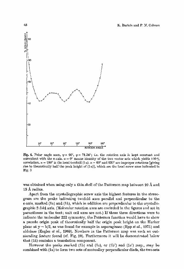
Fig. 4. Polar angle scan, ~-~ 90 ~ q--- 71.34~ i.e. the rotation axis is kept constant and coincident with the e-axis. ~ = 0 ~ means identity of the two vector sets which yields 100 % correlation. ~ = 180 ~ is the local twofold (t a). ~ = 60 ~ and 120 ~ are improper rotations [giving rise to theoretically half the peak height of (1 a)], which are the local screw axes indicated in Fig. 3
was obtained when using only a th in shell of the Pa t te rson map between 10 -~ and t5 -~ radius.
Apa r t f rom the crystallographic screw axis the highest features in the stereo- g ram are the peaks indicating twofold axes parallel and perpendicular to the e-axis, marked (la) and (i~), which in addit ion are perpendienlar to the crystallo- graphic 2-fold axis. (Molecular ro ta t ion axes are encircled in the figures and set in parantheses in the t ex t ; uni t cell axes are not.) I f these three directions were to indicate the molecular 222 symmetry , the Pa t te r son funct ion would have to show a pseudo origin peak of theoret ical ly half the origin peak height on the Harker plane at y = b]2, as was found for example in asparaginase (Epp et al., i971) and aldolase (Eagles et al., 1969). Nowhere in the Pa t te rson map was such an out- s tanding feature found (cf. Fig. 10). Fur thermore it will be demonst ra ted below t h a t (i~) contains a t ranslat ion component .
However the peaks marked (lb) and (1c), or ( tb ' ) and (1c') resp., m a y be combined with (ia) to form two sets of mutua l lay perpendicular diads, the two sets
Subunit Symmetry of Tetrameric Phosphorylase a 49
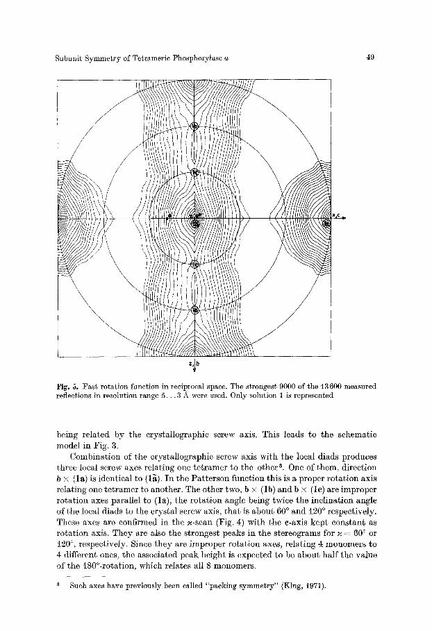
Fig. 5. Fast rotation function in reciprocal space. The strongest 9000 of tile 13600 measured reflections in resolution range 5 . . . 3 A were used. Only solution I is represented
b e i n g re la ted by the c rys ta l lographic screw axis. This leads to the schemat ic model in Fig. 3.
Combina t ion of the c rys ta l lographic screw axis wi th the local d iads produces th ree local screw axes re la t ing one t e t r a m e r to the o ther a. One of them, d i rec t ion b • ( la) is ident ica l to (1~). I n the P a t t e r s o n funct ion th is is a p roper ro t a t i on axis re la t ing one t e t r a m e r to another . The o ther two, b • ( lb) and b • ( lc) are imprope r ro t a t ion axes para l le l to ( la) , the ro t a t i on angle being twice the inc l ina t ion angle of the local d iads to the c rys ta l screw axis, t h a t is abou t 60 ~ and i20 ~ respect ively . These axes are confirmed in the ~-scan (Fig. 4) wi th the c-axis kep t cons tan t as ro t a t ion axis. They are ~lso the s t ronges t peaks in the s te reograms for z = 60 ~ or 120 ~ respect ively . Since t h e y are imprope r ro t a t i on axes, re la t ing 4 monomers to 4 different ones, the associa ted peak he igh t is expec ted to he abou t ha l f the v a l u e of the 180~ which re la tes all 8 monomers .
Such axes have previously been called "packing symmetry" (Klug, 1971).
50 K. Bartels and P. M. Cohnan
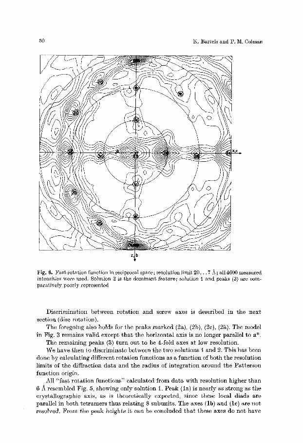
Fig. 6. :Fast rotation function in reciprocal space; resolution limit 20. . . 7/~; all 4600 measured intensities were used. SOlution 2 is the dominant feature; solution ~ and peaks (3) are com- paratively poorly represented
Discrimination between rota t ion and screw axes is described in the next section (disc rotation).
The foregoing also holds for the peaks marked (2a), (2b), (2c), (2~). The model in Fig. 3 remains valid except t ha t the horizontal axis is no longer parallel to a*.
The remaining peaks (3) tu rn out to be 4-fold axes at low resolution. We have then to discriminate between the two solutions ~ and 2. This has been
done by calculating different rota t ion functions as a funct ion of both the resolution limits of the diffraction data and the radius of integrat ion around the Pat te rson funct ion origin.
All "fast ro ta t ion funct ions" calculated from da ta with resolution higher than 6 A resembled Fig. 5, showing only solution i. Peak (In) is nearly as s t rong as the crystallographic axis, as is theoretically expected, since these local diads are parallel in both te t ramers thus relating 8 subunits. The axes (lb) and (lc) are not resolved. F rom the peak heights it can be concluded tha t these axes do no t have
Subunit Symmetry of Tetrameric Phosphorylase a
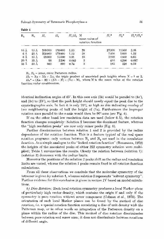
Table 1
51
R1 ~2 17~ ~ H1/;[2 M I71. I]2 * ~1 */82" mean value of rotation function
10 A 15 ~ 209000 135000 1.55 28 27300 11 500 2.38 6 ~- 20 ~ 205000 i78000 1.15 38 7400 5600 1.32 6 ~ 35 A 68000 75000 0.91 25 3100 4800 0.65
20 A 35 A 98 2200 0.045 - 3 4t0 4200 0.097 35 ~ 50 A 660 890 0.74 3 165 320 0.52
R1, R e - inner, outer Patterson radius. Hx = Xa x Xb x Xc, the triple product of associated peak heights where X = I or 2. Hx* = (Xa - M) x (Xb - M) x (Xe - M), where 1~{ is the mean value of the rotation
function under consideration.
identical inclination angles of 45 ~ I n this ease axis (lb) would be parallel to (le'), and (le) to (lb'), so tha t the peak height should nearly equal the peak due to the crystallographic axis. I n fact it is only 70 % as high as this indicating overlap of two neighbouring peaks of half the height of (la). Fur thermore the improper rota t ion axes parallel to the e-axis would then be 90~ (see Fig. 4).
I f on the other hand low resolution data are used (below 6 •), the ro ta t ion funct ion changes completely: Solution 2 becomes the dominant feature, whereas the "high resolution peaks" are now only minor peaks (Fig. 6).
Fur ther discrimination between solution I and 2 is provided by the radius dependence of the ro ta t ion function. This is a feature typical of the real space ro ta t ion programs: only vectors between R 1 and 1~ are used in the correlation function. As a simple analogon to the "locked rota t ion funct ion" (Rossmann, 1972) the heights of the associated peaks of either 222 s y m m e t r y solution were multi- plied; Table t summarizes the results. Clearly the relation between (solution t ) : (solution 2) decreases with the radius limits.
Moreover the positions of the solution 2 peaks shift as the radius and resolution limits are varied, wheras the solution t peaks remain fixed in all rota t ion function calculations.
F r o m all these observations we conclude tha t the molecular s y m m e t r y of the te t ramer is given by solution t, whereas solution 2 represents "solvent symmet ry" . Fur ther evidence for this conclusion is given in section I V based on model calcula- tions.
b) Disc-Rotation: Each local rota t ion s y m m e t r y produces a local I-Iarker-plane of part icular ly high vector density, which contains the origin if and only if the s y m m e t r y is pure ro ta t ion wi thout screw component (Colman et al., t975). The orientat ion of such local Harker planes can be found by the method of disc rotation, i.e. a special rota t ion funct ion correlating a disc of unit densi ty with the Pat te rson map, or in other words an integrat ion of the Pat te rson densi ty on a plane within the radius of the disc. This method of disc rota t ion discriminates between pure ro ta t ion and screw axes; it does not discriminate between rotat ions of different angle.
50 K. Barrels and P. M. Colman
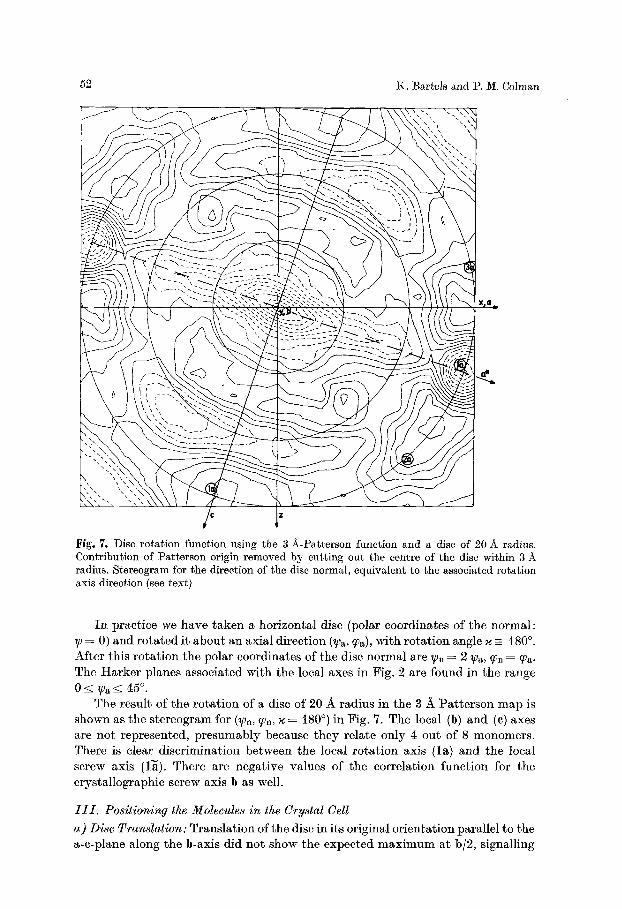
Fig. 7. Disc rotation function using the 3 A-Patterson function and a disc of 20 A radius. Contribution of Patterson origin removed by cutting out the centre of the disc within 3 radius. Stereogram for the direction of the disc normal, equivalent to the associated rotation axis direction (see text)
I n prac t ice we have t a k e n a hor izonta l disc (polar coordina tes of the normal : ~0 = 0) and ro t a t ed i t abou t an axia l d i rec t ion (~a, q0a), wi th ro t a t i on angle ~ - 180 ~ Af te r th is ro t a t i on the polar coordinates of the disc no rma l are ~n = 2 ~Pa, ~n = ~a. The H a r k e r p lanes associa ted wi th the local axes in Fig. 2 are found in the range 0 ~ ~a --~ 45 ~
The resul t of the ro t a t i on of a disc of 20 A rad ius in the 3 A P a t t e r s o n m a p is shown as the s te reogram for (~n, ~n, z = 180 ~ in Fig. 7. The local (b) and (c) axes are no t represented , p r e s u m a b l y because t h e y re la te only 4 out of 8 monomers . There is clear d i sc r imina t ion be tween the local ro t a t ion axis ( la ) and the local screw axis (1~). There are nega t ive values of the corre la t ion funct ion for the c rys ta l lographic screw axis b as well.
I I I . Positioning the Molecules in the Crystal Cell
a) Disc Translation: Trans la t ion of the disc in i ts original o r ien ta t ion para l le l to the a-c-plane along the b-axis d id no t show the expec ted m a x i m u m a t b/2, s ignall ing
Subunit Symmetry of Tetrameric t)hosphorylase a 53
0.5 1 ( 0 , - ) ~ ~ - ~ ' j ~ , ~ "
I . . . . - . \ ~ .i,," , ' - : - - - " ,' . ' , ' . . . . . . . / ' /"" '" '----.----=,'~', ~ ~~ x . ~ ~ "\ "" " " . . . . . . . . . . ' "" '~"~ k". ' ' , " ~',,' "-,,- '"- -.. - ~ h ', ".-: - ; f - ' - ,, _\--~
~ i ' / . i , " ( .'-', . . . . . . . " . . . . . . \ - - ~ . . - " ~ . . . . " ~ .', .~ . . - - ' ~ \ ~ ~ i
t - - ' j . I .; .'~,'" 't ", " ' - - , ' " "-:, " - - : ' - " j ',' "S':: :7---- '" ," <: - ' - - - - -= :J - ' , "~ '~ '~ - -~ O . 5 d ~ / ' ' ' ' : ', ' ', '- _ ' - - - - . ' , " , " , : > - ' - " ' ' " . , ' ; ; _
/ ~ < <:.-~ ',, ,' . . . . . . . \ ",,',,~ t( t..~._~> ~ " ~ . . . . . (,,,','_=:- _ . , . , .,.i
~,, ,' ,' ,,'. ; : : ; . o ~ C ~
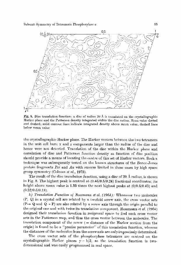
Fi I , 8. D isc t r a n s l a t i o n f u n c t i o n ; a disc o f rad ius 20 .& is t r a n s l a i d on the c r ys ta l l og raph i c t a r k e r p lane a n d the P a t t e r s o n dens i t y i n t e g r a t e d w i t h i n t i e disc rad ius . M e a n va lue d o t t e d and dashed; solid contour lines indicate integrated density above mean value, dashed lines below mean value
the crystallographic Harker plane. The Harker vectors between the two tetramers in the unit cell have x and z components larger than the radius of the disc and hence were not detected. Translation of the disc within the Harker plane and correlation of disc and Patterson function density as function of disc position should provide a means of locating the centre of this set of Harker vectors. Such a technique was subsequently tested on the known structures of the Bence-Jones protein fragments Rei and Au with success limited in these cases by high space group symmetry (Colman et al., 1975).
The result of the disc translation function, using a disc of 20 A radius, is shown in Fig. 8. The highest peak is centred at (0.45/0.5/0.24) fractional coordinates; its height above mean "value is 1.35 times the next highest peaks at (0/0.5/0.43) and (0.32/0.5/0.18).
b) Translation .Function o/ Rossmann et al. (1964): Whenever two molecules (P, Q) in a crystal cell are related by a twofold screw axis, the cross vector sets (P-+ Q and Q-+ P) are also related by a screw axis through the origin parallel to the original one and with twice its translation component. Rossmann ct al. (i964) designed their translation function in reciprocal space to find such cross vector sets in the Patterson map, and thus the cross vector between the molecules. The translation component of the screw ( = distance of the Harker section from the origin) is found to be a "precise parameter" of this translation function, whereas the distances of the molecules from the screw axis are only imprecisely determined.
The cross vector sets of the phosphorylase tetramers are centred on the crystallographic Harker planes y = b/2, so the translation function is two- dimensional and was easily programmed in real space.
54 K. Bartels and P. M. Colmau
I O 5 I ( O j ~ O ) ! . . . . . . . . . / , ' , i ' , , ' , ', ~ " ; ~ ' : , ' , "q " ' , . . . . . ~" ~ "- / "
/ .., : , ~ _ ~ - ~ \ ",_-::~::-:,--::::--,':~d.'-- .,: : -- i -- , - :=-Z:y, ' ,> , ' , " : /
~c
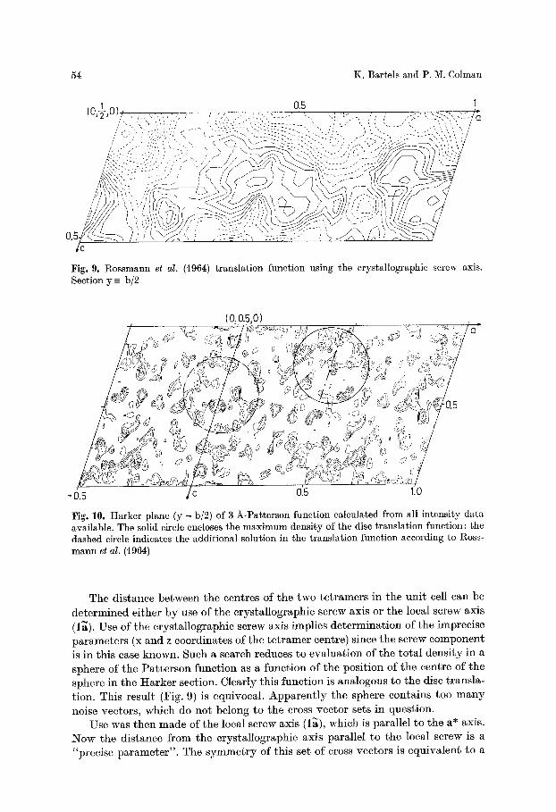
Fig. 9. Rossmann et al. (1964) translation function using the crystallographic screw axis. Section y ~ b/2
(0 .0 .5 .0 )
Fig. 10. Harker plane (y = b/2) of 3 A-Patterson function calculated from all intensity data available. The solid circle encloses the maximum density of the disc translation function; the dashed circle indicates the additional solution in the translation function according to Ross- mann et al. (1964)
The d i s tance be tween the centres of the two t e t r amer s in the un i t cell can be de t e rmined e i ther b y use of the c rys ta l lographic screw axis or the local screw axis (l~). Use of the c rys ta l lographic screw axis implies de t e rmina t ion of the imprecise pa r ame te r s (x and z coordinates of the t e t r a m e r centre) since the screw componen t is in th is case known. Such a search reduces to eva lua t ion of the t o t a l dens i ty in a sphere of the P a t t e r s o n funct ion as a funct ion of the pos i t ion of the centre of the sphere in the H a r k e r section. Clear ly th is funct ion is analogous to t he disc t rans la - t ion. This resul t (Fig. 9) is equivocal . A p p a r e n t l y the sphere conta ins too m a n y noise vectors , which do no t belong to the cross vec tor sets in quest ion.
Use was then m a d e of the local screw axis (1~), which is para l le l to the a* axis. :Now the d i s tance from the c rys ta l lograph ic axis para l le l to the local screw is a "precise p a r a m e t e r " . The s y m m e t r y of th is set os cross vec tors is equ iva len t to a
Subunit Symmetry of Tetrameric Phosphorylase a
. _ t D~
55
1
0 .
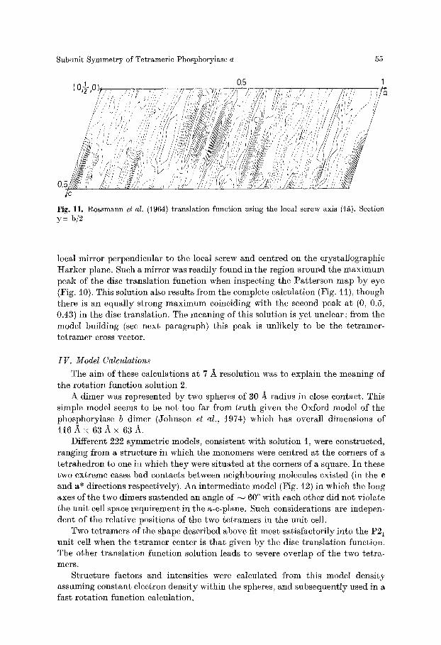
Fig. 11. gossmann et al. (1964) translation function using the local screw axis (1~). Section y _= b/2
local mirror perpendicular to the local screw and centred on the crystallographic Harker plane. Such a mirror was readily found in the region around the maximum peak of the disc translation function when inspecting the Patterson map by eye (Fig. t0). This solution also results from the complete calculation (Fig. 11), though there is an equally strong maximum coinciding with the second peak at (0, 0.5, 0.43) in the disc translation. The meaning of this solution is yet unclear ; from the model building (see next paragraph) this peak is unlikely to be the tetramer- tetramer cross vector.
I V . Model Calculations
The aim of these calculations at 7 A resolution was to explain the meaning of the rotation function solution 2.
A dimer was represented by two spheres of 30 :~ radius in close contact. This simple model seems to be not too far from truth given the Oxford model of the phosphorylase b dimer (Johnson et al., 1974) which has overall dimensions of i l 6 A • 6 3 A • 63A.
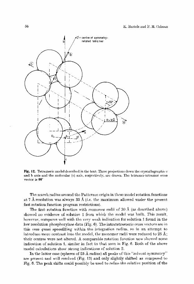
Different 222 symmetric models, consistent with solution l, were constructed, ranging from a structure in which the monomers were centred at the corners of a tetrahedron to one in which they were situated at the corners of a square. In these two extreme cases bad contacts between neighbouring molecules existed (in the c and a* directions respectively). An intermediate model (Fig. t2) in which the long axes of the two dimers sustended an angle of ~ 60 ~ with each other did not violate the unit cell space requirement in the a-c-plane. Such considerations are indepen- dent of the relative positions of the two tetramers in the unit cell.
Two tetramers of the shape described above fit most satisfactorily into the P21 unit cell when the tetramer center is that given by the disc translation function. The other translation function solution leads to severe overlap of the two tetra- m e r s .
Structure factors and intensities were calculated from this model density assuming constant electron density within the spheres, and subsequently used in a fast rotation function calculation.
56 K. Barrels and P, M. Colman
0'= centre of symmetry- related tetramer
�9 |
Fig. 12. Tetrameric model described in the text. Three projections down the crystallographic c and b axis and the molecular (c) axis, respectively, are drawn. The tetramer-tetramer cross vector is 00'
The search radius around the Pat te rson origin in these model rota t ion functions at 7 -~ resolution was always 35/~ (i.e. the max imum allowed under the present fast ro ta t ion funct ion program restrictions).
The first ro ta t ion function with monomer radii of 30 • (as described above) showed no evidence of solution i f rom which the model was built. This result, however, compares well with the very weak indication for solution I found in the low resolution phosphorylase da ta (Fig. 6). The intratetrameric cross vectors are in this case quasi spacefilling within the integrat ion radius, so in an a t t empt to introduce more contrast into the model, the monomer radii were reduced to 25 A ; their centres were not altered. A comparable ro ta t ion funct ion now showed some indication of solution t, similar in fact to t ha t seen in Fig. 6. Bo th of the above model calculations show strong indications of solution 2.
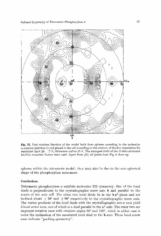
I n the lat ter case (spheres of 25 A radius) all peaks of this "solvent s y m m e t r y " are present and well resolved (Fig. 13) and only slightly shifted as compared to Fig. 6. The peak shifts could possibly be used to refine the relative position of the
Subunit Symmetry of Tetramerie Phosphorylase a 57
Fig. 13. Fast rotation function of the model built from spheres according to the molecular symmetry (solution 1) and placed in the cell according to the solution of the disc translation for resolution limit 20. . . 7 ~; Patterson radius 35 A. The strongest 9000 of the 11600 calculated triclinic structure factors were used. Apart from (3b) all peaks from Fig. 6 show up
spheres wi th in the te t rameric model ; they m a y also be clue to the non spherical shape of the phosphorylase monomers.
Conclusions
Tetrameric phosphorylase a exhibits molecular 222 symmetry . One of the local diads is perpendicular to the crystallographic screw axis b and parallel to the c-axis of the u n i t cell. The other two local diads lie in the b,a*-plane and are incl ined about +_ 30 ~ and + 60 ~ respectively to the crystallographic screw axis. The vector products of the local diads with the crystallographic screw axis yield 3 local screw axes, one of which is a diad parallel to the a*-axis. The other two are improper ro ta t ion axes with ro ta t ion angles 60 ~ and 120 ~ which in either ease is twice the inc l ina t ion of the associated local diad to the b-axis. These local screw axes indicate "packing symmet ry" .
58 K. Bartels and P. M. Colman
I n add i t ion the low resolut ion d a t a show a set of s y m m e t r y axes which have been in t e rp re t ed as " so lven t s y m m e t r y " , i.e. corre la t ion of cross vectors be tween ne ighbour ing monomers which belong to different t e t ramers . I n o ther words, these axes p u t p ro te in onto p ro te in and solvent onto solvent , regardless of the inner s t ruc ture of the prote in . This i n t e rp re t a t i on has been confirmed b y the ro t a t i on funct ion ca lcula t ion using a model consis t ing of homogeneous spheres.
The cons t ruc t ion of the model requi red the knowledge of the pos i t ion of the t e t r amer s re la t ive to the c rys ta l lographic screw axis or equ iva len t ly re la t ive to each other. The d is tance of the centre of the t e t r amers from the c rys ta l lographic d i ad is approx , i /4 the cell wid th in the direction, of the a* axis.
The model t e t r a m e r was bui l t f rom spher ical monomers a r ranged according to the molecular 222 s y m m e t r y ; the re la t ive o r ien ta t ion of the dimcrs wi th in the t e t r a m e r was chosen to provide op t ima l space filling in the a-c-plane. W h e n the s y m m e t r y re la ted t e t r amer i c models were p laced in the cell using the resul t of the disc t r ans l a t i on as t e t r a m e r - t e t r a m e r cross vector , the re is m i n i m u m over lap be tween the t e t ramers .
F u t u r e plans include the de tec t ion of the known s t ruc ture of phosphory lase b ( Johnson et al., 1974) in the phosphory lase a cell which would provide i n d e p e n d a n t conf i rmat ion of the resul ts descr ibed before. There migh t then be a poss ib i l i ty to ex tend the phases using the known molecular s y m m e t r y (Colman, i974; Bricogne, 1974).
Acknowledgements. We wish to thank Dr. R. Huber and our colleagues, especially H. Fehl- hammer, for helpful discussions. The financial assistance of the DFG and SFB 51 is gratefully acknowledged.
Reierences Bricogne, G.: Geometric sources of redundancy in intensity data and their use for phase
determination. Acta Cryst. A30, 395--405 (1974) Campbell, I. D., Dwek, 1~. A., Price, N. C., Radda, G. K. : Studies on the interaction of ligands
with phosphorylase b using a spin-label probe. Europ. J. Biochem. 30, 339--347 (1972) Colman, P. M. : Noncrystallographic symmetry and the sampling theorem. Z. Krist. 149,
344--349 (1974) Colman, P. M., Fehlhammer, H., Barrels, K. : Patterson search methods in protein structure
determination: fl-trypsin and immunoglobulin fragments. In: Crystallographic computing (ed. ~. R. Ahmed). Copenhagen: Munksgaard 1975
Crowther, R. A.: The fast rotation function. In: The molecular replacement method (ed. M. G. l~ossmann), pp. 173--178. ~ew York-London-Paris: Gordon & Breach t972
Dwek, 1~. A., Griffiths, J. R., Radda, G. K., Strauss, U.: A spin label probe for the con- formational change on conversion of phosphorylase b to phyosphorylase a. FEBS Letters 28, 16i--164 (1972)
Eagles, P. A. M., Johnson, L. N., Joynson, M. A., McMurray, C. It., Gutfreund, H. : Subunit structure of aldolase: Chemical and crystallographic evidence. J. molec. Biol. 45, 533--544 (i969)
Epp, 0,, Steigemann, W., Formanek, H., Huber, R.: Crystallographic evidence for the tetrameric subunit structure of L-asparaginase from Escherichia coli. Europ. J. Biochem. 20, 432--437 (197i)
Fasold, It., Ortanderl, F., Huber, 1~., Barrels, K., Schwager, P. : Crystallization and crystallo- graphic data of rabbit muscle phosphorylase a and b. FEBS Letters 21, 229--232 (1972)
Fehlhammer, It. : Dissertation, Technische Universit~t Mfinchen (1975) Griffiths, J. t~., Price, N. C., Radda, G. K.: Conformational changes in phosphorylase a,
studied by a spin label probe. Biochim. biophys. Acta (Amst.) 358, 275--280 (1974)
Subunit Symmetry of Tetrameric Phosphorylase a 59
Hoppe, W.: Die Faltmolekiilmethode und ihre Anwendung in der rSntgenographischen Konstitutionsanalyse yon ]3iflorin (C20Hz004). Elektrochem. 61, 1076--1083 (1957)
Huber, R.: Programmed "Faltmolekiil" method. In: Crystallographic computing (ed. F. R. Ahmed), pp. 96--102. Copenhagen: Munksgaard 1970
Johnson, L. N., Madsen, N. ]3., Mosley, J., Wilson, K. S. : The crystal structure of phosphoryl- ase b at 6/k resolution. J. molec. :Biol. 90, 703--717 (1974)
Kiselev, N. A., Lerner, F. Ya., Livanova, N. ]3. : Electron microscopy of muscle phosphorylase b. J. molec. [Biol. 62, 537--549 (t971)
Kiselev, N. A., Lerner, F. u Livanova, N. ]3. : Electron microscopy of muscle phosphorylase a. J. molec. :Biol. 86, 587--599 (1974)
Klug, A.: Interpretation of the rotation function map of satellite tobacco necrosis virus: Octahedral packing of icosahedral particles. Cold Spr. Harb. Symp. quant. Biol. 36, 483--487 (1971)
Madsen, N. ]3., Honickel, K. 0., James, M. N. G.: Studies on glycogen phosphorylase in solution and in the crystalline state. 2nd Int. Symp. on Metabolic Interconversion of Enzymes (eds. O. Wieland, E. Helmreich, H. Holzer), pp. 55--72. ]3erlin-Heidelberg- New York: Springer 1972
Mathews, F. S. : X-ray crystallographic study of glycogen phosphorylase. Fed. Proc. 26, 831 (1967)
gossmann, M. G., :Blow, D. M.: The detection of sub-units within the crystallographic asymmetric unit. Acta Cryst. 15, 24--31 (1962)
Rossmann, M. G., Blow, D. M., Harding, M. M., Coller, E.: The relative positions of in- dependent molecules within the same asymmetric unit. Acta Cryst. 17, 338--342 (1964)
Rossmann, M. G. : The locked rotation function. Appendix in l~ossmann, M. G., Ford, G. C., Watson, H. C., ]3anaszak, L. J.; J. molec. ]3iol. 64, 237--249 (1972)
Schwager, P., ]3artels, K., Huber, R.: A simple empirical absorption-correction method for X-ray intensity data films. Acta Cryst. A29, 291--295 (1973)
Schwager, P., ]3artels, K., Jones, A. : Refinement of setting angles in screenless film methods. J. appl. Cryst. 8, 275--280 (1975)
Schwager, P., ]3artels, K.: The Munich Program System for X-l~ay Intensity Data Film Evaluation by Off-Line-Computer (Tentative Title). Proceedings of the Groningen symposium on the rotation method (Feb. 75) (eds. U. W. Arndt, A. J. Wonacott). Amster- dam: Elsevier North Holland Publ. Corp. t975
Steigemann, W. : Dissertation, Technische Universitgt Miinchen (1974) Tollin, P., Rossmann, M. G. : Errata in I~ossmann and Blow (1962). Appendix in Acta Cryst. 21,
876 (1966)
Received August 15, J975/Accepted December 18, J975