Embed Size (px)
Citation preview
RESEARCH ARTICLE SUMMARY◥
STRUCTURAL BIOLOGY
Substrate-engaged 26S proteasomestructures reveal mechanisms forATP-hydrolysis–driven translocationAndres H. de la Peña*, Ellen A. Goodall*, Stephanie N. Gates*,Gabriel C. Lander†, Andreas Martin†
INTRODUCTION: As the major protease ineukaryotic cells and the final component ofthe ubiquitin-proteasome system, the 26S pro-teasome is responsible for protein homeostasisand the regulation of numerous vital processes.Misfolded, damaged, or obsolete regulatory pro-teins are marked for degradation by the attach-ment of polyubiquitin chains, which bind toubiquitin receptors of the proteasome.Ahetero-hexameric ring of AAA+ (ATPases associatedwith diverse cellular activities) subunits thenuses conserved pore loops to engage, mecha-nically unfold, and translocate protein sub-strates into a proteolytic core for cleavagewhilethe deubiquitinase Rpn11 removes substrate-attached ubiquitin chains.
RATIONALE: Despite numerous structuraland functional studies, the mechanisms by
which adenosine triphosphate (ATP) hydroly-sis drives the conformational changes respon-sible for protein degradation remained elusive.Structures of related homohexameric AAA+motors, in which bound substrates were stabi-lized with ATP analogs or hydrolysis-eliminatingmutations, revealed snapshots of ATPase sub-units in different nucleotide states and spiral-staircase arrangements of pore loops aroundthe substrate. These structures gave rise to “hand-over-hand” translocation models by inferringhow individual subunits may progress throughvarious substrate-binding conformations. How-ever, the coordination of ATP-hydrolysis stepsand their mechanochemical coupling to pro-pelling substrate were unknown.
RESULTS:We present the cryo–electron mi-croscopy (cryo-EM) structures of the actively
ATP-hydrolyzing, substrate-engaged 26S pro-teasome with four distinct motor conforma-tions. Stalling substrate translocation at adefined position by inhibiting deubiquitinationled to trapped states in which the substrate-attached ubiquitin remains functionally boundto the Rpn11 deubiquitinase, and the scissileisopeptide bond of ubiquitin is aligned withthe substrate-translocation trajectory throughthe AAA+ motor. Our structures suggest aubiquitin capture mechanism, in which me-chanical pulling on the substrate by the AAA+motor delivers ubiquitin modifications directlyinto the Rpn11 catalytic groove and acceler-
ates isopeptide cleavagefor efficient, cotransloca-tional deubiquitination.These structures also
show how the substratepolypeptide traverses fromthe Rpn11 deubiquitinase,
through the AAA+ motor, and into the corepeptidase. The proteasomal motor therebyadopts staircase arrangements with fivesubstrate-engaged subunits and one disen-gaged subunit. Four of the substrate-engagedsubunits are ATP bound, whereas the subunitat the bottom of the staircase and the dis-engaged subunit are bound to adenosine di-phosphate (ADP).
CONCLUSION: Of the four distinct motorstates we observed, three apparently repre-sent sequential stages of ATP binding, hy-drolysis, and substrate translocation and hencereveal the coordination of individual steps inthe ATPase cycle and their mechanochemicalcoupling with translocation. ATP hydrolysisoccurs in the fourth substrate-engaged sub-unit from the top, concomitantly with exchangeof ADP for ATP in the disengaged subunit. Thesubsequent transition, which is likely triggeredby phosphate release from the fourth, posthy-drolysis subunit of the staircase, then involvesmajor conformational changes of the entireATPase hexamer. The bottom ADP-bound sub-unit is displaced and the previously disengagedsubunit binds the substrate at the top of thestaircase, while the four engaged subunitsmove downward as a rigid body and trans-locate substrate toward the peptidase. Ourlikely consecutive proteasome conformations,together with previously determined substrate-free structures, suggest a sequential progressionof ATPase subunits through the ATP-hydrolysiscycle. We hypothesize that, in general, hexa-meric AAA+ translocases function by this se-quential mechanism.▪
RESEARCH
de la Peña et al., Science 362, 1018 (2018) 30 November 2018 1 of 1
The list of author affiliations is available in the full article online.*These authors contributed equally to this work.†Corresponding author. Email: [email protected] (G.C.L.);[email protected] (A.M.)Cite this article as A. H. de la Peña et al., Science 362,eaav0725 (2018). DOI: 10.1126/science.aav0725
Cryo-EM structures of the substrate-engaged 26S proteasome. (A) Substrate paththrough the proteasome, with ubiquitin bound to Rpn11 (left inset) and the substratepolypeptide traversing through the AAA+ motor into the core peptidase. (B) Schematicshowing coordinated ATP hydrolysis and nucleotide exchange observed between consecutivemotor states. (C) Substrate translocation is driven by changes in the spiral-staircasearrangement of pore loops, as indicated by arrows.
ON OUR WEBSITE◥
Read the full articleat http://dx.doi.org/10.1126/science.aav0725..................................................
on May 9, 2020
http://science.sciencem
ag.org/D
ownloaded from
RESEARCH ARTICLE◥
STRUCTURAL BIOLOGY
Substrate-engaged 26S proteasomestructures reveal mechanisms forATP-hydrolysis–driven translocationAndres H. de la Peña1*, Ellen A. Goodall2,3*, Stephanie N. Gates2,3,4*,Gabriel C. Lander1†, Andreas Martin2,3,4†
The 26S proteasome is the primary eukaryotic degradation machine and thus is criticallyinvolved in numerous cellular processes.The heterohexameric adenosine triphosphatase(ATPase) motor of the proteasome unfolds and translocates targeted protein substrates intothe open gate of a proteolytic core while a proteasomal deubiquitinase concomitantlyremoves substrate-attached ubiquitin chains. However, the mechanisms by which ATPhydrolysis drives the conformational changes responsible for these processes haveremained elusive. Here we present the cryo–electron microscopy structures offour distinct conformational states of the actively ATP-hydrolyzing, substrate-engaged26S proteasome. These structures reveal how mechanical substrate translocationaccelerates deubiquitination and how ATP-binding, -hydrolysis, and phosphate-releaseevents are coordinated within the AAA+ (ATPases associated with diverse cellularactivities) motor to induce conformational changes and propel the substrate throughthe central pore.
The 26S proteasome, the final component ofthe ubiquitin-proteasome system, is centralto general proteostasis and the regulationof essential processes in eukaryotic cells (1).Proteins are targeted for proteasomal deg-
radation through the covalent attachment ofpolyubiquitin chains to lysine residues (2). Tosafeguard against indiscriminate degradation,the proteolytic active sites of the proteasome aresequestered within the barrel-shaped 20S coreparticle (CP). Access to these active sites is con-trolled by the 19S regulatory particle (RP), whichbinds to one or both ends of the CP, recruitsubiquitinated proteins, and catalyzes their deu-biquitination, unfolding, and translocation througha central pore into the proteolytic chamber of theCP for degradation (3). The RP can be furthersubdivided into the base and lid subcomplexes.The nine-subunit lid subcomplex fulfills im-portant scaffolding functions and contains theZn2+-dependent deubiquitinase Rpn11, which ispositioned above the central pore of the protea-some to remove ubiquitin chains from substratesbefore degradation (4–8). The base subcomplex
consists of 10 subunits, including three ubiquitinreceptors and six distinct AAA+ATPases (ATPasesassociated with diverse cellular activities), Rpt1to Rpt6 (3, 9). These ATPases (adenosine triphos-phatases) form a heterohexameric ring (in theorder Rpt1, Rpt2, Rpt6, Rpt3, Rpt4, and Rpt5)that is the molecular motor of the proteasome(10). Each Rpt consists of a N-terminal helix, anoligonucleotide binding (OB)–fold domain, and aC-terminal AAA+ motor domain. In the hetero-hexamer, the N-terminal helices of neighboringRpt pairs form a coiled coil, and the six OB-folddomains assemble into a rigid N-ring above theAAA+motor ring (6, 8). After ubiquitin-mediatedsubstrate recruitment, the ATPase motor engagesa flexible initiation region of the substrate forsubsequent mechanical translocation and unfold-ing (11). To facilitate substrate transfer to the CP,the ATPase hexamer also triggers opening of theCP access gate by docking conserved C-terminaltails of Rpt subunits into pockets at the surface ofthe CP a ring (12–14).Like other AAA+ ATPases, the Rpt subunits
contain a highly conserved nucleotide bindingpocket that couples ATP binding and hydrolysiswith conformational changes to produce mecha-nical work (15, 16). This pocket is largely formedby the signature Walker-A and Walker-B motifs,responsible for nucleotide binding and hydroly-sis, respectively, and an arginine finger providedby the clockwise-neighboringATPase subunit thatcoordinates the g phosphate of ATP during hy-drolysis and enables subunit communication(17). Conserved pore-1 loops protrude from eachATPase subunit into the central channel, where
they sterically interact with the substrate poly-peptide and transduce nucleotide-dependent con-formational changes into directional translocation(18–21).The common functional architecture of ring-
shaped hexameric helicases and AAA+ trans-locases gave rise to a “hand-over-hand” modelfor substrate translocation (22–24), which is sup-ported by numerous cryo–electron microscopy(cryo-EM) structures of substrate-bound homo-hexameric AAA+motors (25–30). These prior struc-tures were trapped using hydrolysis-inactivatingWalker-B mutations, nonhydrolyzable ATP ana-logs, or analogs that are slowly hydrolyzed, toreveal series of subunits in the hexamer that re-semble the ATP-bound, adenosine diphosphate(ADP)–bound, and nucleotide-free states. Gener-ally, five nucleotide-bound subunits contact thesubstrate polypeptide in a spiral-staircase arrange-ment of pore loops, whereas one subunit remainsdisengaged and nucleotide-free. The hand-over-hand model stems from inferences regardinghow individual subunits may progress throughthe various nucleotide states and substrate-binding conformations around the ring. The het-erohexameric proteasomal AAA+motor adoptsdistinct spiral-staircase arrangements with indi-vidual Rpts in different vertical positions (6, 31–35)and thus promisesmore detailed insights into theprogression of states during the ATP-hydrolysisand substrate-translocation cycles.However, high-resolution structural studies of the proteasomeduring active substrate translocation have so farbeen unsuccessful.In the absence of substrate, the ATP-hydrolyzing
proteasome primarily adopts the s1 state (6, 36)in which the ATPase domains of Rpt1 to Rpt6form a spiral staircase that is not coaxially alignedwith the CP and Rpn11 is positioned offset fromthe central pore of the motor. A low-resolutionstructure of the proteasome trapped with a stalledprotein in the central pore revealed that uponsubstrate engagement the RP transitions fromthe s1 state to a processing conformation, whichis characterized by a more planar ATPase ring, arotated lid subcomplex, and a coaxial alignmentof Rpn11, the Rpt hexamer, and the CP (31).However, the limited resolution and strong het-erogeneity of the ATPases within these stalledproteasome complexes prevented the visualiza-tion of substrate and the identification of distinctmotor states.States that share structural similarities with
the substrate-processing conformation are alsoobserved for the substrate-free proteasome as asmall subpopulation in the presence of ATP (s2state) and upon ATPase inhibition using eitherATP analogs or Walker-B mutations in individ-ual Rpt subunits (s3, s4, s5, and s6 states and theunnamed state seen in ADP-AlFx) (14, 33–35).Cryo-EM reconstructions of these states revealeddistinct spiral-staircase arrangements and nucle-otide occupancies of Rpt subunits, but the lack ofATP hydrolysis and the absence of substrate lim-ited the conclusions that could be drawn regard-ing the mechanisms for ATP-hydrolysis–coupledtranslocation.
RESEARCH
de la Peña et al., Science 362, eaav0725 (2018) 30 November 2018 1 of 9
1Department of Integrative Structural and ComputationalBiology, The Scripps Research Institute, 10550 N. TorreyPines Road, La Jolla, CA 92037, USA. 2Department ofMolecular and Cell Biology, University of California, Berkeley,CA 94720, USA. 3California Institute for QuantitativeBiosciences, University of California at Berkeley, Berkeley,CA 94720, USA. 4Howard Hughes Medical Institute, Universityof California at Berkeley, Berkeley, CA 94720, USA.*These authors contributed equally to this work.†Corresponding author. Email: [email protected] (G.C.L.);[email protected] (A.M.)
on May 9, 2020
http://science.sciencem
ag.org/D
ownloaded from
We explored the mechanistic details of ATP-hydrolysis–driven substrate translocation by de-termining the structure of the substrate-engaged26S proteasome in the presence of ATP. Unlikeprevious studies that used ATPase inhibition totrap substrate-bound states of other AAA+motors,we stalled substrate translocation in the activelyhydrolyzing motor of the proteasome by inhibi-tingRpn11-mediateddeubiquitination.Wedescribefour cryo-EM structures, depicting four distinctmotor states with the unambiguous assignmentof substrate polypeptide traversing the RP fromthe lysine-attached ubiquitin at the Rpn11 activesite, through the Rpt hexamer, to the gate of theCP. Three of these states appear to representsequential stages of ATP binding, hydrolysis, andsubstrate translocation and hence reveal the co-ordination of individual steps in the ATPasecycle of the AAA+ hexamer and their mechano-chemical coupling with translocation.
Four substrate-bound 26Sproteasome structures
To stall translocation at a defined substrate posi-tion, we inactivated the Rpn11 deubiquitinase ofSaccharomyces cerevisiae 26S proteasomes byincubationwith the inhibitor ortho-phenanthroline(4) and added a globular substrate with a singlepolyubiquitinated lysine flanking an unstructuredC-terminal initiation region. Proteasomes engagedthe flexible initiation region and translocatedthe substrate until the attached ubiquitin chainreached the inhibited Rpn11, preventing furthertranslocation and trapping the substrate in thecentral pore, which is indicated by a complete in-hibition of degradation (fig. S1A). Stalling sub-
strate translocation in the proteasome does notalso stall the AAA+ motor, as we observed a rateof ATP hydrolysis that was even slightly elevatedcompared with that of freely translocating protea-somes (fig. S1B). We posit that this stalled stateresembles the scenario when the proteasomeencounters thermodynamically stable substratedomains that require repeated pulling by theATPase to be unfolded (31, 37).After incubation with substrate, proteasomes
were vitrified for cryo-EM single-particle anal-ysis, which produced reconstructions of the 26Sproteasome in six distinct conformational states.In the initial 3D classification, roughly 42% ofparticles were observed to be substrate free,adopting an s1-like state (fig. S2A and table S1),whereas the rest of the particles were sortedinto reconstructions that showed ubiquitin den-sity adjacent to Rpn11 and adopted non–s1-likeconformations (fig. S2A). Further focused classi-fication of the ATPase motor resulted in four s4-like reconstructions and one reconstruction thatresembled the s2 state but lacked density forsubstrate within the central channel of the AAA+motor. In contrast, the four s4-like reconstructions(ranging from ~4.2 to ~4.7 Å in overall resolution)showed clearly visible substrate density threadedthrough the center of the RP (Fig. 1, figs. S2 andS3A, and table S1).
Proteasome interactions with thetranslocating substrate
The stalled proteasome states not only revealedthe detailed path of the substrate polypeptidefrom the RP to the CP but also resolved the struc-ture of ubiquitin-bound Rpn11 in the context of
the 26S holoenzyme (Fig. 1A and movie S1). Themost proximal, substrate-attached ubiquitin moi-ety of the polyubiquitin chain is positioned inthe catalytic groove of Rpn11, whose ubiquitin-interacting Insert-1 region adopts the same ac-tive b-hairpin conformation previously observedin the crystal structure of the isolated ubiquitin-bound Rpn11-Rpn8 dimer (Fig. 1B and fig. S3B).Although the catalytic Zn2+ ion is not visible inthe Rpn11 active site, likely due to the treatmentwith ortho-phenanthroline, the conformations ofubiquitin and Rpn11 match the active, Zn2+-containing structure (Fig. S3B), with the additionof an intact isopeptide bond to the substratelysine.Upstream (N-terminal) of the ubiquitin-modified
lysine, only two amino acids of the substratewere resolved. The orientation of these residuesdelineates a path near the N-terminal helix ofRpt2 by which substrates may approach the cen-tral pore of the proteasome (Fig. 1A), yet to whatextent this path outside the N-ring is fixed orsubstrate dependent remains unclear. Down-stream (C-terminal) of the ubiquitinated lysine,the substrate is confined to the narrow centralchannel of the Rpt hexamer (Fig. 1A and fig. S3, Cand D). An axial view of the RP reveals that theRpn11 catalytic groove is aligned with the trajec-tory of substrate translocation through thischannel, which follows a straight line from theisopeptide bond into the AAA+ motor (fig. S3D).This alignment explains how vectorial tugging bythe motor can pull ubiquitin directly into thecup-shaped Rpn11 binding site and thus acceler-ate cotranslocational deubiquitination (38). Theactive b-hairpin conformation of Rpn11’s Insert-1
de la Peña et al., Science 362, eaav0725 (2018) 30 November 2018 2 of 9
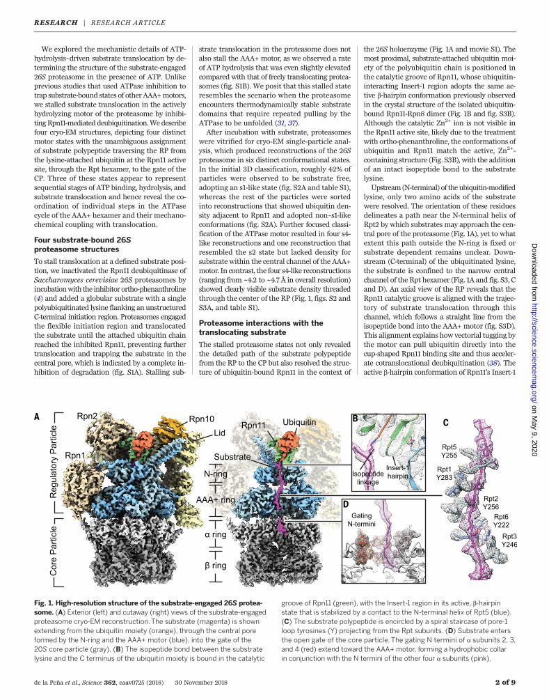
Fig. 1. High-resolution structure of the substrate-engaged 26S protea-some. (A) Exterior (left) and cutaway (right) views of the substrate-engagedproteasome cryo-EM reconstruction. The substrate (magenta) is shownextending from the ubiquitin moiety (orange), through the central poreformed by the N-ring and the AAA+ motor (blue), into the gate of the20S core particle (gray). (B) The isopeptide bond between the substratelysine and the C terminus of the ubiquitin moiety is bound in the catalytic
groove of Rpn11 (green), with the Insert-1 region in its active, b-hairpinstate that is stabilized by a contact to the N-terminal helix of Rpt5 (blue).(C) The substrate polypeptide is encircled by a spiral staircase of pore-1loop tyrosines (Y) projecting from the Rpt subunits. (D) Substrate entersthe open gate of the core particle. The gating N termini of a subunits 2, 3,and 4 (red) extend toward the AAA+ motor, forming a hydrophobic collarin conjunction with the N termini of the other four a subunits (pink).
RESEARCH | RESEARCH ARTICLEon M
ay 9, 2020
http://science.sciencemag.org/
Dow
nloaded from
region appears to be stabilized through additionalcontacts with Rpt5 at the base of the Rpt4-Rpt5coiled coil (Fig. 1B). Our structures suggest thatthe translocation stall originates from ubiquitinbecoming trapped as it is pulled into the catalyticgroove of inactive Rpn11, rather than stericallyclashing with the narrow entrance of the N-ring.Despite having theATPase ring in distinct hydrol-ysis states (see below), all four proteasome struc-tures showubiquitin functionally bound toRpn11,indicating that deubiquitination indeed occurscotranslocationally, after the regulatory particlehas switched to an engaged conformation andwhile the substrate is threaded into the pore.This observation is consistent with a mechanism
in which the regulatory particle does not adopta specific conformation for deubiquitinationbut cleaves off ubiquitin modifications as theyapproach Rpn11 during processive substratetranslocation.Because of the defined stall at the single ubi-
quitin chain, we were able to reliably model theC-terminally inserted substrate and assign a spe-cific sequence to the polypeptide density withinthe AAA+ motor (Fig. 1C). The pore-1 loop Tyrand neighboring Lys residue of individual Rptsubunits form a spiral staircase that tightly en-circles the substrate, consistent with a transloca-tion mechanism that involves steric interactionswith amino acid side chains of the polypeptide
(Fig. 1C). As in many other AAA+ motors, thepore-2 loops (a second loop that protrudesfrom each ATPase subunit into the pore) are ar-ranged in a second staircase that lies in closeproximity to the substrate below the pore-1 loopspiral (fig. S3E). In contrast to the pore-1 loops,the pore-2 loops do not contain bulky residuesand may contribute to translocation through in-teractions of their backbones with the substrate(fig. S3E), as suggested by defects previously ob-served for pore-2 loop mutations (20).After traversing the AAA+ motor, the sub-
strate enters the gate of the CP (Fig. 1D). Ourfour cryo-EM structures reveal two gating con-formations with distinct RP-CP interactions and
de la Peña et al., Science 362, eaav0725 (2018) 30 November 2018 3 of 9
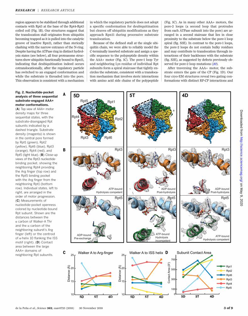
Fig. 2. Nucleotide-pocketanalysis of three sequentialsubstrate-engaged AAA+motor conformations.(A) Top view of AAA+ motordensity maps for threesequential states, with thesubstrate-disengaged Rptsubunits indicated by adashed triangle. Substratedensity (magenta) is shownin the central pore formedby Rpt1 (green), Rpt2(yellow), Rpt6 (blue), Rpt3(orange), Rpt4 (red), andRpt5 (light blue). (B) Close-upviews of the Rpt3 nucleotide-binding pocket, showing theneighboring Rpt4 providingthe Arg finger (top row) andthe Rpt5 binding pocketwith the Arg finger from theneighboring Rpt1 (bottomrow). Individual states, left toright, are arranged in theorder of motor progression.(C) Measurements ofnucleotide-pocket opennesscolored by nucleotide-boundRpt subunit. Shown are thedistances between thea carbon of Walker-A Thrand the a carbon of theneighboring subunit’s Argfinger (left) or the centroidof a-helix 10 flanking the ISSmotif (right). (D) Contactarea between the largeAAA+ domains ofneighboring Rpt subunits.
RESEARCH | RESEARCH ARTICLEon M
ay 9, 2020
http://science.sciencemag.org/
Dow
nloaded from
arrangements for the N termini of CP a subunits(fig. S4). In all structures, the C termini of HbYX(hydrophobic–Tyr–any amino acid)–motif con-taining Rpt subunits (Rpt2, Rpt3, and Rpt5) oc-cupy the intersubunit pockets of the CP a ring,whereas the pockets for the C termini of Rpt1and Rpt6 vary in occupancy (fig. S4A). Two of thestructures show all Rpt tails except for Rpt4docked into the intersubunit pockets and, con-sequently, a completely open gate, similar topreviously described states in substrate-free pro-teasomes (14, 35) (fig. S4, B to D). In this openconformation, the gating N termini of a subunits2, 3, and 4 become directed toward the base sub-complex and interact with the N termini of theother four a subunits through a conserved Tyrresidue (fig. S4E). This results in the formation ofa hydrophobic collar directly beneath the exitfrom the AAA+ motor (fig. S4E). The other twostructures, which exhibit lower levels of Rpt1-and Rpt6-tail occupancies in the respective a-ringpockets, reveal a partially open gate (fig. S4, Cand D). This observation supports a recently pro-posed model in which cooperative gate openingis driven by the tail insertion of Rpt1 and Rpt6,after the three HbYX-containing tails are docked(14, 35).
Distinct nucleotide states give rise tofour ATPase conformations
Our four substrate-engaged proteasome struc-tures show distinct motor conformations withnucleotide density present in all six ATP-bindingpockets and one or two subunits that do notinteract with substrate (Fig. 2A and fig. S5A).To reliably assign nucleotide identities andthereby establish the progression of the ATP-hydrolysis cycle within the actively hydrolyzingRpt hexamer, we assessed not only the occupy-ing nucleotide densities but also the geometriesof the ATPase sites, the structural stability ofallosteric motifs, and the intersubunit contactareas (Fig. 2 and table S2). ATP-bound, hydrolysis-competent subunits form a closed pocket withan increased intersubunit contact area char-acterized by a direct interaction between the gphosphate of ATP and the well-resolved Argfingers of the clockwise neighboring subunit(Fig. 2, B to D, and fig. S5, B to F). In contrast,ADP-bound subunits are more open with a de-creased intersubunit contact area and Arg fin-gers that are more flexible, as indicated by lowerresolvability (Fig. 2, B to D, and fig. S5, B to F).Subunits that are ATP bound but not yet hydro-lysis competent and subunits where ATP hydrol-ysis has just occurred show similar, intermediateArg-finger distances (Fig. 2, B and C). To dis-tinguish between these pre- and posthydrolysisstates, we assessed the pocket openness by mea-suring the intersubunit contact area or the dis-tance between the conservedWalker-Amotif Thrand the helix preceding the intersubunit signal-ing (ISS)motif of the neighboring subunit (Fig. 2,C and D, and fig. S5, B to E) (26, 33). Our analysesrevealed a continuum of nucleotide states withinthe Rpt hexamers, ranging from ATP-bound par-tially open pockets with semi-engaged Arg fin-
gers (hydrolysis incompetent) toATP-bound closedpockets with fully engaged Arg fingers (hydrol-ysis competent), ADP-bound closed pockets withdisengaged Arg fingers (posthydrolysis), and ADP-bound open pockets with disengaged Arg fingers(pre–nucleotide exchange) (Fig. 2B). IndividualRpt subunits show a progression through dis-crete nucleotide states around the hexameric ring(Fig. 3A), indicating that each reconstruction re-flects a distinct snapshot of the proteasomal AAA+motor during the ATPase cycle and that Rptslikely progress sequentially through this cycle.Our structures allow us to correlate the distinctvertical registers and nucleotide states of Rptsubunits and analyze the coupling of individ-ual ATPase steps with each other and with themechanical translocation of substrate.
Sequential motor states reveal themechanism for ATP-hydrolysis–drivensubstrate translocation
The current hand-over-hand translocationmodelfor hexameric AAA+ ATPases is based on ob-servations of the subunits encircling and inter-
acting with substrate in a staircase-like organi-zation, with the exception of the sixth subunit(often referred to as the “seam subunit”) that isdisplaced from the substrate and positioned be-tween the lowest and highest subunits of thestaircase (25–30). Consistent with this previouslyobserved configuration,we see that in all substrate-engaged proteasome states the Rpt subunits in-teract with the substrate through their pore-1loops in a spiral-staircase arrangement, with thepore-2 loops forming a similar staircase under-neath (figs. S6, A and B). A characteristic seam isobserved along the interface between the highestsubstrate-engaged subunit of the staircase and theneighboring substrate-disengaged subunit (Figs. 2Aand 3, A and B, and fig. S5A). We name our fourproteasome states based on the identity andthe nucleotide state of this substrate-disengaged“seam” subunit: Rpt1-ADP, Rpt5-ADP, Rpt5-ATP,and Rpt4-ADP are named as 1D*, 5D, 5T, and 4D,respectively (Fig. 3A).In these four conformations, three different
Rpt subunits occupy the uppermost substrate-bound position of the staircase. In 1D*, Rpt2 is
de la Peña et al., Science 362, eaav0725 (2018) 30 November 2018 4 of 9
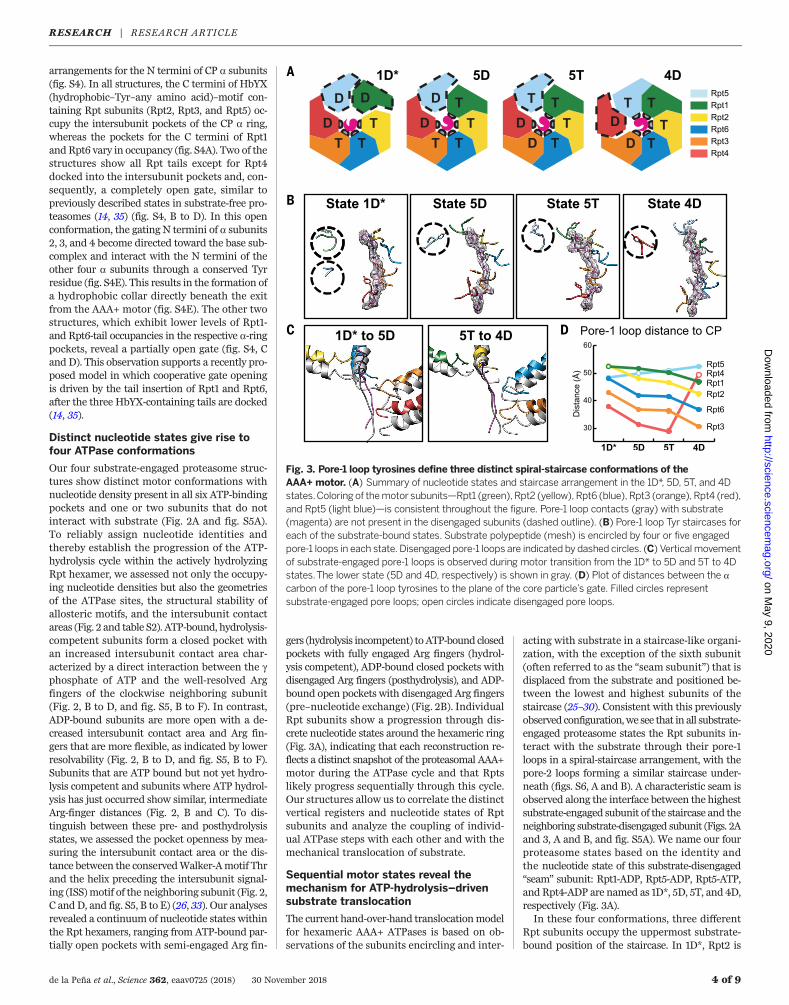
Fig. 3. Pore-1 loop tyrosines define three distinct spiral-staircase conformations of theAAA+ motor. (A) Summary of nucleotide states and staircase arrangement in the 1D*, 5D, 5T, and 4Dstates.Coloring of themotor subunits—Rpt1 (green), Rpt2 (yellow), Rpt6 (blue), Rpt3 (orange), Rpt4 (red),and Rpt5 (light blue)—is consistent throughout the figure. Pore-1 loop contacts (gray) with substrate(magenta) are not present in the disengaged subunits (dashed outline). (B) Pore-1 loop Tyr staircases foreach of the substrate-bound states. Substrate polypeptide (mesh) is encircled by four or five engagedpore-1 loops in each state. Disengaged pore-1 loops are indicated by dashed circles. (C) Verticalmovementof substrate-engaged pore-1 loops is observed during motor transition from the 1D* to 5D and 5T to 4Dstates.The lower state (5D and 4D, respectively) is shown in gray. (D) Plot of distances between the acarbon of the pore-1 loop tyrosines to the plane of the core particle’s gate. Filled circles representsubstrate-engaged pore loops; open circles indicate disengaged pore loops.
RESEARCH | RESEARCH ARTICLEon M
ay 9, 2020
http://science.sciencemag.org/
Dow
nloaded from
in the top substrate-bound position with theseam subunit Rpt1 displaced from the substrate(Fig. 3B). Unexpectedly, Rpt5 is also disengagedin this conformation, indicating that 1D* mayrepresent an off-pathway ATPase configuration(see discussion below).On the basis of their nucleotide occupancies
and spiral-staircase arrangements, we posit thatthe remaining three conformations—5D, 5T, and4D—represent consecutive states whose transi-tions include a nucleotide exchange, a hydrolysisevent, and a translocation step (Fig. 3 and movieS2). In 5D and 5T, the vertical staircase register isshifted by one subunit in the counterclockwisedirection compared with 1D*, such that Rpt1 as-sumes the uppermost substrate-bound position,whereas the other subunits move downwardand only Rpt5 is substrate disengaged (Fig. 3C).During the 5D-to-5T transition, the staircase ar-rangement of Rpt subunits remains largely thesame (fig. S6D), but the density for the Rpt5-boundnucleotide changes concomitantly with a substan-tial closure of the binding pocket that brings theArg fingers of Rpt1 into close proximity (Fig. 2B),which is consistent with an exchange of ADP forATP. This exchange and the resulting shift ofRpt5 toward the central pore likely primes thissubunit by allosterically positioning the poreloops for substrate engagement in the subse-quent 4D state (Fig. 3B). Nucleotide exchangein the disengaged Rpt thus appears prerequisitefor substrate binding at the top of the spiralstaircase, which agreeswith the highest substrate-contacting subunit always being bound to ATP(Fig. 3A). Notably, concurrent with ATP bindingto Rpt5, Rpt3 hydrolyzes ATP during the 5D-to-5T transition, as indicated by correlative changesin the Rpt3-bound nucleotide density and thedisengagement of the neighboring Arg finger(Fig. 2, B and C). Neither the nucleotide exchangenor the hydrolysis event cause substantial con-formational changes in the Rpt hexamer, butthey represent the trigger for themost pronouncedrearrangement of the mechanochemical cycle inthe subsequent transition to 4D.During this 5T-to-4D transition, we observe
an opening of the Rpt3 nucleotide-binding pocketand a disruption of the intersubunit interactionswith the neighboring Rpt4 (Fig. 2, B andD). Rpt4separates from Rpt3, disengages from substrate,and moves from the bottom of the staircase outand upward, which is likely driven by the topo-logically closed ring architecture of the Rpt hex-amer. At the same time, the ATP-bound Rpt5 atthe top of the staircase moves to a more centralposition and binds substrate (Fig. 3B), whereasthe substrate-engaged subunits Rpt1, Rpt2, Rpt6,and Rpt3move as a rigid body downward by oneregister and translocate the substrate toward theCP gate (Fig. 3, C and D, and movie S2).Even though we do not detect concrete
nucleotide-density changes between the 5T and4D conformations, we can postulate based onthe preceding ATP-hydrolysis event in Rpt3 andthe subsequent opening of its pocket that phos-phate release from Rpt3 is responsible for thedisruption of intersubunit interactions with Rpt4
and the consequent conformational changes ofthe entire ATPase ring. This model is consistentwith our observations that the penultimate sub-unit in the staircase exhibits a completely orpartially closed pocket in all proteasome confor-mations, whereas the lowest substrate-engagedsubunit is always ADP bound with an open pock-et (Figs. 3A; and 2, C and D; and fig. S5, B to E).Furthermore, it agrees with previous single-molecule data on the homohexameric ClpXATPase, suggesting that phosphate release rep-resents the force-generating step of the ATPasecycle (39). Similar to the coordinated nucleotide-exchange and ATP-hydrolysis steps in the pre-vious 5D-to-5T transition, the disruption of theRpt3-Rpt4 interface through potential phosphaterelease, the substrate-engagement by Rpt5, andthe movement of a four-subunit rigid body forsubstrate translocation appear to be interdependentand tightly coupled during the 5T-to-4D transition.For the ATP-hydrolysis and substrate-
translocation cycles, our findings suggest that aparticular subunit binds ATP and engages sub-strate at the uppermost position, hydrolyzes ATPwhen at the penultimate position of the staircase,releases phosphate as it moves to the bottom ofthe ring, and disengages from substrate in the nextstep (Fig. 4A). AAA+ motor movements and sub-strate translocation would thus be powered bysequential ATP hydrolysis and phosphate releaseas each Rpt transitions to the bottom of thestaircase. Our observation of four substrate-engaged subunits moving as a rigid body to trans-locate substrate in response to ATP hydrolysisand phosphate release is consistent with pre-vious biochemical studies of the ClpX ATPase,which indicated that several subunits interactsynergistically with substrate, allowing even apore-1 loop–deficient subunit to drive translo-cation (40, 41). The rigid-body movement of fourRpts vertically advances the engaged pore-1 loopTyr residues by ~6Å (Fig. 3C andD), suggesting afundamental step size of twoaminoacidsperhydro-lyzed ATP for proteasomal substrate translocation.We do not observe a vertical movement of
substrate due to the defined stall of translocationuponRpn11 inhibition, and all of our proteasomeconformations show largely the same stretch ofpolypeptide in the central channel. Nevertheless,the substrate responds to staircase rearrange-ments with lateral movements in the ATPasechannel, shifting toward the engaged pore-1loops and away from the disengaged subunits(fig. S6A). The substrate backbone follows thespiral-staircase arrangement of pore loops ratherthan traversing the motor in a straight verticalpath, and its lateral position in the channel ro-tates counterclockwise around the hexamer asthe Rpts progress through the various nucleotidestates (fig. S6A).
Additional states of the proteasomalATPase cycle
Whereas 5D, 5T, and 4D each contain four ATP-bound and two ADP-bound subunits, 1D* showsthree ATP-bound and three ADP-bound subunits(Fig. 3A). We interpret this conformation as an
alternate, potentially off-pathway version of a1D state. A comparison of the Rpt subunit or-ganization in 1D* with those in the 5D, 5T, and4D states, as well as other proteasome and AAA+motor structures (figs. S7, A and B), suggests thatRpt5 has prematurely released from substrate atthe bottom of the spiral after opening of the Rpt4nucleotide-binding pocket, and hence both Rpt5and Rpt1 are disengaged (Fig. 3B and fig. S7A).The Rpt5 pore-1 loop is divergent from the otherRpts, containing a conserved Met rather thana Lys (42), which could result in weaker sub-strate interactions, especially when translocationis stalled, and thus contribute to the prematuredisengagement in the 1D* state. Conversely, the1D* state might be explained by failed nucleotideexchange in Rpt1 at the top of the spiral, whichwould prevent substrate engagement and theconsequent rearrangement of the staircase tothe 5D state. Notably, the previously describeds3 conformation of the substrate-free proteasomeshows the expected staircase arrangement of 1D,with Rpt5 remaining in the lowest position of thestaircase (fig. S7B) (33).Our three distinct spiral-staircase states offer a
view of the discrete events leading to a completestep of hydrolysis-driven substrate translocation,yet the current hand-over-hand model requiresthat every Rpt subunit cycles through all thestates. Previous biochemical studies have indi-cated that ATP hydrolysis in almost all Rpt sub-units contribute to substrate engagement andtranslocation (20, 43, 44). Therefore, the pro-teasome conformations described here likelyrepresent only a subset of states that may becomplemented by the corresponding 4T, 3D, 3T,6D, 6T, 2D, and 2T states, aswell as an additional1T state between 1D and 5D (Fig. 4, B to D, andfig. S7), to complete the ATP-hydrolysis andsubstrate-translocation cycles of the proteasome.Indeed, several additional staircases have been
previously observed for the proteasome, albeitin the absence of substrate and induced by non-hydrolyzable ATP analogs, which hampered robustconclusions about mechanochemical couplingor the ATPase cycle. Their overall similarity toour Rpt staircase arrangements is sufficient todesignate specific spiral-staircase states. Someof these states (e.g., the ADP-AlFx–bound ands2 states) were regarded as unlikely processingconformations of the AAA+ motor, as they wereassociatedwith a partially openCP gate (14, 33, 34).However, two of our engaged states similarlycontain only partially open gates yet clearly showsubstrate being threaded through the centralchannel to the CP gate (fig. S4). This indicatesthat a fully open gate is not required for everystep of substrate translocation, but its opennessmay vary depending on the state of the Rptstaircase and corresponding allosteric subtle-ties in Rpt-tail interactions with the CP. Morepredictive criteria for a processing motor stateare the coaxial alignment of the AAA+ motorwith CP, the rotation of the lid subcomplex, andthe presence of rigid bodies formed between thelarge AAA subdomain of one subunit and thesmall AAA subdomain of its neighbor in all but
de la Peña et al., Science 362, eaav0725 (2018) 30 November 2018 5 of 9
RESEARCH | RESEARCH ARTICLEon M
ay 9, 2020
http://science.sciencemag.org/
Dow
nloaded from
the substrate-disengaged Rpts. On the basis ofthese criteria and their staircase orientation,the s2 and recently described s5 states (14) wouldrepresent 3D or 3T states, and the ADP-AlFx–bound proteasome conformation (34) resemblesa putative 2D* or 2T* state, with two substrate-disengaged subunits similar to 1D* (Fig. 4, Band C). The s4 and SD2 states, which had pre-viously been proposed as potential processingstates (33, 35), show overall staircase similaritieswith our substrate-engaged 4D and 5D states,respectively, even though some of their Rpts aredistorted as a likely consequence of inhibited ATPhydrolysis and the absence of substrate (fig. S7,C and D).Our substrate-engaged proteasome states pro-
vide a structural context for previously describedATP analog–bound conformations, enabling usto approximate all possible Rpt staircases, exceptfor the 6D and 6T states, and thus support themodel of a sequential hand-over-hand mecha-nism wherein each Rpt transitions through theATP-hydrolysis and substrate-translocation cycles(Fig. 4D). Why the proteasome in the presenceof substrate preferentially adopts only the 1D*,5D, 5T, and 4D states remains unclear; however,the likely consecutive 5D, 5T, and 4D states aresufficient to provide us with a complete pictureof subunit transitions during the ATPase cycleand substrate translocation.
Outlook
Weelucidated structures of the substrate-engaged26S proteasome that answer many of the out-standing questions regarding proteasomal deg-radation and the general mechanism by whichAAA+ translocases process their substrates. In-hibitingdeubiquitinationbyRpn11 led to a trappedstate in which the substrate-attached ubiquitinremains functionally bound in the Rpn11 cata-lytic groove and the scissile isopeptide bond islinearly aligned with the translocation trajectorythrough the AAA+ motor (Fig. 1). We concludethat during normal degradation, ubiquitin mod-ifications are pulled directly into the Rpn11 cat-alytic groove. This ubiquitin-capture mechanismexplains how Rpn11 functions as a gatekeeperto efficiently remove all ubiquitin modificationsfrom a substrate during processive translocation,as well as how deubiquitination can be accel-erated by mechanical pulling of the AAA+motoron the substrate polypeptide (38).We resolved three apparently sequential states
of the Rpt heterohexamer, which provide a mod-el for the intersubunit coordination during nu-cleotide exchange, ATP hydrolysis, and phosphaterelease within the AAA+ motor, as well as howthese events are mechanochemically coupled tosubstrate translocation. Consistent with othersubstrate-bound AAA+ ATPase structures, theproteasome motor adopts staircase arrangementsthat encircle the unfolded polypeptide substrate,with one subunit disengaged (25–29, 45). Four ofthe substrate-engaged subunits are ATP bound,whereas the subunit at the bottom of the stair-case and the disengaged subunit are bound toADP. Our structures suggest that nucleotide ex-
change primes the disengaged subunit for sub-strate binding at the top of the staircase and thatthis exchange occurs concomitantly with ATPhydrolysis in the fourth substrate-engaged sub-unit from the top. Both steps of the ATPase cycleare associated with only subtle allosteric rear-rangements, whereas the entire ATPase hexamerundergoesmajor conformational changes duringthe subsequent transition that appears to belinked to phosphate release from the posthydrol-ysis, fourth subunit of the staircase. These rear-
rangements include the displacement of thebottomADP-bound subunit, substrate binding ofthe previously disengaged subunit at the top ofthe staircase, and the downwardmovement of theremaining four substrate-engaged subunits as arigid body. It appears that all of these processesmust happen in a coordinated fashion for sub-strate translocation to occur.The likely consecutive ATPase states that we
observe, together with equivalent staircase ar-rangements in previous substrate-free proteasome
de la Peña et al., Science 362, eaav0725 (2018) 30 November 2018 6 of 9
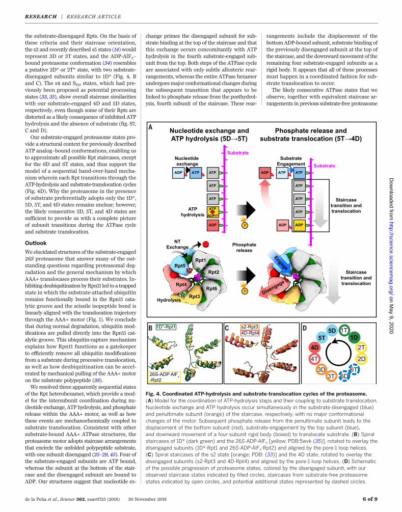
Fig. 4. Coordinated ATP-hydrolysis and substrate-translocation cycles of the proteasome.(A) Model for the coordination of ATP-hydrolysis steps and their coupling to substrate translocation.Nucleotide exchange and ATP hydrolysis occur simultaneously in the substrate-disengaged (blue)and penultimate subunit (orange) of the staircase, respectively, with no major conformationalchanges of the motor. Subsequent phosphate release from the penultimate subunit leads to thedisplacement of the bottom subunit (red), substrate-engagement by the top subunit (blue),and downward movement of a four-subunit rigid body (boxed) to translocate substrate. (B) Spiralstaircases of 1D* (dark green) and the 26S-ADP-AlFx [yellow; PDB:5wvk (35)], rotated to overlay thedisengaged subunits (1D*-Rpt1 and 26S-ADP-AlFx-Rpt2) and aligned by the pore-1 loop helices.(C) Spiral staircases of the s2 state [orange; PDB: (33)] and the 4D state, rotated to overlay thedisengaged subunits (s2-Rpt3 and 4D-Rpt4) and aligned by the pore-1 loop helices. (D) Schematicof the possible progression of proteasome states, colored by the disengaged subunit, with ourobserved staircase states indicated by filled circles, staircases from substrate-free proteasomestates indicated by open circles, and potential additional states represented by dashed circles.
RESEARCH | RESEARCH ARTICLEon M
ay 9, 2020
http://science.sciencemag.org/
Dow
nloaded from
structures, suggest a sequential progression ofindividual Rpt subunits through the ATPasecycle, rather than a burst mechanism, whereseveral subunits hydrolyze in rapid successionbefore nucleotide exchange, as proposed for theClpX motor on the basis of single-moleculemeasurements (46, 47). Given the structuraland functional similarities between the pro-teasomal Rpt hexamer and other AAA+ motors,we hypothesize that this sequential ATP-hydrolysisand substrate-translocation mechanism appliesto hexameric AAA+ translocases in general. Itis reminiscent of a six-subunit conveyor belt, inwhich a four-subunit rigid body grips the sub-strate and moves downward as the bottommostsubunit disengages and the topmost subunitreengages substrate (Fig. 4A). The coordinatedgripping by pore loops of four subunits, whichare stabilized by ATP-bound, closed interfaces,likely enables higher pulling forces and reducedslippage, consistent with previous biochemicalstudies of the ClpX motor (40, 41). Similarconveyor-belt mechanisms have been proposedpreviously for AAA+protein translocases aswellas DNA and RNA helicases (22, 23, 26–29, 45),yet our structures clarify the precise movementof ATPase subunits and their coordination withindividual steps of the ATP-hydrolysis cycle.
Materials and methodsSample preparationPurification of proteasome holoenzyme
26S proteasomes were purified from strain YYS40[MATa leu2-3,112 trp1-1 can1-100 ura3-1ade2-1his3-11,15 RPN11::RPN11-3XFLAG (HIS3)] (48) aspreviously described (49). Briefly, frozen yeastpaste from saturated cultures was lysed in aSpex SamplePrep 6875 Freezer/Mill, and cellpowder was resuspended in 60 mM HEPES,pH 7.6, 20 mMNaCl, 20 mM KCl, 8 mMMgCl2,2.5% glycerol, 0.2%NP-40, and ATP regenerationmix (5 mM ATP, 0.03 mg/ml creatine kinase,16 mM creatine phosphate). Proteasomes werebatch-bound to anti-FLAG M2 Affinity Gel (Milli-pore Sigma), washed with Wash Buffer (60 mMHEPES, pH 7.6, 20 mM NaCl, 20 mM KCl, 8 mMMgCl2, 2.5% glycerol, 5 mM ATP), eluted with3XFLAG peptide, and further separated by size-exclusion chromatography using a Superose 6Increase column in 60mMHEPES, pH 7.6, 20mMNaCl, 20 mM KCl, 10 mM MgCl2, 2.5% glycerol,and 1 mM ATP.
Preparation of ubiquitinatedmodel substrate
Amodel substrate consisting of anN-terminal Cys,lysine-less titin-I27V15P, a single-lysine-containingsequence derived from an N-terminal fragmentof Sphaerechinus granularis cyclinB (residues22 to 42, with Lys-to-Ala substitutions), a Rsp5recognition motif (PPPY), and 6X His-tag, waspurified after expression in Escherichia coliBL21-Star by standard methods. Briefly, In Ter-rific Broth, protein expressionwas induced withIPTG at OD600 = 1.2 to 1.5 for 5 hours at 30°C.Cells were harvested by centrifugation, resus-pended in chilled lysis buffer (60 mM HEPES,
pH 7.6 100 mM NaCl, 100 mM KCl, 15 mM im-idazole) and lysed by sonication. Following clar-ification by centrifugation at 20,000 × g, theprotein was purified usingNi-NTA affinity chro-matography. The substrate was fluorescentlylabeled using 5-fluoroscein maleimide at pH 7.2for 3 hours at room temperature and quenchedwith DTT. Free dye was separated from the sub-strate by size-exclusion chromatography with aSuperdex 200 column (GE Healthcare), bufferexchanging the substrate into 60 mM HEPES,pH 7.6, 20mMNaCl, 20mMKCl, 10mMMgCl2,2.5% glycerol.The substrate at final substrate concentration
of 50 mM was modified with long, K63-linkedubiquitin chains using 5 mMMusmusculusUba1,5 mM S. cerevisiae Ubc1, 20 mM S. cerevisiaeRsp5DWW (20, 50), and 2 mM S. cerevisiaeubiquitin, in 60 mM HEPES, pH 7.6, 20 mMNaCl, 20 mM KCl, 10 mMMgCl2, 2.5% glycerol,and 15 mM ATP for 3 hours at 25°C, followedby incubation overnight at 4°C.
ATPase assay
Proteasome ATPase activity was monitored usinga spectrophotometric assay that couples regen-eration of hydrolyzed ATP to the oxidation ofNADH (51). Reactions contained a final concen-tration of 150 nM 26S proteasome that had beenpreincubated with ortho-phenanthroline andATPase mix for 5 min on ice or mock treatedbefore bringing the sample to 25°C and addingFAM-labeled ubiquitinated substrate to a finalconcentration of 3 mMand ortho-phenanthrolineto a final concentration of 3 mM. Absorbanceat 340 nm was measured for 10 min with 12-sintervals in a 384-well plate (Corning) using aBiotek Synergy Neo2 plate reader. Reactionswere done in 60 mM HEPES, pH 7.6, 20 mMNaCl, 20 mM KCl, 10 mM MgCl2, 2.5% glycerol1 mM TCEP, and 1 X ATPase mix (5 mM ATP,3 U ml−1 pyruvate kinase, 3 U ml−1 lactate de-hydrogenase, 1 mM NADH, and 7.5 mM phos-phoenol pyruvate).
Gel-based and fluorescenceanisotropy–based monitoring ofproteasome degradation
200 nM proteasome was pre-incubated with3mM ortho-phenanthroline as described in theATPase assay. Upon the addition of substrate,fluorescence anisotropy of the substrate-attachedFAM dye was measured with a 5-s interval in a384-well plate (Corning) using a Biotek SynergyNeo2 plate reader. 10 min after the addition ofubiquitinated substrate, samples were quenchedby the addition of 2% SDS and separated on a4 to 20% gradient Tris-Glycine SDS-PAGE gel(BioRad). Fluorescence at 530nm from the FAM-labeled substrate was measured using a BioRadChemiDoc MP imager.
Grid preparation forcryo–electron microscopy
26S proteasomes were diluted to a concentra-tion of 20 mM in a solution containing 20 mMHEPES, pH 7.6, 25 mM NaCl, 25 mM KCl, 10 mM
MgCl2, 1 mM TCEP, 0.05% NP-40, an ATP regen-erationmix (5mMATP, 0.03mg/ml creatine kinase,16 mM creatine phosphate), and 6 mM ortho-phenanthroline. This solution was mixed withan equal volume of 50 mM ubiquitinated modelsubstrate. Three microliters of the holoenzyme-substrate solution were immediately applied toR2/2 400-mesh grids (Quantifoil) that had beenplasma treated for 20 s using a glow discharger(Electron Microscopy Sciences) operated underatmospheric gases. The grids were manually blottedto near dryness with Whatman no. 1 filter paperinside a cold room (4°C) and gravity plungedinto liquid ethane using a home-built system.
Data collection and image processing
Cryo-EM data were acquired using the Leginonsoftware for automated data acquisition (52) anda Titan Krios (Thermo Fisher) equipped with aK2 Summit (Gatan) direct electron detector incounting mode (table S1). Movies were collectedby navigating to the center of a hole and se-quentially image shifting to 10 targets situatedat the periphery of the 2-mm hole (fig. S2F). Tomaximize the number of targets per hole, ananoprobe beam of 597 nm in diameter wasutilized. This resulted in a total acquisition of11,656 movies at an approximate rate of 2200movies per day. Movies were recorded at a nom-inal magnification of 29000x (1.03-Å magnifiedpixel size) and composed of 25 frames (250 msper frame, ~50 e−/Å2 permovie). Movie collectionwas guided by real-time assessment of image andvitrified sample quality using the Appion image-processing software (53). Frame alignment anddoseweightingwere performed in real-timeusingUCSFMotioncor2 (54). CTF estimation on aligned,unweighted, micrographs was performed withGctf (55).All data postprocessing steps were conducted
in RELION 2.1 (56, 57). Holoenzyme particleswere picked using s1 proteasome templatesgenerated from 2D class averages obtainedfrom a prior cryo-EM experiment. This resultedin 579,361 particle picks that were extracted(660 pixels × 660 pixels) and downsampled(110 pixels × 110 pixels) for reference-free 2Dclassification. 298,997 particles, belonging tothe 2D classes demonstrating features charac-teristic of secondary structural elements, weresubjected to 3D refinement and subsequent 3Dclassification (k = 10). A 3D template of an s1proteasome was utilized to guide the initial 3Drefinement and 3D classification, which ensuredthat s4-like or substrate-bound reconstructionsdid not arise from template bias. 238,828 par-ticles corresponding to 3D classes without arte-factual features were chosen for further dataprocessing. To minimize the detrimental effectsof the holoenzyme’s pseudo-symmetry (C2) onresolution, the raw holoenzyme particles wereC2 symmetry expanded, 3D refined, and a py-thon script was used to determine the x and ycoordinates corresponding to the center of theATPase within the regulatory particles (RP). Inthis way, the RPs at each end of every core par-ticle were re-extracted to serve as individual
de la Peña et al., Science 362, eaav0725 (2018) 30 November 2018 7 of 9
RESEARCH | RESEARCH ARTICLEon M
ay 9, 2020
http://science.sciencemag.org/
Dow
nloaded from
asymmetric units without down-sampling. Around of reference-free 2D classification enabledus to remove the ends of core particles thatlacked a regulatory particle. This combined ex-pansion and classification approach netted380,011 distinct RP-containing particles.We performed 3D classification on the RP
dataset and isolated 242,980 particles whoseparent 3D class exhibited a globular ubiquitin-shaped density in the periphery of the Rpn11active site. Further classification aimed to iden-tify substrate in the central pore of the proteasome.To accomplish this, a soft mask encompassingthe AAA+ motor was used to exclude the restof the proteasome for 3D classification and 3Drefinement. This resulted in four distinct AAA+motor reconstructions containing density at-tributed to substrate in the central pore withnominal resolutions ranging from 3.9 to 4.7 Å(fig. S2). To further increase map quality out-side the AAA+ motor, the global maps corre-sponding to each AAA+ motor were subdividedinto 12 regions for focused 3D refinement, anda composite map consisting of all 12 focused3D refinements was then generated for eachreconstruction to facilitate atomic model build-ing (fig. S2E).
Atomic model building
All atomic models were built using the s4 pro-teasome model [PDB ID: 5MPC (33)] as atemplate. The initial template’s subunits wereindividually rigid body fit into each of the fourEM reconstructions (1D*, 5D, 5T, and 4D) withChimera “Fit in Map” (58). The docked tem-plates were then subjected to one cycle ofmorphing and simulated annealing in PHENIX,followed by a total of 10 real-space refinementmacrocycles utilizing atomic displacement pa-rameters, secondary structure restraints (xsdssp),local grid searches, and global minimization (59).After automated PHENIX refinement, manualreal-space refinement was performed in Coot(60). Residue side chains without attributabledensity were truncated at the a-carbon, ionswere removed, and atoms corresponding tothe b-ring, N-ring, and RP lid were removed inthe 1D*, 5D, and 5T models due to redundancyand to accelerate refinement. For the 4D state,a similar approach was followed, but atomscorresponding to the N-ring were not removedto facilitate template-based (PDB ID: 5U4P)(38) modeling of ubiquitin at the Rpn11 activesite. Isopeptide bond-length restraints were man-ually created and implemented during PHENIXrefinement (59). Multiple rounds of real-spacerefinement in PHENIX (five macro cycles, nomorphing, no simulated annealing) and Cootwere performed to address geometric and stericdiscrepancies identified by the RCSB PDB vali-dation server and MolProbity (59–61). To ensureatomic models were not overfit by simulatedannealing, morphing, and real space refinement,map-model FSCs were calculated with PHENIX(fig. S2H).All images were generated using UCSF Chimera
(58) and ChimeraX (62).
REFERENCES AND NOTES
1. G. A. Collins, A. L. Goldberg, The Logic of the 26S Proteasome.Cell 169, 792–806 (2017). doi: 10.1016/j.cell.2017.04.023;pmid: 28525752
2. D. Komander, M. Rape, The ubiquitin code. Annu. Rev.Biochem. 81, 203–229 (2012). doi: 10.1146/annurev-biochem-060310-170328; pmid: 22524316
3. J. A. M. Bard et al., Structure and Function of the 26SProteasome. Annu. Rev. Biochem. 87, 697–724 (2018).doi: 10.1146/annurev-biochem-062917-011931; pmid: 29652515
4. T. Yao, R. E. Cohen, A cryptic protease couplesdeubiquitination and degradation by the proteasome.Nature 419, 403–407 (2002). doi: 10.1038/nature01071;pmid: 12353037
5. R. Verma et al., Role of Rpn11 metalloprotease indeubiquitination and degradation by the 26S proteasome.Science 298, 611–615 (2002). doi: 10.1126/science.1075898;pmid: 12183636
6. G. C. Lander et al., Complete subunit architecture of theproteasome regulatory particle. Nature 482, 186–191 (2012).doi: 10.1038/nature10774; pmid: 22237024
7. M. H. Glickman et al., A subcomplex of the proteasomeregulatory particle required for ubiquitin-conjugate degradationand related to the COP9-signalosome and eIF3. Cell 94,615–623 (1998). doi: 10.1016/S0092-8674(00)81603-7;pmid: 9741626
8. K. Lasker et al., Molecular architecture of the 26S proteasomeholocomplex determined by an integrative approach. Proc.Natl. Acad. Sci. U.S.A. 109, 1380–1387 (2012). doi: 10.1073/pnas.1120559109; pmid: 22307589
9. M. H. Glickman, D. M. Rubin, V. A. Fried, D. Finley, Theregulatory particle of the Saccharomyces cerevisiaeproteasome. Mol. Cell. Biol. 18, 3149–3162 (1998).doi: 10.1128/MCB.18.6.3149; pmid: 9584156
10. R. J. Tomko Jr., M. Funakoshi, K. Schneider, J. Wang,M. Hochstrasser, Heterohexameric ring arrangement of theeukaryotic proteasomal ATPases: Implications for proteasomestructure and assembly. Mol. Cell 38, 393–403 (2010).doi: 10.1016/j.molcel.2010.02.035; pmid: 20471945
11. T. Inobe, S. Fishbain, S. Prakash, A. Matouschek, Defining thegeometry of the two-component proteasome degron.Nat. Chem. Biol. 7, 161–167 (2011). doi: 10.1038/nchembio.521;pmid: 21278740
12. D. M. Smith et al., Docking of the proteasomal ATPases’carboxyl termini in the 20S proteasome’s alpha ring opens thegate for substrate entry. Mol. Cell 27, 731–744 (2007).doi: 10.1016/j.molcel.2007.06.033; pmid: 17803938
13. S. Chen et al., Structural basis for dynamic regulation of thehuman 26S proteasome. Proc. Natl. Acad. Sci. U.S.A. 113,12991–12996 (2016). doi: 10.1073/pnas.1614614113;pmid: 27791164
14. M. R. Eisele et al., Expanded Coverage of the 26S ProteasomeConformational Landscape Reveals Mechanisms of PeptidaseGating. Cell Reports 24, 1301–1315.e5 (2018). doi: 10.1016/j.celrep.2018.07.004; pmid: 30067984
15. J. P. Erzberger, J. M. Berger, Evolutionary relationships andstructural mechanisms of AAA+ proteins. Annu. Rev. Biophys.Biomol. Struct. 35, 93–114 (2006). doi: 10.1146/annurev.biophys.35.040405.101933; pmid: 16689629
16. P. I. Hanson, S. W. Whiteheart, AAA+ proteins: Have engine, willwork. Nat. Rev. Mol. Cell Biol. 6, 519–529 (2005). doi: 10.1038/nrm1684; pmid: 16072036
17. P. Wendler, S. Ciniawsky, M. Kock, S. Kube, Structure andfunction of the AAA+ nucleotide binding pocket. Biochim.Biophys. Acta 1823, 2–14 (2012). doi: 10.1016/j.bbamcr.2011.06.014; pmid: 21839118
18. J. Hinnerwisch, W. A. Fenton, K. J. Furtak, G. W. Farr,A. L. Horwich, Loops in the central channel of ClpA chaperonemediate protein binding, unfolding, and translocation.Cell 121, 1029–1041 (2005). doi: 10.1016/j.cell.2005.04.012;pmid: 15989953
19. A. Martin, T. A. Baker, R. T. Sauer, Pore loops of the AAA+ ClpXmachine grip substrates to drive translocation and unfolding.Nat. Struct. Mol. Biol. 15, 1147–1151 (2008). doi: 10.1038/nsmb.1503; pmid: 18931677
20. R. Beckwith, E. Estrin, E. J. Worden, A. Martin, Reconstitutionof the 26S proteasome reveals functional asymmetries in itsAAA+ unfoldase. Nat. Struct. Mol. Biol. 20, 1164–1172 (2013).doi: 10.1038/nsmb.2659; pmid: 24013205
21. J. Erales, M. A. Hoyt, F. Troll, P. Coffino, Functionalasymmetries of proteasome translocase pore. J. Biol. Chem.287, 18535–18543 (2012). doi: 10.1074/jbc.M112.357327;pmid: 22493437
22. N. D. Thomsen, J. M. Berger, Running in reverse: Thestructural basis for translocation polarity in hexamerichelicases. Cell 139, 523–534 (2009). doi: 10.1016/j.cell.2009.08.043; pmid: 19879839
23. E. J. Enemark, L. Joshua-Tor, Mechanism of DNA translocationin a replicative hexameric helicase. Nature 442, 270–275(2006). doi: 10.1038/nature04943; pmid: 16855583
24. A. Martin, T. A. Baker, R. T. Sauer, Rebuilt AAA + motors revealoperating principles for ATP-fuelled machines. Nature 437,1115–1120 (2005). doi: 10.1038/nature04031; pmid: 16237435
25. S. N. Gates et al., Ratchet-like polypeptide translocationmechanism of the AAA+ disaggregase Hsp104. Science 357,273–279 (2017). doi: 10.1126/science.aan1052;pmid: 28619716
26. C. Puchades et al., Structure of the mitochondrial innermembrane AAA+ protease YME1 gives insight into substrateprocessing. Science 358, eaao0464 (2017). doi: 10.1126/science.aao0464; pmid: 29097521
27. N. Monroe, H. Han, P. S. Shen, W. I. Sundquist, C. P. Hill,Structural basis of protein translocation by the Vps4-Vta1 AAAATPase. eLife 6, e24487 (2017). doi: 10.7554/eLife.24487;pmid: 28379137
28. Z. A. Ripstein, R. Huang, R. Augustyniak, L. E. Kay,J. L. Rubinstein, Structure of a AAA+ unfoldase in the processof unfolding substrate. eLife 6, e25754 (2017). doi: 10.7554/eLife.25754; pmid: 28390173
29. C. Deville et al., Structural pathway of regulated substratetransfer and threading through an Hsp100 disaggregase.Sci. Adv. 3, e1701726 (2017). doi: 10.1126/sciadv.1701726;pmid: 28798962
30. C. Alfieri, L. Chang, D. Barford, Mechanism for remodellingof the cell cycle checkpoint protein MAD2 by the ATPaseTRIP13. Nature 559, 274–278 (2018). doi: 10.1038/s41586-018-0281-1; pmid: 29973720
31. M. E. Matyskiela, G. C. Lander, A. Martin, Conformationalswitching of the 26S proteasome enables substratedegradation. Nat. Struct. Mol. Biol. 20, 781–788 (2013).doi: 10.1038/nsmb.2616; pmid: 23770819
32. P. Śledź et al., Structure of the 26S proteasome with ATP-gSbound provides insights into the mechanism of nucleotide-dependent substrate translocation. Proc. Natl. Acad. Sci. U.S.A.110, 7264–7269 (2013). doi: 10.1073/pnas.1305782110;pmid: 23589842
33. M. Wehmer et al., Structural insights into the functional cycleof the ATPase module of the 26S proteasome. Proc. Natl.Acad. Sci. U.S.A. 114, 1305–1310 (2017). doi: 10.1073/pnas.1621129114; pmid: 28115689
34. Z. Ding et al., High-resolution cryo-EM structure of theproteasome in complex with ADP-AlFx. Cell Res. 27, 373–385(2017). doi: 10.1038/cr.2017.12; pmid: 28106073
35. Y. Zhu et al., Structural mechanism for nucleotide-drivenremodeling of the AAA-ATPase unfoldase in the activatedhuman 26S proteasome. Nat. Commun. 9, 1360 (2018).doi: 10.1038/s41467-018-03785-w; pmid: 29636472
36. F. Beck et al., Near-atomic resolution structural model of theyeast 26S proteasome. Proc. Natl. Acad. Sci. U.S.A. 109,14870–14875 (2012). doi: 10.1073/pnas.1213333109;pmid: 22927375
37. A. Henderson, J. Erales, M. A. Hoyt, P. Coffino, Dependenceof proteasome processing rate on substrate unfolding.J. Biol. Chem. 286, 17495–17502 (2011). doi: 10.1074/jbc.M110.212027; pmid: 21454622
38. E. J. Worden, K. C. Dong, A. Martin, An AAA Motor-DrivenMechanical Switch in Rpn11 Controls Deubiquitination at the26S Proteasome. Mol. Cell 67, 799–811.e8 (2017).doi: 10.1016/j.molcel.2017.07.023; pmid: 28844860
39. M. Sen et al., The ClpXP protease unfolds substrates using aconstant rate of pulling but different gears. Cell 155, 636–646(2013). doi: 10.1016/j.cell.2013.09.022; pmid: 24243020
40. O. Iosefson, A. R. Nager, T. A. Baker, R. T. Sauer, Coordinatedgripping of substrate by subunits of a AAA+ proteolyticmachine. Nat. Chem. Biol. 11, 201–206 (2015). doi: 10.1038/nchembio.1732; pmid: 25599533
41. O. Iosefson, A. O. Olivares, T. A. Baker, R. T. Sauer, Dissectionof Axial-Pore Loop Function during Unfolding and Translocationby a AAA+ Proteolytic Machine. Cell Rep. 12, 1032–1041(2015). doi: 10.1016/j.celrep.2015.07.007; pmid: 26235618
42. F. Zhang et al., Structural insights into the regulatory particleof the proteasome from Methanocaldococcus jannaschii.Mol. Cell 34, 473–484 (2009). doi: 10.1016/j.molcel.2009.04.021; pmid: 19481527
43. D. M. Rubin, M. H. Glickman, C. N. Larsen, S. Dhruvakumar,D. Finley, Active site mutants in the six regulatory particle
de la Peña et al., Science 362, eaav0725 (2018) 30 November 2018 8 of 9
RESEARCH | RESEARCH ARTICLEon M
ay 9, 2020
http://science.sciencemag.org/
Dow
nloaded from
ATPases reveal multiple roles for ATP in the proteasome.EMBO J. 17, 4909–4919 (1998). doi: 10.1093/emboj/17.17.4909; pmid: 9724628
44. Y. C. Kim, X. Li, D. Thompson, G. N. Demartino, ATP-binding byproteasomal ATPases regulates cellular assembly andsubstrate-induced functions of the 26S proteasome. J. Biol.Chem. 288, 3334–3345 (2013). doi: 10.1074/jbc.M112.424788
45. H. Han, N. Monroe, W. I. Sundquist, P. S. Shen, C. P. Hill, TheAAA ATPase Vps4 binds ESCRT-III substrates through arepeating array of dipeptide-binding pockets. eLife 6, e31324(2017). doi: 10.7554/eLife.31324; pmid: 29165244
46. R. A. Maillard et al., ClpX(P) generates mechanical force tounfold and translocate its protein substrates. Cell 145,459–469 (2011). doi: 10.1016/j.cell.2011.04.010;pmid: 21529717
47. P. Rodriguez-Aliaga, L. Ramirez, F. Kim, C. Bustamante,A. Martin, Substrate-translocating loops regulatemechanochemical coupling and power production in AAA+protease ClpXP. Nat. Struct. Mol. Biol. 23, 974–981 (2016).doi: 10.1038/nsmb.3298; pmid: 27669037
48. T. Sone, Y. Saeki, A. Toh-e, H. Yokosawa, Sem1p is a novelsubunit of the 26 S proteasome from Saccharomycescerevisiae. J. Biol. Chem. 279, 28807–28816 (2004).doi: 10.1074/jbc.M403165200; pmid: 15117943
49. D. S. Leggett, M. H. Glickman, D. Finley, Purification ofproteasomes, proteasome subcomplexes, and proteasome-associated proteins from budding yeast. Methods Mol. Biol.301, 57–70 (2005). pmid: 15917626
50. H. C. Kim, J. M. Huibregtse, Polyubiquitination by HECT E3sand the determinants of chain type specificity. Mol. Cell. Biol.29, 3307–3318 (2009). doi: 10.1128/MCB.00240-09;pmid: 19364824
51. J. G. Nørby, Coupled assay of Na+,K+-ATPase activity. MethodsEnzymol. 156, 116–119 (1988). doi: 10.1016/0076-6879(88)56014-7; pmid: 2835597
52. B. Carragher et al., Leginon: An automated system foracquisition of images from vitreous ice specimens. J. Struct.Biol. 132, 33–45 (2000). doi: 10.1006/jsbi.2000.4314;pmid: 11121305
53. G. C. Lander et al., Appion: An integrated, database-drivenpipeline to facilitate EM image processing. J. Struct. Biol. 166,
95–102 (2009). doi: 10.1016/j.jsb.2009.01.002;pmid: 19263523
54. S. Q. Zheng et al., MotionCor2: Anisotropic correction ofbeam-induced motion for improved cryo-electron microscopy.Nat. Methods 14, 331–332 (2017). doi: 10.1038/nmeth.4193;pmid: 28250466
55. K. Zhang, Gctf: Real-time CTF determination and correction.J. Struct. Biol. 193, 1–12 (2016). doi: 10.1016/j.jsb.2015.11.003;pmid: 26592709
56. S. H. Scheres, S. Chen, Prevention of overfitting in cryo-EMstructure determination. Nat. Methods 9, 853–854 (2012).doi: 10.1038/nmeth.2115; pmid: 22842542
57. D. Kimanius, B. O. Forsberg, S. H. Scheres, E. Lindahl,Accelerated cryo-EM structure determination withparallelisation using GPUs in RELION-2. eLife 5, e18722 (2016).doi: 10.7554/eLife.18722; pmid: 27845625
58. E. F. Pettersen et al., UCSF Chimera—a visualizationsystem for exploratory research and analysis. J. Comput.Chem. 25, 1605–1612 (2004). doi: 10.1002/jcc.20084;pmid: 15264254
59. P. V. Afonine et al., Towards automated crystallographicstructure refinement with phenix.refine. Acta Crystallogr.D Biol. Crystallogr. 68, 352–367 (2012). doi: 10.1107/S0907444912001308; pmid: 22505256
60. P. Emsley, B. Lohkamp, W. G. Scott, K. Cowtan, Features anddevelopment of Coot. Acta Crystallogr. D Biol. Crystallogr. 66,486–501 (2010). doi: 10.1107/S0907444910007493;pmid: 20383002
61. V. B. Chen et al., MolProbity: All-atom structure validationfor macromolecular crystallography. Acta Crystallogr.D Biol. Crystallogr. 66, 12–21 (2010). doi: 10.1107/S0907444909042073; pmid: 20057044
62. T. D. Goddard et al., UCSF ChimeraX: Meeting modernchallenges in visualization and analysis. Protein Sci. 27, 14–25(2018). doi: 10.1002/pro.3235; pmid: 28710774
ACKNOWLEDGMENTS
We thank B. M. Gardner, J. A. M. Bard, A. L. Yokom, andC. Puchades for comments and critical evaluation of themanuscript; all members of the Martin and Lander laboratories
for discussions, suggestions, and support; and R. J. Beckwithfor cloning the model substrate. All cryo-EM data werecollected at The Scripps Research Institute (TSRI) electronmicroscopy facility. We thank B. Anderson for microscopesupport and J. C. Ducom at TSRI’s High Performance Computingfacility for computational support. Funding: A.H.P. is afellow of the American Cancer Society (132279-PF-18-189-01-DMC). S.N.G. is a Howard Hughes Medical Institute Fellowof the Damon Runyon Cancer Research Foundation(DRG-2342-18). G.C.L. is supported as a Pew Scholar inthe Biomedical Sciences by the Pew Charitable Trusts. A.M. isan investigator of the Howard Hughes Medical Institute. Thiswork was funded by the National Institutes of Health(DP2EB020402 to G.C.L. and R01-GM094497 to A.M.) andthe Howard Hughes Medical Institute (A.M.). Computationalanalyses of EM data were performed using sharedinstrumentation at SR funded by NIH S10OD021634.Author contributions: A.H.P., E.A.G., and S.N.G. designedexperiments and performed data analysis. A.H.P. performedcryo-EM sample preparation, data collection, and dataprocessing. E.A.G. expressed and purified constructs andperformed biochemical experiments. G.C.L and A.M. designedand supervised the study. All authors wrote the manuscript.Competing interests: The authors declare no competinginterests. Data and materials availability: Structural data areavailable in the Electron Microscopy Databank and the RCSBProtein Databank (EMDB IDs 9042, 9043, 9044, and 9045and PDB IDs 6EF0, 6EF1, 6EF2, and 6EF3 for the proteasomestates 1D*, 5D, 5T, and 4D, respectively). All other data areavailable in the main text or the supplementary materials.
SUPPLEMENTARY MATERIALS
www.sciencemag.org/content/362/6418/eaav0725/suppl/DC1Figs. S1 to S7Tables S1 to S2Reference (63)Movies S1 and S2
10 August 2018; accepted 4 October 2018Published online 30 October 201810.1126/science.aav0725
de la Peña et al., Science 362, eaav0725 (2018) 30 November 2018 9 of 9
RESEARCH | RESEARCH ARTICLEon M
ay 9, 2020
http://science.sciencemag.org/
Dow
nloaded from
translocationdriven− proteasome structures reveal mechanisms for ATP-hydrolysisSSubstrate-engaged 26
Andres H. de la Peña, Ellen A. Goodall, Stephanie N. Gates, Gabriel C. Lander and Andreas Martin
originally published online October 11, 2018DOI: 10.1126/science.aav0725 (6418), eaav0725.362Science
, this issue p. eaav0725Scienceproteasome.between the six subunits of the motor to cause the conformational changes that translocate the substrate through thethree sequential conformational states that show how ATP binding, hydrolysis, and phosphate release are coordinated electron microscopy structures in the presence of substrate and adenosine triphosphate (ATP). The findings distinguish
− trapped the substrate inside the motor by inhibiting removal of ubiquitin. This allowed them to determine cryoet al.Peña proteolytic chamber, while at the same time, a protein located at the entrance of this motor removes the ubiquitin. De lathat have been tagged with ubiquitin. A heterohexameric adenosine triphosphatase motor pulls the substrate into the
The proteasome is a cytosolic molecular machine that recognizes and degrades unneeded or damaged proteinsMolecular-motor coordination
ARTICLE TOOLS http://science.sciencemag.org/content/362/6418/eaav0725
MATERIALSSUPPLEMENTARY http://science.sciencemag.org/content/suppl/2018/10/10/science.aav0725.DC1
REFERENCES
http://science.sciencemag.org/content/362/6418/eaav0725#BIBLThis article cites 63 articles, 16 of which you can access for free
PERMISSIONS http://www.sciencemag.org/help/reprints-and-permissions
Terms of ServiceUse of this article is subject to the
is a registered trademark of AAAS.ScienceScience, 1200 New York Avenue NW, Washington, DC 20005. The title (print ISSN 0036-8075; online ISSN 1095-9203) is published by the American Association for the Advancement ofScience
Science. No claim to original U.S. Government WorksCopyright © 2018 The Authors, some rights reserved; exclusive licensee American Association for the Advancement of
on May 9, 2020
http://science.sciencem
ag.org/D
ownloaded from


![Loss of 26S Proteasome Function Leads to …Loss of 26S Proteasome Function Leads to Increased Cell Size and Decreased Cell Number in Arabidopsis Shoot Organs1[C][W][OA] Jasmina Kurepa,](https://img.pdfslide.us/doc/110x75/5e564ca73cb3c5319d626adb/loss-of-26s-proteasome-function-leads-to-loss-of-26s-proteasome-function-leads-to.jpg)

![1 INHIBITION OF NUCLEAR FAKTOR - KB S. Sagalovsky2 (NF … · E3 ubiquitin kinase complex and degraded by the 26S proteasome [5]. Amino acid residues Ser-32 and Ser-36 of IkBa were](https://img.pdfslide.us/doc/110x75/5ccbd55588c993de558b4fc6/1-inhibition-of-nuclear-faktor-kb-s-sagalovsky2-nf-e3-ubiquitin-kinase-complex.jpg)