Embed Size (px)
DESCRIPTION
important aspect of free radical damage
Citation preview
FEBS 28996 FEBS Letters 577 (2004) 539–544
The direct effect of leptin on skelet
al muscle thermogenesis is mediatedby substrate cycling between de novo lipogenesis and lipid oxidationGiovanni Solinasa,b, Serge Summermattera, Davide Mainieria, Marcel Gublerc, Luciano Pirolad,Matthias P. Wymannb, Sandro Rusconib, Jean-Pierre Montania, Josiane Seydouxe,
Abdul G. Dullooa,*
aDepartment of Medicine, Division of Physiology, University of Fribourg, SwitzerlandbDepartment of Medicine, Division of Biochemistry, University of Fribourg, Switzerland
cDepartment of Vascular and Metabolic Diseases, Hoffmann-La Roche, SwitzerlanddFaculty of Medicine, INSERM, Nice, France
eDepartment of Physiology, Faculty of Medicine, University of Geneva, Switzerland
Received 18 October 2004; accepted 27 October 2004
Available online 4 November 2004
Edited by Vladimir Skulachev
Abstract We report here studies that integrate data of respi-ration rate from mouse skeletal muscle in response to leptin andpharmacological interference with intermediary metabolism,together with assays for phosphatidylinositol 3-kinase (PI3K)and AMP-activated protein kinase (AMPK). Our results suggestthat the direct effect of leptin in stimulating thermogenesis inskeletal muscle is mediated by substrate cycling between de novolipogenesis and lipid oxidation, and that this cycle requires bothPI3K and AMPK signaling. This substrate cycling linkingglucose and lipid metabolism to thermogenesis provides a novelthermogenic mechanism by which leptin protects skeletal musclefrom excessive fat storage and lipotoxicity.� 2004 Federation of European Biochemical Societies. Publishedby Elsevier B.V. All rights reserved.
Keywords: Obesity; Diabetes; Lipotoxicity; Gluco-lipotoxicity;Insulin resistance; Phosphatidylinositol 3-kinase;AMP-activated protein kinase; Sterol regulatory elementbinding protein-1c
1. Introduction
Skeletal muscle, which accounts for 30–40% of body mass in
mammals, is an important site for glucose disposal, lipid oxi-
dation and thermogenesis whose impairments contribute to the
pathogenesis of obesity and type 2 diabetes. It has long been
suspected that these metabolic events are often interdependent
in normal and disease states [1,2], but a mechanistic link be-
tween glucose and lipid metabolism to skeletal muscle ther-
mogenesis is still ill-defined. Leptin, an adipocyte-derived
hormone which is well known for its role in weight regulation,
* Corresponding author. Fax: +41-26-300-9734.E-mail address: [email protected] (A.G. Dulloo).
Abbreviations: MO2, respiration (oxygen consumption) rate; EDL,extensor digitorum longus; PI3K, phosphatidylinositol 3-kinase;AMPK, AMP-activated protein kinase; ObRb, long form of the leptinreceptor; PY, phosphotyrosine; IRS1, insulin receptor substrate 1;IRS2, insulin receptor substrate 2; CPT-1, carnitine palmitoyl trans-ferase-1; ACC, acetyl-CoA carboxylase; araA, adenine 9-b-DD-arabino-furanoside; AICAR, 5-aminoimidazole-4-carboxamide ribonucleoside
0014-5793/$22.00 � 2004 Federation of European Biochemical Societies. Pu
doi:10.1016/j.febslet.2004.10.066
has also been shown to protect insulin-sensitive tissues like
skeletal muscle against excessive fat storage that can lead to
functional impairments known as lipotoxicity [3]. The dem-
onstrations that leptin can act directly on skeletal muscle,
specifically via the long form of the leptin receptor (ObRb), to
stimulate glucose utilization [4], lipid oxidation through AMP-
activated protein kinase (AMPK) [5,6] or thermogenesis in a
phosphatidylinositol 3-kinase (PI3K)-dependent manner [7],
have provided the impetus to investigate the mechanisms by
which muscle substrate metabolism and thermogenesis are
interdependent. Although the mechanisms leading to increased
fatty acid oxidation in skeletal muscle in response to leptin
have been described in molecular details [6], those underlying
its effects on thermogenesis are still unknown, amid continuing
controversies concerning the role of novel uncoupling proteins,
UCP2 and UCP3, as effectors of skeletal muscle thermogenesis
[3,8,9]. Furthermore, the mechanism by which glucose and li-
pid metabolism are linked to thermogenesis in response to
leptin’s direct effect on skeletal muscle is unknown. With the
objective of elucidating the mechanisms by which leptin exerts
its direct effect on skeletal muscle thermogenesis, we report
here a study that integrates data of respiration rate from intact
mouse skeletal muscle ex vivo in response to leptin and
pharmacological interference with key control points of in-
termediary metabolism, together with biochemical measure-
ments for PI3K and AMPK signaling.
2. Materials and methods
2.1. Mice and muscle tissue preparationsIntact muscles were obtained from 7 to 8 week old male BALB/
cByJIco mice (Charles River Laboratories, L’Arbresle, France). For exvivo calorimetric measurements, soleus and/or extensor digitorumlongus (EDL) muscles were carefully dissected out intact together withtheir tendons and freed of loosely attached connective tissue. They werethen placed on a stainless steel frame, at physiological resting length, inthe test chambers of a twin indirect microcalorimeters perifused withKrebs–Ringer bicarbonate buffer at 30 �C, as described previously [7].
2.2. Measurement of tissue respiration rateThe respiratory rate (MO2) of skeletal muscle was measured by a
method involving repeated O2 uptake determinations, as described by
blished by Elsevier B.V. All rights reserved.
540 G. Solinas et al. / FEBS Letters 577 (2004) 539–544
Barde et al. [10]. The O2 partial pressure of a bubble-free liquid phaseenclosed in a thick-walled Lucite chamber was measured by a Clark O2
electrode connected to a polarographic circuit, whose output voltage isdirectly proportional to the O2 partial pressure. At about 10 min in-tervals, a peristaltic pump partially exchanges the solution for a freshone within 2–3 min. All values for MO2 were taken during steady-staterespiration. For each hormone or drug, this corresponded to 90–120min after administration. The basal steady state MO2 was taken 120–150 min after placing the muscle preparations in the experimentalchambers.
2.3. AMPK Thr172 phosphorylation, PI3K assays and leptin receptorantibodies
Soleus and EDL muscles were incubated in the presence of leptin(10 nM) or insulin (100 nM) or AICAR (10 mM) for 15 min, andthen immediately frozen in liquid nitrogen. Frozen muscles werehomogenized and incubated in lysis buffer (20 mM Tris–HCl, 138mM NaCl, 2.7 mM KCl, 5% (v/v) glycerol, NP-40, and proteaseinhibitors) for 15 min. After centrifugation at 13 000 rpm for 15 min,protein concentration was quantified and the protein extracts wereused for measurement of AMPK Thr172 phosphorylation or PI3Kactivity. For AMPK phosphorylation, 200 lg of protein extract wasimmunoprecipitated with phospho-AMPKa (Thr172) polyclonal an-tibodies (Cell Signalling). The samples were then separated on a 10%
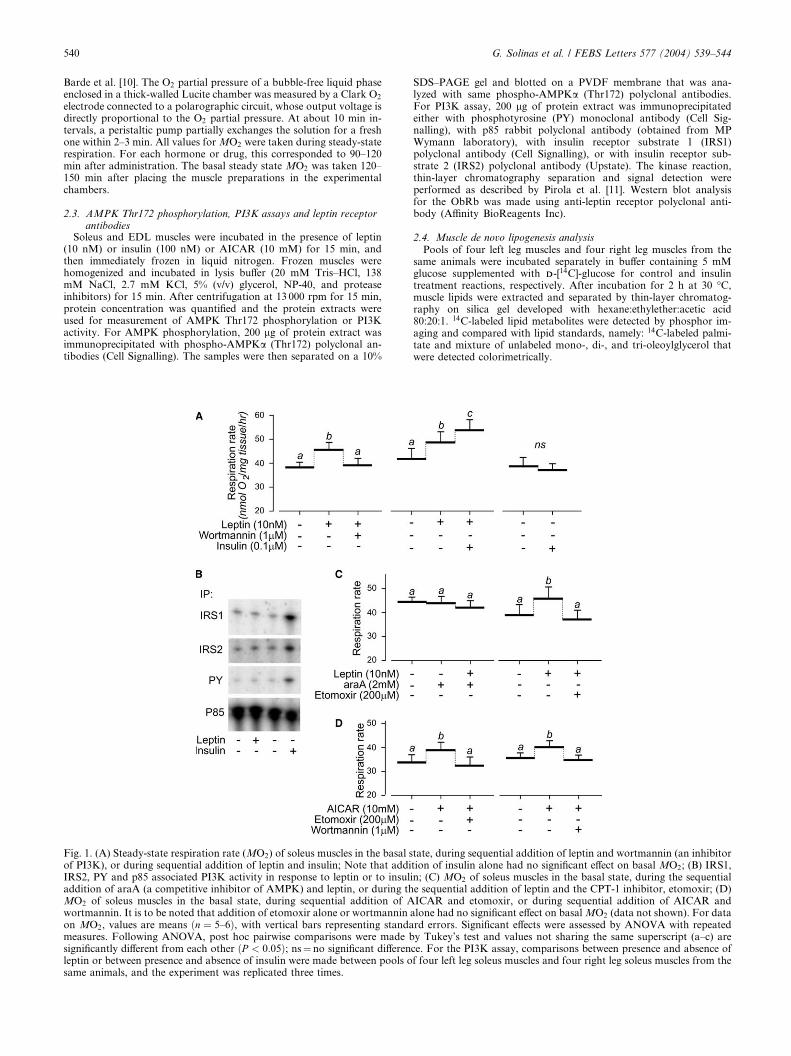
Fig. 1. (A) Steady-state respiration rate (MO2) of soleus muscles in the basal sof PI3K), or during sequential addition of leptin and insulin; Note that addiIRS2, PY and p85 associated PI3K activity in response to leptin or to insuladdition of araA (a competitive inhibitor of AMPK) and leptin, or during thMO2 of soleus muscles in the basal state, during sequential addition of Awortmannin. It is to be noted that addition of etomoxir alone or wortmanninon MO2, values are means ðn ¼ 5–6Þ, with vertical bars representing standameasures. Following ANOVA, post hoc pairwise comparisons were made bsignificantly different from each other ðP < 0:05Þ; ns¼no significant differenleptin or between presence and absence of insulin were made between pools osame animals, and the experiment was replicated three times.
SDS–PAGE gel and blotted on a PVDF membrane that was ana-lyzed with same phospho-AMPKa (Thr172) polyclonal antibodies.For PI3K assay, 200 lg of protein extract was immunoprecipitatedeither with phosphotyrosine (PY) monoclonal antibody (Cell Sig-nalling), with p85 rabbit polyclonal antibody (obtained from MPWymann laboratory), with insulin receptor substrate 1 (IRS1)polyclonal antibody (Cell Signalling), or with insulin receptor sub-strate 2 (IRS2) polyclonal antibody (Upstate). The kinase reaction,thin-layer chromatography separation and signal detection wereperformed as described by Pirola et al. [11]. Western blot analysisfor the ObRb was made using anti-leptin receptor polyclonal anti-body (Affinity BioReagents Inc).
2.4. Muscle de novo lipogenesis analysisPools of four left leg muscles and four right leg muscles from the
same animals were incubated separately in buffer containing 5 mMglucose supplemented with DD-[14C]-glucose for control and insulintreatment reactions, respectively. After incubation for 2 h at 30 �C,muscle lipids were extracted and separated by thin-layer chromatog-raphy on silica gel developed with hexane:ethylether:acetic acid80:20:1. 14C-labeled lipid metabolites were detected by phosphor im-aging and compared with lipid standards, namely: 14C-labeled palmi-tate and mixture of unlabeled mono-, di-, and tri-oleoylglycerol thatwere detected colorimetrically.
tate, during sequential addition of leptin and wortmannin (an inhibitortion of insulin alone had no significant effect on basal MO2; (B) IRS1,in; (C) MO2 of soleus muscles in the basal state, during the sequentiale sequential addition of leptin and the CPT-1 inhibitor, etomoxir; (D)ICAR and etomoxir, or during sequential addition of AICAR andalone had no significant effect on basalMO2 (data not shown). For datard errors. Significant effects were assessed by ANOVA with repeatedy Tukey’s test and values not sharing the same superscript (a–c) arece. For the PI3K assay, comparisons between presence and absence off four left leg soleus muscles and four right leg soleus muscles from the
G. Solinas et al. / FEBS Letters 577 (2004) 539–544 541
2.5. Chemicals and drugsAll chemicals were purchased from Fluka (Buchs, Switzerland).
Recombinant murine leptin was purchased from Insight BiotechnologyLtd. (Middlesex, UK), Wortmannin and Hydroxy-citrate from Cal-biochem (Luzern, Switzerland), Cerulenin from Fluka (Buchs, Swit-zerland), 5-aminoimidazole-4-carboxamide ribonucleoside (AICAR)from Toronto Research Chemicals (TRC, Toronto, Canada) andAdenine 9-b-DD-arabinofuranoside (araA) from Sigma (St. Louis, MO,USA). Etomoxir was a generous gift from Prof. W. Langhans (Zurich,Switzerland).
3. Results
3.1. Requirement for PI3K signaling
Consistent with our previous report [7], leptin (10 nM)
stimulates the respiration rate (MO2) of soleus muscle (by
about 20%), and this effect is abolished by the addition of
wortmannin, an inhibitor of PI3K (Fig. 1A). Since leptin in-
duces associations of PI3K activity to PY residues and to IRS1
and IRS2 in muscle myotubes [12] and in skeletal muscle from
mice injected with leptin [13], we investigate here whether an
increase in PI3K association with these molecules also corre-
lates with the direct effect of leptin on muscle MO2. Using in
vitro kinase assays in ex vivo intact soleus muscles incubated
with leptin, insulin or saline solution as control, we however
found that there is no induction of PY, IRS1, IRS2 and p85
associated PI3K activities in response to leptin, in contrast to
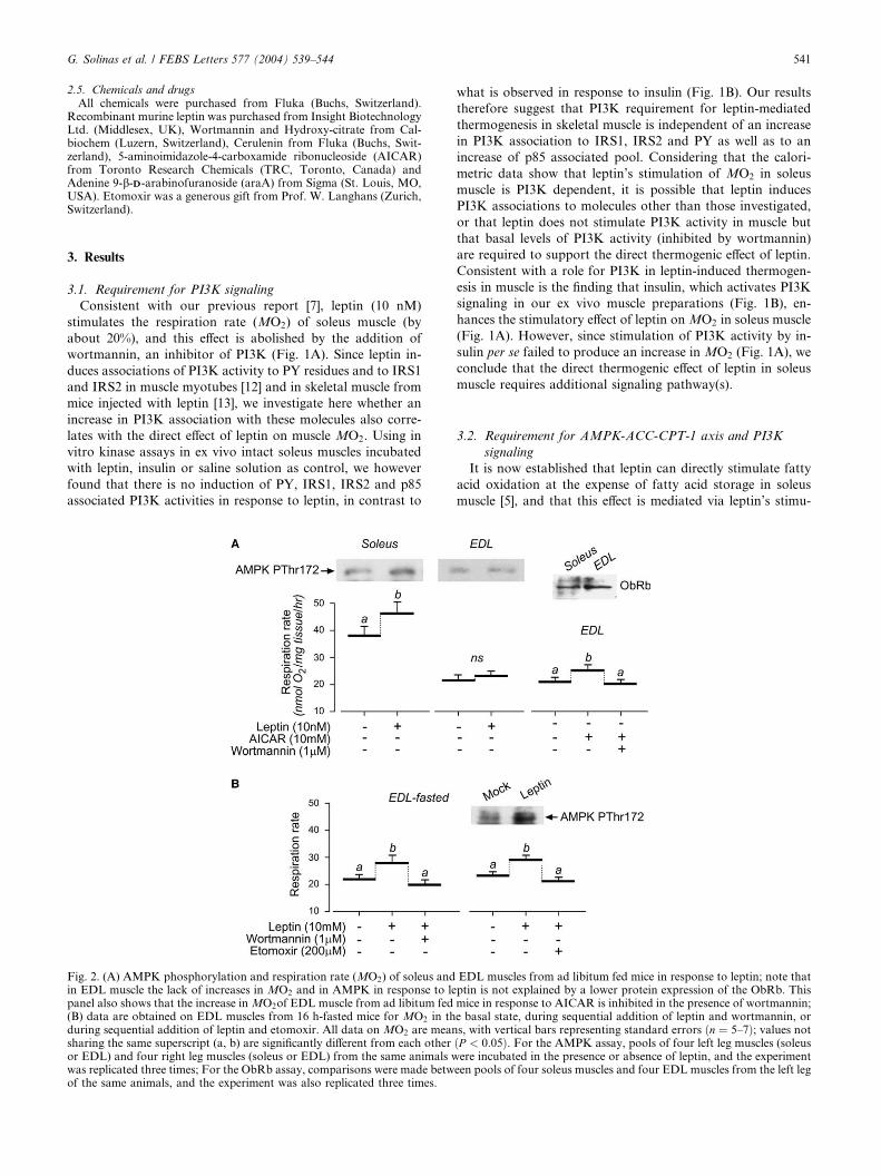
Fig. 2. (A) AMPK phosphorylation and respiration rate (MO2) of soleus andin EDL muscle the lack of increases in MO2 and in AMPK in response to lepanel also shows that the increase inMO2of EDL muscle from ad libitum fed(B) data are obtained on EDL muscles from 16 h-fasted mice for MO2 in thduring sequential addition of leptin and etomoxir. All data on MO2 are meansharing the same superscript (a, b) are significantly different from each otheror EDL) and four right leg muscles (soleus or EDL) from the same animals wwas replicated three times; For the ObRb assay, comparisons were made betwof the same animals, and the experiment was also replicated three times.
what is observed in response to insulin (Fig. 1B). Our results
therefore suggest that PI3K requirement for leptin-mediated
thermogenesis in skeletal muscle is independent of an increase
in PI3K association to IRS1, IRS2 and PY as well as to an
increase of p85 associated pool. Considering that the calori-
metric data show that leptin’s stimulation of MO2 in soleus
muscle is PI3K dependent, it is possible that leptin induces
PI3K associations to molecules other than those investigated,
or that leptin does not stimulate PI3K activity in muscle but
that basal levels of PI3K activity (inhibited by wortmannin)
are required to support the direct thermogenic effect of leptin.
Consistent with a role for PI3K in leptin-induced thermogen-
esis in muscle is the finding that insulin, which activates PI3K
signaling in our ex vivo muscle preparations (Fig. 1B), en-
hances the stimulatory effect of leptin onMO2 in soleus muscle
(Fig. 1A). However, since stimulation of PI3K activity by in-
sulin per se failed to produce an increase in MO2 (Fig. 1A), we
conclude that the direct thermogenic effect of leptin in soleus
muscle requires additional signaling pathway(s).
3.2. Requirement for AMPK-ACC-CPT-1 axis and PI3K
signaling
It is now established that leptin can directly stimulate fatty
acid oxidation at the expense of fatty acid storage in soleus
muscle [5], and that this effect is mediated via leptin’s stimu-
EDL muscles from ad libitum fed mice in response to leptin; note thatptin is not explained by a lower protein expression of the ObRb. Thismice in response to AICAR is inhibited in the presence of wortmannin;e basal state, during sequential addition of leptin and wortmannin, ors, with vertical bars representing standard errors ðn ¼ 5–7Þ; values notðP < 0:05Þ. For the AMPK assay, pools of four left leg muscles (soleusere incubated in the presence or absence of leptin, and the experimenteen pools of four soleus muscles and four EDL muscles from the left leg
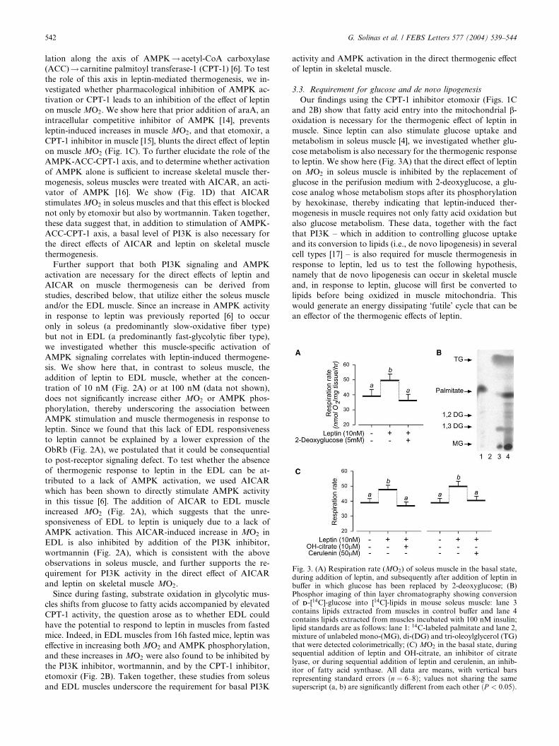
Fig. 3. (A) Respiration rate (MO2) of soleus muscle in the basal state,during addition of leptin, and subsequently after addition of leptin inbuffer in which glucose has been replaced by 2-deoxyglucose; (B)Phosphor imaging of thin layer chromatography showing conversionof DD-[14C]-glucose into [14C]-lipids in mouse soleus muscle: lane 3contains lipids extracted from muscles in control buffer and lane 4contains lipids extracted from muscles incubated with 100 nM insulin;lipid standards are as follows: lane 1: 14C-labeled palmitate and lane 2,mixture of unlabeled mono-(MG), di-(DG) and tri-oleoylglycerol (TG)that were detected colorimetrically; (C) MO2 in the basal state, duringsequential addition of leptin and OH-citrate, an inhibitor of citratelyase, or during sequential addition of leptin and cerulenin, an inhib-itor of fatty acid synthase. All data are means, with vertical barsrepresenting standard errors ðn ¼ 6–8Þ; values not sharing the samesuperscript (a, b) are significantly different from each other ðP < 0:05Þ.
542 G. Solinas et al. / FEBS Letters 577 (2004) 539–544
lation along the axis of AMPK! acetyl-CoA carboxylase
(ACC)! carnitine palmitoyl transferase-1 (CPT-1) [6]. To test
the role of this axis in leptin-mediated thermogenesis, we in-
vestigated whether pharmacological inhibition of AMPK ac-
tivation or CPT-1 leads to an inhibition of the effect of leptin
on muscle MO2. We show here that prior addition of araA, an
intracellular competitive inhibitor of AMPK [14], prevents
leptin-induced increases in muscle MO2, and that etomoxir, a
CPT-1 inhibitor in muscle [15], blunts the direct effect of leptin
on muscle MO2 (Fig. 1C). To further elucidate the role of the
AMPK-ACC-CPT-1 axis, and to determine whether activation
of AMPK alone is sufficient to increase skeletal muscle ther-
mogenesis, soleus muscles were treated with AICAR, an acti-
vator of AMPK [16]. We show (Fig. 1D) that AICAR
stimulatesMO2 in soleus muscles and that this effect is blocked
not only by etomoxir but also by wortmannin. Taken together,
these data suggest that, in addition to stimulation of AMPK-
ACC-CPT-1 axis, a basal level of PI3K is also necessary for
the direct effects of AICAR and leptin on skeletal muscle
thermogenesis.
Further support that both PI3K signaling and AMPK
activation are necessary for the direct effects of leptin and
AICAR on muscle thermogenesis can be derived from
studies, described below, that utilize either the soleus muscle
and/or the EDL muscle. Since an increase in AMPK activity
in response to leptin was previously reported [6] to occur
only in soleus (a predominantly slow-oxidative fiber type)
but not in EDL (a predominantly fast-glycolytic fiber type),
we investigated whether this muscle-specific activation of
AMPK signaling correlates with leptin-induced thermogene-
sis. We show here that, in contrast to soleus muscle, the
addition of leptin to EDL muscle, whether at the concen-
tration of 10 nM (Fig. 2A) or at 100 nM (data not shown),
does not significantly increase either MO2 or AMPK phos-
phorylation, thereby underscoring the association between
AMPK stimulation and muscle thermogenesis in response to
leptin. Since we found that this lack of EDL responsiveness
to leptin cannot be explained by a lower expression of the
ObRb (Fig. 2A), we postulated that it could be consequential
to post-receptor signaling defect. To test whether the absence
of thermogenic response to leptin in the EDL can be at-
tributed to a lack of AMPK activation, we used AICAR
which has been shown to directly stimulate AMPK activity
in this tissue [6]. The addition of AICAR to EDL muscle
increased MO2 (Fig. 2A), which suggests that the unre-
sponsiveness of EDL to leptin is uniquely due to a lack of
AMPK activation. This AICAR-induced increase in MO2 in
EDL is also inhibited by addition of the PI3K inhibitor,
wortmannin (Fig. 2A), which is consistent with the above
observations in soleus muscle, and further supports the re-
quirement for PI3K activity in the direct effect of AICAR
and leptin on skeletal muscle MO2.
Since during fasting, substrate oxidation in glycolytic mus-
cles shifts from glucose to fatty acids accompanied by elevated
CPT-1 activity, the question arose as to whether EDL could
have the potential to respond to leptin in muscles from fasted
mice. Indeed, in EDL muscles from 16h fasted mice, leptin was
effective in increasing both MO2 and AMPK phosphorylation,
and these increases in MO2 were also found to be inhibited by
the PI3K inhibitor, wortmannin, and by the CPT-1 inhibitor,
etomoxir (Fig. 2B). Taken together, these studies from soleus
and EDL muscles underscore the requirement for basal PI3K
activity and AMPK activation in the direct thermogenic effect
of leptin in skeletal muscle.
3.3. Requirement for glucose and de novo lipogenesis
Our findings using the CPT-1 inhibitor etomoxir (Figs. 1C
and 2B) show that fatty acid entry into the mitochondrial b-oxidation is necessary for the thermogenic effect of leptin in
muscle. Since leptin can also stimulate glucose uptake and
metabolism in soleus muscle [4], we investigated whether glu-
cose metabolism is also necessary for the thermogenic response
to leptin. We show here (Fig. 3A) that the direct effect of leptin
on MO2 in soleus muscle is inhibited by the replacement of
glucose in the perifusion medium with 2-deoxyglucose, a glu-
cose analog whose metabolism stops after its phosphorylation
by hexokinase, thereby indicating that leptin-induced ther-
mogenesis in muscle requires not only fatty acid oxidation but
also glucose metabolism. These data, together with the fact
that PI3K – which in addition to controlling glucose uptake
and its conversion to lipids (i.e., de novo lipogenesis) in several
cell types [17] – is also required for muscle thermogenesis in
response to leptin, led us to test the following hypothesis,
namely that de novo lipogenesis can occur in skeletal muscle
and, in response to leptin, glucose will first be converted to
lipids before being oxidized in muscle mitochondria. This
would generate an energy dissipating ‘futile’ cycle that can be
an effector of the thermogenic effects of leptin.
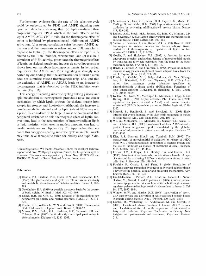
Leptin Insulin
AMPK
PI3K
CPT-
Fatty acids
Citrate
Citrate
AcetylCoAACC
MalonylCoA
Fatty acids
Cytoplasmicmembrane
Glucose
Glucose
Mitochondrialmembrane
Pyruvate
CitrateLyase
Fatty acidSynthase
TG
NADPH
NADPH
G. Solinas et al. / FEBS Letters 577 (2004) 539–544 543
We show here that incubation of our muscle preparations in
buffer containing 14C-labeled glucose resulted in the produc-
tion of 14C-labeled lipids corresponding to free fatty acids,
diacylglycerol and triacylglycerol when analyzed by thin-layer
chromatography (Fig. 3B), and that insulin increased the
synthesis of lipids from glucose in soleus muscle, and to a lesser
extent in EDL muscles (data not shown). This experiment
demonstrates that de novo lipogenesis can occur in skeletal
muscle and that it can be induced by insulin, as in the liver and
adipose tissue [17]. To know whether de novo lipogenesis is
necessary for leptin-induced thermogenesis in skeletal muscle,
we tested whether leptin-induced increases in MO2 in the so-
leus muscle are inhibited by the addition of inhibitors of key
control points in the conversion of glucose to lipids. We show
here that OH-citrate, which inhibits the enzyme citrate lyase,
or cerulenin, an inhibitor of fatty acid synthase, can blunt the
leptin induction of MO2 in soleus muscle (Fig. 3C). Taken
together, these data indicate that both glucose metabolism and
de novo lipogenesis are required for the direct thermogenic
effect of leptin in skeletal muscle.
AcetylCoA
Fatty acids
Krebs cycle
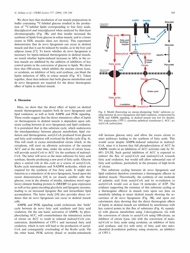
Fig. 4. Model illustrating an energy-dissipating ‘futile’ substrate cy-cling between de novo lipogenesis and lipid oxidation, orchestrated byPI3K and AMPK signaling, in skeletal muscle (see text for details).TG: triglycerides; CPT-1: carnitine palmitoyl transferase-1; ACC: ac-etyl-CoA carboxylase.
4. Discussion
Here, we show that the direct effect of leptin on skeletal
muscle thermogenesis requires both de novo lipogenesis and
lipid oxidation, as well as both AMPK and PI3K signaling.
These results suggest that the direct stimulatory effect of leptin
on thermogenesis in skeletal muscle is dependent upon sub-
strate cycling between de novo lipogenesis and lipid oxidation.
It is postulated that in this substrate cycle, which underscores
the interdependency between glucose metabolism, lipid oxi-
dation and thermogenesis, acetyl-CoA produced from glucose
and fatty acid oxidation will overload the Krebs cycle (Fig. 4).
This will result in excess mitochondrial citrate which, in the
cytoplasm, will exert an allosteric activation of the enzyme
ACC and at the same time, under the action of citrate lyase,
will provide acetyl-CoA to ACC for the synthesis of malonyl-
CoA. The latter will serve as the main substrate for fatty acid
synthase, thereby producing a new pool of fatty acids. Glucose
plays a central role in this cycle as a source of acetyl-CoA,
Krebs cycle intermediates and NADPH molecules, which are
required for the synthesis of free fatty acids. It might also
function as a stimulator of de novo lipogenesis, based upon the
recent demonstration [18] in rat muscle satellite cells that
glucose, even in the absence of insulin, stimulates sterol regu-
latory element binding protein-1c (SREBP-1c) gene expression
as well as key genes encoding glycolytic and lipogenic enzymes,
leading to an increased lipogenic flux and intracellular lipid
accumulation. The latter study [18] also provides direct evi-
dence that de novo lipogenesis can occur in skeletal muscle
cells.
AMPK and PI3K signaling could orchestrate this ‘futile’
cycle between de novo fatty acid synthesis and fatty acid
oxidation. On the one hand, activation of AMPK, by phos-
phorylating ACC, will counterbalance the stimulatory action
of citrate on ACC to result in reduced malonyl-CoA con-
centration, disinhibition of CPT-1 and increased fatty acid
oxidation, which in turn will lead to the production of acetyl-
CoA and consequently overloading of the Krebs cycle. On
the other hand, PI3K activity (basal or insulin-stimulated)
will increase glucose entry and allow the excess citrate to
enter pathways leading to the synthesis of fatty acids. This
would occur despite AMPK-induced reduction in malonyl-
CoA, since it is known that full phosphorylation of ACC by
AMPK results in an inhibition of ACC activities only by 50–
60% [19,20]. Such partial inhibition of ACC is expected to
redirect the flux of acetyl-CoA and malonyl-CoA towards
fatty acid oxidation, but would still allow substantial rate of
fatty acid synthesis, particularly in the presence of high levels
of citrate.
This substrate cycling between de novo lipogenesis and
lipid oxidation therefore constitutes a thermogenic effector in
skeletal muscle. Theoretically, the synthesis of one molecule
of palmitic acid from acetyl-CoA and its re-oxidation to
acetyl-CoA would cost at least 14 molecules of ATP. The
evidence supporting the existence of this substrate cycling as
a thermogenic effector in muscle rests upon our data on
metabolic labeling in intact skeletal muscle showing the oc-
currence of de novo lipogenesis (Fig. 3B), and upon our
calorimetric data showing that the direct thermogenic effects
of leptin in skeletal muscle are inhibited by interference with
key control points in this flux of substrates (Fig. 4), namely:
(i) with glucose metabolism using 2-deoxyglucose, (ii) with
the conversion of citrate to acetyl-CoA using OH-citrate, an
inhibitor of citrate lyase, (iii) with the conversion of malo-
nyl-CoA to fatty acids using cerulenin, an inhibitor of fatty
acid synthase, and (iv) with entry of fatty acid into mito-
chondrial b-oxidation pathway using etomoxir, an inhibitor
of CPT-1.
544 G. Solinas et al. / FEBS Letters 577 (2004) 539–544
Furthermore, evidence that the rate of this substrate cycle
could be orchestrated by PI3K and AMPK signaling rests
upon our data here showing that: (a) leptin-mediated ther-
mogenesis requires CPT-1 which is the final effector of the
leptin-AMPK-ACC-CPT-1 axis, (b) the thermogenic effect of
leptin is inhibited by pharmacological inhibition of AMPK
activation, (c) a strong correlation exists between AMPK ac-
tivation and thermogenesis in soleus and/or EDL muscles in
response to leptin, (d) the thermogenic effects of leptin is in-
hibited by the PI3K inhibitor, wortmannin, and (e) insulin, a
stimulator of PI3K activity, potentiates the thermogenic effects
of leptin on skeletal muscle and induces de novo lipogenesis as
shown from our metabolic labeling experiments. In fact, a dual
requirement for AMPK and PI3K signaling is strongly sup-
ported by our findings that the administration of insulin alone
does not stimulate muscle thermogenesis (Fig. 1A), and that
the activation of AMPK by AICAR leads to an increase in
thermogenesis that is abolished by the PI3K inhibitor wort-
mannin (Fig. 1D).
This energy dissipating substrate cycling linking glucose and
lipid metabolism to thermogenesis provides a novel molecular
mechanism by which leptin protects the skeletal muscle from
ectopic fat storage and lipotoxicity. Although the increase in
muscle metabolic rate induced by leptin (about 20%) may not,
a priori, be considered to be large, it must be emphasized that
peripheral resistance to this thermogenic effect of leptin can,
over time, lead to the accumulation of intramyocellular lipids
or lipid moieties, which even in modest amounts, can lead to
insulin resistance and lipotoxicity [3]. Approaches that en-
hance this energy-dissipating substrate cycle in skeletal muscle
may thus have therapeutic value for obesity and type 2 dia-
betes.
Acknowledgements: We thank Doroth�ee Rohrer for excellent technicalsupport and Prof. Wolfgang Langhans (Zurich) for his generous gift ofetomoxir. This work was supported by Grant Nos. 3157129.991 and3200B0-102156 of the Swiss National Science Foundation.
References
[1] Randle, P.J., Garland, P.B., Hales, C.N. and Newsholme, E.A.(1963) The glucose-fatty acid cycle: its role in insulin sensitivityand metabolic disturbances of diabetes mellitus. Lancet I, 785–789.
[2] Newsholme, E.A. (1980) A possible metabolic basis for the controlof body weight. N. Engl. J. Med. 302, 400–405.
[3] Unger, R.H. and Orci, L. (2001) Diseases of liporegulation: newperspective on obesity and related disorders. FASEB J. 15, 312–321.
[4] Ceddia, R.B., William Jr., W.N. and Curi, R. (2001) The responseof skeletal muscle to leptin. Front. Biosci. 6, D90–97.
[5] Muoio, D.M., Dohn, G.L., Fiedorek, F.T., Tapscott, E.B. andColeman, R.A. (1997) Leptin directly alters lipid partitioning inskeletal muscle. Diabetes 46, 1360–1363.
[6] Minokoshi, Y., Kim, Y.B., Peroni, O.D., Fryer, L.G., Muller, C.,Carling, D. and Kahn, B.B. (2002) Leptin stimulates fatty-acidoxidation by activating AMP-activated protein kinase. Nature415, 339–343.
[7] Dulloo, A.G., Stock, M.J., Solinas, G., Boss, O., Montani, J.P.and Seydoux, J. (2002) Leptin directly stimulates thermogenesis inskeletal muscle. FEBS Letters 515, 109–113.
[8] Samec, S., Seydoux, J. and Dulloo, A.G. (1998) Role of UCPhomologues in skeletal muscles and brown adipose tissue:mediators of thermogenesis or regulators of lipids as fuelsubstrate? FASEB J. 12, 715–724.
[9] Goglia, F. and Skulachev, V.P. (2003) A function for noveluncoupling proteins: antioxidant defense of mitochondrial matrixby translocating fatty acid peroxides from the inner to the outermembrane leaflet. FASEB J. 17, 1585–1591.
[10] Barde, Y., Chinet, A. and Girardier, L. (1975) Potassium-inducedincrease in oxygen consumption of brown adipose tissue from therat. J. Physiol. (Lond.) 252, 523–536.
[11] Pirola, L., Zvelebil, M.J., Bulgarelli-Leva, G., Van Obberg-hen, E., Waterfield, M.D. and Wymann, M.P. (2001) Acti-vation loop sequences confer substrate specificity tophosphoinositide 3-kinase alpha (PI3Kalpha). Functions oflipid kinase-deficient PI3Kalpha in signalling. J. Biol. Chem.276, 21544–21554.
[12] Kellerer, M., Koch, M., Metzinger, E., Mushack, J., Capp, E. andHaring, H.U. (1997) Leptin activates PI-3 kinase in C2C12myotubes via janus kinase-2 (JAK-2) and insulin receptorsubstrate-2 (IRS-2) dependent pathways. Diabetologia 40, 1358–1362.
[13] Maroni, P., Bendinelli, P. and Piccoletti, R. (2003) Earlyintracellular events induced by in vivo leptin treatment in mouseskeletal muscle. Mol. Cell. Endocrinol. 201, 109–121.
[14] Wu, X., Motoshima, H., Mahadev, K., Stalker, T.J., Scalia, R.and Goldstein, B.J. (2003) Involvement of liver AMP-activatedprotein kinase in glucose uptake stimulated by the globulardomain of adiponectin in primary rat adipocytes. Diabetes 52,1355–1363.
[15] Kler, R.S., Sherratt, H.A.S. and Turnbull, D.M. (1992) Themeasurement of mitochondrial b oxidation by release of 3H2Ofrom [9,10-3H]hexadecanoate: application to skeletal muscle andthe use of inhibitors as models of metabolic disease. Biochem.Med. Metab. Biol. 47, 145–156.
[16] Corton, J.M., Gillespie, J.G., Hawley, S.A. and Hardie, D.G.(1995) 5-Aminoimidazole-4-carboxamide ribonucleoside. A spe-cific method for activating AMP-activated protein kinase in intactcells. Eur. J. Biochem. 229, 558–565.
[17] Foufelle, F., Girard, J. and Ferre, P. (1996) Regulation oflipogenic enzyme expression by glucose in liver and adipose tissue:a review of the potential cellular and molecular mechanisms. Adv.Enzyme Regul. 36, 199–226.
[18] Guillet-Deniau, I., Pichard, A.L., Kon�e, A., Esnous, C., Nieru-chalski, M., Girard, J. and Prip-Buus, C. (2004) Glucose inducesde novo lipogenesis in rat muscle satellite cells through a srerol-regulatory-element-binding-protein-1c-dependent pathway. J. CellSci. 177, 1937–1944.
[19] Winder, W.W. and Hardie, D.G. (1996) Inactivation of acetyl-CoA carboxylase and activation of AMP-activated protein kinasein muscle during exercise. Am. J. Physiol. 270, E299–E304.
[20] Gubler, M., Westerberg, R., Andjelkovic, M. and Mizrahi, J.(2003). Functional characterization f human ACC2 enzymeand elucidation of its role in the regulation of mitochondrialfatty acid oxidation. Keystone Conference on Obesity: Newinsights into pathogenesis and treatment, Keystone: Abstract214.