Embed Size (px)
Citation preview

FIRST EDITION WITH A FOCUS ONFRIDAY & SATURDAY
S C I E N T I F I C H I G H L I G H T S O F C H I C A G O 2 0 1 2EyeNet
M A G A Z I N E
SUBSPECIALTY DAY: The Directors Pick Their Must-See Sessions

EyeNet A
cademy N
ews
Cataract Surgery is Changing in a Femtosecond.
With Alcon’s LenSx® Laser, the Possibilities Have Just Begun.Delivering the precision of a femtosecond laser to Refractive Cataract Surgery, the LenSx® Laser is designed to reproducibly
perform many of the most challenging aspects of traditional cataract surgery. Creating highly reproducible capsulotomy, lens fragmentation and all corneal incisions including arcuate incisions with image-guided surgeon control, Alcon’s LenSx® Laser is Putting the Future in Motion.
For Important Safety Information about the LenSx® Laser, please refer to the adjacent page.
To learn more about LenSx® Laser technology forLaser Refractive Cataract Surgery, visit lensxlasers.comor visit us at Booth 2808 at AAO
© 2012 Novartis 9/12 LSX12137JAD
Laser Refractive Cataract Surgery is a Reality with Alcon’s LenSx® Laser.
80099 LSX12137JAD ENAN.indd 1 9/19/12 2:34 PM
e y e n e t ’ s a c a d e m y n e w s 3
ON THE COVER“Erupting” Avellino Dystrophy
Photo by Michael Stanley
Medical College of Georgia
Georgia Health Sciences University
Augusta, Ga.
IN THIS ISSUE
Welcome to Subspecialty Day 2012! This year’s presentations will cover
the latest developments in diagnosis, treatments, and procedures by world-renowned ophthalmologists in disci-
plines ranging from cornea to uveitis. On Saturday, there will be Subspecialty Day meetings in cor-
nea, glaucoma, oculofacial plastic surgery, pediatric ophthalmol-ogy, refractive surgery, retina, and uveitis. Refractive surgery and retina are also covered today (Friday).
As always, I urge you to take time to explore disciplines other than your own. Often, pearls from one subspecialty can be applied to a completely different arena in surprising and useful ways. Please refer to this issue of Academy News to find presenta-tions that might be of interest to you. Look for the second edition of Academy News on Sunday and check your e-mail each evening for Academy Live, a daily roundup of news from Subspecialty Day and the Joint Meeting.
Richard P. Mills, MD, MPHChief Medical Editor, EyeNet Magazine
FROM THE EDITOR
ANNUAL BUSINESS MEETING. Notice is hereby given that the Annual Business Meeting of the American Academy of Ophthalmology will be held on Sunday, Nov. 11, in North Hall B of the McCormick Place Convention Center in Chicago, from 10 to 10:30 a.m.
The order of business shall be: Call to order New business Report of the president Announcements and notices Report of the executive vice president Adjournment Election of fellows and members
As stated in the bylaws of the Academy, the order of business of each Annual Business Meeting may be amended by an affirmative vote of a majority of the voting fellows and members present and voting at the meeting.
F O R T H E R E C O R D
Guide to Subspecialty Day The program direc-tors provide an insider’s perspective on what’s hot this year.
Introducing the APAO See how the Asia-Pacific Academy of Ophthalmology is charting a unique course in ophthalmology.
Electronic Health Records These two tables of EHR system specs will help you “shop smart” when you visit vendors in the exhibit hall.
A Novel Approach to Pterygium Surgery Inspired by a 2011 Best of Show Video, EyeNet developed this story that was popular with readers.
Cataract Spotlight Redux, Part One: Looking forward to the Spotlight on Cataract Surgery Session? Until then, relive the excitement from last year’s meeting.
4-8
11-12
14-17
18-21
22-26
NOTICE: This publication was printed in advance of Subspecialty Day and the Joint Meeting. Check the Online Program (www.aao.org/2012) for the most up-to-date information.
Indication: The LenSx® Laser is indicated for use in patients undergoing cataract surgery for removal of the crystalline lens. Intended uses in cataract surgery include anterior capsulotomy, phacofragmentation, and the creation of single plane and multi-plane arc cuts/incisions in the cornea, each of which may be performed either individually or consecutively during the same procedure.
Caution: United States Federal Law restricts this device to sale and use by or on the order of a physician or licensed eye care practitioner. United States Federal Law restricts the use of this device to practitioners who have been trained in the operation of this device.
Restrictions: • Patientsmustbeabletolieflatandmotionlessinasupineposition.
• Patientmustbeabletounderstandandgiveaninformedconsent.
• Patientsmustbeabletotoleratelocalortopicalanesthesia.
• PatientswithelevatedIOPshouldusetopicalsteroidsonlyunderclosemedicalsupervision.
Contraindications: • Cornealdiseasethatprecludesapplanationofthecorneaortransmissionoflaserlightat1030nm
wavelength
• Descemetocelewithimpendingcornealrupture
• Presenceofbloodorothermaterialintheanteriorchamber
• Poorlydilatingpupil,suchthattheirisisnotperipheraltotheintendeddiameterforthecapsulotomy
• Conditionswhichwouldcauseinadequateclearancebetweentheintendedcapsulotomydepthandtheendothelium (applicable to capsulotomy only)
• Previouscornealincisionsthatmightprovideapotentialspaceintowhichthegasproducedbytheprocedure can escape
• Cornealthicknessrequirementsthatarebeyondtherangeofthesystem
• Cornealopacitythatwouldinterferewiththelaserbeam
• Hypotony,glaucoma,orthepresenceofacornealimplant
• Residual,recurrent,activeocularoreyeliddisease,includinganycornealabnormality(forexample,recurrent corneal erosion, severe basement membrane disease)
• Thisdeviceisnotintendedforuseinpediatricsurgery
• Ahistoryoflenswithzonularinstability.
• Anycontraindicationtocataractor keratoplastysurgery.
Attention:ReferencetheDirectionsforUselabelingforacompletelistingofindications,warningsandprecautions.
Warnings: The LenSx® Laser System should only be operated by a physician trained in its use.
TheLenSx®LaserdeliverysystememploysonesteriledisposableLenSx®LaserPatientInterfaceconsistingofanapplanationlensandsuctionring.ThePatientInterfaceisintendedforsingleuseonly.ThedisposablesusedinconjunctionwithALCON®instrumentproductsconstituteacompletesurgicalsystem.UseofdisposablesotherthanthosemanufacturedbyAlconmayaffectsystemperformanceandcreatepotentialhazards.
The physician should base patient selection criteria on professional experience, published literature, and educationalcourses.Adultpatientsshouldbescheduledtoundergocataractextraction.
Precautions: • DonotusecellphonesorpagersofanykindinthesameroomastheLenSx®Laser.
• DiscardusedPatientInterfacesasmedicalwaste.
AEs/Complications: • Capsulotomy,phacofragmentation,orcutorincisiondecentration
• Incompleteorinterruptedcapsulotomy,fragmentation,orcornealincisionprocedure
• Capsulartear
• Cornealabrasionordefect
• Pain
• Infection
• Bleeding
• Damagetointraocularstructures
• Anteriorchamberfluidleakage,anteriorchambercollapse
• Elevatedpressuretotheeye
©2012Novartis 9/12 LSX12137JAD
80099 LSX12137JAD_PI ENAN.indd 1 9/21/12 12:39 PM

To provide you with an insider’s perspective on Subspecialty Day, we contacted a program director from
each meeting and asked them to identify presentations that 1) may cause physi-cians within the subspecialty to recon-sider a clinical practice, 2) ophthalmolo-gists outside the subspecialty particularly need to know about, and 3) may surprise attendees.
Please note: This article was written in advance of Subspecialty Day.
CLINICAL PRACTICES TO RECONSIDER
CORNEAGrand Ballroom S100abn Top 10 Reasons Why You Should Be Per-forming Descemet Membrane Endothelial Keratoplasty in 2012, presented by Gerrit R. J. Melles, MD, PhD, from 9:50 to 10 a.m.
The section “Anterior Segment Surgery–Expanding Your Reach” (9:30 to 10:45 a.m.) is aimed at expanding the repertoire of cornea specialists. One presentation that reflects the timeliness of this section is “Top 10 Reasons Why You Should Be Performing Descemet Membrane Endo-thelial Keratoplasty in 2012” by Gerrit R. J. Melles, MD, PhD.
Recent research has showed numerous advantages of DMEK, a form of selective endothelial transplantation in which only the Descemet membrane and endotheli-um are transplanted. These include lower risk of endothelial rejection, less induced hyperopic shift, and elimination of the requirement for a microkeratome to prepare the donor cornea, which enables surgeons to perform the procedure with-out needing to purchase any specialized instruments or equipment.
While the learning curve for DMEK is steep, Dr. Melles will provide 10 reasons why it is worth the climb.
—Anthony J. Aldave, MDCornea program codirector
GLAUCOMARoom E354n Pressure Fluctuation: In the Lab and in the Clinic, presented by Arthur J. Sit, MD, from 9:11 to 9:18 a.m.n Effect of Cerebrospinal Pressure, pre-sented by Jost B. Jonas, MD, from 9:18 to 9:25 a.m.n Blood Pressure and Sleep Apnea, present-ed by Donald L. Budenz, MD, MPH, from 9:32 to 9:39 a.m.
Glaucoma specialists traditionally have focused their efforts on keeping patients within an optimal range of target pres-sure. Yet this strategy does not account for those patients who appear to be getting worse despite having pressures that are
well within the normal range (Fig. 1). Some ophthalmologists are looking at the possible role of fluctuations in pres-sure readings and how they could affect the optic nerve. Arthur J. Sit, MD, in his presentation, “Pressure Fluctuation: In the Lab and in the Clinic,” will address this emerging area of research and discuss how it may impact clinical practice.
Presenters in the same section will also shed light on some nonophthalmic sources of pressure on the optic nerve and how they might affect glaucoma man-agement. Jost B. Jonas, MD, will discuss “Effect of Cerebrospinal Pressure,” and Donald L. Budenz, MD, MPH, will cover “Blood Pressure and Sleep Apnea.”
—Wallace L. M. Alward, MDGlaucoma program codirector
OCULOFACIAL PLASTIC SURGERYVista Room S406an Thyroid Eye Disease—Case Presentation and Structured Discussion, led by Petros Perros, MD, from 2:30 to 3:30 p.m.
Graves disease continues to pose chal-lenges to oculofacial plastic surgeons be-cause treatment options remain limited, and much of the supporting data is weak and conflicting (Fig. 2). To tackle the complexities of thyroid eye disease, we are devoting an entire section to one case pre-sentation. Petros Perros, MD, will present three decision points: “1: For Treatment of Thyrotoxicosis” (2:30 to 2:35 p.m.); “2: For Initial Treatment of the Orbitopathy” (2:50 to 2:55 p.m.); and “3: After Pulsed IV Steroid Course, Patient Relapses, Then What?” (3:10 to 3:14 p.m.). Each of these presentations will be followed by point-counterpoint discussions.
—Julian D. Perry, MDOculofacial Plastic Surgery program
codirector
PEDIATRIC OPHTHALMOLOGYGrand Ballroom S100cn Strategies for Prevention of ROP, pre-sented by William V. Good, MD, from 10:31 to 10:36 a.m.
Our “ROP Mini-Symposium” (10:10 to 10:40 a.m.) promises to be of particular interest to pediatric retina specialists who treat retinopathy of prematurity. In addition to sharing treatment pearls, the section will focus on preventing the condition in the first place. This is a revolutionary concept for our field, and William V. Good, MD, in his talk “Strate-gies for Prevention of ROP,” will discuss identifying those children who are at risk and then creating approaches to prevent them from developing ROP.
Dr. Good has a long-standing interest in ROP prevention. In a recent research letter, he and his colleagues observed the relative immunity to severe ROP seen in many African-American infants.1 They pointed to the existence of beta-blocker receptor polymorphisms, which exist in many African-Americans, and cited the possibility of using this to develop a preventive strategy.
—Stephen P. Christiansen, MDPediatric Ophthalmology program
codirector
1 Good WV et al. Arch Ophthalmol. 2012;
130(1):117-118.
REFRACTIVE SURGERYNorth Hall Bn Point: Limbal Relaxing Incisions, present-ed by Louis D. “Skip” Nichamin, MD, from 8:02 to 8:07 a.m.n Counterpoint: Toric IOLs, presented by John A. Hovanesian, MD, from 8:07 to 8:12 a.m.
Now that lens surgery is becoming increasingly important in managing
refractive error, we have scheduled two full hours on Saturday morning on the subject.
One highlight is the point-counter-point on astigmatism correction, which continues to be a challenge. Louis D. “Skip” Nichamin, MD, will present “Point: Limbal Relaxing Incisions,” followed by John A. Hovanesian, MD, who will offer “Counterpoint: Toric IOLs.”
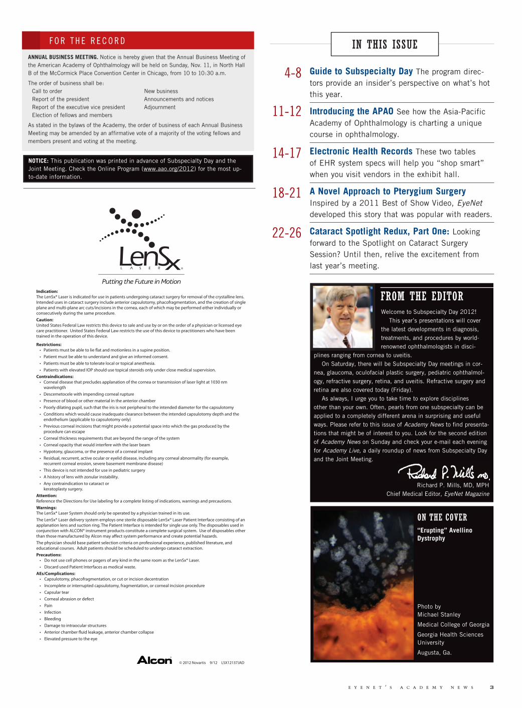
As we do not yet have a presbyopia-correcting IOL for patients with astig-matism, we are seeing a resurgence in the popularity of limbal relaxing inci-sions (Fig. 3, page 6). Interestingly, the advent of the femtosecond laser has also prompted surgeons to revisit limbal relax-ing incisions.
Toric lenses are designed to provide both spherical and astigmatic correction, and they are widely used for cataract patients with preexisting astigmatism. These two discussions will no doubt give specialists insight into clinical options for their astigmatic presbyopic patients.
—David R. Hardten, MDRefractive Surgery program codirector
RETINAArie Crown Theatern How Do I Incorporate What I Just Heard Into My Practice? presented by Peter K. Kai-ser, MD, from 8:47 to 8:54 a.m.
With three anti-VEGF drugs now avail-able to treat neovascular AMD and several ongoing trials that have generated reams of data, it is not surprising that retina specialists may feel challenged to keep up with the latest research find-ings and treatment alternatives. In the “Neovascular AMD” section (8:05 to 9:56 a.m.), Peter K. Kaiser, MD, will help frame the section’s discussions about various approaches in his talk—“How Do
4 j o i n t m e e t i n g 2 0 1 2
DIRECTORS’ PICKSPROGRAM
PROGRAM DIRECTORS RECOMMEND MUST-SEE PRESENTATIONS
An Insider’s Guide to Saturday’s Meetingsby lori baker schena, contributing writer
Wa
lla
ce L
. M
. A
lw
ard
, M
D;
BC
SC
, S
ec
tio
n 6
Continued on page 6
GLAUCOMA. Optic nerve of a patient pro-gressing despite normal IOP readings.
1
OCULOFACIAL PLASTIC SURGERY. The complexities of treating Graves disease will be discussed in a series of point-counterpoint discussions.
2

Visit us at AAO/APAO Booth #1571
Dedicated to advancing the treatment of eye diseases with unmet medical need
ThromboGenics, Inc. 101 Wood Avenue South, 6th Floor, Iselin, NJ 08830 - USA ©2012 ThromboGenics, Inc. All rights reserved. THROMBOGENICS and the THROMBOGENICS logo are trademarks or registered trademarks
of ThromboGenics in the United States, European Union, Japan, and other countries.
THRCOR002 A11/12
ThromboGenics™, a biopharmaceutical company focused on developing innovative ophthalmic medicines.
I Incorporate What I Just Heard Into My Practice?”—and provide insight into how practitioners can incorporate these recent advances into their practice.
For example, two-year data from the Comparison of Age-Related Macular Degeneration Treatments Trial (CATT) suggest that bevacizumab and ranibizu-mab may be equivalent in treating AMD; as a result, specialists are feeling more comfortable using either of these two drugs. In addition, now with findings from the VIEW Year 2 study and the FDA approval of aflibercept, we have a third alternative.
Dr. Kaiser will help attendees gain some clarity into the optimal treatment for our patients.
—Joan W. Miller, MDRetina program codirector
UVEITISRoom E450n How to Consider a Uveitic Entity and When to Refer a Patient With Uveitis, pre-sented by Douglas A. Jabs, MD, from 8 to 8:10 a.m.
In his presentation, “How to Consider a Uveitic Entity and When to Refer a Pa-tient With Uveitis,” Douglas A. Jabs, MD, will introduce a model that will provide practical guidelines for managing new patients with uveitis. This is a paradigm that all ophthalmologists will find helpful. Moreover, it is likely to be the single most important avenue to reducing the burden of blindness secondary to ocular inflam-matory diseases.
The first part of the model involves assigning the patient to one of four broad categories (trauma, infectious disease, autoimmunity, or malignancy) to help determine the underlying cause of the uveitis and start appropriate treatment in a timely manner.
The second part of the model involves helping physicians determine whether to have a specialist help comanage the patient. Four possible referral triggers are as follows: 1) if the patient has a poten-tially lethal disease heralded by certain ocular lesions, such as peripheral ulcer-ative keratitis or necrotizing scleritis; 2) if inflammation returns repeatedly follow-ing the cessation of corticosteroids; 3) if
the physician sees evidence that immuno-modulatory therapy needs to be adminis-tered rapidly; and 4) if the physician faces an atypical entity that he or she does not feel comfortable managing.
This model provides clear, useful guide-lines for managing a complex disease.
—C. Stephen Foster, MDUveitis program codirector
OF INTEREST ACROSS SUBSPECIALTIES
CORNEAGrand Ballroom S100abn New Tests for Dry Eye: Should I Incorpo-rate Them Into My Practice? presented by Stephen C. Pflugfelder, MD, from 8:15 to 8:25 a.m.
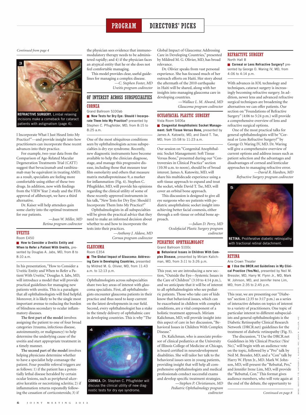
One of the most ubiquitous conditions seen by ophthalmologists across subspe-cialties is dry eye syndrome. Recently, new diagnostic instruments have become available to help the clinician diagnose, stage, and manage this progressive dis-ease, including those that measure tear film osmolarity and others that measure matrix metalloproteinase-9, a marker for inflammation (Fig. 4). Stephen C. Pflugfelder, MD, will provide his opinions regarding the clinical utility of some of these recently approved instruments in his talk, “New Tests for Dry Eye: Should I Incorporate Them Into My Practice?”
Ophthalmologists in all subspecialties will be given the practical advice that they need to make an informed decision about whether to and how to incorporate the tests into their practices.
—Anthony J. Aldave, MDCornea program codirector
GLAUCOMARoom E354n The Global Impact of Glaucoma: Address-ing Care in Developing Countries, presented by Mildred M. G. Olivier, MD, from 11:43 a.m. to 12:13 p.m.
Ophthalmologists across subspecialties share two key areas of interest with glau-coma specialists. First, all ophthalmolo-gists encounter glaucoma patients in their practice and thus need to keep current on the latest developments in our field. Second, every ophthalmologist has a stake in the timely delivery of ophthalmic care in developing countries. This is why “The
Global Impact of Glaucoma: Addressing Care in Developing Countries,” presented by Mildred M. G. Olivier, MD, has broad relevance.
Dr. Olivier speaks from vast personal experience. She has focused much of her outreach efforts on Haiti. Her story about the aftermath of the 2010 earthquake in Haiti will be shared, along with her insights into managing glaucoma care in developing countries.
—Wallace L. M. Alward, MDGlaucoma program codirector
OCULOFACIAL PLASTIC SURGERYVista Room S406an Congenital Anophthalmic Socket Manage-ment: Soft Tissue Versus Bone, presented by James A. Katowitz, MD, and David T. Tse, MD, from 10:58 to 11:23 a.m.
Our session on “Congenital Anophthal-mic Socket Management: Soft Tissue Versus Bone,” presented during our “Con-troversies in Clinical Practice” section (10:30 a.m. to noon), should be of broad interest. James A. Katowitz, MD, will share his multidecade experience using a soft-tissue approach to expand and treat the socket, while David T. Tse, MD, will cover an orbital bone approach.
This discussion is designed to give all eye surgeons who see patients with pe-diatric anophthalmic socket insight into achieving better facial cosmesis, either through a soft-tissue or orbital bone ap-proach.
—Julian D. Perry, MDOculofacial Plastic Surgery program
codirector
PEDIATRIC OPHTHALMOLOGYGrand Ballroom S100cn Behavioral Issues in Children With Com-plex Disease, presented by Miriam Kalich-man, MD, from 3:11 to 3:26 p.m.
This year, we are introducing a new sec-tion, “Outside the Eye—Systemic Issues in the Care of Children” (3:10 to 4:14 p.m.), and we anticipate that it will be of interest to all ophthalmologists who see pediat-ric patients. Those who take care of kids know that behavioral issues, which can be exacerbated in children with complex conditions, are a vital component of a holistic treatment approach. Miriam Kalichman, MD, will provide insight into this aspect of care in her discussion, “Be-havioral Issues in Children With Complex Disease.”
Dr. Kalichman, who is associate profes-sor of clinical pediatrics at the University of Illinois College of Medicine at Chicago, is board certified in neurodevelopment disabilities. She will tailor her talk to the behavioral issues seen in young patients, providing insight that will help all com-prehensive ophthalmologists and medical professionals conduct successful exams and develop specific treatment strategies.
—Stephen P. Christiansen, MDPediatric Ophthalmology program
codirector
REFRACTIVE SURGERYNorth Hall Bn Corneal or Lens Refractive Surgery? pre-sented by George O. Waring IV, MD, from 4:06 to 4:14 p.m.
With advances in IOL technology and techniques, cataract surgery is increas-ingly becoming refractive surgery. In ad-dition, newer lens and advanced refractive surgical techniques are broadening the alternatives we can offer patients. Our section on “Foundations of Refractive Surgery” (4:06 to 5:24 p.m.) will provide a comprehensive overview of lens and refractive surgical options.
One of the most practical talks for general ophthalmologists will be “Cor-neal or Lens Refractive Surgery?” by George O. Waring IV, MD. Dr. Waring will give a comprehensive overview of current options and address issues such as patient selection and the advantages and disadvantages of corneal and lenticular approaches to managing refractive error.
—David R. Hardten, MDRefractive Surgery program codirector
RETINAArie Crown Theatern I Use the DRCR.net Guidelines in My Clini-cal Practice (Yes/No), presented by Neil M. Bressler, MD, Harry W. Flynn Jr., MD, Mark W. Johnson, MD, and Jennifer Irene Lim, MD, from 2:35 to 2:45 p.m.
This year, we are presenting our “Diabe-tes” section (2:35 to 3:17 p.m.) as a series of interactive debates on topics of interest to a variety of practitioners. One topic of particular interest to different subspecial-ists and general ophthalmologists is the Diabetic Retinopathy Clinical Research Network (DRCR.net) guidelines for the treatment of diabetic retinopathy (Fig. 5).
The discussion, “I Use the DRCR.net Guidelines in My Clinical Practice (Yes/No),” will begin with an audience vote on the topic, followed by a “Pro” talk by Neil M. Bressler, MD, and a “Con” talk by Harry W. Flynn Jr., MD. Mark W. John-son, MD, will present the “Rebuttal, Pro,” and Jennifer Irene Lim, MD, will provide the “Rebuttal, Con.” This format gives audience members, who will vote again at the end of the debate, the opportunity to
6 j o i n t m e e t i n g 2 0 1 2
DIRECTORS’ PICKSPROGRAM
bc
sc
, s
ec
tio
n 1
3;
an
th
on
y j
. a
ld
ave,
md
; jo
an
w.
mil
ler
, m
d
Continued from page 4
Continued on page 8
REFRACTIVE SURGERY. Limbal relaxing incisions make a comeback for cataract patients with astigmatism (page 4).
3
CORNEA. Dr. Stephen C. Pflugfelder will discuss the clinical utility of new diag-nostic tests for dry eye syndrome.
4
RETINA. Proliferative diabetic retinopathy with tractional retinal detachment.
5

©2012 Allergan, Inc., Irvine, CA 92612 ™ mark owned by Allergan, Inc. eyebuzz is a registered service mark owned by Eyetechs, Inc. www.allergan.com APC80TC12 122482 Presentation times and speakers are subject to change. This event is not affiliated with the official program of the 2012 Joint Meeting.
Catch the leading experts in eye care at Allergan Booth #1408
FALL INTO THEWINDY CITY
Saturday, November 109:30 am Treatment of Macular Edema Due to Retinal Vein OcclusionShree Kurup, MD
10:00 am Treatment of Allergic ConjunctivitisRajesh Rajpal, MD
10:30 am Management of the Post-operative Cataract Surgery PatientKarl Stonecipher, MD
11:00 amTreatment of HypotrichosisSteve Yoelin, MD
12:00 pm Detecting and Managing Glaucoma ProgressionLouis B. Cantor, MD
12:30 pm RESCUE ME!—Interactive CasesRobert Osher, MD
1:00 pm IOP Lowering: Options for Starting or Replacing TherapyJonathan Myers, MD
1:30 pm Conquering Capsule Complications— Strategies for Complicated CataractsDavid Chang, MD
2:00 pm Treatment of Macular Edema Due to Retinal Vein Occlusion Ron Gallemore, MD, PhD
3:00 pm Focus on Dry Eye DiseaseChristopher Starr, MD, FACS
3:30 pmMaking Social Media “Work” for Your PracticeJoe Casper, MBA, COE, OCS, Senior Eye Care Business Advisor, Allergan, Inc. Eric Abrantes, Marketing Director, Advanced Eye Centers
Sunday, November 119:30 am Management of the Small Pupil in Cataract SurgeryEric Donnenfeld, MD, FACS
10:30 am REFRESH OPTIVE™ AdvancedMarguerite McDonald, MD, FACS
11:00 am IOP Reduction With Adjunctive TherapyNathan Radcliffe, MD
12:00 pm Treatment of HypotrichosisSteve Yoelin, MD
1:00 pm A Versatile Option in Adjunctive IOP Lowering E. Randy Craven, MD
1:30 pm Treatment of Macular Edema Due to Retinal Vein OcclusionMichael Singer, MD
2:00 pmHealthcare Reform: What Every Practice Should KnowMike Driscoll, OCS, Eye Care Business Advisor, Allergan, Inc.Jeffrey Lemay, Director, Healthcare Reform Initiative, Allergan, Inc.
3:00 pmAdventures in DarknessTom Sullivan
Monday, November 129:30 am Protecting Your Practice From Theft: Lessons LearnedJill Maher, MA, OCS, Eye Care Business Advisor, Allergan, Inc.
11:00 am Successful Strategies for Effective EMR ImplementationSherri Boston, MBA, COE, OCS, Eye Care Business Advisor, Allergan, Inc.Jane T. Shuman, COT, COE, OCS, EyeTechs and eyebuzz ®
Jeff Grant, President & Founder, Healthcare Management & Automation Systems, Inc.
12:30 pm Why You Can’t Ignore Social Media: As Featured in Ophthalmology ManagementGreg Raeman, COE, CCOA, OCS, Eye Care Business Advisor, Allergan, Inc.
2:00 pmKeys to Attracting & Managing Talented EmployeesJim Rienzo, OCS, Senior Eye Care Business Advisor, Allergan, Inc.Tom Pannullo, COO, Ophthalmic Consultants of Long Island
122482 AAO News Ad_ST.indd 1 8/28/12 10:48 AM

8 j o i n t m e e t i n g 2 0 1 2
HONORARY LECTURESPROGRAM DIRECTORS’ PICKSPROGRAM
decide whether and how the guidelines fit into their own practices.
—Joan W. Miller, MDRetina program codirector
UVEITISRoom E450n How to Orchestrate Comanagement for Im-munomodulatory Therapy, presented by Jus-tine R. Smith, MD, from 8:20 to 8:30 a.m.
Given the dearth of fellowship-trained uveitis experts, ophthalmologists across the country are finding themselves in a challenging predicament: They know their patient needs immunomodulatory therapy, but there is no ocular immunolo-gist or fellowship-trained uveitis special-ist in the area who can serve as a referral source. In many instances, these oph-thalmologists will seek a rheumatologist or another specialist who is comfortable with prescribing steroid-sparing drugs. In her presentation, “How to Orchestrate Comanagement for Immunomodulatory Therapy,” Justine R. Smith, MD, will pro-vide valuable guidelines for those situa-tions in which rheumatologists and other specialists are monitoring patients for systemic toxicity while the ophthalmolo-gist checks on ocular health (Fig. 6).
—C. Stephen Foster, MDUveitis program codirector
EXCITING DEVELOPMENTS
CORNEAGrand Ballroom S100abn Cross-Linking in the Year 20/20: What Will the Future Hold? presented by Theo Seiler, MD, PhD, from 2:10 to 2:30 p.m.
One of the most exciting advances in the field of cornea in the last several years is the introduction of corneal collagen cross-linking (CXL), a technique that uses ultraviolet light and a photosensitizer to strengthen chemical bonds in the cornea and prevent the progression of ectasia. Theo Seiler, MD, PhD, will give the Dohl-man Lecture during the Cornea Subspe-cialty Day. Dr. Seiler, who will present
“Cross-Linking in the Year 20/20: What Will the Future Hold?” is credited for hav-ing developed the procedure in the late 1990s, along with Eberhard Spoerl, PhD.
This is a talk that should not be missed, as we will hear from a true vision-ary on what he sees as the evolving role of CXL over the next decade. Dr. Seiler will discuss a variety of aspects of the pro-cedure, including its use for traditional indications such as progressive keratoco-nus and ectasia following keratorefractive surgery, as well as expanding indications such as infectious keratitis.
—Anthony J. Aldave, MDCornea program codirector
GLAUCOMARoom E354n The Glaucoma Filtration Device Mini-Shunt Has Been a Positive Development, presented by Marlene R. Moster, MD, from 1:57 to 2:03 p.m.n The Glaucoma Filtration Device Mini-Shunt: I Don’t Get It, presented by Robert M. Feldman, MD, from 2:03 to 2:09 p.m.
Each year, discussions focused on glau-coma surgeries constitute an exciting aspect of Subspecialty Day. This year, a talk by Marlene R. Moster, MD, “The Glaucoma Filtration Device Mini-Shunt Has Been a Positive Development,” will focus on the positives of the ExPress device, which shunts aqueous from the anterior chamber to a subconjunctival reservoir in a manner similar to trabecu-lectomy without removing any sclera or iris tissue. Dr. Moster uses the ExPress in her practice to help manage patients with uncontrolled glaucoma. In contrast, Rob-ert M. Feldman, MD, will offer a counter-point in his presentation, “The Glaucoma Filtration Device Mini-Shunt: I Don’t Get It,” exploring whether this device adds genuine value.
We expect this approach to our section on glaucoma surgery (1:30 to 3:10 p.m.) will not only introduce participants to the latest developments in the field but also help them decide whether or not they would be better off using them.
—Wallace L. M. Alward, MDGlaucoma program codirector
OCULOFACIAL PLASTIC SURGERYVista Room S406an Increasingly Routine Use of Thyroid- Stimulating Immunoglobulin, Müller Muscle–Conjunctival Resection, Semicircular Flaps, and Stentless Dacryocystorhinostomy, pre-sented by Stuart R. Seiff, MD, from 8:14 to 8:24 a.m.
Our section, “Lessons From the Masters: What I Am Doing Differently Today” (8:02 to 9:01 a.m.) spotlights several exciting developments in the field of ocu-lofacial plastic surgery.
Stuart R. Seiff, MD, a past president of the American Society of Ophthalmic Plastic & Reconstructive Surgery, will be discussing his current practice and what he is doing differently today. The title
of his talk, “Increasingly Routine Use of Thyroid-Stimulating Immunoglobulin, Müller Muscle–Conjunctival Resection, Semicircular Flaps, and Stentless Dacryo-cystorhinostomy,” illustrates the breadth of his discussion. Each of these topics will reveal the insights of a master clinician into treatment of common conditions.
—Julian D. Perry, MDOculofacial Plastic Surgery program
codirector
PEDIATRIC OPHTHALMOLOGYGrand Ballroom S100cn Gene Therapy for Inherited Retinal Dis-eases, presented by Edwin M. Stone, MD, PhD, from 2:17 to 2:24 p.m.
We are excited to have Edwin M. Stone, MD, PhD, who has been at the forefront of ocular genetics for years, speak on “Gene Therapy for Inherited Retinal Diseases” in our “Emerging Technologies in Pediatric Ophthalmology and Strabis-mus” section (1:40 to 2:40 p.m.).
One goal of treating blinding inher-ited retinal disease is to genetically alter the retina so that children can preserve function over the course of a lifetime. In addition, it is hoped that genetic therapy will eventually be able to reverse disease to recover function. Although this work is still in its infancy, Dr. Stone will shed light on the latest advances and show meeting attendees where the field is heading.
—Stephen P. Christiansen, MDPediatric Ophthalmology program
codirector
REFRACTIVE SURGERYNorth Hall Bn Can Laser Refractive Lens Surgery Really Work for You and Your Practice? presented by Stephen G. Slade, MD, from 3:11 to 3:19 p.m.n Technique Pearls for Success in Laser Re-fractive Lens Surgery, presented by William W. Culbertson, MD, from 2:07 to 2:15 p.m.
No recent innovation has the potential to change the field of refractive surgery as much as the use of lasers does. A few years ago, the topic was covered in one five-minute presentation. This year, we are devoting 90 minutes to the field (1:59 to 3:30 p.m.).
In his talk, “Can Laser Refractive Lens Surgery Really Work for You and Your Practice?” Stephen G. Slade, MD, will focus on understanding how this new technology fits in patient care. Femtosec-ond lasers have the potential to revolu-tionize lens refractive surgery, offering more precise and reproducible incisions. However, according to presenter William W. Culbertson, MD, achieving the full benefits is dependent upon mastering surgical technique. In his talk, “Technique Pearls for Success in Laser Refractive Lens Surgery,” Dr. Culbertson will share in-sights from his extensive experience with femtosecond technology.
—David R. Hardten, MDRefractive Surgery program codirector
RETINAArie Crown Theatern Intraoperative OCT: Is It Actually Useful? presented by Sunil K. Srivastava, MD, from 11:01 to 11:08 a.m.n Simple Estimation of Fluid Volumes in Neovascular AMD, presented by Alexander C. Walsh, MD, from 11:15 to 11:22 a.m.n Practical Approaches to Needle Biopsy and Genetic Diagnosis for Ocular Melanoma, presented by Thomas M. Aaberg Jr., MD, from 11:49 to 11:56 a.m.
Two exciting areas will be explored at Retina Subspecialty Day this year. The first is our “Imaging” section (10:40 to 11:41 a.m.), which will highlight two relatively new aspects of optical coherence tomography: “Intraoperative OCT: Is It Actually Useful?” presented by Sunil K. Srivastava, MD, and “Simple Estimation of Fluid Volumes in Neovascular AMD,” presented by Alexander C. Walsh, MD.
The “Oncology” section (11:40 a.m. to 12:30 p.m.) will also feature advances in our field, including information on the genetics of tumors. Thomas M. Aaberg Jr., MD, will present “Practical Approaches to Needle Biopsy and Genetic Diagnosis for Ocular Melanoma,” to give attendees a glimpse of a future that includes truly personalized treatment and adjuvant therapy options for patients.
—Joan W. Miller, MDRetina program codirector
UVEITISRoom E450n Systemic Pharmacologic Agents in De-velopment for Uveitis: Voclosporin, Adalim-umab, and AIN457, presented by Eric Suhler, MD, from 4:40 to 4:50 p.m.
One of the most exciting developments in uveitis—new drugs in development—will be covered by Eric Suhler, MD, in his talk, “Systemic Pharmacologic Agents in Development for Uveitis: Voclosporin, Adalimumab, and AIN457.”
Voclosporin is a calcineurin inhibi-tor that appears to be more potent and less toxic than cyclosporine. Researchers have completed a trial with the drug, and a replicate study is being wrapped up. If the drug proves efficacious, as it was in the first study, then it is expected to be the first systemic drug to be approved by the FDA for treating uveitis.
Adalimumab, which inhibits tumor necrosis factor-alpha, is currently being used to treat rheumatoid arthritis, anky-losing spondylitis, and psoriatic arthritis. Researchers have mounted a controlled trial to determine whether the drug can effectively treat posterior uveitis.
Finally, AIN457 is a monoclonal antibody that neutralizes interleukin-17, a proinflammatory cytokine secreted by activated T cells. This drug, which can be given intravenously or subcutaneously, is in clinical trials for posterior uveitis.
—C. Stephen Foster, MDUveitis program codirector c
. s
teph
en
fo
ster
, m
d
Continued from page 6
UVEITIS. This patient with uveitis and scleritis has dilated iris blood vessels, posterior synechiae, and a hazy view back through the pupil, thanks to a developing cataract and inflammatory cells in the vitreous body. Inappropriate, chronic use of corticosteroid therapy led to the cataract formation.
6

Brief Summary of the Prescribing Information for ZIOPTAN.
INDICATIONS AND USAGE
ZIOPTAN is indicated for reducing elevated intraocular pressure in patients with open-angle glaucoma or ocular hypertension.
DOSAGE AND ADMINISTRATION
The recommended dose is 1 drop of ZIOPTAN in the conjunctival sac of the affected eye(s) once daily in the evening.
The dose should not exceed once daily since it has been shown that more frequent administration of prostaglandin analogs may lessen the intraocular pressure-lowering effect.
Reduction of the intraocular pressure starts approximately 2 to 4 hours after the first administration with the maximum effect reached after 12 hours.
ZIOPTAN may be used concomitantly with other topical ophthalmic drug products to lower intraocular pressure. If more than 1 topical ophthalmic product is being used, each 1 should be administered at least 5 minutes apart.
The solution from 1 individual unit is to be used immediately after opening for administration to 1 or both eyes. Since sterility cannot be maintained after the individual unit is opened, the remaining contents should be discarded immediately after administration.
CONTRAINDICATIONS
None.
WARNINGS AND PRECAUTIONS
PigmentationTafluprost ophthalmic solution has been reported to cause changes to pigmented tissues. The most frequently reported changes have been increased pigmentation of the iris, periorbital tissue (eyelid), and eyelashes. Pigmentation is expected to increase as long as tafluprost is administered. The pigmentation change is due to increased melanin content in the melanocytes rather than to an increase in the number of melanocytes. After discontinuation of tafluprost, pigmentation of the iris is likely to be permanent, while pigmentation of the periorbital tissue and eyelash changes have been reported to be reversible in some patients. Patients who receive treatment should be informed of the possibility of increased pigmentation. The long-term effects of increased pigmentation are not known.
Iris color change may not be noticeable for several months to years. Typically, the brown pigmentation around the pupil spreads concentrically towards the periphery of the iris and the entire iris or parts of the iris become more brownish. Neither nevi nor freckles of the iris appear to be affected by treatment. While treatment with ZIOPTAN can be continued in patients who develop noticeably increased iris pigmentation, these patients should be examined regularly. [See Patient Counseling Information.]
Eyelash Changes ZIOPTAN may gradually change eyelashes and vellus hair in the treated eye. These changes include increased length, color, thickness, shape, and number of lashes. Eyelash changes are usually reversible upon discontinuation of treatment.
Intraocular InflammationZIOPTAN should be used with caution in patients with active intraocular inflammation (eg, iritis/uveitis) because the inflammation may be exacerbated.
Macular EdemaMacular edema, including cystoid macular edema, has been reported during treatment with prostaglandin F2 analogs. ZIOPTAN should be used with caution in aphakic patients, in pseudophakic patients with a torn posterior lens capsule, or in patients with known risk factors for macular edema.
ADVERSE REACTIONS
Clinical Studies ExperienceBecause clinical studies are conducted under widely varying conditions, adverse reaction rates observed in the clinical studies of a drug cannot be directly compared to rates in the clinical studies of another drug and may not reflect the rates observed in practice.
Preservative-containing or preservative-free tafluprost 0.0015% was evaluated in 905 patients in 5 controlled clinical studies of up to 24-months’ duration. The most common adverse reaction observed in patients treated with tafluprost was conjunctival hyperemia which was reported in a range of 4% to 20% of patients. Approximately 1% of patients discontinued therapy due to ocular adverse reactions.
Ocular adverse reactions reported at an incidence of ≥2% in these clinical studies included ocular stinging/irritation (7%), ocular pruritus including allergic conjunctivitis (5%), cataract (3%), dry eye (3%), ocular pain (3%), eyelash darkening (2%), growth of eyelashes (2%), and blurred vision (2%).
Nonocular adverse reactions reported at an incidence of 2% to 6% in these clinical studies in patients treated with tafluprost 0.0015% were headache (6%), common cold (4%), cough (3%), and urinary tract infection (2%).
Postmarketing ExperienceThe following adverse reactions have been identified during postapproval use of tafluprost. Because postapproval adverse reactions are reported voluntarily from a population of uncertain size, it is not always possible to reliably estimate their frequency or establish a causal relationship to drug exposure.
Eye disorders: iritis/uveitis
In postmarketing use with prostaglandin analogs, periorbital and lid changes, including deepening of the eyelid sulcus, have been observed.
USE IN SPECIFIC POPULATIONS
PregnancyPregnancy Category C.Teratogenic effects: In embryo-fetal development studies in rats and rabbits, tafluprost administered intravenously was teratogenic. Tafluprost caused increases in post-implantation losses in rats and rabbits and reductions in fetal body weights in rats. Tafluprost also increased the incidence of vertebral skeletal abnormalities in rats and the incidence of skull, brain, and spine malformations in rabbits. In rats, there were no adverse effects on embryo-fetal development at a dose of 3 μg/kg/day corresponding to maternal plasma levels of tafluprost acid that were 343 times the maximum clinical exposure based on Cmax. In rabbits, effects were seen at a tafluprost dose of 0.03 μg/kg/day corresponding to maternal plasma levels of tafluprost acid during organogenesis that were approximately 5 times higher than the clinical exposure based on Cmax. At the no-effect dose in rabbits (0.01 μg/kg/day), maternal plasma levels of tafluprost acid were below the lower level of quantification (20 pg/mL).
In a pre- and postnatal development study in rats, increased mortality of newborns, decreased body weights, and delayed pinna unfolding were observed in offsprings. The no observed adverse effect level was at a tafluprost intravenous dose of 0.3 μg/kg/day, which is greater than 3 times the maximum recommended clinical dose based on body surface area comparison.
There are no adequate and well-controlled studies in pregnant women. Although animal reproduction studies are not always predictive of human response, ZIOPTAN should not be used during pregnancy unless the potential benefit justifies the potential risk to the fetus.
Women of childbearing age/potential should have adequate contraceptive measures in place.
Nursing MothersA study in lactating rats demonstrated that radio-labeled tafluprost and/or its metabolites were excreted in milk. It is not known whether this drug or its metabolites are excreted in human milk. Because many drugs are excreted in human milk, caution should be exercised when ZIOPTAN is administered to a nursing woman.
Pediatric UseUse in pediatric patients is not recommended because of potential safety concerns related to increased pigmentation following long-term chronic use.
Geriatric UseNo overall clinical differences in safety or effectiveness have been observed between elderly and other adult patients.
PATIENT COUNSELING INFORMATION
See FDA-Approved Patient Labeling (Patient Information).
Nightly ApplicationPatients should be advised to not exceed once-daily dosing since more frequent administration may decrease the intraocular pressure-lowering effect of ZIOPTAN.
Handling the Single-Use ContainerPatients should be advised that ZIOPTAN is a sterile solution that does not contain a preservative. The solution from 1 individual unit is to be used immediately after opening for administration to 1 or both eyes. Since sterility cannot be maintained after the individual unit is opened, the remaining contents should be discarded immediately after administration.
Potential for Pigmentation Patients should be advised about the potential for increased brown pigmentation of the iris, which may be permanent. Patients should also be informed about the possibility of eyelid skin darkening, which may be reversible after discontinuation of ZIOPTAN.
Potential for Eyelash ChangesPatients should also be informed of the possibility of eyelash and vellus hair changes in the treated eye during treatment with ZIOPTAN. These changes may result in a disparity between eyes in length, thickness, pigmentation, number of eyelashes or vellus hairs, and/or direction of eyelash growth. Eyelash changes are usually reversible upon discontinuation of treatment.
When to Seek Physician AdvicePatients should be advised that if they develop a new ocular condition (eg, trauma or infection), experience a sudden decrease in visual acuity, have ocular surgery, or develop any ocular reactions, particularly conjunctivitis and eyelid reactions, they should immediately seek their physician’s advice concerning the continued use of ZIOPTAN.
Use with Other Ophthalmic DrugsIf more than 1 topical ophthalmic drug is being used, the drugs should be administered at least five (5) minutes between applications.
Storage InformationPatients should be instructed on proper storage of cartons, unopened foil pouches, and opened foil pouches [see How Supplied/Storage and Handling]. Recommended storage for cartons and unopened foil pouches is to store refrigerated at 2-8°C (36-46°F). After the pouch is opened, the single-use containers may be stored in the opened foil pouch for up to 28 days at room temperature: 20-25°C (68-77°F). Protect from moisture.
For more detailed information, please read the Prescribing Information.
Rx only.
Manufactured for: Merck Sharp & Dohme Corp., a subsidiary of
Whitehouse Station, NJ 08889, USA
Manufactured by: Laboratoire UnitherZI de la GuerieF-50211 COUTANCES CedexFrance
Revised: 08/2012
USPI-OS-24521207R003
ZIOPTANTM (tafluprost ophthalmic solution) 0.0015%
Copyright © 2012 Merck Sharp & Dohme Corp., a subsidiary of Merck & Co., Inc. All rights reserved. OPHT-1044142-0013 09/12
OPHT-1044142-0013.indd 2 9/27/12 9:39 AM

ASIA-PACIFIC
OVERVIEWAPAO
BECOMING A REALITY. The APAO was formed from one man’s dream. More than 50 years ago, Dr. William John Holmes called on Asian ophthalmic leaders to unite, as he realized that eye surgeons in Asia, Australasia, and Oceania were faced with the same ophthalmologi-cal problems as their U.S. and European counterparts. A Hungarian ophthalmolo-gist living in Hawaii, Dr. Holmes believed
that an organization should be created to foster closer relationships among ophthalmologists and ophthalmological societies in the Asia-Pacific region to bet-ter combat preventable blindness.
A response to his call came in Sep-tember 1958. At the 19th International Congress of Ophthalmology in Brussels, a decision was made to form the APAO. A month later, Dr. Holmes met with a num-
ber of ophthalmic leaders, including Drs. Germiniano de Ocampo and Jesus Tamesis from the Philippines and Dr. Robert F. Lowe from Australia, and drew up the organization’s con-stitution. Their en-
deavors also paved the way for the APAO’s first meeting, a four-day congress held in Manila, Philippines, in October 1960.
MEETING THE DEMANDS OF A VAST REGION. Since its inception, the APAO has sought to promote the science and art of oph-thalmology in the Asia-Pacific region; eliminate preventable blindness through teaching, research, and service; foster cooperation among various ophthalmo-logical societies in different countries; and encourage collaboration with other international and regional ophthalmo-logical organizations. Accordingly, the APAO continues to organize congresses and co-organizes and promotes scientific meetings and conferences.
To meet the increasing demands of ophthalmologists and visual scientists in the Asia-Pacific region, the APAO moved from holding its congress once every four years to every two years in 1972. In 2006, the congress became an annual event to further advance ophthalmology in the region.
That year also marked the APAO’s first collaboration with the Academy—the AAO-APAO Joint Meeting in Las Vegas. The APAO is glad that the first joint meeting was well received and that we now have the opportunity to present this year’s Joint Meeting in Chicago. We are fortunate to have the support of over 130 leading eye experts in organizing three joint symposia and more than 20 instruc-tion courses.
Early next year, the American Academy of Ophthalmology will join together with the International Council of Ophthal-mology and the South Asian Association for Regional Cooperation Academy of Ophthalmology to cosponsor the 28th APAO Congress in Hyderabad, India. With closer collaboration among vari-ous ophthalmological organizations, the APAO hopes ophthalmologists and visual scientists worldwide will have more and more platforms for learning, teaching, meeting, and networking.
ADVANCING APAO’S MISSION THROUGH PUBLICATION AND RESEARCH. Another major endeavor of the APAO is its initia-tive to establish a three-tier publication system.
JOURNAL. The APAO’s official publica-tion, the Asia-Pacific Journal of Ophthal-mology (APJO), was launched in January 2012. Published online every two months, the APJO is a peer-reviewed scientific journal representing the ophthalmic and visual-science developments of the Asia-Pacific region and beyond. Covering as many as 16 subspecialties, the APJO pub-lishes original studies, clinically relevant laboratory investigations, and review articles, as well as perspectives on new technologies, critical issues, and models that are worth replicating in the region. The APJO is grateful for the support it has received from various eye experts who have agreed to join its Advisory and Editorial Board and have chosen it as their publishing platform. It is the APAO’s hope that we can continue to push for-ward to the frontiers of ophthalmology through publication and research.
NEWSPAPER. Also launched in January 2012, the Ocular Surgery News—APAO
PRESERVING AND PROTECTING VISION IN THE
GETTING TO KNOW THE APAO. The area served by the
Asia-Pacific Academy of Ophthalmology (APAO) is home to more than half of
the world’s population and more than half of the world’s visually impaired.
The organization formed in 1958 to meet the region’s unique education and
training needs, with the principal objective of fostering closer relations
among ophthalmologists and ophthalmological societies in the fight against
vision loss. Today, the APAO continues to serve as a platform to promote
the restoration of sight and the prevention of blindness through service,
research, and teaching. Member societies include those in Australia, Ban-
gladesh, Cambodia, China, Chinese Taipei, Hong Kong, India, Indonesia,
Japan, Malaysia, Mongolia, Nepal, New Zealand, Pakistan, the Philippines,
Singapore, South Korea, Sri Lanka, Thailand, and Vietnam. This article takes
a look at the organization’s past, present, and future.
FROM PAKISTAN TO JAPAN
e y e n e t ’ s a c a d e m y n e w s 11
by dennis s. c. lam, md, and frank j. martin, md
Inauguration of the APAO in Manila, Philippines, 1960.
Asia-Pacific Journal of Ophthalmology and Ocular Surgery News—APAO Edition, the APAO’s official journal and newspaper.
Asia-PacificJournal of
Ophthalmologywww.apjo.org

12 j o i n t m e e t i n g 2 0 1 2
(OSN-APAO) Edition is the APAO’s of-ficial newspaper. It is published online 10 times a year as a joint venture with Slack and provides ophthalmologists and visual scientists with the latest ophthalmic news in the form of leisure reading. There are also a few pages dedicated to report-ing the latest news at the APAO. It is the organization’s hope that the OSN-APAO Edition will facilitate communication between the APAO and its members.
NEWSLETTER. Finally, the APAO launched its official monthly newsletter in Octo-ber with a similar desire: to forge closer connections among its member societies. Not only does the newsletter report on APAO news but also member societies are invited to share news and activities from their own nations.
LEADING THE WAY FOR THE FUTURE. The APAO is also dedicated to nurturing fu-ture leaders in ophthalmology through its leadership development program (LDP). The Academy introduced the LDP to the APAO in 2007; and, after years of plan-ning, the first LDP class had its orienta-tion sessions during the 2009 APAO-AAO Congress in Bali, Indonesia, and graduat-ed during the 2010 APAO-AAO Congress in Beijing. The APAO is now organizing its fourth LDP class and has already of-fered training to nearly 70 future leaders who actively engage in the society affairs of either the APAO or its member societ-ies. With the support of these leaders, the first Asia-Pacific Eye Care Week was held in January 2012 to raise public awareness about the importance of eye care and eye health.
Other APAO plans under way include an expansion of its online education efforts, the second Asia-Pacific Eye Care Week, the launch of the APAO Fellowship Program, a residency curriculum com-mitted to strengthening and augmenting postgraduate training for eye care profes-sionals, and a visiting scholar program.
For more information about the APAO and details regarding future events and programs, be sure to stop by the APAO’s booth (#1200) in Chicago or visit www.apaophth.org.
OVERVIEWAPAO
This year’s Joint Meeting of the Acad-emy and the APAO promises to give Academy members a rare opportunity to learn firsthand about eye diseases and pathologies unique to the regions served by the APAO.
Visit the online Program Search at www.aao.org/2012 for specific details about APAO symposia and instruc-tion courses. Be sure to select “APAO Sponsored” under the Special Interest dropdown menu.
APAO IN CHICAGO
ABOUT THE AUTHORS
This past April, APAO Secretary-General Dennis S. C. Lam, MD (left), and APAO President Frank J. Martin, MD (right), met with Academy Past President Rich-ard L. Abbott, MD, during the APAO/European Society of Ophthalmology Joint Meeting in Busan, Korea, to discuss fu-ture collaborations.
2 9 7 5 B r o t h e r B l v d B a r t l e t t T N 3 8 1 3 3 U S A 8 8 8 . 9 0 5 . 7 7 7 0 o d y s s e y m e d . c o m
© 2012 Odyssey Medical, Inc. All rights reserved. †McCabe, C. (2009). Punctal occlusion reduces dry eye symptoms and improves vision. Review of Ophthalmology, 16(11), 55-58 *Certain conditions apply; call for details.
S i m p l e S i z i n g e a S y i n S e r t i o n g u a r a n t e e D r e t e n t i o n *
Treat your patients with the Parasol punctal occluder, the permanent application
for chronic dry eye.
PARASOL®
92% Retention Rate†
ORDER NOW
Odyssey_AppAd-AN.indd 1 9/14/12 12:33 PM
Dennis S. C. Lam, MD, is secretary-general of the APAO, editor-in-chief of the Asia-Pacific Journal of Ophthalmology, and director of the State Key Laboratory and honorary director of Zhongshan Oph-thalmic Center at Sun Yat-Sen University in Guangzhou, China.Frank J. Martin, MD, is president of the APAO and clinical professor of ophthal-mology and pediatrics and child health at the University of Sydney.
As they return to Washington, D.C., following Tuesday’s elections, federal legislators face major challenges, including passing a Medicare physician pay fix to derail threatened cuts in reimbursement. The Academy is pushing members of Congress to provide oph-thalmologists with a stable, fair pay solution that ensures patients’ continued access to quality eye care. Please visit the Advocacy desk in the Resource Center (Booth 508) to send a letter to your representative and senators urging swift passage of Medicare pay reforms.
In a special session, the 2013 Medicare Update (Spe11), Academy leaders will provide updates on topics including the various Medicare incentive programs for physician reim-bursement. Hear about changes that will impact payments and the latest on Medicare’s Physician Quality Reporting System and e-Rx programs. When: Sunday, 12:15-1:45 p.m. Where: Grand Ballroom S100c.
THE F IGHT FOR MED ICARE PHYS IC IAN PAY F IX

14 j o i n t m e e t i n g 2 0 1 2
VENDORSEXHIBITS
Ophthalmology EHR Checklist. In 2011, the Academy’s Medical Information Technology Committee published “Spe-cial Requirements for Electronic Health Record Systems in Ophthalmology.” The article included a checklist of 23 EHR features—17 of them deemed essential; six desirable—for accommodating the workflow and data management needs of
ophthalmology practices. As a service to members, the committee contacted EHR vendors earlier this fall to ask which of these features their products currently offer. The checklist and vendor responses appear on pages 16 and 17. When you visit these vendors in the exhibit hall, ask for a demonstration of the features on the checklist that you consider the most valu-
Which EHR Systems COMPARE EHR SYSTEMS. If you plan to research EHR systems
in the exhibit hall, consider using the table below and on pages 16 and 17
to prepare before meeting with vendors. They provide useful compara tive
information that will help you zero in on the systems most likely to meet your
needs and may spark questions of your own. (Not all of the vendors included
in the tables have a booth in the exhibit hall.)
ELECTRONIC HEALTH RECORDS
COMPANY NAME Compulink Business Solutions
EMRlogic Epic Eyefinity EyeMD EMR First Insight GE Healthcare
Health Care Intranet Technolo-
gies (HCIT)
ifa systems
iMedicWare Insight Software
Integrity Digital
Solutions
IO Practice-
ware
Keiser Computers
KeyMedical Software
MD-Intelle-
Sys
MDoffice Medflow NexTech NextGen Healthcare
Penn Medical Informatics Systems
SRSsoft VersaSuite
PRODUCT NAME AND VERSION Advantage/ EHR (V 10)
ActivEHR (V 2012.2)
Epic 2012
OfficeMate/ExamWRITER
(V 10.6)
EyeMD EMR (V 1.1.1.3)
MaximEyes SQL (V 1.1.0.0)
Centricity CPS
(V 10.1)
HCIT Total Eye/Retina+
(V 6.1)
ifa (V 6.3)
iDoc (V 5.0)
My Vision Express (V 10.0)
Integrity EMR for Eyes
(V 3.7)
IO Prac-ticeware (V 7.1)
Drs Enterprise
(V 9.0)
KeyChart (V 4.0)
Intelle-Chart
(V 6.3)
MDoffice (V 8.10.12)
Medflow (V 7.6.3)
NexEMR (V 10.1)
NextGen Ambu-latory
(V 8.0.1)
EyeDoc EMR (V 9.7)
SRS EHR (V 8)
VersaSuite (V 8.1)
GENERAL INFORMATIONModules available (PMS for practice management system, EP for electronic prescribing)
PMS, EP PMS, EP PMS, EP PMS, EP PMS, EP PMS, EP PMS, EP PMS, EP PMS, EP PMS, EP EP PMS, EP EP PMS, EP EP PMS, EP PMS, EP PMS, EP PMS, EP PMS, EP PMS, EP PMS, EP
Do you offer an application server provider (ASP) option?
Y Y Y N N Y Y N Y Y Y Y Y Y Y Y Y Y Y Y Y Y
How long have you been in the ophthalmology elec-tronic health record market? (in years)
18 2 20 3.5 18 5 14 26 7 12 3 11 10 10 9 8 14 6 18 15 15 17
How many ophthalmology electronic health record practice installations have you completed?
851 5 400 153 110 100+ 160 19 151 50 200 50+ 140 495 600 55+
• Academic 1 0 0 0 15 3 1 1 0 0 4 0 14 2 2
• Private practice 850 5 400 153 110 100+ 157 18 150 50 200 50+ 180 140 481 55+ 50
How many total ophthalmologists are represented by those installations listed above?
2,000+ 7 358 350-400 200+ 500+ 220 120 700 80+ 600+ 420 2,700 > 4,000 * 325+ 162
Do you offer an ASC module? Y Y N N N Y Y Y Y Y Y Y Y Y N Y Y Y Y Y N Y
Do you offer an optical module? Y Y Y Y Y Y N N Y Y N Y Y Y Y Y Y Y Y Y N Y
Does your contract guarantee your software will meet future Meaningful Use criteria (Stage 2 and Stage 3)?
Y Y Y N (plan to meet)
Y Y Y Y Y Y Y Y N (plan to meet)
Y Y Y Y Y Y Optional Y Y
How many of your ophthalmologist customers have attested to Meaningful Use Stage 1 successfully?
0 60 40+ 100 110 130 22 2 336 750 200 104 15
Have you provided AAOE with an IHE Eye Care Inte-gration Statement?
Y N N Y N N N Y N Y N Y N N N Y Y N N N Y
Is 24-hour support available? Y Y N Y N Y Y Y N Y Y Y Y Y Y N Y Y Y Y Y Y
What are your standard support hours during the week?
5 a.m.–5:30 p.m. PT
5 a.m.– 8 p.m. PT
6 a.m.– 6 p.m. PT
8:30 a.m.– 5:30 p.m. ET
5 a.m.–5 p.m. PT
8 a.m.– 5 p.m. ET
8:30 a.m.– 5:30 p.m. ET
7 a.m.–7 p.m. CT
7 a.m.– 7:30 p.m.
ET
8 a.m.– 7 p.m. ET
24 hours 24 hours 8:30 a.m. –6 p.m. ET
8 a.m.– 8 p.m. ET
24 hours 8 a.m.– 8 p.m. ET
7:30 a.m.– 9 p.m. ET
7 a.m.– 9 p.m. ET
8:30 a.m.– 8:30 p.m.
ET
24 hours 24 hours
24 hours
Booth number 2315 3039, Kowa Optimed;
2340, Wal-man Instru-ment (dis-tributors)
N/A 344 357 4417 1676, Virtual
Officeware (reseller)
N/A 3862 2969 N/A 4353 3868 158 152 1971 4034 3152 2757 3456 931 3367 1065
Y = Yes, our current product version includes this; N = No, we do not include this feature in current products and have no immediate plan to include;
Blank = No re sponse (the company may provide more information if contacted). * = This figure includes some optometrists.

e y e n e t ’ s a c a d e m y n e w s 15
able to your practice. Product Specifics. The committee also
asked vendors about other features (be-low) that ophthalmologists might want to know about, including the following:• Techsupporthours.• WhetherthevendorhassuppliedtheAcademy with an IHE Eye Care Integra-tion Statement. (Physicians/offices should
review the statement to make sure that their needs for interoperability are ad-dressed).• Howmanycurrentcustomerswereable to successfully attest to Meaning-ful Use Stage 1 with the system. (Note that the number of ophthalmologists represented by EHR installations ex-ceeds the number of ophthalmologists
implementing EHRs. This may be due to double-counting of ophthalmologists who practice in multiple locations or who hold concurrent academic medical center and hospital positions as well as private practice positions; or to the inclusion of eye care providers other than ophthal-mologists, etc. Please be sure to look also at the number of installations, and ask the
vendor for references of practices in your area and practices similar to yours.)
DISCLAIMER: All information and claims are
those of the vendors and have not been verified,
nor does the appearance of the product constitute
an endorsement of the company or product by the
American Academy of Ophthalmology, EyeNet
Magazine, or Academy News.
Meet Your Needs?CHECK THE SPECS
COMPANY NAME Compulink Business Solutions
EMRlogic Epic Eyefinity EyeMD EMR First Insight GE Healthcare
Health Care Intranet Technolo-
gies (HCIT)
ifa systems
iMedicWare Insight Software
Integrity Digital
Solutions
IO Practice-
ware
Keiser Computers
KeyMedical Software
MD-Intelle-
Sys
MDoffice Medflow NexTech NextGen Healthcare
Penn Medical Informatics Systems
SRSsoft VersaSuite
PRODUCT NAME AND VERSION Advantage/ EHR (V 10)
ActivEHR (V 2012.2)
Epic 2012
OfficeMate/ExamWRITER
(V 10.6)
EyeMD EMR (V 1.1.1.3)
MaximEyes SQL (V 1.1.0.0)
Centricity CPS
(V 10.1)
HCIT Total Eye/Retina+
(V 6.1)
ifa (V 6.3)
iDoc (V 5.0)
My Vision Express (V 10.0)
Integrity EMR for Eyes
(V 3.7)
IO Prac-ticeware (V 7.1)
Drs Enterprise
(V 9.0)
KeyChart (V 4.0)
Intelle-Chart
(V 6.3)
MDoffice (V 8.10.12)
Medflow (V 7.6.3)
NexEMR (V 10.1)
NextGen Ambu-latory
(V 8.0.1)
EyeDoc EMR (V 9.7)
SRS EHR (V 8)
VersaSuite (V 8.1)
GENERAL INFORMATIONModules available (PMS for practice management system, EP for electronic prescribing)
PMS, EP PMS, EP PMS, EP PMS, EP PMS, EP PMS, EP PMS, EP PMS, EP PMS, EP PMS, EP EP PMS, EP EP PMS, EP EP PMS, EP PMS, EP PMS, EP PMS, EP PMS, EP PMS, EP PMS, EP
Do you offer an application server provider (ASP) option?
Y Y Y N N Y Y N Y Y Y Y Y Y Y Y Y Y Y Y Y Y
How long have you been in the ophthalmology elec-tronic health record market? (in years)
18 2 20 3.5 18 5 14 26 7 12 3 11 10 10 9 8 14 6 18 15 15 17
How many ophthalmology electronic health record practice installations have you completed?
851 5 400 153 110 100+ 160 19 151 50 200 50+ 140 495 600 55+
• Academic 1 0 0 0 15 3 1 1 0 0 4 0 14 2 2
• Private practice 850 5 400 153 110 100+ 157 18 150 50 200 50+ 180 140 481 55+ 50
How many total ophthalmologists are represented by those installations listed above?
2,000+ 7 358 350-400 200+ 500+ 220 120 700 80+ 600+ 420 2,700 > 4,000 * 325+ 162
Do you offer an ASC module? Y Y N N N Y Y Y Y Y Y Y Y Y N Y Y Y Y Y N Y
Do you offer an optical module? Y Y Y Y Y Y N N Y Y N Y Y Y Y Y Y Y Y Y N Y
Does your contract guarantee your software will meet future Meaningful Use criteria (Stage 2 and Stage 3)?
Y Y Y N (plan to meet)
Y Y Y Y Y Y Y Y N (plan to meet)
Y Y Y Y Y Y Optional Y Y
How many of your ophthalmologist customers have attested to Meaningful Use Stage 1 successfully?
0 60 40+ 100 110 130 22 2 336 750 200 104 15
Have you provided AAOE with an IHE Eye Care Inte-gration Statement?
Y N N Y N N N Y N Y N Y N N N Y Y N N N Y
Is 24-hour support available? Y Y N Y N Y Y Y N Y Y Y Y Y Y N Y Y Y Y Y Y
What are your standard support hours during the week?
5 a.m.–5:30 p.m. PT
5 a.m.– 8 p.m. PT
6 a.m.– 6 p.m. PT
8:30 a.m.– 5:30 p.m. ET
5 a.m.–5 p.m. PT
8 a.m.– 5 p.m. ET
8:30 a.m.– 5:30 p.m. ET
7 a.m.–7 p.m. CT
7 a.m.– 7:30 p.m.
ET
8 a.m.– 7 p.m. ET
24 hours 24 hours 8:30 a.m. –6 p.m. ET
8 a.m.– 8 p.m. ET
24 hours 8 a.m.– 8 p.m. ET
7:30 a.m.– 9 p.m. ET
7 a.m.– 9 p.m. ET
8:30 a.m.– 8:30 p.m.
ET
24 hours 24 hours
24 hours
Booth number 2315 3039, Kowa Optimed;
2340, Wal-man Instru-ment (dis-tributors)
N/A 344 357 4417 1676, Virtual
Officeware (reseller)
N/A 3862 2969 N/A 4353 3868 158 152 1971 4034 3152 2757 3456 931 3367 1065
ASC = ambulatory surgical centerIHE = Integrating the Healthcare Enterprise
Data current as of Sept. 4, 2012.

16 j o i n t m e e t i n g 2 0 1 2
VENDORSEXHIBITS
S P E C I A L R E Q U I R E M E N T S F O R E L E C T R O N I C H E A L T H R E C O R D ( E H R ) S Y S T E M S I N O P H T H A L M O L O G YCOMPANY NAME Compulink
Business Solutions EMRlogic Epic Eyefinity
EyeMD EMR
First Insight
GE Healthcare
Health Care Intranet
Technologiesifa
systems iMedicWare Insight
Software
Integrity Digital
Solutions
IO Practice-
ware Keiser
ComputersKeyMedical
Software
MD-Intelle-
Sys MDoffice Medflow NexTech NextGen
Healthcare
Penn Medical Informatics
Systems SRS-soft VersaSuite
PRODUCT NAME AND VERSION Essential (E)Desirable (D)
Advantage/ EHR (V 10)
ActivEHR (V 2012.2)
Epic 2012
OfficeMate/ ExamWRITER
(V 10.6)
EyeMD EMR
(V 1.1.1.3)
MaximEyes SQL
(V 1.1.0.0)
Centricity CPS
(V 10.1)
HCIT Total Eye/Retina+
(V 6.1)
ifa (V 6.3)
iDoc (V 5.0)
My Vision Express (V 10.0)
Integrity EMR for Eyes
(V 3.7)
IO Practiceware
(V 7.1)
Drs Enterprise
(V 9.0)
KeyChart (V 4.0)
Intelle-Chart
(V 6.3)
MDoffice (V 8.10.12)
Medflow (V 7.6.3)
NexEMR (V 10.1)
NextGen Ambulatory (V 8.0.1)
EyeDoc EMR (V 9.7)
SRS EHR (V 8)
VersaSuite (V 8.1)
CLINICAL DOCUMENTATIONEnable entry and storage of all ophthalmology-specific data required to support Academy Preferred Practice Patterns
E Y Y Y Y Y Y Y Y Y Y Y Y Y Y Y Y Y Y Y N Y
Organize ophthalmology-specific elements separately (e.g., past ocular history, ocular medications)
E Y Y Y Y Y Y Y Y Y Y Y Y Y Y Y Y Y Y Y Y Y N Y
Conform or map to vendor-neutral standard terminol-ogies (e.g., SNOMED CT, ICD) to represent problem lists
E Y Y Y Y Y Y Y Y Y Y Y Y Y Y Y Y Y Y Y Y Y Y
Conform or map to RxNorm to represent medications E Y Y Y P Y Y Y Y Y Y Y Y Y P Y Y Y Y Y Y Y Y Y
Conform or map to vendor-neutral standard termi-nologies (e.g., SNOMED CT) to represent:
• Diagnoses and procedures E Y Y Y Y Y Y Y Y Y Y Y Y Y Y Y Y Y Y P Y Y Y Y
• Allergies and clinical findings D Y Y Y Y Y Y Y Y Y Y Y Y Y Y N Y P Y P Y Y Y Y
Enable physicians and technicians to keep multiple records open simultaneously and securely in different rooms, with easy reauthentication
E Y Y Y Y Y Y Y Y Y Y Y Y Y Y Y Y Y Y Y Y Y Y Y
Provide tools for incorporating color drawing, includ-ing ocular templates
E Y Y Y Y Y Y Y Y Y Y Y Y Y Y Y Y Y Y Y Y Y Y Y
Analyze clinical workflow before and after EHR implementation
E Y Y Y N Y Y Y Y Y Y Y Y Y Y Y Y Y Y Y Y Y Y
Exchange full set of ophthalmic clinical data with EHRs from other vendors
D Y Y N N Y Y Y Y Y Y Y P Y Y Y P Y Y Y Y N Y
Link clinical documentation to billing and charge capture and integrate with practice management
D Y Y Y Y Y Y Y Y Y Y Y Y Y P Y Y Y Y Y Y Y N Y
Allow physician to review patient information easily before entering room
D Y Y Y Y Y Y Y Y Y Y Y Y Y Y Y Y Y Y Y Y Y Y Y
OPHTHALMIC VITAL SIGNS AND LABORATORY STUDIESRecord visual acuity and refractive discrete elements in accordance with DICOM Supplement 130
E Y Y Y Y Y Y Y Y Y Y Y Y Y Y Y Y Y P Y Y N Y
Record IOP as a discrete data element E Y Y Y Y Y Y Y Y Y Y Y Y Y Y Y Y Y Y Y Y Y Y Y
Display and graph visual acuity and IOP over time E Y P IOP, Y; VA, N Y Y Y Y Y Y Y Y Y N Y Y Y Y Y Y Y Y Y
MEDICAL AND SURGICAL MANAGEMENTElectronically associate all preoperative, operative, and postoperative documents
E Y P Y Y Y Y Y Y Y Y Y Y Y Y Y Y Y Y Y Y Y N Y
Support documentation of office-based and operat-ing room procedures
E Y Y Y N Y Y Y Y Y Y Y Y Y Y Y Y Y Y Y Y Y N Y
Allow physician to generate operative report at time of surgery
D Y Y Y N Y Y Y Y Y Y P Y Y Y Y Y Y Y Y Y Y Y Y
OPHTHALMIC MEASUREMENT AND IMAGING DEVICESConform to vendor-neutral standards (e.g., DICOM) for receipt and representation of data from all oph-thalmic instruments and devices
E Y Y Y P P P Y P Y Y Y Y Y N Y Y Y Y Y Y Y Y Y
Conform to vendor-neutral standards and profiles for ordering ophthalmic imaging and measurement stud-ies (e.g., DICOM Modality Worklist and IHE Eye Care Workflow)
E Y Y Y P P P Y P Y Y Y Y Y N Y P Y Y P Y Y Y Y
Document completion and interpretation of ophthal-mic imaging and measurement studies
E Y Y Y Y Y Y Y P Y Y Y Y Y Y Y Y Y Y Y Y Y Y Y
Request, retrieve, display, and communicate all im-aging and measurement data generated by ophthal-mic instruments in a standard vendor-neutral format (e.g., DICOM)
E Y Y Y Y P P Y P Y Y Y Y Y Y Y Y Y Y Y Y Y Y
Manage all ophthalmic imaging data in vendor-neu-tral format (e.g., DICOM), or provide tight integration with external PACS in vendor-neutral format
D Y Y Y N P P Y P Y Y Y Y Y Y Y Y Y Y Y Y Y Y
Items are classified either as “Essential” (E) for current systems or as “Desirable” (D) for current systems and essential for future systems.
Certification by the Office of the National Coordinator for Meaningful Use as an EHR system is a given essential.
PACS = Picture Archiving and Communication System

e y e n e t ’ s a c a d e m y n e w s 17
S P E C I A L R E Q U I R E M E N T S F O R E L E C T R O N I C H E A L T H R E C O R D ( E H R ) S Y S T E M S I N O P H T H A L M O L O G YCOMPANY NAME Compulink
Business Solutions EMRlogic Epic Eyefinity
EyeMD EMR
First Insight
GE Healthcare
Health Care Intranet
Technologiesifa
systems iMedicWare Insight
Software
Integrity Digital
Solutions
IO Practice-
ware Keiser
ComputersKeyMedical
Software
MD-Intelle-
Sys MDoffice Medflow NexTech NextGen
Healthcare
Penn Medical Informatics
Systems SRS-soft VersaSuite
PRODUCT NAME AND VERSION Essential (E)Desirable (D)
Advantage/ EHR (V 10)
ActivEHR (V 2012.2)
Epic 2012
OfficeMate/ ExamWRITER
(V 10.6)
EyeMD EMR
(V 1.1.1.3)
MaximEyes SQL
(V 1.1.0.0)
Centricity CPS
(V 10.1)
HCIT Total Eye/Retina+
(V 6.1)
ifa (V 6.3)
iDoc (V 5.0)
My Vision Express (V 10.0)
Integrity EMR for Eyes
(V 3.7)
IO Practiceware
(V 7.1)
Drs Enterprise
(V 9.0)
KeyChart (V 4.0)
Intelle-Chart
(V 6.3)
MDoffice (V 8.10.12)
Medflow (V 7.6.3)
NexEMR (V 10.1)
NextGen Ambulatory (V 8.0.1)
EyeDoc EMR (V 9.7)
SRS EHR (V 8)
VersaSuite (V 8.1)
CLINICAL DOCUMENTATIONEnable entry and storage of all ophthalmology-specific data required to support Academy Preferred Practice Patterns
E Y Y Y Y Y Y Y Y Y Y Y Y Y Y Y Y Y Y Y N Y
Organize ophthalmology-specific elements separately (e.g., past ocular history, ocular medications)
E Y Y Y Y Y Y Y Y Y Y Y Y Y Y Y Y Y Y Y Y Y N Y
Conform or map to vendor-neutral standard terminol-ogies (e.g., SNOMED CT, ICD) to represent problem lists
E Y Y Y Y Y Y Y Y Y Y Y Y Y Y Y Y Y Y Y Y Y Y
Conform or map to RxNorm to represent medications E Y Y Y P Y Y Y Y Y Y Y Y Y P Y Y Y Y Y Y Y Y Y
Conform or map to vendor-neutral standard termi-nologies (e.g., SNOMED CT) to represent:
• Diagnoses and procedures E Y Y Y Y Y Y Y Y Y Y Y Y Y Y Y Y Y Y P Y Y Y Y
• Allergies and clinical findings D Y Y Y Y Y Y Y Y Y Y Y Y Y Y N Y P Y P Y Y Y Y
Enable physicians and technicians to keep multiple records open simultaneously and securely in different rooms, with easy reauthentication
E Y Y Y Y Y Y Y Y Y Y Y Y Y Y Y Y Y Y Y Y Y Y Y
Provide tools for incorporating color drawing, includ-ing ocular templates
E Y Y Y Y Y Y Y Y Y Y Y Y Y Y Y Y Y Y Y Y Y Y Y
Analyze clinical workflow before and after EHR implementation
E Y Y Y N Y Y Y Y Y Y Y Y Y Y Y Y Y Y Y Y Y Y
Exchange full set of ophthalmic clinical data with EHRs from other vendors
D Y Y N N Y Y Y Y Y Y Y P Y Y Y P Y Y Y Y N Y
Link clinical documentation to billing and charge capture and integrate with practice management
D Y Y Y Y Y Y Y Y Y Y Y Y Y P Y Y Y Y Y Y Y N Y
Allow physician to review patient information easily before entering room
D Y Y Y Y Y Y Y Y Y Y Y Y Y Y Y Y Y Y Y Y Y Y Y
OPHTHALMIC VITAL SIGNS AND LABORATORY STUDIESRecord visual acuity and refractive discrete elements in accordance with DICOM Supplement 130
E Y Y Y Y Y Y Y Y Y Y Y Y Y Y Y Y Y P Y Y N Y
Record IOP as a discrete data element E Y Y Y Y Y Y Y Y Y Y Y Y Y Y Y Y Y Y Y Y Y Y Y
Display and graph visual acuity and IOP over time E Y P IOP, Y; VA, N Y Y Y Y Y Y Y Y Y N Y Y Y Y Y Y Y Y Y
MEDICAL AND SURGICAL MANAGEMENTElectronically associate all preoperative, operative, and postoperative documents
E Y P Y Y Y Y Y Y Y Y Y Y Y Y Y Y Y Y Y Y Y N Y
Support documentation of office-based and operat-ing room procedures
E Y Y Y N Y Y Y Y Y Y Y Y Y Y Y Y Y Y Y Y Y N Y
Allow physician to generate operative report at time of surgery
D Y Y Y N Y Y Y Y Y Y P Y Y Y Y Y Y Y Y Y Y Y Y
OPHTHALMIC MEASUREMENT AND IMAGING DEVICESConform to vendor-neutral standards (e.g., DICOM) for receipt and representation of data from all oph-thalmic instruments and devices
E Y Y Y P P P Y P Y Y Y Y Y N Y Y Y Y Y Y Y Y Y
Conform to vendor-neutral standards and profiles for ordering ophthalmic imaging and measurement stud-ies (e.g., DICOM Modality Worklist and IHE Eye Care Workflow)
E Y Y Y P P P Y P Y Y Y Y Y N Y P Y Y P Y Y Y Y
Document completion and interpretation of ophthal-mic imaging and measurement studies
E Y Y Y Y Y Y Y P Y Y Y Y Y Y Y Y Y Y Y Y Y Y Y
Request, retrieve, display, and communicate all im-aging and measurement data generated by ophthal-mic instruments in a standard vendor-neutral format (e.g., DICOM)
E Y Y Y Y P P Y P Y Y Y Y Y Y Y Y Y Y Y Y Y Y
Manage all ophthalmic imaging data in vendor-neu-tral format (e.g., DICOM), or provide tight integration with external PACS in vendor-neutral format
D Y Y Y N P P Y P Y Y Y Y Y Y Y Y Y Y Y Y Y Y
Y = Yes, our current product version includes this; P = We plan to include this feature (please ask us about the time frame); N = No, we do not include this feature in current products and have no immediate plan to include; Blank = No response
(the company may provide more information if contacted).
Data current as of Sept. 4, 2012.
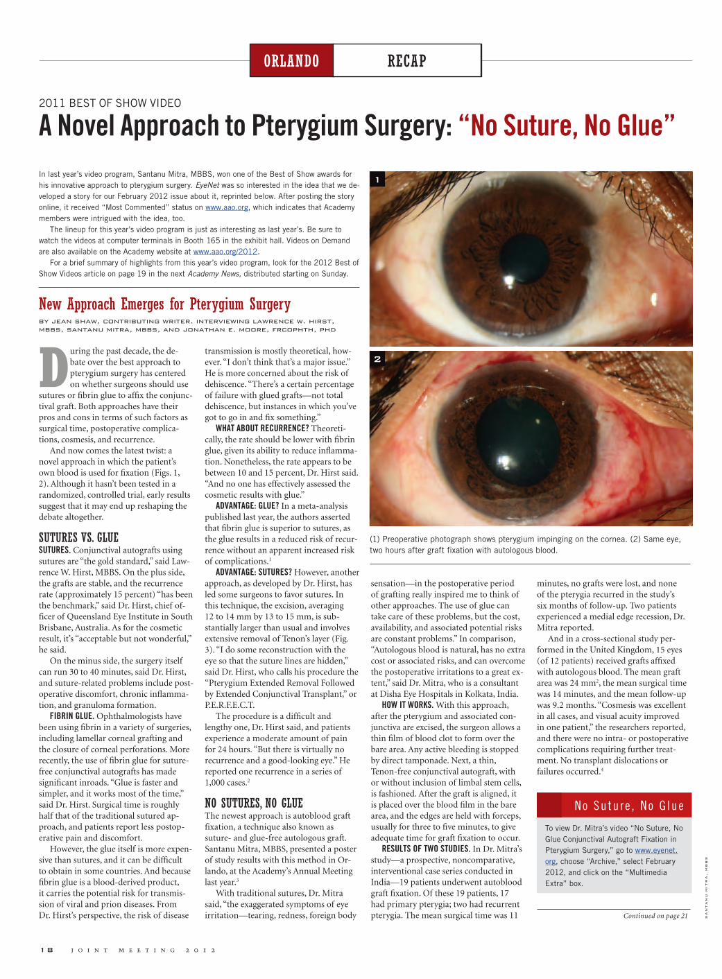
18 j o i n t m e e t i n g 2 0 1 2
RECAPORLANDO RECAPORLANDO
2011 BEST OF SHOW VIDEO
A Novel Approach to Pterygium Surgery: “No Suture, No Glue”
New Approach Emerges for Pterygium Surgeryby jean shaw, contributing writer. interviewing lawrence w. hirst, mbbs, santanu mitra, mbbs, and jonathan e. moore, frcophth, phd
During the past decade, the de-bate over the best approach to pterygium surgery has centered on whether surgeons should use
sutures or fibrin glue to affix the conjunc-tival graft. Both approaches have their pros and cons in terms of such factors as surgical time, postoperative complica-tions, cosmesis, and recurrence.
And now comes the latest twist: a novel approach in which the patient’s own blood is used for fixation (Figs. 1, 2). Although it hasn’t been tested in a randomized, controlled trial, early results suggest that it may end up reshaping the debate altogether.
SUTURES VS. GLUE SUTURES. Conjunctival autografts using sutures are “the gold standard,” said Law-rence W. Hirst, MBBS. On the plus side, the grafts are stable, and the recurrence rate (approximately 15 percent) “has been the benchmark,” said Dr. Hirst, chief of-ficer of Queensland Eye Institute in South Brisbane, Australia. As for the cosmetic result, it’s “acceptable but not wonderful,” he said.
On the minus side, the surgery itself can run 30 to 40 minutes, said Dr. Hirst, and suture-related problems include post-operative discomfort, chronic inflamma-tion, and granuloma formation.
FIBRIN GLUE. Ophthalmologists have been using fibrin in a variety of surgeries, including lamellar corneal grafting and the closure of corneal perforations. More recently, the use of fibrin glue for suture-free conjunctival autografts has made significant inroads. “Glue is faster and simpler, and it works most of the time,” said Dr. Hirst. Surgical time is roughly half that of the traditional sutured ap-proach, and patients report less postop-erative pain and discomfort.
However, the glue itself is more expen-sive than sutures, and it can be difficult to obtain in some countries. And because fibrin glue is a blood-derived product, it carries the potential risk for transmis-sion of viral and prion diseases. From Dr. Hirst’s perspective, the risk of disease
transmission is mostly theoretical, how-ever. “I don’t think that’s a major issue.” He is more concerned about the risk of dehiscence. “There’s a certain percentage of failure with glued grafts—not total dehiscence, but instances in which you’ve got to go in and fix something.”
WHAT ABOUT RECURRENCE? Theoreti-cally, the rate should be lower with fibrin glue, given its ability to reduce inflamma-tion. Nonetheless, the rate appears to be between 10 and 15 percent, Dr. Hirst said. “And no one has effectively assessed the cosmetic results with glue.”
ADVANTAGE: GLUE? In a meta-analysis published last year, the authors asserted that fibrin glue is superior to sutures, as the glue results in a reduced risk of recur-rence without an apparent increased risk of complications.1
ADVANTAGE: SUTURES? However, another approach, as developed by Dr. Hirst, has led some surgeons to favor sutures. In this technique, the excision, averaging 12 to 14 mm by 13 to 15 mm, is sub-stantially larger than usual and involves extensive removal of Tenon’s layer (Fig. 3). “I do some reconstruction with the eye so that the suture lines are hidden,” said Dr. Hirst, who calls his procedure the “Pterygium Extended Removal Followed by Extended Conjunctival Transplant,” or P.E.R.F.E.C.T.
The procedure is a difficult and lengthy one, Dr. Hirst said, and patients experience a moderate amount of pain for 24 hours. “But there is virtually no recurrence and a good-looking eye.” He reported one recurrence in a series of 1,000 cases.2
NO SUTURES, NO GLUEThe newest approach is autoblood graft fixation, a technique also known as suture- and glue-free autologous graft. Santanu Mitra, MBBS, presented a poster of study results with this method in Or-lando, at the Academy’s Annual Meeting last year.3
With traditional sutures, Dr. Mitra said, “the exaggerated symptoms of eye irritation—tearing, redness, foreign body
sensation—in the postoperative period of grafting really inspired me to think of other approaches. The use of glue can take care of these problems, but the cost, availability, and associated potential risks are constant problems.” In comparison, “Autologous blood is natural, has no extra cost or associated risks, and can overcome the postoperative irritations to a great ex-tent,” said Dr. Mitra, who is a consultant at Disha Eye Hospitals in Kolkata, India.
HOW IT WORKS. With this approach, after the pterygium and associated con-junctiva are excised, the surgeon allows a thin film of blood clot to form over the bare area. Any active bleeding is stopped by direct tamponade. Next, a thin, Tenon-free conjunctival autograft, with or without inclusion of limbal stem cells, is fashioned. After the graft is aligned, it is placed over the blood film in the bare area, and the edges are held with forceps, usually for three to five minutes, to give adequate time for graft fixation to occur.
RESULTS OF TWO STUDIES. In Dr. Mitra’s study—a prospective, noncomparative, interventional case series conducted in India—19 patients underwent autoblood graft fixation. Of these 19 patients, 17 had primary pterygia; two had recurrent pterygia. The mean surgical time was 11
minutes, no grafts were lost, and none of the pterygia recurred in the study’s six months of follow-up. Two patients experienced a medial edge recession, Dr. Mitra reported.
And in a cross-sectional study per-formed in the United Kingdom, 15 eyes (of 12 patients) received grafts affixed with autologous blood. The mean graft area was 24 mm2, the mean surgical time was 14 minutes, and the mean follow-up was 9.2 months. “Cosmesis was excellent in all cases, and visual acuity improved in one patient,” the researchers reported, and there were no intra- or postoperative complications requiring further treat-ment. No transplant dislocations or failures occurred.4
In last year’s video program, Santanu Mitra, MBBS, won one of the Best of Show awards for his innovative approach to pterygium surgery. EyeNet was so interested in the idea that we de-veloped a story for our February 2012 issue about it, reprinted below. After posting the story online, it received “Most Commented” status on www.aao.org, which indicates that Academy members were intrigued with the idea, too.
The lineup for this year’s video program is just as interesting as last year’s. Be sure to watch the videos at computer terminals in Booth 165 in the exhibit hall. Videos on Demand are also available on the Academy website at www.aao.org/2012.
For a brief summary of highlights from this year’s video program, look for the 2012 Best of Show Videos article on page 19 in the next Academy News, distributed starting on Sunday.
To view Dr. Mitra’s video “No Suture, No Glue Conjunctival Autograft Fixation in Pterygium Surgery,” go to www.eyenet.org, choose “Archive,” select February 2012, and click on the “Multimedia Extra” box.
No Su tu r e , No G lue
(1) Preoperative photograph shows pterygium impinging on the cornea. (2) Same eye, two hours after graft fixation with autologous blood.
1
2
sa
nta
nu
mit
ra
, m
bb
s
Continued on page 21

Surface Protection and More
SOME SURFACES ARE WORTH PROTECTING
THE OCULAR SURFACE IS ONE.
© 2012 Novartis 2/12 SYS11179JAD
References1. Christensen MT, Blackie CA, Korb DR, et al. An evaluation of the performance of a novel lubricant eye drop. Poster D692 presented at: The Association for Research in Vision and Ophthalmology Annual Meeting; May 2-6, 2010; Fort Lauderdale, FL. 2. Lane S, Paugh JR, Webb JR, Christensen MT. An evaluation of the in vivo retention time of a novel artifi cial tear as compared to a placebo control. Poster D923 presented at: The Association for Research in Vision and Ophthalmology Annual Meeting; May 3-7, 2009; Fort Lauderdale, FL. 3. Davitt WF, Bloomenstein M, Christensen M, et al. Effi cacy in patients with dry eye after treatment with a new lubricant eye drop formulation. J Ocul Pharmacol Ther. 2010;26(4):347-353. 4. Alejandro A. Effi cacy of a Novel Lubricant Eye Drops in Reducing Squamous Metaplasia in Dry Eye Subjects. Presented at the 29th Pan-American Congress of Ophthalmology in Buenos Aires, Argentina, July 7-9, 2011. 5. Wojtowica JC., et al. Pilot, Prospective, Randomized, Double-masked, Placebo-controlled Clinical Trial of an Omega-3 Supplement for Dry Eye. Cornea 2011:30(3) 308-314. 6. Geerling G., et al. The International Workshop on Meibomian Gland Dysfunction: Report of the Subcommittee on Management and Treatment of Meibomian Gland Dysfunction. IOVS 2011:52(4).
EyeNet Academ
y New
s
80152 SYS11179JAD ENAN.indd 1 9/24/12 1:22 PM


e y e n e t ’ s a c a d e m y n e w s 21
RECAPORLANDO
FINE POINTS TO CONSIDERAs with any new procedure, auto blood graft fixation involves a learning curve, said Jonathan E. Moore, FRCOphth, PhD, who participated in the U.K. study. “But this approach is probably less challenging [than sutures or glue] once one over-comes the fear of not suturing,” said Dr. Moore, a consultant ophthalmologist and clinical director at Cathedral Eye Clinic in Belfast, Northern Ireland.
He and Dr. Mitra noted several factors to consider.
PATIENT SELECTION. Patients who regularly take aspirin or other blood thin-ners—or who suffer from a coagulation factor deficiency—would not be good candidates for autoblood graft fixation, Dr. Mitra said.
SURGICAL CHALLENGES. “You need a slightly oversized [about 0.5 mm] Tenon-free graft that is as thin as possible,” Dr. Mitra said. This prevents the risk of graft retraction and/or rejection. The surgeon will also need “a lot of patience,” he said, “giving more than adequate time—three to five minutes on average—during graft apposition.” Dr. Moore concurred with the need for thin grafts and “meticulous dissection to remove the Tenon’s layer” and cautioned, “Do not leave subgraft blood.”
RISK OF DEHISCENCE. “The main disad-vantage is the risk of graft loss in the im-mediate postoperative period,” Dr. Mitra said. “But once the graft stays in place for the first 24 to 48 hours, it is going to stick around.”
He added that he has seen no graft loss in his ongoing case series, which now numbers 63 patients who have been fol-lowed for at least one year.
COSMETIC RESULTS. “Cosmesis is excel-lent with this procedure—as good as with fibrin glue, if not better,” Dr. Moore said. “There is nothing to promote inflamma-tion, and patients achieve a good appear-ance very quickly, within six weeks.” He added that the graft is not noticeable in many of his patients.
RISK OF RECURRENCE. If properly performed, the procedure appears to carry a low risk of recurrence. To date, Dr. Moore has performed 50 autoblood graft
procedures, which have been followed for up to four years. None of his patients has experienced a recurrence.
However, Dr. Moore reported that a surgeon in training at his institution used a graft that was too thick and did not cover the limbus, resulting in a small recurrence within the first two months following surgery.
Dr. Mitra reported one case of recur-rence after the excision of a previously recurrent pterygium. “This probably was due to inadequate dissection of pterygial tissue rather than to autoblood fixation of the graft,” he said, and it was detected “just past the three-month postoperative follow-up examination.”
VISUAL OUTCOMES. Although one of Dr. Moore’s patients experienced a two-line gain in VA, Drs. Moore and Mitra say that any effect on vision is independent of the graft fixation technique used. Instead, it will be influenced by factors such as the size and location of the pterygium, the presence of astigmatism, and other preoperative issues.
Drs. Hirst, Mitra, and Moore reported no related financial interests, although Dr. Hirst owns the trademark “P.E.R.F.E.C.T. for Pterygium.”
1 Pan HW et al. Ophthalmology. 2011;118(6):
1049-1054.
2 Hirst LW. Extensive Incision and Conjunc-
tival Transplantation for Pterygium: Results
of 1,000 Surgeries. Presented at World Cornea
Congress; April 7-9, 2010; Boston.
3 Mitra S et al. Autoblood as Tissue Adhesive
for Conjunctival Autograft Fixation in Pte-
rygium Surgery. Poster presented at the Annual
Meeting of the American Academy of Ophthal-
mology; Oct. 22 and 23, 2011; Orlando, Fla.
4 de Wit D et al. Eye. 2010;24(9):1474-1477.
Recurrence is “the bugbear” in pterygium surgery, said Dr. Hirst. “For patients, the No. 1 question is, ‘Will it come back?’ Everything else is secondary.”
And that should be the surgeon’s first question as well, he said. “If a pterygium comes back, it’s a real problem—it can be worse than the first occurrence. Yet many surgeons have no idea what their recurrence rate is.”
To get an accurate sense of recurrence—and, especially, to identify the earliest stag-es of recurrence—you need to follow patients for a full year, Dr. Hirst said. “That’s not a magical figure. If you want to pick up most of them, you’ll be able to do so within six months.” Still, you would miss a few if you stopped following patients at the six-month mark. “It’s a year if you really want to know what’s going on.”
Do You Know You r Recu r r ence Ra te?
EXTENDED EXCISION. Illustration shows final average size of conjunctival defect in the P.E.R.F.E.C.T. technique.
3
hir
st l
w.
oph
th
alm
olo
gy 2
00
8;1
15
(10
):1
66
8.
Treat the cause.
86% of dry eye patients have signs of Meibomian Gland Dysfunction (MGD).1
1 Lemp MA, et al. Distribution of aqueous deficient and evaporative dry eye in a clinic-based population. Cornea. 2012 May;31(5):472-8
2 Lane SS, et al. A New System, the LipiFlow, for the treatment of Meibomian Gland Dysfunction (MGD). Cornea. 2012;31:396-404
LipiFlow is a registered trademark of TearScience, Inc. Copyright © 2012 TearScience, Inc. All rights reserved.
LipiFlow® is indicated for treatment of Meibomian Gland Dysfunction and evaporative dry eye.
In clinical trials, gland secretions more than doubled, on average, following a single LipiFlow® treatment.2
Visit tearscience.com for complete product and safety information.
Visit us at AAO 2012 Booth #4362
Continued from page 18

22 j o i n t m e e t i n g 2 0 1 2
RECAPORLANDO RECAPORLANDO
THE ORLANDO CATARACT SPOTLIGHT SESSION: PART ONE
Review 2011’s “M&M Rounds: Learning From My Mistakes”
CASE 1: NOT THE USUAL BEVERLY HILLS CATARACTUday Devgan’s patient had a short eye with a mature white cataract and a crowded anterior chamber. Iris prolapse occurred suddenly following hydrodissection.
At this point, assuming that you cannot reposition the iris in the small eye, what is your very next step?
Administer an osmotic agent (e.g., IV mannitol) . . . . . . .31%Perform a pars plana vitreous tap 38%Burp the anterior chamber (AC) via another incision . . . . . . .18%Create a new phaco incision . . . . 5%Abort surgery (in case of a suprachoroidal hemorrhage) . . . 9%
CASE PRESENTER UDAY DEVGAN This is a tricky question because the primary issue is the sudden pressure gradient, which could be at least partially addressed by more than one of these answers. In this case, the 80-year-old patient has a short eye (axial length, 20 mm) with a dense white cataract and moderate dilation. The principal surgical error was excessive hydrodissection because the fluid wave behind the lens could not be visualized. Balanced salt solution became trapped behind the nucleus, which was then unable to prolapse out of the bag be-cause of the smaller-than-desired 4-mm anterior capsulorrhexis. This created a pressure gradient with high pressure in the posterior chamber and lower pres-sure in the anterior chamber, leading to the resultant iris prolapse out of the clear corneal phaco incision. This challenge was resolved by releasing the trapped balanced salt solution from behind the cataract by using a blunt cannula to rock the nucleus. Once the pressure gradient was equalized, the iris prolapse resolved,
and the surgery could be continued. The take-home message is that the cause of the pressure gradient must be addressed in order to resolve iris prolapse.
STEVE LANE’S PERSPECTIVE A patient with a short axial length and a white lens that prevents visualization of the retina and does not even allow a red reflex dur-ing surgery poses special challenges. In this case, the IOP increased suddenly dur-ing hydrodissection and was accompanied by iris prolapse. Without a red reflex, the cause and treatment are not obvious. A suprachoroidal hemorrhage (SCH) due to nanophthalmos must be considered because of its dire consequences. Admin-istration of an osmotic agent is a reason-able treatment if an SCH is suspected, but the pressure-lowering effect may take 30 minutes or longer. Performing a pars plana vitreous tap would be risky without knowing whether an SCH is present because a more severe hemor-rhage might ensue. In the absence of a red reflex, a B-scan ultrasound is the only way to know whether an SCH is present. This specialized instrumentation is not readily available but would have been helpful to rule an SCH in or out.
Burping the anterior chamber or creating another incision are similar alternatives. Given that the pressure is
greater in the posterior chamber than in the anterior chamber, these actions would be of little value and would create more prolapse. Aborting the case is a possibil-ity, but the rhexis has been completed, and delay in nuclear removal can lead to significant inflammation. An SCH, if present, might take several days to resolve. Equalizing the posterior and anterior pressure is the key to solving the problem. Decompression of the pressure inside the capsular bag is an important goal in every cataract procedure. Compressing the lens during hydrodissection and allowing the trapped balanced salt solution within the capsular bag to escape are important maneuvers that should be performed rou-tinely. In this case, it solved the problem and ruled out SCH.
CASE 2: PEARLS FOR POSTERIOR CAPSULORRHEXISMark Packer had a refractive lens exchange patient for whom a multifocal intraocular lens had been planned. During phaco-emulsification with biaxial microincisional instrumentation, the posterior capsule tore.
What should your next step be?Withdraw the irrigating chopper to avoid hydrating the vitreous . . . . . . 8%Inject viscoelastic while maintaining irrigation through the irrigating chopper. . . . . . . . . . . . .91%Perform a pars plana vitrectomy while maintaining irrigation via the irrigating chopper . . . . . . 2%Close the eye and refer the patient 0%
CASE PRESENTER MARK PACKER Ninety-one percent of the audience correctly identi-fied the conventional management wis-dom. Withdrawing irrigation is precisely what should not be done. Doing so would allow the vitreous to prolapse anteriorly and the lens fragments to dislocate poste-riorly. Vitrectomy is not indicated unless one or both of these untoward events occurs.
Converting the posterior capsular tear into a continuous posterior capsulor-rhexis under irrigation or viscoelastic prevents further extension of the tear and will allow in-the-bag implantation of the multifocal IOL as planned. One of the advantages of biaxial phacoemulsifica-tion is the ability to maintain irrigation while removing the phacoemulsification needle, performing the posterior rhexis and injecting viscoelastic. The patient in this example demonstrated uncorrected distance visual acuity of 20/20 and uncor-rected near visual acuity of J1 on postop day 1.
ABHAY VASAVADA’S PERSPECTIVE The audience recommendation is very logical. I would also recommend lowering the irrigating bottle height and foot pedal po-
sition and injecting dispersive viscoelastic anterior to the rupture area and then withdrawing the irrigating chopper. If the surgeon is very patient and the surgical team is very efficient, preservative-free triamcinolone can be injected before the viscoelastic. This would allow the surgeon to recognize the presence of vitreous in the event that it had prolapsed into the anterior chamber.
CASE 3: WHEN DID IT TEAR? KEEP YOUR EYE ON THE BALL!In this case from Barry Seibel, his patient had undergone prior pneumatic retinopexy during which the needle shaft might have contacted the posterior lens capsule. Dur-ing cortical cleanup with coaxial irrigation and aspiration (I&A) instrumentation, a posterior capsular tear is noted.
During which step do you think the pos-terior capsular tear occurred?
A posterior capsular defect orweakness was preexisting . . . . .14%Hydrodissection. . . . . . . . . .11%Nuclear emulsification . . . . . . 6%Cortical I&A . . . . . . . . . . .47%Not sure . . . . . . . . . . . . .23%
CASE PRESENTER BARRY SEIBEL Because of the posterior subcapsular cataract’s un-usual appearance and traumatic etiology, posterior capsular fragility or discontinu-ity was anticipated. Therefore, appropri-ate prophylactic steps were taken, such as refraining from significant hydrodissec-tion in favor of a complete hydrodelin-eation, and using viscodissection of the epinucleus so that if a posterior capsular rent were present, the ophthalmic visco-surgical device (OVD) would tamponade the vitreous. However, attention was diverted from the eye during the switch from phaco to I&A as the surgeon looked at the handpieces, tubing, etc. During this time, the unblinking eye of the camcorder revealed the presence of an obvious fusi-form red reflex rent progressively opening up in the posterior capsule.
However, the surgeon’s mindset was frozen on the last image seen through the operating microscope—an intact capsule. This mindset likely obscured the brief visualization of this break before it was obliterated by the repressurization of the anterior chamber upon insertion of the I&A tip. Vitreous was soon evident during I&A, but it might have been ap-preciated even sooner had there been less complacency about the posterior capsular integrity. Conceivably, the surgeon could have maintained constant observation through the microscope even during handpiece switching. However, I find direct visualization of this step useful in
Again this year, the Spotlight on Cataracts Session takes place on Monday, 8:15 a.m. to 12:15 p.m., in North Hall B. The title is “Clinical Decision-Making With Cataract Complications,” and it promises to be as exciting as previous years’ sessions.
As you gear up to attend the 2012 session, you might enjoy a look back at the audience poll results from last year’s Cataract Spotlight, along with the presenter’s description of the case in question and a second comment from another expert discussing his or her own thoughts about the case. The following is reprinted from the February 2012 EyeNet Maga-zine. Part Two will appear in the next issue of Academy News available starting on Sunday.
Introduction From the Session CochairmanEvery cataract surgeon makes mistakes and suffers complications, but it is what we learn from these mistakes that makes us better ophthalmologists.
With this in mind, Bruce Wallace and I cochaired last year’s annual Spotlight on Cataract Surgery Session at the Academy’s Annual Meeting. The four-hour case-based video symposium entitled “M&M Rounds: Learning From My Mistakes” was focused on cataract surgical complications.
Cataract experts presented video cases in which something had gone wrong, result-ing in a complication that taught them valuable lessons. At critical points during each case, the video was paused and the attendees made clinical decisions using their elec-tronic audience response pads. —David F. Chang, MD
CASE 1. Iris prolapse.
Continued on page 24 Ud
ay D
evg
an
, M
D

Saturday, Nov. 109:30 AM Blepharitis: The New ConsensusStephen V. Scoper, MD
11:00 AM The LenSx® Laser: Sphere and Cylinder Are Not EnoughPaul Ernest, MD
11:30 AM Alcon Advances for Today’s LASIK SurgerySonny Goel, MD Charles Moore, MD
12:00 PM IOL Injection You’ve Always Wanted. Simple, Elegant, Automated. Introducing the AutoSert® IOL InjectorRobert Osher, MD
12:30 PM Advanced Optical Biometry: Using the LENSTAR LS 900®* Optical Biometer with Toric IOLs, Strategies for SuccessWarren Hill, MD
1:00 PM Methods to Manage Pre-Existing Corneal Astigmatism with Toric IOLsEdward J. Holland, MD Samuel Masket, MD
1:30 PM Rethinking the Role of IOP in the Diagnosis and Management of Open-angle GlaucomaMatthew McMenemy, MD
2:00 PM The LenSx® Laser: A New Cataract ProcedureStephen Lane, MD Satish Modi, MD Dan Tran, MD
3:00 PM Multifocal IOLs: Setting Expectations for Presbyopic PatientsRandy Epstein, MDCathleen McCabe, MD
3:30 PM Clinical Pearls to Adopting the EX-PRESS® GFDSteve Vold, MD
Presentations, presenters and times are subject to change.
These presentations are not affiliated with the official program of the 2012 AAO-APAO Joint Meeting.
For important safety information about the ALCON® products discussed in these presentations, please visit the Alcon booth.
Sunday, Nov. 1111:00 AM Maximizing Success with the EX-PRESS® Glaucoma Filtration DeviceIke Ahmed, MD
12:30 PMMultifocal IOLs: Setting Expectations for Presbyopic PatientsWilliam J. Lahners, MD Andrew Maxwell, MD
1:00 PMAlcon Advances for Today’s LASIK SurgeryVance Thompson, MD
1:30 PMIntegrating the LenSx® Laser into Our PracticeMichael P. Jones, MD Christa Garner, BA, CRC
3:00 PM Methods to Manage Pre-Existing Corneal Astigmatism with Toric IOLsGary Foster, MD Ehsan Sadri, MD
3:30 PM Blepharitis: The New ConsensusStephen V. Scoper, MD
Monday, Nov. 1210:00 AM The LenSx® Laser: A New Cataract ProcedureJerry Hu, MD Robert Lehmann, MD
12:30 PMAlcon Advances for Today’s LASIK SurgeryJoseph L. Parisi, MD
1:30 PMMy Experience with the EX-PRESS® Glaucoma Filtration DeviceJeff Goldberg, MD
2:00 PMOptically Measured Lens Thickness in IOL Power CalculationSheridan Lam, MD
2:30 PMTransitioning to Femtosecond Cataract SurgeryGerard Sutton, MD
The Alcon Speakers Forum in ChicagoNovember 10 – 12, 2012
DuriNg the AAO-APAO JOiNt MeetiNgHere is just a sampling of the presentations that showcase the latest
technologies in ophthalmology brought to you by Alcon, booth #2808.
*LENSTAR® is a registered trademark of Haag-Streit ©2012 Novartis 9/12 MIX12422JAS-B
Scan for Alcon at the AAO Information

properly draping tubing over my forearm to relieve traction, adjust irrigating sleeves as needed, etc.
The moral of this case is one we al-ready know: We must maintain constant, unrelenting vigilance during surgery; always be open to the possibility of com-plications, particularly in high-risk cases; and suppress our natural tendency to believe that everything is fine even if there are hints to the contrary.
STEVE DEWEY’S PERSPECTIVE The audi-ence’s response is appropriate, as the tear in the posterior capsule could not be visu-alized until after the I&A was performed. But this is the perfect example of a teach-ing case that would have remained a mys-tery if it had not been caught on video-tape. The tear corresponds exceptionally well to the posterior subcapsular cataract. During the video, the only manipulation Dr. Seibel performed in the area of the tear was his hydrodissection. The tear might have been made worse during this step, but it is much more likely that the tear was preexisting. The softer nature of the cataract and the solid chamber stability prevented the capsule from being challenged during phacoemulsification and allowed the case to proceed as it did.
During my informed consent for cataract surgery, I explain that the greatest barrier to achieving a good outcome is something the patient has brought with him or her into the operating room, such as macular degeneration or diabetic reti-nopathy. This simplifies the concept of a preexisting comorbidity to a basic level of understanding and allows the discussion to continue.
In this challenging case, I would coun-sel the patient that the previous ocular surgery may present unknowns that could potentially affect the outcome. A focal cataract, or one that has progressed exu-berantly over a shorter period following pars plana vitrectomy, certainly raises the question of preexisting capsular damage.
As with a patient with potential zonular laxity, the first step is to reduce the infusion pressure prior to beginning phaco. A larger capsulorrhexis (5.5 mm) is useful, and gentle hydrodelineation will allow the nucleus to be brought into the anterior chamber. A smaller-gauge phaco needle will act as a flow restrictor and will further maintain a stable anterior chamber during phacoemulsification. Size and location of the capsular defect will determine whether the IOL can then be placed in the bag or captured in the capsulorrhexis. Although it is unusual to have vitreous presenting at this point, a touch of triamcinolone in the anterior chamber after IOL placement will identify any unexpected vitreous strands for exci-sion if the posterior capsule was indeed compromised.
CASES 1 TO 3. “Grand Rounds Award” voted for the surgeon who presented the best teaching case:
Uday Devgan: Iris prolapse . . . . 66%Mark Packer: PC rupture . . . . . 25%Barry Seibel: PC rupture. . . . . . 9%
CASE 4: FUN WITH ZONULES (NOT)In Alan Crandall’s case, the nucleus abruptly tilted posteriorly during an at-tempt at prechopping, indicating the pres-ence of a sizable zonular dialysis.
The entire lens appears to be loose.What now?
Insert a capsular tension ring (CTR) and carefully continue phaco . .49%Elevate the nucleus with a posterior-
assisted levitation (PAL) technique, and phaco it in the anterior chamber . . . . . . . . . . . .20%Convert to a manual extracapsular cataract extraction (ECCE) . . . .21%Close the eye and refer the patient 3%Other . . . . . . . . . . . . . . . 6%
CASE PRESENTER ALAN CRANDALL This case reminds us that in phacoemulsification cataract extraction no steps are inconse-quential. In this case of pseudoexfoliation (PXF), poor wound construction led to a small capsulorrhexis after the hydrodis-section; the prechopper movement was too rapid; and the tip tore more than 180 degrees of the already weakened zonules. However, capsular support systems (in this case, Mackool hooks, FCI Ophthal-mics) can allow the surgeon to safely proceed with phaco using a low-flow technique; and, finally, a CTR or Cionni- Modified CTR (Morcher) can allow for in-the-bag implantation of the IOL. To use any of the capsular support systems (I now prefer the MST capsule retrac-tors [MicroSurgical Technology]), a stab incision is made. Then viscoelastic is used to create space in the capsule for easier insertion of the support. Usually two or three capsule hooks will be enough to stabilize the lens. Low flow indicates that the bottle height is lowered to reduce posterior pressure. It is also important to reduce the aspiration flow rate on a peristaltic system to 25 mm/minute, and I use linear vacuum with a maximum of 350 mmHg. Another important decision involves lens placement and the use of CTRs. If the bag is intact and the rhexis is continuous, a CTR can be used. Assuming that the remaining zonules are intact and the dehiscence is less than 120 degrees, a standard CTR can be used. Larger zonular defects need a Cionni-Modified CTR or an Ahmed Capsular Tension Segment (FCI Ophthalmics) sewn to the sclera with 9-0 Prolene or 8-0 Gore-Tex sutures (off-label use). Other options would be iris fixation or an anterior chamber lens.
WALTER STARK’S PERSPECTIVE Zonular dehiscence or dialysis has many causes, the most likely being pseudoexfoliation of the lens capsule. This should be recog-nized or suspected preoperatively so that measures can be taken to prevent further detachment of the zonules. Often, the first evidence of weak zonules is move-ment of the entire lens at the start of the
capsulotomy. In these cases, I perform a wider capsulotomy, which enables me to make a more complete hydrodissection. This separates the lens nucleus away from the capsular bag so that the chopping or manipulation of the nucleus no longer exerts traction on the capsule or, in turn, on the zonules. A wider capsulotomy also reduces the chances of later phimosis of the anterior capsule. With good hydrodis-section and separation of the nucleus from the capsule, phacoemulsification can usually be performed safely within the iris plane.
In the presence of extensive zonular weakness, as in cases of Marfan syndrome or traumatic weakness of the zonules, iris retractors can be used to hold the anterior capsular leaf peripherally, thereby stabi-lizing the capsular bag. I do not use CTRs because I do not believe they offer any better support than a three-piece lens. My preference for these complicated cases is the MA50BM acrylic lens made by Alcon with a 13.5-mm loop. The MA50BM lens is 6.5 mm in diameter, which is 18 per-cent larger than a 6-mm optic. This exerts peripheral tension on the capsular bag and, in my opinion, provides essentially the same support as would be provided by a CTR. If necessary, with the three-piece lens or the CTR, scleral fixation can be created with a 10-0 Prolene suture with a CIF4 or CT6 needle, both of which are made by Ethicon.
The audience poll indicates that 49 percent of respondents would place a CTR and then continue with careful phacoemulsification. I suspect that inser-tion of a CTR without good hydrodissec-tion might lead to further weakness of the zonules and/or a capsular tear. Some people routinely use CTRs in patients with PXF, and I wonder how often this causes a complication. Twenty percent of the audience would use the PAL technique. If the injection of a disper-sive viscoelastic behind the lens did not elevate the nucleus to the pupillary plane, the PAL technique could be used in these more complex cases.
With the nucleus dropping posteriorly, it is very difficult to do a manual extra-capsular cataract extraction; 21 percent of the audience suggested this as an option. To do an extracapsular cataract extrac-tion, one would need to increase the size of the incision to 11 to 13 mm and be
able to exert enough vitreous pressure to push the lens forward. I would not choose this approach out of concern that it would only further sublux the nucleus.
With a preoperative suspicion of weak zonules, the surgeon can properly handle these cases with a wider capsulotomy, good hydrodissection to bring the nucleus forward and then very careful phaco-emulsification. Iris retractors are some-times needed to stabilize the capsular bag. The intraocular lens can then be placed in the ciliary sulcus. If support for the IOL appears inadequate, pupillary capture of the optic can be accomplished; and iris fixation of the haptic can be performed at least superiorly and, if necessary, inferi-orly.
CASE 5: GOT BURNED: SO YOU SAY YOU WON’T WEAR GLASSES?In Lisa Arbisser’s case, a significant wound burn occurred, and the surgeon was faced with the problem of how to close the incision. The wound burn also resulted in an initial large degree of induced astigmatism in a patient who did not want to wear eyeglasses.
How many serious wound burns have you experienced?
None. . . . . . . . . . . . . . . 48%1 . . . . . . . . . . . . . . . . 26%2-3 . . . . . . . . . . . . . . . 15%>3 . . . . . . . . . . . . . . . 11%
CASE PRESENTER LISA ARBISSER A burn can occur anytime absent flow coexists with ultrasound. I learned that my usual technique with a brunescent lens—verti-cal chop, 2.2-mm incision, dispersive OVD and burst-mode high power—could create a burn if OVD was not adequately cleared before embedding the tip in the nucleus. The situation was perhaps ag-gravated by the absence of an ABS port, which might have given an extra margin of safety. This, my first burn, coincided with my first use of a reusable phaco tip featuring no hole in the shaft. Burns are biphasic, progressing from transient tissue shrinkage with remodeling to per-manent destruction.
Happily, after the first chop I fully established flow, clearing OVD along with nucleus as always before the second chop. I realized intraoperatively that interrupt-ed anterior-to-posterior lip sutures repair coagulated tissue poorly. Instead, I closed the tunnel floor and roof in their native position with three interrupted horizon-tal sutures (two from normal tissue to the middle of the burn and one within the tunnel). A fornix-based conjunctival flap sutured tightly over the limbus provided further protection. A more severe burn would have required a split scleral flap to gain tissue without having banked sclera, or a substitute, to secure the anterior chamber. Stable at four months and after suture removal, this toric lens–implanted
RECAPORLANDO
CASE 4. (A) Mackool capsule retractor. (B) MST capsule retractor.
A b
24 j o i n t m e e t i n g 2 0 1 2
Continued from page 22
Continued on page 26 Ala
n C
ra
nd
all,
MD

Patient Support Program
Many will seek more information or look for help in remembering to take their drops. That’s why there is the OPENINGS™ Patient Support Program from Alcon.
• Educational mailings help to ensure disease awareness and understanding
• OpeningsProgram.com provides practical tools and other resources to help patients establish a daily dosing regimen
• Program savings card makes it easier for eligible patients to pay for their medicine
A TRUE PARTNER IN PROVIDING GLAUCOMA PATIENT SUPPORT.
You’ve diagnosed your patient, provided advice and presented a treatment plan.
But what happens when he or she goes home?
WORKING TOGETHER, WE CAN HELP ADDRESS THE CRITICAL ISSUE OF IMPROVING PATIENT COMPLIANCE AND ADHERENCE.
© 2012 Novartis 9/12 MG12097JAD
EyeNet Academ
y New
s
80153 MG12097JAD ENAN.indd 1 9/24/12 1:22 PM

RECAPORLANDO
26 j o i n t m e e t i n g 2 0 1 2
eye’s 2.5 D residual cylinder diminished with astigmatic keratotomy. My patient’s final outcome: uncorrected 20/30 and best-corrected 20/20+ with 1.25 D cylin-der residual manifest refraction.
ELIZABETH DAVIS’ PERSPECTIVE For-tunately, according to the audience, the incidence of serious wound burns is low, with nearly half of respondents reporting never having experienced even one. This is good news, as a severe wound burn can induce considerable astigmatism, require a patch graft to close, or even necessitate a corneal transplant. I believe this complication is uncommon because of major advances in technology. Today’s phaco machines have enhanced designs that reduce the risk of blockage or loss of the cooling inflow of irrigating fluid, as well as advanced power modulations that reduce phaco energy and thus thermal increases. I would also suspect that most cataract surgeons are well versed in ways to avoid wound burns, such as minimiz-ing continuous ultrasound and using techniques of mechanical disassembly.
CASE 6: SPLITTING HEADACHE (PHACO AFTER RK)Sonia Yoo’s case involved difficulty in closing the clear corneal incision in an eye with many prior radial keratotomy (RK) scars. The incision was not completely watertight at the conclusion of surgery.
For cataract surgery in an eye with mul-tiple RK scars, I prefer:
Coaxial phaco with a scleral pocket incision . . . . . . . . . . . . .41%Coaxial phaco with a clear corneal incision . . . . . . . . . . . . .46%Biaxial microphaco . . . . . . . . .4%Manual ECCE . . . . . . . . . . . 1%I would refer this patient . . . . . .8%
CASE PRESENTER SONIA YOO This case of ra-dial keratotomy wound dehiscence during cataract extraction highlights one of the intraoperative challenges of performing cataract surgery in an eye with previous RK. Apart from the problem with biom-etry and intraocular lens calculations in post-RK patients, RK wound dehiscence is always a risk during cataract surgery because RK incisions typically run almost the full thickness of the cornea and extend to the corneal limbus circumfer-entially. Even in cases in which the RK was performed many years prior to the cataract surgery, these wounds can open up. This patient had undergone his RK more than 15 years earlier.
I try to construct my primary cata-ract wound and paracentesis so as not to transect the RK incisions. This can be dif-ficult for cases in which the RK incisions are close together (eight-incision RK or more). In cases in which it is impossible to avoid transecting the RK incisions with a clear corneal cataract wound, I consider a scleral tunnel approach, which
allows for a slightly more posterior entry into the anterior chamber, or a uniplanar limbal approach, which requires suturing for closure at the end of the case.
It is noteworthy that in this case, it was not until the infusion pressure was high that the RK incision dehisced. “Low-flow, slow-mo, small-incision phaco,” which would keep the infusion pressure rela-tively low, should be considered for such cases to reduce the risk of RK wound dehiscence. Finally, when facing the need to suture corneal wounds that transect dehisced RK wounds, I prefer to use an X or a figure-of-eight suture to close these. Interrupted radial or horizontal sutures tend to make the incisions gape and may be ineffective or even exacerbate the dehiscence in this situation.
KERRY SOLOMON’S PERSPECTIVE Cata-ract surgery in an eye that has undergone RK presents many challenges. I always review these with the patient as part of the informed consent. Certainly, cataract surgery involves a risk of these incisions opening. I inform patients that if this oc-curs, sutures or glue/tissue adhesive might be necessary to close the incision(s). Visu-al recovery can be delayed due to induced astigmatism from the wound closure. My rule of thumb is that if my primary clear corneal incision can readily fit between the RK incisions, I can proceed with clear corneal surgery. If my primary incision cannot fit between the RK scars (because of a larger incision or multiple [16+] RK incisions), I prefer a scleral tunnel ap-proach with a more posterior entry into the cornea. The presence of the scleral flap and a sutured conjunc tival closure of-ten facilitates wound closure if the RK in-cisions split open. These patients are also informed about the challenges regard-ing the IOL calculation and the delayed visual recovery (with early postoperative hyperopia), which is a normal occurrence in the setting of preexisting RK incisions. Refractive options are considered once things stabilize. The options are typically photorefractive keratectomy with mito-mycin C or an IOL exchange.
CASES 4 TO 6. “Chinese Water Tor-ture Award” voted for the surgeon who endured the most pain during his or her case:
Alan Crandall: Large zonular dialysis . . . . . . . . . . . . .13%Lisa Arbisser: Incision burn . . . .66%Sonia Yoo: Clear corneal incisions intersecting with RK incisions . .21%
CASE 7: NOW WHAT? I TORE THE CAPSULE AND CAN’T IMPLANT AN ARTIFICIAL IRISIn Kevin Miller’s patient with a traumatic mydriasis, the posterior capsule tore, mak-ing it impossible to implant the Morcher artificial iris segment rings as planned. In-stead, a three-piece PC IOL was implanted into the ciliary sulcus.
Apart from sunglasses, how could this
patient’s severe glare be managed? Prescribe a colored contact lens . .54%Consider corneal tattooing . . . . .15%Exchange the PC IOL with an artificial iris IOL (Morcher, Ophtec) . . . 21%Choose another option . . . . . . 7%Refer the patient . . . . . . . . . 3%
CASE PRESENTER KEVIN MILLER Eyes that experience blunt trauma severe enough to produce a permanent 11-mm mydriasis probably have concomitant zonular in-stability, no matter how subtle it appears at the slit lamp. That was the situation with this patient. I was expecting some trampolining of the lens– iris diaphragm, and I had dialed down the aspiration flow rate and vacuum limit in anticipation. Despite these efforts, I managed to pop a hole in the peripheral posterior capsule at the end of quadrant removal, which was precipitated by an occlusion break surge. At the moment it happened, I did not have a backup or alternative aniridia device to implant in the sulcus.
I had gone to the operating room with two Morcher 50F modified CTRs that had to be placed inside an intact capsular bag. I had a “Plan A” only. A backup iris reconstruction lens would have required a second set of FDA and IRB approvals, and implantation of such a device would have required opening an 11-mm incision. I finished the procedure by performing a limited anterior vitrectomy and implant-ing a Staar Surgical AQ2010V lens in the ciliary sulcus (Fig. A).
Fortunately, several months later I became aware of a beautiful new foldable artificial iris device that is being manufac-tured by Dr. Schmidt Intraocularlinsen. After extensive discussion, the patient consented to implantation of this device, and the results were spectacular (Fig. B). One year after this surgery, his vision was 20/20 uncorrected, and at a typical con-versational distance, it was impossible to tell which eye had experienced the problem.
KEN ROSENTHAL’S PERSPECTIVE In this case, the old adage “to fail to prepare is to prepare to fail” is somewhat overstated. As it turns out, the delay in the repair of the iris afforded the surgeon the oppor-tunity to use a newer, more esthetically pleasing and less invasive method of iris repair. However, I learned early on that one should always have a sulcus fixa-tion device available as a backup. When I request compassionate use for these non–
FDA-approved devices, the alternative device is included in our IRB-submitted protocol, and we obtain a single-piece IOL/iris prosthesis (such as the Ophtec Model 311) as a backup.
I have since determined, however, that in cases of incomplete bag tears, the iris prosthesis (especially the Ophtec Iris Prosthetic System) can be placed in a stable fashion. In addition, the Rasch-Rosenthal modified iris prosthesis (Morcher) can be placed in the ciliary sulcus and fixated to the sclera or, when present, to the iris remnants.
The audience response is interesting in that a majority would favor a contact lens rather than additional surgery. Although I always present this option to the patient when discussing solutions to remaining photosensitivity, only a handful of pa-tients have been successful contact lens wearers.
This is due in part to the optical properties of an iris-painted contact lens, which is at the corneal plane (and in which the pupil position moves with each blink), as well as to the fact that many patients with iris defects have significant ocular surface disease. These lenses are also less comfortable than normal con-tacts because they are thicker and contain a texturized layer of pigment on the surface. If the surgical risk is appropri-ate, implantation of an iris prosthesis is a much better option.
Some audience participants suggested an IOL exchange. I would not recom-mend this because the iris prosthesis de-signed for sulcus/sutured fixation can be obtained without an IOL and implanted over the existing, stable IOL. It is impor-tant to take a careful history: A significant minority (perhaps 10 percent) of pa-tients with iris defects do not have visual symptoms such as glare, photophobia, and starbursts. These patients, of course, require no additional treatment.
The excellent result that Dr. Miller’s patient enjoyed shows how amazing the HumanOptics iris prosthetic im-plants are. I have been very gratified to have implanted these devices, through a compassionate protocol, in patients who otherwise might not have been candidates for the older devices.
NOTE: For financial disclosure informa-tion, please see page 26 of the next issue of Academy News.
RECAPORLANDO
CASE 7. (A) This slit-lamp biomicroscope photograph, which was taken without the in-stillation of dilating drops, shows a rent in the posterior capsule of the right eye and a Staar Surgical AQ2010V IOL in the ciliary sulcus. (B) This photograph shows both eyes after artificial iris implantation into the ciliary sulcus of the right eye.
A bContinued from page 24
Ke
vin
Mil
ler
, M
D
In femtosecond technology…
Exceptional versatility without compromise
introducingThe VICTUS platform is cleared in the United States for creation of a corneal flap in patients undergoing LASIK surgery or other treatment requiring initial lamellar resection of the cornea and anterior capsulotomy during cataract surgery.
©2012 Bausch & Lomb Incorporated. VICTUS and Intelligence meets the eye are trademarks of Bausch & Lomb Incorporated or its affiliates. TECHNOLAS is a trademark of Technolas Perfect Vision GmbH. SU6802 08/12
NOW AVAILABLEAdvanced technology that extends your capabilities. Experience the leading edge of femtosecond performance.
See better. Live better.
Find out how VICTUS™ can fit into your practice. Call Bausch + Lomb at 1-800-338-2020 or Technolas Perfect Vision GmbH at 1-888-704-3601. Or contact your representative today.
See us at booth #3126
keeps you ahead of the curve.
123377_VICTUS_AAO_daily-Show_Ad_ST.indd 1 10/8/12 9:39 AM

See what we’re revealing.
Introducing world-class visualization from the leader in cataract surgery.Experience Alcon’s latest commitment to you, the LuxOR™ Ophthalmic Microscope. It’s the only one of its kind to provide:
• Superior red reflex stability1
• Greater depth of focus1
• An improved surgeon experience
To see how Alcon is expanding its ophthalmic surgery expertise to microscope innovation, contact your sales representative
today or visit AlconSurgical.com.
© 2012 Novartis 7/12 DIA12005JAD AlconSurgical.com
1. Data on file, Alcon Laboratories, Inc.
EyeNet Academ
y New
s
80087 DIA12005JAD ENAN.indd 1 9/19/12 2:35 PM