Embed Size (px)
Citation preview
P120-CATENIN AND P190RHOGAP REGULATE CELL-CELL ADHESION BY
COORDINATING ANTAGONISM BETWEEN
RAC AND RHO
By
Gregg Anthony Wildenberg
Dissertation
Submitted to the Faculty of the
Graduate School of Vanderbilt University
in partial fulfillment of the requirements
for the degree of
DOCTOR OF PHILOSOPHY
in
Cancer Biology
May, 2007
Nashville, Tennessee
Approved:
Professor Albert B. Reynolds
Professor Lynn Matrisian
Professor Vito Quaranta
Asst. Professor Alissa Weaver
Asst. Professor Anne Kenworthy
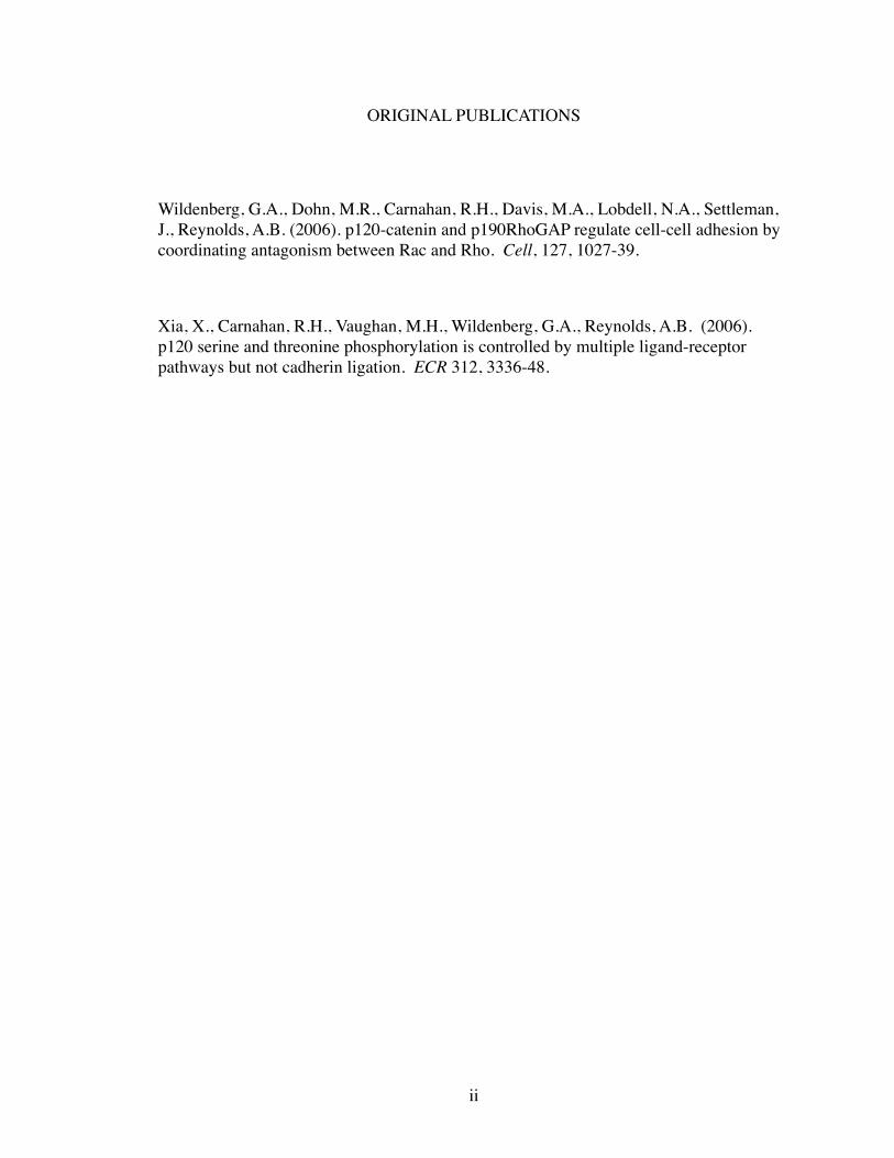
ORIGINAL PUBLICATIONS
Wildenberg, G.A., Dohn, M.R., Carnahan, R.H., Davis, M.A., Lobdell, N.A., Settleman, J., Reynolds, A.B. (2006). p120-catenin and p190RhoGAP regulate cell-cell adhesion by coordinating antagonism between Rac and Rho. Cell, 127, 1027-39.
Xia, X., Carnahan, R.H., Vaughan, M.H., Wildenberg, G.A., Reynolds, A.B. (2006). p120 serine and threonine phosphorylation is controlled by multiple ligand-receptor pathways but not cadherin ligation. ECR 312, 3336-48.
ii

DEDICATION
To truth, understanding,
&
the humility to know the difference
between the two
iii

ACKNOWLEDGMENT
Life is defined by moments of sadness and joy, moments of successes and fail-
ures, moments of wrong and right. If moments provide definition, then it is the people
around us that give us purpose. My graduate work has been no exception to this. While
the complete number of people and the extent of their influence is beyond words and
limit, there are a few that need to be acknowledged.
First, I would like to thank my mentor, Al Reynolds. He has taught me as much
about being a scientist as he has being a person. Al has been by my side not as an in-
structor or overseer, but rather as one who praises abilities and encourages to challenge
weaknesses. In that simple guidance, I have learned an immeasurable skill in the neces-
sity to challenge oneself before challenging a scientific question. The extent of his influ-
ence on me will go far beyond graduate school and display itself to a far greater circum-
ference than my scientific career to which I can offer my thanks and appreciation.
I also need to thank my wife, Mary. She is the love of my life, and has supported
me through so many days of frustration, stress, anxiety, and exuberance. Through her
thoughtfulness and sincerity, I have acquired balance in my life that everyone needs to be
successful with any endeavor. She challenges me to be a better person, and to continually
grow with each day. Without her, not only would I have little to show for my accom-
plishments, but I would be without my best friend to share all of my happiness with.
iv

Lastly, I would like to thank the support of my committee, Lynn Matrisian, Vito
Quaranta, Alissa Weaver, and Anne Kenworthy. Although our interactions were few, they
have served as models of success in science. I have paid careful attention and understood
how they have each applied their own perspectives and unique talents to their respective
passions. From them, I have learned a great deal on how to apply my perspectives and
my passions to my future in science.
My graduate work has been supported by the Training Program in Breast Cancer
Research, and p120 Cell-Cell Adhesion NIH RO1.
v
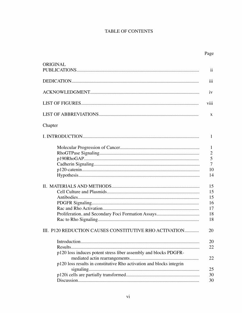
TABLE OF CONTENTS
Page
ORIGINAL PUBLICATIONS....................................................................................................... ii
DEDICATION........................................................................................................... iii ACKNOWLEDGMENT............................................................................................ iv
LIST OF FIGURES................................................................................................... viii
LIST OF ABBREVIATIONS.................................................................................... x
Chapter I. INTRODUCTION.................................................................................................. 1
Molecular Progression of Cancer................................................................... 1 RhoGTPase Signaling.................................................................................... 2 p190RhoGAP................................................................................................. 5 Cadherin Signaling......................................................................................... 7 p120-catenin................................................................................................... 10 Hypothesis...................................................................................................... 14
II. MATERIALS AND METHODS.......................................................................... 15 Cell Culture and Plasmids.............................................................................. 15 Antibodies...................................................................................................... 15 PDGFR Signaling.......................................................................................... 16 Rac and Rho Activation................................................................................. 17 Proliferation, and Secondary Foci Formation Assays.................................... 18 Rac to Rho Signaling..................................................................................... 18 III. P120 REDUCTION CAUSES CONSTITUTIVE RHO ACTIVATION............ 20 Introduction.................................................................................................... 20 Results............................................................................................................ 22 p120 loss induces potent stress fiber assembly and blocks PDGFR- mediated actin rearrangements.......................................................... 22 p120 loss results in constitutive Rho activation and blocks integrin signaling............................................................................................. 25 p120i cells are partially transformed.............................................................. 30 Discussion...................................................................................................... 30
vi
IV. P120 IS ESSENTIAL FOR RAC TO RHO SIGNALING................................. 37
Introduction.................................................................................................... 37 Results............................................................................................................ 40 Rac needs p120 to inhibit Rho....................................................................... 40 Rac induces p190 localization to N-cadherin complexes.............................. 42 Rac-induced recruitment of p190 to N-cadherin is dependent on p120................................................................................................... 46 p190 and p120 interact................................................................................... 53 p190 is necessary for Rac-induced adherens junction formation.................. 53 Discussion...................................................................................................... 56
V. ROLE OF P120 AND P190 IN CONTACT INHIBITION OF GROWTH AND CELLULAR TRANSFORMATION............................................................. 77
Introduction.................................................................................................... 77 Results............................................................................................................ 79 p120i cells have increased MAPK signaling................................................. 79 Confluency regulates Rho activity................................................................. 79 Src regulates the interaction between p120 and p190.................................... 82 Discussion...................................................................................................... 85
VI. FUTURE DIRECTIONS.................................................................................... 88
Introduction.................................................................................................... 88 p190 localization in live cells........................................................................ 89 Determine the binding sites of p120 and p190.............................................. 89 Role of p120/p190 complex in Src transformation........................................ 90 Role of AJ formation in Ras signaling........................................................... 91
VIII. CONCLUDING REMARKS........................................................................... 93
REFERENCES.......................................................................................................... 95
vii
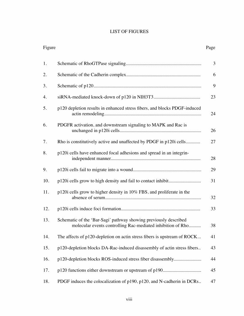
LIST OF FIGURES
Figure Page
1. Schematic of RhoGTPase signaling............................................................... 3
2. Schematic of the Cadherin complex.............................................................. 6
3. Schematic of p120.......................................................................................... 9
4. siRNA-mediated knock-down of p120 in NIH3T3....................................... 23
5. p120 depletion results in enhanced stress fibers, and blocks PDGF-induced actin remodeling................................................................................. 24
6. PDGFR activation, and downstream signaling to MAPK and Rac is unchanged in p120i cells.................................................................... 26
7. Rho is constitutively active and unaffected by PDGF in p120i cells............ 27
8. p120i cells have enhanced focal adhesions and spread in an integrin- independent manner........................................................................... 28
9. p120i cells fail to migrate into a wound......................................................... 29
10. p120i cells grow to high density and fail to contact inhibit........................... 31
11. p120i cells grow to higher density in 10% FBS, and proliferate in the absence of serum................................................................................ 32
12. p120i cells induce foci formation.................................................................. 33
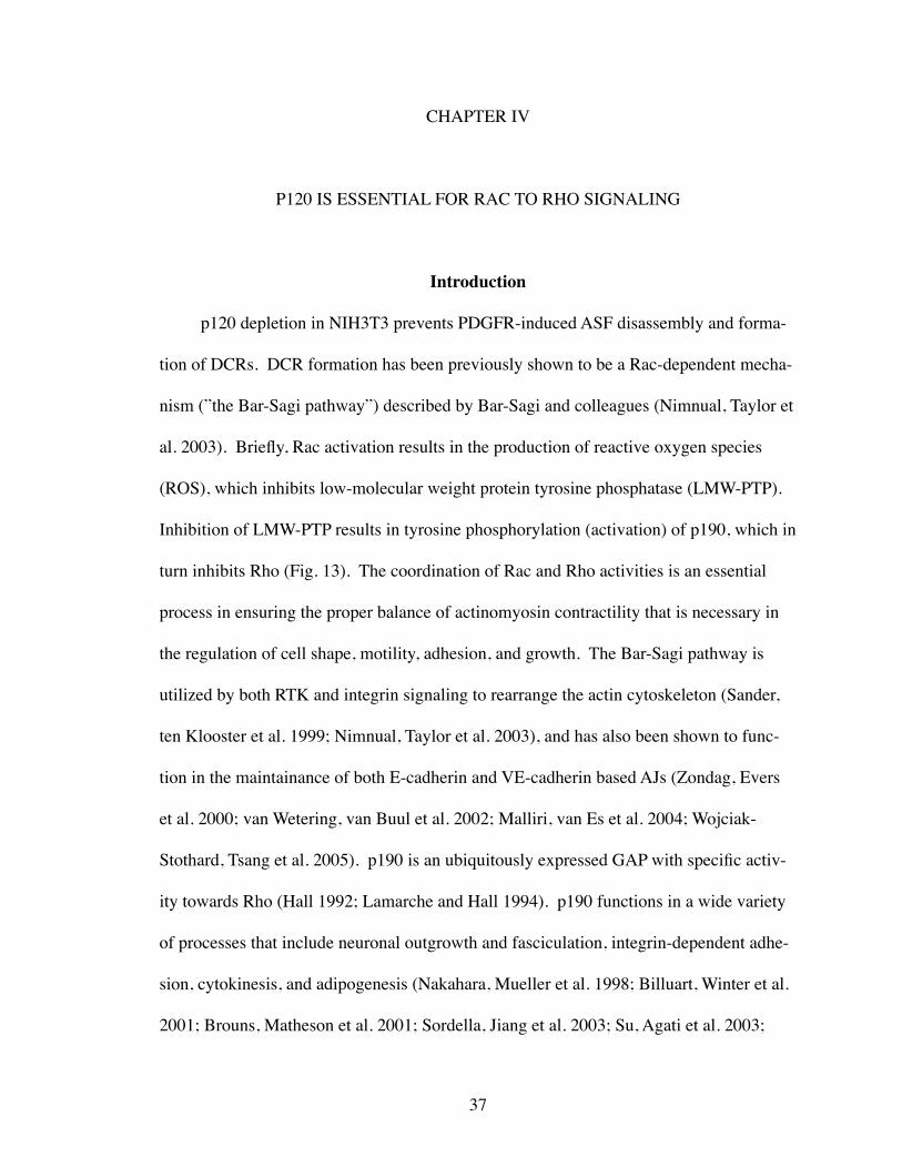
13. Schematic of the ‘Bar-Sagi’ pathway showing previously described molecular events controlling Rac-mediated inhibition of Rho.......... 38
14. The affects of p120-depletion on actin stress fibers is upstream of ROCK... 41
15. p120-depletion blocks DA-Rac-induced disassembly of actin stress fibers.. 43
16. p120-depletion blocks ROS-induced stress fiber disassembly....................... 44
17. p120 functions either downstream or upstream of p190................................ 45
18. PDGF induces the colocalization of p190, p120, and N-cadherin in DCRs.. 47
viii
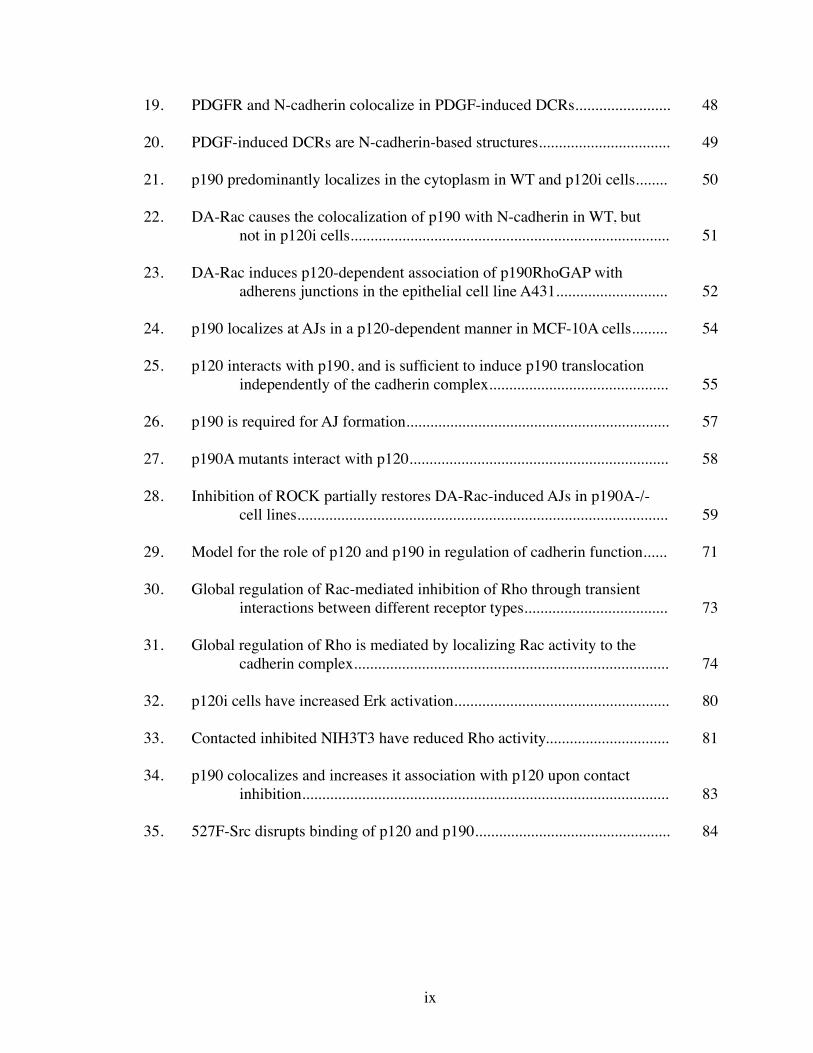
19. PDGFR and N-cadherin colocalize in PDGF-induced DCRs........................ 48
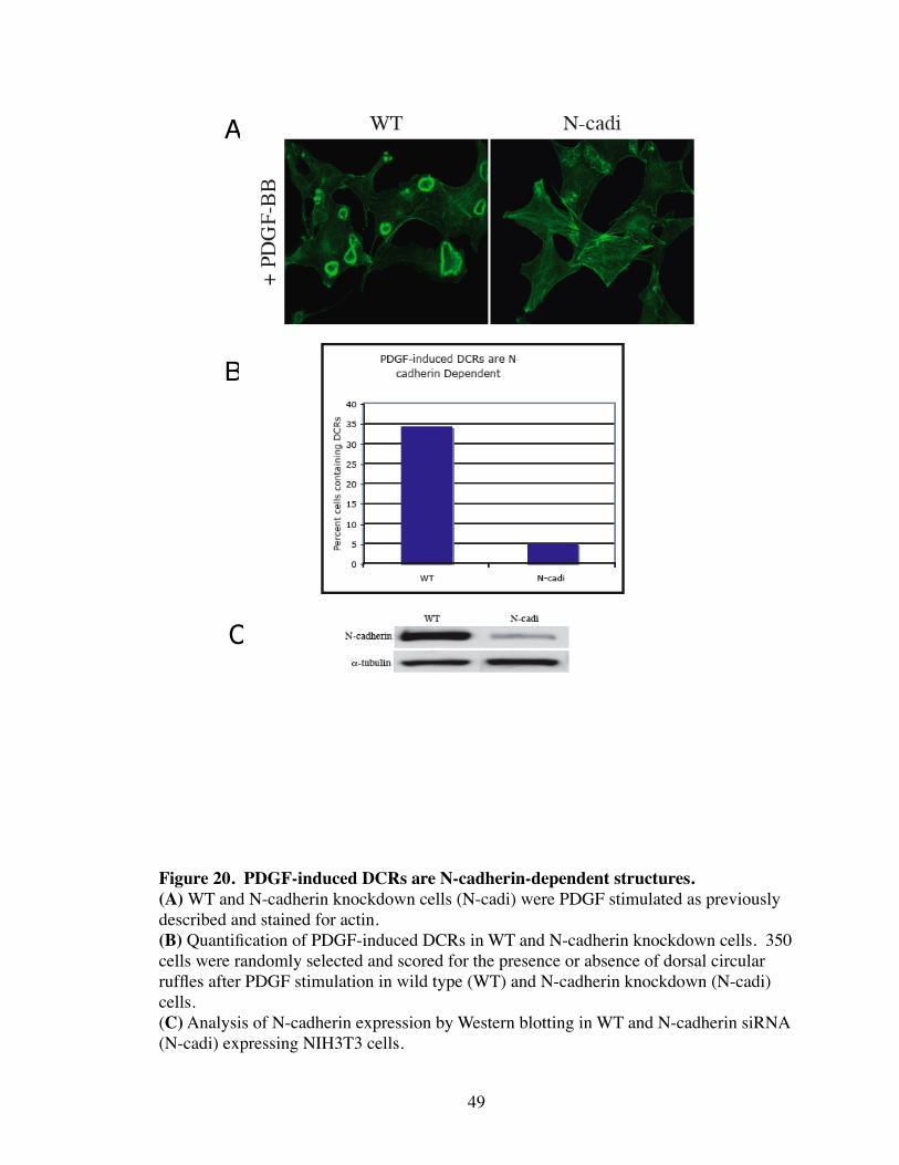
20. PDGF-induced DCRs are N-cadherin-based structures................................. 49
21. p190 predominantly localizes in the cytoplasm in WT and p120i cells........ 50
22. DA-Rac causes the colocalization of p190 with N-cadherin in WT, but not in p120i cells................................................................................ 51
23. DA-Rac induces p120-dependent association of p190RhoGAP with adherens junctions in the epithelial cell line A431............................ 52
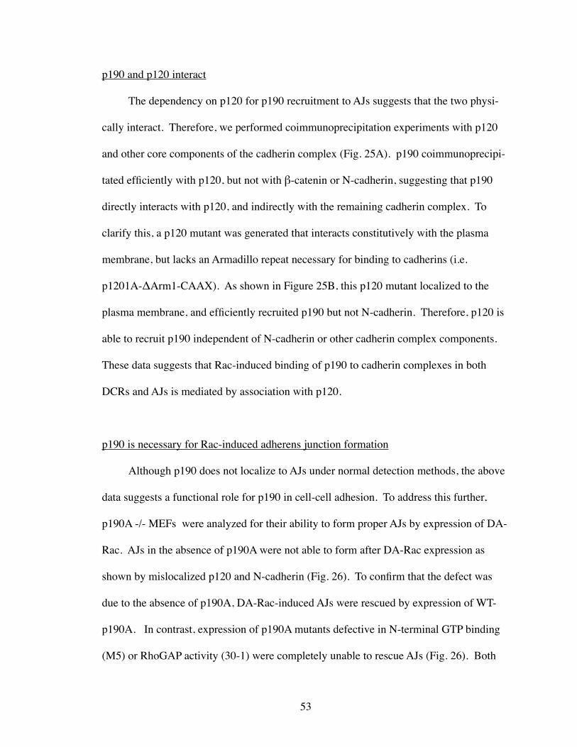
24. p190 localizes at AJs in a p120-dependent manner in MCF-10A cells......... 54
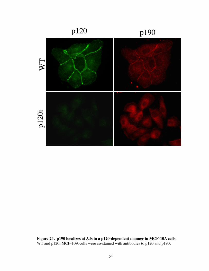
25. p120 interacts with p190, and is sufficient to induce p190 translocation independently of the cadherin complex............................................. 55
26. p190 is required for AJ formation.................................................................. 57
27. p190A mutants interact with p120................................................................. 58
28. Inhibition of ROCK partially restores DA-Rac-induced AJs in p190A-/- cell lines............................................................................................. 59
29. Model for the role of p120 and p190 in regulation of cadherin function...... 71
30. Global regulation of Rac-mediated inhibition of Rho through transient interactions between different receptor types.................................... 73
31. Global regulation of Rho is mediated by localizing Rac activity to the cadherin complex............................................................................... 74
32. p120i cells have increased Erk activation...................................................... 80
33. Contacted inhibited NIH3T3 have reduced Rho activity............................... 81
34. p190 colocalizes and increases it association with p120 upon contact inhibition............................................................................................ 83
35. 527F-Src disrupts binding of p120 and p190................................................. 84
ix
LIST OF ABBREVIATIONS
AJ Adherens junction
ASF Actin stress fiber
DCR Dorsal circular ruffles
EC Extracellular domain
ECM Extracellular matrix
EMT Epithelial-mesynchemal transistion
FA Focal adhesion
GAP GTPase activating protein
GEF Guanine exchange factor
GDI Guanine nucleotide dissociation inhibitor
GCPR G-coupled protein receptor
HGF Hepatocyte growth factor
LMW-PTP Low molecular weight protein tyrosine phosphatase
LPA Lysophosphatidic acid
MAPK Mitogen activated protein kinase
MEF Murine embryonic fibroblasts
p120 p120-catenin
p190 p190RhoGAP
PAK p21-activated kinase
PDGFR Platelet derived growth factor receptor
Rac Rac1
x

Rho RhoA
ROS Reactive oxygen species
RTK Receptor tyrosine kinase
xi
CHAPTER I
INTRODUCTION
Molecular Progression of Cancer
Tumorigenesis is a multistep process that involves genetic variations that drive the
transformation of a normal cell to malignancy (Hanahan and Weinberg 2000). During
this process tumor cells undergo clonal expansion whereby cells harboring certain genetic
changes are selected for their ability to grow in an unregulated fashion. Eventually, se-
lected populations of cells invade the surrounding basement membrane and enter the vas-
culature where they metastasize systemically. Meanwhile, the primary tumor also devel-
ops its own vascular system to supply itself with the necessary nutrients for continued
growth. Although there are numerous distinct types of cancer, they are all governed by
six general principles. These are self-sufficiency in growth signals, insensitivity to anti-
growth signals, evasion of apoptosis, immortalization, angiogenesis, and invasion/
metastasis (Hanahan and Weinberg 2000). Conversely, the tight regulation of these
processes is essential for normal cellular function.
The cancer phenotype is generally represented by a misregulation in growth factor
and cell adhesion (cell-cell and cell-extracellular matrix (ECM)) signaling. Historically,
a reductionist approach has been taken to isolate the individual contributions of different
receptors in mediating the biological responses essential to normal cellular function.
Through this approach, major contributions have been made in eliciting key mechanisms
in the regulation of growth and motility. However, it has become apparent that biological
1
responses are not simply products of individual and/or parallel signaling pathways re-
layed from multiple receptors. Rather, receptor systems mediate cross-talk in order to
carry out the multiple events that are simultaneously necessary for cellular motility,
growth, and adhesion. For example. in suspended cells (i.e. without cell-ECM adhesion),
activation of Rac1 (Rac) by growth factors is unable to activate its downstream effector,
p21-activated kinase (PAK), suggesting that integrins are necessary to properly localize
the Rac activity that comes from receptor tyrosine kinase (RTK) signaling (del Pozo,
Price et al. 2000). Similarly, E-cadherin-mediated activation of Rac and mitogen acti-
vated protein kinase (MAPK) depends on a functional epidermal growth factor receptor
(EGFR) (Pece and Gutkind 2000; Betson, Lozano et al. 2002). Finally, Src-induced de-
regulation of adherens junctions (AJs) requires integrin assembly of focal adhesion com-
plexes (Avizienyte, Wyke et al. 2002). Thus, a cellular event such as AJ formation does
not occur autonomously through cadherin clustering, but rather requires the rearrange-
ment and cooperation of multiple receptor systems.
RhoGTPase Signaling
The small Rho family GTPases Rac and RhoA (Rho) play critical roles in coupling
growth factor and adhesion receptor signaling to rearrangements in the actin cytoskeleton
during motility, adhesion, and growth (Burridge and Wennerberg 2004). RhoGTPases
cycle from the inactive, GDP bound form, to the active, GTP bound form, and localize
from the cytoplasm to the cell membrane, respectively (Hall 1992). The activity of
RhoGTPases is tightly controlled by three groups of proteins. These are Guanine Ex-
change Factors (GEFs), GTPase Activating Proteins (GAPs), and Guanine nucleotide
2
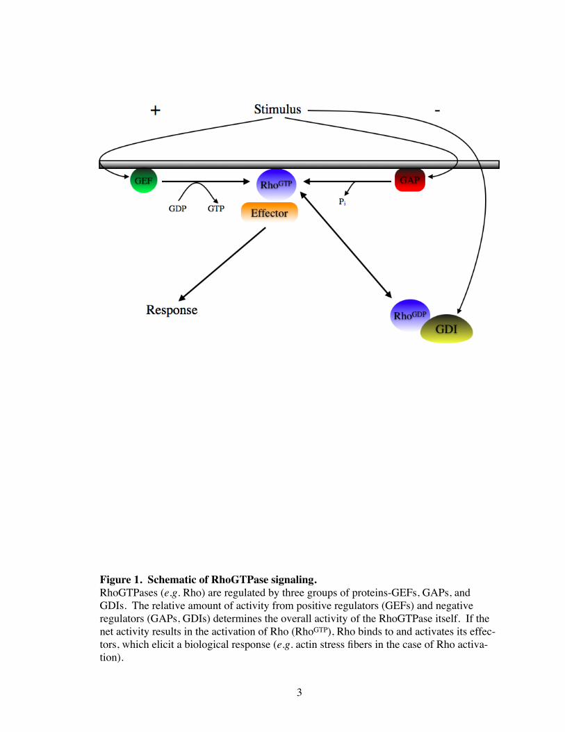
3
Figure 1. Schematic of RhoGTPase signaling.RhoGTPases (e.g. Rho) are regulated by three groups of proteins-GEFs, GAPs, and GDIs. The relative amount of activity from positive regulators (GEFs) and negative regulators (GAPs, GDIs) determines the overall activity of the RhoGTPase itself. If the net activity results in the activation of Rho (RhoGTP), Rho binds to and activates its effec-tors, which elicit a biological response (e.g. actin stress fibers in the case of Rho activa-tion).
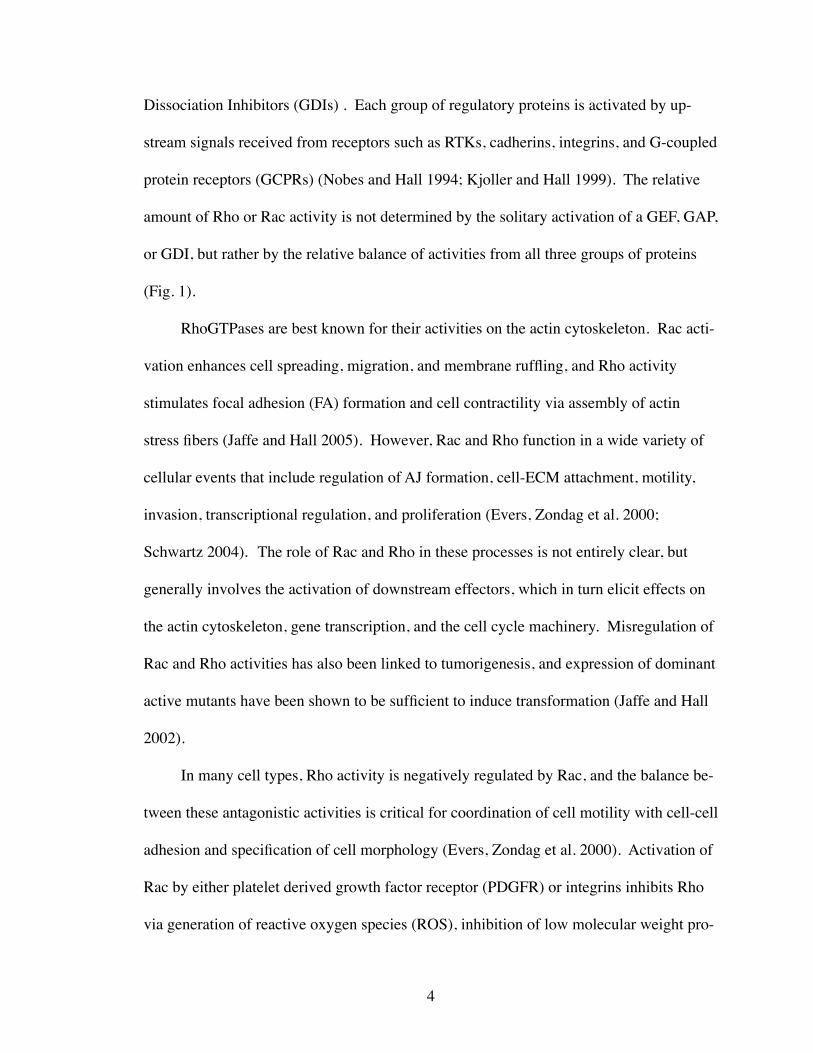
Dissociation Inhibitors (GDIs) . Each group of regulatory proteins is activated by up-
stream signals received from receptors such as RTKs, cadherins, integrins, and G-coupled
protein receptors (GCPRs) (Nobes and Hall 1994; Kjoller and Hall 1999). The relative
amount of Rho or Rac activity is not determined by the solitary activation of a GEF, GAP,
or GDI, but rather by the relative balance of activities from all three groups of proteins
(Fig. 1).
RhoGTPases are best known for their activities on the actin cytoskeleton. Rac acti-
vation enhances cell spreading, migration, and membrane ruffling, and Rho activity
stimulates focal adhesion (FA) formation and cell contractility via assembly of actin
stress fibers (Jaffe and Hall 2005). However, Rac and Rho function in a wide variety of
cellular events that include regulation of AJ formation, cell-ECM attachment, motility,
invasion, transcriptional regulation, and proliferation (Evers, Zondag et al. 2000;
Schwartz 2004). The role of Rac and Rho in these processes is not entirely clear, but
generally involves the activation of downstream effectors, which in turn elicit effects on
the actin cytoskeleton, gene transcription, and the cell cycle machinery. Misregulation of
Rac and Rho activities has also been linked to tumorigenesis, and expression of dominant
active mutants have been shown to be sufficient to induce transformation (Jaffe and Hall
2002).
In many cell types, Rho activity is negatively regulated by Rac, and the balance be-
tween these antagonistic activities is critical for coordination of cell motility with cell-cell
adhesion and specification of cell morphology (Evers, Zondag et al. 2000). Activation of
Rac by either platelet derived growth factor receptor (PDGFR) or integrins inhibits Rho
via generation of reactive oxygen species (ROS), inhibition of low molecular weight pro-
4
tein tyrosine phosphatase (LMW-PTP), and tyrosine phosphorylation (activation) of
p190RhoGAP (p190), a major GAP for Rho (Sander, ten Klooster et al. 1999; Nimnual,
Taylor et al. 2003). In the case of PDGFR, this pathway mediates rapid disassembly of
actin stress fibers (ASFs) and formation of dorsal circular ruffles (DCRs). Additionally,
Rac inhibition of Rho induces “epithelialization” of cells by promoting AJ assembly and
suppression of stress fiber-induced contractility (Sander, ten Klooster et al. 1999; Malliri,
van Es et al. 2004; Wojciak-Stothard, Tsang et al. 2005).
p190RhoGAP
p190 is a ubiquitously expressed GAP that exhibits specificity for Rho. It was
originally discovered as a binding partner of p120RasGAP (RasGAP) whose association
is induced by tyrosine phosphorylation (Bernards and Settleman 2005). Although asso-
ciation of p190 with RasGAP does not directly affect the catalytic GAP activity of p190,
recent evidence suggests that it may affect the subcellular localization of p190 (Bradley,
Hernandez et al. 2006). In addition to a GAP domain, p190 contains a N-terminal GTP
binding domain. The function of this domain is not known, but the ability of p190 to
bind GTP has been shown to be required for its activity on Rho (Foster, Hu et al. 1994;
Tatsis, Lannigan et al. 1998; Roof, Dukes et al. 2000).
Activation of p190 is mediated by RTKs, integrins, and cadherins (Ellis, Moran et
al. 1990; Arthur and Burridge 2001; Noren, Arthur et al. 2003). Upon activation, p190
becomes phosphorylated and translocates from the cytoplasm to membranes and/or actin
cytoskeletal structures where it can access activated Rho (Sharma 1998; Arthur and Bur-
ridge 2001; Sordella, Jiang et al. 2003). Thus, both p190 tyrosine phosphorylation and
5
6
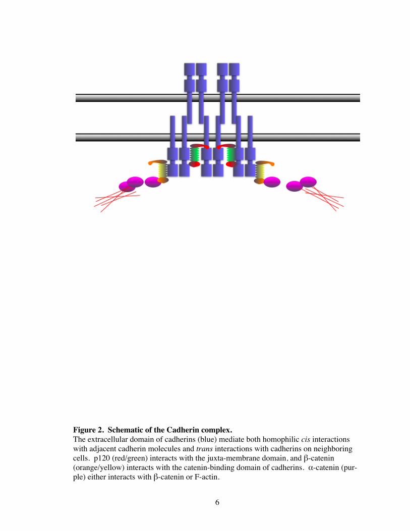
Figure 2. Schematic of the Cadherin complex.The extracellular domain of cadherins (blue) mediate both homophilic cis interactions with adjacent cadherin molecules and trans interactions with cadherins on neighboring cells. p120 (red/green) interacts with the juxta-membrane domain, and β-catenin (orange/yellow) interacts with the catenin-binding domain of cadherins. α-catenin (pur-ple) either interacts with β-catenin or F-actin.
translocation are necessary for proper p190 function. p190 has been localized to focal
adhesions and lipid rafts, but has not been associated with any other cellular structures
(Sharma 1998; Sordella, Jiang et al. 2003). p190 has been shown to function in integrin-
mediated spreading, neuron outgrowth, and fasciculation, but the contribution of p190 to
RTK and cadherin signaling remains largely unexplored (Arthur and Burridge 2001;
Brouns, Matheson et al. 2001).
Cadherin Signaling
Cadherin function
Cadherins comprise a superfamily of transmembrane adhesion receptors that medi-
ate Ca+2-dependent cell-cell adhesion. Classical cadherins (i.e. E-, N-, VE-, and R-
cadherin) are the most widely studied of the cadherin superfamily, and are often associ-
ated with various forms of AJs. Cadherins play critical roles in development, motility,
growth suppression, polarization, and tissue morphogenesis (Yap, Brieher et al. 1997).
Cadherins form lateral dimers, which then mediate homophilic interactions through re-
peating extracellular (EC) domains with cadherins of a nearby cell (Gumbiner 2005). Al-
though cell-cell adhesion is mediated by the extracellular domain of cadherins, it is not
sufficient to induce the strong adhesion observed by cadherin clustering (Yap, Brieher et
al. 1997; Yap, Niessen et al. 1998). Instead, the catenins (α, β, and p120-catenin), which
directly associate with the cadherin cytoplasmic tail, are thought to regulate AJ assembly
through functional but poorly understood interactions with RhoGTPases and the actin
cytoskeleton (Yap, Niessen et al. 1998; Braga 2002) (Fig 2).
7
Cadherin Regulation
Regulation of AJ assembly is a dynamic process that involves post-translational
modification of the cadherin and associated catenins. Though poorly understood, cellular
signals such as those from growth factors, transiently influence the ability of cadherins to
form stable AJs. Phosphorylation of the catenins by RTKs or non-receptor tyrosine ki-
nases such as Src, Fyn, or Abl have been implicated in this process as phosphorylation of
catenins have been correlated to either a loss or strengthening of cell-cell adhesion
(Roura, Miravet et al. 1999; Piedra, Miravet et al. 2003). Likewise, phosphatases have
been linked to this process (Xu, Craig et al. 2004; Sallee, Wittchen et al. 2006). Activity
by kinases or phosphatases on catenins is associated with changes in the ability of the
cadherins to cluster, though the mechanisms are not known.
Loss of E-cadherin expression in epithelial-derived tumors is closely correlated
with the transition to metastasis. Restoration of E-cadherin expression has been shown to
reduce or block metastasis both in vitro and in vivo (Frixen, Behrens et al. 1991; Oka,
Shiozaki et al. 1993). In addition to the direct loss of E-cadherin expression, cell-cell ad-
hesion can be dysregulated through posttranslational mechanisms such as phosphoryla-
tion. Aberrant signaling from oncogenes such as Src, or different growth factors such as
hepatocyte growth factor (HGF), and platelet derived growth factor (PDGF) can nega-
tively affect the ability to form stable AJs (Matsuyoshi, Hamaguchi et al. 1992; Watabe,
Matsumoto et al. 1993; Piedra, Miravet et al. 2003; Uglow, Slater et al. 2003). Altera-
tions in these signaling pathways in transformed cells, therefore, could result in a loss of
cadherin-dependent adhesion and increased invasiveness. Thus, misregulation of the
8
9
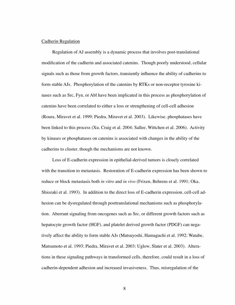
Figure 3. Schematic of p120.p120 contains a central Armadillo repeat domain containing nine Arm repeats. Several different isoforms of p120 are expressed due to four alternative start codons in the N-terminus, and three C-terminal exons. Isoform 1 possesses a coiled-coil domain, which mediates protein-protein interactions. Tyrosine and serine/threonine phosphorylation sites are concentrated in the N-terminal regulatory domain. p120 also possess nuclear localization sequences (NLS) and a nuclear export sequence (NES).
cadherin function can occur through a variety of means, and represents a major signaling
defect during tumor progression.
p120-catenin
p120 family and localization
p120-catenin (p120) was originally identified as a Src substrate, and is the proto-
typic member of an Arm domain subfamily that includes ARVCF, δ-catenin, and p0071.
All family members are ubiqutiously expressed with the exception of δ-catenin, which is
expressed specifically in neuronal cells. Structurally, all of these proteins contain an N-
terminal coiled-coiled motif, and all but p120 have a C-terminal PDZ-binding motif. The
internal Armadillo domain contains nine Arm repeats, which mediates protein-protein
interactions (Anastasiadis and Reynolds 2000) (Fig. 3). In contrast to β-catenin which
binds to the catenin binding domain (CBD) of cadherins, p120 binds to the juxtamem-
brane domain (JMD) (Thoreson, Anastasiadis et al. 2000). Since p120 does not physi-
cally link cadherins to the actin cytoskeleton, p120 appears to modulate assembly and
disassembly of AJs by an indirect mechanism. Additionally, unlike β-catenin, which is
rapidly degraded when sequestered in the cytoplasm, cytoplasmic p120 is stable. It has
been proposed that cytoplasmic p120 inhibits Rho, but the mechanism is not known (see
below) (Anastasiadis, Moon et al. 2000; Noren, Liu et al. 2000; Grosheva, Shtutman et al.
2001). Lastly, p120 can localize to the nucleus where it negatively regulates the tran-
scriptional repressor, Kaiso (Daniel and Reynolds 1999; Daniel, Spring et al. 2002;
Spring, Kelly et al. 2005).
10
p120 isoforms
p120 expresses multiple isoforms, which are derived by alternative splicing of a
single gene. Splicing occurs at the N-terminus due to different start codons (1, 2, 3), and
C-terminally spliced at sites of 3 exons (A, B, C) (Reynolds, Daniel et al. 1994; Keirse-
bilck, Bonne et al. 1998). Interestingly, fibroblasts predominantly express the full length
1A isoform, whereas epithelial cells express the slightly shorter, 3A isoform. These iso-
forms differ by the presence in isoform 1A of an N-terminal coiled-coiled domain, which
typically mediate protein-protein interactions (Fig. 3). The N-terminus has been linked to
a role in promoting motility and invasion (Cozzolino, Stagni et al. 2003; Yanagisawa and
Anastasiadis 2006). However, in many tumor cell lines, p120 isoform expression is quite
heterogeneous (Mo and Reynolds 1996), which may reflect a role for isoform switching
promoting tumor progression.
p120 phosphorylation
Phosphorylation undoubtedly regulates the function of p120 most likely by con-
necting various signaling pathways to the dynamic regulation of cell-cell adhesion.
However, due to the multiple phosphorylation sites (both tyrosine, and serine/threonine)
on p120, and the multiple kinases that can phosphorylate p120, it has been difficult to de-
termine the role of p120 phosphorylation (Reynolds, Roesel et al. 1989; Downing and
Reynolds 1991; Mariner, Anastasiadis et al. 2001). Probably, individual phosphorylation
sites serve as substrates for specific kinases, and the net phosphorylation of p120 received
by multiple signals ultimately regulates the the adhesive strength of a cell. Interestingly,
11
expression of isoform 4A, which lacks almost all of the tyrosine and serine/threonine
sites, in the SW48 cell line strengthens adhesion better than isoform 1A or 3A, which
contain all of the phosphorylation sites (Ireton, Davis et al. 2002). This indicates that
phosphorylation is not necessary for p120 to stabilize cadherins, but perhaps it is neces-
sary for negative regulation of adhesion.
p120 function
We and others have recently demonstrated that p120 is required for cadherin stabil-
ity. Using the p120-deficient colon carcinoma cell line, SW48, it was found that re-
expression of p120 increased E-cadherin levels and restored an epithelial morphology
(Ireton, Davis et al. 2002). Structure-function analysis revealed that restoration of E-
cadherin levels requires direct p120-cadherin interaction. Further studies demonstrated
that p120 mediates cadherin stability, thus dynamically regulating the amount of cadherin
available at the cell surface for adhesion (Xiao, Allison et al. 2003). Follow up studies
using siRNA-mediated depletion of p120 showed that this is indeed a general mechanism
for p120 in stabilizing all classical cadherin types in different cell lines (Davis, Ireton et
al. 2003). In vivo, p120-targeted ablation in the salivary gland also leads to about a 50%
reduction in E-cadherin protein levels, and is associated with severe alterations in epithe-
lial morphology (Davis and Reynolds 2006). These observations suggest that p120 me-
diates cadherin turnover, and that adhesion may be controlled by phosphorylation events
that impact p120 to dynamically regulate AJ assembly and disassembly.
In addition to its role in stabilizing cadherins, overexpression of p120 causes exten-
sive, atypical branching through the inhibition of Rho (Anastasiadis, Moon et al. 2000;
12
Noren, Liu et al. 2000; Grosheva, Shtutman et al. 2001). These data indicated that p120
inhibits Rho in the cytoplasm, and a GDI-like mechanism was proposed based on the
ability of purified p120 to stabilize the GDP-bound form of Rho in vitro (Anastasiadis,
Moon et al. 2000). Developmental studies in Xenopus lavis as well as conditional knock-
out studies done in the epidermis of mice confirm a functional relationship between p120
and Rho, but the mechanism for this interaction is not known (Fang, Ji et al. 2004; Perez-
Moreno, Davis et al. 2006) . Furthermore, the Drosophila homologue of p120 interacts
with the GDP-bound form of Rho1 (Drosophilia RhoA) (Magie, Pinto-Santini et al.
2002). Recently, work done by Yanagisawa et al in the invasive adenocarcinoma cell line
MDA-MB-231, demonstrated that p120 can promote Rac activation through interaction
with cadherin-11, and Rho inhibition in a cadherin-independent manner (Yanagisawa and
Anastasiadis 2006), however, no mechanism was defined. Although little is known re-
garding the mechanism or functional consequence, p120 clearly regulates the activity of
Rho.
Presently, the roles of p120 in adhesion and Rho inhibition appear to be separate
functions. It has been postulated that the relative amount of p120 bound to a cadherin
may regulate the degree of cytoplasmic p120 available to inhibit Rho. Alternatively,
p120 may function to deliver inactive Rho from the cytoplasm to the plasma membrane
where it becomes activated locally by a GEF. Phosphorylation of p120 would ultimately
serve then to regulate the ratio of cadherin bound/cytoplasmic p120 and thus adhesion
and Rho inhibition. These models suggests that the role of p120 in cadherin-stabilization
and Rho-inhibition are mutually exclusive. However, it has not been ruled out that these
two processes are functionally linked. Indeed, RhoGTPases play critical roles in AJ as-
13
sembly and disassembly (Kuroda, Fukata et al. 1997; Sander, van Delft et al. 1998; Lo-
zano, Betson et al. 2003). The contribution of either Rac or Rho to AJ formation is com-
plex, and apparently depends on the maturity of cell-cell contacts. For example, Rac lo-
calizes to and is necessary for nascent cell-cell adhesions, but does not localize to more
mature contacts (Braga, Del Maschio et al. 1999; Ehrlich, Hansen et al. 2002). As fore
mentioned, Rac can “epithelialize” fibroblasts by stabilizing N-cadherin-based AJs sug-
gesting that Rac drives the formation and stabilization of cell-cell adhesions. Indeed,
other reports have suggested a similar role for Rac in epithelial cells (Kuroda, Fukata et
al. 1997; Takaishi, Sasaki et al. 1997). On the other hand, Rho activity has been associ-
ated with a more fibroblastic phenotype that is associated with poor cell-cell adhesions
(Zhong, Kinch et al. 1997). Although the activities of Rac and Rho clearly affect AJ for-
mation, how these activities are regulated at the cadherin complex remains unknown.
Given the affect p120 has on both cell-cell adhesion and Rho activity, it is likely that
p120 may coordinate these two processes.
Hypothesis
My working hypothesis is that p120 inhibition of Rho functions in the regulation of
AJ assembly.
14
CHAPTER II
MATERIALS AND METHODS
Cell Culture and plasmids
NIH3T3 and p190A-/- MEF cells were cultured in DMEM/High Glucose, 10% Fe-
tal Bovine Serum (FBS), and 1% penicillin/streptomycin. Clonal p120-knockdown cell
lines were generated by serial dilution. Rac1 and C3 constructs were PCR amplified from
PcDNA3 and subcloned into LZRS-ms-GFP. p190-A cDNAs were subcloned from Rc-
HAp190-A or MFG into LZRS-ms-GFP or LZRS-ms-neo. LZRS-ms-neo/N-cadherin was
a gift from Dr. Margaret Wheelock. p190A constructs and null MEFs were a gift from
Jeff Settleman. Retroviral vectors and siRNA for p120 and N-cadherin have been de-
scribed (Davis, Ireton et al. 2003).
Antibodies
Secondary antibodies were Alexa-Fluor conjugates from Molecular Probes. mAbs
15D2, KT3, and 8D11 were generated in our lab. mAbs pp120 (cat # 610136), N-
cadherin (610921), Rac1 (610650), p190 (610150), and anti-phosphotyrosine (PY20)
(610000) were from BD Biosciences. mAb anti-RhoA (sc-418) and pab anti-N-cadherin
(sc-7939) was from Santa Cruz. Y-27632 (Y0503), fibronectin (F2006), H202 (H1009), and
mAbs anti-p120RasGAP (B4F8), α-tubulin (T5168), and vinculin (V4139) were from
Sigma-Aldrich. mAbs p-p38 (9216), p38 (9212), MAPK (9102), and p-MAPK (9106)
15
were from Cell Signaling. PDGF-BB (GF018) was from Chemicon, and PDGFRβ (97A)
antibody was from Dr. Andrius Kazlauskas.
PDGFR Signaling
Immunoflourescent (IF) methods have been described (Davis, Ireton et al. 2003).
Cells were fixed/permeabilized in 3% paraformaldehyde/0.2% Triton X-100, and stained
for 30 min. For Dorsal Circular Ruffle formation, cells were serum starved for 24 hrs
(0.2% FBS), and stimulated with 20-50 ng/ml of PDGF-BB for 7 min. Immunoprecipita-
tion (IP) and Western Blot (WB) methods were as described (Davis, Ireton et al. 2003).
Briefly, cells were lysed in RIPA buffer, protein concentrations equilibrated by BCA as-
say. For IP, cells were lysed, nutated at 40C for 5 min. in a 1.5 ml eppendorf tube, and
then passed through a 20 1.1/2 gauge syringe 15 times. Cell debris was pelleted by cen-
trifugation, and supernatant was transferred to a new eppendorf tube. Equal amounts of
protein were IP’ed with indicated antibodies overnight at 4oC. Complexes were collected
with Protein G-Sepharose, for 1.5 hrs, transferred to a new eppendorf tube, washed, and
processed for WB. For p38 analysis, serum starved cells were stimulated with 50ng/ml
of PDGF-BB for 7 min. PDGFRβ was immunoprecipitated from serum starved cells after
stimulation with 50 ng/ml of PDGF-BB for 7 min.
16
Rac and Rho Activation
Rhotekin/ PAK Assay and ROCK inhibition
Rhotekin and PAK assays were performed as described (Anastasiadis, Moon et al.
2000). Briefly, serum starved cells were stimulated with 20 ng/ml of PDGF-BB for 3-5
min or 5µM of LPA for 15 min. Cells were lysed in Lysis Buffer (50 mM Tris, pH 7.2,
1% Triton X-100, 0.5% sodium deoxycholate, 0.1% SDS, 500mM NaCL, 10mM MgCl2,
1mM PMSF, 5 µg/ml leupeptin, and 2 µg/ml aprotinin), 15 µl of lysate was used for
analysis of total protein levels, and remaining lysate was incubated with 30 µg of GST-
RBD or GST-PBD (Cytoskeleton Inc.) to IP activated forms of RhoA or Rac1. IPs were
washed three times in Wash Buffer (50mM Tris, pH 7.2, 1% Triton X-100, 150 mM
NaCl, 10mM MgCl2, 1mM PMSF, 5 µg/ml leupeptin, and 2 µg/ml aprotinin). Levels of
total and activated RhoA or Rac1 were analyzed by WB. To inhibit ROCK, 10 µM Y-
27632 was added to cells for 30 min.
RhoA affects on integrin signaling
In spreading assays, cells were serum starved, trypsinized, washed, and replated on
uncoated or coated glass coverslips (10 µg/ml of Fibronectin) for 20 min. For wound as-
says, confluent cells were scratched with a plastic pipette tip, and washed in PBS. Nor-
mal growth media was added and cells were incubated under normal growth conditions.
Images of multiple wound regions were taken every 12 hours, and the distance of each
wound was calculated, and averaged together for statistical significance. 10 µg/ml Mito-
mycin C (MMC) was added for 3 hours prior to wounding, and present throughout.
17
Proliferation, and Secondary Foci Formation Assays
To characterize morphology of subconfluent and contact inhibited cells, cells were
grown at indicated density and bright field images captured. For proliferation assays,
cells were seeded at equal numbers, grown for 24 hours, and then cultured in medium
with or without FBS. At indicated times, cells were trypsinized and counted. For secon-
dary focus formation assays, p120i cells were co-cultured with WT cells at a ratio of
1:100 (300 p120i-GFP: 30,000 WT), and seeded into 35 mm dishes. Cells were cultured
for 10 days changing media every 3 days, washed in PBS, fixed in 3% PFA, and visual-
ized for GFP expression. WT cells were also cultured alone, or with p120i cells at ratios
of 1:6 and 1:30, or with p120i cells infected with LZRS-ms-GFP, LZRS-ms-GFP/C3,
LZRS-ms-neo, or LZRS-ms-neo/N-cadherin at a 1:30 ratio. After 10 days, cells were
fixed, stained with 0.5% crystal violet / 70% EtOH, and washed with PBS.
Rac to Rho Signaling
Rac inhibition of Rho
For Rac inhibition of Rho, cells were infected with LZRS-ms-GFP or LZRS-ms-
GFP/DA-Rac and visualized with indicated antibodies. For ROS generation, serum
starved cells were treated with 1mM of H202 for 10 min. For p190 analysis, cells were
infected with LZRS-ms-GFP or LZRS-ms-GFP/p190A, serum starved, and visualized for
actin. For p190 analysis, cells were infected with LZRS-ms-GFP or LZRS-ms-GFP/DA-
18
Rac, and serum starved. Cells were then lysed in RIPA buffer, and p190 immunoprecipi-
tates were subjected to western blot with indicated antibodies.
p190 -/- MEFS analyses
For analysis of AJ formation, p190A -/- cells were infected with LZRS-ms-neo
p190A constructs, and then reinfected with LZRS-ms-GFP/DA-Rac1. Cells were then
either incubated with control or 10 µM Y-27632 for 30 min, and visualized with indicated
antibodies. Adherens junctions were quantified by scoring the number of cells forming
obvious junction formation out of 200 total cells, and expressed as a ratio of positive cells
to total cells.
19
CHAPTER III
P120 REDUCTION CAUSES CONSTITUTIVE RHO ACTIVATION
Introduction
In addition to its role in stabilizing cadherins, p120 can regulate Rho activity.
Overexpression of p120 in NIH3T3 fibroblasts has been shown to cause extensive
branching through the inhibition of Rho (Anastasiadis, Moon et al. 2000; Noren, Liu et
al. 2000; Grosheva, Shtutman et al. 2001). In vitro analysis suggested that p120 may
function as a GDI by binding to and sequestering the inactive form of Rho in the cyto-
plasm (Anastasiadis, Moon et al. 2000). Additionally, p120-induced branching was sup-
pressed by co-expression of C-cadherin, which further suggests a cytoplasmic role for
p120 in Rho inhibition (Noren, Liu et al. 2000). Overall, these observations suggested
a model in which the degree of Rho inhibition by p120 is ultimately controlled by the ra-
tio of cadherin bound and cytoplasmic p120.
From these data, it is clear that p120 is able to inhibit Rho both in vivo (in cells) and
in vitro. However, the mechanism and physiological context of p120-mediated Rho inhi-
bition is unclear. Overexpression bypasses the spatial and temporal constraints that are
required for normal signal transduction, and makes it difficult to interpret the physiologi-
cal context in which an event takes place. When p120 localization was analyzed under
conditions of increasing density in MDCK cells, p120 redistributed from the cytoplasm to
AJs as density increased (Grosheva, Shtutman et al. 2001). Thus, the model derived from
overexpression data would predict that upon increasing density, more p120 is sequestered
20
from the cytoplasm to the cadherin, resulting in the activation of Rho. However, when
Rho activity was monitored in MDCK cells at increasing densities, Rho activity was sig-
nificantly lower when cells were at a higher density (Noren, Niessen et al. 2001). Thus,
the current model of p120-mediated Rho inhibition is incomplete, and is lacking physio-
logical context and mechanism.
The permanent or transient loss of cadherin expression is often correlated with in-
creased invasion of numerous cancer types (Birchmeier and Behrens 1994). When this
occurs, p120 becomes exclusively localized to the cytoplasm where it presumably inhib-
its Rho. Invasion of cancer cells has been linked in part to a misregulation of RhoGT-
Pase signaling (Sahai and Marshall 2002; Lozano, Betson et al. 2003), and recent evi-
dence has suggested that cytoplasmic p120 in the breast adenocarcinoma cell line, MDA-
MD-231, promotes invasion through the inhibition of Rho (Yanagisawa and Anastasiadis
2006). Thus, loss of cadherin expression during tumor progression could promote inva-
sion and metastases by sequestering p120 in the cytoplasm. Alternatively, if p120 regu-
lates Rho at the cadherin complex, loss of cadherin expression would result in Rho acti-
vation. In either regard, the effects of cadherin loss observed in cancer is likely due in
part to the misregulation of p120 signaling to Rho.
Recently, siRNA technology has allowed for the targeted degradation of mRNA
(Fire, Xu et al. 1998) in cell culture. This allows for the analysis of the functional conse-
quences of loss rather than gain of a specific protein, thereby eliminating the artifactual
products of overexpression. Additionally, siRNA allows for a cell culture-based, epige-
netic analysis of signaling pathways, which is often difficult to do in knock-out animal
studies. Since overexpression of p120 leads to Rho inhibition, it is predicted that reduc-
21
tion of p120 will result in the activation of Rho, which will have effects on signaling
pathways that inhibit Rho in a p120-dependent manner.
Results
p120 loss induces potent stress fiber assembly and blocks PDGFR-mediated actin rear-
rangements
To investigate the effect of p120 on Rho signaling, we induced stable siRNA-
mediated knockdown of p120 in NIH3T3 fibroblasts (hereafter “p120i” cells) (Fig. 4).
Three different siRNA sequences to murine p120 as well as a control mismatched siRNA
(pRs-hp120) (Davis, Ireton et al. 2003) were tested to ensure that the effects observed
were due to specific targeting of p120. Upon initial analysis, p120i cells contained en-
hanced actin stress fibers (ASFs), and the effects of PDGF stimulation on actin remodel-
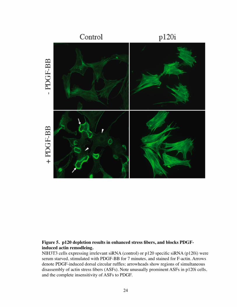
ing were completely blocked (Fig. 5). Specifically, ASFs and DCRs failed to form in the
absence of p120.
We next sought to determine if the PDGFR was nonfunctional in the p120i cells.
However, both PDGFR and p38 activation (as measured by tyrosine phosphorylation) in
response to PDGF was identical in WT and p120i cells (Fig. 6). Lastly, PDGFR-
mediated DCR formation has been shown to be mediated by a Rac-dependent mechanism
(Hooshmand-Rad, Claesson-Welsh et al. 1997; Sander, ten Klooster et al. 1999; Nimnual,
Taylor et al. 2003). Therefore, p120 loss could affect the ability of the PDGFR to acti-
vate Rac. To test this, PAK assays were performed to determine the ability of PDGF to
22
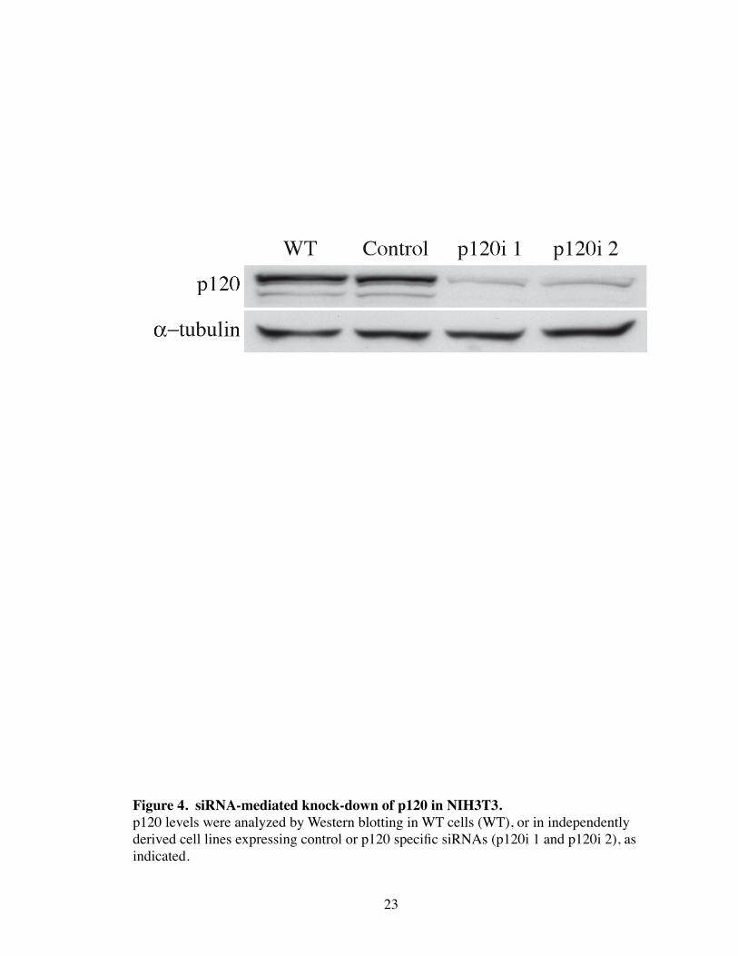
23
Figure 4. siRNA-mediated knock-down of p120 in NIH3T3. p120 levels were analyzed by Western blotting in WT cells (WT), or in independently derived cell lines expressing control or p120 specific siRNAs (p120i 1 and p120i 2), as indicated.
24
Figure 5. p120 depletion results in enhanced stress fibers, and blocks PDGF-induced actin remodleing.NIH3T3 cells expressing irrelevant siRNA (control) or p120 specific siRNA (p120i) were serum starved, stimulated with PDGF-BB for 7 minutes, and stained for F-actin. Arrows denote PDGF-induced dorsal circular ruffles; arrowheads show regions of simultaneous disassembly of actin stress fibers (ASFs). Note unusually prominent ASFs in p120i cells, and the complete insensitivity of ASFs to PDGF.
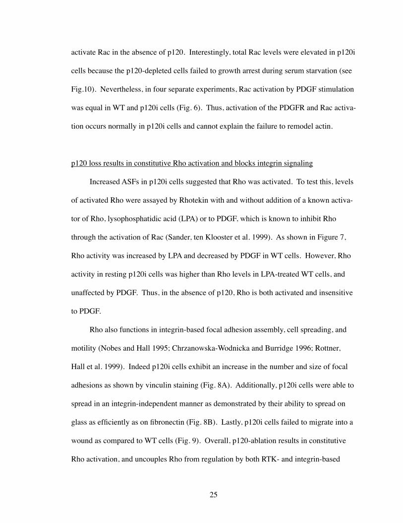
activate Rac in the absence of p120. Interestingly, total Rac levels were elevated in p120i
cells because the p120-depleted cells failed to growth arrest during serum starvation (see
Fig.10). Nevertheless, in four separate experiments, Rac activation by PDGF stimulation
was equal in WT and p120i cells (Fig. 6). Thus, activation of the PDGFR and Rac activa-
tion occurs normally in p120i cells and cannot explain the failure to remodel actin.
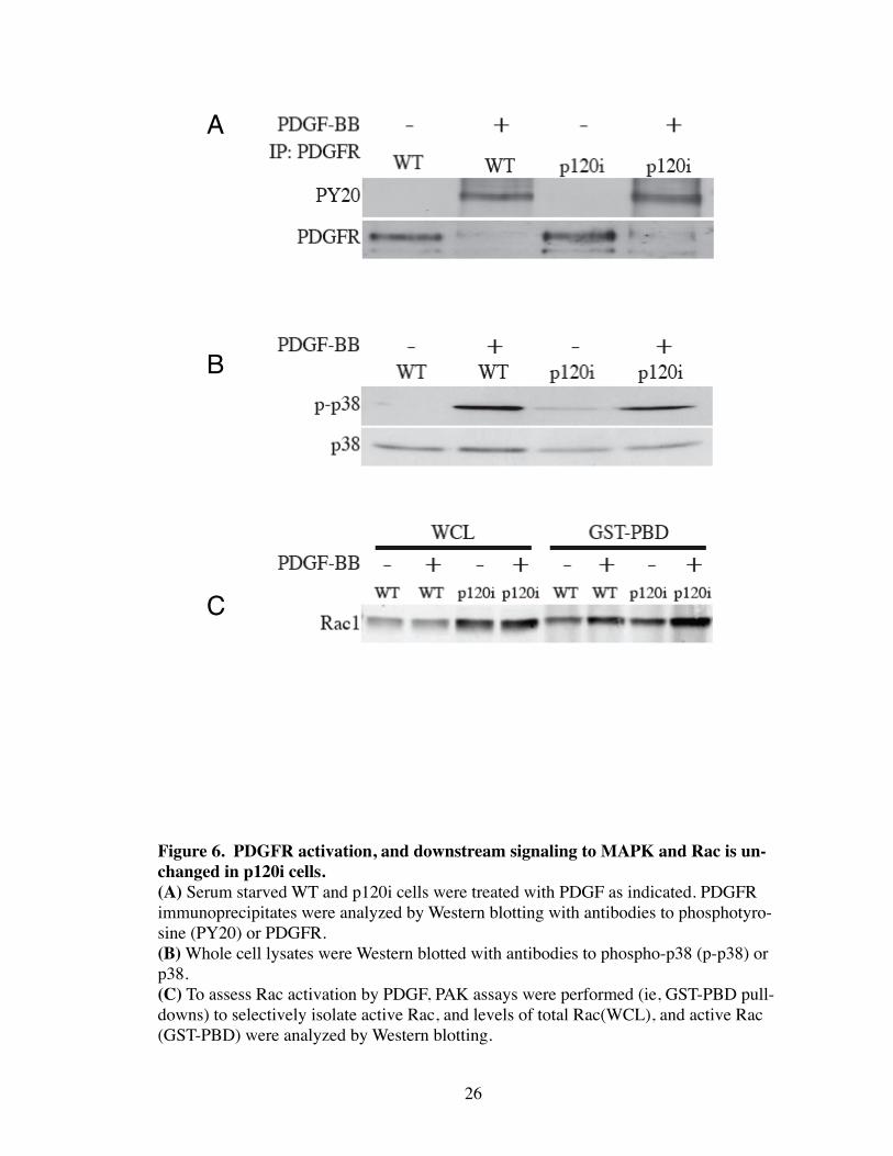
p120 loss results in constitutive Rho activation and blocks integrin signaling
Increased ASFs in p120i cells suggested that Rho was activated. To test this, levels
of activated Rho were assayed by Rhotekin with and without addition of a known activa-
tor of Rho, lysophosphatidic acid (LPA) or to PDGF, which is known to inhibit Rho
through the activation of Rac (Sander, ten Klooster et al. 1999). As shown in Figure 7,
Rho activity was increased by LPA and decreased by PDGF in WT cells. However, Rho
activity in resting p120i cells was higher than Rho levels in LPA-treated WT cells, and
unaffected by PDGF. Thus, in the absence of p120, Rho is both activated and insensitive
to PDGF.
Rho also functions in integrin-based focal adhesion assembly, cell spreading, and
motility (Nobes and Hall 1995; Chrzanowska-Wodnicka and Burridge 1996; Rottner,
Hall et al. 1999). Indeed p120i cells exhibit an increase in the number and size of focal
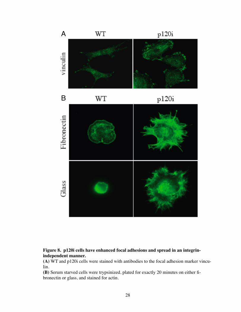
adhesions as shown by vinculin staining (Fig. 8A). Additionally, p120i cells were able to
spread in an integrin-independent manner as demonstrated by their ability to spread on
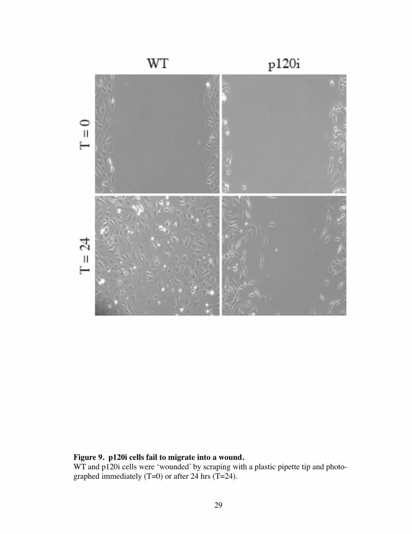
glass as efficiently as on fibronectin (Fig. 8B). Lastly, p120i cells failed to migrate into a
wound as compared to WT cells (Fig. 9). Overall, p120-ablation results in constitutive
Rho activation, and uncouples Rho from regulation by both RTK- and integrin-based
25
26
A
B
C
Figure 6. PDGFR activation, and downstream signaling to MAPK and Rac is un-changed in p120i cells.(A) Serum starved WT and p120i cells were treated with PDGF as indicated. PDGFR immunoprecipitates were analyzed by Western blotting with antibodies to phosphotyro-sine (PY20) or PDGFR.(B) Whole cell lysates were Western blotted with antibodies to phospho-p38 (p-p38) or p38.(C) To assess Rac activation by PDGF, PAK assays were performed (ie, GST-PBD pull-downs) to selectively isolate active Rac, and levels of total Rac(WCL), and active Rac (GST-PBD) were analyzed by Western blotting.
27
Figure 7. Rho is constitutively active and unaffected by PDGF in p120i cells. Serum starved cells were treated as indicated and assayed directly for Rho activity by GST-Rhotekin pulldown. Results are expressed as the ratio of activated to total Rho. Re-sults were expressed as a ratio of WT control (unstimulated WT NIH3T3). Error bars represent standard error of the mean. Student’s T-test was performed between WT and p120i cells, and WT+PDGF and p120i+PDGF to analyze statistical difference within the two groups (n = 3 independent experiments).
28
A
B
Figure 8. p120i cells have enhanced focal adhesions and spread in an integrin-independent manner.(A) WT and p120i cells were stained with antibodies to the focal adhesion marker vincu-lin.(B) Serum starved cells were trypsinized, plated for exactly 20 minutes on either fi-bronectin or glass, and stained for actin.
29
Figure 9. p120i cells fail to migrate into a wound.WT and p120i cells were ‘wounded’ by scraping with a plastic pipette tip and photo-graphed immediately (T=0) or after 24 hrs (T=24).
mechanisms.
p120i cells are partially transformed
Increased Rho activation has also been associated with cell transformation and
changes in cell proliferation (Qiu, Chen et al. 1995; del Peso, Hernandez-Alcoceba et al.
1997; Fritz, Just et al. 1999; Jaffe and Hall 2002). Because Rho was constitutively acti-
vated in p120i cells, we examined if these cells were transformed. Interestingly, whereas
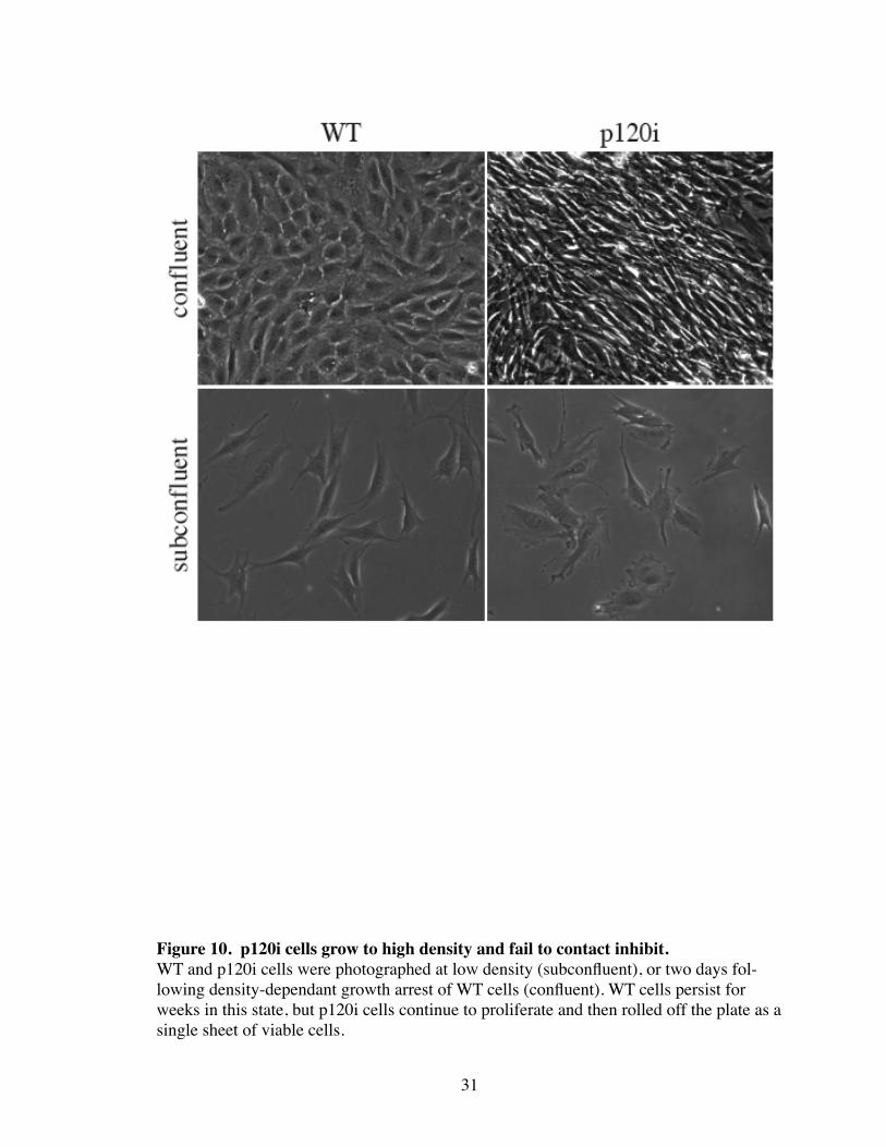
WT cells growth arrested normally at high density, p120i cells continued to proliferate to
extremely high density (Fig. 10) until they rolled off the plate as a single sheet of viable
cells (not shown). Consistent with this, p120i cells grew to 4 fold higher density than
WT cells when grown in the presence of serum. Surprisingly, when grown in the absence
of serum, WT cells died after 7 days, but p120i cells continued to grow, albeit slower than
normal (Fig. 11). p120i cells were also able to induce foci in secondary foci formation
assays (Fig. 12). p120i-induced foci was blocked by expression of the Rho-inhibitor C3,
and suppressed (but not blocked) by expression of N-cadherin. Overall, these data are
consistent with the effects of activating Rho mutants or Rho-GEF oncogenes, which also
induce foci formation and loss of contact inhibition (Jaffe and Hall 2002).
Discussion
p120 regulates RTK- and integrin-dependent Rho inhibition
These data demonstrate that ablation of p120 in NIH3T3 results in the constitutive
activation of Rho. Furthermore, upstream signals from RTKs (i.e. PDGFR) and integrins
30
31
Figure 10. p120i cells grow to high density and fail to contact inhibit.WT and p120i cells were photographed at low density (subconfluent), or two days fol-lowing density-dependant growth arrest of WT cells (confluent). WT cells persist for weeks in this state, but p120i cells continue to proliferate and then rolled off the plate as a single sheet of viable cells.
32
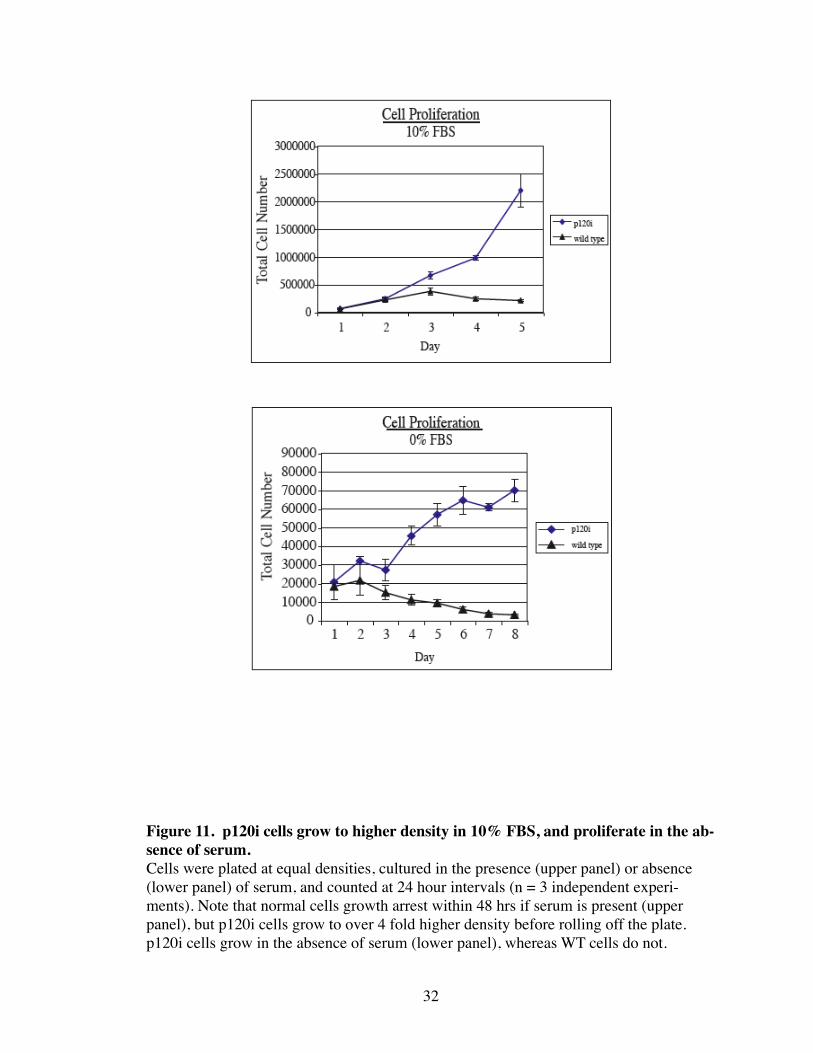
Figure 11. p120i cells grow to higher density in 10% FBS, and proliferate in the ab-sence of serum.Cells were plated at equal densities, cultured in the presence (upper panel) or absence (lower panel) of serum, and counted at 24 hour intervals (n = 3 independent experi-ments). Note that normal cells growth arrest within 48 hrs if serum is present (upper panel), but p120i cells grow to over 4 fold higher density before rolling off the plate. p120i cells grow in the absence of serum (lower panel), whereas WT cells do not.
33
A
B
C
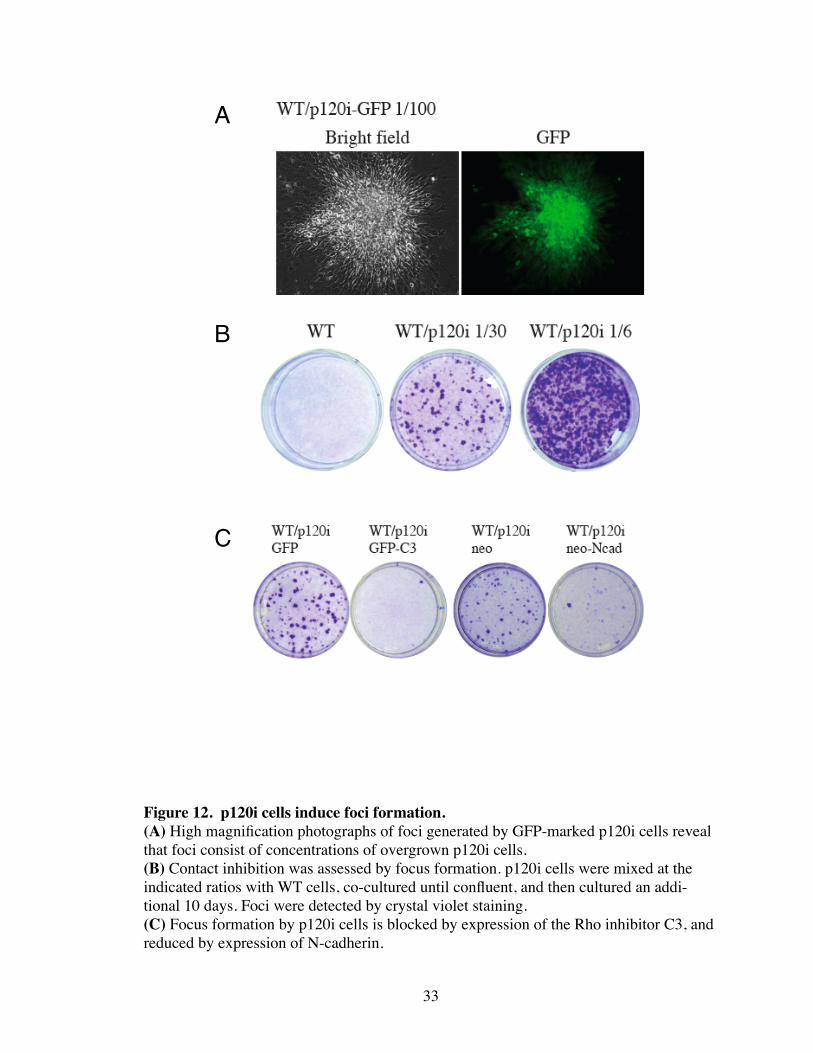
Figure 12. p120i cells induce foci formation.(A) High magnification photographs of foci generated by GFP-marked p120i cells reveal that foci consist of concentrations of overgrown p120i cells. (B) Contact inhibition was assessed by focus formation. p120i cells were mixed at the indicated ratios with WT cells, co-cultured until confluent, and then cultured an addi-tional 10 days. Foci were detected by crystal violet staining. (C) Focus formation by p120i cells is blocked by expression of the Rho inhibitor C3, and reduced by expression of N-cadherin.
are unable to inhibit Rho activity in p120-depleted cells. This is particularly surprising
given that p120 interacts exclusively with N-cadherin, and is not known to be involved in
RTK or integrin signaling. This suggests that RTKs or integrins either utilize p120 in a
cadherin-independent manner, or that p120 mediates crosstalk between these receptors
and the cadherin complex to regulate Rho. p120 is tyrosine phosphorylated by various
growth factors (Kanner, Reynolds et al. 1991), and these data suggest that signaling to
p120 is essential for Rho inactivation.
Interestingly, PDGFR-mediated DCRs have been shown to be formed through Rac
inhibition of Rho. p120 reduction does not affect Rac activation by the PDGFR, but
blocks signaling to Rho. Therefore, it appears that p120 may function in relaying Rac
activity to Rho inhibition. Because Rac activation by PDGFR is unaffected in p120i
cells, it also suggests that Rac activity alone is insufficient in mediating changes on the
actin cytoskeleton (e.g. membrane ruffles). Rather, it is the combined effect of Rac acti-
vation and Rho inhibition that drives this process. Local Rac activation and Rho inhibi-
tion has been documented in a variety of processes such as RTK signaling, EMT, integrin
spreading, motility, and contact inhibition, and thus represents a major arm of Rac signal-
ing (Sander, van Delft et al. 1998; Ren, Kiosses et al. 1999; Zondag, Evers et al. 2000;
Noren, Niessen et al. 2001; Wojciak-Stothard, Potempa et al. 2001; Nimnual, Taylor et al.
2003). The extraordinary level of Rho activation caused by p120-depletion is consistent
with a prominent role for p120 in regulating the overall activity of Rho. Presumably,
GEFs, GDIs, and GAPs should be able to intertact with Rho in p120i cells. It is surpris-
ing, therefore, that these alternative mechanisms are apparently insufficient to compen-
sate for the reduction of p120. Nevertheless, the fact that multiple signaling pathways
34

(i.e. RTKs, integrins) apparently must signal through p120 to inhibit Rho may explain the
potency of p120 loss. Interestingly, the increase in the resting level of Rho activity in
p120i cells reinforces the concept that the activity of RhoGTPases is determined by the
balance of positive and negative signals (see Fig. 1). Therefore, depletion
of p120 presumably uncouples a major negative stimulus and shifts the balance towards
Rho activation.
Initial observations of p120 depletion confirms that p120 plays a major role in regu-
lation of Rho that previous overexpression experiments demonstrated. This approach has
also revealed a surprising and novel role for p120 in controlling RTK and integrin-
dependent inhibition of Rho. An outstanding question is how p120 coordinates signals
from RTKs and integrins to inhibit Rho. Presently, it is difficult to interpret this data
since p120 has never been functionally or physically associated with either system. It
also remains to be determined whether the effects seen on Rho activity in p120-depleted
cells is cadherin-dependent or independent. Nevertheless, it is clear from these data that
p120 plays a major role in regulating the overall activity of Rho, and appears to globally
affect signaling from multiple receptor systems to Rho.
p120-depletion partially transforms NIH3T3
Along with its role in actin stress fiber assembly, Rho is a critical regulator of the
cell cycle. Overexpression of RhoGEFs or activating mutants of Rho lead to oncogenic
transformation in many cell types (Jaffe and Hall 2002). Therefore, the affects on growth
in the p120i cells are not surprising given that Rho is constitutively active. However, it is
surprising that this is caused by the loss of p120 expression alone. These data suggest a
35
major role for p120 as a tumor suppressor through the regulation of Rho. Foci formation
assays were previously utilized to determine if overexpressing certain genes could func-
tion as oncogenes by assaying for their ability to form foci and transform NIH3T3. How-
ever, with the advent of siRNA, a similar approach in unveiling tumor supressor capabili-
ties can be utilized.
36
CHAPTER IV
P120 IS ESSENTIAL FOR RAC TO RHO SIGNALING
Introduction
p120 depletion in NIH3T3 prevents PDGFR-induced ASF disassembly and forma-
tion of DCRs. DCR formation has been previously shown to be a Rac-dependent mecha-
nism (”the Bar-Sagi pathway”) described by Bar-Sagi and colleagues (Nimnual, Taylor et
al. 2003). Briefly, Rac activation results in the production of reactive oxygen species
(ROS), which inhibits low-molecular weight protein tyrosine phosphatase (LMW-PTP).
Inhibition of LMW-PTP results in tyrosine phosphorylation (activation) of p190, which in
turn inhibits Rho (Fig. 13). The coordination of Rac and Rho activities is an essential
process in ensuring the proper balance of actinomyosin contractility that is necessary in
the regulation of cell shape, motility, adhesion, and growth. The Bar-Sagi pathway is
utilized by both RTK and integrin signaling to rearrange the actin cytoskeleton (Sander,
ten Klooster et al. 1999; Nimnual, Taylor et al. 2003), and has also been shown to func-
tion in the maintainance of both E-cadherin and VE-cadherin based AJs (Zondag, Evers
et al. 2000; van Wetering, van Buul et al. 2002; Malliri, van Es et al. 2004; Wojciak-
Stothard, Tsang et al. 2005). p190 is an ubiquitously expressed GAP with specific activ-
ity towards Rho (Hall 1992; Lamarche and Hall 1994). p190 functions in a wide variety
of processes that include neuronal outgrowth and fasciculation, integrin-dependent adhe-
sion, cytokinesis, and adipogenesis (Nakahara, Mueller et al. 1998; Billuart, Winter et al.
2001; Brouns, Matheson et al. 2001; Sordella, Jiang et al. 2003; Su, Agati et al. 2003;
37
38
Figure 13. Schematic of the ‘Bar-Sagi’ pathway showing previously described mo-lecular events controlling Rac-mediated inhibition of Rho.
Barberis, Casazza et al. 2005). p190 contains a C-terminal GAP domain, which catalyzes
the hydrolysis of GTP bound to Rho. The N-terminal end of p190 contains a GTP bind-
ing domain that is necessary for its activity on Rho, though the mechanism is not known
(Tatsis, Lannigan et al. 1998; Roof, Dukes et al. 2000). p190 is tyrosine phosphorylated
by numerous kinases such as EGFR, Src family members, and Arg/Abl (Chang, Gill et al.
1995; Haskell, Nickles et al. 2001; Wolf, Wilkes et al. 2001; Hernandez, Settleman et al.
2004). Upon tyrosine phosphorylation, p190 translocates from the cytoplasm to mem-
branes and/or cytoskeletal structures such as ruffles where it can access pools of activated
Rho (Arthur and Burridge 2001) (Sharma 1998). Thus, p190 phosphorylation and
membrane translocation are tightly linked and are both necessary for its subsequent roles
in cell signaling and Rho inhibition. Lastly, p190 forms a low level, constitutive complex
with p120RasGAP (RasGAP), which increases upon tyrosine phosphorylation of p190.
The role of RasGAP in p190 signaling is not clear, but studies suggest that RasGAP is
necessary for phosphorylation of p190, and localization of p190 at focal adhesions. The
mechanisms are not well understood (DeClue, Vass et al. 1993; Ellis, Measday et al.
1995; van der Geer, Henkemeyer et al. 1997; Fincham, Chudleigh et al. 1999).
Cadherin engagement has also been shown to induce src-dependent phosphoryla-
tion of p190 (Noren, Arthur et al. 2003). The functional consequence of this event, or
where p190 localizes upon cadherin ligation, however, is not known. Several lines of
evidence suggest that alleviation of contractility through the inhibition of Rho is neces-
sary for proper AJ formation, and the reciprocal disassembly of AJs may be due to in-
creased contractility through Rho activation (Dudek and Garcia 2001; Garcia, Liu et al.
2001; Wojciak-Stothard, Potempa et al. 2001). p190, therefore, may be involved in alle-
39
viating contractility through Rho inhibition during AJ formation. Interestingly, growth
factor and integrin signaling, through regulation of Rac and Rho, can promote either AJ
assembly or disassembly (Ridley, Comoglio et al. 1995; Monier-Gavelle and Duband
1997; Potempa and Ridley 1998; Chen and Gumbiner 2006). Although how regulation of
RhoGTPases by RTKs or integrins is translated into AJ assembly/disassembly is not
known, p120 is likely to be involved in this process due to its dual role in cell-cell adhe-
sion and Rho regulation. In chapter three, we demonstrated that depletion of p120 blocks
both RTK and integrin signaling at the level of Rho. Therefore, since both receptor sys-
tems utilize the Bar-sagi pathway to activate Rac and inhibit Rho, p120 may play a role in
this pathway in relaying crosstalk between RTKs, integrins, and AJs.
Results
Rac needs p120 to inhibit Rho
We have previously shown in chapter III that p120-depletion in NIH3T3 results in
the constitutive activation of Rho, which surprisingly blocks both RTK- and integrin-
mediated rearrangements in the actin cytoskeleton. To determine the function behind
these observations, we carried out a series of experiments to isolate the precise mecha-
nism of p120-dependent Rho inhibition. To ensure that elevated ASFs in p120i cells was
due to aberrant Rho signaling, we inhibited ROCK, a downstream effector of Rho that
functions in stress fiber assembly (Leung, Chen et al. 1996). As seen in Figure 14, inhibi-
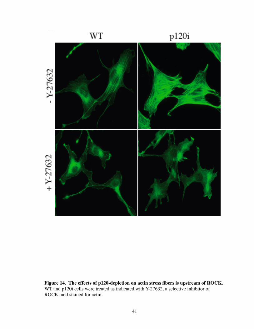
tion of ROCK with the specific inhibitor, Y-27632, caused the complete loss of ASFs in
both WT and p120i cells. Thus, effects of p120 loss occur upstream of Rho and ROCK,
40
41
Figure 14. The effects of p120-depletion on actin stress fibers is upstream of ROCK.WT and p120i cells were treated as indicated with Y-27632, a selective inhibitor of ROCK, and stained for actin.
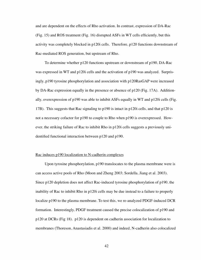
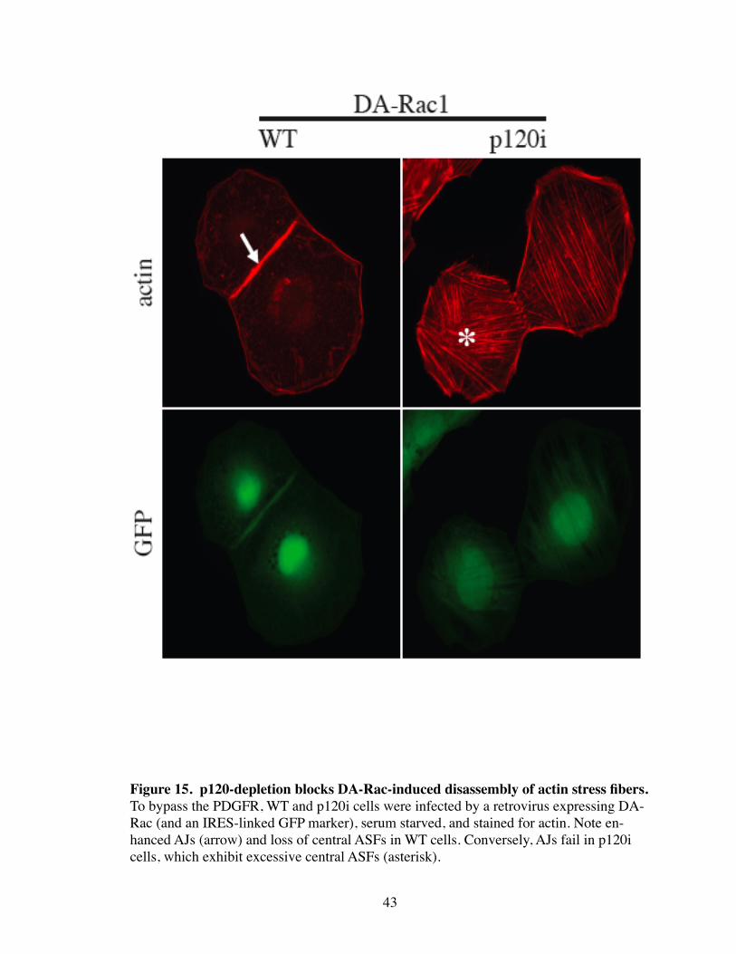
and are dependent on the effects of Rho activation. In contrast, expression of DA-Rac
(Fig. 15) and ROS treatment (Fig. 16) disrupted ASFs in WT cells efficiently, but this
activity was completely blocked in p120i cells. Therefore, p120 functions downstream of
Rac-mediated ROS generation, but upstream of Rho.
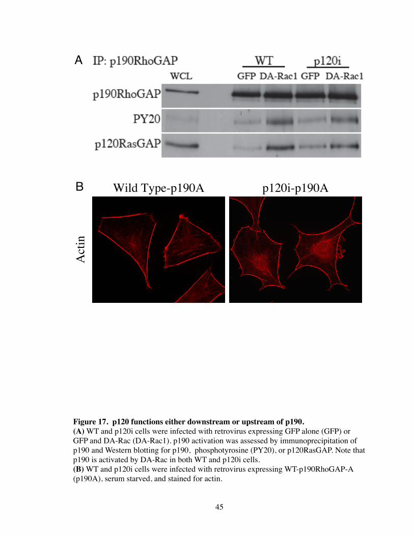
To determine whether p120 functions upstream or downstream of p190, DA-Rac
was expressed in WT and p120i cells and the activation of p190 was analyzed. Surpris-
ingly, p190 tyrosine phosphorylation and association with p120RasGAP were increased
by DA-Rac expression equally in the presence or absence of p120 (Fig. 17A). Addition-
ally, overexpression of p190 was able to inhibit ASFs equally in WT and p120i cells (Fig.
17B). This suggests that Rac signaling to p190 is intact in p120i cells, and that p120 is
not a necessary cofactor for p190 to couple to Rho when p190 is overexpressed. How-
ever, the striking failure of Rac to inhibit Rho in p120i cells suggests a previously uni-
dentified functional interaction between p120 and p190.
Rac induces p190 localization to N-cadherin complexes
Upon tyrosine phosphorylation, p190 translocates to the plasma membrane were is
can access active pools of Rho (Moon and Zheng 2003; Sordella, Jiang et al. 2003).
Since p120 depletion does not affect Rac-induced tyrosine phosphorylation of p190, the
inability of Rac to inhibit Rho in p120i cells may be due instead to a failure to properly
localize p190 to the plasma membrane. To test this, we re-analyzed PDGF-induced DCR
formation. Interestingly, PDGF treatment caused the precise colocalization of p190 and
p120 at DCRs (Fig 18). p120 is dependent on cadherin association for localization to
membranes (Thoreson, Anastasiadis et al. 2000) and indeed, N-cadherin also colocalized
42
43
Figure 15. p120-depletion blocks DA-Rac-induced disassembly of actin stress fibers.To bypass the PDGFR, WT and p120i cells were infected by a retrovirus expressing DA-Rac (and an IRES-linked GFP marker), serum starved, and stained for actin. Note en-hanced AJs (arrow) and loss of central ASFs in WT cells. Conversely, AJs fail in p120i cells, which exhibit excessive central ASFs (asterisk).
44
Figure 16. p120-depletion blocks ROS-induced stress fiber disassembly.To bypass Rac1, WT or p120i cells were H2O2 treated (ROS) as indicated and stained for actin. Note that p120i cells are unresponsive.
45
Figure 17. p120 functions either downstream or upstream of p190.(A) WT and p120i cells were infected with retrovirus expressing GFP alone (GFP) or GFP and DA-Rac (DA-Rac1). p190 activation was assessed by immunoprecipitation of p190 and Western blotting for p190, phosphotyrosine (PY20), or p120RasGAP. Note that p190 is activated by DA-Rac in both WT and p120i cells.(B) WT and p120i cells were infected with retrovirus expressing WT-p190RhoGAP-A (p190A), serum starved, and stained for actin.
p120i-p190AWild Type-p190A
Act
in
B
A
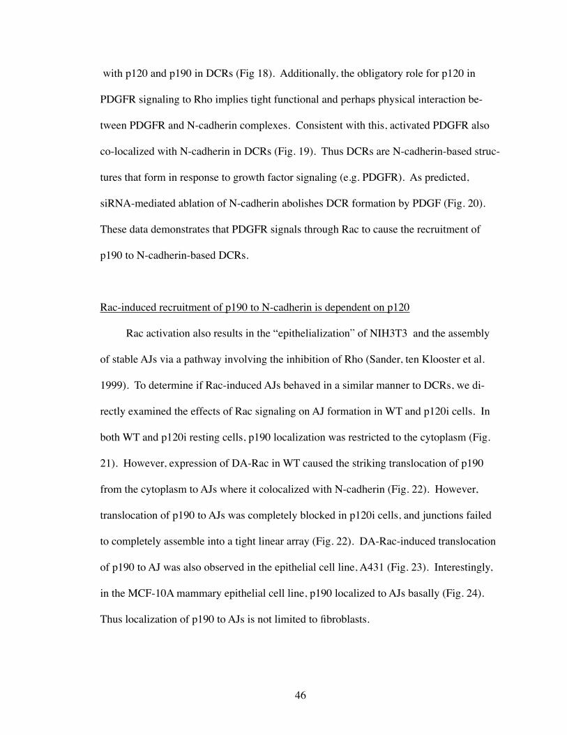
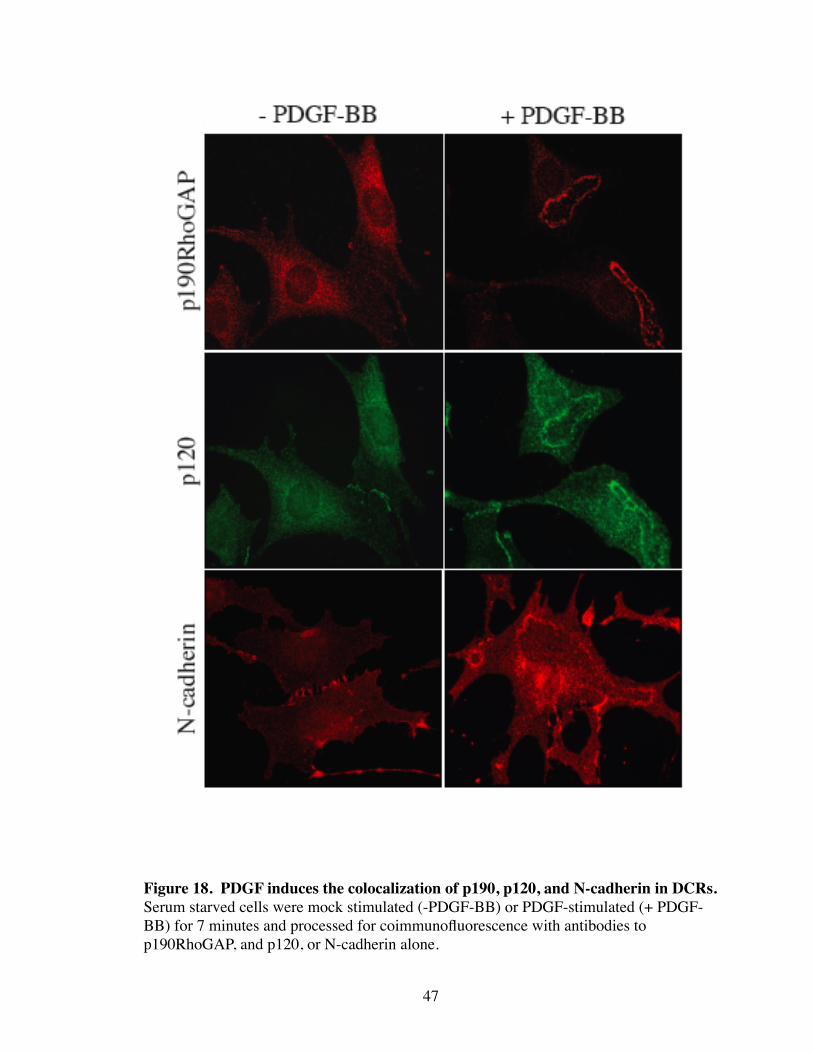
with p120 and p190 in DCRs (Fig 18). Additionally, the obligatory role for p120 in
PDGFR signaling to Rho implies tight functional and perhaps physical interaction be-
tween PDGFR and N-cadherin complexes. Consistent with this, activated PDGFR also
co-localized with N-cadherin in DCRs (Fig. 19). Thus DCRs are N-cadherin-based struc-
tures that form in response to growth factor signaling (e.g. PDGFR). As predicted,
siRNA-mediated ablation of N-cadherin abolishes DCR formation by PDGF (Fig. 20).
These data demonstrates that PDGFR signals through Rac to cause the recruitment of
p190 to N-cadherin-based DCRs.
Rac-induced recruitment of p190 to N-cadherin is dependent on p120
Rac activation also results in the “epithelialization” of NIH3T3 and the assembly
of stable AJs via a pathway involving the inhibition of Rho (Sander, ten Klooster et al.
1999). To determine if Rac-induced AJs behaved in a similar manner to DCRs, we di-
rectly examined the effects of Rac signaling on AJ formation in WT and p120i cells. In
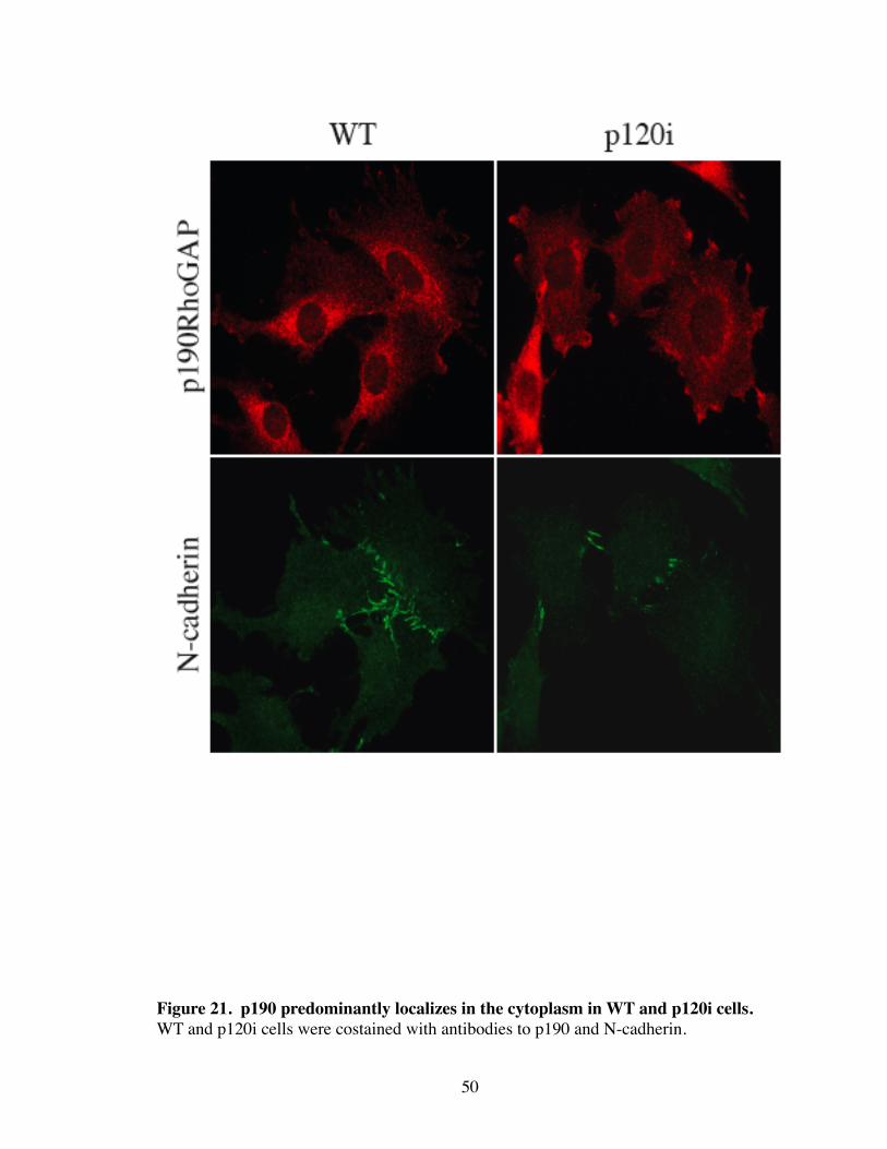
both WT and p120i resting cells, p190 localization was restricted to the cytoplasm (Fig.
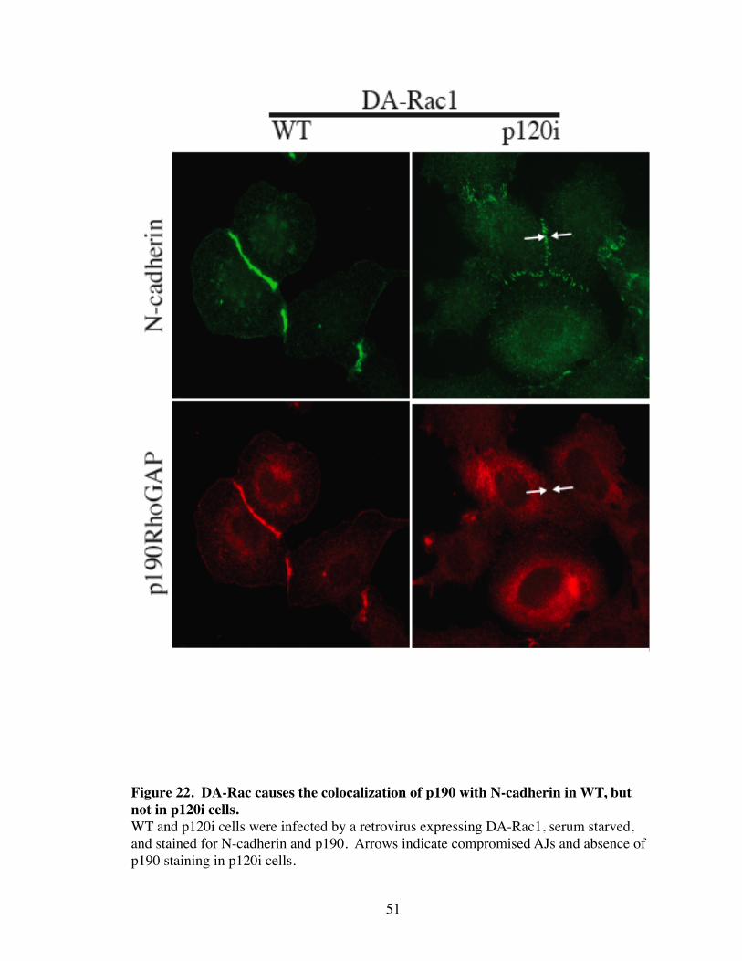
21). However, expression of DA-Rac in WT caused the striking translocation of p190
from the cytoplasm to AJs where it colocalized with N-cadherin (Fig. 22). However,
translocation of p190 to AJs was completely blocked in p120i cells, and junctions failed
to completely assemble into a tight linear array (Fig. 22). DA-Rac-induced translocation
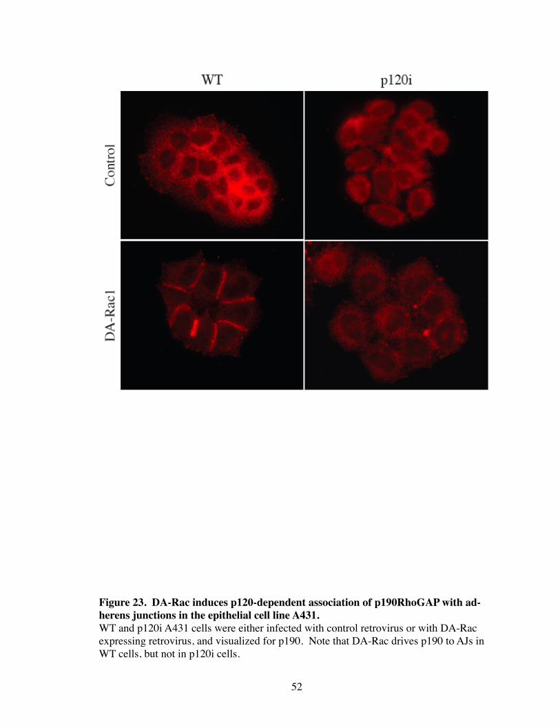
of p190 to AJ was also observed in the epithelial cell line, A431 (Fig. 23). Interestingly,
in the MCF-10A mammary epithelial cell line, p190 localized to AJs basally (Fig. 24).
Thus localization of p190 to AJs is not limited to fibroblasts.
46
47
Figure 18. PDGF induces the colocalization of p190, p120, and N-cadherin in DCRs. Serum starved cells were mock stimulated (-PDGF-BB) or PDGF-stimulated (+ PDGF-BB) for 7 minutes and processed for coimmunofluorescence with antibodies to p190RhoGAP, and p120, or N-cadherin alone.
48
Figure 19. PDGFR and N-cadherin colocalize in PDGF-induced DCRs. Cells were serum starved, PDGF-stimulated, and costained for N-cadherin and PDGFR.
49
Figure 20. PDGF-induced DCRs are N-cadherin-dependent structures.(A) WT and N-cadherin knockdown cells (N-cadi) were PDGF stimulated as previously described and stained for actin. (B) Quantification of PDGF-induced DCRs in WT and N-cadherin knockdown cells. 350 cells were randomly selected and scored for the presence or absence of dorsal circular ruffles after PDGF stimulation in wild type (WT) and N-cadherin knockdown (N-cadi) cells. (C) Analysis of N-cadherin expression by Western blotting in WT and N-cadherin siRNA (N-cadi) expressing NIH3T3 cells.
B
C
A
50
Figure 21. p190 predominantly localizes in the cytoplasm in WT and p120i cells.WT and p120i cells were costained with antibodies to p190 and N-cadherin.
51
Figure 22. DA-Rac causes the colocalization of p190 with N-cadherin in WT, but not in p120i cells.WT and p120i cells were infected by a retrovirus expressing DA-Rac1, serum starved, and stained for N-cadherin and p190. Arrows indicate compromised AJs and absence of p190 staining in p120i cells.
52
Figure 23. DA-Rac induces p120-dependent association of p190RhoGAP with ad-herens junctions in the epithelial cell line A431. WT and p120i A431 cells were either infected with control retrovirus or with DA-Rac expressing retrovirus, and visualized for p190. Note that DA-Rac drives p190 to AJs in WT cells, but not in p120i cells.
p190 and p120 interact
The dependency on p120 for p190 recruitment to AJs suggests that the two physi-
cally interact. Therefore, we performed coimmunoprecipitation experiments with p120
and other core components of the cadherin complex (Fig. 25A). p190 coimmunoprecipi-
tated efficiently with p120, but not with β-catenin or N-cadherin, suggesting that p190
directly interacts with p120, and indirectly with the remaining cadherin complex. To
clarify this, a p120 mutant was generated that interacts constitutively with the plasma
membrane, but lacks an Armadillo repeat necessary for binding to cadherins (i.e.
p1201A-ΔArm1-CAAX). As shown in Figure 25B, this p120 mutant localized to the
plasma membrane, and efficiently recruited p190 but not N-cadherin. Therefore, p120 is
able to recruit p190 independent of N-cadherin or other cadherin complex components.
These data suggests that Rac-induced binding of p190 to cadherin complexes in both
DCRs and AJs is mediated by association with p120.
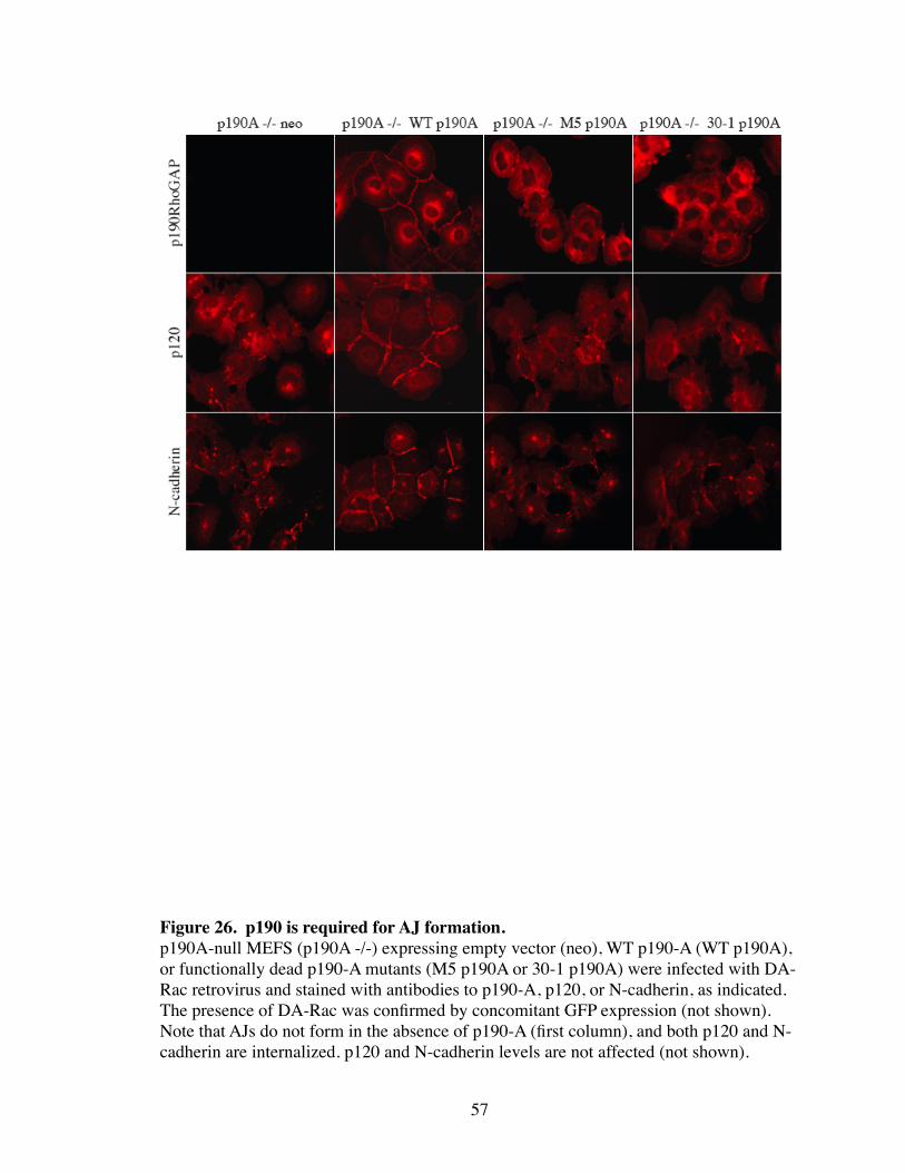
p190 is necessary for Rac-induced adherens junction formation
Although p190 does not localize to AJs under normal detection methods, the above
data suggests a functional role for p190 in cell-cell adhesion. To address this further,
p190A -/- MEFs were analyzed for their ability to form proper AJs by expression of DA-
Rac. AJs in the absence of p190A were not able to form after DA-Rac expression as
shown by mislocalized p120 and N-cadherin (Fig. 26). To confirm that the defect was
due to the absence of p190A, DA-Rac-induced AJs were rescued by expression of WT-
p190A. In contrast, expression of p190A mutants defective in N-terminal GTP binding
(M5) or RhoGAP activity (30-1) were completely unable to rescue AJs (Fig. 26). Both
53
54
Figure 24. p190 localizes at AJs in a p120-dependent manner in MCF-10A cells.WT and p120i MCF-10A cells were co-stained with antibodies to p120 and p190.
p120 p190WT
p120i
55
Figure 25. p120 interacts with p190, and is sufficient to induce p190 translocation independently of the cadherin complex.(A) Immunoprecipitation with p120, N-cadherin, β-catenin, or irrelevant control (NS) antibodies was followed by Western blotting with antibodies to p190. Whole cell lysate (WCL, left) was included as a marker for p190. (B) A murine p120 mutant (mp120-1A-ΔArm1-CAAX) was generated that cannot bind to cadherins but localizes to membranes by virtue of a fused CAAX box. The construct con-tains a silent mutation within the siRNA targeted region of p120 to allow its expression in the murine p120 siRNA background. p120i cells expressing the p120 mutant (mp120-1A-ΔArm 1-CAAX), or neomycin alone (neo), were costained with antibodies to p120 and p190 (left panels) or p120 and N-cadherin (right panels).
B
A
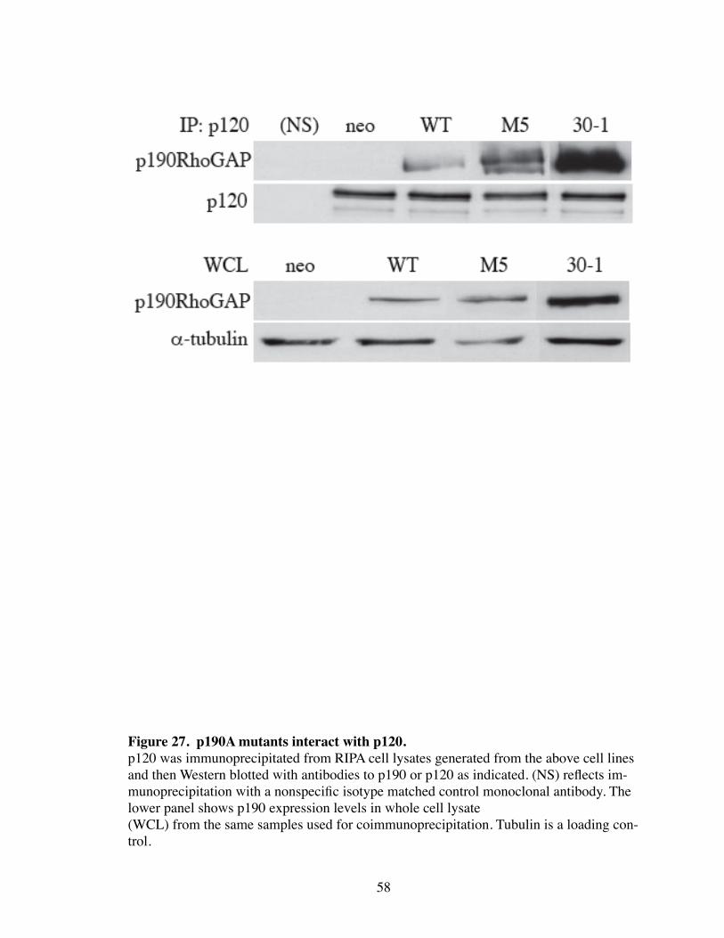
mutations are unable to inhibit Rho through different mechanisms, which suggests that
the activity on Rho by p190 is essential in this process. These mutants, however, were
able to efficiently coimmunoprecipitate with p120, and therefore do not fail to permit AJ
formation through a failure to bind p120 (Fig. 27).
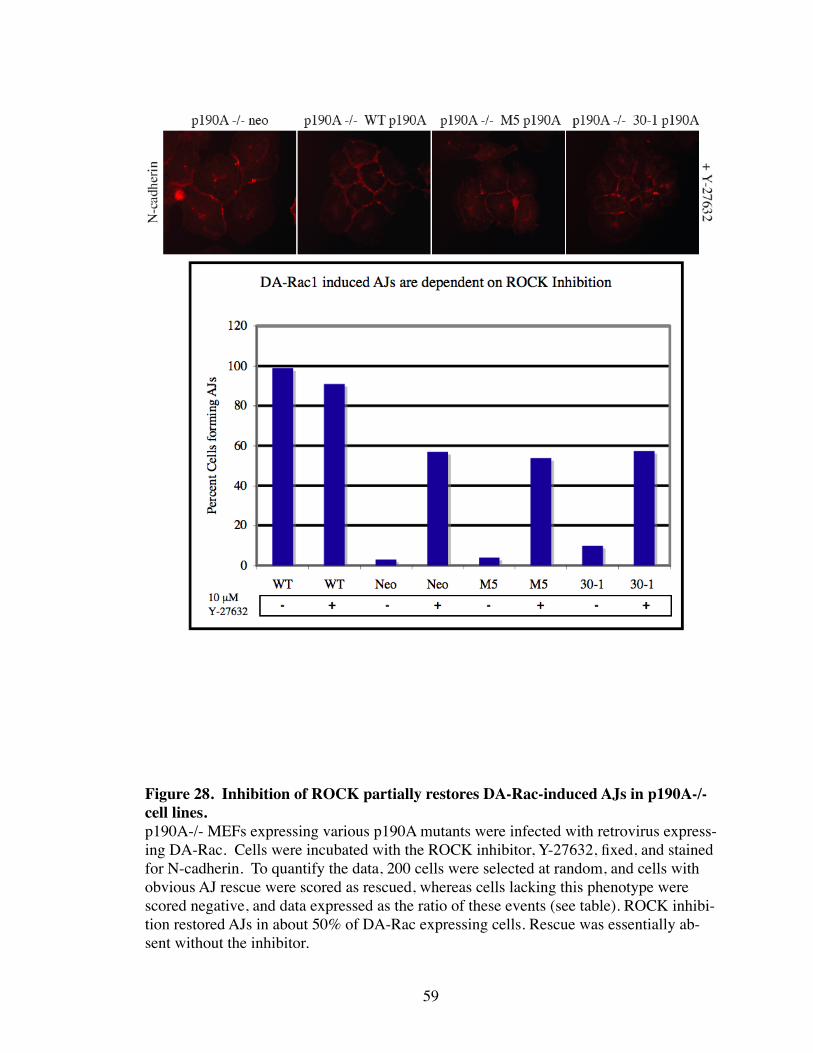
These data show that p120 and p190 are essential in the formation of stable AJs.
Without either p120 or p190, there is a failure to effectively inactivate Rho thereby pre-
venting AJ formation. To understand the relationship between Rho signaling and AJ as-
sembly further, ROCK was inhibited in DA-Rac expressing p190A -/- cell lines. ROCK
inhibition in all cell lines was able to partially rescue AJ formation as shown in Figure 28.
Thus, it is likely that Rac coordinates AJ assembly by promoting the recruitment of p190
to N-cadherin through p120 binding. There, p190 is able to inhibit Rho and suppress the
contractility required for AJ formation.
Discussion
Rac requires p120 to inhibit Rho
Here we utilized an epistasis approach to identify where p120 lies in the Bar-Sagi
pathway. To this end, we were able to identify p120 as an essential regulator of p190 lo-
calization at the plasma membrane in Rac-mediated inhibition of Rho. Although p120
was not necessary for phosphorylation of p190 by Rac, p120 was absolutely required to
localize p190 to the cell surface where it could access activated pools of Rho. Because
p120 only localizes to the plasma membrane through interacting with N-cadherin (or E-
cadherin in A431 and MCF-10A cells), the activation of p190 is concentrated at cadherin
56
57
Figure 26. p190 is required for AJ formation.p190A-null MEFS (p190A -/-) expressing empty vector (neo), WT p190-A (WT p190A), or functionally dead p190-A mutants (M5 p190A or 30-1 p190A) were infected with DA-Rac retrovirus and stained with antibodies to p190-A, p120, or N-cadherin, as indicated. The presence of DA-Rac was confirmed by concomitant GFP expression (not shown). Note that AJs do not form in the absence of p190-A (first column), and both p120 and N-cadherin are internalized. p120 and N-cadherin levels are not affected (not shown).
58
Figure 27. p190A mutants interact with p120.p120 was immunoprecipitated from RIPA cell lysates generated from the above cell lines and then Western blotted with antibodies to p190 or p120 as indicated. (NS) reflects im-munoprecipitation with a nonspecific isotype matched control monoclonal antibody. The lower panel shows p190 expression levels in whole cell lysate(WCL) from the same samples used for coimmunoprecipitation. Tubulin is a loading con-trol.
59
Figure 28. Inhibition of ROCK partially restores DA-Rac-induced AJs in p190A-/- cell lines. p190A-/- MEFs expressing various p190A mutants were infected with retrovirus express-ing DA-Rac. Cells were incubated with the ROCK inhibitor, Y-27632, fixed, and stained for N-cadherin. To quantify the data, 200 cells were selected at random, and cells with obvious AJ rescue were scored as rescued, whereas cells lacking this phenotype were scored negative, and data expressed as the ratio of these events (see table). ROCK inhibi-tion restored AJs in about 50% of DA-Rac expressing cells. Rescue was essentially ab-sent without the inhibitor.
complexes. Although we have not ruled out the possibility that p190 can translocate to
the membrane through p120-independent means, it is apparent that p120 and the cadherin
complex is necessary for Rac to couple to Rho. Likewise, these data do not rule out other
means for negatively regulating Rho such as through inhibition of a GEF or activation of
a GDI. However, given that the Bar-Sagi pathway represents a major pathway in a vari-
ety of several receptor types, and that cells become transformed without proper Rac to
Rho signaling, the translocation of p190 to the cadherin complex represents a major as-
pect of p190 function and overall regulation of Rho. Recently, it was reported that p190
localizes to focal adhesions in a RasGAP-dependent manner (Bradley, Hernandez et al.
2006). Since p120 does not localize to focal adhesions, it is possible, therefore, that
RasGAP and p120 could compete for p190 localization to focal adhesion or AJs, respec-
tively. However, integrins fail to regulate Rho in p120i cells, which suggests that p190
must translocate to the cadherin complex for integrins to properly inhibit Rho. Thus it is
tempting to speculate that proper focal adhesion formation depends on both organization
of integrins and cadherins.
p120/p190 and the cadherin complex regulates overall inhibition of Rho
The complete failure of Rac to inhibit Rho in p120i cells establishes p120 and the
cadherin complex as obligatory intermediates in Rac to Rho signaling (i.e. the Bar-Sagi
pathway). By localizing the Bar-Sagi pathway to cadherin complexes, a cellular context
is established for Rac to Rho signaling. Given that the Bar-Sagi pathway is utilized by
multiple receptor systems (e.g. RTKs, integrins), these data define the cadherin complex
as a major gateway through which signals must be relayed to rearrange the actin cyto-
60
skeleton. Our initial observations postulated that the effects of p120 depletion on RTK
and integrin signaling could be through a cadherin independent or dependent mechanism
(see chapter III). These data now favor the latter interpretation since inhibition of Rho by
p120 appears to occur when p120 and p190 are localized to N-cadherin. Furthermore,
PDGFR-induced DCRs are cadherin based structures that cannot form in N-cadherin-
depleted cells. This highlights a surprising and novel role for p120 in not only organizing
Rho activity at the cadherin complex, but also being the rate limiting step in the ability of
RTKs and integrins to inhibit Rho. Alternatively, Rho may be so highly activated in
p120i cells that other systems simply cannot suppress Rho enough to elicit their respec-
tive biological response. It is admittedly difficult to determine absolutely whether RTKs,
integrins, and cadherins regulate Rho through separate pathways, or if all signaling to
Rho is channeled through the cadherin complex. However, functionally placing p120 in
the Bar-Sagi pathway, and the complete failure of RTKs and integrins to reorganize the
actin cytoskeleton in p120i cells suggests that p120 and the cadherin complex plays a far
greater role in coordinating Rac-mediated inhibition of Rho in general.
Our model also predicts that the loss of cadherin expression would result in a simi-
lar Rho-dependent phenotype seen by the loss of p120 (e.g. p120i cells). Since p190
translocation to the plasma membrane depends on the cadherin complex, both p120 and
cadherin expression are theoretically rate limiting for functionally coupling of Rac to
Rho. Though Rho activity was not directly tested by Rhotekin assay, N-cadherin knock-
down NIH3T3 (N-cadi) did not appear to have prominent ASFs as seen in p120i cells.
siRNA-mediated knockdown efficiency for N-cadherin was not as high as for p120, and
therefore the remaining levels of N-cadherin may have been sufficient to regulate Rho
61
activity by some reduced measure. It has recently been shown that in the E-cadherin de-
ficient cell line, MBA-MD-231, p120 inhibits Rho in the cytoplasm (Yanagisawa and An-
astasiadis 2006). This suggests that p120 can regulate Rho both at the plasma membrane
when associated with cadherins, or in the cytoplasm when cadherin expression is lost.
Therefore, in N-cadi cells, cytoplasmic p120 may still inhibit Rho by another unknown
mechanism. Drosophila p120 has been shown to directly interact with GDP-bound Rho,
and others have shown a weak interaction in mammalian cell lines (Magie, Pinto-Santini
et al. 2002; Bellovin, Bates et al. 2005). Therefore, in N-cadi cells, p120 may still inhibit
Rho, but in a Rac-independent manner by physically interacting with and sequestering
Rho in the cytoplasm. Thus Rac-dependent mechanisms such as PDGFR-induced DCRs
would still be blocked by N-cadherin loss, but inhibition of Rho by cytoplasmic p120
would be sufficient to suppress ASFs.
Likewise, p190A -/- MEFs would be predicted to have elevated Rho activity. How-
ever, this cell line also does not exhibit phenotypes of increased Rho activity such as
strong ASFs. This cell line does express the p190A homologue, p190B, which also func-
tions as a GAP for Rho. p190B is also necessary for AJ formation by Rac as p190B -/-
MEFs do not form AJs upon DA-Rac expresssion (data not shown). Therefore, both
forms of p190 are necessary for coupling Rac to Rho, but the presence of one gene may
be sufficient to partially suppress Rho levels to a degree in which, at basal levels, ele-
vated ASFs are not observed. Alternatively, p190A -/- or p190B -/- cell lines may have
evolved mechanisms during clonal expansion and immortalization to suppress Rho activ-
ity to compensate for the lack of these genes. Nevertheless, p190A -/- or p190B -/- MEFs
are functionally uncoupled from signaling events that require Rho inhibition as shown by
62
our data, and several other reports that have demonstrated in p190A-/- or B-/- MEFs, the
requirement of p190-dependent inhibition of Rho in various processes such as adipogene-
sis, neuronal fasciculation, and semaphorin-mediated axon guiding (Brouns, Matheson et
al. 2001; Sordella, Jiang et al. 2003; Barberis, Casazza et al. 2005).
Cadherin-bound p120 stabilizes AJs by inhibiting Rho
Our lab has shown previously that p120 is necessary for the stablization of cadherin
levels (Ireton, Davis et al. 2002). This mechanism requires direct p120-cadherin interac-
tion, which is consistent with the hypothesis that p120 and p190 coordinately regulate
cadherin and AJ stability by localizing and controlling Rho-mediated contractility. Like-
wise, control of Rac activity by RTKs and integrins is likely to be a major mechanism in
regulating AJ assembly. Although the functional interactions between RTKs and cadher-
ins are not well understood, post-translational regulation of cadherin stability and AJ as-
sembly by RTK activity is well documented (Wrobel, Debnath et al. 2004). Thus, it is
exciting to speculate that regulation of AJ assembly/disassembly by growth factor signal-
ing occurs through Rac-inhibition of Rho and the p120/p190 complex. Although not ex-
tensively tested, this appears to be particularly important in MCF-10A cells in which
p190 localizes to AJs basally. Thus, MCF-10A may be an ideal epithelial cell line to in-
vestigate the role of p120 and p190 in epithelial cells.
This data suggests that the major function of p120 is to inhibit Rho at the site of the
cadherin complex. This is contrary to our original hypothesis, which suggested that p120
predominantly inhibits Rho in the cytoplasm. Indeed, recent evidence has suggested that
p120 regulates Rac through a cadherin-dependent manner and Rho through a cadherin-
63
independent mechanism (Yanagisawa and Anastasiadis 2006). Though, no mechanism
was identified, their observations suggests that p120-depletion by siRNA induces Rho
activation, and can be rescued by a p120 mutant that cannot associate with cadherins.
However, a cytoplasmic role for p120-mediated inhibition of Rho would predict that as
more AJs are formed and hence more p120 is recruited from the cytoplasm to the cad-
herin complex, there would be a porportional elevation in the amount of activated Rho.
This is contrary to data that demonstrates upon high cell density, Rho activity is sup-
pressed by cadherin engagement (Noren, Niessen et al. 2001). Furthermore, the cell line
utilized in this study is the highly invasive MDA-MB-231 breast adenocarcinoma cell
line, which has lost normal E-cadherin expression and undergone epithelial to mesen-
chymal transition (EMT). Therefore, it is difficult to determine if normal signaling
mechanisms are intact in their model system. Attempts to re-express E-cadherin in MD-
MB-231 and examine DA-Rac-induced localization of p190 were not successful. Simi-
larily, expression of E-cadherin in other cadherin-deficient tumorigenic cell lines also
failed to localize p190 by DA-Rac expression (data not shown). However, as noted
above, cell lines which have an intact cadherin complex (e.g. A431, MCF-10A) are able
to efficiently localize p190 in a p120-dependent manner. Therefore, it is possible that cell
lines that have lost cadherin expression have also lost critical signaling pathways that
regulate p190 translocation to the cadherin complex, which is consistent with a role for
p190 in regulating AJ assembly. We do not eliminate the possibility that there is an addi-
tional mechanism for Rho regulation by cytoplasmic p120, but our data strongly suggest
a prominent role for p120 in regulating Rho at the cadherin complex. The discrepancies
between our data and those of another lab raise the possibility of different roles for p120
64
in the inhibition of Rho depending on whether or not the cell has undergone oncogenic
transformation. This highlights an interesting paradigm in p120 signaling that requires
further exploration to appreciate the role of p120 during tumor progression.
p120 is a tumor suppressor
The striking loss of contact inhibition and acquired ability to grow without serum in
p120i cells suggests that p120 is a tumor suppressor. Here we demonstrate that loss of
p120 expression results in the concomitant downregulation of cadherin expression and
constitutive activation of Rho. E-cadherin is frequently downregulated in epithelial tu-
mors (Birchmeier and Behrens 1994), and loss of E-cadherin expression has been ob-
served in both early and late stages of tumor progression indicating a role as both a tumor
and invasion suppressor (Berx, Nollet et al. 1998; Berx and Van Roy 2001). This concept
has been widely tested both in vitro and in vivo demonstrating that E-cadherin expression
and function is critical in suppressing tumor progression (Derksen, Liu et al. 2006; Ol-
meda, Jorda et al. 2006; Zhang, Alt-Holland et al. 2006). Although our research de-
scribes the effects of p120 loss in fibroblasts (i.e. N-cadherin expressing cells), accumu-
lating evidence in our lab suggests that the mechanism described here is conserved in
epithelial cell lines. The Rip1Tag2 mouse model for beta-cell carcinogenesis has been
used to demonstrate the E-cadherin loss is the rate-limiting step in progression from ade-
noma to carcinoma (Perl, Wilgenbus et al. 1998). Interestingly, it was recently shown
that the effects on E-cadherin loss in tumor progression of the Rip1Tag2 mouse are not
due to β-catenin-mediated Wnt signaling. Furthermore, tumor progression in this model
is not affected by loss of Tcf-1, a major target of β-catenin/Wnt signaling (Herzig, Sa-
65
varese et al. 2006). Alternatively, the effects of E-cadherin loss on tumor progression
could be due to misregulation of p120-dependent signaling processes including regulation
of Rho.
Likewise, alterations in Rho signaling have been implicated in various processes of
tumor progression including increased proliferation, de-differentiation, invasion, migra-
tion, and metastasis(Lozano, Betson et al. 2003). Although there are few cancers where
Rho is directly mutated, aberrant Rho signaling can also result from either increased ex-
pression of Rho, or increased GTP loading (activation). There are numerous examples of
both instances in various tumors, although the latter is more readily observed (Lozano,
Betson et al. 2003). For example, chromosomal rearrangements to the gene locus of
p190A has been documented in a number of tumors including pancreatic carcinomas and
gliomas (Tikoo, Czekay et al. 2000). Loss of p190A function in turn results in the unbal-
anced activation of Rho. Likewise, mutational activation of various RTKs results in the
upregulation of Rho signaling (Boettner and Van Aelst 2002). Lastly, Rho activity is nec-
essary for cellular transformation by other oncogenes including Ras (Qiu, Chen et al.
1995). Thus, although Rho is not directly mutated in cancers, the abilities of an onco-
genic signal to transform cells appears to depend in part on Rho activation.
Since both cadherins and Rho both play important roles in tumor progression, p120
loss would result in the misregulation of two protiens involved in tumorigenesis. p120
loss in human tumors has been documented in a variety of cancers including colon, gas-
tric, and lung carcinoma (Thoreson and Reynolds 2002). Therefore, based on the effects
observed in this thesis, p120 loss would significantly contribute to tumor progression
through the downregulation of cadherin expression, and aberrant Rho activation. The
66
acquired capabilities outlined by Hanahan and Weinberg highlight six basic principles
that are characteristic to most cancers. These are evading apoptosis, self-sufficiency in
growth signals, insensitivity to anti-growth signals, tissue invasions and metastasis, limit-
less replicative potential, and sustained angiogenesis (Hanahan and Weinberg 2000). We
observe in our data, that p120-depletion results in at least three of the six oncogenic char-
acteristics. Namely, insensitivity to anti-growth signals (i.e. failure to contact inhibit),
and self-sufficiency in growth signals (i.e. growth without serum).
In addition to the affects of direct loss on tumor progression, the role of p120 in co-
ordinating crosstalk between different receptors suggests that p120 represents a major
signaling nexus that may be affected by various receptors that are often causal in tumor
progression. Our data demonstrates that p120 regulates the dynamic assembly/
disassembly of AJs by integrating signals from multiple receptors (e.g. RTKs, integrins)
to Rho regulation. Misregulation of cell-cell adhesion is a critical step in promoting tu-
morigenesis and invasion, and in addition to direct loss of cadherin expression, AJ forma-
tion may be downregulated by aberrant signaling. Many RTKs as well as different types
of integrins are missexpressed or mutated in almost all cancers (Kolibaba and Druker
1997; Porter and Vaillancourt 1998; Blume-Jensen and Hunter 2001), and promote the
disassembly of AJs (Birchmeier, Birchmeier et al. 2003; Mareel and Leroy 2003).
Mechanistically, this may reflect a misregulation of p120-dependent Rac inhibition of
Rho. For example, HGF and TGF-B-induced disruption of AJs in epithelial cells corre-
lates with Rho activation. Likewise, in Ras-transformed MDCK cells, elevated Rho ac-
tivity results in the breakdown of cell-cell adhesions (Takaishi, Sasaki et al. 1994;
Zondag, Evers et al. 2000; Bhowmick, Ghiassi et al. 2001). Similarily, inhibition of Rac
67
signaling through transcriptional downregulation of the Rac-GEF, Tiam1, promotes dis-
assembly of AJs (Sander, van Delft et al. 1998). Lastly, Src-mediated disruption of AJs
requires FAK and integrin signaling (Avizienyte, Wyke et al. 2002). Interestingly, p120
is tyrosine phosphorylated by numerous RTKs including EGFR, PDGFR, and CSFR
(Reynolds, Roesel et al. 1989) indicating that growth factor-induced phosphorylation of
p120 may ultimately regulate its ability to interact with p190. Thus, the misregulation of
cell-cell adhesion often observed in tumor cell lines may be due to aberrant signaling
from a mutated RTK or integrin that affects the ability of p120 to properly regulate Rho
and AJ assembly. Although not directly tested, our data suggests that p120 function may
be relevant to other characteristics of transformation outlined by Hanahan and Weinberg.
For example, there is a functional, but poorly understood relationship between the pro-
angiogenic Eph receptors and cadherin signaling (Zantek, Azimi et al. 1999; Orsulic and
Kemler 2000) that suggests a role for p120 and the cadherin complex in angiogenesis.
Our data sheds new light on the role of the cadherin complex in being a critical interme-
diate between various receptors and their biological responses, and provides new avenues
to explore through which p120 and the cadherin complex may affect tumor progression
far beyond cell-cell adhesion.
p120 cooperates with β-catenin and α-catenin to regulate cell-cell adhesion
Overall, our data demonstrates that p120 functions to regulate cell-cell adhesion
through both stabilization and inhibition of Rho. In addition, the other core components
of the cadherin complex, β-catenin and α-catenin, play important roles in the regulation
of cell-cell adhesion. The catenin binding domain (CBD) of cadherins mediates direct
68
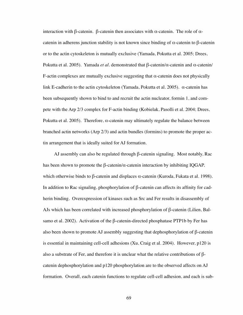
interaction with β-catenin. β-catenin then associates with α-catenin. The role of α-
catenin in adherens junction stability is not known since binding of α-catenin to β-catenin
or to the actin cytoskeleton is mutually exclusive (Yamada, Pokutta et al. 2005; Drees,
Pokutta et al. 2005). Yamada et al. demonstrated that β-catenin/α-catenin and α-catenin/
F-actin complexes are mutually exclusive suggesting that α-catenin does not physically
link E-cadherin to the actin cytoskeleton (Yamada, Pokutta et al. 2005). α-catenin has
been subsequently shown to bind to and recruit the actin nucleator, formin 1, and com-
pete with the Arp 2/3 complex for F-actin binding (Kobielak, Pasolli et al. 2004; Drees,
Pokutta et al. 2005). Therefore, α-catenin may ultimately regulate the balance between
branched actin networks (Arp 2/3) and actin bundles (formins) to promote the proper ac-
tin arrangement that is ideally suited for AJ formation.
AJ assembly can also be regulated through β-catenin signaling. Most notably, Rac
has been shown to promote the β-catenin/α-catenin interaction by inhibiting IQGAP,
which otherwise binds to β-catenin and displaces α-catenin (Kuroda, Fukata et al. 1998).
In addition to Rac signaling, phosphorylation of β-catenin can affects its affinity for cad-
herin binding. Overexpression of kinases such as Src and Fer results in disassembly of
AJs which has been correlated with increased phosphorylation of β-catenin (Lilien, Bal-
samo et al. 2002). Activation of the β-catenin-directed phosphatase PTP1b by Fer has
also been shown to promote AJ assembly suggesting that dephosphorylation of β-catenin
is essential in maintaining cell-cell adhesions (Xu, Craig et al. 2004). However, p120 is
also a substrate of Fer, and therefore it is unclear what the relative contributions of β-
catenin dephosphorylation and p120 phosphorylation are to the observed affects on AJ
formation. Overall, each catenin functions to regulate cell-cell adhesion, and each is sub-
69
ject to regulation by multiple signaling events. Interestingly, many of these signaling
events appear to simultaneously impact the function of all three catenins. For example,
Rac activation promotes the formation of both the β-catenin/α-catenin and the p120/p190
complex. Therefore, Rac may promote AJ assembly by globally arranging all of the cat-
enins under the cadherin complex. However, the relative contribution of each catenin in
cell-cell adhesion is not well appreciated, which is primarily due to the fact that the pre-
cise role of each catenin in regulating cell-cell adhesion is just now being elucidated. As
in the case with Rac signaling, all three catenins are essential to the dynamic process of
AJ assembly and disassembly. Misregulation of one catenin is likely to disrupt AJs by its
specific contribution to cell-cell adhesion.
Spatial versus global regulation of Rho
We conclude that cadherin complexes are an obligatory intermediate in the Bar-
Sagi pathway, which is essential in regulation of the actin cytoskeleton by RTKs, in-
tegrins, and cadherins (Noren, Niessen et al. 2001; Nimnual, Taylor et al. 2003) (Fig. 29).
In fibroblasts, AJ formation is blocked by strong inhibition of Rho (Sander, ten Klooster
et al. 1999). On the other hand, too much Rho activity also disrupts AJs and leads to cell
transformation (Jaffe and Hall 2002), as illustrated here in cells lacking p120. Thus, ex-
quisite control of Rho activity is essential for proper cell-cell adhesion and to prevent un-
controlled cell growth. The transient targeting of otherwise cytoplasmic p190 to cadherin
complexes is ideally suited for this role, and provides an extremely sensitive means of
regulating local, and perhaps overall Rho activity in cells. RhoGTPase-mediated cadherin
internalization and/or turnover allows for rapid modulation of cell-cell adhesion by post-
70
71
Figure 29. Model for the role of p120 and p190 in regulation of cadherin function. Signaling input to Rac occurs through multiple receptor types, including RTKs, integrins, and cadherins. Rac activity induces translocation of cytoplasmic p190 to membranes via a pathway involving generation of reactive oxygen species (ROS), inhibition of LMW-PTP, and subsequent activation/tyrosine phosphorylation of p190 by associated tyrosine kinases (probably Src and/or Abl family kinases). p190 interacts transiently with cadherin-bound p120, which in turn targets Rho inactivation to membrane domains in-volved in cadherin activity. Organization of the pathway around the cadherin complex reflects the importance of linked Rac and Rho activities for successful AJ formation.
transcriptional signaling mechanisms, as would be required during wound healing,
changes in endothelial cell permeability, and many growth factor-induced alterations in
cell behavior. Thus, the novel mechanisms described herein may be essential for many
morphologic and adhesive changes associated with the activation of receptor systems.
The dependancy of the cadherin complex on actin remodeling by other receptors
suggests that these receptors are functionally connected. How they are functionally con-
nected remains an open question. One possibility is that different receptor types physi-
cally and transiently couple to one another in response to some stimulus (Fig. 30). Alter-
natively, the local activation of one receptor transmits a signal across the cell to a differ-
ent receptor type (Fig. 31). In p120i NIH3T3, Rho is uncoupled from multiple receptors
including RTKs and integrins. PDGFR and N-cadherin co-localize in DCRs upon stimu-
lation with PDGF-BB, and PGFR-induced actin rearrangements depend on a functional
cadherin complex (see Fig. 19-20). Likewise, others have reported that E-cadherin and
activated EGFR co-localize at AJs (Pece and Gutkind 2000; Qian, Karpova et al. 2004).
Certain types of integrins have also been reported to co-localize with cadherins, although
this concept is currently under debate (Chattopadhyay, Wang et al. 2003) (personal com-
munication, Vito Quaranta). Lastly, RTKs have also been shown to co-localize and are
functionally coupled with integrin receptors (Brunton, MacPherson et al. 2004). This
evidence suggests that crosstalk between apparently distant receptors occurs through
transient signaling events that bring these receptors together spatially. Thus the global
affects of p120 depletion on Rho activation would be either primarily due to the fact that
multiple receptors must transiently couple with the cadherin complex to regulate Rho or a
lack of inhibition in the cytoplasm. Depending on which receptor pairs with the cadherin
72
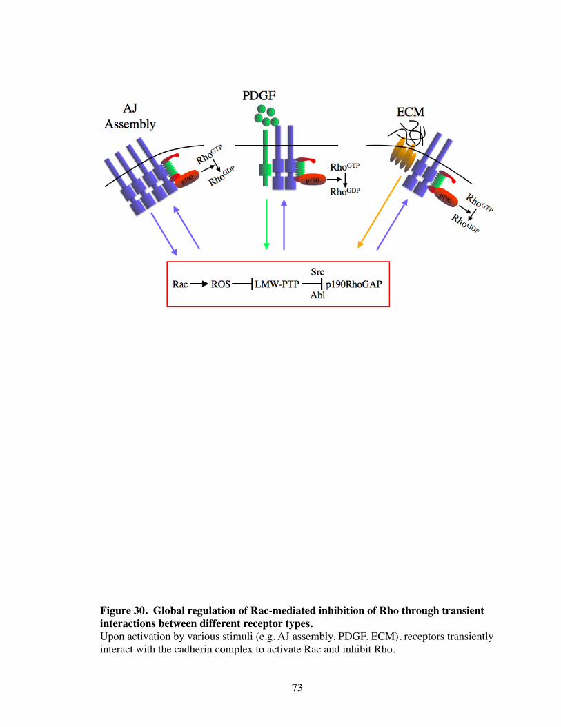
73
Figure 30. Global regulation of Rac-mediated inhibition of Rho through transient interactions between different receptor types.Upon activation by various stimuli (e.g. AJ assembly, PDGF, ECM), receptors transiently interact with the cadherin complex to activate Rac and inhibit Rho.
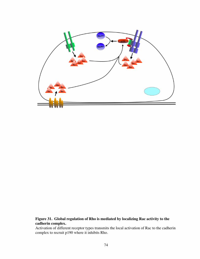
74
Figure 31. Global regulation of Rho is mediated by localizing Rac activity to the cadherin complex. Activation of different receptor types transmits the local activation of Rac to the cadherin complex to recruit p190 where it inhibits Rho.
complex, a different biological response is elicited. For example, RTK coupling with the
cadherin complex mediates DCRs and integrin coupling permits cell spreading on ECM.
On the other hand, the distinct localization, but functional dependency of different
receptor types suggests that certain signals may be transmitted over distances within the
cell. Our data shows that the regulation of Rho by integrins, RTKs, and cadherins are
functionally linked through the p120/p190 complex. Indeed, p120i cells fail to migrate
into a wound reflecting a putative inability of integrins to properly regulate Rho. There-
fore, focal adhesions on the basal surface must transmit signals to laterally localized cad-
herins to inhibit Rho to permit motility. Thus, it is difficult to imagine transient interac-
tions between integrins and cadherins when they are localized to distinct regions within
the cell. Actin-based contractility extends throughout the entire cell, and globally affects
both integrin- and cadherin-based complexes in an apparent antagonistic manner
(D'Souza-Schorey 2005; Bershadsky, Ballestrem et al. 2006). Increased contractility
promotes integrin-mediated focal adhesions, and the disassembly of AJs. On the other
hand, a reduction in contractility results in the opposite effect. Perhaps integrin signaling
locally activates Rac which then initiates low level changes in contractility. Once initi-
ated by focal adhesions, changes in contractility could permit cadherin clustering and
p120-dependent Rac-inhibition of Rho at sites of cell-cell adhesions. In this scenario de-
termining the relative contribution to integrin-dependent and cadherin-dependent activa-
tion of Rac would be critical in understanding how these receptors mediate cross-talk.
Likewise, when a cell is globally stimulated with a growth factor such as PDGF,
cadherin-dependent DCRs form in spatially distinct regions. Local activation of Rac by
PDGFR may initiate the changes in contractility to permit cadherin-dependent Rho inac-
75

tivation and organization of the cadherin complex. How different receptors communicate
to one another remains an open question. Regardless of how cross-talk is mediated, our
data highlights the biological significance of these cross-talk events, and suggests that
interactions between different receptor types represents a major component of general
cellular signaling.
76
CHAPTER V
ROLE OF P120 AND P190 IN CONTACT INHIBITION OF GROWTH AND CELLULAR TRANSFORMATION
Introduction
Contact inhibition is a process in which increased cell density negatively regulates
cell proliferation. Loss of contact inhibition of growth is a hallmark of most cancers, but
the mechanisms that regulate this process are not understood. Contact inhibition is medi-
ated by cadherin-based cell-cell adhesions, and loss of contact inhibition is paralleled
with a disruption in cell-cell adhesion (Takahashi and Suzuki 1996; Bracke, Depypere et
al. 1997; St Croix, Sheehan et al. 1998). Specific cadherin ligation between sparsely
plated fibroblasts and latex beads coated with the extracellular domain of N-cadherin is
sufficient to induce cell cycle arrest involving regulation of the cell cycle kinase inhibi-
tors (CKIs) p21 and p27 (Levenberg, Yarden et al. 1999; Gavard, Marthiens et al. 2004).
Although the mechanism is unknown, these observations suggest that signaling through
N-cadherin alone is sufficient to inhibit proliferation.
In addition to their role in regulating the actin cytoskeleton, Rac and Rho have im-
portant roles in regulating cell proliferation. Rac can stimulate the expression of cyclin
D1 in a stress fiber independent manner whereas Rho negatively regulates p21 and p27 in
a stress fiber dependent pathway (Olson, Paterson et al. 1998; Roovers and Assoian
2003). Activation of Erk signaling by Rho was shown to be dependent on stress fiber
formation and integrin clustering (Roovers and Assoian 2003) suggesting that regulation
of cell adhesion and growth by Rho are functionally related. On the other hand, too much
77
Rho activity can transform cells since overexpression of RhoGEFs or dominant-active
mutants of Rho are able to cause partial transformation and a complete loss of contact
inhibition (Jaffe and Hall 2002). Therefore, maintaining proper levels of Rho activity is
crucial for both promoting and inhibiting cell growth. The parallels between cadherin-
dependent regulation of the cell cycle and RhoGTPase signaling, however, remains
largely unexplored.
Given the duel role for p120 in stabilizing cadherins and regulating Rho, p120 is a
likely candidate for mediating contact inhibition of growth. As shown in chapter III, re-
duction of p120 expression results in the loss of contact inhibition due to the constitutive
activation of Rho (Fig. 10-12). Furthermore, our data localizes p120-mediated inhibition
of Rho at sites of cadherin engagement suggesting that AJ assembly and Rho inhibition
by p120 are functionally related. Therefore, it is likely that as cell density increases,
there would be a concomitant increase in Rho inhibition by p120.
Interestingly, both p120 and p190 are prominent Src substrates. Transformation by
Src results in a loss of contact inhibition as well as dramatic alterations to the actin cyto-
skeleton. The mechanism of Src transformation is largely unknown, but involves both
misregulation of adhesion (cell-cell and cell-ECM) and RhoGTPase signaling (Mat-
suyoshi, Hamaguchi et al. 1992; Fincham, Wyke et al. 1995; Avizienyte, Wyke et al.
2002; Frame, Fincham et al. 2002). Given the requirement of p120 and the cadherin
complex to recruit p190 to the plasma membrane to inhibit Rho, our hypothesis is that as
cell density increases, so does the number of cell-cell contacts, which in turn recruits
more p190 to the cell surface. This functionally translates into a reduction of Rho activ-
78
ity to levels low enough to suppress growth. Lastly, the role of the p120/p190 complex
may have an important role in Src-induced misregulation of cell-cell adhesion.
Results
p120i cells have increased MAPK signaling
Rho induces cell proliferation through activation of Erk signaling pathways (Roo-
vers and Assoian 2003; Roovers, Klein et al. 2003). Therefore, we analyzed MAPK ac-
tivities to determine if the growth abnormalities observed in p120i cells are due to ele-
vated Rho signaling. Indeed, basal levels of Erk1/2 phosphorylation is increased in
p120i cells as compared to WT (Fig. 32). This observation is consistent with constitu-
tively active Rho driving uncontrolled proliferation in p120i cells.
Confluency regulates Rho activity
Upon contact inhibition, cadherins become maximally engaged to suppress cell
growth (St Croix, Sheehan et al. 1998). p120i cells fail to contact inhibit due to the con-
stitutive activation of Rho, which may reflect a failure to recruit p190 to the plasma
membrane upon increasing cell density. Rho activity was first analyzed under conditions
of contact inhibition to confirm that Rho activity was suppressed when WT NIH3T3 cells
underwent contact inhibition. Indeed, contact inhibited cells displayed about a 2-fold de-
crease in Rho activity as compared to, subconfluent, proliferating cells (Fig. 33). Inter-
estingly, when p190 localization was analyzed, there was a noticeable shift from the cy-
toplasm in dense, proliferating cells to the plasma membrane in contact inhibited cells
79
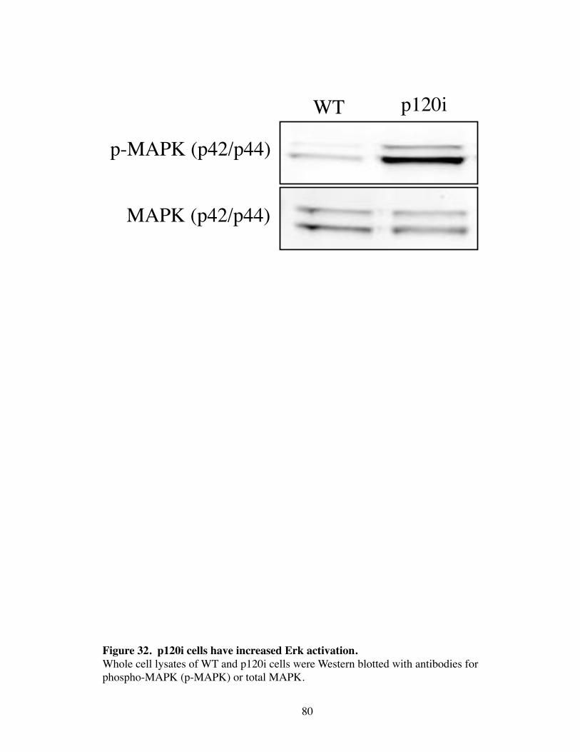
80
Figure 32. p120i cells have increased Erk activation.Whole cell lysates of WT and p120i cells were Western blotted with antibodies for phospho-MAPK (p-MAPK) or total MAPK.
p-MAPK (p42/p44)
MAPK (p42/p44)
WT p120i
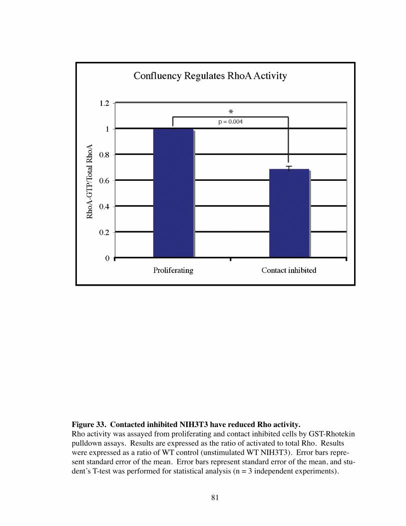
81
Figure 33. Contacted inhibited NIH3T3 have reduced Rho activity.Rho activity was assayed from proliferating and contact inhibited cells by GST-Rhotekin pulldown assays. Results are expressed as the ratio of activated to total Rho. Results were expressed as a ratio of WT control (unstimulated WT NIH3T3). Error bars repre-sent standard error of the mean. Error bars represent standard error of the mean, and stu-dent’s T-test was performed for statistical analysis (n = 3 independent experiments).
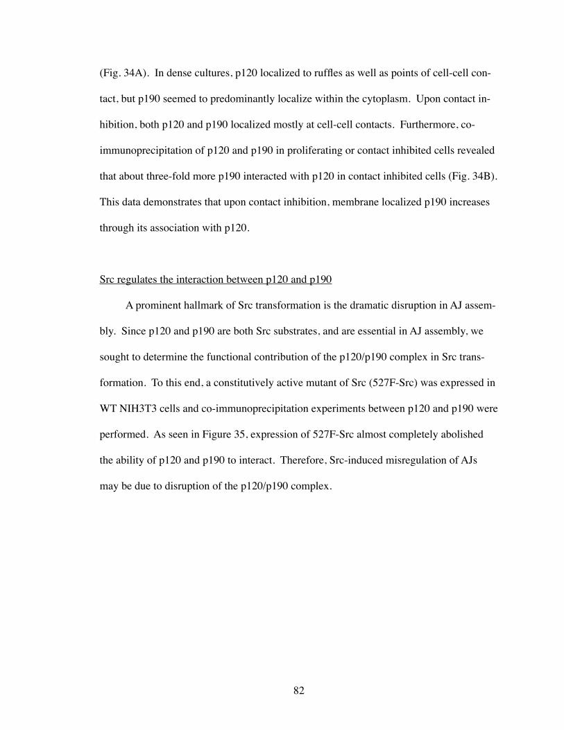
(Fig. 34A). In dense cultures, p120 localized to ruffles as well as points of cell-cell con-
tact, but p190 seemed to predominantly localize within the cytoplasm. Upon contact in-
hibition, both p120 and p190 localized mostly at cell-cell contacts. Furthermore, co-
immunoprecipitation of p120 and p190 in proliferating or contact inhibited cells revealed
that about three-fold more p190 interacted with p120 in contact inhibited cells (Fig. 34B).
This data demonstrates that upon contact inhibition, membrane localized p190 increases
through its association with p120.
Src regulates the interaction between p120 and p190
A prominent hallmark of Src transformation is the dramatic disruption in AJ assem-
bly. Since p120 and p190 are both Src substrates, and are essential in AJ assembly, we
sought to determine the functional contribution of the p120/p190 complex in Src trans-
formation. To this end, a constitutively active mutant of Src (527F-Src) was expressed in
WT NIH3T3 cells and co-immunoprecipitation experiments between p120 and p190 were
performed. As seen in Figure 35, expression of 527F-Src almost completely abolished
the ability of p120 and p190 to interact. Therefore, Src-induced misregulation of AJs
may be due to disruption of the p120/p190 complex.
82
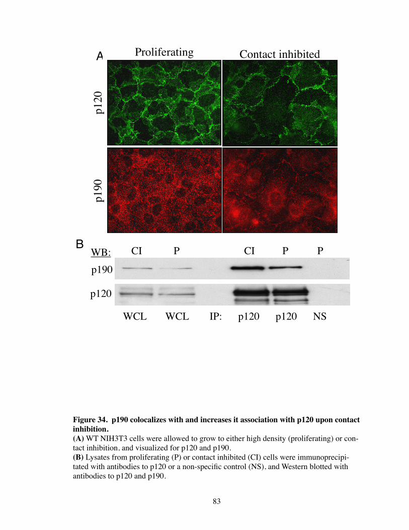
83
Figure 34. p190 colocalizes with and increases it association with p120 upon contact inhibition.(A) WT NIH3T3 cells were allowed to grow to either high density (proliferating) or con-tact inhibition, and visualized for p120 and p190.(B) Lysates from proliferating (P) or contact inhibited (CI) cells were immunoprecipi-tated with antibodies to p120 or a non-specific control (NS), and Western blotted with antibodies to p120 and p190.
Proliferating Contact inhibited
p120
p190
A
p190
p120
WCL WCL IP: p120 p120 NS
WB: CI P PCI PB
84
Figure 35. 527F-Src disrupts binding of p120 and p190.(A) Immunoprecipitation with p120 or irrelevant antibodies (NS) from WT or 527F-Src expressing NIH3T3 lysates was followed by Western blotting with antibodies to p190 and p120.(B) Whole cell lysates from WT or 527F-Src NIH3T3 were analyzed by Western blotting with antibodies to p190 and p120.
B
A
Discussion
p120/p190 interaction increases upon contact inhibition
As shown in chapter III, siRNA-mediated depletion of p120 caused the dramatic
loss of contact inhibition of growth, which could be rescued by inhibiting Rho through
expression of C3 (see Fig. 12). This suggests that the ability of p120 to suppress Rho ac-
tivity is critical in maintaining contact inhibition. We have traced the mechanism of p120
inhibition of Rho to the p120-dependent recruitment of p190 to the cadherin complex un-
der conditions that induce AJ formation. Therefore, if this model is correct, more AJs
should translate into more membrane bound p190. Indeed, upon contact inhibition, more
p190 is observed to localize to the cell surface, as well as about a three-fold increase in
the amount of p190 bound to p120. When p120 and p190 were localized in dense cul-
tures, p120 was enriched at both sites of cell-cell junctions but also within lamellipodia,
whereas p190 was cytoplasmic. However, in contact inhibited cells, AJs are maximally
engaged, and a majority of p120 is recruited along with p190 to cell-cell contacts. Thus
cells in the dense, proliferating population appear to be smaller due to the more diffuse
p120 staining relative to the contact inhibited cells in which most of the p120 is localized
to precise cell-cell junctions. Overlapping lamellipodia from neighboring cells has been
shown to be precursors to AJs (Ehrlich, Hansen et al. 2002). This suggests that p120 may
have a role in establishing new AJs, and that although cells in dense cultures appear to
have stable AJs, they are continually establishing new cell-cell contacts until the cell
population undergoes contact inhibition. Interestingly, strong localization of p190 to the
cell surface was only observed when cells became contact inhibited as opposed to merely
85
crowded. This observation demonstrates an intriguing correlation between the amount of
p190 that localizes to the cadherin complex and the ability to suppress growth. As shown
in previous chapters, p190 association with p120 at the cadherin complex stabilizes AJs
through the inhibition of Rho. Thus our working model is that during a proliferative
state, p190 translocation to AJs (and AJ assembly) must be transient to allow for the
proper level of Rho activity to promote cell growth. Upon contact inhibition, AJs become
stabilized and more p190 is recruited to the cell surface to suppress Rho and proliferation.
Therefore, this model predicts that stabilization of AJs through the p120/p190 complex
and stabilization of cell growth is a functionally identical process. Moreover, the degree
of cellular proliferation is ultimately controlled by the regulation of p190 recruitment to
the cadherin complex by either RTK, integrin, or direct cadherin signaling.
These data also suggest that AJ formation alone is sufficient to both recruit and ac-
tivate p190. However, we do not rule out the possibility that upon contact inhibition an-
other receptor system cross-talks with the cadherin complex to mediate the activation of
p190, and induce translocation of p190 to p120 and the cadherin complex. Crosstalk be-
tween RTKs and cadherins is an important process in contact inhibition, but it is not clear
how these two systems regulate each other upon increasing density (Pece and Gutkind
2000; Qian, Karpova et al. 2004). Adhesion of C-cadherin expressing cells to a surface
coated with the extracellular domain of C-cadherin was sufficient to activate p190
(Noren, Arthur et al. 2003). Therefore, cadherins are indeed sufficient to both activate
and recruit p190 to the cadherin complex, however, understanding the relationship be-
tween RTK- and cadherin-mediated p190 activation under condition of increasing density
will be necessary to put these observations in the context of contact inhibition.
86
p120/p190 complex is regulated by Src
Expression of 527F-Src prevents the p120/p190 interaction, which presents a poten-
tially exciting role for p120 and p190 in Src transformation. Furthermore, both p120 and
p190 are prominent Src substrates, which suggests that tyrosine phosphorylation regu-
lates the interaction between p120 and p190. This would provide for a highly sensitive
means for regulating AJ assembly during processes such as wound healing and cell per-
meability. Identifying the phosphorylation sites on p120 and p190 necessary for regulat-
ing their interaction will be useful in determining their contribution to Src signaling.
Overall, these data demonstrate that the p120/p190 complex may have important
roles in translating increasing cell density to inhibition of growth. Furthermore, regula-
tion of the p120/p190 complex by tyrosine phosphorylation allows for the fine tuning of
AJs that is critical in many biological processes.
87
CHAPTER VI
FUTURE DIRECTIONS
Introduction
Illuminating a possible mechanism for the inhibition of Rho by p120 has raised as
many questions as it has answered. Targeting p120-mediated inhibition of Rho as a func-
tion of AJ assembly provides a general model that unifies the dual roles of p120 in Rho
regulation and cadherin-based adhesion. This model establishes a mechanistic platform
from which several questions can be tested. For example, there is wealth of literature on
p190 signaling that now must be subjected to scrutiny from the perspective of cell-cell
adhesion. Furthermore, since both p120 and p190 are prominent Src substrates, our data
reveals a new avenue in exploring the role of p120 in Src signaling.
However, there are specific questions that must first be addressed directly. One of
the surprising elements of this work is that p190 has not previously been localized to
cadherin complexes. With the exception of MCF-10A cells, p190 does not obviously lo-
calize to the cadherin complex unless driven there by expression of DA-Rac. Addition-
ally, determining the binding domains of p120 and p190 and selectively uncoupling the
two proteins will be vital to a more detailed analysis of the role of this interaction in cel-
lular signaling.
88
p190 localization in live cells
Our model for p120 and p190 signaling predicts that the association between p190
and p120 at the cadherin complex must be highly regulated and transient in order to
maintain the proper level of Rho activity at any given time. Therefore, the reason why
we do not detect p190 at the cadherin complex may be due to the fact that static meas-
urements fail to capture this highly dynamic process. Since simple static images cannot
represent this event, the most direct way to test this model is through live cell imaging of
p190. Expression of GFP or RFP tagged genes have yielded new insights to cellular sig-
naling networks with the increasing capacity of live cell imaging microscopy techniques.
Through these approaches, an increasing appreciation for the roles of spatial and tempo-
ral constraints in signal transduction has emerged. Time lapse movies of fluorescently-
tagged p190 under conditions of cadherin signaling such as wound repair, growth factor-
induced cell scattering, and increasing cell density may reveal novel insights to the role
of p190 in cadherin signaling.
Determine the binding sites of p120 and p190
Mapping the binding domains between p120 and p190 will be critical in moving
forward with this project. Selectively uncoupling p120 and p190 will not only allow us
to confirm some of our previous observations, but it will provide an essential tool for fu-
ture investigations in p120 and Rho signaling. There are several approaches that can be
used to map the binding sites between two proteins. Some of these include shotgun yeast
two-hybrid, in vitro binding, and “mislocalization assays”. Briefly, shotgun yeast two-
hybrid involves screening the full length cDNA of one gene against a library of randomly
89
sheared cDNA of another gene. By comparing the overlapping sequences of the gene
fragments that interact with the full length cDNA, a discrete binding region can be iden-
tified. in vitro binding assays involve purification of recombinant proteins containing se-
quential deletions and assaying for binding. Lastly, “mislocalization assays” target one
protein to the plasma membrane through a C-terminal CAAX fusion, and then assay for
the co-localization of another protein. This approach was effective in demonstrating that
p120 is able to co-localize with p190 independently of the cadherin complex (see Fig.
24). By introducing small mutations in this p120 mutant and assaying for p190 co-
localization, the regions of p120 necessary for binding p190 will be identified.
Role of p120/p190 complex in Src transformation
Both p120 and p190 are major Src substrates. The function of tyrosine phosphory-
lation of p120 by Src remains unknown, and Src phosphorylation of p190 has been
shown to both activate and inactivate p190 (Chang, Gill et al. 1995; Billuart, Winter et al.
2001). The interaction between p120 and p190 raises a new and exciting possibility for
understanding the role of p120 in Src signaling both under physiological conditions and
in Src transformation.
As shown in chapter V, expression of 527F-Src disrupts the interaction between
p120 and p190. Interestingly, a 527F-Src mutant that cannot translocate to the plasma
membrane (2A/527F-Src) is non-transforming. Additionally, this mutant retains its abil-
ity to phosphorylate many of its substrates. However, one of the substrates that is not
phosphorylated by this Src mutant is p120. Co-expression of 2A/527F-Src with the N-
terminus of RasGAP restores the transforming capabilities of this Src mutant (DeClue,
90
Vass et al. 1993). This N-terminal fragment of RasGAP encodes the SH2-SH3-SH2 (2-
3-2) motif that mediates binding between RasGAP and p190. Expression of 2-3-2 has
been shown subsequently to affect the activity of p190 (McGlade, Brunkhorst et al. 1993;
Bradley, Hernandez et al. 2006). Therefore, the synergistic effects between 2A/527F-Src
and the N-terminus of RasGAP could be due regulation of the p120/p190 complex that
527F-Src is able to mediate alone. Analysis of binding between p120 and p190 in the
presence of 2A/527F-Src and 2A/527F-Src with the 2-3-2 peptide could help determine if
this is so. Lastly, a mutational analysis of tyrosine residues in p120 and p190 to block the
Src-induced disruption in p120/p190 binding should determine the functional significance
of the p120/p190 complex in Src transformation.
Role of AJ formation in Ras signaling
p190 was first characterized by its constitutive association with p120RasGAP
(RasGAP), a major GAP for Ras (Ellis, Moran et al. 1990). RasGAP is among three
known GAPs for Ras, but its contribution to Ras signaling is not well understood. Activa-
tion of p190 by tyrosine phosphorylation increases the association between p190 and
RasGAP, and is necessary for overall p190 function (Bradley, Hernandez et al. 2006).
The novel role for p190 in AJ assembly described here in chapter IV establishes an excit-
ing model for examining the relationships between RasGAP signaling and AJ dynamics.
Recent evidence regarding a role for RasGAP in anchoring p190 at focal adhesions
bares a striking resemblance to our observations for p120 and AJ assembly (Bradley,
Hernandez et al. 2006). It is therefore tempting to speculate antagonistic roles for p120
and RasGAP in competing for p190 recruitment to AJs and focal adhesions, respectively.
91
This would suggest that antagonism between integrin-mediated motility and cadherin-
mediated AJ assembly may be functionally regulated by p190 recruitment. Thus it would
be necessary to determine the different signals that induces p190 translocation to these
two sites. Alternatively, p120-dependent recruitment of p190 to AJs may also cause
RasGAP to localize to AJs. Localization of RasGAP at sites of AJ assembly would dem-
onstrate a novel role for p120 in the negative regulation of Ras signaling. Implementa-
tion of several tools to uncouple RasGAP from p190 will help determine the role of Ras-
GAP in p190 recruitment to AJs.
92
CHAPTER VII
CONCLUDING REMARKS
These data make a significant step forward in understanding the role of p120 in the
regulation of Rho and AJ assembly. Additionally, they suggest a much broader role for
p120 in communicating signaling cues from other receptor systems (e.g. RTKs, integrins)
to the cadherin complex as they pertain to growth, motility and adhesion. Indeed it is
difficult to build adequate models for these cellular functions without considering the sig-
nals that come simultaneously from RTKs, integrins, and cadherin suggesting that these
receptors are functionally inseparable. Our model predicts that the ability of one receptor
to regulate any of these processes depends on signaling through the cadherin complex,
and that the cadherin complex is central in coordinating overall Rho activity (see Fig. 29).
Furthermore, AJ assembly/disassembly is not merely an endpoint of RTK or integrin sig-
naling, but rather crosstalk involves signaling through the cadherin complex to inhibit
Rho. It is also likely that there is a reciprocal dependency of the cadherin complex on
integrin and RTK signaling, and the relative balance of activity from each receptor drives
the cell into a state of cell-cell adhesion, motility, or growth. This model dramatically
simplifies how multiple signaling events (i.e. changes in cell-cell and cell-ECM adhesion,
growth factor signaling) must coordinate their activities on the actin cytoskeleton for one
unified event to occur (i.e. motility). Determining the relative role of p120 in coordinat-
ing these events both spatially and temporally will be an exciting advancement in under-
93

standing crosstalk between these receptor systems as it pertains to normal physiology as
well as cancer.
94
REFERENCES
Anastasiadis, P. Z., S. Y. Moon, et al. (2000). "Inhibition of RhoA by p120 catenin." Nat Cell Biol 2(9): 637-44.
Anastasiadis, P. Z. and A. B. Reynolds (2000). "The p120 catenin family: complex roles in adhesion, signaling and cancer." J Cell Sci 113 ( Pt 8): 1319-34.
Arthur, W. T. and K. Burridge (2001). "RhoA inactivation by p190RhoGAP regulates cell spreading and migration by promoting membrane protrusion and polarity." Mol Biol Cell 12(9): 2711-20.
Avizienyte, E., A. W. Wyke, et al. (2002). "Src-induced de-regulation of E-cadherin in colon cancer cells requires integrin signalling." Nat Cell Biol 4(8): 632-8.
Barberis, D., A. Casazza, et al. (2005). "p190 Rho-GTPase activating protein associates with plexins and it is required for semaphorin signalling." J Cell Sci 118(Pt 20): 4689-700.
Bellovin, D. I., R. C. Bates, et al. (2005). "Altered localization of p120 catenin during epithelial to mesenchymal transition of colon carcinoma is prognostic for aggres-sive disease." Cancer Res 65(23): 10938-45.
Bernards, A. and J. Settleman (2005). "GAPs in growth factor signalling." Growth Fac-tors 23(2): 143-9.
Bershadsky, A. D., C. Ballestrem, et al. (2006). "Assembly and mechanosensory function of focal adhesions: experiments and models." Eur J Cell Biol 85(3-4): 165-73.
Berx, G., F. Nollet, et al. (1998). "Dysregulation of the E-cadherin/catenin complex by irreversible mutations in human carcinomas." Cell Adhes Commun 6(2-3): 171-84.
Berx, G. and F. Van Roy (2001). "The E-cadherin/catenin complex: an important gate-keeper in breast cancer tumorigenesis and malignant progression." Breast Cancer Res 3(5): 289-93.
Betson, M., E. Lozano, et al. (2002). "Rac activation upon cell-cell contact formation is dependent on signaling from the epidermal growth factor receptor." J Biol Chem 277(40): 36962-9.
Bhowmick, N. A., M. Ghiassi, et al. (2001). "Transforming growth factor-beta1 mediates epithelial to mesenchymal transdifferentiation through a RhoA-dependent mecha-nism." Mol Biol Cell 12(1): 27-36.
95
Billuart, P., C. G. Winter, et al. (2001). "Regulating axon branch stability: the role of p190 RhoGAP in repressing a retraction signaling pathway." Cell 107(2): 195-207.
Birchmeier, C., W. Birchmeier, et al. (2003). "Met, metastasis, motility and more." Nat Rev Mol Cell Biol 4(12): 915-25.
Birchmeier, W. and J. Behrens (1994). "Cadherin expression in carcinomas: role in the formation of cell junctions and the prevention of invasiveness." Biochim Biophys Acta 1198(1): 11-26.
Blume-Jensen, P. and T. Hunter (2001). "Oncogenic kinase signalling." Nature 411(6835): 355-65.
Boettner, B. and L. Van Aelst (2002). "The role of Rho GTPases in disease development." Gene 286(2): 155-74.
Bracke, M. E., H. Depypere, et al. (1997). "Functional downregulation of the E-cadherin/catenin complex leads to loss of contact inhibition of motility and of mitochon-drial activity, but not of growth in confluent epithelial cell cultures." Eur J Cell Biol 74(4): 342-9.
Bradley, W. D., S. E. Hernandez, et al. (2006). "Integrin Signaling through Arg Activates p190RhoGAP by Promoting Its Binding to p120RasGAP and Recruitment to the Membrane." Mol Biol Cell.
Braga, V. M. (2002). "Cell-cell adhesion and signalling." Curr Opin Cell Biol 14(5): 546-56.
Braga, V. M., A. Del Maschio, et al. (1999). "Regulation of cadherin function by Rho and Rac: modulation by junction maturation and cellular context." Mol Biol Cell 10(1): 9-22.
Brouns, M. R., S. F. Matheson, et al. (2001). "p190 RhoGAP is the principal Src substrate in brain and regulates axon outgrowth, guidance and fasciculation." Nat Cell Biol 3(4): 361-7.
Brunton, V. G., I. R. MacPherson, et al. (2004). "Cell adhesion receptors, tyrosine kinases and actin modulators: a complex three-way circuitry." Biochim Biophys Acta 1692(2-3): 121-44.
Burridge, K. and K. Wennerberg (2004). "Rho and Rac take center stage." Cell 116(2): 167-79.
Chang, J. H., S. Gill, et al. (1995). "c-Src regulates the simultaneous rearrangement of actin cytoskeleton, p190RhoGAP, and p120RasGAP following epidermal growth factor stimulation." J Cell Biol 130(2): 355-68.
96
Chattopadhyay, N., Z. Wang, et al. (2003). "alpha3beta1 integrin-CD151, a component of the cadherin-catenin complex, regulates PTPmu expression and cell-cell adhe-sion." J Cell Biol 163(6): 1351-62.
Chen, X. and B. M. Gumbiner (2006). "Crosstalk between different adhesion molecules." Curr Opin Cell Biol 18(5): 572-8.
Chrzanowska-Wodnicka, M. and K. Burridge (1996). "Rho-stimulated contractility drives the formation of stress fibers and focal adhesions." J Cell Biol 133(6): 1403-15.
Cozzolino, M., V. Stagni, et al. (2003). "p120 Catenin is required for growth factor-dependent cell motility and scattering in epithelial cells." Mol Biol Cell 14(5): 1964-77.
D'Souza-Schorey, C. (2005). "Disassembling adherens junctions: breaking up is hard to do." Trends Cell Biol 15(1): 19-26.
Daniel, J. M. and A. B. Reynolds (1999). "The catenin p120(ctn) interacts with Kaiso, a novel BTB/POZ domain zinc finger transcription factor." Mol Cell Biol 19(5): 3614-23.
Daniel, J. M., C. M. Spring, et al. (2002). "The p120(ctn)-binding partner Kaiso is a bi-modal DNA-binding protein that recognizes both a sequence-specific consensus and methylated CpG dinucleotides." Nucleic Acids Res 30(13): 2911-9.
Davis, M. A., R. C. Ireton, et al. (2003). "A core function for p120-catenin in cadherin turnover." J Cell Biol 163(3): 525-34.
Davis, M. A. and A. B. Reynolds (2006). "Blocked acinar development, E-cadherin re-duction, and intraepithelial neoplasia upon ablation of p120-catenin in the mouse salivary gland." Dev Cell 10(1): 21-31.
DeClue, J. E., W. C. Vass, et al. (1993). "Functional role of GTPase-activating protein in cell transformation by pp60v-src." Mol Cell Biol 13(11): 6799-809.
del Peso, L., R. Hernandez-Alcoceba, et al. (1997). "Rho proteins induce metastatic prop-erties in vivo." Oncogene 15(25): 3047-57.
del Pozo, M. A., L. S. Price, et al. (2000). "Adhesion to the extracellular matrix regulates the coupling of the small GTPase Rac to its effector PAK." Embo J 19(9): 2008-14.
Derksen, P. W., X. Liu, et al. (2006). "Somatic inactivation of E-cadherin and p53 in mice leads to metastatic lobular mammary carcinoma through induction of anoikis re-sistance and angiogenesis." Cancer Cell 10(5): 437-49.
97
Downing, J. R. and A. B. Reynolds (1991). "PDGF, CSF-1, and EGF induce tyrosine phosphorylation of p120, a pp60src transformation-associated substrate." Onco-gene 6(4): 607-13.
Drees, F., S. Pokutta, et al. (2005). "Alpha-catenin is a molecular switch that binds E-cadherin-beta-catenin and regulates actin-filament assembly." Cell 123(5): 903-15.
Dudek, S. M. and J. G. Garcia (2001). "Cytoskeletal regulation of pulmonary vascular permeability." J Appl Physiol 91(4): 1487-500.
Ehrlich, J. S., M. D. Hansen, et al. (2002). "Spatio-temporal regulation of Rac1 localiza-tion and lamellipodia dynamics during epithelial cell-cell adhesion." Dev Cell 3(2): 259-70.
Ellis, C., V. Measday, et al. (1995). "Phosphorylation-dependent complexes of p120 Ras-specific GTPase-activating protein with p62 and p190." Methods Enzymol 255: 179-92.
Ellis, C., M. Moran, et al. (1990). "Phosphorylation of GAP and GAP-associated proteins by transforming and mitogenic tyrosine kinases." Nature 343(6256): 377-81.
Evers, E. E., G. C. Zondag, et al. (2000). "Rho family proteins in cell adhesion and cell migration." Eur J Cancer 36(10): 1269-74.
Fang, X., H. Ji, et al. (2004). "Vertebrate development requires ARVCF and p120 caten-ins and their interplay with RhoA and Rac." J Cell Biol 165(1): 87-98.
Fincham, V. J., A. Chudleigh, et al. (1999). "Regulation of p190 Rho-GAP by v-Src is linked to cytoskeletal disruption during transformation." J Cell Sci 112 ( Pt 6): 947-56.
Fincham, V. J., J. A. Wyke, et al. (1995). "v-Src-induced degradation of focal adhesion kinase during morphological transformation of chicken embryo fibroblasts." On-cogene 10(11): 2247-52.
Fire, A., S. Xu, et al. (1998). "Potent and specific genetic interference by double-stranded RNA in Caenorhabditis elegans." Nature 391(6669): 806-11.
Foster, R., K. Q. Hu, et al. (1994). "p190 RhoGAP, the major RasGAP-associated protein, binds GTP directly." Mol Cell Biol 14(11): 7173-81.
Frame, M. C., V. J. Fincham, et al. (2002). "v-Src's hold over actin and cell adhesions." Nat Rev Mol Cell Biol 3(4): 233-45.
Fritz, G., I. Just, et al. (1999). "Rho GTPases are over-expressed in human tumors." Int J Cancer 81(5): 682-7.
98
Frixen, U. H., J. Behrens, et al. (1991). "E-cadherin-mediated cell-cell adhesion prevents invasiveness of human carcinoma cells." J Cell Biol 113(1): 173-85.
Garcia, J. G., F. Liu, et al. (2001). "Sphingosine 1-phosphate promotes endothelial cell barrier integrity by Edg-dependent cytoskeletal rearrangement." J Clin Invest 108(5): 689-701.
Gavard, J., V. Marthiens, et al. (2004). "N-cadherin activation substitutes for the cell con-tact control in cell cycle arrest and myogenic differentiation: involvement of p120 and beta-catenin." J Biol Chem 279(35): 36795-802.
Grosheva, I., M. Shtutman, et al. (2001). "p120 catenin affects cell motility via modula-tion of activity of Rho-family GTPases: a link between cell-cell contact formation and regulation of cell locomotion." J Cell Sci 114(Pt 4): 695-707.
Gumbiner, B. M. (2005). "Regulation of cadherin-mediated adhesion in morphogenesis." Nat Rev Mol Cell Biol 6(8): 622-34.
Hall, A. (1992). "Ras-related GTPases and the cytoskeleton." Mol Biol Cell 3(5): 475-9.Hall, A. (1992). "Signal transduction through small GTPases--a tale of two GAPs." Cell
69(3): 389-91.
Hanahan, D. and R. A. Weinberg (2000). "The hallmarks of cancer." Cell 100(1): 57-70.
Haskell, M. D., A. L. Nickles, et al. (2001). "Phosphorylation of p190 on Tyr1105 by c-Src is necessary but not sufficient for EGF-induced actin disassembly in C3H10T1/2 fibroblasts." J Cell Sci 114(Pt 9): 1699-708.
Hernandez, S. E., J. Settleman, et al. (2004). "Adhesion-dependent regulation of p190RhoGAP in the developing brain by the Abl-related gene tyrosine kinase." Curr Biol 14(8): 691-6.
Herzig, M., F. Savarese, et al. (2006). "Tumor progression induced by the loss of E-cadherin independent of beta-catenin/Tcf-mediated Wnt signaling." Oncogene.
Hooshmand-Rad, R., L. Claesson-Welsh, et al. (1997). "Involvement of phosphatidyli-nositide 3'-kinase and Rac in platelet-derived growth factor-induced actin reor-ganization and chemotaxis." Exp Cell Res 234(2): 434-41.
Ireton, R. C., M. A. Davis, et al. (2002). "A novel role for p120 catenin in E-cadherin function." J Cell Biol 159(3): 465-76.
Jaffe, A. B. and A. Hall (2002). "Rho GTPases in transformation and metastasis." Adv Cancer Res 84: 57-80.
99
Jaffe, A. B. and A. Hall (2005). "Rho GTPases: biochemistry and biology." Annu Rev Cell Dev Biol 21: 247-69.
Kanner, S. B., A. B. Reynolds, et al. (1991). "Tyrosine phosphorylation of a 120-kilodalton pp60src substrate upon epidermal growth factor and platelet-derived growth factor receptor stimulation and in polyomavirus middle-T-antigen-transformed cells." Mol Cell Biol 11(2): 713-20.
Keirsebilck, A., S. Bonne, et al. (1998). "Molecular cloning of the human p120ctn catenin gene (CTNND1): expression of multiple alternatively spliced isoforms." Genom-ics 50(2): 129-46.
Kjoller, L. and A. Hall (1999). "Signaling to Rho GTPases." Exp Cell Res 253(1): 166-79.
Kobielak, A., H. A. Pasolli, et al. (2004). "Mammalian formin-1 participates in adherens junctions and polymerization of linear actin cables." Nat Cell Biol 6(1): 21-30.
Kolibaba, K. S. and B. J. Druker (1997). "Protein tyrosine kinases and cancer." Biochim Biophys Acta 1333(3): F217-48.
Kuroda, S., M. Fukata, et al. (1997). "Regulation of cell-cell adhesion of MDCK cells by Cdc42 and Rac1 small GTPases." Biochem Biophys Res Commun 240(2): 430-5.
Kuroda, S., M. Fukata, et al. (1998). "Role of IQGAP1, a target of the small GTPases Cdc42 and Rac1, in regulation of E-cadherin- mediated cell-cell adhesion." Sci-ence 281(5378): 832-5.
Lamarche, N. and A. Hall (1994). "GAPs for rho-related GTPases." Trends Genet 10(12): 436-40.
Leung, T., X. Q. Chen, et al. (1996). "The p160 RhoA-binding kinase ROK alpha is a member of a kinase family and is involved in the reorganization of the cytoskele-ton." Mol Cell Biol 16(10): 5313-27.
Levenberg, S., A. Yarden, et al. (1999). "p27 is involved in N-cadherin-mediated contact inhibition of cell growth and S-phase entry." Oncogene 18(4): 869-76.
Lilien, J., J. Balsamo, et al. (2002). "Turn-off, drop-out: functional state switching of cadherins." Dev Dyn 224(1): 18-29.
Lozano, E., M. Betson, et al. (2003). "Tumor progression: Small GTPases and loss of cell-cell adhesion." Bioessays 25(5): 452-63.
100
Magie, C. R., D. Pinto-Santini, et al. (2002). "Rho1 interacts with p120ctn and alpha-catenin, and regulates cadherin-based adherens junction components in Droso-phila." Development 129(16): 3771-82.
Malliri, A., S. van Es, et al. (2004). "The Rac exchange factor Tiam1 is required for the establishment and maintenance of cadherin-based adhesions." J Biol Chem 279(29): 30092-8.
Mareel, M. and A. Leroy (2003). "Clinical, cellular, and molecular aspects of cancer in-vasion." Physiol Rev 83(2): 337-76.
Mariner, D. J., P. Anastasiadis, et al. (2001). "Identification of Src phosphorylation sites in the catenin p120ctn." J Biol Chem 276(30): 28006-13.
Matsuyoshi, N., M. Hamaguchi, et al. (1992). "Cadherin-mediated cell-cell adhesion is perturbed by v-src tyrosine phosphorylation in metastatic fibroblasts." J Cell Biol 118(3): 703-14.
McGlade, J., B. Brunkhorst, et al. (1993). "The N-terminal region of GAP regulates cyto-skeletal structure and cell adhesion." Embo J 12(8): 3073-81.
Mo, Y. Y. and A. B. Reynolds (1996). "Identification of murine p120 isoforms and het-erogeneous expression of p120cas isoforms in human tumor cell lines." Cancer Res 56(11): 2633-40.
Monier-Gavelle, F. and J. L. Duband (1997). "Cross talk between adhesion molecules: control of N-cadherin activity by intracellular signals elicited by beta1 and beta3 integrins in migrating neural crest cells." J Cell Biol 137(7): 1663-81.
Moon, S. Y. and Y. Zheng (2003). "Rho GTPase-activating proteins in cell regulation." Trends Cell Biol 13(1): 13-22.
Nakahara, H., S. C. Mueller, et al. (1998). "Activation of beta1 integrin signaling stimu-lates tyrosine phosphorylation of p190RhoGAP and membrane-protrusive activi-ties at invadopodia." J Biol Chem 273(1): 9-12.
Nimnual, A. S., L. J. Taylor, et al. (2003). "Redox-dependent downregulation of Rho by Rac." Nat Cell Biol 5(3): 236-41.
Nobes, C. and A. Hall (1994). "Regulation and function of the Rho subfamily of small GTPases." Curr Opin Genet Dev 4(1): 77-81.
Nobes, C. D. and A. Hall (1995). "Rho, rac, and cdc42 GTPases regulate the assembly of multimolecular focal complexes associated with actin stress fibers, lamellipodia, and filopodia." Cell 81(1): 53-62.
101
Noren, N. K., W. T. Arthur, et al. (2003). "Cadherin engagement inhibits RhoA via p190RhoGAP." J Biol Chem 278(16): 13615-8.
Noren, N. K., B. P. Liu, et al. (2000). "p120 catenin regulates the actin cytoskeleton via Rho family GTPases." J Cell Biol 150(3): 567-80.
Noren, N. K., C. M. Niessen, et al. (2001). "Cadherin engagement regulates Rho family GTPases." J Biol Chem 276(36): 33305-8.
Oka, H., H. Shiozaki, et al. (1993). "Expression of E-cadherin cell adhesion molecules in human breast cancer tissues and its relationship to metastasis." Cancer Res 53(7): 1696-701.
Olmeda, D., M. Jorda, et al. (2006). "Snail silencing effectively suppresses tumour growth and invasiveness." Oncogene.
Olson, M. F., H. F. Paterson, et al. (1998). "Signals from Ras and Rho GTPases interact to regulate expression of p21Waf1/Cip1." Nature 394(6690): 295-9.
Orsulic, S. and R. Kemler (2000). "Expression of Eph receptors and ephrins is differen-tially regulated by E-cadherin." J Cell Sci 113 ( Pt 10): 1793-802.
Pece, S. and J. S. Gutkind (2000). "Signaling from E-cadherins to the MAPK pathway by the recruitment and activation of epidermal growth factor receptors upon cell-cell contact formation." J Biol Chem 275(52): 41227-33.
Perez-Moreno, M., M. A. Davis, et al. (2006). "p120-catenin mediates inflammatory re-sponses in the skin." Cell 124(3): 631-44.
Perl, A. K., P. Wilgenbus, et al. (1998). "A causal role for E-cadherin in the transition from adenoma to carcinoma." Nature 392(6672): 190-3.
Piedra, J., S. Miravet, et al. (2003). "p120 Catenin-associated Fer and Fyn tyrosine ki-nases regulate beta-catenin Tyr-142 phosphorylation and beta-catenin-alpha-catenin Interaction." Mol Cell Biol 23(7): 2287-97.
Porter, A. C. and R. R. Vaillancourt (1998). "Tyrosine kinase receptor-activated signal transduction pathways which lead to oncogenesis." Oncogene 17(11 Reviews): 1343-52.
Potempa, S. and A. J. Ridley (1998). "Activation of both MAP kinase and phosphatidyli-nositide 3-kinase by Ras is required for hepatocyte growth factor/scatter factor-induced adherens junction disassembly." Mol Biol Cell 9(8): 2185-200.
102
Qian, X., T. Karpova, et al. (2004). "E-cadherin-mediated adhesion inhibits ligand-dependent activation of diverse receptor tyrosine kinases." Embo J 23(8): 1739-48.
Qiu, R. G., J. Chen, et al. (1995). "A role for Rho in Ras transformation." Proc Natl Acad Sci U S A 92(25): 11781-5.
Ren, X. D., W. B. Kiosses, et al. (1999). "Regulation of the small GTP-binding protein Rho by cell adhesion and the cytoskeleton." Embo J 18(3): 578-85.
Reynolds, A. B., J. Daniel, et al. (1994). "Identification of a new catenin: the tyrosine ki-nase substrate p120cas associates with E-cadherin complexes." Mol Cell Biol 14(12): 8333-42.
Reynolds, A. B., D. J. Roesel, et al. (1989). "Transformation-specific tyrosine phosphory-lation of a novel cellular protein in chicken cells expressing oncogenic variants of the avian cellular src gene." Mol Cell Biol 9(2): 629-38.
Ridley, A. J., P. M. Comoglio, et al. (1995). "Regulation of scatter factor/hepatocyte growth factor responses by Ras, Rac, and Rho in MDCK cells." Mol Cell Biol 15(2): 1110-22.
Roof, R. W., B. D. Dukes, et al. (2000). "Phosphorylation of the p190 RhoGAP N-terminal domain by c-Src results in a loss of GTP binding activity." FEBS Lett 472(1): 117-21.
Roovers, K. and R. K. Assoian (2003). "Effects of rho kinase and actin stress fibers on sustained extracellular signal-regulated kinase activity and activation of G(1) phase cyclin-dependent kinases." Mol Cell Biol 23(12): 4283-94.
Roovers, K., E. A. Klein, et al. (2003). "Nuclear translocation of LIM kinase mediates Rho-Rho kinase regulation of cyclin D1 expression." Dev Cell 5(2): 273-84.
Rottner, K., A. Hall, et al. (1999). "Interplay between Rac and Rho in the control of sub-strate contact dynamics." Curr Biol 9(12): 640-8.
Roura, S., S. Miravet, et al. (1999). "Regulation of E-cadherin/Catenin association by ty-rosine phosphorylation." J Biol Chem 274(51): 36734-40.
Sahai, E. and C. J. Marshall (2002). "RHO-GTPases and cancer." Nat Rev Cancer 2(2): 133-42.
Sallee, J. L., E. S. Wittchen, et al. (2006). "Regulation of cell adhesion by protein-tyrosine phosphatases: II. Cell-cell adhesion." J Biol Chem 281(24): 16189-92.
103
Sander, E. E., J. P. ten Klooster, et al. (1999). "Rac downregulates Rho activity: recipro-cal balance between both GTPases determines cellular morphology and migratory behavior." J Cell Biol 147(5): 1009-22.
Sander, E. E., S. van Delft, et al. (1998). "Matrix-dependent Tiam1/Rac signaling in epithelial cells promotes either cell-cell adhesion or cell migration and is regu-lated by phosphatidylinositol 3-kinase." J Cell Biol 143(5): 1385-98.
Schwartz, M. (2004). "Rho signalling at a glance." J Cell Sci 117(Pt 23): 5457-8.
Sharma, S. V. (1998). "Rapid recruitment of p120RasGAP and its associated protein, p190RhoGAP, to the cytoskeleton during integrin mediated cell-substrate interac-tion." Oncogene 17(3): 271-81.
Sordella, R., W. Jiang, et al. (2003). "Modulation of Rho GTPase signaling regulates a switch between adipogenesis and myogenesis." Cell 113(2): 147-58.
Spring, C. M., K. F. Kelly, et al. (2005). "The catenin p120ctn inhibits Kaiso-mediated transcriptional repression of the beta-catenin/TCF target gene matrilysin." Exp Cell Res 305(2): 253-65.
St Croix, B., C. Sheehan, et al. (1998). "E-Cadherin-dependent growth suppression is mediated by the cyclin-dependent kinase inhibitor p27(KIP1)." J Cell Biol 142(2): 557-71.
Su, L., J. M. Agati, et al. (2003). "p190RhoGAP is cell cycle regulated and affects cyto-kinesis." J Cell Biol 163(3): 571-82.
Takahashi, K. and K. Suzuki (1996). "Density-dependent inhibition of growth involves prevention of EGF receptor activation by E-cadherin-mediated cell-cell adhe-sion." Exp Cell Res 226(1): 214-22.
Takaishi, K., T. Sasaki, et al. (1994). "Involvement of Rho p21 small GTP-binding pro-tein and its regulator in the HGF-induced cell motility." Oncogene 9(1): 273-9.
Takaishi, K., T. Sasaki, et al. (1997). "Regulation of cell-cell adhesion by rac and rho small G proteins in MDCK cells." J Cell Biol 139(4): 1047-59.
Tatsis, N., D. A. Lannigan, et al. (1998). "The function of the p190 Rho GTPase-activating protein is controlled by its N-terminal GTP binding domain." J Biol Chem 273(51): 34631-8.
Thoreson, M. A., P. Z. Anastasiadis, et al. (2000). "Selective uncoupling of p120(ctn) from E-cadherin disrupts strong adhesion." J Cell Biol 148(1): 189-202.
104
Thoreson, M. A. and A. B. Reynolds (2002). "Altered expression of the catenin p120 in human cancer: implications for tumor progression." Differentiation 70(9-10): 583-9.
Tikoo, A., S. Czekay, et al. (2000). "p190-A, a human tumor suppressor gene, maps to the chromosomal region 19q13.3 that is reportedly deleted in some gliomas." Gene 257(1): 23-31.
Uglow, E. B., S. Slater, et al. (2003). "Dismantling of cadherin-mediated cell-cell con-tacts modulates smooth muscle cell proliferation." Circ Res 92(12): 1314-21.
van der Geer, P., M. Henkemeyer, et al. (1997). "Aberrant Ras regulation and reduced p190 tyrosine phosphorylation in cells lacking p120-Gap." Mol Cell Biol 17(4): 1840-7.
van Wetering, S., J. D. van Buul, et al. (2002). "Reactive oxygen species mediate Rac-induced loss of cell-cell adhesion in primary human endothelial cells." J Cell Sci 115(Pt 9): 1837-46.
Watabe, M., K. Matsumoto, et al. (1993). "Effect of hepatocyte growth factor on cadherin-mediated cell-cell adhesion." Cell Struct Funct 18(2): 117-24.
Wojciak-Stothard, B., S. Potempa, et al. (2001). "Rho and Rac but not Cdc42 regulate endothelial cell permeability." J Cell Sci 114(Pt 7): 1343-55.
Wojciak-Stothard, B., L. Y. Tsang, et al. (2005). "Rac and Rho play opposing roles in the regulation of hypoxia/reoxygenation-induced permeability changes in pulmonary artery endothelial cells." Am J Physiol Lung Cell Mol Physiol 288(4): L749-60.
Wolf, R. M., J. J. Wilkes, et al. (2001). "Tyrosine phosphorylation of p190 RhoGAP by Fyn regulates oligodendrocyte differentiation." J Neurobiol 49(1): 62-78.
Wrobel, C. N., J. Debnath, et al. (2004). "Autocrine CSF-1R activation promotes Src-dependent disruption of mammary epithelial architecture." J Cell Biol 165(2): 263-73.
Xiao, K., D. F. Allison, et al. (2003). "Cellular levels of p120 catenin function as a set point for cadherin expression levels in microvascular endothelial cells." J Cell Biol 163(3): 535-45.
Xu, G., A. W. Craig, et al. (2004). "Continuous association of cadherin with beta-catenin requires the non-receptor tyrosine-kinase Fer." J Cell Sci 117(Pt 15): 3207-19.
Yamada, S., S. Pokutta, et al. (2005). "Deconstructing the cadherin-catenin-actin com-plex." Cell 123(5): 889-901.
105
Yanagisawa, M. and P. Z. Anastasiadis (2006). "p120 catenin is essential for mesenchy-mal cadherin-mediated regulation of cell motility and invasiveness." J Cell Biol 174(7): 1087-96.
Yap, A. S., W. M. Brieher, et al. (1997). "Molecular and functional analysis of cadherin-based adherens junctions." Annu Rev Cell Dev Biol 13: 119-46.
Yap, A. S., W. M. Brieher, et al. (1997). "Lateral clustering of the adhesive ectodomain: a fundamental determinant of cadherin function." Curr Biol 7(5): 308-15.
Yap, A. S., C. M. Niessen, et al. (1998). "The juxtamembrane region of the cadherin cy-toplasmic tail supports lateral clustering, adhesive strengthening, and interaction with p120ctn." J Cell Biol 141(3): 779-89.
Zantek, N. D., M. Azimi, et al. (1999). "E-cadherin regulates the function of the EphA2 receptor tyrosine kinase." Cell Growth Differ 10(9): 629-38.
Zhang, W., A. Alt-Holland, et al. (2006). "E-cadherin loss promotes the initiation of squamous cell carcinoma invasion through modulation of integrin-mediated adhe-sion." J Cell Sci 119(Pt 2): 283-91.
Zhong, C., M. S. Kinch, et al. (1997). "Rho-stimulated contractility contributes to the fi-broblastic phenotype of Ras-transformed epithelial cells." Mol Biol Cell 8(11): 2329-44.
Zondag, G. C., E. E. Evers, et al. (2000). "Oncogenic Ras downregulates Rac activity, which leads to increased Rho activity and epithelial-mesenchymal transition." J Cell Biol 149(4): 775-82.
106