Embed Size (px)
Citation preview

The Legionella Autoinducer Synthase LqsA Produces an�-Hydroxyketone Signaling Molecule*□S
Received for publication, March 10, 2008, and in revised form, April 10, 2008 Published, JBC Papers in Press, April 14, 2008, DOI 10.1074/jbc.M801929200
Thomas Spirig‡, Andre Tiaden‡, Patrick Kiefer‡, Carmen Buchrieser§, Julia A. Vorholt‡, and Hubert Hilbi‡1
From the ‡Institute of Microbiology, ETH Zurich, 8093 Zurich, Switzerland and §Unite de Biologie des Bacteries Intracellulaires andCNRS URA 2171, Institute Pasteur, 75724 Paris, France
The opportunistic pathogen Legionella pneumophila repli-cates in human lung macrophages and in free-living amoebae.To accommodate the transfer between host cells, L. pneumo-phila switches from a replicative to a transmissive phase.L. pneumophila harbors a gene cluster homologous to theVibrio cholerae cqsAS quorum sensing system, encoding a puta-tive autoinducer synthase (lqsA) and a sensor kinase (lqsS),which flank a response regulator (lqsR). LqsR is an element ofthe L. pneumophila virulence regulatory network, which pro-motes pathogen-host cell interactions and inhibits entry intothe replicative growth phase. Here, we show that lqsA function-ally complements aV. cholerae cqsA autoinducer synthase dele-tion mutant and, upon expression in L. pneumophila or Esche-richia coli, produces the diffusible signaling molecule LAI-1(Legionella autoinducer-1). LAI-1 is distinct from CAI-1 (Chol-erae autoinducer-1) and was identified as 3-hydroxypentade-can-4-one using liquid chromatography coupled to high resolu-tion tandem mass spectrometry. The activity of both LqsA andCqsA was abolished upon mutation of a conserved lysine, andcovalent binding of the cofactor pyridoxal 5�-phosphate to thislysine was confirmed by mass spectrometry. Thus, LqsA andCqsA belong to a family of pyridoxal 5�-phosphate-dependentautoinducer synthases, which produce the �-hydroxyketonesignaling molecules LAI-1 and CAI-1.
Legionella pneumophila is a ubiquitous bacterium that per-sists in biofilms and replicates within environmental predatorsincluding amoebae (1, 2). Upon inhalation of aerosols fromcontaminated water sources, the Gram-negative bacteria repli-cate within macrophages and may cause the severe pneumoniaLegionnaires disease, which was first recognized 30 years ago
(3). The Icm/Dot type IV secretion system (T4SS) governsinteractions between L. pneumophila and phagocytes (4–10),ultimately leading to a replication-permissive Legionella-con-taining vacuole, which does not communicate with the endo-somal pathway but, rather, intercepts the early secretory path-ways and possibly other trafficking routes (11, 12). At currentcountmore than 40 “effector” proteins secreted by the Icm/Dottype IV secretion system have been identified, some of whichinterfere with host cell trafficking bymodulating small host cellGTPases or phosphoinositide metabolism (13–15).L. pneumophila is a facultative intracellular bacterium and,
thus, needs to control gene regulation in response to a variety ofdifferent environments. The transition of L. pneumophila froman intracellular to an extracellular environment coincides witha transition from a replicative growth phase to a transmissive(virulent) phase (16). Upon entry of L. pneumophila into sta-tionary growth phase, motility and virulence genes required fortransmission are expressed. This transition is also reflected inthe gene expression pattern observed in the course of growth inbroth orwithinAcanthamoeba castellanii (17). Although in thereplicative phase constituents of aerobic amino acid and carbo-hydrate catabolism are up-regulated, in the stationary phasegenes required for transmission and host cell infection areexpressed. The latter include genes encoding the flagellar appa-ratus, type IV pili, as well as Icm/Dot-dependent and -inde-pendent virulence factors.Alternative� factors are crucial regulators ofL. pneumophila
virulence. In addition to the expression of the flagellar regulon,the flagellar � factor FliA (�28) regulates contact-dependentcytotoxicity, infectivity, and lysosome avoidance in macro-phages (18, 19) and intracellular replication in Dictyosteliumdiscoideum (20) as well as biofilm formation (21). The stationaryphase � factor RpoS (�S/�38) promotes growth withinA. castella-nii (22) and in primary macrophages (23) by up-regulating theexpression of transmission genes in stationary phase while inhib-iting these genes in the replicative growth phase (24).L. pneumophila response regulators such as LetA (GacA)
(18, 25, 26), CpxR (27), and PmrA (28) have also been impli-cated in the regulation of transmissive traits including viru-lence. Recently, we characterized the putative response regula-tor LqsR as a novel element of the L. pneumophila virulenceregulatory network controlled by RpoS and LetA (29). LqsRpromotes pathogen-host cell interactions such as phagocytosis,formation of the Legionella-containing vacuole, intracellularreplication and toxicitywhile inhibiting the entry of L. pneumo-phila into the replicative growth phase. The lqsR gene is flankedby lqsA and lqsS, encoding a putative autoinducer synthase and
* This work was supported, in whole or in part, by National Institutes of HealthGrant AI044212 (to C. B.). This work was also supported by grants from theSwiss National Science Foundation (631-065952; PP00A-112592), the ETHZurich (TH 17/02-3), the Commission for Technology and Innovation(6629.2 BTS-LS), and the Swiss Federal Agency for Energy (to H. H.), and byfunding from the Agence Francaise de Securite Sanitaire de l’Environmentet du Travail (ARCL-2005-002) (to C. B.). The group of H. H. participates inthe NEMO (Non-mammalian Experimental Models for the Study of Bacte-rial Infections) Network supported by the Swiss 3R foundation. The costs ofpublication of this article were defrayed in part by the payment of pagecharges. This article must therefore be hereby marked “advertisement” inaccordance with 18 U.S.C. Section 1734 solely to indicate this fact.
□S The on-line version of this article (available at http://www.jbc.org) containssupplemental Figs. S1 and S2 and Tables S1 and S2.
1 To whom correspondence should be addressed: Wolfgang-Pauli-Strasse 10,8093 Zurich, Switzerland. Tel.: 41-44-632-4782; Fax: 41-44-632-1137;E-mail: [email protected].
THE JOURNAL OF BIOLOGICAL CHEMISTRY VOL. 283, NO. 26, pp. 18113–18123, June 27, 2008© 2008 by The American Society for Biochemistry and Molecular Biology, Inc. Printed in the U.S.A.
JUNE 27, 2008 • VOLUME 283 • NUMBER 26 JOURNAL OF BIOLOGICAL CHEMISTRY 18113
by guest on April 14, 2019
http://ww
w.jbc.org/
Dow
nloaded from

a sensor kinase (see Fig. 1). LqsA and LqsS are homologous tothe CqsAS “quorum sensing” system identified in Vibrio chol-erae and other marine Vibrio spp., which is involved in theregulation of virulence, biofilm formation, and biolumines-cence (30, 31).Quorum sensing designates bacterial cell-cell communica-
tion via endogenously produced and secreted small moleculestermed autoinducers. These signaling molecules regulate geneexpression by directly binding to cytoplasmic transcription fac-tors or indirectly by binding to sensor kinases that transmit thesignal via phospho-relays (32–34). Autoinducers belong to dis-tinct chemical classes, includingN-acyl-L-homoserine lactones(AHLs),2 linear and cyclic peptides, quinolones, and the furano-syl borate diester AI-2. Recently, the signaling molecule CAI-1(Cholerae autoinducer-1) produced by the autoinducer syn-thase CqsA has been identified as (S)-3-hydroxytridecan-4-one(35). Specific AHLs, peptides, or CAI-1 are synthesized by indi-vidual bacterial species and, thus, are proposed to promoteintraspecies communication. In contrast, AI-2 is produced byLuxS-type synthases and detected by a wide variety of Gram-negative and Gram-positive bacteria. Therefore, this autoin-ducermight serve as an interspecies signal. AHLs aswell asAI-2are derived from S-adenosylmethionine, thus intimately linkingbacterial metabolism with the production of a diffusible signal.L. pneumophila apparently lacks an AI-2 signaling system
and AHL-based quorum sensing circuits. Here, we report thatthe expression of lqsA partially complements aV. cholerae cqsAmutant strain and produces the diffusible signaling molecule3-hydroxypentadecan-4-one as the major product (Legionellaautoinducer, LAI-1). Furthermore, the L. pneumophila autoin-ducer synthase LqsA was found to be a pyridoxal 5�-phosphate(PLP)-dependent enzyme.
EXPERIMENTAL PROCEDURES
Media andGrowth Conditions—The bacterial strains used inthis study are listed in supplemental Table S1. L. pneumophilaand other Legionella spp. were grown on charcoal yeast extractagar plates (36) inAYEbroth supplementedwith chloramphen-icol (5�g/ml) or kanamycin (50�g/ml) if necessary.V. choleraestrains harboring plasmid pBB1were cultured in LB containingtetracycline (5 �g/ml). Escherichia coli was grown in LBmedium supplemented with chloramphenicol (30 �g/ml),kanamycin (50 �g/ml), or ampicillin (100 �g/ml) if required.All reagents were from Sigma unless specified otherwise.Cloning andReverse Transcription-PCR—TheBLASTP algo-
rithm (37) was used to identify orthologues of V. cholerae O1biovar El Tor CqsA (NP_232914) and CqsS (NP_232913),encoded in the genomes of theL. pneumophila strains Philadel-phia-1 (38), Paris, Lens (39), and Corby (40). DNA manipula-tions were performed according to standard protocols, and
plasmids were isolated using commercially available kits (Qia-gen, Macherey-Nagel). Point mutations were introduced byusing the QuikChange kit (Stratagene). The oligonucleotideslisted in supplemental Table S2 were used for cloning.Broad host range expression vectors containing lqsA (pTS-2)
or cqsA (pTS-6) under the control of Ptac were constructed byPCR amplification of the putative open reading frame of lqsA orcqsA from plasmid pNT-1 or genomic DNA of V. choleraestrain El Tor VC2740, respectively. The PCR products wereligated into plasmid pGEM-T-Easy, liberated by digestion withNdeI and BamHI, and cloned into pMMB207C-RBS-lcsC cutwith the same restriction enzymes.To express a His-tagged LqsA fusion protein in E. coli strain
BL21(DE3), plasmid pTS-21 was constructed by amplifyinglqsA by PCR using the primers TS-21-fo/TS-21-re and pNT-1as a template. The 1250-bp PCR fragment was cut with EcoRIandNotI and ligated into the same sites of plasmid pET-28a(�).Plasmid pTS-22 expressing a His-tagged CqsA fusion proteinwas constructed by releasing cqsA from pTS-6 using BamHIand NdeI and cloning the 1170-bp fragment into pET-28a(�).The conserved lysine residues representing the putative PLPbinding sites in LqsA (Lys-258) and CqsA (Lys-236) werereplaced by alanine (serine) by site-directed mutagenesis ofplasmid pTS-2 or pTS-6, yielding the plasmids pTS-25 (pTS-24) and pTS-26, respectively.The presence of lqsA in Legionella spp. was assessed by low
stringency PCR. DNA from resuspended bacteria (L. pneumo-phila strains AA100, Corby, 502, 509, 514) or prepared by a kit(remaining strains; GenElute, Sigma) was used as template, andthe genes of interest were amplified at 45 °C with the primerpairs LqsA-fo/LqsA-re and oUA64/oUA65 for lqsA and 16 SrRNA, respectively. LqsA gene expression was determined byreverse transcription-PCR in replicative phase cultures (OD6000.6) and stationary phase cultures (OD600 3.5) grown in AYEbroth (41). To quantify RNA from bacteria grown intracellu-larly in amoebae, A. castellanii were harvested 2 or 17 h post-infection with L. pneumophila JR32.Determination and Characterization of LqsA Activity—
L. pneumophila lqsA or V. cholerae cqsA were expressed undercontrol of the Ptac promoter in the V. cholerae CAI-1 reporterstrain MM920 (30). The emission of light (relative units) wasquantified by a luminometer (Victor3 reader; Wallac 1420,PerkinElmer Life Sciences). Production of diffusible autoin-ducer signals was assessed with E. coli BL21(DE3) harboringpTS-2 (lqsA), pTS-6 (cqsA), or pTS-10 (control) streaked out oncharcoal yeast extract agar in 24-well plates. V. choleraeMM920 was streaked out in a parallel line after 1 day, and theautoinducer-producing strain was impregnated with 10 �l of a100 �M isopropyl 1-thio-�-D-galactopyranoside solution. Afteranother day, bioluminescence was determined with theFluorChem 8900 reader (Alpha Innotech Corp.).The activity of LAI-1 or CAI-1 released into the supernatant
of E. coli BL21(DE3) producing either His-LqsA (pTS-21) orHis-CqsA (pTS-22) under the control of the T7 promoter after4 h of induction with 1 mM isopropyl 1-thio-�-D-galactopyran-oside was determined by bioluminescence in 96-well plates(B&W Isoplate, Wallac) using a Victor3 plate reader. To this endE. coli was removed by centrifugation, and 100 �l of (sterile fil-
2 The abbreviations used are: AHL, N-acyl-L-homoserine lactone; AYE, N-(2-acetamido)-2-aminoethanesulfonic acid yeast extract; APCI, atmosphericpressure chemical ionization; CAI-1, Cholerae autoinducer-1; cqs, choleraequorum sensing; Icm/Dot, intracellular multiplication/defective organelletrafficking; LAI-1, Legionella autoinducer-1; LC, liquid chromatography; lqs,Legionella quorum sensing; MS, mass spectrometry; PLP, pyridoxal5�-phosphate; O-PFB, O-(2,3,4,5,6-pentafluorobenzyl) hydroxylaminehydrochloride.
Legionella Autoinducer Synthase LqsA
18114 JOURNAL OF BIOLOGICAL CHEMISTRY VOLUME 283 • NUMBER 26 • JUNE 27, 2008
by guest on April 14, 2019
http://ww
w.jbc.org/
Dow
nloaded from
tered) supernatant were added to 100 �l of V. cholerae MM920(overnight culture diluted to an OD600 of 0.5) and incubated for4 h. Supernatant containing LAI-1 or CAI-1 activity was alsopassed over a PD-10 size exclusion column (Amersham Bio-sciences) before testing autoinducer activity in 1-ml fractions.To determine whether LAI-1 or CAI-1 are volatile, the
four central wells of a 96-well plate were inoculated withE. coli BL21(DE3) harboring plasmid pTS-21 (His-LqsA),pTS-22 (His-CqsA), pTS-25 (His-LqsAK258A), pTS-26 (His-CqsAK236A), or the corresponding vector (pET-28a(�)). Thesurrounding wells containedV. choleraeMM920, and biolumi-nescence was determined after 4 h of incubation at room tem-perature. As a control, the wells were covered with adhesiveplastic foil (PVC foil MP30A, MaProline GmbH, Starrkirch-Wil, Switzerland).Mass Spectrometry—Liquid chromatography-mass spec-
trometry (LC-MS) analyses were carried out with a Rheos 2200high performance liquid chromatography (HPLC) system (FluxInstruments, Basel, Switzerland) coupled to an LTQ-Orbitrap(Thermo Fisher Scientific, Waltham), equipped with an atmo-spheric pressure chemical ionization (APCI) probe. The com-pounds were separated with acetonitrile using a Gemini C18analytical column (150 � 2.0 mm, particle size 3; Phenomenex,Torrance, CA) at a flow rate of 200 �l min�1 and an injectionvolume of 10 �l. MS analysis was done in the positive FTMSmode at a resolution of 60,000. The analytes were identified bythe exactmasses of the corresponding [M�H]� ions. Fragmen-tationwas performed in the linear ion trap by collision-induceddissociation at a normalized collision energy of 30 using heliumas collision gas. Fragment ions were subsequently detected inthe positive FTMSmode at a resolution of 30,000.MS/MS spec-tra of CAI-1 and LAI-1 were analyzed and interpreted usingMassFrontier 5.0 software (HighChem, Slovak Republik).To prepare samples for mass spectrometry, L. pneumophila
containing pTS-2 (lqsA) or no plasmid and E. coli containingpTS-21 (lqsA), pTS-22 (cqsA), or pET-28a (vector) were grownat 37 °C under vigorous shaking in 1 liter of AYE/chloramphen-icol orM9/kanamycin, respectively. At an OD600 of 2.0 the cul-tures were induced with 0.5 mM isopropyl 1-thio-�-D-galacto-pyranoside for 4 h. V. cholerae MM227 was grown at 30 °C inLB medium to an optical density of 2.9. Cell-free supernatantswere prepared by centrifugation (3800 � g, 30 min) andextracted with dichloromethane (supernatant:dichlorometh-ane 2:3) in a separation funnel. The organic phase was evapo-rated to dryness, reconstituted with 200 �l of acetonitrile, andstored at �80 °C.For oximation, 40 �l of supernatant extract were mixed with
10 �l of saturated O-(2,3,4,5,6-pentafluorobenzyl) hydroxyl-amine hydrochloride (O-PFB; �98%, Sigma-Aldrich) in aceto-nitrile. The mixture was incubated for 10 min at room temper-ature before analysis. Partition coefficients were predicted byChemBioDraw Ultra 11.0. To test the activity of signals, 100-�lfractions eluting from the LC column were collected. 2 �l ofeach fraction was incubated with V. choleraeMM920 in dupli-cates as described above.Identification of Pyridoxal 5�-Phosphate as the Cofactor of
LqsA and CqsA—Supernatants of E. coli BL21(DE3) harboringplasmid pTS-21 (His-LqsA), pTS-22 (His-CqsA), pTS-25 (His-
LqsAK258A), pTS-26 (His-CqsAK236A), or an empty plasmid(pET-28a(�)) were assayed for autoinducer activity by biolumi-nescence using V. cholerae MM920 as described above. ThePLP precursor pyridoxine was added to growing bacteria whereindicated.Covalent binding of PLP to purified His-LqsA, His-
LqsAK258A, His-CqsA, and His-CqsAK236A was determined bymass spectroscopy using the above E. coli strains. To purify theHis-tagged proteins, the strains were grown at 30 °C (LB, 50mgml�1 kanamycin, 0.2 mM pyridoxine) to an OD600 of 0.7,induced with 1 mM isopropyl 1-thio-�-D-galactopyranoside for4 h, centrifuged (8000� g), and resuspended in 50mMNaPi, pH8.0, 15 mM imidazole. After lysis of the bacteria by a Frenchpress, cell debris were removed by centrifugation (180,000� g),and the supernatant was loaded onto a nickel-nitrilotriaceticacid-agarose column (Qiagen). After washingwith 50mMNaPi,pH 8.0, 60 mM imidazole, 300 mMNaCl, the protein was elutedwith 4 ml of the same buffer containing 250 mM imidazole andimmediately diluted with the same amount of distilled water.The samples were concentrated (Amicon Ultrafiltration Cell,10 kDa cutoff), purified on C4-ZipTip (Millipore) using thestandard protocol, and eluted with 50% acetonitrile, 0.1% for-mic acid. The purified samples were injected through a fusedsilica capillary (inner diameter 75 um) at a flow rate of 0.3�l/min and analyzed by nanoelectrospray ionization-MS on aquadrupole-time of flight Ultima API mass spectrometer(Micromass). Mass spectra were acquired by scanning an m/zrange from 50 to 1500 with a scan duration of 1 s and an inter-scan delay of 0.1 s. Spray voltage was set to 2.1 kV, cone voltagewas 35 V, and radio frequency lens 1 energy was 50 V. Massspectra were deconvolved using theMaxEnt 1 software (Micro-mass) with an accuracy of �1 Da.
RESULTS
Prevalence of lqsA in L. pneumophila Strains and GeneExpressionAnalysis—The lqs gene cluster (Fig. 1A) is present inall fourL. pneumophila genomes sequenced to date: strain Phil-adelphia (lpg2731-2734), Paris (lpp2787-2790), and Lens(lpl2656-2659) as well as Corby (lpc0402-0401-0399-0396)(38–40). However, the putative autoinducer synthase and sen-sor kinase genes lqsA and lqsS genes are distributed morewidely than the other genes of the cluster and are also prevalentamong marine Vibrio spp. (cqsA, cqsS), where they originallywere discovered (30, 31). To further analyze the L. pneumo-phila lqs putative quorum sensing system, we focused on lqsA.The lqsA gene was found by PCR amplification to be present inlaboratory wild-type L. pneumophila strains (JR32, AA100,Corby) as well as in all clinical and environmental L. pneumo-phila isolates tested (Fig. 2A). In contrast, the lqsA genewas notamplified using as a PCR template the genomic DNA of otherLegionella spp., such as L. bozemanii, L. rubrilucens, and L. tau-rinensis, indicating that these species either do not contain thelqsA gene or harbor a gene too diverse to be amplified by PCRunder the low stringency conditions used. The expression oflqsA in L. pneumophila strain JR32 was determined by semi-quantitative reverse transcription-PCR (Fig. 2B). In AYE broth,L. pneumophila expressed lqsA preferentially in the replicative(OD600 � 0.6) rather than in the stationary growth phase
Legionella Autoinducer Synthase LqsA
JUNE 27, 2008 • VOLUME 283 • NUMBER 26 JOURNAL OF BIOLOGICAL CHEMISTRY 18115
by guest on April 14, 2019
http://ww
w.jbc.org/
Dow
nloaded from
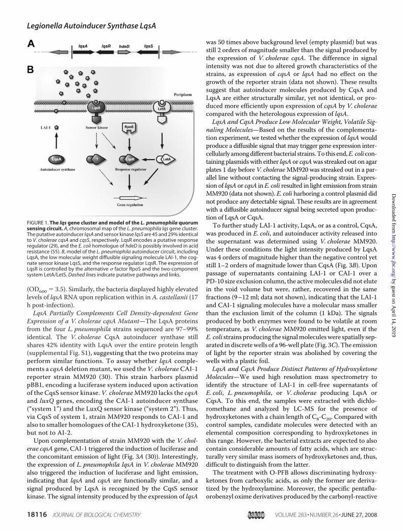
(OD600 � 3.5). Similarly, the bacteria displayed highly elevatedlevels of lqsA RNA upon replication within in A. castellanii (17h post-infection).LqsA Partially Complements Cell Density-dependent Gene
Expression of a V. cholerae cqsA Mutant—The LqsA proteinsfrom the four L. pneumophila strains sequenced are 97–99%identical. The V. cholerae CqsA autoinducer synthase stillshares 42% identity with LqsA over the entire protein length(supplemental Fig. S1), suggesting that the two proteins mayperform similar functions. To assay whether lqsA comple-ments a cqsA deletion mutant, we used the V. choleraeCAI-1reporter strain MM920 (30). This strain harbors plasmidpBB1, encoding a luciferase system induced upon activationof the CqsS sensor kinase. V. choleraeMM920 lacks the cqsAand luxQ genes, encoding the CAI-1 autoinducer synthase(“system 1”) and the LuxQ sensor kinase (“system 2”). Thus,via CqsS of system 1, strain MM920 responds to CAI-1 andalso to smaller homologues of the CAI-1 hydroxyketone (35),but not to AI-2.Upon complementation of strain MM920 with the V. chol-
erae cqsA gene, CAI-1 triggered the induction of luciferase andthe concomitant emission of light (Fig. 3A (30)). Interestingly,the expression of L. pneumophila lqsA in V. cholerae MM920also triggered the induction of luciferase and light emission,indicating that lqsA and cqsA are functionally similar, and asignal produced by LqsA is recognized by the CqsS sensorkinase. The signal intensity produced by the expression of lqsA
was 50 times above background level (empty plasmid) but wasstill 2 orders of magnitude smaller than the signal produced bythe expression of V. cholerae cqsA. The difference in signalintensity was not due to altered growth characteristics of thestrains, as expression of cqsA or lqsA had no effect on thegrowth of the reporter strain (data not shown). These resultssuggest that autoinducer molecules produced by CqsA andLqsA are either structurally similar, yet not identical, or pro-duced more efficiently upon expression of cqsA by V. choleraecompared with the heterologous expression of lqsA.LqsA and CqsA Produce LowMolecular Weight, Volatile Sig-
naling Molecules—Based on the results of the complementa-tion experiment, we tested whether the expression of lqsAwouldproduce a diffusible signal that may trigger gene expression inter-cellularly amongdifferentbacterial strains.To this end,E. colicon-taining plasmidswith either lqsA or cqsAwas streaked out on agarplates 1 day before V. choleraeMM920 was streaked out in a par-allel line without contacting the signal-producing strain. Expres-sion of lqsA or cqsA inE. coli resulted in light emission from strainMM920 (data not shown). E. coli harboring a control plasmid didnot produce any detectable signal. These results are in agreementwith a diffusible autoinducer signal being secreted upon produc-tion of LqsA or CqsA.To further study LAI-1 activity, LqsA, or as a control, CqsA,
was produced in E. coli, and autoinducer activity released intothe supernatant was determined using V. cholerae MM920.Under these conditions the light intensity produced by LqsAwas 4 orders of magnitude higher than the negative control yetstill 1–2 orders of magnitude lower than CqsA (Fig. 3B). Uponpassage of supernatants containing LAI-1 or CAI-1 over aPD-10 size exclusion column, the activemolecules did not elutein the void volume but were, rather, recovered in the samefractions (9–12 ml; data not shown), indicating that the LAI-1and CAI-1 signaling molecules have a molecular mass smallerthan the exclusion limit of the column (1 kDa). The signalsproduced by both enzymes were found to be volatile at roomtemperature, as V. cholerae MM920 emitted light, even if theE. coli strains producing the signalmoleculeswere spatially sep-arated in discretewells of a 96-well plate (Fig. 3C). The emissionof light by the reporter strain was abolished by covering thewells with a plastic foil.LqsA and CqsA Produce Distinct Patterns of Hydroxyketone
Molecules—We used high resolution mass spectrometry toidentify the structure of LAI-1 in cell-free supernatants ofE. coli, L. pneumophila, or V. cholerae producing LqsA orCqsA. To this end, the samples were extracted with dichlo-romethane and analyzed by LC-MS for the presence ofhydroxyketones with a chain length of C8-C20. Compared withcontrol samples, candidate molecules were detected with anelemental composition corresponding to hydroxyketones inthis range. However, the bacterial extracts are expected to alsocontain considerable amounts of fatty acids, which are struc-turally very similar mass isomers of hydroxyketones and, thus,difficult to distinguish from the latter.The treatment with O-PFB allows discriminating hydroxy-
ketones from carboxylic acids, as only the former are deriva-tized by the hydroxylamine. Moreover, the specific pentaflu-orobenzyl oxime derivatives produced by the carbonyl-reactive
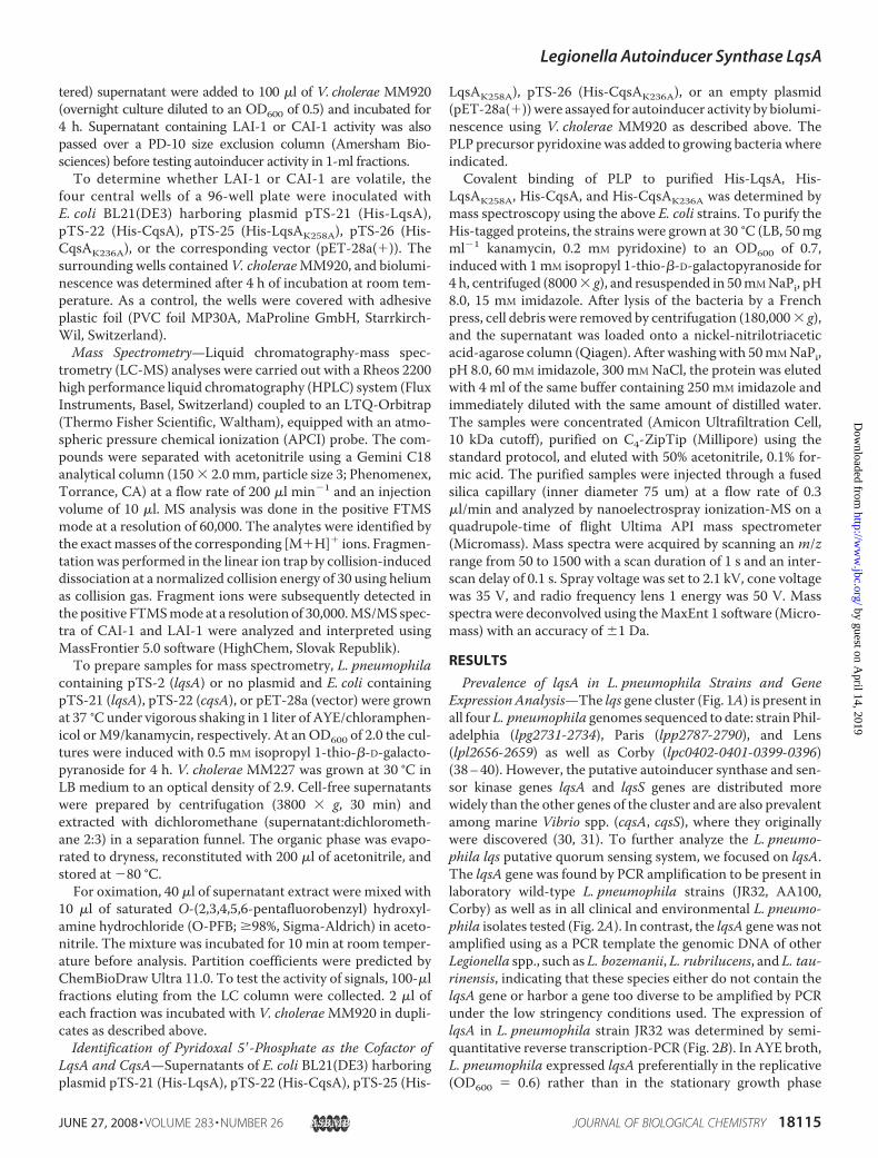
FIGURE 1. The lqs gene cluster and model of the L. pneumophila quorumsensing circuit. A, chromosomal map of the L. pneumophila lqs gene cluster.The putative autoinducer lqsA and sensor kinase lqsS are 45 and 29% identicalto V. cholerae cqsA and cqsS, respectively. LqsR encodes a putative responseregulator (29), and the E. coli homologue of hdeD is possibly involved in acidresistance (55). B, model of the L. pneumophila autoinducer circuit, includingLqsA, the low molecular weight diffusible signaling molecule LAI-1, the cog-nate sensor kinase LqsS, and the response regulator LqsR. The expression ofLqsR is controlled by the alternative � factor RpoS and the two-componentsystem LetA/LetS. Dashed lines indicate putative pathways and links.
Legionella Autoinducer Synthase LqsA
18116 JOURNAL OF BIOLOGICAL CHEMISTRY VOLUME 283 • NUMBER 26 • JUNE 27, 2008
by guest on April 14, 2019
http://ww
w.jbc.org/
Dow
nloaded from
agent O-PFB enhance the detection sensitivity of carbonyl-compounds (42, 43). LC-MS/MS analysis of the different bac-terial supernatant extracts identified hydroxyketones with achain length from 10 to 17 carbon units (Fig. 4). The moleculeswere not detected in supernatants of E. coli or L. pneumophilain the absence of plasmids encoding the autoinducer synthasesCqsA or LqsA (data not shown).An estimation of the relative abundance of the hydroxy-
ketones produced by E. coli expressing lqsA or cqsA, L. pneu-mophila expressing lqsA, or V. cholerae is shown in Table 1.Upon expression of CqsA in E. coli, the masses for both CAI-1(C13) and its C11 homologue were detected in the supernatant,and a similar hydroxyketone product pattern was determinedin supernatants of wild-type V. choleraeMM227. In the super-natants of E. coli- or L. pneumophila-producing LqsA, severalsignals with masses exactly matching the corresponding C13-C17 hydroxyketones were detected. Interestingly, however, thesignal intensity in the pattern was shifted toward longer chainlengths compared with supernatants of bacteria producingCqsA. The major product of LqsA, designated as LAI-1, elutedlater from the LC column than the CqsA product (data not
shown) and was identified by highresolution MS as the C15 hydroxy-ketone homologue of CAI-1.Identification of LAI-1 as
3-Hydroxypentadecan-4-one—Thecharacteristic fragment ions ofO-PFB oximes greatly facilitate thestructural assignment of carbonylcompounds (44). Therefore, the for-mation of O-PFB derivatives isexpected to also specifically shift themass of LqsA- or CqsA-producedhydroxyketones, thus allowing thedetection of structure-determiningfragment ions. Using LC-MS, theC13 hydroxyketone compoundsproduced by either CqsA or LqsAwere found to elute with the sameretention time (data not shown).SubsequentMS/MSanalysis yieldedidentical spectra, including the spe-cific fragment ions at m/z 152.143,194.190, and 350.154 (Fig. 5). Thestrong signals at m/z 152.143 and350.154 can be attributed to thecleavage of the C3-C4 bond upon afavored collision-induced dissocia-tion between the adjacent hydroxyland oxime moieties. In summary,the fragmentation patterns of theC13 hydroxyketone produced byCqsA and LqsA were indistin-guishable and indicated that LqsAsynthesizes the molecule 3-hy-droxytridecan-4-one (CAI-1) as aminor product (Table 1).For the major signal present in
the LqsA sample (LAI-1), the fragmentation pattern of theselected [M�H]� ion correlatedwell with the theoretically pre-dicted fragments for a C15 �-hydroxyketone homologue. Inagreement with this notion, the loss of water from the C15 com-pound yielded the dominating ion at m/z 420.233 (Fig. 5). Thefragmentation spectrum with the specific ions at m/z 180.174,222.222, 378.185, and 420.233 aswell as their relative intensitiesare analogous to the fragmentation products seen for CAI-1.This pattern indicates that an ethyl-�-hydroxyketone moi-ety is present in LAI-1 as well as CAI-1. Furthermore, thefragment ions m/z 180.174 and 378.185 allow determiningthe position of the ketone moiety relative to the C15 back-bone in LAI-1. Themass of both fragment ions was shifted by28.03 atomic mass units, relative to the corresponding frag-ment ions m/z 152.143 and m/z 350.154 of CAI-1, respec-tively. Thus, the ethyl moiety additionally present in LAI-1compared with CAI-1 is located at the far end of the mole-cule with regard to the ketone group. Taken together, thesefragmentation characteristics strongly support the classifi-cation of LAI-1 as an �-hydroxyketone and its identificationas 3-hydroxypentadecan-4-one.
FIGURE 2. Prevalence of lqsA in L. pneumophila strains and gene expression analysis. A, the presence oflqsA was assessed by PCR amplification of the genes from genomic DNA of laboratory wild-type L. pneumophilastrains (JR32, AA100, Corby) and clinical (502, 509, 514) and environmental (529, 534, 535) L. pneumophilaisolates as well as L. bozemanii, L. rubrilucens, and L. taurinensis. As a positive control, the 16 S rRNA gene wasamplified. B, the expression of lqsA in L. pneumophila JR32 was determined by semiquantitative reverse trans-cription-PCR. In AYE broth lqsA was expressed preferentially in the replicative (R) rather than in the stationary(S) growth phase, and in A. castellanii lqsA was expressed during replication (17 h). gDNA, genomic DNA. Similarresults were obtained in at least two independent experiments. kb, kilobase.
Legionella Autoinducer Synthase LqsA
JUNE 27, 2008 • VOLUME 283 • NUMBER 26 JOURNAL OF BIOLOGICAL CHEMISTRY 18117
by guest on April 14, 2019
http://ww
w.jbc.org/
Dow
nloaded from

V. cholerae Does Not Respond to LAI-1—Next, we wanted tocorrelate specific hydroxyketone homologues produced byLqsA to their autoinducer activity. To this end cell-free super-
natants of E. coli expressing CqsA or LqsA were extracted withdichloromethane and analyzed by LC-MS. Treatment withO-PBF was omitted to retain the biological activity of the sam-
FIGURE 3. Production of diffusible, volatile signaling molecules by LqsA and CqsA. A, LqsA partially complements cell density-dependent gene expression of aV. cholerae cqsA mutant. L. pneumophila lqsA or V. cholerae cqsA was expressed in the V. cholerae CAI-1 reporter strain MM920 by introducing pTS-2 (pLqsA) or pTS-6(pCqsA), respectively. The emission of light (relative units) was quantified by luminescence. B, signal activity is produced in E. coli upon heterologous expression of LqsAor CqsA. His-LqsA or His-CqsA were produced under the control of the PT7 promoter in E. coli BL21(DE3) harboring plasmid pTS-21 (pLqsA) or pTS-22 (pCqsA), and thebacterial supernatants were assayed for autoinducer activity by bioluminescence using V. cholerae MM920. C, the signaling molecules produced by LqsA and CqsA arepartially volatile at room temperature. E. coli BL21(DE3) harboring plasmid pTS-21 (pLqsA) or pTS-22 (pCqsA) was placed in the 4 central wells of a 96-well platesurrounded by V. cholerae MM920 and incubated for 4 h before the determination of bioluminescence. As a control the wells were covered with plastic foil. Means andS.D. of triplicates are shown (A and B). Similar results were obtained in at least three independent experiments.
FIGURE 4. LqsA and CqsA produce a distinct pattern of hydroxyketone molecules. Selected-ion mass spectrometry chromatograms display the patterns of hydroxy-ketone molecules produced by E. coli BL21(DE3) harboring pTS-21 (pLqsA) or pTS-22 (pCqsA), L. pneumophila containing pTS-2 (pLqsA), or wild-type V. cholerae MM227.ExtractedsupernatantsweretreatedwithO-PFBandanalyzedbyLC-APCI-MS. InsupernatantsofE. coliorL. pneumophilaproducingLqsA,theC15 compoundwasdetectedasthe major product and termed LAI-1. Supernatants of E. coli producing CqsA or supernatants of V. cholerae contained high levels of CAI-1 (C13) and its C11 homologue.
Legionella Autoinducer Synthase LqsA
18118 JOURNAL OF BIOLOGICAL CHEMISTRY VOLUME 283 • NUMBER 26 • JUNE 27, 2008
by guest on April 14, 2019
http://ww
w.jbc.org/
Dow
nloaded from

ples. The retention times of CAI-1 and LAI-1 were determinedfrom the chromatograms of the corresponding [M�H]� masspeaks atm/z 215.201 and 243.241, respectively (Fig. 6A). Iden-tities were confirmed by MS/MS analysis (data not shown).In parallel, LC fractions of the same samples were collected
and tested using the V. choleraeCAI-1 reporter strainMM920.The activity eluted as a single peak around 3.3 min for both theLqsA product as well as the CqsA product and correlated wellwith the elution time of the CAI-1 molecule (Fig. 6B). For thefraction containing LAI-1, no reporter activity was found.These data indicate that CAI-1 produced by either CqsA or
FIGURE 5. Identification of LAI-1 as 3-hydroxypentadecan-4-one. A, the chemical structures of CAI-1 and LAI-1 are shown. Treatment with O-PFB leads to theoxime adducts depicted, which will yield the proposed fragment ions upon collision-induced dissociation. B, the MS/MS spectra for the CAI-1 and LAI-1 oximederivatives are depicted. The complete fragmentation spectra including the dominating but unspecific [M-18�H]� fragment ion are shown in the insets. Themajor peaks of the LqsA sample correspond well to the predicted fragmentation products and are analogous to the ions produced by fragmentation of CAI-1,confirming the identity of LAI-1. Each mass peak is labeled with the exact measured m/z value, the predicted elemental composition, and the differencebetween the measured and calculated m/z values in ppm, respectively.
TABLE 1Estimation of the relative amounts of �-hydroxyketone signalmoleculesThe peak areas of the corresponding mass peaks were determined from LC-MSchromatograms and normalized to the sum of peak areas detected in a chromato-gram. Ion suppression and concurrence effects cannot be excluded.
C10 C11 C12 C13 CAI-1 C14 C15 LAI-1 C16 C17
% % % % % % % %E. coli/pCqsAa 1 36 2 61 – – – –V. cholerae –b 43 4 53 – – – –E. coli/pLqsA – – – 22 – 66 6 6L. pneumophila/pLqsA – – – 3 4 68 16 9a The strains used are E. coli BL21(DE3) containing pTS-21 (lqsA) or pTS-22 (cqsA),L. pneumophila JR32 containing pTS-2 (lqsA), and V. choleraeMM227.
b –, not detectable.
Legionella Autoinducer Synthase LqsA
JUNE 27, 2008 • VOLUME 283 • NUMBER 26 JOURNAL OF BIOLOGICAL CHEMISTRY 18119
by guest on April 14, 2019
http://ww
w.jbc.org/
Dow
nloaded from

LqsA triggers the reporter strain MM920, whereas the C15�-hydroxyketone LAI-1 does not. Thus, the activity of LqsAsamples on the V. cholerae reporter strain results from the syn-thesis of CAI-1 as a byproduct.LqsA and CqsA Are Pyridoxal 5�-Phosphate-dependent
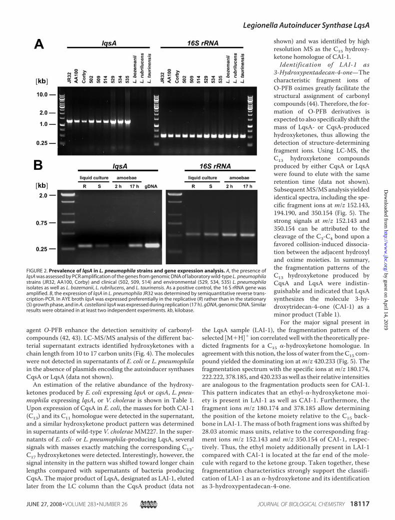
Enzymes—LqsA is 27 or 23% homologous to the PLP-contain-ing E. coli enzymes Kbl (2-amino-3-ketobutyrate-CoA ligase(45)) or BioF (8-amino-7-oxononanoate synthase (46, 47)),respectively. Moreover, LqsA shares a number of conservedamino acids forming the active site of E. coli BioF, including thePLP-binding site Lys-236 (supplemental Fig. S1 (46, 47)). Toinitially test whether PLP might be a cofactor required for theproduction of signalingmolecules, we added the PLP precursorpyridoxine to growing cultures of E. coli expressing lqsA.Reporter activity increased 3.5-fold upon the addition of 10 �Mpyridoxine, suggesting that PLP might be indeed a cofactor ofLqsA (Fig. 7A).Next, we addressed the question genetically by replacing
with alanine the putative PLP binding amino acid, a conservedlysine residue. Neither the LqsAK258A mutant enzyme (orLqsAK258S; data not shown) nor the CqsAK236A mutant pro-
duced any activity detectable by the V. cholerae reporter strainMM920 (Fig. 7B). The observed lack of signal activity was notdue to an impaired production or stability of the mutant auto-inducer synthases, as the proteins were produced at similar lev-els as the corresponding wild-type enzymes (supplemental Fig.S2).Moreover, in the 96-well plate setting described above (Fig.3C), the emission of light by V. choleraeMM920 was abolishedupon expression of either an LqsAK258A or CqsAK236A mutantenzyme by E. coli in the central wells (data not shown).
Finally, we confirmed a covalent binding of PLP to the con-served lysine residues of LqsA or CqsA by electrospray ioniza-tion-time of flight MS (Fig. 7C). Peaks corresponding to puri-fied His-tagged LqsA or CqsA covalently binding PLP wereidentified for the wild-type enzymes but not for the mutantenzymes, where the conserved lysine was replaced by alanine.Compared with His-CqsA, lower amounts of His-LqsA wereproduced by E. coli (supplemental Fig. S2), and affinity-purifiedHis-LqsA was unstable. The low amounts of His-LqsA-PLPavailable forMS yielded signals close to the detection limit, andtherefore, additional peaks likely originating from contaminantproteins were detected. In contrast, purified His-CqsA was sta-
FIGURE 6. Synthesis of active CAI-1 by CqsA and LqsA. A, selected-ion chromatograms for CAI-1 and LAI-1 display the main products of CqsA and LqsA uponexpression in E. coli BL21(DE3). The peaks representing the two main autoinducer molecules are labeled, and their elution times are indicated. The identity ofCAI-1 and LAI-1 as hydroxyketones was confirmed by MS/MS (data not shown). B, activity of the eluted fractions was determined with the V. cholerae CAI-1reporter strain MM920. In both samples the activity was exclusively retained in the fraction containing CAI-1, which indicates that LAI-1 does not contribute tothe activation of the reporter. The error bars represent S.D. for duplicates.
Legionella Autoinducer Synthase LqsA
18120 JOURNAL OF BIOLOGICAL CHEMISTRY VOLUME 283 • NUMBER 26 • JUNE 27, 2008
by guest on April 14, 2019
http://ww
w.jbc.org/
Dow
nloaded from
ble, yielding a strong and specific signal with the exact masscalculated for His-CqsA-PLP.LqsA and CqsA might not only contain PLP and share
sequence homologies with the PLP-dependent aminotrans-ferases Kbl and BioF but also display functional similarities.Both Kbl and BioF catalyze the condensation of a small aminoacid (Kbl, L-glycine; BioF, L-alanine) with an acyl-CoA moiety(Kbl, acetyl-CoA; BioF, pimeloyl-CoA), thereby liberatingCoA-SH. In an attempt to identify the substrates and productsof LqsA and CqsA, we assayed in lysates of E. coli producingHis-LqsA or His-CqsA the release of CoA-SH upon incubationwith small amino acids (L-glycine, L-alanine) and acyl-CoAmoieties (acetyl-CoA, succinyl-CoA). However, none of thesubstrate combinations used led to the release of the putativeproduct of the condensation reaction, CoA-SH (data notshown). Attempts using purified His-CqsA or His-LqsA werealso not successful.
DISCUSSION
In this study we demonstrate that L. pneumophila lqsA func-tionally complements aV. cholerae cqsAmutant strain and pro-duces a diffusible low molecular weight molecule, which isdetected by a V. cholerae CAI-1 reporter strain (Figs. 3 and 4).These findings suggest that LqsA together with the putativecognate sensor kinase LqsS constitutes the first autoinducer
system identified in L. pneumophila. Bioinformatic data basesearches failed to identify in L. pneumophila several autoin-ducer systems present in other bacteria. These include systemssignaling via AHLs, such as the homologues LuxR-LuxI (Vibriofisheri), LasR-LasI (Pseudomonas aeruginosa), RhlR-RhlI (P.aeruginosa) or TraR-TraI (Agrobacterium tumefaciens) (32–34). In the simplest systems, the AHL autoinducer is directlybound by a LuxR-type response regulator containing a DNAbinding helix-turn-helix motive. More complex quorum sens-ing systems that are also missing in L. pneumophila include theAHL-based LuxR-LuxN-LuxM system (Vibrio harveyi), con-sisting of a response regulator, a sensor kinase, and an autoin-ducer synthase, respectively. Finally, the widespread AI-2 sys-tem signaling via a furanosyl borate diester is apparently alsoabsent from L. pneumophila. Components of the latter systeminclude the autoinducer synthase LuxS, the periplasmic bindingprotein LuxP, and the sensor kinase LuxQ (32–34).Only recently theV. choleraeCAI-1 activity was identified as
(S)-3-hydroxytridecan-4-one, and the molecule was synthe-sized, thus proving to be the biologicallymost active formof theautoinducer (35). To identify LAI-1, we used LC coupled tohigh resolutionMS. LAI-1 was identified as 3-hydroxypentade-can-4-one based on its retention time on a C18 column, specificion fragmentation patterns, and comparison to data obtained
FIGURE 7. LqsA and CqsA are pyridoxal 5�-phosphate-dependent enzymes. A, the PLP precursor pyridoxine was added at the concentrations indicated toE. coli BL21(DE3) harboring plasmid pTS-21 (His-LqsA), and autoinducer activity in cell supernatants was determined by bioluminescence using the V. choleraeCAI-1 reporter strain MM920. The data shown are the means and S.D. of 10 samples. Asterisks denote the significance of differences relative to the untreatedsample (*, p � 0.01; **, p � 0.001; two-tailed Student’s t test). Similar results were obtained in three independent experiments. B, supernatants of E. coli strainBL21(DE3) harboring plasmid pTS-21 (pLqsA), pTS-22 (pCqsA), pTS-25 (pLqsAK258A), pTS-26 (pCqsAK236A), or plasmid pET-28a(�) (vector) were assayed bybioluminescence using V. cholerae MM920. The data shown are the means and S.D. of triplicates and are representative of three independent experiments.C, covalent binding of PLP to purified His-LqsA, His-LqsAK258A, His-CqsA, or His-CqsAK236A was determined by electrospray ionization-time of flight-MS.
Legionella Autoinducer Synthase LqsA
JUNE 27, 2008 • VOLUME 283 • NUMBER 26 JOURNAL OF BIOLOGICAL CHEMISTRY 18121
by guest on April 14, 2019
http://ww
w.jbc.org/
Dow
nloaded from
for CAI-1 (Fig. 5). Supernatants of E. coli or L. pneumophilaproducing LqsA triggered signaling by a V. cholerae CAI-1reporter strain, and therefore, it was likely that moleculesrelated or identical to CAI-1 are present in these samples.Reporter strain assays are highly sensitive andwidely used toolsfor the detection of autoinducer molecules. However, due totheir intrinsic specificity, these bioassays are often very selec-tive and can neither provide a comprehensive profile nor anaccurate quantification of signaling molecules (48, 49). To trig-ger signaling of theCAI-1 reporter strain, both backbone lengthand stereochemistry of �-hydroxyketones were found to beimportant, yet in addition to CAI-1 (C13) smaller homologues(C12 and C11), activated the V. cholerae quorum sensing circuitas well (35). In contrast, the C15 homologue of these hydroxy-ketones, LAI-1, did not trigger signaling by the CAI-1 reporterstrain, and the active compound in samples containing LqsAproducts was CAI-1 (C13) (Fig. 6). Therefore, V. cholerae CqsSapparently recognizes �-hydroxyketones with shorter but notlonger linear hydrocarbon backbones than CAI-1.LC-APCI-MS analysis of extracted supernatants allowed an
estimation of the relative amounts of different �-hydroxy-ketones synthesized by LqsA and CqsA (Fig. 4, Table 1). Wedetected significant levels of the C11 homologue of CAI-1 (C13)not only upon heterologous expression of cqsA in E. coli butalso in the supernatant of wild-type V. cholerae. Thus, CqsAapparently shows a broad specificity and yields byproducts ofdifferent hydrocarbon length not only upon heterologous pro-duction in E. coli as reported previously (35) but also in theendogenous V. cholerae background. In supernatants of bacte-rial cultures expressing LqsA, LAI-1 (C15) was the predominantmolecule identified (66–68%). However, minor amounts of theC13, C14, C16, and C17 homologues were also detected. Note-worthy, although upon expression of lqsA in L. pneumophilathe C16 �-hydroxyketone was the second most abundant spe-cies (16%), upon expression of lqsA in E. coli the C13 compound(CAI-1) was the second most abundant signal molecule (22%).Taken together, both CqsA and LqsA do not show an exquisitespecificity concerning the chain length of their products.To our knowledge, 3-hydroxypentadecan-4-one has previ-
ously not been described in biology. Together with V. choleraeCAI-1, L. pneumophila LAI-1 forms a family of �-hydroxy-ketone autoinducer signaling molecules. Like the homoserinelactones, these �-hydroxyketones might represent two exam-ples of an extended autoinducer family, and the substituents aswell as the length of the side chainmight define the signal spec-ificity. Based on bioassay analysis and data base searches, CAI-1-mediated cell-cell communication was found to be predom-inant among the genus Vibrio (31). Yet in addition toL. pneumophila (Fig. 2) and Vibrio spp., lqsA and lqsS homo-logues are found in a number of environmental bacteria,including Nitrococcus mobilis, Burkholderia xenovorans, andPolaromonas spp. (29). Accordingly, intercellular signalinginvolving �-hydroxyketones might be common among differ-ent bacterial species and genera.L. pneumophilapersists in bio-films in the environment (1, 50), where interspecies communi-cation with any of these or other bacteria might be relevant. Inthis context the extended product spectrum of autoinducer
synthases of the LqsA/CqsA family might allow interspeciescommunication with a more diverse group of bacteria.Although �-hydroxyketone signaling molecules were readily
detected by reporter assays and LC-MS in supernatants ofE. coli and L. pneumophila expressing LqsA, these signals werenot identified in wild-type L. pneumophila grown on plate or inliquid broth. Possible explanations for this observation includethat (i) the amount of LAI-1 secreted by L. pneumophila is gen-erally very low, (ii) LAI-1 secretion is tightly regulated and lowonly under the experimental conditions used, or (iii) LAI-1 isnot secreted at all.Perhaps, the threshold for LAI-1 signaling via LqsS is low,
and thus, small amounts of LAI-1 are sufficient to efficientlypromote cell-cell communication in a confined, possibly signal-impermeable environment such as the intracellular Legionella-containing vacuole. At the same time, the concentration of bac-teria within Legionella-containing vacuoles is high, andaccordingly, a considerable concentration of small signalingmolecules might be achieved by a relatively small number ofbacteria. Moreover, the production of LAI-1 and/or LqsAmight be tightly regulated. It has been postulated that Vibriospp. repress the production ofCqsA (and consequently, CAI-1),as the expression of the cqsA gene alone yielded a strongerautoinducer signal than expression of a 25-kilobase chromo-somal region including cqsA (31). Along this line, many geneswere found to be poorly expressed in L. pneumophila brothcultures but are up-regulated upon infection of host cells (17).This is also true for lqsA, which is strongly expressed uponintracellular replication of L. pneumophila in amoebae (Fig. 2).Finally, under physiological conditions LAI-1 might representan intracellular metabolite rather than, or in addition to, asecreted signaling molecule. A dual role in cell-cell signalingand metabolism has been proposed for the LuxS autoinducersynthase and its furanosyl borate product AI-2 (51, 52). In anycase the characterization of metabolic, signaling and virulencephenotypes of an L. pneumophila lqsA mutant strain will shedlight on the physiological functions of the correspondingenzyme.LqsA is 27 or 23% identical to the PLP-containing E. coli
enzymes Kbl (2-amino-3-ketobutyrate CoA ligase) and BioF(8-amino-7-oxononanoate synthase), respectively, and theautoinducer synthase shares a number of conserved aminoacids forming the active site of E. coli BioF (supplemental Fig.S1). Based on biochemical and genetic approaches, both LqsAand CqsAwere shown to also contain PLP as a cofactor (Fig. 7).The efficient synthesis of signalingmolecules by E. coli express-ing LqsA suggests that common metabolic intermediates areused as substrates for LAI-1 or that E. coli harbors additionalenzymes required in a putative multistep enzymatic pathwayleading to the production of the signaling molecule. Similarobservations have been made for V. cholerae CqsA (31). How-ever, using different small amino acids (L-glycine, L-alanine)and acyl-CoA moieties (acetyl-CoA, succinyl-CoA), we failedto identify substrates or products of a condensation reactioncatalyzed by LqsA in vitro. PLP-dependent enzymes perform avast repertoire of reactions, which is expected to also includenovel catalytic activities (53, 54). Further in vitro studies arerequired to gain more insight into the biosynthetic mechanism
Legionella Autoinducer Synthase LqsA
18122 JOURNAL OF BIOLOGICAL CHEMISTRY VOLUME 283 • NUMBER 26 • JUNE 27, 2008
by guest on April 14, 2019
http://ww
w.jbc.org/
Dow
nloaded from
of �-hydroxyketone production by bacterial autoinducer syn-thases of the LqsA/CqsA family.
Acknowledgments—We thank Dr. Bonnie Bassler and Doug Higgins(Princeton University) for providing V. cholerae strains and advice onisolation of hydroxyketones as well as Dr. Christoph von Ballmoos(ETH Zurich) for help with extractions and Serge Chesnov (Func-tional Genomics Center Zurich) for electrospray ionization-time offlight-MS measurements of PLP-protein samples.
REFERENCES1. Fields, B. S., Benson, R. F., and Besser, R. E. (2002)Clin.Microbiol. Rev. 15,
506–5262. Hilbi, H., Weber, S. S., Ragaz, C., Nyfeler, Y., and Urwyler, S. (2007) Envi-
ron. Microbiol. 9, 563–5753. McDade, J. E., Shepard, C. C., Fraser, D. W., Tsai, T. R., Redus, M. A., and
Dowdle, W. R. (1977) N. Engl. J. Med. 297, 1197–12034. Segal, G., Purcell, M., and Shuman, H. A. (1998) Proc. Natl. Acad. Sci.
U. S. A. 95, 1669–16745. Vogel, J. P., Andrews, H. L., Wong, S. K., and Isberg, R. R. (1998) Science
279, 873–8766. Albers, U., Reus, K., Shuman,H. A., andHilbi, H. (2005)Microbiology 151,
167–1827. Hilbi, H., Segal, G., and Shuman,H. A. (2001)Mol.Microbiol. 42, 603–6178. Otto, G. P., Wu, M. Y., Clarke, M., Lu, H., Anderson, O. R., Hilbi, H.,
Shuman, H. A., and Kessin, R. H. (2004)Mol. Microbiol. 51, 63–729. Segal, G., Feldman, M., and Zusman, T. (2005) FEMS Microbiol. Rev. 29,
65–8110. Weber, S. S., Ragaz, C., Reus, K., Nyfeler, Y., and Hilbi, H. (2006) PLoS
Pathog. 2, e4611. Kagan, J. C., and Roy, C. R. (2002) Nat. Cell Biol. 4, 945–95412. Dorer, M. S., Kirton, D., Bader, J. S., and Isberg, R. R. (2006) PLoS Pathog.
2, e3413. Bruggemann, H., Cazalet, C., and Buchrieser, C. (2006) Curr. Opin. Mi-
crobiol. 9, 86–9414. Hilbi, H. (2006) Cell. Microbiol. 8, 1697–170615. Ninio, S., and Roy, C. R. (2007) Trends Microbiol. 15, 372–38016. Molofsky, A. B., and Swanson, M. S. (2004)Mol. Microbiol. 53, 29–4017. Bruggemann, H., Hagman, A., Jules, M., Sismeiro, O., Dillies, M. A.,
Gouyette, C., Kunst, F., Steinert, M., Heuner, K., Coppee, J. Y., andBuchrieser, C. (2006) Cell. Microbiol. 8, 1228–1240
18. Hammer, B. K., Tateda, E. S., and Swanson, M. S. (2002) Mol. Microbiol.44, 107–118
19. Molofsky, A. B., Shetron-Rama, L. M., and Swanson, M. S. (2005) Infect.Immun. 73, 5720–5734
20. Heuner, K., Dietrich, C., Skriwan, C., Steinert, M., and Hacker, J. (2002)Infect. Immun. 70, 1604–1608
21. Mampel, J., Spirig, T.,Weber, S. S., Haagensen, J. A. J., Molin, S., andHilbi,H. (2006) Appl. Environ. Microbiol. 72, 2885–2895
22. Hales, L. M., and Shuman, H. A. (1999) J. Bacteriol. 181, 4879–488923. Bachman, M. A., and Swanson, M. S. (2001) Mol. Microbiol. 40,
1201–121424. Bachman,M.A., and Swanson,M. S. (2004) Infect. Immun. 72, 2468–247625. Gal-Mor, O., and Segal, G. (2003)Microb. Pathog. 34, 187–19426. Lynch, D., Fieser, N., Gloggler, K., Forsbach-Birk, V., andMarre, R. (2003)
FEMS Microbiol. Lett. 219, 241–24827. Gal-Mor, O., and Segal, G. (2003) J. Bacteriol. 185, 4908–491928. Zusman, T., Aloni, G., Halperin, E., Kotzer, H., Degtyar, E., Feldman, M.,
and Segal, G. (2007)Mol. Microbiol. 63, 1508–152329. Tiaden, A., Spirig, T., Weber, S. S., Bruggemann, H., Bosshard, R.,
Buchrieser, C., and Hilbi, H. (2007) Cell. Microbiol. 9, 2903–292030. Miller, M. B., Skorupski, K., Lenz, D. H., Taylor, R. K., and Bassler, B. L.
(2002) Cell 110, 303–31431. Henke, J. M., and Bassler, B. L. (2004) J. Bacteriol. 186, 6902–691432. Bassler, B. L. (2002) Cell 109, 421–42433. Fuqua, C., andGreenberg, E. P. (2002)Nat. Rev.Mol. Cell Biol. 3, 685–69534. Camilli, A., and Bassler, B. L. (2006) Science 311, 1113–111635. Higgins, D. A., Pomianek, M. E., Kraml, C. M., Taylor, R. K., Semmelhack,
M. F., and Bassler, B. L. (2007) Nature 450, 883–88636. Feeley, J. C., Gibson, R. J., Gorman, G. W., Langford, N. C., Rasheed, J. K.,
Mackel, D. C., and Baine, W. B. (1979) J. Clin. Microbiol. 10, 437–44137. Altschul, S. F., Madden, T. L., Schaffer, A. A., Zhang, J., Zhang, Z., Miller,
W., and Lipman, D. J. (1997) Nucleic Acids Res. 25, 3389–340238. Chien, M., Morozova, I., Shi, S., Sheng, H., Chen, J., Gomez, S. M., Asa-
mani, G., Hill, K., Nuara, J., Feder, M., Rineer, J., Greenberg, J. J., Stesh-enko, V., Park, S. H., Zhao, B., Teplitskaya, E., Edwards, J. R., Pampou, S.,Georghiou, A., Chou, I. C., Iannuccilli, W., Ulz, M. E., Kim, D. H.,Geringer-Sameth, A., Goldsberry, C.,Morozov, P., Fischer, S. G., Segal, G.,Qu, X., Rzhetsky, A., Zhang, P., Cayanis, E., De Jong, P. J., Ju, J., Kalachikov,S., Shuman, H. A., and Russo, J. J. (2004) Science 305, 1966–1968
39. Cazalet, C., Rusniok, C., Bruggemann, H., Zidane, N., Magnier, A., Ma, L.,Tichit, M., Jarraud, S., Bouchier, C., Vandenesch, F., Kunst, F., Etienne, J.,Glaser, P., and Buchrieser, C. (2004) Nat. Genet. 36, 1165–1173
40. Glockner, G., Albert-Weissenberger, C., Weinmann, E., Jacobi, S., Schun-der, E., Steinert, M., Hacker, J., and Heuner, K. (2007) Int. J. Med. Micro-biol., in press
41. Albers, U., Tiaden, A., Spirig, T., Al Alam,D., Goyert, S.M., Gangloff, S. C.,and Hilbi, H. (2007)Microbiology 153, 3817–3829
42. Tholey, A., Wittmann, C., Kang, M. J., Bungert, D., Hollemeyer, K., andHeinzle, E. (2002) J. Mass Spectrom. 37, 963–973
43. Jakober, C. A., Charles,M. J., Kleeman,M. J., andGreen, P. G. (2006)Anal.Chem. 78, 5086–5093
44. Schulze, B., Lauchli, R., Sonwa,M.M., Schmidt, A., and Boland,W. (2006)Anal. Biochem. 348, 269–283
45. Schmidt, A., Sivaraman, J., Li, Y., Larocque, R., Barbosa, J. A., Smith, C.,Matte, A., Schrag, J. D., andCygler,M. (2001)Biochemistry 40, 5151–5160
46. Webster, S. P., Alexeev, D., Campopiano, D. J., Watt, R. M., Alexeeva, M.,Sawyer, L., and Baxter, R. L. (2000) Biochemistry 39, 516–528
47. Alexeev, D., Alexeeva, M., Baxter, R. L., Campopiano, D. J., Webster, S. P.,and Sawyer, L. (1998) J. Mol. Biol. 284, 401–419
48. Kumari, A., Pasini, P., Deo, S. K., Flomenhoft, D., Shashidhar, H., andDaunert, S. (2006) Anal. Chem. 78, 7603–7609
49. Cataldi, T. R., Bianco, G., andAbate, S. (2008) J. Mass Spectrom. 43, 82–9650. Sheehan, K. B., Henson, J. M., and Ferris, M. J. (2005) Appl. Environ.
Microbiol. 71, 507–51151. Vendeville, A., Winzer, K., Heurlier, K., Tang, C. M., and Hardie, K. R.
(2005) Nat. Rev. Microbiol. 3, 383–39652. Winzer, K., Hardie, K. R., andWilliams, P. (2003)Adv. Appl.Microbiol. 53,
291–39653. Percudani, R., and Peracchi, A. (2003) EMBO Rep. 4, 850–85454. Eliot, A. C., and Kirsch, J. F. (2004) Annu. Rev. Biochem. 73, 383–41555. Masuda, N., and Church, G. M. (2003)Mol. Microbiol. 48, 699–712
Legionella Autoinducer Synthase LqsA
JUNE 27, 2008 • VOLUME 283 • NUMBER 26 JOURNAL OF BIOLOGICAL CHEMISTRY 18123
by guest on April 14, 2019
http://ww
w.jbc.org/
Dow
nloaded from

Hubert HilbiThomas Spirig, André Tiaden, Patrick Kiefer, Carmen Buchrieser, Julia A. Vorholt and
Signaling Molecule-Hydroxyketoneα Autoinducer Synthase LqsA Produces an LegionellaThe
doi: 10.1074/jbc.M801929200 originally published online April 14, 20082008, 283:18113-18123.J. Biol. Chem.
10.1074/jbc.M801929200Access the most updated version of this article at doi:
Alerts:
When a correction for this article is posted•
When this article is cited•
to choose from all of JBC's e-mail alertsClick here
Supplemental material:
http://www.jbc.org/content/suppl/2008/04/16/M801929200.DC1
http://www.jbc.org/content/283/26/18113.full.html#ref-list-1
This article cites 54 references, 15 of which can be accessed free at
by guest on April 14, 2019
http://ww
w.jbc.org/
Dow
nloaded from