Embed Size (px)
Citation preview
S•NRINE
K-kx- A0
Submarine Base, Groto.i Conn.
REPORT NUMBER 660
A REVIEW OF D u"- xTHE PATHOGENESIS OF SKIN B---
by
LT. W. Landon Dennison, Jr., Mcq ._N, C
Bureau of Medicine and Surgery, Navy DepartmentResearch Work Unit MF099.01.01.08
Released by:- '-HNICAL
J. E. Staw.k, CAPT MC USN "I'i -RVICECOMMANDING OFFICERNaval Submarine Medical Center
22 April 1971 C1
Approved for public release; distribution unlimited ,)
UNCLASSIFIEDSe•curitv C"lassification,
DOCUMENT CONTROL DATA • R & D
S•.'rMrtt ins-4kation, of title b.,d, ut a. itr. I a.nd Indexng,) innotlarkn titI be entered tiher fipt. overall report Is clsslfled)
I ORIGINATING ACTIVITY (Corporilteoalthor) 2a. REPORT SECURITY CLASSIFICATION
Naval Submarine Medical Center, Naval Submarine J Unclassified
Medical Research Laboratory 2b. GROUP
3 REPOR 7 TITLE
A REVIEW OF THE PATHOGENESIS OF SKIN BENDS
0 DESCRIPTIVE NOTES (7)'pe of report and icluitive dates)
Interim Report5 AUTHORISI (Filst name. middle inlitial, last nAme)
W. Landon DENNISON, Jr., LT, MC, USN
6. REPORT DATE 70. TOTAL NO. OF PAGES j7b. NO. OF REFS
22 April 1971 15 3164. CONTRACT OR GRANT NO 9a. ORIGINATOR-S REPORT NUMBERIS)
b. PROJECT NO NSMRL Report Number 660MF099.01.0ý1.08
C. 9b. OTHER REPORT NOMSI (Any other numbers that may be aessined
this report)
d.
10 OISTRIOUTION JITATEMENT
Approved for public release; distribution unlimited.
1I SUPPLEMEN) ARY NOTES 12. SPONSORING MILITARY ACTIVITY.
Naval Submarine Medical CenterBox 600 Naval Submarine Base
" _ _ _ _Groton, Connecticut 0684013 ADSTACT
• The literature concerning "skin bends" was searched, especially as regards thecauses of this condition. This investigator concluded (by compilation of the theories fromthe literature and application of physiological and anatomical data) that gases reach thevarious layers of the skin by perfusion from the blood and by penetration from without.The route by which the gases reach the skin and the histological area in which they formbubbles is variable, but the site where they form is important in production of the variousclinical varieties of "skin bends".
DD FORM"(PGIDD Nov 17 UNGC LASS I9EDS/N t012 014.6500 Secuitv Ciassification
iiI
i~r . ,UNCLASSFEseC curity clelication
14. LINK A LINK S LINK CKEY WORDS-
ROLE WT ROLE WT ROLE WT
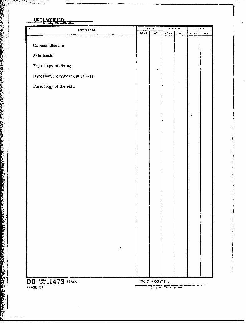
Caisson disease
Skin bends
Phby'ology of diving
Hyperbaric environment effects
Physiology of the skin
DD , .FORM1473 (BACK) uLI .A lsill' 1, -
(PAGE 2) ., , ( .... ,e
A REVIEW OFTHE PATHOGENESIS OF SKIN BENDS
by
LT. W. Landon Dennison, Jr., MC, USN
NAVAL SUBMARINE MEDICAL RESEARCH LABORATORYNAVAL SUBMARINE MEDICAL CENTER REPORT No. 660
Bureau of Medicine and Surgery, Navy DepartmentResearch Work Unit MF099. 01. 01. 08'
Transmitted by:
J. H. Baker, CDR, MC, USNDirector, School of Submarine Medicine
Reviewed and Approved by: Reviewed and Approved by:
I
Charles F. Cell, M.D., D.Sc. (Med.) J. D. Bloom, CDR, MC, USNScientific Director 0 fficer-in- ChargeNavSubMedRschLab NavSubMedRschLab
Approved and Released by:
J, MC USNCOMMANDING OFFICER
Naval Submarine Medical Ccnter
Approved for public release; distribution unlimited
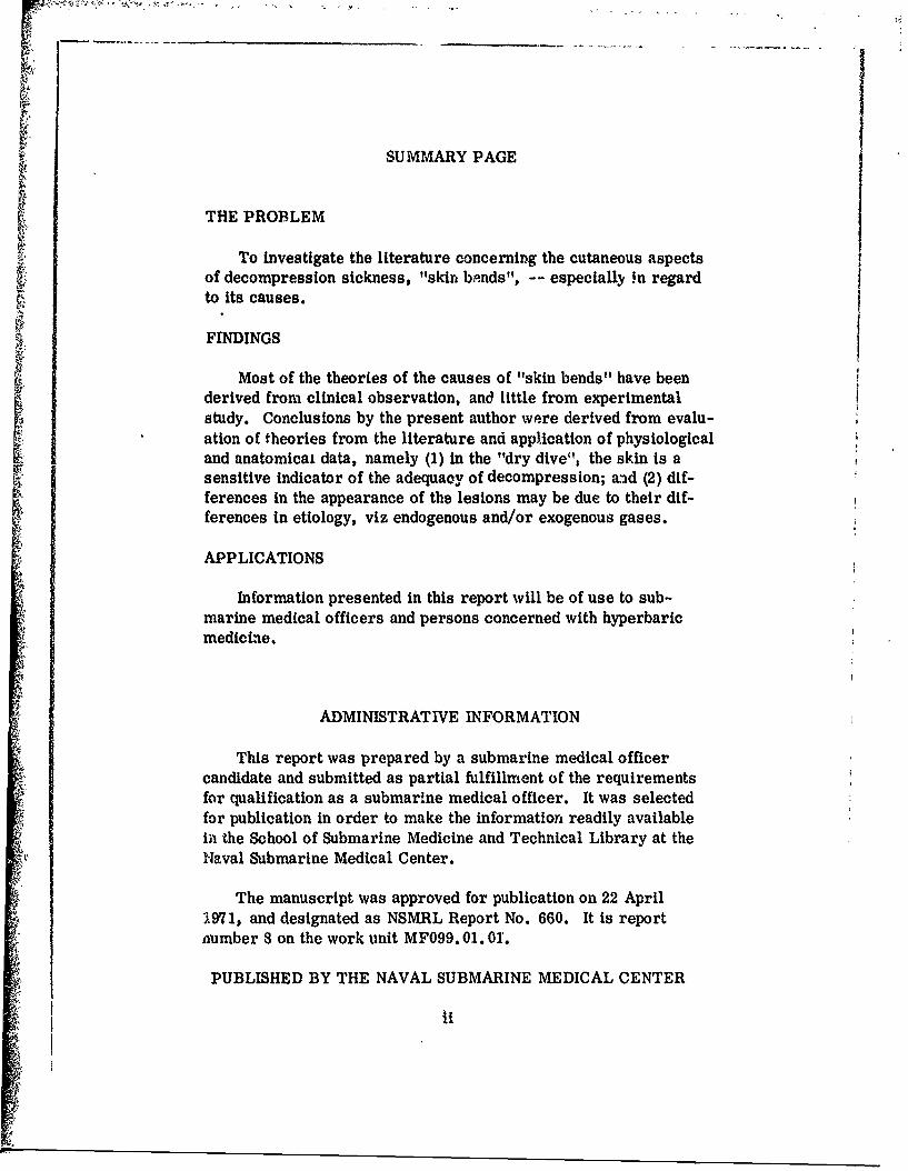
SUMMARY PAGE
THE PROBLEM
To investigate the literature concerning the cutaneous aspectsof decompression sickness, "skin bnds", -- especially in regard
to its causes.
FINDINGS
Most of the theories of the causes of "skin bends" have beenderived from clinical observation, and little from experimentalstudy. Conclusions by the present author were derived from evalu-ation of theories from the literature and application of physiologicaland anatomicaL data, namely (1) in the "dry dive", the skin is asensitive indicator of the adequacy of decompression; and (2) dif-ferences in the appearance of the lesions may be due to their dif-ferences in etiology, viz endogenous and/or exogenous gases.
APPLICATIONS
Information presented In this report will be of use to sub-marine medical officers and persons concerned with hyperbaricmedickie.
ADMINISTRATIVE INFORMATION
This report was prepared by a submarine medical officercandidate and submitted as partial fulfillment of the requirementsfor qualification as a submarine medical officer. It was selectedfor publication in order to make the information readily availablein the School of Submarine Medicine and Technical Library at theNaval Submarine Medical Center.
The manuscript was approved for publication on 22 April197 1, and designated as NSMRL Report No. 660. It is reportnumber 8 on the work unit MF099. 01. 01.
PUBLISHED BY THE NAVAL SUBMARINE MEDICAL CENTER
ttI
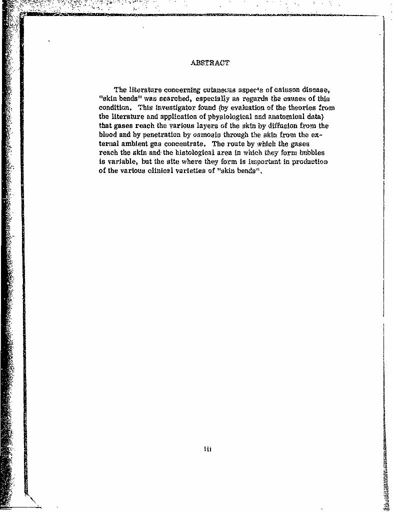
ABSTRACT
The litertur.e concerning cutanmus aspecs of caisson disease,"skin bends" was searched, especially as regards the causes of thia
condition. This Investigator found (by evaluation of the theories fromthe literature and application of pbysiological and anatomical data)that gases reach the. various layers of the skin by diffusion from theblood and by penetration by osmosis through the skirt from the ex-ternal ambient gas concentrate. The route by which the gasesreach the skin and, the histological area in which they form bubblesis variable, but the site where they form is important in productlonof the various clinical varieties of "skin bends".
Li
+!I!
iii
AREVIEWOFTHE PATHOGENISIS OF SKIN BENDS
INTRODUCTION Skin manifestations of decompres-sion sickness develop during or afteýr
Many of those who have been exposed decompression. , !though it occursto extreme changes in atmospheric pres- both in aviators and deep-sea divers,sure, be it at high altitudes or the depths the bulk of this paper will deal withof th,- sea, may have experienced a mild deep sea divers or hyperbaric chambercase of praritus. This dermatologic subjects. Skin bends develop only insymptom will be called "skin bends" in "dry dives" (with a few exceptions)2 .this paper. Skin bends, a mild form of A dry dive is one where the diver'sdecompression sickness, has been skin is dry as in "hard-hat divers" withvariously called "les pouces" (fleas) by water tight suits or in hyperbaric chain-Paul Bert or "the itch" by the English bers. SCUBA divers, on the other hand,speaking peoples. Most persons asso- have dire,'t skin exposure to the water.ciated w~ith diving limilt the te)rm "skin3
bends" t a transient "rash" with or hi Rivera's3 analysis of decompres-without pruritus, however, this paper sion sickness taken from U. S. Navywill consider all skin manifestations files, 14.9 per cent of bends casesassociated with decompression as "skil (935 total cases) had skin manifestations;
Sbends". The phenomenon has been Duffner's series 0. 9 per cent; Behnke,known since men began to expose them- 13.6 per cent; Thorne, working withselves to great alteratione in barometric caisson workers, found tMat 10 per centpressure1 . of all bends cases developed had skin
bends. Again, many more probablyAlthough it has been long recognized had very minor signs or symptoms
as an entity, little has been written which werc not reported, and many ofspecifically on skin bends, presumably the divers in the above series hadbecause the signs and symptoms are direct skin contact with water (as infleeting and only mildly annoying. When SCUBA divers) and therefore would notthe patient has other symptoms of de- deveicp skin bends anyway.compression sickness, these symptomsare often severe, thus most injury re- Skin betics take on several differentports list only a "rash" and then go on forms. Aldao 4 lists six categories:to describe the graver symptoms andsigns. The intention of this paper is to (I' Pruritvs only - others5 mentionattempt to determine tne pathogenesis that a pricking sensation may also beof the disease because it is felt that t.he present.skin, being exposed to both the divingmedium and the blood, may react a (2) Sca ria tina form rash - this islittle differently from. the way other perharps the most frequent sign. It isorgansq react to compression and de- mnos- frequent o'ver V. chest, shoulders,compression. back, upper abdomen, and thighs, in
oI
, , , - .--6 -
that order. 5, This form is usually area becomes tender to palr ation which
accompanied by mild pruritus. The increases in intensity for 24 to 36rash disappears within a few hours of hours, after which the tenderness de-surfacing and is not usually accompanied creases and is gone in two to threeby more serious forms of decompres- days. In most cases there is no changesion sickness. in the color of the skin during this
period of tenderness, except for occa-(3) Frysipelas form - Has the same siunal erythema. Infiltration of the
general distribution as the scarlatina- deep layers of skin with an anestheticform rash. The lesions vary from (subcutaneous tissues?) causes rellefsmall papules to very large plaques of the teliderness.which cover the whole chest and shoul-ders. The borders are flat and firm (5) Serious form--In this form theand violet in color showing a character- condition of the patient becomes towi'yistic venous network on the surface, cyanotic--here the patient is in shockCoughing or performing the Valsava and has signs of emboli in vital tissues.maneuver will accentuate the venous Aldao states a great quantity of bloodmarkings (Mellinghoff's sign). This is trapped in the cutaneous blood ves-may accomptny or precede the more sels, but he does not give any refer-serious forms of decompression sick- ence and furthermore states that heness. has never seen such a case.
(4) Curls marmorata marbleization-- (6) Emphysema:Thi3 is one of the danger signs in divLng,because frequently this precedes major (a) Intracutaneous-- Ferris andcomplications of decompression. Engel 5 describe minute intracutaneousFerris and Engel in Fultcn's Decom- blebs in aviators. These blebs arenression Sickness 5 describe this lesion accompanied by mild pruritus and/orin great detail, from aviation studies, pricking sensations. This pehnomenonbut there is no reason to suspect that has not been described in the divingthe process differs from that in diving, literature seen by this author.They state that the lesion begins as asmall pale area with cyanotic mottling. (b) Subcutaneous emphysema--The lesion may or may not spread Is seen frequently in air embolismperipherally becoming erythematous associated with rapid ascent in thewith superimposed mottling. Continued water and rupture of air into the pul-exposure to relatively decreased at- monary circulation or dissection of airmospheric pressure causes spread over into the neck and chest from pneu-a large area. The temperature of the mothorax. It is only seen in severeskin is 1-.2 C. warmer in the erythe- decompression sickness,matous stage. When recompressed theraottled area becomes diffusely red and In further consideration of thehot and then disappears completely pathogenesis of skin bends the funda-shortly after recompression is com- mental of diving physiology and aplete. However, 4-6 hours later the review of skin anatomy and physiology
2
will follow, with special emphasis to the has certain advantages over nitrogen asbehavior of the skin in relation to gases. the inert portion of the breathing mix-In research for this paper, the author ture, among which are its lower nar-has found little to explain the above cotic effect and faster elimination fromphenomena; many explanations have the body.been offered. In the ensuing sectionsthese hypotheses will be reviewed and Therefore, in the process of diving,discussed. gases are driven and dissolved Into the
bndy as the atmospheric pressure isFUNDAMENTALS OF DIVING increased, the reverse occurs whenPHYSICS AND PHYSIOLOGY the barometric pressure is reduced.
In order to present a clear discussionof this subject, it is important to have Decompression, the act oi ikeducingan understanding of the physics and atmc.pheric pressure must be per-physiology of compression and decom- formed carefully. If done too rspidiypression 7 . Manon the earth's surface is the gases, especially the inert ones,
continually exposed to the weight of the will come out of solution and formatmosphero above and around him. This bubbles. Bubbles usually do notatmosphere exerts a pressure of 14.7 form until the partial pressure ol thepounds per square inch (psi) which is dissolved gases exceed the partialequivalent to the pressure exerted by pressure of the ambient gases by aapproximately 33 feet of sea water, factor of two ("Haldane's 2:1 Ratio").
Once they are formed, they grow asWhen an individual is subjected to the barometric pressure is reduced.
greater or Lesser pressures, the at- The expanding bubbles cause symp-mospheric gases (or those of the diving toms, such as pain if they are inmedium) react according to the various tendons and muscles and CNS symp-physical gas laws. The gas laws, al- toms result from bubbles arising in
though important, do not act alone. The fatty tiss-ne or vital blood vessels inprinciples of dynamic equilibrium and those organs. Here one can see thesolubility are important forces. Solu- imrortance of adequate blood supplybility of gases, especially the bio- (high capillary-tissue ratio andlogically inert gases such as nitrogen adequate venous drainage) to clearand helium, determine how much more the tissue of gases approaching theof a gas will be dissolved into the body's point of super-saturation for a par-tissues than would occur with the sim- ticular pressure.ple application of Henry's Law, andperhaps more important, where that Symptoms resulting from bubbles thusgas will be dissolved. For instance, occurring result in similar syndromesnitrogen is highly soluble in fatty tis- known as Caisson's Disease, Deccmsues, thus a great quantity of nitrogen pression Sickness or simply a-? Bends.will be dissolved into tCio fatty portionof the organism (Table I), and since Since the skin is exposed to these
heliu-i is less soluble than nitrogen it gas forces from within (the blood) and
3
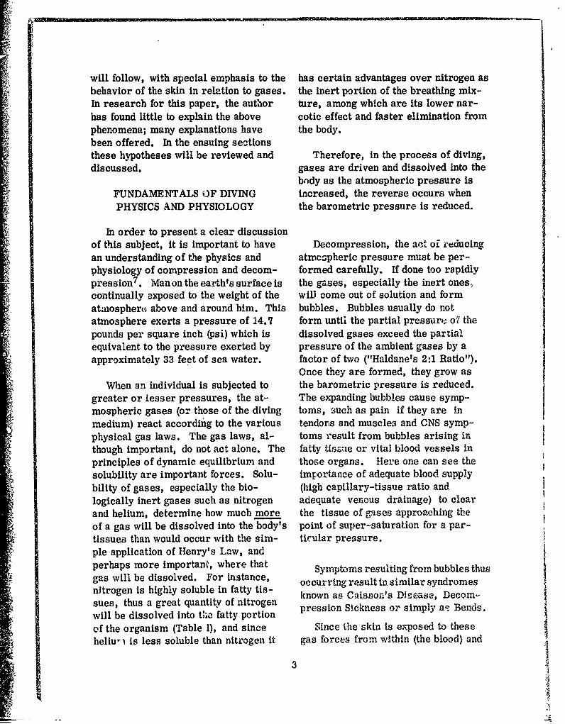
Table 1. Solubility Coefficients of Gases in Oil and in Water at 380 C.(from Behnke2 9 )
Nitrogen Argon Helium Oxygen
In olive oil 0.0667 0.1395 0.0148 0.0113
In water 0.0125 0.0262 0.0087 0.023
Ratio 5.24:1 5.32:1 1.7:1 5.0:1
from without (the atmosphere), the inert desquamation. Covering this layer is agases may originate from either source, thin, oily layer of material secretedi. e., by direct penetration in osmotic from the sebaceous glands. Below theex•change through the integument or stratum corneum one finds successively,from vascular diffusion. 0', 31 the stratum lucidumr the stratum
granulosurn, the prickle cell layer, andANATOMY AND PHYSIOLOGY lastly the stratum germinativum. The
OF THE SKIN cells of the stratum germinativumn(basal layer) are the metabolically
Considering the skin in relation to active cells which migrate upwards asthe rest of the body, one should recall aging occurs to the layers above arid
4 that it is the largest organ in the body are finally desquamated from thoby weighl and its surface area niea- stratum corneum. The epidermalsures about 1.7 square meters, which appendages (hair follicles, sweatis one-twenty-fifth of the lung's surface glands and sebaceous g.nds) are seenarea 8 . The function of this integument as invaginationa of epiderrr is into theis that of protectton; secondarily, it dermis. These invaginatio "s are veryaids In temperature regulation and to a thin walled in comparison with the restminor extent some excretory processes of the epidermis. The epidermis hastake place. no nerve or blood supply per se, but
nutrition diffuses from the vessels ofThe skin 9 can be considered as a the dermis. Note that the derr,,al sur-
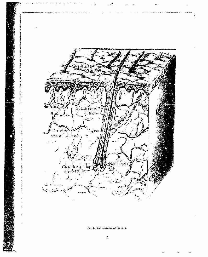
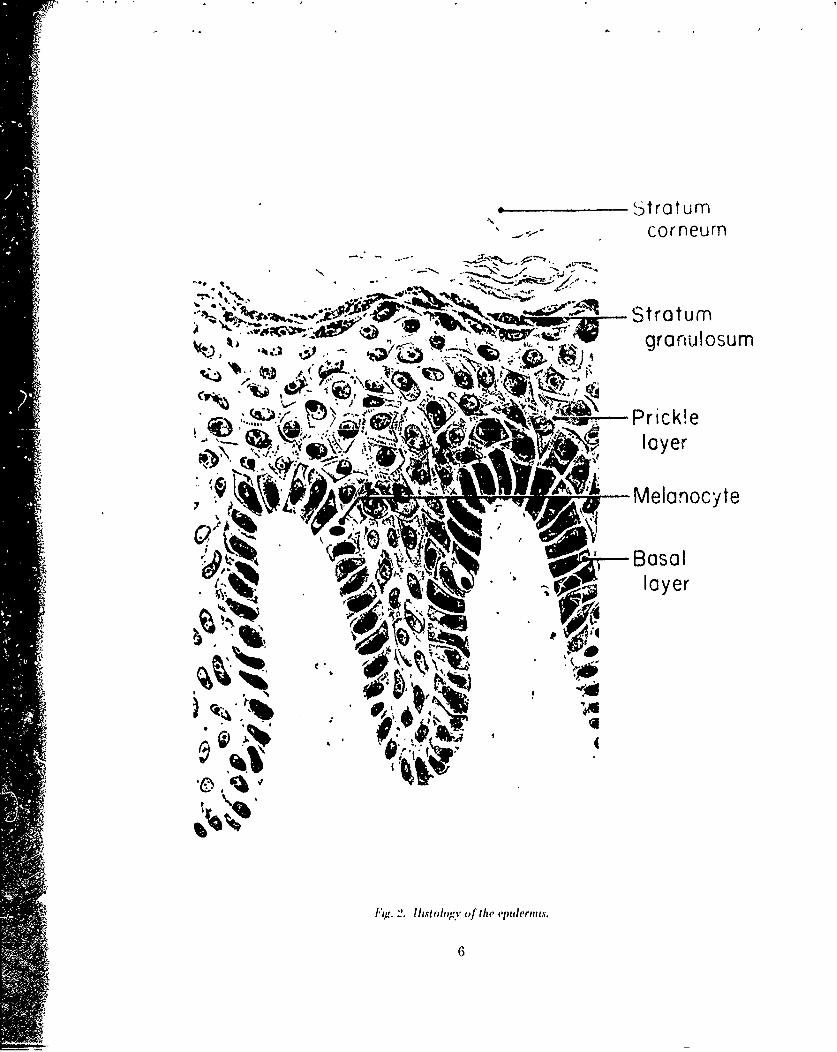
sheet covering the body, rnodffied in face is not smooth but has many small,ertain places to accomrnodate paitteu- ele,,ations which protrude into the epi-lar functions and steesses. Cr(cssly it dermis. The blood supply o" the dermisI,,s divided into the epidermi3 and dermis is quite intricate (Figure 3), P-i slnce(Figure 1); the epiderlais LB tho unpur it is germaine to the subject of zkinlayer and is composed of five l.ayere o&" bends, it will be considercd :n c:n aii,cells (Figure 2). The outernmost layer,the stratum corneum, is a compact, (a, The cutaneous arterial network.dead, horny layer undergoing 3onbtant This is a richly anastarnosirg network
4
fI
~i~c~o 10
jlAil- -1,
Sbc'c'c ....
)r~ - UY A'
v!S -L 1
0lie
cor neurn
~ ~~-* ~Stratum- U ~ gronulosumn
laye
Melanocyte
~rBoso Ip layer
6
3 leoext
cua- eenoIu ne
Sub it filan~c~lx adiosuIMg. .1diarai oftheski an it vesel, S~ulg t e aran eme t o theartria andven llsple tiss a
tIro slri atrS ith l0 ro t.f.le o- 2 ) R p o u edb e msi no p ig r ra .
deep in the corium, from this, rela- Skin color depends largely on thistively straight arterioles ascend to the circulation. The predominant role isupper corium to form the sub papillarj, played by the 3ubpapillary venousarterial plexus. This plexus has ob- plexu-.. Table II shows the skin colorlong meshes running parallel to the in various circulatory states.papillary ridges. Branching from thesubpapillary plexus, smaller arterioles The nerve supply of the skin is rich,called the papillary arterioles supply yet no nerves enter the epidermalthe papillary capillary network. Much layer; all nerve endings are in theof the nutrition of the epidermis comes upper levels of the corium and aroundfrom these capillaries by diffusion, the epidermal appendages. The nerves
are very responsive to noxious stimuli,(b) Venous circulation. The venous both local and general. Alterations of
circulation roughly parallels the arterial the environment are often reflected incirculation, but the venules are char- the local circulation, which in turnacteristicaily narrow until they descend changes the skin color or appearance,into the depths of the corium where a good example of which is the Triplelarge meshes are formed. Between the Re3ponse of Lewis.dermis and the subdermis are largeveins, many of which contain valves. Chemically, the integument of theSome of these veins are very thin- human body complements the role ofwalled and a certain amount of exchange protection. The granular layer con-takes place through the walls of tie tains granules of keratohyaline andveins. eleidin which are probably the begin-
ning of transformation of the proto-(c) Anastomoses. The papillary plasm of the cell to the waxy and
arterioles anastomose with muscular fatty material with which the surfacevenules. The muscular metaarterioles cells are loaded. Sebaceous glandsregulate the flow through the capillary supply a thin lipoidal film whichbed. covers the stratum corneum and hair.
Table II. Comparison of Skin Color with Cutaneous C:' L. c :tion(from Krogh! 1)
SSkin Color Arterioles Capillaries Venules B31u-A Flow
Red dilated dilated dilated normal
Scarlet normal normal normal increased
Lt. pink constricted constricted constricted increased
White constricted constricted constrictPd decreased
Deep Blue constricted dilated dilated decreasedi8
This film is composed of free and indicated that liquids diffused throughesterified fatty acids and ýnsaponafiable the cells rather than around them. Gasmaterial, diffusion is dependent on concentration
on both sides of the membrane, molecu-Thus we see that the skin is a well lar size, temperature and solubility.
designed armour protecting the "milieuinterieur', however there appears to be Behnke , found that completesome chinks in this armour, which will nitrogen desaturation could not occurbe considered in more detail, during oxygen breathing because the
entrance of nitrogen through the skinoccurred at a rate of about 15-20 cc.
PERMEABILITY OF THE SKIN per hour. Diffusion of nitrogen wouldincrease if the temperature were in-
Generally, it can be stated that the creased or if an incision were made inskin is relatively impermeable to most the skin. In ancther report, Behnkesubstances, except for fluids and some and Willmon 15 discovered that heliumvapors 9, 12. The way in which gases was absorbed at a rate of 170 cc. perpenetrate the skin has been studied in hour. These findings have been sub-some detail with particulate substances stantiated in more recent experimentsand liquids; but litt,,e is known in re- by Klocke] 6 who also correlated thegard to how gases penetrat6 the skin. increased diffusion with an increaseMacKee, 9 using heavy metals as in cutaneous blood flow. Shaw andtracers, found that the route of pene- Messer17 and Chambers and Gold-tration was through the follicular pores schmidt' 8 have made some interestiigand follicles and thence into the corium observations which postulate a dualvia the walls of the sebaceous glands blood supply for the skin--personsand follicles. The metals then diffused enclosed in a box filled with nitrogenfrom the corium in all directions. The developed~an increase in the respira-exact layer of the skin whicý acts as tory uptake of oxygen.the barrier to penetration is now known.The stratum corneum does not act as a When one considers the changesbarrier. rhe closely packed layers of that occur about the skin when thethe stratum granulosum is the area atmospheric pressure is increased,where most of the barrier action takes we can say that grossly, the uptake ofplace. This is known as the Szakall gas through the skin is enhanced onlayer. compression and that the release of
9 12 26 gas from the skin during decompres-Many authorities ' ' state that sion is variable, viz. cooling decreases
fluids do not necessarily follow the gas transfer, because of decreasedroute as outlined by MacKee, but cutaneous circulation, but increased be-merely diffuse through the epidermis cause of the decreased exterior concen-as gases diffuse through any other tration of the gas. Whether the skin ismembrane. Scheupleini'l recently re- wet or dry probably makes some dif-ported that his data from electron ference; radon was found to passmicroscopy and diffusion studies through the skin faster when thc subject
9
was in a water bath, in the same experi- felt that the rash ater decompressionments radon penetrated the skin faster was due to emoblization df vessels in thewhen grease was applied to the skin9 . subcutaneous fat. But Erdman 21 when
commenting on Hill's findings, notedThus, it is seen that there are many that the rash was more firequent when
variables and unknowns in this problem, sweating was less frde, 'and thereforebut with this information, one has bet- concluded that the eruption was due toter insight into the problem of skin the expansion of gases* in the sebaceousbends. glands. This opiqion.was also voiced
by Shilling. 22 Behnke1 4 and Anthony 23
also noted that rash and pruritusTHEORIES ON THE CAUSE OF occurred with regularity if the skin is
SKIN BENDS chilled during decompression.Harvey2 7 in experimiental studies,
The author has found very few histo- found that vasoconstriction favoredlogical descriptions of skin bends, and bubble formation, although Duffnerl
those that have been found are usually concluded that bubbles in the capillariesof massive decompression sickness, stimulated the contractile cells of thewhere gross subcutaneous emphysema capillaries.and severe gas embolization of thelarger blood vessels were found. Rashbass 2 conducted a series ofNothing has been found regarding the experiments to determine the etioiogyless serious, less spectacular lesions, of the itch, which was the only experi-
mental publication found on this subject.
Most of the proposed explanations He conducted a series of chamber divesfor production of skin bends fall into to a simulated depth of 240 feet for 18three categories: (1) Expansion of minutes. This dive produced the itchgases in the sebaceous glands and sweat in all subjects, but no itch occurredglands. (2) Gaseous embolization of when a portion or all of the body wasblood vessels and (3) Reflex vasocon- immersed in water. This indicatedstriction, vasodilatation ard piloerec- that gas entered the skin directly fromtion due to trauma in the skin and sub- the outside. Repeated exposure usingcutaneous tissues. barrier creams, glycerine, etc.
showed no effect on the severity of thePaul Bert, in his classic Barometric pruritus. The portions of the body with
Pressure', was of the opinion that the the highest sebaceous content were the"fleas" (itch) was due to irritation of most common sites for the rash or itch.the tissues by fine bubbles e",n if the It should be noted that the decompres-
ciculation was not stopped. Hill and sion schedule which Rashbass usedWM-Loed' 9 wrote that they had difficulty differs from that of the U.S. Navyapplying the cupping glass when treat- Schedules 7 : (1) The depth and timeing patients with Caisson's Disease, falls into the category of Exceptionalconcluding that great amounts of dls- Exposure and (2) The stage decompres-solved gases were present in the skin. sion was considerably shorter thanHill and Greenwood 20 in andther paper called for in the U.S. Navy
10
41
Decompression Tables for Exceptional Thus the theories for production ofExposures. This confirms the opinion skin bends are varied, and probably allof others 26 that the rash and itch is of them are functioning to some extentpresent more frequently when marginal at one time o: another. In summarydecompression is used. Behnke2 4 has then, we have the following propositionsnoted that bubbles have been seen in the on rash formation:cutaneous arteries and veins afterrapid decompression. Aldao 4 states (1) Expansion of gases in thethat all the eruptions seen are a mani- sebaceous and sweat glands.festation of bubbles in the cutaneousvessels, and Wright et a125 surmise (2) Blood vessel gas thrombosis.the mottling of the skin is due tocapillary embolization with gas. (3) Reflex changes in the blood ves-
sels, secondary to trauma of gas ex-At this juncture we find fairly gen- pansion.
eral agreement on the fact that the"rashes" (which probably represent The following discussion will dealsome of those descriptive entities men- with the facts heretofore presented.tioned, viz: pruritus scarlatinaformrash, and intracutaneous emphysema)are manifestations of expansion of gas DISCUSSIONwhich has entered the body from theoutside directly into the skin, the To review some of the facets coveredsebaceous glands or sweat glands. thus far, in the pathogenesis of skinFerris and Engel have written some bends one must consider the variousinteresting thoughts that may account alterations in the diver's environment:for formation of the other types of skin Increased air pressur. , dissolution ofbends. Writing about cutis marmorata, gas into the tissues, especially highlythey state that the pale, cool, cyanotic soluble gases such as nitrogen, chillingmottling that occurs is caused by the during decompression with subsequentobstruction of terminal arterioles and vasoconstriction and release of gasesvenules. The transformation •o from the tissues. Bends do not usuallyerythema suggests that the obstruction occur until Haldane's 2:1 ratio Is ex-is overcome by vasodilatation, akin to ceeded, although this may not be truereactive hyperemia. The concentric with skin bends, i.e., the ratio may bespreading of the cutis marmorata may lower. What then, is the cause of skinbe secondary to a spreading factor in bends? Rashbass's work leads one totissues, since the spread cannot be suspect that the itching and the tran-explained anatomically. The deep pain sient rashen are due to the rapid ex-which is nresent following the disap- pansion and evolution of gases whichpearance of the marbleization suggests originated from the outside. However,trauma as the potential spreading the observations of Ferris ana Lgelfactor. The trauma is probably due to on the spread of cutis marmorata pointbubble formation in the subcutaneous fat. to trauma deep in the skin which would

make one suspect that the trauma wan over the areas of high sebaceous glandfrom bubbles expanding in the loose content leads one to conclude that the
connective tissue of the corium or sub- dermis is reached via the sebaceouscutaneous tissues. Probably both glands.etiologies are correct, and thatexogenous and endogenous gases have a (2) Scarlatinaform rash: In allrole in the production of different likelihood, this is an extension of themorphologic changes. process which occurs in pruritus alone.
The irritant gas is present in greaterThe author proposes the following as concentration and takes longer for it
the etiology of the various manifesta- to evolve so that secondary vasculartions of skin bends, these proposals are reaction occurs as well as stimulationnot proven but seem logical in view of of the pilo-erectory apparatus andthe findings already presented. perhaps some tissue damage occurs
with release of histamine.(1) Skin bends are produced by the
expansion of both exegenous and endo- (3) The erysipelas form leads intogenous gas bubbles. the category of endogenous gases as
the etiologic source. Here the vascular(2) The morphological pattern of the pattern and the positive Mellinghof's
lesion depends largely on where the sign indicate that the deeper veins aregreatest quantity of gas is present (the involved either directly by gaseousinitial lesion), embolism or because of reflex con-
striction of the musculature of the"(3) Subsequent to the initial lesion veins, due to irritation elsewhere. It
(#2.) secondary vascular reaction may is not likely that enough gas from anpredominate, exogenous source could wholly account
for this type of lesion.Using the above proposals, let us
examine the different categories of skin (4) Cutis marmorata: Since this isbends as outlined by Aldao. seen only with severe bends, the source
_ _ of the gas is endogenous and here bub-(1) Pruritus alone: this probably bles are formed either in the tissues or
represents small amounts of gases in in the adjacent blood vessels. This is
the very superficial layers of the dermis, a cutaneous manifestation of what is orthe irritant* is probably not present in will be occurring in the rest of the body.large enough quantities or present longenough to cause secondary vascular re- (5) The serious form: Again, thisaction, but is deposited in an area is most likely an extension of thesupply by nerves, i.e., below the epi- marbleization phenomenon.dermis. The frequency of the symptom
(6) Emphysema: The intracutaneousforms described in aviators represents
*-IrrtanS "does rot amply that the gas is e.. .ni.ally expansion of gas below one of the majoroffensive, rather, a gas bubble is the "'Irritant." "skin barriers" such as the Szakall
* 12
* • $ 5•• L:-••,' • • :• •.... :•. . •.. :" - • .. .. .. : o .. ,. .
layer. Subcutaneous emphysema is due some cause arising from the concentration
to either pneumothorax or massive de- gradient maintained between cutaneous bloodco-pression sickness where is and chamber atmosphere in the affected areas
w gas rather than considered a manifestation of de.
found in all the organs and tissues of conapreision sickness. Gas-induced osmosis is
the body. discusscd as a possible initiating mechanismand is shown to be quantitatively consistent
In cwith the clinical observations. This evidencenappears to support the author of this report
to be a sensitive indicator of the ade- in his contention that there is an exterior
quacy of decompression, and when in- gaseous osmotic exchange in the skin with
adequate this is reflected by changes in subsequent gas expansion as a contributingthe skin. These changes appear to be factor to the urticaria. Similar lesions were
noted under the same conditions on subjectsdue to gas bubbles whose origin is gas in the recent saturation dives conducted at
induced osmosis from the surrounding the University of Pennsylvania Medical Shool
medium and diffusion from the Environmental Institute, although the theoreti.
blood. 30,31 cal observation as to the cause of the lesionsb dhas not been defined to the satisfaction of the
University of Pennsylvania investigators.
"Sci. Dir'.s Note: In March 1970, at the F. G.Hall Environmental Laboratory, Duke Uni. CONCLUSIONS
versity Medical Center, Durham, NorthCarolina, during a "saturation" dive at a simu- Little experimental evidence waslated depth of 200 feet in a gaseous environ- found to account for the phenomenon ofient of helium and oxygen, 0 all three ex-perimental subjects developed urticaria during skin bends, and most of the conclusionsor!shortly after breathing a normoxic nitrogen were drawn from the clinical appear-
4 gas mixture. 7o experienced observers these ance of the lesions. This author con-cutaneous symptoms and signs were similar to cludes that certain types of skin bendsthose frequently observed following decom-pression, and therefore considered a mani- are caused by expansion of gases thatfestation of decompression sickness. However, were dissolved into the skin duringno decompression preceded Mhe above sympto- compression and that other lesions werematri reactions, suggesting an association ofthe .'ticaria with some inherent difference iin due to expansion of gases that were de-
the inert gases such as a physical property livered to the tissues via the bloodwhose effect upon the tissue is enhanced under stream. The patterns of the eruptionshyperbaric conditions. As stated before, the vary depending on secondary vascularlesions occurred before the subjects had ex- . r ea in.
perienced decompression, and although they reaction.appeared very similar to the cutaneousreactions frequently termed "skin bends",they were not observed upon skin within the Further inestigation on the phy-hcad.tent and hence over those areas exposedto the breathing mixture at all times. The siology of the skin during hyperbaric
urticaria has therefore been attributed to conditions is indicated.
13
REFERENCES 9. Rothman, S.: Physiology andBiochemistry of the Skin, Univ. of
1. Bert, P.: Barometric Pressure. Chicago Press, 1954, Chicago.Researches in Experimental Phy-siology (1878) Hitchcock, M. A. 10. Duffier, G. J.: The Capillaryand Hitchcock, F. A., trans. Col- Circulation - Its Role in Compressedlege Book Co., Columbus, 1943. Air Illness. Submarine Medical
Officer Qualification Thesis.2. Rashbass, C.: The Aetiology of
Itching on Decompression, Royal 11. Krogh, A.: The Anatomy and Phy-Navy Physiologic Laboratory siology of the Capillaries, YaleReports, 1-957. Univ. Press, 1924 (quoted from
Duffner (10)).3. Rivera, J.C.: Decompression
Sickness Among Divers: An Analy- 12. Pillsbury, D. M., et al: Derma-sis of 935 Cases, EDU Research tology, W. B. Saunders Co.,Report 1-63, February 1963. Philadelphia, 1956.
4. Aldao, C.N.G.: Cutaneous Forms 13. Scheuplein, R. J. Routes of Pene-of Caisson's Disease, Rev. Argent. tration Through the Skin fromDermatosif., 33: 20-25, 1949. Permeability Data. Report read to
the Society of Investigative Derma-5. Ferris, E. B. and Engel, G. L.: tology, Inc., 20 June 1965, New
p35 in Decompression Sickness, York City.John F. Fulton, Ed., W. B.Saunders Co., Philadelphia, 1951. 14. Behnke, A. R.: The Absorbtion
and Elimination of Gases of the6. Golding, F. C.: Decompression Body in Relation to its Fat and
Sickness Occurring During the Water Content, Medicine, 24: De-Construction of the Dartford Tunnel, cember, 1945.Brit. Jour. Indust. Med., 17:167, 1960. 15, Behnke, A. R. and Willmon, T.L.:
Cutaneous Diffusion of Helium in7. U. S. Navy Diving Manual Relation to Peripheral Blood Flow
(NAVSHIPS 250-538), Bureau of and Absorbtion Atmospheric Nitro-Ships, Navy Department, G. P. 0., gen Through the Skin, Am. J.July, 1963. Physiol., 13: 627-632, 1941.
8. Best, C. H. and Taylor, N. B.: 16. Klocke, R. A., et al: GasThe Physiologic Basis of Medical Transfer Across the Skin in
* Practice, Williams and Wilkins Co., Man, J. Appl. Physiol., 18: 311-Baltimore, 1955, p345. 317, 1963.
14
17. Shaw, L. A. and Messer, A. C.: 25. Wright, W. and Brady, J. W. S.:.Cutaneous Respiration in Man: H1. Compressed Air illness orThe Effects of Temperature and Caisson's Disease, in Billings-Humidity upon the Rate of C02 Forehheimerla Therausis ozfElimination and Oxygen Absorbtion, Internal Diseases, George Blumer,Am. J. Physiol., 90: 107-118, ed., Appleton and Co., New York1929. 1929.
18. Chambers, A.H. and Goldschmidt, 26. Van der Aue, 0. E., Keller, R. J.S.: The Influence of Atmospheric and Brinton, E. S.: E.D.U. ReportOxygen Absorbtlon upon Trans- No. 1, March 1949.parent Total Oxyger Utilization ofthe Body, Am. J. Physiol., 12-5: ?7. Harvey, E. N., et al: The M4echan-331-332, 1940. Ism of Bubble Formation in the Blood
of Animals in Relation to Decom-19. Hill, L. and McLoed, J., Jr.: pression Sickness, National Re-
Caisson's Disease and Diver's search Council Report No., 229,Palsy; Experimental Study, J. 19.3.Hygiene, 3: 401-445, 1903.
28, Caivery, H. 0., et al: The
20. Hill, L. and Greenwood, M.: In- Metabolism and Permeability offluence of Increased Barometric Normal Skin, Physiological Re-Pressure on Man, Proc. Roy. views, 26: 495-540, 1946. (ThiaSoc., 77: 442-453, 1906. '•.z an excellent review of the phy-
siology of the skin).
21. Erdman, S.: The Acute Effects ofCaisson's Disease, Int. Cong 29. Beklke, A. R, and Stephenson,Hyg. and Demog., 3: 619-625, C. S.: Applied Phy.iology, Annual1913. Review of Physio2lo Vol. IV, 1942.
22. Shilling, C. W.: Compressed Air 30. Blenkarn, G. D., Aquadro, C.,Illness, U. S. Naval Med. Bull., Hills, B. A. and Saltzman, H. A.:36: 235-259, 1938. Urticaria Following the Sequential
Breathing of Various Inert Gases at
23. Anthony, R., et ah Temperature a Constant Ambient Pressure of 7and Decompression Sickness, ATA: A Possible Manifestation oiNational Research Council Report Gas Induced Osmosis, AerospaceNo. 136, 1943. Med., 42: 141-146, Feb, 1971.
24. Behnke, A. R.: A Review of Phy- 31. Unpublished data.sioiogical and Clinical Data Per-taining to Decompression Sickness,NMRI Report No, 4, 1947.
15