Embed Size (px)
Citation preview
Subclinical seizures identified by postoperative electroencephalographic monitoring are common after neonatal cardiac surgery
Maryam Y. Naim, MDa, J. William Gaynor, MDb, Jodi Chen, MDa, Susan C. Nicolson, MDc, Stephanie Fuller, MDb, Thomas L. Spray, MDb, Dennis J. Dlugos, MDd, Robert R. Clancy, MDd, Livia Vianez Costa, MDd, Daniel J. Licht, MDd, Rui Xiao, PhDe, Heather Meldrum, RNf, and Nicholas S. Abend, MDd
aDivision of Cardiac Critical Care Medicine, Department of Anesthesiology, Critical Care Medicine, and Pediatrics, The Children’s Hospital of Philadelphia, Perelman School of Medicine at the University of Pennsylvania, Philadelphia, Pa
bDivision of Cardiothoracic Surgery, Department of Surgery, The Children’s Hospital of Philadelphia, Perelman School of Medicine at the University of Pennsylvania, Philadelphia, Pa
cDivision of Cardiothoracic Anesthesiology, Department of Anesthesiology and Critical Care Medicine, The Children’s Hospital of Philadelphia, Perelman School of Medicine at the University of Pennsylvania, Philadelphia, Pa
dDivision of Neurology, Departments of Neurology and Pediatrics, The Children’s Hospital of Philadelphia, Perelman School of Medicine at the University of Pennsylvania, Philadelphia, Pa
eDepartment of Biostatistics and Epidemiology, The Children’s Hospital of Philadelphia, Perelman School of Medicine at the University of Pennsylvania, Philadelphia, Pa
fDepartment of Nursing, The Children’s Hospital of Philadelphia, Perelman School of Medicine at the University of Pennsylvania, Philadelphia, Pa
Abstract
Objectives—The American Clinical Neurophysiology Society recommends continuous
electroencephalographic monitoring after neonatal cardiac surgery because seizures are common,
often subclinical, and associated with worse neurocognitive outcomes. We performed a quality
improvement project to monitor for postoperative seizures in neonates with congenital heart
disease after surgery with cardiopulmonary bypass.
Address for reprints: Maryam Y. Naim, MD, Department of Anesthesiology and Critical Care Medicine, The Children’s Hospital Of Philadelphia and Perelman School of Medicine at the University of Pennsylvania, 34th and Civic Center Blvd, Philadelphia, PA 19104, [email protected].
Read at the 94th Annual Meeting of The American Association for Thoracic Surgery, Toronto, Ontario, Canada, April 26–30, 2014.
You can watch a Webcast of this AATS meeting presentation by going to: http://webcast.aats.org/2014/files/Monday/20140428_435_455pm_Maryam_Naim.mp4
Conflict of Interest StatementAuthors have nothing to disclose with regard to commercial support.
HHS Public AccessAuthor manuscriptJ Thorac Cardiovasc Surg. Author manuscript; available in PMC 2016 July 07.
Published in final edited form as:J Thorac Cardiovasc Surg. 2015 July ; 150(1): 169–180. doi:10.1016/j.jtcvs.2015.03.045.
Author M
anuscriptA
uthor Manuscript
Author M
anuscriptA
uthor Manuscript
Methods—We implemented routine continuous electroencephalographic monitoring and
reviewed the results for an 18-month period. Clinical data were collected by chart review, and
continuous electroencephalographic tracings were interpreted using standardized American
Clinical Neurophysiology Society terminology. Electrographic seizures were classified as
electroencephalogram-only or electroclinical seizures. Multiple logistic regression was used to
assess associations between seizures and potential clinical and electroencephalogram predictors.
Results—A total of 161 of 172 eligible neonates (94%) underwent continuous
electroencephalographic monitoring. Electrographic seizures occurred in 13 neonates (8%)
beginning at a median of 20 hours after return to the intensive care unit after surgery. Neonates
with all types of congenital heart disease had seizures. Seizures were electroencephalogram only
in 11 neonates (85%). Status epilepticus occurred in 8 neonates (62%). In separate multivariate
models, delayed sternal closure or longer deep hypothermic circulatory arrest duration was
associated with an increased risk for seizures. Mortality was higher among neonates with than
without seizures (38% vs 3%, P<.001).
Conclusions—Continuous electroencephalographic monitoring identified seizures in 8%of
neonates after cardiac surgery with cardiopulmonary bypass. The majority of seizures had no
clinical correlate and would not have been otherwise identified. Seizure occurrence is a marker of
greater illness severity and increased mortality. Further study is needed to determinewhether
seizure identification and management lead to improved outcomes.
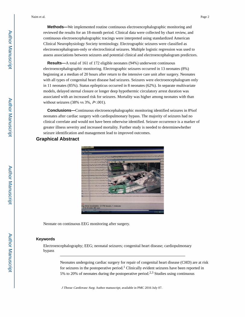
Graphical Abstract
Neonate on continuous EEG monitoring after surgery.
Keywords
Electroencephalography; EEG; neonatal seizures; congenital heart disease; cardiopulmonary bypass
Neonates undergoing cardiac surgery for repair of congenital heart disease (CHD) are at risk
for seizures in the postoperative period.1 Clinically evident seizures have been reported in
5% to 20% of neonates during the postoperative period.2,3 Studies using continuous
Naim et al. Page 2
J Thorac Cardiovasc Surg. Author manuscript; available in PMC 2016 July 07.
Author M
anuscriptA
uthor Manuscript
Author M
anuscriptA
uthor Manuscript
electroencephalographic (CEEG) monitoring have reported that electroencephalogram
(EEG)-only seizures (also termed “nonconvulsive seizures”) may be more common,
occurring in 5% to 26% of neonates.3–9 Postoperative seizures are associated with worse
neurodevelopmental outcomes, abnormal neurologic examination results, and abnormal
neuroimaging study results.10–14 On the basis of these data, the American Clinical
Neurophysiology Society’s (ACNS) guideline on neonatal EEG monitoring recommends
consideration of CEEG monitoring after neonatal cardiac surgery.15 We implemented this
recommendation and performed a single-center quality-improvement project to monitor for
postoperative EEG seizures in a contemporary cohort of neonates with CHD after cardiac
surgery with cardiopulmonary bypass (CBP). We aimed to determine the incidence of
postoperative EEG seizures and to identify risk factors for seizures.
MATERIALS AND METHODS
Patient Population
By using a clinical pathway in which CEEG was recommended for all neonates (≤30 days of
age, corrected gestational age ≤44 weeks) after cardiac surgery with CBP, we implemented
the ACNS clinical recommendation on June 15, 2012, and reviewed the results for the period
ending on December 31, 2013. If a patient underwent multiple surgeries during the neonatal
period, only the index surgery was included. The Institutional Review Board of the
Children’s Hospital of Philadelphia approved review of the Quality Improvement project.
Surgical Strategy
Operations were performed by 4 cardiac surgeons using a pH-stat blood gas management
strategy. Deep hypothermic circulatory arrest (DHCA) was used at the discretion of the
surgeon after induction of hypothermia to 18°C. Modified ultrafiltration was performed on
all patients. Delayed sternal closure was not routinely used.
Electroencephalogram Monitoring
CEEG was performed by the encephalography service and initiated within 6 hours of
returning to the cardiac intensive care unit (CICU) after surgery. The encephalography
service includes acquisition and review software, network infrastructure, licensed EEG
technologists, and expert encephalographers. CEEG was performed using a Grass-Telefactor
video-EEG system (Grass Technologies, West Warwick, RI) with a portable acquisition
machine networked to the main EEG server, allowing EEG review at the bedside, from
multiple sites in the hospital, and remotely. EEG technologists were present in the hospital
24 hours per day and 7 days per week. CEEG was performed with 12 gold-over-silver scalp
electrodes affixed according to the international 10–20 system (modified for neonates), with
collodion adhesive (or paste for neonates on extracorporeal membrane oxygenation
[ECMO]). Abnormal movements or vital sign fluctuations noted by the clinical team were
marked on the recording as push-button events. If an electrographic seizure was identified,
the CICU team was alerted by the EEG technologist or encephalographer, and neurologic
consultation was obtained. CEEG was continued for 48 hours if no seizures occurred. If
seizures were identified, CEEG was continued until 24 hours after the end of the last seizure.
The initial antiseizure medications administered were phenobarbital or levetiracetam at the
Naim et al. Page 3
J Thorac Cardiovasc Surg. Author manuscript; available in PMC 2016 July 07.
Author M
anuscriptA
uthor Manuscript
Author M
anuscriptA
uthor Manuscript
discretion of the on-call neurologist and cardiac inten-sivist. Phenobarbital (20 mg/kg bolus)
was administered as a first-line agent in the majority of patients. For patients in whom there
were concerns for hemodynamic instability, clinicians sometimes used phenobarbital in
divided doses (5 mg/kg × 4 doses over 1 hour) or levetiracetam (20 mg/kg bolus). Patients
with seizures usually underwent clinically indicated magnetic resonance imaging (MRI) of
the brain or head ultrasound.
Data Collection
Clinical data were obtained from the medical record, including anesthesia and perfusion
records. Patients were categorized according to a classification that incorporates cardiac
anatomy and perioperative physiology, which has been shown to predict perioperative
mortality. Class I is 2 ventricles with no aortic arch obstruction, class II is 2 ventricles with
aortic arch obstruction, class III is a single ventricle with no aortic arch obstruction, and
class IV is a single ventricle with aortic arch obstruction.16
For purposes of this review, EEG tracings were reinterpreted by a single encephalographer
(NSA), blinded to clinical information (except conceptional age), using standardized ACNS
neonatal EEG terminology.17 EEG seizures were defined as abnormal, paroxysmal EEG
events that were different from the background, lasted more than 10 seconds (or less if
associated with a clinical seizure), had a plausible electrographic field, and evolved in
frequency, voltage, morphology, and often spatial distribution. EEG seizures were classified
as electrographic status epilepticus if any single seizure lasted more than 30 minutes or if
recurrent seizures together lasted for more than 30 minutes in any 1-hour epoch (50%seizure
burden). EEG seizures were classified as EEG-only seizures (no clinical signs observed by
bedside caregivers or on video review) or electroclinical seizures. All available
neuroimaging studies were reviewed by a neurologist (DJL) to examine associations
between neuroimaging abnormalities and seizure localization.
Data were collected and managed using Research Electronic Data Capture (REDCap), a
web-based electronic data application hosted at the Children’s Hospital of Philadelphia
Research Institute.18
Statistical Analysis
Summary statistics are reported as medians and interquartile ranges (IQRs) for continuous
data and counts and proportions for categoric data. The association of each clinical and
interictal EEG variable with seizures was examined using the chi-square test for categoric
variables and Wilcoxon’s rank-sum test for continuous variables.
Multiple logistic regression was used for association of seizures with clinical and interictal
EEG variables. To avoid collinearity, correlation between predictors was examined by using
Pearson correlation coefficients for continuous predictors, and association between
continuous and categoric predictors was examined by 2-sample test or analysis of variance.
Variables associated with seizures with a P value less than .2 on univariable analysis were
included in multivariable analyses. An initial model used only clinical data, whereas a
subsequent model also used data obtained from the initial hour of CEEG. All statistics were
performed with Stata 10.0 (StataCorp LP, College Station, Tex).
Naim et al. Page 4
J Thorac Cardiovasc Surg. Author manuscript; available in PMC 2016 July 07.
Author M
anuscriptA
uthor Manuscript
Author M
anuscriptA
uthor Manuscript
RESULTS
Demographics and Surgical Details
During the 18-month study period, 172 neonates with CHD underwent cardiac surgery with
CPB. Postoperative CEEG was obtained in 161 of 172 eligible neonates (94%). The reasons
for not undergoing CEEG included unavailability of EEG machine in 1 neonate, CICU team
not ordering CEEG (mainly in the initial phase of implementation) in 5 neonates, death in
the operating room in 1 neonate, and CICU team deciding not to monitor in 2 neonates (cutis
aplasia in 1 and decision that monitoring was not warranted in 1). Pathway adherence
improved over time. For the initial period of monitoring June 15 to December 31, 2012, 48
of 55 neonates (87%) who underwent surgery with CPB were monitored. For the middle
period from January 1 to June 30, 2013, 54 of 56 neonates (96%) were monitored. For the
later period of monitoring from July 1 to December 31, 2013, 57 of 59 neonates (97%) were
monitored.
Of the 161 neonates who underwent CEEG, 92 (57%) were male. The median gestational
age was 39 weeks (IQR, 38–39), and 26 neonates (16%) were premature (<37 weeks
gestational age). The median head circumference at birth was 34 cm (IQR, 32–35). Genetic
defects were identified in 21 neonates (13%). The median age at surgery was 5 days (IQR,
3–7). Five neonates had 2 operations with CPB during the neonatal period, and 1 neonate
had 3 operations with CPB during the neonatal period. Both were monitored after each
surgery.
The cardiac defects were class I in 68 neonates (42%), class II in 35 neonates (22%), class
III in 15 neonates (9%), and class IV in 43 neonates (27%). The 5 most common operations
were the stage I Norwood operation in 43 neonates (27%), the arterial switch operation in 25
neonates (16%), the systemic to pulmonary artery shunt in 17 neonates (11%), complete
repair of tetralogy of Fallot in 14 neonates (9%), and truncus arteriosus repair in 12 neonates
(8%). The median duration of CPB was 46 minutes (IQR, 38–62). DHCA was used in 96
neonates (60%), with a median duration of 41 minutes (IQR, 32–50). Twenty-six neonates
(16%) had delayed sternal closure. Eleven neonates (7%) required ECMO (2 were placed on
ECMO in the operating room before initiation of CEEG, 8 were placed on ECMO during
CEEG, and 1 was placed on ECMO while CEEG had been temporarily discontinued during
a diagnostic cardiac catheterization). Fifteen neonates (9%) had a cardiac arrest (2 in the
operating room before initiation of CEEG, 1 in the CICU before initiation of CEEG, and 12
in the CICU during CEEG). Table 1 summarizes the demographic and clinical
characteristics.
Nonseizure Events
Bedside clinicians identified events concerning for clinically evident seizures in 32 neonates
(push-button events). None of these events had an EEG correlate, and they were deemed
nonepileptic. Push-button events included episodes of abnormal body movement in 14
neonates, hypertension in 7 neonates, tachycardia in 6 neonates, abnormal face movement in
6 neonates, desaturation in 5 neonates, slow respiratory rate in 2 neonates, bradycardia in 1
Naim et al. Page 5
J Thorac Cardiovasc Surg. Author manuscript; available in PMC 2016 July 07.
Author M
anuscriptA
uthor Manuscript
Author M
anuscriptA
uthor Manuscript
neonate, and hypotension in 1 neonate. Events occurred during the initial 12 hours in 22
neonates, later than 12 hours in 10 neonates, and during both time periods in 2 neonates.
Seizures and Electroencephalogram Characteristics
EEG seizures occurred in 13 of 161 neonates (8%). The proportion with seizures has not
changed since our previous report4 (14% previously vs 8% currently, P = .10). The median
seizure onset was 20 hours (IQR, 15–34) after return to the CICU postoperatively. EEG
seizures were EEG only in 11 neonates (85%) and electroclinical in 2 neonates (15%).
Electrographic status epilepticus occurred in 8 neonates (62%). Seizures were spatially
diffuse in 2 neonates (15%), lateralized in 2 neonates (15%), and focal in 9 neonates (69%).
Seizure characteristics are provided in Table 2. The encephalographer performing
reinterpretation blinded to clinical information (except conceptional age) was consistent with
initial clinical interpretation for seizure occurrence in all patients.
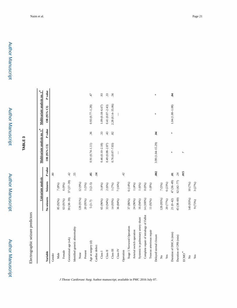
Univariable and multivariable analyses using clinical variables for seizure prediction are
shown in Table 3. On univariable analysis, seizures were more common in younger
neonates, with longer CBP and DHCA times, in patients with single-ventricle defects with
arch obstruction, in patients returned to the CICU with delayed sternal closure, in patients
with postoperative cardiac arrests, and in patients who were placed on ECMO. Variables that
were not known on return to the CICU and occurred later would not be useful in identifying
patients at higher risk for seizures and in need of CEEG on postoperative CICU admission.
Thus, cardiac arrest and ECMO were not included in the multivariable model.
Delayed sternal closure and DHCA duration were strongly associated (P < .0001).
Therefore, to avoid collinearity, separate multivariate analyses were performed for delayed
sternal closure and DHCA duration to examine their respective associations with seizure
occurrence. Both multivariate models are provided in Table 3. Both had similar performance
characteristics. The goodness-of-fit for the multivariable logistic regression models indicated
that both had a pseudo-R2 of 0.11. Receiver operating characteristic curves (Figure 1) and
the c-statistics were not significantly different (P = .8). We performed the leave-one-out
cross validation to calculate the predictive probability of seizure occurrence for each
individual using the multivariate logistic model and again observed similar results between
the 2 models. In their respective models, only delayed sternal closure (odds ratio [OR] 3.99;
95% confidence interval [CI], 1.04–15.29; P = .04) and DHCA duration (OR, 1.04; 95% CI,
1.00–1.08; P = .04) were significantly associated with seizures. Because DHCA was
analyzed as a continuous variable, the increase in seizure risk is provided per minute of
DHCA. We assessed the potential collinearity of variables. Lower weight was not associated
with ECMO (P = .77) or DHCA use (P = .86). Younger age at the time of surgery was not
associated with ECMO (P = .41) or DHCA use (P = .23). Arch obstruction was not
associated with ECMO (P = .29) but, as expected, was associated with DHCA (P = .0000).
When DHCA duration was divided into 3 categories, there was a trend toward higher seizure
occurrence with longer DHCA duration (no DHCA 3%; DHCA <40 minutes 7% with OR,
2.48; 95% CI, 0.40–15.56; DHCA ≥40 minutes 15% with OR, 5.36; 95% CI, 1.09–26.42;
chi-square P = .07). When DHCA was dichotomized to less than 40 minutes (including no
DHCA) or 40 minutes or more, neonates with DHCA 40 minutes or more had a significantly
Naim et al. Page 6
J Thorac Cardiovasc Surg. Author manuscript; available in PMC 2016 July 07.
Author M
anuscriptA
uthor Manuscript
Author M
anuscriptA
uthor Manuscript
increased seizure risk (15% vs 5%, chi-square P = .03; OR, 3.4; 95% CI, 1.07–11.08; P = .
04). When DHCA was changed into a 10-minute scale for subjects with DHCA more than 0
minutes, for every 10-minute increase in DHCA duration time, the OR of developing seizure
was 1.47 (95% CI, 0.96–2.15).
Several alternative analyses were performed. First, an alternative regression model was
explored with the cardiac defect type grouped as 1 versus 2 ventricles, and similar results
were observed. Second, an alternative regression model was explored including EEG
variables known in the initial hour of EEG recording, including background category,
excessive interictal epileptiform discharge presence, and reactivity. The EEG background
category was continuous in 30 neonates (19%), appropriately discontinuous in 31 neonates
(19%), and excessively discontinuous in 99 neonates (62%). EEG background category did
not predict seizure occurrence on univariable analysis (P = .85). Seizures occurred in 5 of 46
neonates (11%) with excessive interictal epileptiform discharges and in 7 of 105 neonates
(6%) without excessive epileptiform discharges (P = .06). Among the 112 neonates in whom
reactivity could be assessed, seizures occurred in 4 of 96 neonates (4%) with reactivity and 2
of 16 neonates (13%) without reactivity (P = .08). Multivariable analyses including these
EEG variables were not substantially different than the model described previously. On
multivariable analysis using delayed sternal closure, the only predictor of seizures was
delayed sternal closure (OR, 4.09; 95%CI, 1.04–15.99; P = .04). On multivariable analysis
using DHCA duration, the only predictor of seizure was DHCA duration (OR, 1.04; 95% CI,
1.00–1.08; P = .04). We compared the receiver operating characteristic curve c-statistics
between the model containing only clinical variables and the model containing EEG
variables, and they were not significantly different.
Seizure descriptions and management are summarized in Table 2. The most commonly
administered antiseizure medication was phenobarbital, followed by a combination of
levetiracetam or phenobarbital. The choice of antiseizure medication and bolus parameters
were left to the clinician’s discretion. No patients had hemodynamic instability related to
medication administration.
Nine neonates (6%) died. Four neonates had multiorgan system dysfunction and withdrawal
of technologic support on ECMO, 1 neonate had a large intracranial hemorrhage and
withdrawal of technologic support on ECMO, 1 neonate had multiorgan system dysfunction
and withdrawal of ventilatory and inotropic support, 1 neonate had a large intracranial
hemorrhage and withdrawal of ventilatory and inotropic support, 1 neonate had a cardiac
arrest and was deemed to not be an ECMO candidate, and 1 neonate had cardiac arrest and
was unable to be cannulated for ECMO. Mortality was higher among neonates with than
without seizures (38% vs 3%, P <.01). Neonates with and without seizures did not have
significantly different postoperative lengths of stay in the CICU (11 days for both groups, P = .65) or hospital (18 vs 17 days, P = .85). When the 9 neonates who died were excluded,
neonates with and without seizures still did not have significantly different lengths of stay in
the CICU (11 days for both groups, P = .69) or hospital (17 vs 19 days, P = .40).
Naim et al. Page 7
J Thorac Cardiovasc Surg. Author manuscript; available in PMC 2016 July 07.
Author M
anuscriptA
uthor Manuscript
Author M
anuscriptA
uthor Manuscript
Brain Imaging
Brain MRI scans were obtained in 8 of 13 neonates with seizures. Findings included diffuse
periventricular leukomalacia in 5 neonates, subdural hematomas in 3 neonates, infarction in
2 neonates, microhemorrhages in 2 neonates, and intraparenchymal hemorrhage in 1
neonate. Five neonates did not undergo MRI (death in 4 and transfer to another hospital in 1)
but underwent head ultrasounds that showed intraventricular hemorrhage in 2, infarction in
2, intraparenchymal hemorrhage in 2, and subdural hematoma in 1. Imaging characteristics
are provided in Table 2. All neonates with seizures had diffuse or multifocal imaging
lesions. Four patients had seizures arising predominantly from one of their injury sites
(subjects 4, 5, 10, and 12 in Table 2), but none had a single site of injury with focal seizures
arising solely from that region.
DISCUSSION
This is the first report describing the impact of implementation of the ACNS guideline on
routine postoperative CEEG among neonates with CHD after surgery with CPB. We
identified an EEG seizure incidence of 8% (13/161). The seizure burden was often high
(status epilepticus in 62%). Seizures were often EEG only (85%), indicating that CEEG was
required for identification because bedside clinical assessment for seizures without CEEG
monitoring would be unreliable. The only clinical predictors of seizure occurrence available
on return to the CICU postoperatively were delayed sternal closure and longer DHCA
duration. Seizures also were more likely in neonates who subsequently required ECMO or
experienced cardiac arrest, and seizures were associated with higher mortality.
The 8% incidence of EEG seizures in our current cohort was not significantly different from
our previous report of CEEG among neonates who received care from 2001 to 2003 (14%,
15/110).4 However, unlike our previous cohort in which all patients with seizures had EEG-
only seizures, 15% of our current cohort of neonates had some clinical correlate to their
seizures. These data are similar to the Boston Circulatory Arrest Study of children with
transposition of the great arteries in which the incidences of EEG-only and electroclinical
seizures were 20% and 6%, respectively.3 Likewise, a study of a heterogeneous CHD cohort
of neonates and infants reported perioperative (preoperative, intraoperative, and
postoperative) electrographic seizures in 30% of neonates, of which 16% were
electroclinical.19 In comparison, Andropoulos and associates20 examined the occurrence of
preoperative and postoperative seizures in neonates undergoing surgery with CBP and found
that only 1 patient with a single ventricle had an EEG seizure, leading to an overall
incidence of 1.5%. The lower incidence of seizures in this last study is likely due to the
routine intraoperative and postoperative administration of benzodiazepines, which were not
used routinely in the other studies.
Similar to our previous report,4 electrographic seizures were most common in neonates with
single-ventricle defects with arch obstruction (16% in current report, 18% in previous
report). In our prior study of clinical seizures after infant cardiac surgery, the risk of clinical
seizures was also highest in those with single-ventricle defects with arch obstruction.2 This
contrasts with other studies that have reported the highest seizure incidence in patients with
2-ventricle defects with aortic arch obstruction.19 In addition, we found that increasing
Naim et al. Page 8
J Thorac Cardiovasc Surg. Author manuscript; available in PMC 2016 July 07.
Author M
anuscriptA
uthor Manuscript
Author M
anuscriptA
uthor Manuscript
DHCA duration predicted seizure occurrence. This finding is similar to that of the Boston
Circulatory Arrest Study, in which identified risk factors for seizures included increasing
duration of DHCA, the presence of a ventricular septal defect, and older age at the time of
surgery.9 Likewise, we have previously reported risk factors that include coexisting genetic
defects, aortic arch obstruction,2 and increasing duration of DHCA.2,4 Since our previous
report, the incidence of seizures in patients who had DHCA more than 40 minutes has
decreased from 24% to 15%. Of note, avoidance of DHCA does not prevent seizures. In the
current study, seizures occurred in 3% of patients in whom DHCA was not used. A recent
study by Gunn and associates19 described a high incidence of perioperative seizures (30%).
The operative strategy consisted primarily of antegrade cerebral perfusion at one center for
all patients; in the second center, DHCAwas used with only brief periods (median duration, 8
minutes [IQR 5–17]) in patients with biventricular circulation during arch reconstruction and
during surgery to the atrial septum.19
In this study, on univariable analysis, seizures occurred more often in neonates who were
younger at the time of surgery compared with those who were older (aged 3 vs 5 days). This
variable was not a seizure predictor in our previous report4 and contradicts the previous
finding of older age at the time of surgery in the Boston Circulatory Arrest Study, although it
was not possible to separate age and diagnosis of ventricular septal defects as predictors for
seizures.3 Seizure onset occurred at median of 20 hours after return to the CICU
postoperatively. This is similar to our previous report, in which the median seizure onset
time was 21 hours after surgery.5 In the Boston Circulatory Arrest Study, most seizures
occurred 13 to 36 hours after surgery.3
Status epilepticus was common in our cohort, occurring in 62% of neonates with seizures.
This is consistent with the Boston Circulatory Arrest Study that identified a high occurrence
of status epilepticus.3 Patients who had seizures were medically sicker than patients without
seizures, as indicated by being more likely to return to the CICU with an open chest, to have
longer DHCA durations, to experience a cardiac arrest, and to require ECMO. The
occurrence of seizures in our cohort was ominous because 38% (5/13) of neonates with
postoperative seizures died. In the Boston Circulatory Arrest Study, 2% (n = 3) of infants
died within 1 month of surgery, but no association with postoperative seizures was reported.9
Gunn and colleagues8 reported a 44% early mortality in neonates with hypoplastic left heart
syndrome and variants who had seizures after Norwood type operations compared with 13%
without postoperative seizures.
Given increasing attention to quality assessment and cost-effective healthcare strategies,
physicians must determine whether the seizure incidence and available data regarding the
association between seizures and worse neurodevelopmental outcomes justify the routine
use of CEEG for all neonates who undergo cardiac surgery with CBP. The ACNS guideline
recommends routine monitoring for several neonatal populations who have been identified
as having an especially high risk for seizures.15 Neonates at high risk of seizures include
34% to 65% of neonates treated with therapeutic hypothermia for hypoxic ischemic
encephalopathy,21,22 90% of neonates with stroke,23 10% to 30% of neonates undergoing
ECMO,24–26 and 85% of neonates with meningitis.27 The incidence of seizures in these at-
risk populations is higher than has been reported in the population with CHD,3,4,9,19
Naim et al. Page 9
J Thorac Cardiovasc Surg. Author manuscript; available in PMC 2016 July 07.
Author M
anuscriptA
uthor Manuscript
Author M
anuscriptA
uthor Manuscript
although in many of those populations the impact of seizures on neurodevelopmental
outcomes has not been studied. In adult populations, CEEG has not been shown to
significantly increase hospital costs,28,29 but cost-effectiveness analyses have not been
performed in neonates with CHD. With implementation of the ACNS guideline at our
institution, we were aiming to identify a population of neonates to target for routine
postoperative CEEG monitoring. Both delayed sternal closure and longer DHCA durations
predicted seizures in our multivariable model, but both ORs included a lower bounds of
approximately 1, suggesting the statistically significant findings may not be useful in
focusing CEEG implementation on a high-risk group.
Over the last 3 decades, surgical and medical care improvements have increased the survival
of neonates, with CHD leading to emphasis on improving functional outcome and quality of
life among survivors. Outcome studies have described neurodevelopmental dysfunction in
half of all survivors, characterized by mild cognitive impairment, impaired executive
function, inattention and impulsive behavior, and impaired language and social skills.30 In
the Boston Circulatory Arrest Study, postoperative seizure occurrence was the medical
variable most consistently related to worse neuropsychologic outcomes at 16-year follow-up,
including lower scores on reading and math composites, general memory index, executive
function, and visual special testing.13 In that study, only clinical seizures were treated with
antiseizure medications. Most seizures were EEG-only seizures and were untreated, which
may have contributed to these unfavorable outcomes. In our previous evaluation of the
neurodevelopmental impact of postoperative EEG seizures that were detected and treated
with antiseizure medications, seizures were associated with less severe deficits at 4 years of
age, including impaired executive function and social interactions,31 compared with the
Boston Circulatory Arrest Study. These studies suggest that identifying and treating seizures
may reduce secondary brain injury and improve outcomes. Gunn and colleagues8,19 did not
find neurodevelopmental impairment at 2-year follow-up in neonates who had perioperative
seizures, but testing at this age is limited and does not test higher functions such as memory
and executive function. Further, there may have been some misclassification of patients with
and without seizures because the study used amplitude-integrated EEG and not conventional
full-array EEG.32 High seizure burdens have been associated with worse outcome in older
critically ill children.33–35 Although the occurrence of a seizure is a marker of brain injury,
there may also be secondary injury if the seizure activity is not terminated. The association
between seizures and worse outcomes is consistent with animal models. In baboons with
pharmacologically induced seizures and paralysis, thus producing nonconvulsive status
epilepticus, severe brain injury occurred.36 Further, seizures in the immature brain have been
shown to induce a cascade of events resulting in synaptic changes, altered long-term
potentiation, cell injury, and cell death.37–39
Given the association between seizures and worse neurodevelopment outcomes,
postoperative CEEG to identify seizures is warranted. Our investigation showed that patients
who had delayed sternal closure and longer duration of DHCA had an increased risk of
seizures. However, given that neonates with all categories of CHD were at risk of
development of seizures and that the majority of seizures in our study were EEG only,
widespread monitoring strategies are indicated in the neonatal post-CBP population.
Furthermore, push-button events by bedside clinicians, including abnormal movements and
Naim et al. Page 10
J Thorac Cardiovasc Surg. Author manuscript; available in PMC 2016 July 07.
Author M
anuscriptA
uthor Manuscript
Author M
anuscriptA
uthor Manuscript
hypertensive episodes concerning for possible seizures, did not have any EEG correlate
indicating that bedside clinical assessment for seizures without CEEG monitoring is
unreliable.
Study Limitations
First, we did not monitor patients preoperatively or intraoperatively, as has been done in
previous reports.19,20 Second, we did not evaluate neurodevelopmental outcomes and thus
cannot determine whether seizure occurrence was associated with adverse
neurodevelopmental outcomes among survivors.
CONCLUSIONS
We implemented routine postoperative CEEG in neonates with CHD who underwent surgery
with CPB in accordance with the ACNS guideline and identified an 8% incidence of
postoperative EEG seizures. In the majority of neonates, the seizures were EEG only.
Bedside clinical assessment for seizures without CEEG monitoring was unreliable. Neonates
with all classifications of CHD had postoperative seizures. The only risk factors for
electrographic seizures were delayed sternal closure and longer DHCA duration. Seizure
occurrence was associated with more severe illness, as indicated by associations with
delayed sternal closure, longer DHCA durations, postoperative cardiac arrest, and need for
ECMO. Seizures were markers of brain injury, characterized by multifocal neuroimaging
lesions and an association with higher mortality. Further study is needed to determine
whether identification and management of seizures improve neurodevelopmental outcomes.
Acknowledgments
DJL is funded by National Institutes of Health RO1 NS-072338 and the June and Steve Wolfson Family Foundation. NSA is funded by National Institutes of Health K23NS076550.
The authors thank Elizabeth McBride who participated in data collection.
Abbreviations and Acronyms
ACNS American Clinical Neurophysiology Society
CEEG continuous electroencephalographic
CHD congenital heart disease
CI confidence interval
CICU cardiac intensive care unit
CPB cardiopulmonary bypass
DHCA deep hypothermic circulatory arrest
ECMO extracorporeal membrane oxygenation
EEG electroencephalogram
Naim et al. Page 11
J Thorac Cardiovasc Surg. Author manuscript; available in PMC 2016 July 07.
Author M
anuscriptA
uthor Manuscript
Author M
anuscriptA
uthor Manuscript
IQR interquartile range
MRI magnetic resonance imaging
OR odds ratio
References
1. Abend NS, Dlugos D, Clancy RR. A review of long term EEG monitoring in critically ill children with hypoxic ischemic brain encephalopathy, congenital heart disease, ECMO, and stroke. J Clin Neurophysiol. 2013; 30:134–42. [PubMed: 23545764]
2. Clancy RR, McGaurn SA, Wernovsky G, Gaynor JW, Spray TL, Norwood WI, et al. Risk of seizures in survivors of newborn heart surgery using deep hypothermic circulatory arrest. Pediatrics. 2003; 111:592–601. [PubMed: 12612242]
3. Helmers SL, Wypij D, Constantinou JE, Newburger JW, Hickey PR, Carrazana EJ, et al. Perioperative electroencephalographic seizures in infants undergoing repair of complex congenital cardiac defects. Electroencephalogr Clin Neurophysiol. 1997; 102:27–36. [PubMed: 9060852]
4. Gaynor JW, Nicolson SC, Jarvik GP, Wernovsky G, Montenegro LM, Burnham NB, et al. Increasing duration of deep hypothermic circulatory arrest is associated with an increased incidence of postoperative electroencephalographic seizures. J Thorac Cardiovasc Surg. 2005; 130:1278–86. [PubMed: 16256779]
5. Clancy RR, Sharif U, Ichord R, Spray TL, Nicolson S, Tabbutt S, et al. Electrographic neonatal seizures after infant heart surgery. Epilepsia. 2005; 46:84–90. [PubMed: 15660772]
6. Chock VY, Reddy VM, Bernstein D, Madan A. Neurologic events in neonates treated surgically for congenital heart disease. J Perinatol. 2006; 26:237–42. [PubMed: 16496014]
7. Schmitt B, Finckh B, Christen S, Lykkesfeldt J, Schmid ER, Bauersfeld U, et al. Electroencephalographic changes after pediatric cardiac surgery with cardiopulmonary bypass: is slow wave activity unfavorable? Pediatr Res. 2005; 58:771–8. [PubMed: 16189208]
8. Gunn JK, Beca J, Penny DJ, Horton SB, d’Udekem YA, Brizard CP, et al. Amplitude-integrated electroencephalography and brain injury in infants undergoing Norwood-type operations. Ann Thorac Surg. 2012; 93:170–6. [PubMed: 22075220]
9. Newburger JW, Jonas RA, Wernovsky G, Wypij D, Hickey PR, Kuban KC, et al. A comparison of the perioperative neurologic effects of hypothermic circulatory arrest versus low-flow cardiopulmonary bypass in infant heart surgery. N Engl J Med. 1993; 329:1057–64. [PubMed: 8371727]
10. Bellinger DC, Jonas RA, Rappaport LA, Wypij D, Wernovsky G, Kuban KC, et al. Developmental and neurologic status of children after heart surgery with hypothermic circulatory arrest or low-flow cardiopulmonary bypass. N Engl J Med. 1995; 332:549–55. [PubMed: 7838188]
11. Rappaport LA, Wypij D, Bellinger DC, Helmers SL, Holmes GL, Barnes PD, et al. Relation of seizures after cardiac surgery in early infancy to neurodevelopmental outcome. Boston Circulatory Arrest Study Group. Circulation. 1998; 97:773–9. [PubMed: 9498541]
12. Bellinger DC, Wypij D, Kuban KC, Rappaport LA, Hickey PR, Wernovsky G, et al. Developmental and neurological status of children at 4 years of age after heart surgery with hypothermic circulatory arrest or low-flow cardiopulmonary bypass. Circulation. 1999; 100:526–32. [PubMed: 10430767]
13. Bellinger DC, Wypij D, Rivkin MJ, DeMaso DR, Robertson RL Jr, Dunbar-Masterson C, et al. Adolescents with d-transposition of the great arteries corrected with the arterial switch procedure: neuropsychological assessment and structural brain imaging. Circulation. 2011; 124:1361–9. [PubMed: 21875911]
14. Gaynor JW, Jarvik GP, Bernbaum J, Gerdes M, Wernovsky G, Burnham NB, et al. The relationship of postoperative electrographic seizures to neurodevelopmental outcome at 1 year of age after neonatal and infant cardiac surgery. J Thorac Cardiovasc Surg. 2006; 131:181–9. [PubMed: 16399310]
Naim et al. Page 12
J Thorac Cardiovasc Surg. Author manuscript; available in PMC 2016 July 07.
Author M
anuscriptA
uthor Manuscript
Author M
anuscriptA
uthor Manuscript
15. Shellhaas RA, Chang T, Tsuchida T, Scher MS, Riviello JJ, Abend NS, et al. The American Clinical Neurophysiology Society’s Guideline on continuous electroencephalography monitoring in neonates. J Clin Neurophysiol. 2011; 28:611–7. [PubMed: 22146359]
16. Clancy RR, McGaurn SA, Wernovsky G, Spray TL, Norwood WI, Jacobs ML, et al. Preoperative risk-of-death prediction model in heart surgery with deep hypothermic circulatory arrest in the neonate. J Thorac Cardiovasc Surg. 2000; 119:347–57. [PubMed: 10649211]
17. Tsuchida TN, Wusthoff CJ, Shellhaas RA, Abend NS, Hahn CD, Sullivan JE, et al. American Clinical Neurophysiology Society Standardized EEG Terminology and Categorization for the Description of Continuous EEG Monitoring in Neonates: Report of the American Clinical Neurophysiology Society Critical Care Monitoring Committee. J Clin Neurophysiol. 2013; 30:161–73. [PubMed: 23545767]
18. Wechsler LR, Tsao JW, Levine SR, Swain-Eng RJ, Adams RJ, Demaerschalk BM, et al. Teleneurology applications: report of the Telemedicine Work Group of the American Academy of Neurology. Neurology. 2013; 80:670–6. [PubMed: 23400317]
19. Gunn JK, Beca J, Hunt RW, Olischar M, Shekerdemian LS. Perioperative amplitude-integrated EEG and neurodevelopment in infants with congenital heart disease. Intensive Care Med. 2012; 38:1539–47. [PubMed: 22653373]
20. Andropoulos DB, Mizrahi EM, Hrachovy RA, Stayer SA, Stark AR, Heinle JS, et al. Electroencephalographic seizures after neonatal cardiac surgery with high-flow cardiopulmonary bypass. Anesth Analg. 2010; 110:1680–5. [PubMed: 20435942]
21. Wusthoff CJ, Dlugos DJ, Gutierrez-Colina A, Wang A, Cook N, Donnelly M, et al. Electrographic seizures during therapeutic hypothermia for neonatal hypoxic-ischemic encephalopathy. J Child Neurol. 2011; 26:724–8. [PubMed: 21447810]
22. Nash KB, Bonifacio SL, Glass HC, Sullivan JE, Barkovich AJ, Ferriero DM, et al. Video-EEG monitoring in newborns with hypoxic-ischemic encephalopathy treated with hypothermia. Neurology. 2011; 76:556–62. [PubMed: 21300971]
23. Laugesaar R, Kolk A, Tomberg T, Metsvaht T, Lintrop M, Varendi H, et al. Acutely and retrospectively diagnosed perinatal stroke: a population-based study. Stroke. 2007; 38:2234–40. [PubMed: 17585082]
24. Hahn JS, Vaucher Y, Bejar R, Coen RW. Electroencephalographic and neuroimaging findings in neonates undergoing extracorporeal membrane oxygenation. Neuropediatrics. 1993; 24:19–24. [PubMed: 8474607]
25. Streletz LJ, Bej MD, Graziani LJ, Desai HJ, Beacham SG, Cullen J, et al. Utility of serial EEGs in neonates during extracorporeal membrane oxygenation. Pediatr Neurol. 1992; 8:190–6. [PubMed: 1622514]
26. Piantino JA, Wainwright MS, Grimason M, Smith CM, Hussain E, Byron D, et al. Nonconvulsive seizures are common in children treated with extracorporeal cardiac life support. Pediatr Crit Care Med. 2013; 14:601–9. [PubMed: 23823196]
27. ter Horst HJ, van Olffen M, Remmelts HJ, de Vries H, Bos AF. The prognostic value of amplitude integrated EEG in neonatal sepsis and/or meningitis. Acta Paediatr. 2010; 99:194–200. [PubMed: 19889102]
28. Vespa PM, Nenov V, Nuwer MR. Continuous EEG monitoring in the intensive care unit: early findings and clinical efficacy. J Clin Neurophysiol. 1999; 16:1–13. [PubMed: 10082088]
29. Ney JP, van der Goes DN, Nuwer MR, Nelson L, Eccher MA. Continuous and routine EEG in intensive care: utilization and outcomes, United States 2005–2009. Neurology. 2013; 81:2002–8. [PubMed: 24186910]
30. Shillingford AJ, Glanzman MM, Ittenbach RF, Clancy RR, Gaynor JW, Wernovsky G. Inattention, hyperactivity, and school performance in a population of school-age children with complex congenital heart disease. Pediatrics. 2008; 121:e759–67. [PubMed: 18381503]
31. Gaynor JW, Jarvik GP, Gerdes M, Kim DS, Rajagopalan R, Bernbaum J, et al. Postoperative electroencephalographic seizures are associated with deficits in executive function and social behaviors at 4 years of age following cardiac surgery in infancy. J Thorac Cardiovasc Surg. 2013; 146:132–7. [PubMed: 23768805]
Naim et al. Page 13
J Thorac Cardiovasc Surg. Author manuscript; available in PMC 2016 July 07.
Author M
anuscriptA
uthor Manuscript
Author M
anuscriptA
uthor Manuscript
32. Shellhaas RA, Soaita AI, Clancy RR. Sensitivity of amplitude-integrated electroencephalography for neonatal seizure detection. Pediatrics. 2007; 120:770–7. [PubMed: 17908764]
33. Topjian AA, Gutierrez-Colina AM, Sanchez SM, Berg RA, Friess SH, Dlugos DJ, et al. Electrographic status epilepticus is associated with mortality and worse short-term outcome in critically ill children. Crit Care Med. 2013; 31:215–23. [PubMed: 23164815]
34. Wagenman KL, Blake TP, Sanchez SM, Schultheis MT, Radcliffe J, Berg RA, et al. Electrographic status epilepticus and long-term outcome in critically ill children. Neurology. 2014; 82:396–404. [PubMed: 24384638]
35. Payne ET, Zhao XY, Frndova H, McBain K, Sharma R, Hutchison JS, et al. Seizure burden is independently associated with short term outcome in critically ill children. Brain. 2014; 137:1429–38. [PubMed: 24595203]
36. Meldrum BS, Vigouroux RA, Brierley JB. Systemic factors and epileptic brain damage. Prolonged seizures in paralyzed, artificially ventilated baboons. Arch Neurol. 1973; 29:82–7. [PubMed: 4197956]
37. Ben-Ari Y, Holmes GL. Effects of seizures on developmental processes in the immature brain. Lancet Neurol. 2006; 5:1055–63. [PubMed: 17110286]
38. Holmes GL, Gairsa JL, Chevassus-Au-Louis N, Ben-Ari Y. Consequences of neonatal seizures in the rat: morphological and behavioral effects. Ann Neurol. 1998; 44:845–57. [PubMed: 9851428]
39. Zhou C, Lippman JJ, Sun H, Jensen FE. Hypoxia-induced neonatal seizures diminish silent synapses and long-term potentiation in hippocampal CA1 neurons. J Neurosci. 2011; 31:18211–22. [PubMed: 22171027]
Naim et al. Page 14
J Thorac Cardiovasc Surg. Author manuscript; available in PMC 2016 July 07.
Author M
anuscriptA
uthor Manuscript
Author M
anuscriptA
uthor Manuscript
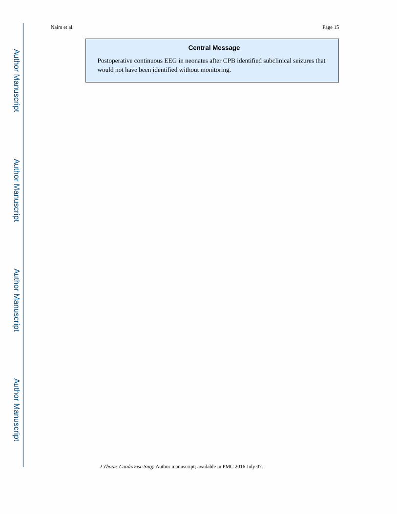
Central Message
Postoperative continuous EEG in neonates after CPB identified subclinical seizures that
would not have been identified without monitoring.
Naim et al. Page 15
J Thorac Cardiovasc Surg. Author manuscript; available in PMC 2016 July 07.
Author M
anuscriptA
uthor Manuscript
Author M
anuscriptA
uthor Manuscript
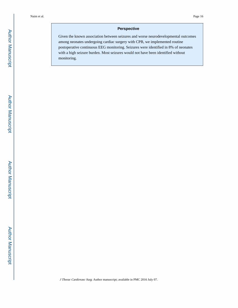
Perspective
Given the known association between seizures and worse neurodevelopmental outcomes
among neonates undergoing cardiac surgery with CPB, we implemented routine
postoperative continuous EEG monitoring. Seizures were identified in 8% of neonates
with a high seizure burden. Most seizures would not have been identified without
monitoring.
Naim et al. Page 16
J Thorac Cardiovasc Surg. Author manuscript; available in PMC 2016 July 07.
Author M
anuscriptA
uthor Manuscript
Author M
anuscriptA
uthor Manuscript
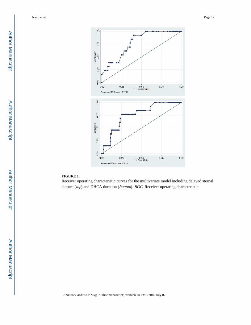
FIGURE 1. Receiver operating characteristic curves for the multivariate model including delayed sternal
closure (top) and DHCA duration (bottom). ROC, Receiver operating characteristic.
Naim et al. Page 17
J Thorac Cardiovasc Surg. Author manuscript; available in PMC 2016 July 07.
Author M
anuscriptA
uthor Manuscript
Author M
anuscriptA
uthor Manuscript
Author M
anuscriptA
uthor Manuscript
Author M
anuscriptA
uthor Manuscript
Naim et al. Page 18
TABLE 1
Demographic and clinical characteristics
Male, n (%) 92 (57%)
Birth weight (kg), median (IQR) 3.2 (2.8–3.6)
Head circumference (cm), median (IQR) 34 (32–35)
Gestational age (wk), median (IQR) 39 (38, 39)
Premature neonates (<37 wk gestational age), n (%) 26 (16%)
Identified genetic defects, n (%) 21 (13%)
Age at surgery (d) median (IQR) 5 (3–7)
Cardiac defect, n (%)
Class I 43 (27%)
Class II 15 (9%)
Class III 35 (22%)
Class IV 68 (42%)
Duration of CPB (min), median (IQR) 46 (38–62)
DHCA used, n (%) 96 (60%)
DHCA duration (min), median (IQR) 41 (32–50)
Delayed sternal closure, n (%) 26 (16%)
ECMO used, n (%) 11 (7%)
Cardiac arrest, n (%) 15 (9%)
Number (%) and median (IQR) are reported as appropriate. IQR, Interquartile range; CPB, cardiopulmonary bypass; DHCA, deep hypothermic circulatory arrest; ECMO, extracorporeal membrane oxygenation.
J Thorac Cardiovasc Surg. Author manuscript; available in PMC 2016 July 07.
Author M
anuscriptA
uthor Manuscript
Author M
anuscriptA
uthor Manuscript
Naim et al. Page 19
TAB
LE
2
Seiz
ure
desc
ript
ion,
trea
tmen
t, an
d br
ain
imag
ing
Subj
ect
Stat
us e
pile
ptic
us
Any
sei
zure
s w
ith
clin
ical
co
rrel
ates
Seiz
ure
desc
ript
ion
Ant
isei
zure
med
icat
ions
adm
inis
tere
dIm
agin
g
1Y
esY
es25
foc
al R
cen
tral
sei
zure
s la
stin
g 0.
5–3.
5 m
inPB
Bra
in M
RI
Bila
tera
l dee
p an
d pe
rive
ntri
cula
r fo
ci o
f PV
L
2Y
esN
o29
bi-
occi
pita
l sei
zure
s la
stin
g 2–
30 m
inPB
Bra
in M
RI
Bila
tera
l dee
p an
d pe
rive
ntri
cula
r fo
ci o
f he
mor
rhag
ic
PVL
3Y
esN
o>
100
bi-c
entr
al a
nd v
erte
x se
izur
es la
stin
g 0.
5–3
min
LE
V, P
BH
ead
US
Bila
tera
l IV
H, p
eriv
entr
icul
ar h
emor
rhag
e vs
hem
orrh
agic
in
farc
t
4N
oN
o1
R c
entr
al a
nd 1
L o
ccip
ital s
eizu
re la
stin
g 1
min
Non
eB
rain
MR
ISm
all S
DH
alo
ng f
lax
and
tent
oriu
m, s
mal
l in
trap
aren
chym
al h
emor
rhag
e in
R c
ereb
ella
r he
mis
pher
e an
d R
pos
teri
or p
arie
tal l
obe
5N
oN
o5
L c
entr
al s
eizu
res
last
ing
0.25
–1 m
inPB
Bra
in M
RI
L p
ost f
ront
al a
nd a
nter
ior
pari
etal
infa
rctio
n, R
fro
ntal
lo
be in
farc
tion
6Y
esN
o13
R c
entr
al s
eizu
res
last
ing
1–3
min
PB, L
EV
Hea
d U
S2
larg
e in
trap
aren
chym
al le
sion
s in
L h
emis
pher
e su
gges
tive
of I
CH
7N
oN
o9
L o
ccip
ital 1
-sei
zure
s la
stin
g 5
min
PBB
rain
MR
IC
ystic
PV
L c
entr
al s
emio
vale
and
cor
ona
radi
ate
bila
tera
lly
8Y
esN
o5
bi-o
ccip
ital s
eizu
res
last
ing
2–13
min
PBB
rain
MR
IPV
L L
par
ieta
l whi
te m
atte
r re
gion
, L te
ntor
ial S
DH
, bi
late
ral c
ereb
ral m
icro
hem
orrh
ages
9Y
esN
o34
L o
ccip
ital s
eizu
res
last
ing
2–4
min
LE
VB
rain
MR
IPV
L c
orpu
s ca
llosu
m, p
eriv
entr
icul
ar d
eep
whi
te m
atte
r, m
icro
hem
orrh
ages
R f
ront
al a
nd p
arie
tal d
eep
whi
te
mat
ter,
R la
tera
l med
ulla
and
cer
ebel
lar
pedu
ncle
, SD
H
post
erio
r fl
ax a
nd te
ntor
ium
10Y
esN
o>
100
R f
ront
al-c
entr
al-t
empo
ral t
empo
ral
seiz
ures
last
ing
0.25
–1.5
min
PB, L
EV
Bra
in M
RI
Suba
cute
infa
rctio
ns in
the
R f
ront
al lo
be a
nd L
fro
ntal
lo
be
11N
oN
o3
L o
ccip
ital s
eizu
res
last
ing
1–20
min
PB, L
EV
Hea
d U
SIn
crea
sed
echo
geni
city
bila
tera
l tha
lam
i, bi
late
ral S
DH
12Y
esY
es~1
0 se
izur
es p
er h
our
for
3 d
from
mul
tifoc
al
loca
tions
last
ing
0.5–
5 m
inPB
, LE
VH
ead
US
Lar
ge a
rea
of in
crea
sed
pare
nchy
mal
ech
ogen
icity
in th
e L
par
ieta
l-oc
cipi
tal r
egio
n, c
ompa
tible
with
hem
orrh
agic
in
farc
tion,
IV
H le
ft g
reat
er th
an r
ight
13N
oN
o8
diff
use
seiz
ures
last
ing
0.1–
2 m
inPB
Hea
d U
S
J Thorac Cardiovasc Surg. Author manuscript; available in PMC 2016 July 07.
Author M
anuscriptA
uthor Manuscript
Author M
anuscriptA
uthor Manuscript
Naim et al. Page 20
Subj
ect
Stat
us e
pile
ptic
us
Any
sei
zure
s w
ith
clin
ical
co
rrel
ates
Seiz
ure
desc
ript
ion
Ant
isei
zure
med
icat
ions
adm
inis
tere
dIm
agin
g
Evo
lvin
g le
ft c
audo
thal
amic
gro
ove
germ
inal
mat
rix
hem
orrh
age
with
intr
aven
tric
ular
syn
echi
a
PB, P
heno
barb
ital;
MR
I, m
agne
tic r
eson
ance
imag
ing;
PV
L, p
eriv
entr
icul
ar le
ukom
alac
ia; L
EV
, lev
etir
acet
am; U
S, u
ltras
ound
; IV
H, i
ntra
vent
ricu
lar
hem
orrh
age;
SD
H, s
ubdu
ral h
emat
oma;
R, r
ight
; IC
H,
intr
acra
nial
hem
orrh
age.
J Thorac Cardiovasc Surg. Author manuscript; available in PMC 2016 July 07.
Author M
anuscriptA
uthor Manuscript
Author M
anuscriptA
uthor Manuscript
Naim et al. Page 21
TAB
LE
3
Ele
ctro
grap
hic
seiz
ure
pred
icto
rs
Var
iabl
e
Uni
vari
ate
anal
ysis
Mul
tiva
riat
e an
alys
is n
o. 1
*M
ulti
vari
ate
anal
ysis
no.
2*
No
seiz
ures
Seiz
ures
P v
alue
OR
(95
% C
I)P
val
ueO
R (
95%
CI)
P v
alue
Gen
der
.80
M
ale
85 (
92%
)7
(8%
)
Fe
mal
e63
(91
%)
6 (9
%)
Ges
tatio
nal a
ge (
wk)
39 (
38–3
9)37
(37
–39)
.42
Iden
tifie
d ge
netic
abn
orm
ality
.55
N
one
128
(91%
)12
(9%
)
Pr
esen
t20
(95
%)
1 (5
%)
Age
at s
urge
ry (
d)5
(3–7
)3
(2–5
).0
50.
91 (
0.74
–1.1
1).3
60.
93 (
0.77
–1.2
8).4
7
Car
diac
def
ect
.14
C
lass
I65
(96
%)
3 (4
%)
0.46
(0.
10–2
.18)
.33
1.09
(0.
18–6
.67)
.93
C
lass
II
33 (
94%
)2
(6%
)0.
49 (
0.08
–2.8
7).4
30.
41 (
0.07
–2.4
3).3
3
C
lass
III
14 (
93%
)1
(7%
)0.
76 (
0.07
–7.8
3).8
22.
28 (
0.14
–35.
06)
.56
C
lass
IV
36 (
84%
)7
(16%
)—
—
Ope
ratio
n.4
2
St
age
1 N
orw
ood
Ope
ratio
n37
(86
%)
6 (1
4%)
A
rter
ial s
witc
h op
erat
ion
24 (
96%
)1
(4%
)
Sy
stem
ic to
pul
mon
ary
arte
ry s
hunt
16 (
94%
)1
(6%
)
C
ompl
ete
repa
ir o
f te
tral
ogy
of F
allo
t14
(10
0%)
0 (0
%)
T
runc
us a
rter
iosu
s re
pair
11 (
92%
)1
(8%
)
Del
ayed
ste
rnal
clo
sure
.002
3.99
(1.
04–1
5.29
).0
4*
*
N
o12
8 (9
5%)
7 (5
%)
Y
es20
(77
%)
6 (2
3%)
Dur
atio
n of
DH
CA
(m
in)
21 (
0–42
)47
(36
–49)
.01
**
1.04
(1.
00–1
.08)
.04
Dur
atio
n of
CPB
(m
in)
45 (
38–6
0)62
(42
–77)
.24
EC
MO
*.0
15†
N
o14
0 (9
3%)
10 (
7%)
Y
es9
(73%
)3
(27%
)
J Thorac Cardiovasc Surg. Author manuscript; available in PMC 2016 July 07.
Author M
anuscriptA
uthor Manuscript
Author M
anuscriptA
uthor Manuscript
Naim et al. Page 22
Var
iabl
e
Uni
vari
ate
anal
ysis
Mul
tiva
riat
e an
alys
is n
o. 1
*M
ulti
vari
ate
anal
ysis
no.
2*
No
seiz
ures
Seiz
ures
P v
alue
OR
(95
% C
I)P
val
ueO
R (
95%
CI)
P v
alue
Car
diac
arr
est*
.006
†
N
o13
7 (9
4%)
9 (6
%)
Y
es11
(73
%)
4 (2
7%)
Num
ber
(%)
and
med
ian
(IQ
R)
are
repo
rted
as
appr
opri
ate.
Bol
dfac
e in
dica
tes
stat
istic
al s
igni
fica
nce.
OR
, Odd
s ra
tio; C
I, co
nfid
ence
inte
rval
; DH
CA
, dee
p hy
poth
erm
ic c
ircu
lato
ry a
rres
t; C
PB,
card
iopu
lmon
ary
bypa
ss; E
CM
O, e
xtra
corp
orea
l mem
bran
e ox
ygen
atio
n.
* Del
ayed
ste
rnal
clo
sure
and
DH
CA
dur
atio
n w
ere
high
ly c
orre
late
d, s
o m
ultiv
aria
te a
naly
sis
incl
uded
onl
y de
laye
d st
erna
l clo
sure
(m
ultiv
aria
ble
anal
ysis
no.
1)
or D
HC
A d
urat
ion
(mul
tivar
iabl
e an
alys
is
no. 2
).
† EC
MO
and
car
diac
arr
est w
ere
not i
nclu
ded
in th
e m
ultiv
aria
ble
anal
ysis
bec
ause
thes
e va
riab
les
wou
ld n
ot b
e kn
own
at th
e tim
e of
ret
urn
to th
e C
ICU
and
ther
efor
e co
uld
not b
e us
ed to
hel
p de
cide
w
heth
er E
EG
mon
itori
ng w
as in
dica
ted.
J Thorac Cardiovasc Surg. Author manuscript; available in PMC 2016 July 07.