Embed Size (px)
Citation preview

Study No.: RADIC200318-01
Assessment of Radic8 VK102 device to reduce
airborne pathogens: Testing with Cystovirus Phi6 as the challenge
Page 1 of 12
STUDY TITLE
Assessment of Radic8 VK102 device to reduce airborne pathogens: Testing with Cystovirus
Phi6 (ATCC 21781-B1) as the challenge
TEST ORGANISM
Cystovirus Phi6 (ATCC 21781-B1):
Host: Pseudomonas syringae (ATCC 19310).
TEST PRODUCT IDENTITY
Radic8 VK102
TEST Method
Air Decontamination Protocol based on U.S. EPA Guidelines OCSPP 810.2500 for Efficacy Test
Recommendations on Air Sanitizers
AUTHOR
Bahram Zargar, PhD
STUDY COMPLETION DATE
April/19/20
PERFORMING LABORATORY CREM Co. Labs. Units 1-2, 3403 American Dr., Mississauga, Ontario, Canada L4V 1T4
SPONSOR
Radic8
STUDY NUMBER
RADIC200318-01
STUDY PERSONNEL

Study No.: RADIC200318-01
Assessment of Radic8 VK102 device to reduce
airborne pathogens: Testing with Cystovirus Phi6 as the challenge
Page 2 of 12
STUDY DIRECTOR: Bahram. Zargar, PhD
PROFESSIONAL PERSONNEL INVOLVED: Sepideh Khoshnevis, MSc
STUDY REPORT
Study No.: RADIC200318-01
Assessment of Radic8 VK102 device to reduce
airborne pathogens: Testing with Cystovirus Phi6 as the challenge
Page 3 of 12
GENERAL STUDY INFORMATION Study Title: Assessment of Radic8 VK102 to reduce airborne pathogens:
Testing with Cystovirus Phi6 (ATCC 21781-B1) as the challenge Study Number: RADIC200318-01 Sponsor RADIC8
Testing Facility CREM Co Labs Units 1-2, 3403 American Drive, Mississauga, ON, Canada
TEST SUBSTANCE IDENTITY
Test Substance Name: RADIC8 Device STUDY DATES Date Device Received: Study initiation date: March/03/18 Experimental Start Date: March/03/20 Experimental End Date: April/18/20 Study Completion Date: April/19/20
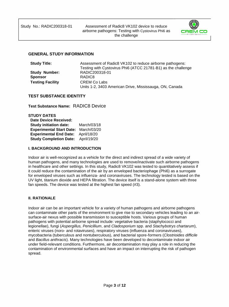
I. BACKGROUND AND INTRODUCTION Indoor air is well-recognized as a vehicle for the direct and indirect spread of a wide variety of human pathogens, and many technologies are used to remove/inactivate such airborne pathogens in healthcare and other settings. In this study, Radic8 VK102 was tested to quantitatively assess if it could reduce the contamination of the air by an enveloped bacteriophage (Phi6) as a surrogate for enveloped viruses such as influenza- and coronaviruses. The technology tested is based on the UV light, titanium dioxide and HEPA filtration. The device itself is a stand-alone system with three fan speeds. The device was tested at the highest fan speed (#3).
II. RATIONALE
Indoor air can be an important vehicle for a variety of human pathogens and airborne pathogens can contaminate other parts of the environment to give rise to secondary vehicles leading to an air-surface-air nexus with possible transmission to susceptible hosts. Various groups of human pathogens with potential airborne spread include: vegetative bacteria (staphylococci and legionellae), fungi (Aspergillus, Penicillium, and Cladosporium spp. and Stachybotrys chartarum), enteric viruses (noro- and rotaviruses), respiratory viruses (influenza and coronaviruses), mycobacteria (tuberculous and nontuberculous), and bacterial spore-formers (Clostrioides difficile and Bacillus anthracis). Many technologies have been developed to decontaminate indoor air under field-relevant conditions. Furthermore, air decontamination may play a role in reducing the contamination of environmental surfaces and have an impact on interrupting the risk of pathogen spread.
Study No.: RADIC200318-01
Assessment of Radic8 VK102 device to reduce
airborne pathogens: Testing with Cystovirus Phi6 as the challenge
Page 4 of 12
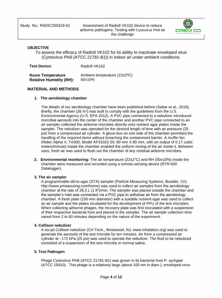
OBJECTIVE
To assess the efficacy of Radic8 VK102 for its ability to inactivate enveloped virus (Cystovirus Phi6 (ATCC 21781-B1)) in indoor air under ambient conditions.
Test Device:
Radic8 VK102
Room Temperature Relative Humidity (RH):
Ambient temperature (22±2ºC) 50±10%
MATERIAL AND METHODS
1. The aerobiology chamber The details of our aerobiology chamber have been published before (Sattar et al., 2016). Briefly, the chamber (26 m3) was built to comply with the guidelines from the U.S. Environmental Agency (U.S. EPA 2012). A PVC pipe connected to a nebulizer introduced microbial aerosols into the center of the chamber and another PVC pipe connected to an air sampler collected the airborne microbes directly onto nutrient agar plates inside the sampler. The nebulizer was operated for the desired length of time with air pressure (25 psi) from a compressed air cylinder. A glove-box on one side of the chamber permitted the handling of the required items without breaching the containment barrier. A muffin fan (Nidec Alpha V, TA300, Model AF31022-20; 80 mm X 80 mm, with an output of 0.17 cubic meters/minute) inside the chamber enabled the uniform mixing of the air inside it. Between uses, fresh air was used to flush out the chamber of any residual airborne microbes.
2. Environmental monitoring: The air temperature (22±2°C) and RH (50±10%) inside the chamber were measured and recorded using a remote-sensing device (RTR-500 Datalogger).
3. The air sampler A programmable slit-to-agar (STA) sampler (Particle Measuring Systems, Boulder, CO; http://www.pmeasuring.com/home) was used to collect air samples from the aerobiology chamber at the rate of 28.3 L (1 ft3)/min. The sampler was placed outside the chamber and the sampler’s inlet was connected via a PVC pipe to withdraw air from the aerobiology chamber. A fresh plate (150 mm diameter) with a suitable nutrient agar was used to collect an air sample and the plates incubated for the development of PFU of the test microbes. When collecting airborne phages, the recovery plate was first inoculated with a suspension of their respective bacterial host and placed in the sampler. The air sample collection time varied from 2 to 60 minutes depending on the nature of the experiment.
4. Collison nebulizer A six-jet Collison nebulizer (CH Tech., Westwood, NJ; www.inhalation.org) was used to generate the aerosols of the test microbe for ten minutes. Air from a compressed air cylinder at ~172 kPa (25 psi) was used to operate the nebulizer. The fluid to be nebulized consisted of a suspension of the test microbe in normal saline.
5. Test Pathogen Phage Cystovirus Phi6 (ATCC 21781-B1) was grown in its bacterial host P. syringae (ATCC 19310). This phage is a relatively large (about 100 nm in diam.), enveloped virus

Study No.: RADIC200318-01
Assessment of Radic8 VK102 device to reduce
airborne pathogens: Testing with Cystovirus Phi6 as the challenge
Page 5 of 12
that is frequently used as a surrogate for human pathogenic viruses. This virus was a gift from the Laval University, Laval, Quebec, Canada.
6. Test Medium
The vegetative microbial growth and recovery media in this study were Luria Broth (LB) and Luria Broth Agar (LBA).
7. Preparation of Test Pathogen Suspension To prepare a broth culture of P. syringae, a loopful of the stock culture was streaked on a LB agar and was incubated for 18±2 h at 28±1°C. A colony was inoculated in 25 mL of LB broth and incubated in at 28±1°C. When the optical density (OD) reached around 0.7, the bacterial suspension was used for the test.
8. Preparation of Phage Inocula for aerosolization The test phage suspended in saline and nebulized into the aerobiology chamber (Sattar et al., 2016) using a six-jet Collison nebulizer.
TEST METHOD
1. Experimental setup Flowchart 1 provides the sequence of steps in a typical experiment for testing the air-decontamination device. As control, the study included testing the natural decay of the test organism over time while the fan of the device was on without turning on the device. Table 1 and Table 2 list the times at which the air samples from the chamber were collected and the duration of sampling for each in control and efficacy test, respectively.

Study No.: RADIC200318-01
Assessment of Radic8 VK102 device to reduce
airborne pathogens: Testing with Cystovirus Phi6 as the challenge
Page 6 of 12
Flowchart 1. Sequence of steps in a typical experiment.
Air decontamination
Turn on muffin fan in aerobiology chamber at least 10 min before testing and placing 10-150 mm plates on the floors in five different locations
Nebulize test microorganism for 10 minutes
Allow 5 minutes for uniform distribution of aerosols
Collect a two-minute air sample
Turning on the device (in control test no action is required)
Collect an air sample based on Tables 1 and Table 2
Count PFU on plates after 24 hour of incubation
Calculate reduction in the level of viable microbes in air and surface
Table 1: Time interval of air sampling for control test
Sampling point (min) Sampling duration
(min)
0 (Baseline) 2 15 2 30 6 45 10 60 20
70-100 30 100-160 60
160-220 60

Study No.: RADIC200318-01
Assessment of Radic8 VK102 device to reduce
airborne pathogens: Testing with Cystovirus Phi6 as the challenge
Page 7 of 12
Table 2: Time interval of air sampling for efficacy test
Sampling point (min) Sampling duration (min)
0 (Baseline) 2
15 (0-30) 30
45 (30-60) 30
75 (60-90) 30
115 (90-120) 30
In efficacy, all plates were divided to the sections with 3.75 min sampling period and the PFU in each area was counted and used for calculating the concentration of the bacteriophage in the chamber at the median of that interval.
Experimental Design
Two control tests were performed, with the device OFF, and the muffin fan ON. 150 mm plates with agar and host bacteria were placed in in the STA machine to sample the air. Two multi-challenge efficacy tests were performed. In efficacy test after sampling the baseline, the device turned ON and kept ON until the end of the test. STUDY ACCEPTANCE CRITERIA No product acceptance criterion was specified for this range-finding study.
RESULTS Testing phage survival: Any meaningful assessment of air decontamination requires that the aerosolized challenge microorganisms remain viable in the experimentally-contaminated air long enough to allow for proper differentiation between biological decay and inactivation/removal by the technology being tested. Such airborne viability of the microorganism used in this study was tested in the aerobiology chamber with two control tests without turning on the device while muffin fan was ON. The average of the two control tests was used to calculate the efficacy of Radic8 VK102. Efficacy test of the Radic8 VK102 against Cystovirus Phi6: This part of the report represents data from the efficacy experiments on the Radic8 VK102 against Phi6 at RH 50±10%. The raw data are tabulated in Appendix A. Figure 1 shows the average log10 PFU/m3 recoveries for the two control tests (biological decay) with the corresponding standard deviation at each sampling interval. The concentration of Phage becomes undetectable after 2 hours.
Study No.: RADIC200318-01
Assessment of Radic8 VK102 device to reduce
airborne pathogens: Testing with Cystovirus Phi6 as the challenge
Page 8 of 12
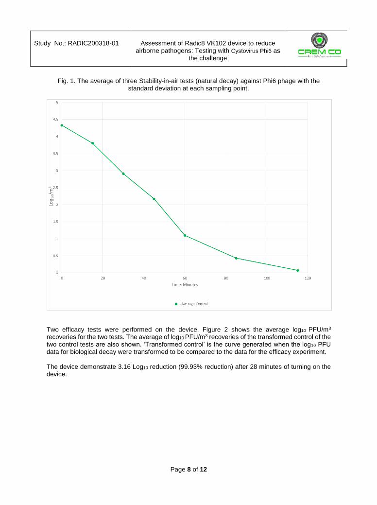
Fig. 1. The average of three Stability-in-air tests (natural decay) against Phi6 phage with the standard deviation at each sampling point.
Two efficacy tests were performed on the device. Figure 2 shows the average log10 PFU/m3 recoveries for the two tests. The average of log10 PFU/m3 recoveries of the transformed control of the two control tests are also shown. ‘Transformed control’ is the curve generated when the log10 PFU data for biological decay were transformed to be compared to the data for the efficacy experiment. The device demonstrate 3.16 Log10 reduction (99.93% reduction) after 28 minutes of turning on the device.

Study No.: RADIC200318-01
Assessment of Radic8 VK102 device to reduce
airborne pathogens: Testing with Cystovirus Phi6 as the challenge
Page 9 of 12
Fig 2. Efficacy of Radic8 VK102 in reducing microbial contamination of air. The average of two
control and two efficacy tests. Reductions were calculated using the % recovery formula for the
determination of the biological decay with log10 and % reductions at each time point for Phi6.
3.1
6 L
og
10

Study No.: RADIC200318-01
Assessment of Radic8 VK102 device to reduce
airborne pathogens: Testing with Cystovirus Phi6 as the challenge
Page 10 of 12
Appendix A:
Table 4. Natural decay of bacteriophage Phi6 without soil load, Reductions were calculated using the % recovery formula for the determination of the biological decay with log10 and % reductions at each time point for Phi6.
Sampling Time Points (minutes)
Sampling Time Points (minutes)
0 15 30 45 60 85 115
Sampling Period (minutes)
2 2 6 10 20 30 60
To
tal C
olo
ny in
the
ro
om
PF
U
Control#1
21431 6357 716 89 5 2 1
Control #2
32067 8819 1727 327 53 2 1
Reco
ve
red
on
Pla
tes
PF
U
Control #1
1213 359 121 25 3 2 1
Control #2
1815 498 292 92 30 2 1
log
10 r
ed
uctio
n**
log
10
Control #1
4.33 3.80 2.85 1.95 0.73 0.38 0.077
Control #2
4.51 3.94
992 3.24 2.52 1.73 0.38 0.077

Study No.: RADIC200318-01
Assessment of Radic8 VK102 device to reduce
airborne pathogens: Testing with Cystovirus Phi6 as the challenge
Page 11 of 12
Table 5. Efficacy of Radic8 VK102 in reducing microbial contamination of air. Reductions were calculated using the % recovery formula for the determination of the biological decay with log10 and % reductions at each time point for Phi6.
Radic8 VK102 Sampling Time Points (minutes)
Sampling Time Points (minutes)
0 5.625 13.125 16.875 20.625 24.375 28.125 31.875
Sampling Period (minutes)
2 3.75 3.75 3.75 3.75 3.75 3.75 3.75
To
tal P
FU
in
the
ro
om
PF
U
Test #1 32650 671 549 218 10 0 0 0
Test #2 35830 689 341 161 67 38 0 0
Reco
ve
red
on
Pla
tes
PF
U Test #1 1848 71 58 23 1 0 0 0
Test #2 2028 73 36 17 7 4 0 0
log
10 r
ed
uctio
n**
log
10
Test #1 4.55 2.84 2.53 2.21 1.82 1.58 0 0
Test #2 4.51 2.83 2.74 2.34 0.98 0 0 0

Study No.: RADIC200318-01
Assessment of Radic8 VK102 device to reduce
airborne pathogens: Testing with Cystovirus Phi6 as the challenge
Page 12 of 12
References Environ. Protection Agency (Dec. 2012). Air Sanitizers – Efficacy Data Recommendations. OCSPP
810.2500. Sattar, S.A., Kibbee, R.J., Zargar, Z., Wright, K.E., Rubino, J.R., Khalid, M.K. (2016).
Decontamination of indoor air to reduce the risk of airborne infections: Studies on survival and inactivation of airborne pathogens using an aerobiology chamber. Am. J. Infect. Control. 44: e177-e182.
The use of the CREM Co. Labs’ name, logo or any other representation of CREM Co. Labs without the written approval of CREM Co., Inc. is prohibited. In addition, CREM Co Labs may not be referred to any form of promotional materials, press release, advertising or similar materials (whether by print, broadcast, communication or electronic means) without the expressed written permission of CREM Co., Inc.