Embed Size (px)
Citation preview
European Randomized Study of Screening
for Prostate Cancer (ERSPC)
Study Book
Originally designed for the Rotterdam section
1st version: July 5, 1994
2nd version: August 25, 1994
3rd version: January 2, 1997
4th version: September 10, 1998
TABLE OF CONTENTS
1.0 INTRODUCTION
2.0 PURPOSE OF THE STUDY AND ENDPOINTS
2.1 Purpose
2.2 Endpoints
2.3 Tumor classification
3.0 STATISTICAL CONSIDERATIONS
3.1 Determination of population and sample size for the age group 55-74
3.1.1 Prostate cancer mortality in the study populations
3.1.2 Sample size calculations with screening age 55-74
3.2 Determination of population and sample size for the age group 55-70
4.0 DEFINITIONS
4.1 Age groups
4.2 Prostate cancer
4.3 Prostate cancer death
4.4 Stopping/flagging rules
4.5 Re-screening intervals
5.0 OVERVIEW OF STRUCTURES AND PROCEDURES
5.1 Study team
5.1.1 Administrative Unit
5.1.2 Research coordinators
5.2 Randomization, screening and evaluation procedures
5.2.1 Current algorithm of the screening procedure
5.2.2 Current assumptions made for the purpose of this plan of investigation
5.2.3 Procedures and timing
5.3 Recruitment
5.4 Informed consent
5.5 Screening tests
5.5.1 PSA assay
5.5.2 Digital rectal examination (DRE)
5.5.3 Transrectal ultrasonography (TRUS)
5.6 Indication for and technique of biopsy
5.7 Treatment
5.8 Pathology evaluation
5.8.1 Processing of biopsy specimens
5.8.2 Processing of radical prostatectomy specimens
5.9 Re-screening
5.9.1 Re-screening interval
5.9.2 Re-evaluation after 1 year
5.10 Follow-up (period, causes of death)
5.11 Evaluation (endpoints)
5.12 Biological samples
5.13 Contamination of the control arm (by use of screening tests)
5.14 General practitioners and urologists
6.0 INTERNATIONAL COLLABORATION

7.0 COMMITTEE STRUCTURE
7.1 Scientific Committee
7.2 Data Monitoring Committee
7.3 Causes of Death Committee
7.4 Quality Control Committee
7.5 International Epidemiology Committee
7.6 PSA Committee
7.7 Pathology Committee
8.0 ADDENDA

1.0
1.0 INTRODUCTION
The randomized study of screening for adenocarcinoma of the prostate described in this study book and
carried out and coordinated in the region of Rotterdam, the Netherlands, is part of the "European
Randomized Study of Screening for Adenocarcinoma of the Prostate" (ERSPC). As such it is part of a
major European effort, which is made to show or to rule out whether there is benefit or harm associated
with the application of screening tests for prostate cancer to the general population. This study should
result in advice to governments around the world as to whether screening for prostate cancer should be a
part of the general provision of health care to our populations.
2.0
2.0 PURPOSE OF THE STUDY AND ENDPOINTS
2.1 Purpose of the study
The purpose of the study is:
1. To establish or to disprove an effect of active screening on prostate cancer mortality
2. To evaluate the efficiency of the screening tests: serum prostate specific antigen (PSA), digital
rectal examination (DRE) and transrectal ultrasonography (TRUS) in the early detection of prostate
cancer and to study the morbidity related to the screening procedure.
3. To predict life years gained and to calculate the costs related to different screening policies.
4. To carry out side studies related to:
a. quality of life in the two screening populations
b. prognostic factors related to the tumours detected in the screened, and if possible, also the
control population
c. to study prostate cancer morbidity and tumor extent at the time of diagnosis
d. evaluation of the socio-economic status related to prostate cancer, factors influencing
participation, process evaluation, symptom distribution in the group of refusers, the screening
and control group (GGD Rotterdam, Dr. H.G.T. Nijs).
2.2 Endpoints of the study
The major endpoint of the study is prostate cancer mortality which is to be compared between the
screened and the non-screened group of men at risk. The study result will be considered positive if a
difference in prostate cancer mortality of 20% or more is shown. In case of a negative outcome, however,
intermediate endpoints may be of great importance. As far as one can foresee at this time, such
endpoints are: progression-free (tumor-free) survival, survival free of metastatic disease, quality of life in
the screened and non-screened populations. Separate protocols will have to be worked out for the side
studies and for the determination of intermediate endpoints. As far as morbidity and progression of
disease is concerned, the criteria of response and progression to treatment of the EORTC Genitourinary
Group and of the WHO should be applied (D.W.W. Newling, Baillière's Clinical Urology, 1988; 2: 505-
520).
2.3 Tumor classification
Throughout the study the TNM system of 1992 will be applied (see addendum 22).

3.0
3.0 STATISTICAL CONSIDERATIONS
Two detailed sample size calculations are presented under 3.1 and 3.2, 'Determination of population and
sample size'. These calculations are based on the age groups 55-74 and 55-70 years at the time of entry.
With a 20% reduction in mortality at least 49,142 participants are required in both study and control group
to reach a 90% power (in total 90,284 participants). For evaluation within the European study the age
group 55-70 years (including the age of 70) is considered in 3.2. Sample size is estimated to be 130,000
(65,000 randomized to each of the study groups) to be able to show a 20% reduction in mortality and to
reach a 90% power.
3.1.1
3.1 Determination of population and sample size for the age group 55-74
3.1.1 Prostate cancer mortality in the study population
In sample size calculations one attempts to assess the minimum size of a study population with which an
expected reduction in prostate cancer mortality (due to screening) can be shown significantly. Starting
point is the prostate cancer mortality in the unscreened part of the study population (control group). Using
CBS prostate cancer mortality figures would result in an over-estimation of this mortality since diagnosed
cases of prostate cancer will not be entering the study. The exclusion of these cases will result in a
substantially lower prostate cancer mortality in the first years of the study than if based on national
registered mortality. Another difficulty that arises is that men who participate in the study may be healthier
than the average population (selection).
The prostate cancer mortality in the study population can be calculated from age-specific incidence rates,
and survival data after diagnosis. We used the 1989 incidence data from the Netherlands Cancer
Registry (NCR, 1992):
Table 1.
Prostate cancer incidence rates per 100,000 men (1989)
Age 45-49 50-54 55-59 60-64 65-69 70-74 75-79 80-84
Incidence 3.5 11.5 43.1 121.9 234.8 423.4 639.4 800.0
NCR, 1992
Survival rates are based on the Norwegian Cancer Registry (1980) and Dutch SOOZ-data (Coebergh,
1991)
Although there is still some lethality after ten years, it does not contribute much to prostate cancer
mortality at a certain age, since incidence increases rapidly.
Because of this increase the age-specific CBS incidence (5 year categories) are interpolated.
3.1.1
Table 2.
Relative one- to ten years survival after diagnosis of prostate cancer (=prostate specific mortality)
age group
survival -64 65-74 75+
1 year .887 .880 .839
2 years .789 .778 .746
3 years .708 .695 .659
4 years .642 .626 .589
5 years .586 .568 .532
6 years .538 .519 .485
7 years .498 .477 .446
8 years .462 .440 .412
9 years .431 .409 .383
10 years .404 .381 .357
Thus, in our approach of sample size calculations, the prostate cancer mortality at a certain age comes
from incidence in the previous ten years, and so for each study year the age-specific prostate cancer
mortality rates can be assessed. Since there will be no deaths from prostate cancer of men already
diagnosed before the start of the screening trial, this will result in a rather low prostate cancer mortality
rate in the first years. The prostate cancer mortality in the control group in the first year of the study
comes from newly diagnosed cases in that year, given the 1-year relative survival. The prostate cancer
mortality in the 2nd year can be calculated from newly diagnosed cases in that year and 1-year survival +
cases diagnosed in the 1st year of the study, given the 2-year survival, etc.
During the study this rate will gradually increase to the level that would have appeared if diagnosed cases
had not been excluded from the study. In the tenth year of the study the prostate cancer mortality will be
'complete' again, so we can check if it corresponds to the CBS-data. Table 3 shows that this is the case
for the 70-74 age group; within the 65-69 group, however, the calculated mortality is too high. This can be
explained by the fact that the survival figures used are based on non-recent data; also, there is an
increasing trend for incidence over time.
3.1.1
Table 3.
Calculated prostate cancer mortality rates per 100,000 men in year 10, compared to CBS 1990 (5-year
age groups).
Age 65 66 67 68 69 70 71 72 73 74
calculated 66.9 78.2 90.9 105.1 120.5 137.6 156.9 177.6 198.8 221.1
CBS 75.8 174.8
Further calculations will be based on CBS-data for two reasons: first, these data will be more reliable (see
above reasons on incidence and survival changes), and second, for sample size calculations it is safest
to assume the lowest possible mortality (conservative estimate).
The procedure of calculating consists of four steps in which we consider the age-distribution in the study
population (Table 4) and the CBS lifetables (1986-1990):
First, in the same way as before, we calculate the prostate cancer mortality when diagnosed cancers are
not excluded.
Subsequently, by comparing this to the situation with exclusion, we estimate what the effect of exclusion
is on prostate cancer mortality.
The third step is to determine the weighted average CBS prostate cancer mortality per year in the study
population, where we have to take into account that this population will get older every year and people
will die from other causes. (For the age-specific prostate cancer mortality rates we used an average over
the years 1988-1990)
Finally, the prostate cancer mortality in the study population in each year of the study can be calculated
as a percentage of this average CBS prostate cancer mortality (CBS, average over 1988-1990).
Table 4.
Age distribution of population in Rotterdam, Dordrecht, Spijkenisse and Vlaardingen
Age 50-54 55-59 60-64 65-69 70-74
Male population 37886 36828 32414 27926 21671
Source: Comprehensive Cancer Centre
Figure 1 en Table 5 display how prostate cancer mortality in the study population (with exclusion of
diagnosed cases) gradually increases from approximately 15 percent of CBS prostate cancer mortality
without exclusion in year 1, to 100 percent in the 10th year of the study.
3.1.1
Figure 1
Prostate cancer mortality in the study population as a percentage of the CBS national prostate cancer
mortality during the study.
Not electronically available
Table 5 shows the average prostate cancer mortality rates in the study population for each of the 10
study years.
Table 5.
Prostate cancer mortality in the study population (55-74) as a percentage of CBS prostate cancer
mortality, rates per 100,000 men
Year 1 2 3 4 5 6 7 8 9 10
CBS rate 60.7 71.9 83.1 94.5 106.0 117.7 134.1 150.7 167.7 185.2
Percentage 15.4 40.3 58.3 70.6 79.5 86.4 91.2 94.9 97.7 100.0
Study rate 9.4 29.0 48.5 66.7 84.2 101.6 122.3 143.0 163.9 185.2
Study numbers* 9 28 46 61 75 87 101 113 124 134
* rounded numbers of prostate cancer deaths in a population of 100,000 men 55-74 at entry
With these average mortality rates the number of prostate cancer deaths per year can be determined in a
population of 100,000 men at entry. The total number of prostate cancer deaths in this group during the
10 years of the study is 780. In other words, the average prostate cancer mortality per year in the
unscreened group will be 78.0 per 100,000 men aged 55-74 at entry.
3.1.2
3.1.2 Sample size calculations with screening age 55-74 years
For these sample size calculations the same method as in the American PLCO (prostate, lung, colorectal
and ovarium) screening trial is used [Taylor, Cancer 30, November 1972; Kramer et al., 1991]. Different
aspects like impact of screening, different proportions of study- and control group sizes, and variable
levels of compliance in both groups can be included.
Let Nc be the number of men assigned to the control group, and Ns the number in the screen group, with
Ns = fNc. The compliance value is to what extent the screen group actually receives screening (Ps), and to
what extent the control group does not receive screening (Pc).
The assumed reduction in the cumulative prostate cancer mortality during the study is (1-r)x 100%. This
reduction applies to the situation with 100% compliance: Ps = Pc = 1.
The significance level is α, i.e. the probability to coincidently show a reduction in mortality while in fact
there is no screening effect, is smaller than or equal to α.
The power is 1-ß, i.e. if screening does have a reducing effect on mortality, then the probability to show a
significant (α-level) mortality reduction in the study equals 1-ß.
The total number of prostate cancer deaths needed for a one-sided α-level significance test with power 1-
ß then is:
with θ1= r + (1-r)Pc en θ2= 1 - (1-r)Ps
The required number of participants in the control group is :( ) YR f +
D=N
c21
c
θθ2, with Y is the
duration of the study in years from the start of the study until the end of the follow-up, and Rc is the
average prostate cancer mortality rate in the control group expressed as deaths per year.
( ){( )θθ
θθθθ α
21
2
21-121
- f
Z ) f+(1 -Z f + = D
3.1.2
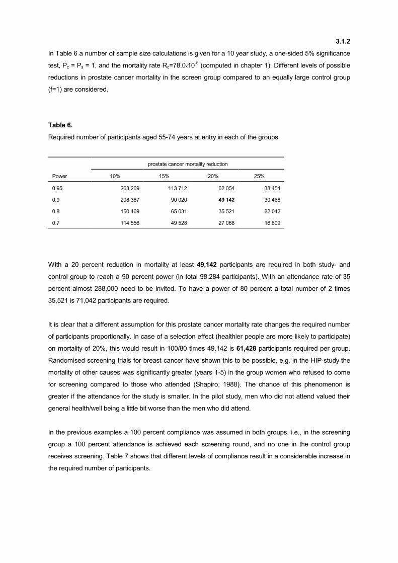
In Table 6 a number of sample size calculations is given for a 10 year study, a one-sided 5% significance
test, Pc = Ps = 1, and the mortality rate Rc=78.0x10⁻⁵ (computed in chapter 1). Different levels of possible
reductions in prostate cancer mortality in the screen group compared to an equally large control group
(f=1) are considered.
Table 6.
Required number of participants aged 55-74 years at entry in each of the groups
prostate cancer mortality reduction
Power 10% 15% 20% 25%
0.95 263 269 113 712 62 054 38 454
0.9 208 367 90 020 49 142 30 468
0.8 150 469 65 031 35 521 22 042
0.7 114 556 49 528 27 068 16 809
With a 20 percent reduction in mortality at least 49,142 participants are required in both study- and
control group to reach a 90 percent power (in total 98,284 participants). With an attendance rate of 35
percent almost 288,000 need to be invited. To have a power of 80 percent a total number of 2 times
35,521 is 71,042 participants are required.
It is clear that a different assumption for this prostate cancer mortality rate changes the required number
of participants proportionally. In case of a selection effect (healthier people are more likely to participate)
on mortality of 20%, this would result in 100/80 times 49,142 is 61,428 participants required per group.
Randomised screening trials for breast cancer have shown this to be possible, e.g. in the HIP-study the
mortality of other causes was significantly greater (years 1-5) in the group women who refused to come
for screening compared to those who attended (Shapiro, 1988). The chance of this phenomenon is
greater if the attendance for the study is smaller. In the pilot study, men who did not attend valued their
general health/well being a little bit worse than the men who did attend.
In the previous examples a 100 percent compliance was assumed in both groups, i.e., in the screening
group a 100 percent attendance is achieved each screening round, and no one in the control group
receives screening. Table 7 shows that different levels of compliance result in a considerable increase in
the required number of participants.
3.1.2
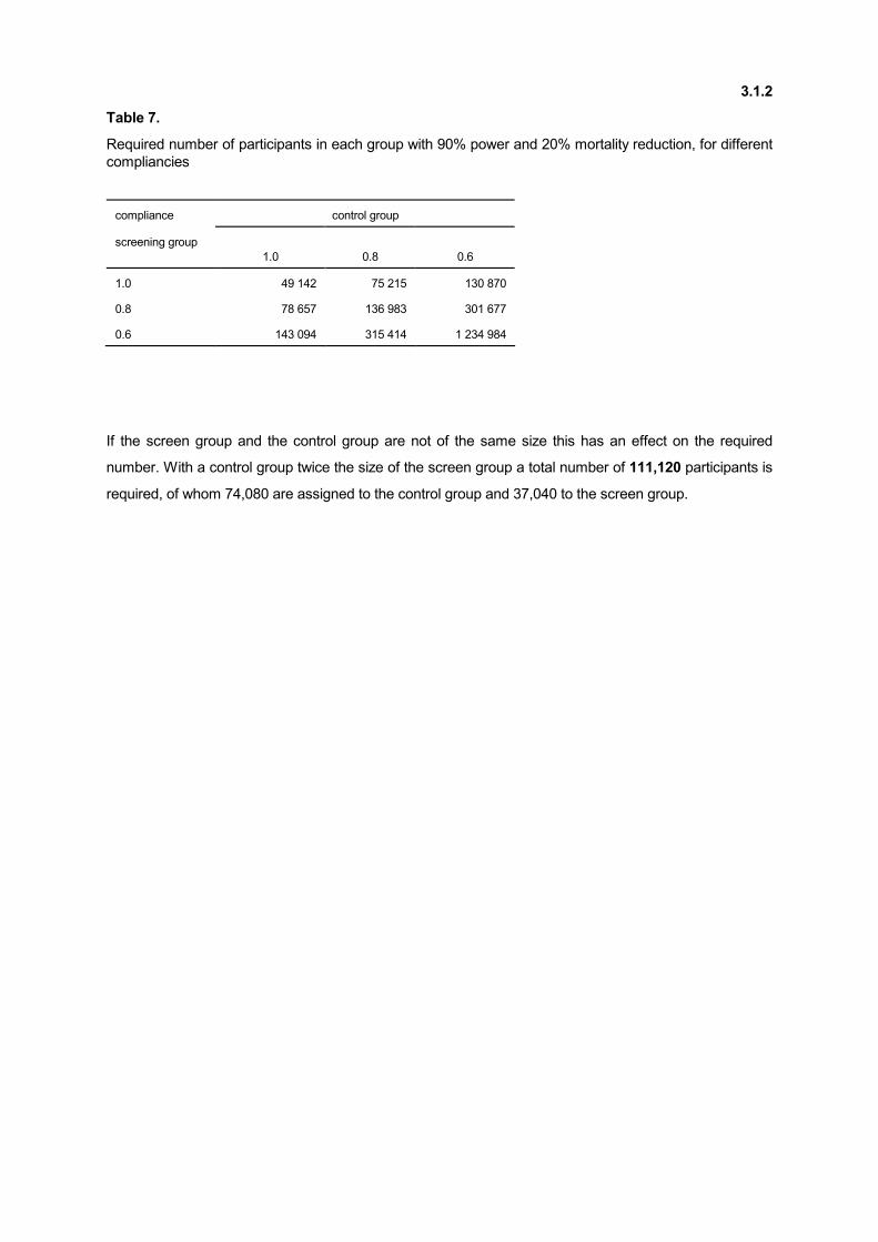
Table 7.
Required number of participants in each group with 90% power and 20% mortality reduction, for different
compliancies
compliance control group
screening group
1.0
0.8
0.6
1.0 49 142 75 215 130 870
0.8 78 657 136 983 301 677
0.6 143 094 315 414 1 234 984
If the screen group and the control group are not of the same size this has an effect on the required
number. With a control group twice the size of the screen group a total number of 111,120 participants is
required, of whom 74,080 are assigned to the control group and 37,040 to the screen group.
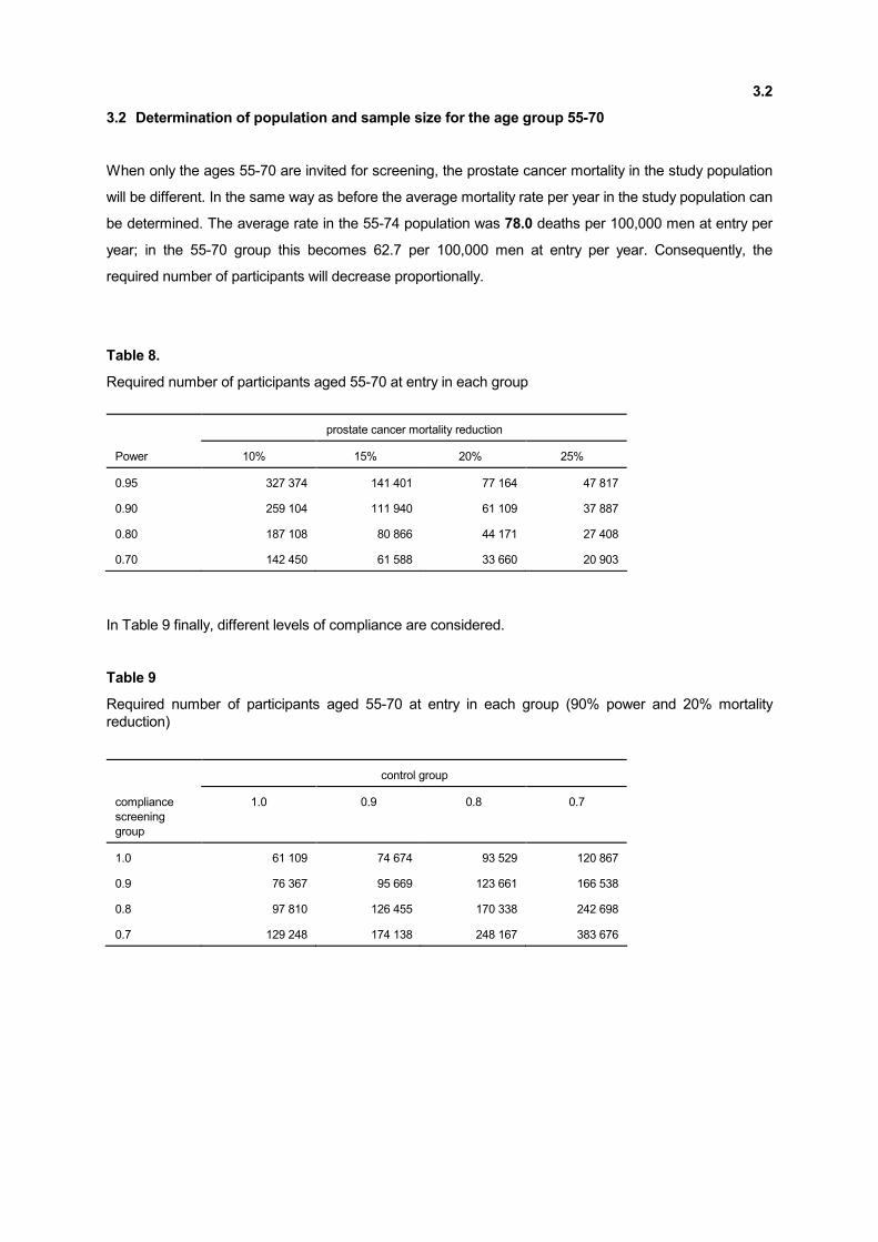
3.2
3.2 Determination of population and sample size for the age group 55-70
When only the ages 55-70 are invited for screening, the prostate cancer mortality in the study population
will be different. In the same way as before the average mortality rate per year in the study population can
be determined. The average rate in the 55-74 population was 78.0 deaths per 100,000 men at entry per
year; in the 55-70 group this becomes 62.7 per 100,000 men at entry per year. Consequently, the
required number of participants will decrease proportionally.
Table 8.
Required number of participants aged 55-70 at entry in each group
prostate cancer mortality reduction
Power 10% 15% 20% 25%
0.95 327 374 141 401 77 164 47 817
0.90 259 104 111 940 61 109 37 887
0.80 187 108 80 866 44 171 27 408
0.70 142 450 61 588 33 660 20 903
In Table 9 finally, different levels of compliance are considered.
Table 9
Required number of participants aged 55-70 at entry in each group (90% power and 20% mortality
reduction)
control group
compliance
screening
group
1.0 0.9 0.8 0.7
1.0 61 109 74 674 93 529 120 867
0.9 76 367 95 669 123 661 166 538
0.8 97 810 126 455 170 338 242 698
0.7 129 248 174 138 248 167 383 676
4.1-4.2
4.0 DEFINITIONS
4.1 Age groups
The Scientific Committee of ERSPC has determined that within the European protocol males aging 55-70
(including age 70) will be recruited. However it has been left to the individual national study groups to
recruit different age groups. Within the European protocol only males aged 55-70 years at the time of
entry into the randomized study will be included.
Considerations.
The screening study should cover those age groups in which, in clinical practice, the evaluable treatment
procedures would be considered to be applicable. With a re-screening interval of 4 years, those men who
were 70 years old at entry into the study, and are discovered to have prostate cancer at the time of
re-screening, would then be 74 years old. It is likely, that within this age group, radiotherapy will be the
preferred treatment, since there is agreement that for the application of radical prostatectomy a life
expectancy of 10-15 years should be present.
Extensive discussion has circled around the younger age groups. While the group realizes that the gain
in survival would be potentially the largest in men between 50 and 55 years of age, the relative
scarceness of prostate cancer in this age group would lead to a dramatic increase of the sample size
which was not considered acceptable.
4.2 Prostate cancer
Prostate cancer is defined as adenocarcinoma of the prostate. Other prostatic malignancies, such as
sarcomas, transitional cell carcinomas, mucinous carcinomas and metastatic malignancies to the
prostate, will be recorded but will not be registered as "prostate cancer" in this study. They will not be
included in the evaluation of prostate cancer mortality.
4.3
4.3 Prostate cancer death
A definition of prostate cancer death and procedures have been worked out by the Causes of Death
Committee, which has been established in Rotterdam and will be adapted for use within the European
study.
A preliminary definition of prostate cancer death was given in the Rotterdam protocol of the pilot studies
under 6.1.0.: "Patients will be considered to have died of prostate carcinoma if there is clinical evidence of
metastatic disease in absence of an obvious unrelated cause of death. All prostate cancer deaths will be
reviewed by the "Causes of Death committee".
Cause of death are defined as:
'death from prostate cancer'
'death from prostate cancer screening'
'death from prostate cancer treatment'
'death from unrelated disease(s)'
Subclasses will be provided:
'autopsy performed/not performed'
'metastasis present/not present/unknown'
'recurrence/residual prostate cancer present/not present/unknown'
'natural death/unnatural death'
For further explanation see forms and procedures of the Causes of Death Committee (section 7.3).
4.4
4.4 Stopping rules/flagging rules
Stopping rules c.q flagging rules have been established by the Data Monitoring Committee. Obviously,
the study will have to be discontinued if a significant difference is reached between the screening and
control arm with regard to the main endpoint of the study: prostate cancer mortality. The protocol of the
Rotterdam section foresees in an annual evaluation. The results of this evaluation will be made available
to the Data Monitoring Committee (DMC). These data will also include information on the complications
of the screening procedures and of treatment.
The stopping rules will cover the ethical aspects of the study, also those that may not be foreseeable at
the time of initiation (see addendum 23).
The information necessary for the DMC will be provided by each study center in "empty tables" to make a
uniform evaluation possible. The evaluation of the study by the DMC will be done on a local and on an
aggregated level, covering all information of the European studies together.
More information can be provided by the secretary of the DMC; Dr. H. de Koning (NL).
(See par. 7.2; Data Monitoring Committee)
4.5
4.5 Re-screening intervals
The re-screening interval within the European study has been determined to be 4 years.
Males in whom suspicion exists that a biopsy was inadequate or not representative may be re-
biopsied as this is considered to be indicated. Decision making will be formalised depending on PSA and
suspicion on DRE and/or TRUS.
The decision will be made by the urologist discussing the result of the pathological examination with the
participant. The administrative unit of the screening study can and will give an indication of the necessity
of a repeated biopsy based upon the written pathology report of the biopsies.
Also the pathologist can suggest or request a repeated biopsy, if with the available material the diagnosis
of prostate cancer cannot be made or rejected.
In case of high grade PIN a repeated biopsy is indicated if no prostate cancer is detected by the
pathologist in the biopsy specimen.
Males in the screening group who have presented with suspicious findings on rectal examination or
TRUS or who have a PSA of ≥ 4.0 ng/ml at the time of the first or second screen, and who have negative
biopsies, they were re-examined after 1 year until August 1996.
Due to a discussion with the committee of the Ministry of Health for the Population Based Screening for
Prostate cancer (Wet Bevolkingsonderzoek) no rescreen after 1 year will be offered after August 1996.
Men who were re-examined after 1 year and who have negative findings at that time will be re-screened
3 years later.
Since 1997 a PSA cut off of ≥ 3.0 ng/ml is being used. The screening procedure has been simplified,
based on an interim analysis of the screening tests. As a result the number of false positive biopsy
indications has decreased.
5.1
5.0 OVERVIEW OF STRUCTURES AND PROCEDURES
5.1 The Rotterdam study team
Recruitment and evaluation is achieved by a study team consisting of 3 data-managers, 2 doctors
assistants, 1 x-ray technologist and 2 AIO/AGIKO. This team is supervised by the principal investigator,
Prof. dr. F.H. Schröder, and his associate study coordinator, dr. W.J. Kirkels. They are assisted by two
clinical research coordinators of the Department of Urology who are in charge of the actual daily
supervision and coordination of the study. Ries Kranse (Bsc) is especially responsible for all aspects of
information technology, in collaboration with one associate of the Central Information Processing
Department (CDAI) (04. fte). Mathilde Dijk (Drs.) is mainly concerned in coordinating and organizing the
financial, legal and ethical aspects.
As of January 1996, at least temporarily, the 0.4 fte (programmer) is available for a necessary increase in
manpower of the data management/screening team. This is in agreement with the Dutch Cancer
Foundation Progress Report of December 1995 (addendum 20b).
As of January 1998, the x-ray technologist has left his position in the study team, due to the protocol
change less DRE’s were performed.
The organogram is included as the next page.
5.1.1
5.1.1 Administrative Unit (AU)
In collaboration with the hospital information department (CDAI) and the city of Rotterdam population
registry, the administrative unit coordinates all mailing activities. All resulting personal and/or telephone
contacts related to the recruitment of study participants are a task of the AU. The administrative
procedures allows an assessment of the response rate. Other parameters concerning participation,
attendance and acceptance, necessary to record the progress of the study are readibly available (see
addendum 7 and 8).
If eligible for the study, the participants are randomized to the control group or to the screen-
ing/intervention group by the data-managers. All participants are notified of the outcome of this
randomization procedure (addendum 9). The AU takes care of the initial invitations (addendum 10) and
of subsequent re-screening invitations of participants randomized to the intervention group (addendum
12). The re-screening time interval (if applicable) is determined by means of the algorithm listed in section
5.2. The AU receives the participants randomized to screening and answers initial questions.
The doctors assistant collects the blood sample for PSA analysis and assists in taking biopsies (if
necessary). Biopsy specimens are submitted to the pathology lab and the biopsy evaluation forms (that
contain the examination results) are collected by the AU.
The data-managers collect all screening results, i.e. the general questionnaire, the WHO-PSS
questionnaire, the PSA, DRE, TRUS results and biopsy results (if present). The results are entered into
the study data base by the data-managers.
The AU assists in side studies where applicable.
The AU manages the study archive that contains all written correspondence.
5.1.2
5.1.2 The Research Coordinators
- The research coordinators have a part-time task within the European Screening Study of Prostate
Cancer. It is estimated that they spend 40% of their time on this project.
- The research coordinators report to the chairman, Department of Urology, Prof.Dr. F.H. Schröder
and, in given situations, also to other staff members.
- Within the Rotterdam section of the European screening study they coordinate the communication
between the disciplines involved in the study in accordance with the organogram, given under 5.1.
- They are responsible for the development and maintenance of appropriate software together with
the Central Information Processing Department (CDAI).
- They function as the direct supervisors of the Administrative Unit (AU) and research coordination
Unit.
- They are responsible for the information content of the study data base and the related quality
control as well as backup procedures. They make sure that data are related to the study
coordinators and the participating disciplines as needed.
- They cooperate with the Data Monitoring Committee, the Quality Control Committee and the Causes
of Death Committee and respond together with the cancer registry unit in the Comprehensive
Cancer Center (IKR) and with the statistical unit in the Department of Public Health to their
requirements of interim information.
- They are involved in the statistical evaluation of the study.
- They are responsible for the coordination & organisation of financial, legal and ethical aspects of the
study.
Procedures related to the function of the administrative unit, data collection and management as well as
the feedback with the Comprehensive Cancer Center (IKR) will be described in separate chapters.
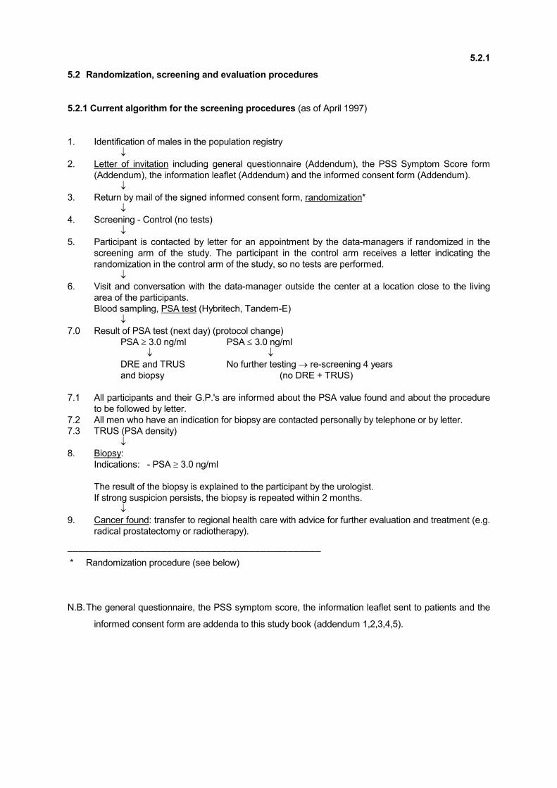
5.2.1
5.2 Randomization, screening and evaluation procedures
5.2.1 Current algorithm for the screening procedures (as of April 1997)
1. Identification of males in the population registry
↓ 2. Letter of invitation including general questionnaire (Addendum), the PSS Symptom Score form
(Addendum), the information leaflet (Addendum) and the informed consent form (Addendum).
↓ 3. Return by mail of the signed informed consent form, randomization*
↓ 4. Screening - Control (no tests)
↓ 5. Participant is contacted by letter for an appointment by the data-managers if randomized in the
screening arm of the study. The participant in the control arm receives a letter indicating the
randomization in the control arm of the study, so no tests are performed.
↓ 6. Visit and conversation with the data-manager outside the center at a location close to the living
area of the participants.
Blood sampling, PSA test (Hybritech, Tandem-E)
↓ 7.0 Result of PSA test (next day) (protocol change)
PSA ≥ 3.0 ng/ml PSA ≤ 3.0 ng/ml
↓ ↓ DRE and TRUS No further testing → re-screening 4 years
and biopsy (no DRE + TRUS)
7.1 All participants and their G.P.'s are informed about the PSA value found and about the procedure
to be followed by letter.
7.2 All men who have an indication for biopsy are contacted personally by telephone or by letter.
7.3 TRUS (PSA density)
↓ 8. Biopsy:
Indications: - PSA ≥ 3.0 ng/ml
The result of the biopsy is explained to the participant by the urologist.
If strong suspicion persists, the biopsy is repeated within 2 months.
↓ 9. Cancer found: transfer to regional health care with advice for further evaluation and treatment (e.g.
radical prostatectomy or radiotherapy).
──────────────────────────────────────────────
* Randomization procedure (see below)
N.B. The general questionnaire, the PSS symptom score, the information leaflet sent to patients and the
informed consent form are addenda to this study book (addendum 1,2,3,4,5).
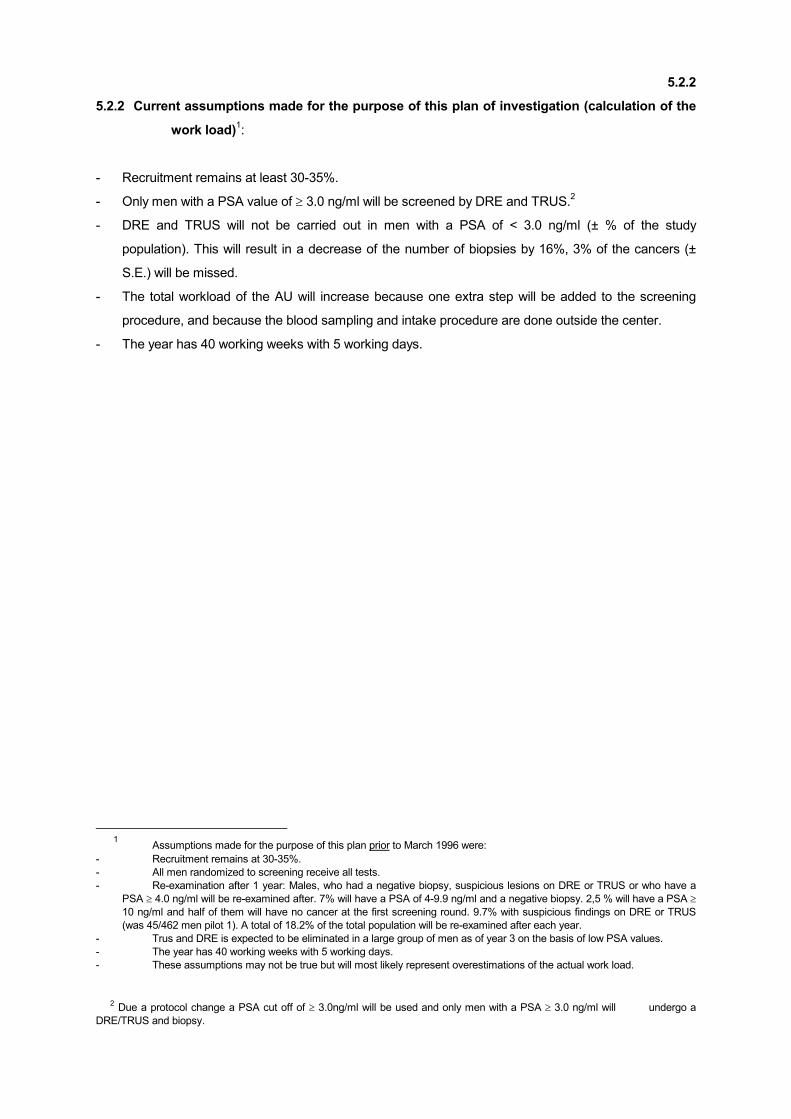
5.2.2
5.2.2 Current assumptions made for the purpose of this plan of investigation (calculation of the
work load)1:
- Recruitment remains at least 30-35%.
- Only men with a PSA value of ≥ 3.0 ng/ml will be screened by DRE and TRUS.2
- DRE and TRUS will not be carried out in men with a PSA of < 3.0 ng/ml (± % of the study
population). This will result in a decrease of the number of biopsies by 16%, 3% of the cancers (±
S.E.) will be missed.
- The total workload of the AU will increase because one extra step will be added to the screening
procedure, and because the blood sampling and intake procedure are done outside the center.
- The year has 40 working weeks with 5 working days.
1 Assumptions made for the purpose of this plan prior to March 1996 were:
- Recruitment remains at 30-35%.
- All men randomized to screening receive all tests.
- Re-examination after 1 year: Males, who had a negative biopsy, suspicious lesions on DRE or TRUS or who have a
PSA ≥ 4.0 ng/ml will be re-examined after. 7% will have a PSA of 4-9.9 ng/ml and a negative biopsy. 2,5 % will have a PSA ≥ 10 ng/ml and half of them will have no cancer at the first screening round. 9.7% with suspicious findings on DRE or TRUS
(was 45/462 men pilot 1). A total of 18.2% of the total population will be re-examined after each year.
- Trus and DRE is expected to be eliminated in a large group of men as of year 3 on the basis of low PSA values.
- The year has 40 working weeks with 5 working days.
- These assumptions may not be true but will most likely represent overestimations of the actual work load.
2 Due a protocol change a PSA cut off of ≥ 3.0ng/ml will be used and only men with a PSA ≥ 3.0 ng/ml will undergo a
DRE/TRUS and biopsy.
5.2.3
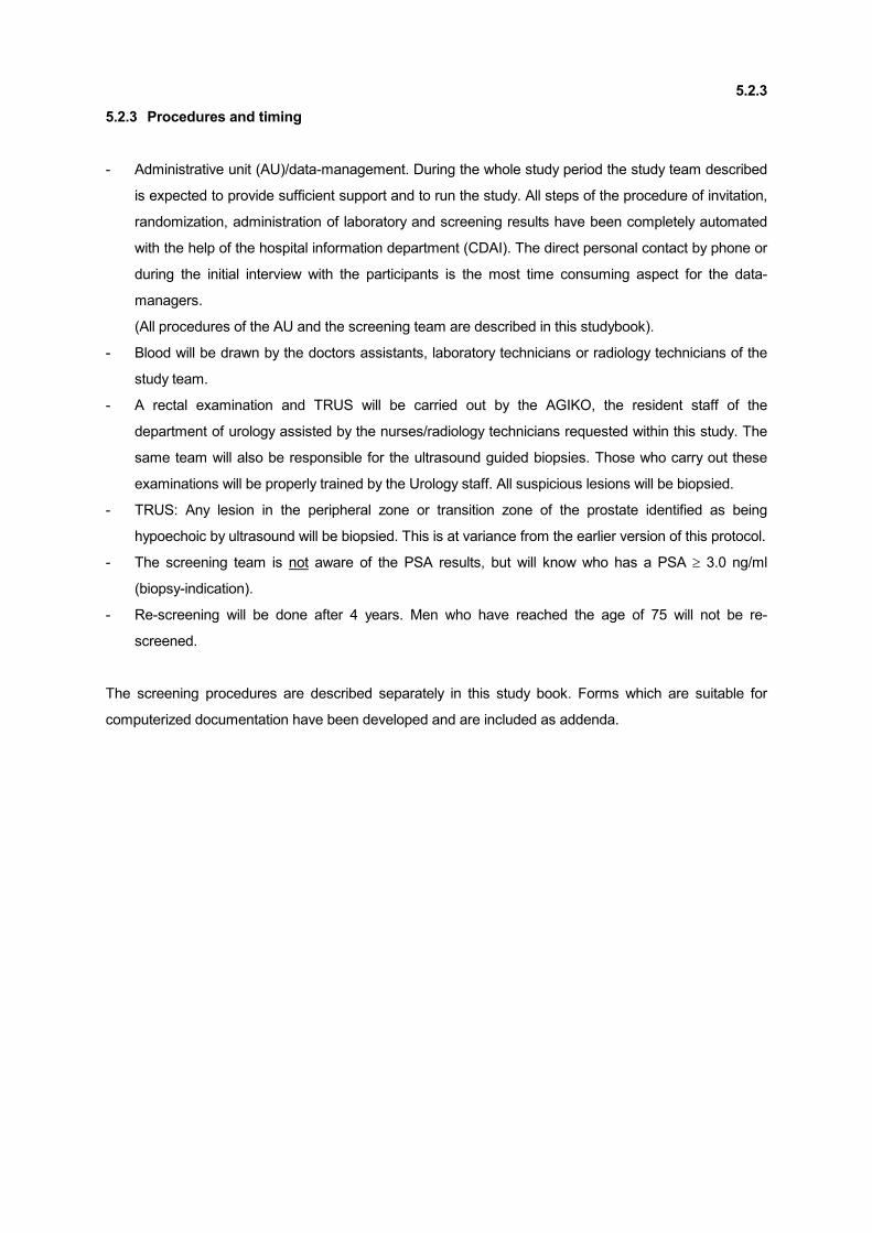
5.2.3 Procedures and timing
- Administrative unit (AU)/data-management. During the whole study period the study team described
is expected to provide sufficient support and to run the study. All steps of the procedure of invitation,
randomization, administration of laboratory and screening results have been completely automated
with the help of the hospital information department (CDAI). The direct personal contact by phone or
during the initial interview with the participants is the most time consuming aspect for the data-
managers.
(All procedures of the AU and the screening team are described in this studybook).
- Blood will be drawn by the doctors assistants, laboratory technicians or radiology technicians of the
study team.
- A rectal examination and TRUS will be carried out by the AGIKO, the resident staff of the
department of urology assisted by the nurses/radiology technicians requested within this study. The
same team will also be responsible for the ultrasound guided biopsies. Those who carry out these
examinations will be properly trained by the Urology staff. All suspicious lesions will be biopsied.
- TRUS: Any lesion in the peripheral zone or transition zone of the prostate identified as being
hypoechoic by ultrasound will be biopsied. This is at variance from the earlier version of this protocol.
- The screening team is not aware of the PSA results, but will know who has a PSA ≥ 3.0 ng/ml
(biopsy-indication).
- Re-screening will be done after 4 years. Men who have reached the age of 75 will not be re-
screened.
The screening procedures are described separately in this study book. Forms which are suitable for
computerized documentation have been developed and are included as addenda.
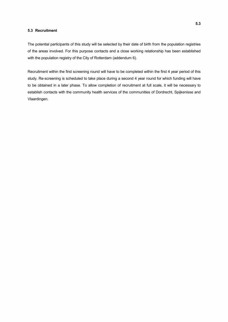
5.3
5.3 Recruitment
The potential participants of this study will be selected by their date of birth from the population registries
of the areas involved. For this purpose contacts and a close working relationship has been established
with the population registry of the City of Rotterdam (addendum 6).
Recruitment within the first screening round will have to be completed within the first 4 year period of this
study. Re-screening is scheduled to take place during a second 4 year round for which funding will have
to be obtained in a later phase. To allow completion of recruitment at full scale, it will be necessary to
establish contacts with the community health services of the communities of Dordrecht, Spijkenisse and
Vlaardingen.
5.4
5.4 Informed consent
An informed consent form has been developed and approved by the Medical Ethical Committee (MEC)
of the Academic Hospital. The revision of the form after adapting the screening procedure in March 1996
has been elaborated in cooperation with the PR department of the NKB (Drs. C. Honing). It was
approved by the MEC. Informed consent is obtained prior to randomization. The informed consent form is
mailed to the potential participants together with the general information concerning the study, the PSS
form (Prostate Symptom Score) and a general health questionnaire (see addendum 4 and 5).
The informed consent forms are included as addenda to this study book (addendum 3)
If necessary update versions will be added to the studybook, because ongoing discussions can result in
changes in the consent form.
5.5
5.5. The screening tests
The evaluation of the screening tests is one of the main purposes of this study. The study regimen is
based on the experience of the pilot studies and on a published literature review by the working group
(Bentvelsen and Schröder: Modalities available for screening for prostate cancer. EJC 1993; 29: 804-
811). Extensive argumentation is given in the original grant application to the Dutch Cancer Society
(addendum 20). The preliminary results indicate that a large proportion of negative biopsies is carried out
on the basis of false-positive TRUS and DRE studies. On the other hand, if PSA with a "normal value" of
4.0 ng/ml would be used as a parameter excluding males from biopsy if they have normal PSA values,
about 20% of cancers would be missed. Exact information on the differential role of the 3 screening tests
with regard to the detection of this considerable group of cancers, is not available. It is for this reason that
the study group has decided to proceed as follows:
- Evaluate all 3 screening tests until clear indications for their use in the group of males with a PSA <
4.0 ng/ml emerges, probably for a duration of 2 years.
- Streamline the screening tests after this information has been obtained to the most suitable
screening algorithm.
N.B. In March 1996 the screening procedure was changed as follows:
Men with a PSA value of/or less than 1.0 ng/ml (34,6% of the population) are excluded from DRE
and TRUS. This will lead to a reduction of the number of biopsies by 17,5% (S.E. 1,2).
Approximately 4% of cancers will be missed at the first screening round.
After evaluation of the screeningsprocudure, in 1997 the screening procedure has been simplified.
Men with a PSA value of/or less than 3.0 ng/ml ( %) are excluded from DRE and TRUS, and will
be re-screened after 4 years. Only men with a PSA value of/or more than 3.0 ng/ml will undergo
DRE/TRUS and a biopsy. It is anticipated that 19% of all the participants will be biopsied (and
simultameously undergo DRE and TRUS) 81% will have a PSA test as the only screening
measure. It is expected that the rate of false positive biopsy indications will drop to 14.2%.
5.5.1-5.5.3
5.5.1 The PSA assay
PSA assay: In the pilot study the Hybritech Stratus assay was used. Results were available within 1 hour.
Unfortunately, this assay was withdrawn from the market. Comparative studies of different available PSA
assays are available (references 12, 15, 20, 22, 23, 27 in addendum 20a, and doctoral thesis Bangma).
On the basis of scientific evidence (best evidence of equimolarity) the Hybritech Tandem E assay was
chosen.
PSA density (PSA/prostatic volume by TRUS), free/total PSA ratio, age-specific reference ranges have
all been included in the evaluation of the screening procedure (see references above). Their application
was shown to be less useful than the pre-screen by PSA in terms of maintaining sensitivity.
5.5.2 Digital rectal examination (DRE)
Digital Rectal Examination (DRE) will be carried out by a resident urologist or an AGIKO, assisted by staff
and residents of the Department of Urology.
The DRE will be carried out and documented prior to doing the TRUS.
The documentation form for DRE is included as addendum 15.
5.5.3 Transrectal ultrasonography (TRUS)
Transrectal ultrasonography will be carried out by the AGIKO, or in case of illness and vacation by one of
the residents/AGNIO's of the Department of Urology.
The ultrasound machines by Bruell & Kjäer, model 1846 mainframe and a 7 MHz biplanar endorectal
transducer, with the subject in the left lateral decubitus position, will be used.
The ultrasound findings will be documented on forms which are suited for computerized administration.
The evaluation form is included as addendum 15.
A transrectal ultrasonography is considered suspicious if a hypoechogenic lesion is seen in the peripheral
zone or if other pathology, such as capsular disruption, is present.
5.6
5.6 Indication for and technique of biopsy
Biopsies will be carried out by the staff and residents of the Department of Urology and by the
AIO/AGIKO employed within this study. Biopsies are carried out by ultrasound guidance making use of
an automated biopsy device (biopsy gun or newer revised models).
Systematic sextant biopsies are carried out in participants who have a PSA ≥ 3.0 ng/ml. In this case 6
biopsies will be taken numbered and documented according to the biopsy form which is included as
addendum 16. The capsular end of the biopsies is inked. Biopsies are submitted for pathological
examination after fixation together with the special pathology form for biopsy evaluation.
In case of a visible lesion, this lesion is also biopsied in addition to the systematic sextant biopsies.
The rationale and statistics of the biopsies is described on page 5.3 and 5.4 of the Dutch Cancer Society
grant application, which is part of this study book (addendum 20a). Based on these considerations, it was
estimated that during the initial period with use of all 3 screening tests, about 18% of all males were to be
biopsied. In fact this figure was 21%. A drop to ± 18% during the subsequent years of recruitment was
anticipated. Estimates of numbers of biopsies are given for the years 2-4 in Table 1 of the continuation
application, Dutch Cancer Society (addendum 20b).
Biopsy specimens are formalin fixed and submitted to Pathology in separately numbered containers. The
numbers will refer to the biopsy scheme (see addendum 16). Evaluation of biopsies will be semi-quant-
itative with respect to the amount of cancer present. The findings will be documented on special biopsy
evaluation forms (addendum 17).
No biopsy specimens will be included into the biological sample bank. Formalin fixed material will be
stored according to routine pathology procedures.
After a biopsy has been carried out, participants will be seen in consultation by a staff urologist to
communicate the result of the pathology exam (addendum 18).
5.7
5.7 Treatment
The treatment of locally confined prostate cancer which is expected to be diagnosed in about 70% of all
PC cases identified within this study, will have to follow community practice patterns on one side but will
also have to be "state of the art" in every respect. Effectiveness and a minimal amount of complications
must be guaranteed. Within this study radical prostatectomy will be offered as the first treatment option.
Radiotherapy is the second and a wait-and-see policy or endocrine treatment will be reserved to patients
who are not eligible to potentially curative forms of management or who present with metastatic disease.
A major effort will have to be made to make sure that eligible cases in the control arm who will present to
the regional urologist, will also be managed according to the same decision making pattern. All urologists
will be contacted in this respect, the necessity of following a certain treatment pattern within the study
protocol will be pointed out and stressed.
It is made clear that screening for prostate cancer is performed with an 'intention to treat'. Watchful
waiting is in this respect a 'treatment option'. If necessary the local study coordinator will discuss this
option in case a disproportional number of prostate cancer patients are observed to be put on 'watchful
waiting'.
The techniques of radical prostatectomy and radiotherapy will follow standard protocols. For the time
being, a lymph node dissection will be carried out in conjunction with radical prostatectomy but not
routinely with radiotherapy.

5.8
5.8 Pathology evaluation
The pathologist plays an important role in the evaluation of prostate specimens. Histo-pathologic
examination includes grading and staging, and investigations for potential prognostic factors.
The participation of different hospitals in a central analysis of prostate specimens calls for standardisation
of protocols for tissue preservation, histo-pathologic examination and immuno-histochemical stainings.
Furthermore, such analysis requires reference pathologists at each center to determine inter-observer
variation of histo-pathologic examination between the different centers and as a method of quality control.
5.8.1
5.8.1 Processing of biopsy specimens
Biopsy collection and processing
Directly following the sextant needle biopsy procedure, all biopsy specimens are numbered according to
their site of origin and inked at their capsular site by the urologist. The specimens are then immersed in
10% neutral-buffered formalin and sent to the department of Pathology. After the specimens are blocked
in paraffin, 4-5µm haematoxylin and eosin stained slides are prepared for routine histologic examination.
Biopsy evaluation
After examination, for each biopsy, the extent of AAH (atypical adenomatous hyperplasia), Prostate
Intraepithelial Neoplasia (PIN) and adenocarcinoma are estimated as a percentage of the total biopsy
volume with 10% increments. In the carcinomatous biopsies, the Gleason score and a modified M.D.
Anderson-grade (Table 1) are determined, as well as possible capsular penetration and perineurial
invasion. The findings are noted schematically in a biopsy evaluation form (Addendum 17).
Table 1
Modified MD Anderson grades
Grade Histological features
I 75%-100% of the tumor forms glands
0%-25% does not form glands Does not include cribriform growth patterns
II* 25%-75% of the tumor forms glands
12%-75% does not form glands Does not include cribriform growth patterns
III 0%-25% of the tumor forms glands
75%-100% does not form glands
* Combined MD Anderson grades II and III
5.8.2
5.8.2 Processing of radical prostatectomy specimens
Processing radical prostatectomy specimens according to a standardized protocol is very important. The
comparability of data like tumor multifocallity and heterogeneity and gleason scores dictates that all
specimens are processed in a similar way and evaluated by a single pathologist. In addition, a
standardized protocol for fixation is important for comparison of immuno-histochemical stains between
different centers.
Specimen collection and processing
Fixation of radical prostatectomy specimens is initiated within 30 minutes after surgical resection. After
general inspection, the prostate specimen is weighed and measured. To enhance an optimal fixation, the
prostate is injected with approximately 400cc 10% neutral-buffered formalin at multiple sites using a 0.7
mm hypodermic needle. The specimen is then allowed to fix for 4 hours. Lymph nodes are fixed routinely
without formalin injection.
Sectioning technique
To accurately determine tumor characteristics like volume, position, site and extent of capsular
penetration and multifocallity, the specimen has to be sectioned according to the following scheme (Fig
1).
After inking of the specimen, the apex is removed
by a transverse cut 5mm from the apical site of the
specimen, perpendicular to the rectal surface. The
apical tissue is then sliced into 4 to 8 parts by
parasagittal cuts at 4mm intervals.
After removal of the apex, the rest of the specimen
is sectioned into 4 to 10 parts (according to the
size of the specimen) by transverse cuts at 4mm
intervals perpendicular to the rectal surface. Each
transverse section is numbered according to origin,
and sagittally and coronally divided into two to four
parts that are blocked in paraffin. The seminal
vesicles are blocked in paraffin separately. In this
way the whole prostatectomy specimen is available
for histo-pathologic examination.
Lymph node chain dissection specimens are
examined for the presence of lymph nodes,
5.8.2
Figure 1 : Prostate Sectioning Scheme
Not electronically available
which are then separately blocked in paraffin.
5µ Haematoxylin and eosin-stained histologic slides are prepared of the inner side of each apical block
and of the cranial side of each transverse block.
Histological examination
Each slide of the prostatectomy specimen is examined microscopically for presence and extent of PIN or
tumor. Based on the presence of nucleoli and nuclear crowding PIN-lesions are divided into high grade
PIN and low grade PIN.
To get a better insight in tumor heterogeneity and multifocality, tumor fields on different slides are
separately given a gleason growth pattern. After this, the Gleason score as well as the modified MD
Anderson grade (Table 1) is established for the entire specimen.
All different tumor areas are outlined with a waterproof marker and visualized in the schematic drawing of
the prostate specimen on the radical prostatectomy evaluation scheme form (Addendum). On the same
form, the occurrence of other characteristics, such as capsular invasion or penetration, perineurial or
vaso-invasive infiltration and many more are denoted according to the 1992 TNM classification system
(addendum 22).
5.9.1-5.9.2
5.9 Re-screening
5.9.1 Re-screening interval
The re-screening interval has been determined to be 4 years. This is based on the considerations which
are indicated on page 5.5 under F of the grant application of the Dutch Cancer Society (addendum 20).
The discussion on the duration of the re-screening interval may not be terminated and may be influenced
by information which becomes available during the first years of running this study.
5.9.2 Re-evaluation after 1 year
As discussed in 4.5, after August 1996, no rescreening or re-evaluation after 1 year is offered anymore,
provided the biopsies were of sufficient quality and the level of suspicion was not so high that a repeated
biopsy within 1-3 months was advised.
5.10
5.10 Follow-up
Follow-up with regard to the main endpoint, prostate cancer death, is partially established through
cooperation with the Comprehensive Cancer Center (IKR), Prof.Dr. D.W. van Bekkum, Drs. R.A.M.
Damhuis. Follow-up will include prostate cancer incidence with respect to the screening and control arm.
Through the informed consent it will be possible to retrieve charts of prostate cancer patients to support
the follow-up procedure through the cancer registry of the Comprehensive Cancer Center.
The follow-up through the cancer registry will have to include information on the screening and control
group, at least:
- The time at which prostate cancer was diagnosed clinically
- The time at which progression of prostate cancer to metastases was diagnosed
- The event of a (prostate cancer) death
- The time at which (prostate cancer) death occurred
- The incidence and time of death from other forms of cancer within the study population
The Causes of Death Committee will review all deaths in prostate cancer patients. An effort will be made
to establish through the Central Buro of Statistics (CBS) an inventory of other causes of death within the
study population.
An inventory of follow-up data will be made every year and presented after evaluation to the Data
Monitoring Committee, which has been made within this study. The Data Monitoring Committee will have
to decide every year whether the study is to be continued or modified in any way.
Further follow-up information will be collected through chart review. The standard data sheets of AZR will
be used as much as possible. Permission for chart review is granted through the informed consent.
5.11
5.11 Evaluation
Interim evaluations are carried out (every other year) through the Department of Public Health, Erasmus
University Rotterdam in cooperation with the screening working group and the study coordinator. This
evaluation is funded within this study. The evaluation will include the major endpoints and intermediate
outcome measures of this study as well as an inventory of interval cancers in both study groups. It will
also include the preparation of the cost-effectiveness analysis and modelling by making use of the
MISCAN program, which was developed for screening for cervical and breast cancer and which is to be
modified for prostate cancer within this protocol. Interim results related to the main endpoint (PC
mortality) will be confidential at the level of the Department of Public Health and the DMC. The DMC
applies the flagging rules if necessary.
A yearly analysis of secondary measures will be carried out (tests, data quality, etc.). This will be related
to the value of the screening tests, side effects of screening and biopsy, internal quality control and other
parameters.
Further details of the epidemiological evaluation and cost-effectiveness analysis are described under G
on page 5.6 and 5.7 of the grant application of the Dutch Cancer Society (addendum).
Protocols of the evaluation will be included within this study book (addendum).
Major endpoints: prostate cancer mortality.
Some intermediate outcome measures are:
1. The value of screening tests
2. Complications of the screening procedure
3. Progression free (tumor free) survival
4. Survival free of metastasis
5. Serious side effects of the screening procedure and of treatment
6. Quality of Life
5.12
5.12 Biological samples
Tissue Repository
The collection and storage of tissue samples and blood samples is a very important issue. Tissue and
blood samples of participating patients must be stored in a way that assures accessibility of this tissue for
purposes like review investigation and possible side studies. A central tissue repository for the E.R.S.P.C.
could improve the accessibility of these specimens from across Europe and create the possibility of
conducting studies on tissue samples of the European population as a whole.
However, there are some restrictions. According to Dutch law paraffin blocks have to stay in the central
archive of the pathology department in question for at least 25 years. This rule has been set up to ensure
the possibility of the patient or his direct family needing further -mostly genetic - research later in their
lives. The law forces us to keep at least some paraffin blocks in the central archive of the department of
Pathology at the Rotterdam center and disables us to send all our prostatectomy tissue blocks to a
central European tissue repository.
Therefore, the storage of solely the information about the location, pathology numbers and some general
characteristics of the specimens seems the best way in setting up a virtual central tissue repository.
A database structure has been created in Rotterdam, to contain data on all tissue an blood samples that
are collected.
Serum samples
Of all participating patients in the screening program four 250 µl vials of serum collected at the initial
screening date are stored at -80°C at the Central Clinical Lab of the Dijkzigt Hospital.
All serum collected at a later date in the program is stored under the same circumstances.
Tissue specimens
Tissue material that is currently collected consists mainly of sextant transrectal core biopsies and radical
prostatectomy specimens. Later in the study, we can expect a growing number of transurethral
resections (T.U.R) of prostate tissue for palliative purposes. Most of this tissue is paraffin embedded and
stored in the central archives of the Pathology Department of the Dijkzigt Hospital.
Transrectal core biopsies
Paraffin embedded material from core biopsies comprises a very important part of material that needs to
be stored for later usage. Often the core biopsy specimen will contain the only prostate 5.12
tumor tissue we will ever get from patients at initial diagnosis and eventually, in the other patient
populations this tissue can serve us well in comparative studies on tumor progression. In Rotterdam
blanc slides of all positive biopsy specimens are stored.
Radical prostatectomy specimens
All radical prostatectomy specimens in the ERSPC coming from screened men or from controls need to
be processed according to protocol, which ensures an accurate assessment of tumor volume and of all
other relevant parameters. This implicates, that the specimen is eventually totally embedded into paraffin
and stored in the central archives of the pathology department. For routine histology and also for volume
assessment and mapping purposes, 25-35 paraffin blocks are made from each prostate.
Other tissue material
Tissue material from palliative transurethral resections or autopsies is not yet available in large quantities.
It is however the expectation, that the amount of these tissue samples will rise during the course of the
study. Next to storing representative paraffin embedded material it seems important to try to store also
fresh frozen tissue from these samples.

5.13
5.13 Contamination of the control arm by use of screening tests
Contamination of both arms by use of the screening tests outside the study has a major influence on the
necessary sample size. It will be important to obtain data on the contamination of the control arm by the
use of PSA, DRE or TRUS or any combination of these tests on a yearly basis. A procedure has to be
developed by the study group and will be included later on in this study book.
5.14
5.14 General practitioners and urologists
Both, general practitioners and urologists in the region of Rotterdam have a role within this study. The
general practitioners will be informed about the outcome of the PSA assay and of biopsies. They will be
encouraged to refer males with a positive biopsy for proper staging procedures and treatment to one of
the regional urology centers. Contacts with the general practitioners in Rotterdam have been established.
They have been renewed after completion of the pilot phase. GP's were sent an informative letter (see
addendum 14) and a short description of the screening protocol. GP's in regional communities which will
be included are to be involved in the same fashion.
The urologists in the region have a major function within the screening study. They will receive referrals
from GP's for further staging and treatment of prostate cancer cases identified within the screening
group. They will also have a major function in managing those cancers that are detected clinically within
the control group. It is of importance that staging procedures and treatment are similar throughout the
urology centers and that documentation on this is available. The latter is assured through regional
protocols (IKR) and through direct contacts at the level of the regional working group of the
comprehensive Cancer Center Rotterdam.
Urologists will be briefed orally during the regional grand rounds in urology. In specific situations direct
contact of the local study coordinator with the regional GP or urologist can be necessary (e.g. treatment
differences for prostate cancer in control arm and screening arm).
In addition, study summaries and a letter indicating the exact role of the urologists within this protocol will
be sent. Specimens of this information are included as addenda.
6.0
6.0 INTERNATIONAL CO-OPERATION
As described in chapter 1.0, the ERSPC is a coordinated effort of several European centers; with their
own protocol but putting the data in one centralized database. At the moment the following centers
participate in the European Randomised Study for Prostate Cancer (ERSPC).
Belgium; chairman is Prof.dr. Louis Denis
Finland; chairman is Dr. Anssi Auvinen
The Netherlands; chairman is Prof.dr. Fritz H. Schröder (International Coordinator)
Italy; chairman is Dr. Stefano Ciatto
Sweden; chairman is Prof.dr. Jonas Hugosson
Spain; chairman is Dr. Antonio Berenguer
Portugal; chairman is Dr. Fernando Calais da Silva
The following centers will probably participate soon:
Switserland; chairman is Dr. Recker
Norway; chairman is Dr. Eri
Each partner in the project will be responsible for the local organisation of the study including selection of
men from population registries, invitations for participation in the trial, randomisation, blood sampling,
PSA level assessment, ultrasound-guided biopsies, pathology of biopsy specimens, follow-up of the
screening and control group, and the collection of data on costs and quality of life.
7.0
7.0 COMMITTEE STRUCTURE
Within the ERSPC-study, several committees have been installed for the surveillance of quality and
progress of the trial. The following committees will be active in the surveillance of the project's quality and
progress:
1. Scientific Committee (SC)
2. Data Monitoring Committee (DMC)
3. Causes of Death Committee (CoD)
4. Quality Control Committee (QCC)
5. International Epidemiology Committee (EC)
6. PSA Committee
7. Pathology Committee (PaC)

7.1
7.1 Scientific Committee
The Scientific Committee (SC) consists two persons per participating center and is chaired by Prof. dr.
Fritz H. Schröder, Urologist in Rotterdam (NL). Twice a year there will be a meeting with all the members
of the Scientific Committee. At these meetings each participating center will give an update of their data.
7.2
7.2 Data Monitoring Committee
The Data Monitoring Committee (DMC) has regular 2-yearly meetings and is chaired by Dr. Phillip Smith,
Urologist in Leeds (GB).
General tasks of the committee:
The DMC will monitor the work and protocols of all participating study centers as well as the work of the
other committees active in the study.
Information to the DMC will be provided in two ways, as 'raw data' annually and as 'filtered data'
(information that has passed certain committees) for review.
The DMC has the power to review interim data and to determine whether the study (centre) needs
change, discontinuation or to proceed, and to monitor all ethical aspects of the study. It has established
flagging rules to do both. The secretary will coordinate information exchange between the study centers
and the DMC.
Furthermore the DMC has composed the following tables which are to be completed annually by each
study centre.
Table 1: Recruitment
Table 1B: Age distribution of men
Table 2: Uptake of screening
Table 3A: Assessment, biopsy, and cancer detection rates (1st screen)
Table 3B: Assessment, biopsy, and cancer detection rates (routine screen)
Table 4A: Clinical stage distribution of prostate cancers
Table 4B: Pathology stage distribution of prostate cancers
Table 4C: Gleason score distribution of prostate cancers
Table 5: Treatment by stage
Table 6: All-cause mortality rates
Table 7: Prostate cancer incidence rates
Table 8: Prostate cancer mortality rates
7.3
7.3 Causes of Death Committee
The Causes of Death Committee (CoD) functions at each study center and focus on the following
question:
'Would this man have died at this moment if prostate cancer had not been present?'
Therefore, all death among patients who are diagnosed with PC within the study population are
considered deaths due to prostate cancer unless another cause of death can be proven. This includes
patients who have died as a result of the screening procedure, biopsy procedure, and treatment.
Each case will be discussed by the members of the committee who are not involved in the study. The
cases will be presented to the committee anonymously, and blinded towards screening or control group.
Evaluation of each case is done by means of completion of two standardised forms.
7.4
7.4 Quality Control Committee
The Quality Control Committee (QCC) is chaired by Dr. Stefano Ciatto, head of the department of
Diagnostic Medical Imaging in Florence (I). The QCC meets on a 6-months regular basis or exceptionally
on chairman or member request.
The QCC has been appointed by the ERSPC to:
1. verify that active centers fulfil to the admission criteria of ERSPC, by means of a
questionnaire/checklist (see addendum 25) and of site visits, if needed.
2. verify that active centers have available (and agree to provide) a minimal data set to the centralized
DMC, by means of questionnaire/checklist (see addendum 26), and of site visits if needed.
3. verify the compliance to the ERSPC protocol and the internal consistency of data according to a
periodic check, based on the data set provided every year by active centers. Performance indicators
will be provided to the QCC by DMC.
4. report to the SC the results of checks 1 and 2, suggesting that single centers are eligible to participate
to ERSPC. Report to the SC any result from check 3, which suggests major deviations from protocol
or data inconsistency, apparently not compatible with ERSPC requirements.
7.5
7.5 International Epidemiology Committee
The Epidemiology Committee (EC) is chaired by Dr. Freda Alexander, Epidemiologist in Edinburgh (GB).
Later replaced by Dr. Sue Moss, London UK.
The tasks of the EC are:
1. To monitor the conduct of the study, adherence to trial design insofar as these are relevant to final co-
ordinated analyses, which are;
(i) the analysis of prostate cancer mortality and the interpretation of these results
(ii) analyses of sensitivity, sojourn time and survival leading to recommendations regarding choice
of modalities for screening, frequency of screening and interim end-points for use in the future.
2. To be responsible on behalf of the SC for the administration of the centralised data-base. This
authority will be exercised primarily by the chairperson of the epidemiology committee. Each center
will supply data at six-monthly intervals.
3. To report to the SC of ERSPC to advise it on all matters relevant to the centralised statistical analysis
and the appropriate use, protection and ownership of data held on behalf of the SC at the data centre.
This must satisfy the dataprotection regulations of each country. Reports to be provided six-monthly.
4. To produce and evaluate routine reports on the centralised data. These will be presented to the SC of
ERSPC, local trialists and other relevant sub-committees. The chairperson, on behalf of the
committee, shall be responsible for the validity of those reports.
5. To scrutinise all trial protocols with respect to epidemiology aspects. To consider all protocols for
'grafted' studies (i.e., epidemiological and other studies which are independent but use the facilities
affunded by trial framework).
6. To liaise in all these activities with the quality control committee and the data monitoring committee.
7.6
7.6 P.S.A. Committee
The PSA-standardisation committee is coordinated by Dr. Bert Blijenberg, Clinical Chemist in Rotterdam
(NL). The first aim of this committee is the assessment of laboratory performance among all partners in
the study.
For 1997 a quality control programme for total prostate-specific antigen (PSA) is proposed. The
organisation of this programme is in the hands of the Dutch Quality Assessment Foundation. Six times
per year two human serum samples will be sent to all partners. The elaboration of the results will be done
statistically as well as graphically by using Youden plots.
In the future other activities will be focused on the standardisation of PSA assays and on the assessment
of free and total PSA.
7.7
7.7 Pathology Committee
The chairman of the Pathology Committee is Prof. Theo van der Kwast, Pathologist in Rotterdam (NL).
The Pathology Committee will guard the uniformity in tissue processing and nomenclature of diagnosis
and staging terms in the histopathological reporting of sextant needle biopsies, enhance the quality of
histopathological diagnosis, and reduce the inter-observer variation particularly in grading and staging of
prostatic adenocarcinomas. The Pathology Committee will use the following methods:
1. The pathology departments of all participating centers will report on sextant needle biopsies by using
exclusively the diagnostic terms as mentioned at the Consensus Workshop on Prostatic Screening
held in Antwerp (see Denis L, Murphy GP, Schröder FH. Cancer 1995; 75: 1178-1207). Ambiguous
terms like atypia, suspect or "consistent with" may not be used.
2. All centers use a standardised reporting form.
3. Each center appoints a reference pathologist who is responsible for the completion of the prostatic
needle biopsy forms. In addition he/she can be consulted by his/her colleagues.
4. The reference pathologist reviews all needle biopsies of patients reported to have lesion of
indeterminate malignancy, a precancerous lesion or an overt malignancy.
5. The reference pathologist of all centers meet once a year and participate in a slide panel discussion.
For this, each reference pathologist submits slides with:
- PC adenocarcinoma Gleason score 2-4 (N=2)
- PC adenocarcinoma Gleason score 5-6 (N=2)
- PC adenocarcinoma Gleason score 7-8 (N=2)
- PC adenocarcinoma Gleason score > 8 if any (N=1)
- dubious malignancy (N=4, if possible)
- low grade Prostatic Intra-epithelial Neoplasia (PIN) (N=2, if possible)
- high grade PIN (N=2, if possible)
- non-adenocarcinomas (if possible)
During the annual conference each slide will be evaluated (blindly) by each pathologist independently
and their diagnosis will be compared.
The Pathology Committee consists of all reference pathologists and two or three external expert
pathologists. The Committee reports to the Quality Control Committee.
8.0
8.0 ADDENDA
1. "Prostaatscreening", uitnodigingsbrief (letter of invitation to participants)
2. "Informatie vroegopsporing prostaatkanker" formulier (participant information form)
3. "Toestemmingsformulier vroegopsporing prostaatkanker" (informed consent form)
4. "Vragenlijst prostaatkankerscreening onderzoek" (intake questions, filled in by all participants)
5. Prostatic Symptom Score (PSS) form
6. "Adressen bestand Gemeente" (procedure for request of addresses from the City of
Rotterdam)
7. "Uitnodig procedure" (procedure of invitation)
8. "Responsmeting" (measurement of response to invitations)
9. "Behandeling van de respons" (action after agreement to participate)
10. "Loting in de screeninggroep", "Loting in de controle groep" (action after randomization into the
control group or screening group)
11. "Bezoek aan polikliniek" (visit to the screening unit)
12. "Biopsie afspraken" + procedure (appointment for biopsy and biopsy administration)
13. "Controle op invoerfouten" (quality check data base)
14. Correspondentie (correspondence) (12 letters)
15. "Screening studie prostaatcarcinoom, rectaal toucher, transrectale echo" (rectal examination
DRE and transrectal ultrasonography)
16.* "Punctieschema" (biopsy scheme)
17. "Naaldbiopten prostaatscreening" (prostatic biopsy evaluation form)
18. "Consult na biopsie" (consultation after biopsy)
19.* "Radicale prostatectomie rapportage" (evaluation scheme of radical prostatectomy specimens)
20a.* The 'Dutch Cancer Society' grant application (1994-1998)
20b.* Continuation application 'Dutch Cancer Society'
20c.* The 'Dutch Cancer Society' grant application (1998-2004)
21. "Overzicht van de pilot-studies Rotterdam" (General survey of the pilotstudies Rotterdam)
22.* TNM-classification
23.* Stopping/flagging rules (BJUI 2003)
24.* Checklist Causes of Death Committee (BJUI 2003)
25.* Admission Criteria ( separate document)
26.* Minimal data set ( separate document)
N.B. Only the addenda with an asterix are in English. The other addenda have not been translated.