Embed Size (px)
Citation preview
FULL PAPER
DOI: 10.1002/ejic.200600058
Study on the Photochromism of Ni–Al Layered Double Hydroxides ContainingNitrate Anions
Min Wei,[a] Xiangyu Xu,[a] Xinrui Wang,[a] Feng Li,[a] Hui Zhang,[a] Yanluo Lu,[a] Min Pu,[a]
David G. Evans,[a] and Xue Duan*[a]
Keywords: Layered compounds / Photochromism / Host–guest systems
The photochromism of nitrate-containing nickel–aluminumlayered double hydroxides (NiAl-NO3-LDHs) has beenstudied. Powder X-ray diffraction (PXRD), FTIR, UV/Vis,XPS, ESR, EXAFS, and elemental analysis were used to in-vestigate the structure, composition, and photochromic be-
Introduction
Layered double hydroxides (LDHs) consist of metal bru-cite type layers – Mg(OH)2 sheets where octahedra of Mg2+
ions, sixfold coordinated to OH–, share edges to form two-dimensional infinite sheets. LDHs constitute a vast familyof compounds of general formula [MII
1–xMIIIx(OH)2]-
Ay–x/y·nH2O, where x typically varies between 0.20 and
0.33. They are stacks of positively charged, partially substi-tuted brucite-like layers that are spaced by exchangeable Ay–
anions and water molecules.[1,2] LDHs are attractive materi-als for various fields of application such as catalysis,[3] op-tical materials,[4,5] biomimetic catalysis,[6,7] separation sci-ence,[8,9] medical science,[10,11] and electrochemistry.[12,13]
Recently, research on photochromic materials has re-ceived much attention because of the potential use of thesematerials in photonic applications. Practical applicationsare also well established, for example, in optical trans-mission.[14] Photochromism is also widely utilized from themolecular level up to complete devices; representative ex-amples are molecular switching of host–guest aggre-gates,[15,16] sol–gel thin films,[17] and pigment–protein com-plexes involved in proton pumping.[18] However, there arevery few reports of LDHs being used as candidates for pho-tochromic materials. It has been reported that a sulfonatedspiropyran dye (SP-SO3
–) intercalated in Mg–Al, Zn–Al,and Li–Al LDHs exhibited photochromism due to inter-layer reversible photoisomerization between SP-SO3
– andphotoinduced merocyanine.[19,20]
Although photochromism resulting from the propertiesof the interlayer organic guest has been observed in LDHcomposites, the mechanism related to the host–guest inter-
[a] State Key Laboratory of Chemical Resource Engineering, Beij-ing University of Chemical Technology,Beijing 100029, P. R. ChinaE-mail: [email protected]
Eur. J. Inorg. Chem. 2006, 2831–2838 © 2006 Wiley-VCH Verlag GmbH & Co. KGaA, Weinheim 2831
havior of NiAl-NO3-LDHs. A possible photochromic mecha-nism in NiAl-NO3-LDHs has been proposed.
(© Wiley-VCH Verlag GmbH & Co. KGaA, 69451 Weinheim,Germany, 2006)
action has not been reported. In this paper, we report forthe first time photochromism in a NiAl-NO3-LDH. PXRD,FTIR, UV/Vis, ESR, XPS, EXAFS, and elemental analysiswere used to investigate the photochromic behavior, and itspossible mechanism has also been proposed.
Results and Discussion
The Crystalline Structure and Chemical Composition ofNiAl-NO3-LDHs
Figure 1 shows the PXRD pattern for NiAl-NO3-LDHs.A typical PXRD pattern was found for NiAl-NO3-LDHsprepared by a hydrothermal method (a profile similar tothose observed in hydrotalcite-like compounds): sharp andsymmetric peaks related to 00l reflections and broad andless symmetric peaks related to 0kl reflections (Figure 1).The peaks were indexed in a hexagonal cell with rhombohe-dral symmetry, where c = 26.79 Å and a = 3.00 Å. The d003
parameter, which corresponds to an interlayer distance of8.93 Å, is typical for hydrotalcite-like materials containingnitrate anions.[2]
On the basis of elemental analysis, the chemical composi-tion of this particular NiAl-NO3-LDH (Ni/Al = 2) wasfound to be [Ni0.643Al0.357(OH)2](NO3
–)0.357·0.7H2O (calcd.H 2.92, Al 8.29, N 4.30, Ni 32.62; found H 3.01, Al 8.15,N 4.23, Ni 33.49).
Figure 2A shows the Ni K-edge k1χ(k) spectrum andFourier transform magnitude of the powdered NiAl-NO3-LDH, while Figure 2B displays the experimental (solid) andfitted (dotted) EXAFS curves with k1-weight for Ni–O co-ordination in the powdered NiAl-NO3-LDH. The fittingparameters are listed in Table 1 [Ni(OH)2 was used as areference sample]. Data analysis suggests a Ni–O distanceof 2.04 Å and a Ni–O coordination number (CNNi–O) of six

X. Duan et al.FULL PAPER
Figure 1. Powder XRD patterns of NiAl-NO3-LDHs with differentaging times at 180 °C.
for the NiAl-NO3-LDH, which corresponds well to the datareported by Scheidegger et al.[21] This reveals that the firstcoordination shell of Ni comprises six oxygen atoms at2.04 Å, indicating that NiII is coordinated in an octahedralenvironment.
Figure 2. (A) Ni K-edge k1χ(k) spectrum and Fourier transformmagnitude of powdered NiAl-NO3-LDH. (B) Experimental (solidline) and fitted (dotted line) EXAFS curves with k1-weight for Ni–O coordination in powdered NiAl-NO3-LDH.
www.eurjic.org © 2006 Wiley-VCH Verlag GmbH & Co. KGaA, Weinheim Eur. J. Inorg. Chem. 2006, 2831–28382832
Table 1. Local coordination parameters around Ni in powderedNiAl-NO3-LDH.
Sample CNNi–O RNi–O [Å] σ2Ni–O [Å]2
Ni(OH)2 6.0 2.05 0.002NiAl-NO3-LDH 6.0 2.04 0.003
Photochromism in NiAl-NO3-LDHs
Photochromism in NiAl-NO3-LDHs has been observedfor the first time in this work. Figure 3c shows the UV/Visabsorption spectra of NiAl-NO3-LDH both before and af-ter UV irradiation. For the as-synthesized NiAl-NO3-LDHsample before UV irradiation, two absorption peaks (at ca.376 and 652 nm) were observed, which were assigned to the(d–d) spin-allowed electronic transitions expected for a Ni2+
ion in an octahedral crystal field.[22] Upon irradiation ofNiAl-NO3-LDH with UV light, the green samples rapidlyturned black. Furthermore, there was a remarkable en-hancement of absorption in the wavelength range 300–650 nm (Figure 3c). No change in color could be observed,even when the black-colored NiAl-NO3-LDH was kept atroom temperature for a week. However, upon heating to70 °C for 2 h, the color returned to green. The UV/Vis ab-sorption spectrum also recovered its original profile (dottedline shown in Figure 3c, almost coincident with the initialspectrum), demonstrating the reversibility of the photo-chromism. Moreover, both the reversibility and reproducib-ility of the photochromism were investigated. The inset inFigure 3 displays the UV absorbance of this material at500 nm, before and after ten irradiation cycles, indicating arather high reversibility and reproducibility.
Figure 3. UV/Vis absorption spectra of (a) MgAl-NO3-LDH, (b)ZnAl-NO3-LDH and (c) NiAl-NO3-LDH, before and after UV ir-radiation [absorption curves of before and after UV irradiation for(a) and (b) are coincident]. Inset: the relationship between the UVabsorbance of NiAl-NO3-LDH at 500 nm (before and after irradia-tion) and cycle number.
The Photochromic Mechanism in NiAl-NO3-LDHs
Influence of Nickel IonsIn order to study the effect of the nickel ions of the host
layers on photochromic behavior, MgAl-NO3-LDH and
Photochromism of Ni–Al Layered Double Hydroxides Containing Nitrate Anions FULL PAPERZnAl-NO3-LDH were prepared as samples for comparison.All three samples exhibit a PXRD pattern characteristic ofLDH-like materials, and the interlayer distance of the threeintercalates is about 0.88 nm, indicating well-defined LDHscontaining nitrate anions.
Figure 3 displays the UV/Vis absorption spectra of thethree samples, both before and after UV irradiation. Noobvious change could be observed in the spectra of MgAl-and ZnAl-NO3-LDHs after UV-light irradiation (Figure 3aand Figure 3b, respectively). This observation is in agree-ment with their invariable white color, and in contrast withthe photochromic behavior that was observed in the case ofNiAl-NO3-LDH, as described above. This indicates that thenickel ions in the host layer play an important role in thephotochromic behavior. This is possibly related to the octa-hedral coordination environment of the nickel ion.
Influence of Aging Conditions
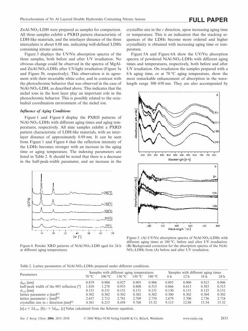
Figure 1 and Figure 4 display the PXRD patterns ofNiAl-NO3-LDHs with different aging times and aging tem-peratures, respectively. All nine samples exhibit a PXRDpattern characteristic of LDH-like materials, with an inter-layer distance of approximately 0.89 nm. It can be seenfrom Figure 1 and Figure 4 that the reflection intensity ofthe LDHs becomes stronger with an increase in the agingtime or aging temperature. The indexing parameters arelisted in Table 2. It should be noted that there is a decreasein the half-peak-width parameter, and an increase in the
Figure 4. Powder XRD patterns of NiAl-NO3-LDH aged for 24 hat different aging temperatures.
Table 2. Lattice parameters of NiAl-NO3-LDHs prepared under different conditions.
Samples with different aging temperatures Samples with different aging timesParameters 70 °C 100 °C 130 °C 150 °C 180 °C 6 h 12 h 18 h 24 h
d003 [nm] 0.879 0.904 0.927 0.903 0.906 0.893 0.900 0.912 0.906half-peak width of the 003 reflection [°] 1.810 1.270 0.933 0.808 0.515 0.866 0.613 0.583 0.515d110 [nm] 0.151 0.151 0.151 0.151 0.151 0.150 0.151 0.152 0.151lattice parameter a [nm][a] 0.302 0.302 0.302 0.302 0.302 0.300 0.302 0.304 0.302lattice parameter c [nm][b] 2.637 2.712 2.781 2.709 2.718 2.679 2.700 2.736 2.718crystallite size in c direction [nm][c] 4.361 6.215 8.458 9.768 15.32 9.115 12.88 13.54 15.32
[a] a = 2d110. [b] c = 3d003. [c] Value calculated from the Scherrer equation.
Eur. J. Inorg. Chem. 2006, 2831–2838 © 2006 Wiley-VCH Verlag GmbH & Co. KGaA, Weinheim www.eurjic.org 2833
crystallite size in the c direction, upon increasing aging timeor temperature. This is an indication that the stacking se-quences of the LDHs become more ordered and highercrystallinity is obtained with increasing aging time or tem-perature.
Figure 5A and Figure 6A show the UV/Vis absorptionspectra of powdered NiAl-NO3-LDHs with different agingtimes and temperatures, respectively, both before and afterUV irradiation. On irradiation the samples prepared with a6 h aging time, or at 70 °C aging temperature, show themost remarkable enhancement of absorption in the wave-length range 300–650 nm. They are also accompanied by
Figure 5. (A) UV/Vis absorption spectra of NiAl-NO3-LDHs withdifferent aging times at 180 °C, before and after UV irradiation.(B) Background correction for the absorption spectra of the NiAl-NO3-LDHs from (A) before and after UV irradiation.

X. Duan et al.FULL PAPERthe most obvious change in sample color, which indicatesthe most significant photochromism. Moreover, the en-hancement of the absorption in the range 300–650 nm de-creased upon increasing aging time or temperature. As theaging time was increased to 24 h, or the aging temperaturewas increased to 180 °C, no change could be observed ineither the absorption spectra or the sample color followingUV-light irradiation, which implied the absence of photo-chromic behavior. On the basis of the PXRD characteriza-tion above, it can be concluded that the ordered stackingsequences and the crystallinity of NiAl-NO3-LDH have amarked influence on its photochromism. Moreover, the ef-fect of the wavelength of the excitation light has beenstudied, and it was found that UV light with a wavelengthbelow 376 nm could cause photochromism in the material.
Figure 6. (A) UV/Vis absorption spectra of NiAl-NO3-LDHs withdifferent aging temperatures and aged for 24 h, before and afterUV irradiation. (B) Background correction for the absorption spec-tra of the NiAl-NO3-LDHs from (A) before and after UV irradia-tion.
In order to gain direct insight into photochromism inNiAl-NO3-LDH, the NiAl-NO3-LDH samples which werenonphotochromic (aging time 24 h and aging temperature180 °C) were used as the baseline reference for furtheranalysis of the UV/Vis spectra. Background correction wasperformed by subtracting the absorption spectrum of thenonphotochromic sample from the photochromic NiAl-NO3-LDH samples, both before and after UV irradiation.
www.eurjic.org © 2006 Wiley-VCH Verlag GmbH & Co. KGaA, Weinheim Eur. J. Inorg. Chem. 2006, 2831–28382834
The results are shown in Figure 5B and Figure 6B. Com-paring the UV/Vis absorption spectra before and after UVirradiation, it can be seen that there is an obvious peak atapproximately 500 nm. This indicates that there might besome specific structural unit, with Ni in an octahedrallycoordinated environment, which results in photochromismin NiAl-NO3-LDH. As the aging time or temperature in-creases, the stacking sequences of the LDHs become moreordered, leading to the loss of the specific structural unitand thus to the loss of photochromic behavior.
Influence of Host–Guest Interactions
In order to study the influence of host–guest interactions,two samples of NiAl-CO3-LDH were prepared: one by theion-exchange method from a NiAl-NO3-LDH precursorand the other by direct coprecipitation. Both samples exhi-bit a PXRD pattern characteristic of LDH-like materials,with an interlayer distance of approximately 0.76 nm, indi-cating well-defined LDHs containing carbonate anions.Figure 7 displays a comparison of the FTIR spectra of thetwo NiAl-CO3-LDH samples. Both show the strong, sharpband at approximately 1363 cm–1 due to the absorption ofinterlayer carbonate anions. However, it can be seen that apeak at approximately 1384 cm–1 was observed in the sam-ple prepared by ion-exchange (Figure 7a), which indicatesthe presence of a small amount of nitrate anions.
Figure 7. FTIR spectra of NiAl-CO3-LDHs prepared by (a) ionexchange and (b) coprecipitation.
On the basis of elemental analysis, the chemical composi-tion of the NiAl-CO3-LDH that was prepared by coprecipi-tation was found to be [Ni0.656Al0.344(OH)2](CO3
2–)0.172·0.8H2O (calcd. C 2.06, H 3.37, Al 8.70, Ni 36.27; foundC 2.12, H 3.31, Al 8.73, Ni 36.19). However, the chemicalcomposition of the NiAl-CO3-LDH that was prepared bythe ion-exchange method from a NiAl-NO3-LDH precursorwas found to be [Ni0.647Al0.353(OH)2](CO3
2–)0.151(NO3–)0.051·
0.7H2O (calcd. C 1.70, H 3.19, Al 8.95, N 0.67, Ni 35.83;found C 1.62, H 3.13, Al 8.86, N 0.71, Ni 35.76). It can beseen that the sample prepared by coprecipitation was a pureCO3
2– LDH, while the other one, prepared by ion-exchangefrom a NiAl-NO3-LDH precursor, contained both CO3
2–
and a little unexchangeable NO3–.

Photochromism of Ni–Al Layered Double Hydroxides Containing Nitrate Anions FULL PAPERFigure 8a and b show the UV/Vis absorption spectra of
the two powdered NiAl-CO3-LDH samples before and afterUV irradiation. It was found that almost no change in theabsorption spectrum, after UV-light irradiation, could beobserved for the NiAl-CO3-LDH prepared by coprecipi-tation (Figure 8b), which means that photochromism is notan intrinsic property of NiAl-CO3-LDHs. However, in thecase of the sample prepared by ion exchange, a considerableenhancement in the 300–650 nm range (Figure 8a) was ob-served after UV irradiation. This is an indication that theinteraction between Ni2+ and co-intercalated nitrate anionsis indispensable for the photochromism in NiAl-CO3-LDHsprepared by ion exchange. This is possibly related to theremaining nitrate anions that interact strongly with Ni2+
ions in NiAl-NO3-LDHs through chemical bonding andthus cannot be exchanged by CO3
2–.
Figure 8. UV/Vis absorption spectra of powdered NiAl-CO3-LDHs, both before and after UV irradiation, prepared by (a) ionexchange and (b) coprecipitation.
XPS Data
In order to confirm whether the photochromic behaviorof NiAl-NO3-LDHs is related to the redox of Ni2+, XPSwas used to determine the chemical environment of Ni, N,and O before and after UV irradiation. The XPS Ni2p3/2,N1s, and O1s core level spectra of NiAl-NO3-LDHs, beforeand after UV irradiation, are shown in Figure 9. The bind-ing energy (BE) value of the main Ni2p3/2 peak, at around856.4/856.5 eV, is assigned to the Ni2+ ion by comparisonwith the known BE value for Ni2p3/2 photoelectrons inNi(OH)2 (855.6–856.6 eV).[23] The XPS N1s peak at around406.6/406.7 eV is attributed to N5+ from the nitrate anions,and the XPS O1s peak at around 531.9/531.8 eV is due toO2– from the nitrate anions. As no remarkable change inthe values of the BE can be observed after UV irradiation,it is reasonable to deduce that the oxidation states of Ni,N, and O in NiAl-NO3-LDHs do not change, and hence amechanism based on the redox of Ni2+ can be excluded.
Eur. J. Inorg. Chem. 2006, 2831–2838 © 2006 Wiley-VCH Verlag GmbH & Co. KGaA, Weinheim www.eurjic.org 2835
Figure 9. XPS spectra of Ni2p3/2, N1s, and O1s for NiAl-NO3-LDHs before and after UV irradiation.
ESR Data
ESR was used to study the coordination environment ofNi2+ in NiAl-NO3-LDHs to obtain a further understandingof the photochromic mechanism. It has been reported thatNi2+ ions in Ni(OH)2 are ESR silent at room temperature,most probably because of the regular octahedral crystalfield.[22] Figure 10 displays the ESR spectra of powderedNiAl-NO3-LDH before and after UV irradiation, as well asthat of NiAl-CO3-LDH prepared by coprecipitation, as areference sample. It can be seen from Figure 10a that NiAl-CO3-LDH is ESR silent at room temperature and at liquidnitrogen temperature, because of its Ni–OH regular octahe-dral crystal field. However, in the case of NiAl-NO3-LDH,distinct signals with g = 2.163 (Figure 10b) and g = 2.643

X. Duan et al.FULL PAPER(Figure 10c) were observed for the sample before and afterUV irradiation, respectively. These can be attributed toNi2+ ions (3d8 electronic configuration) in a strongly dis-torted octahedral crystal field.[22] The difference in the gvalues indicates that the distortion in the octahedral crystalfield for NiAl-NO3-LDH is not completely the same beforeand after UV irradiation.
Figure 10. ESR spectra of powdered NiAl-CO3-LDH prepared bycoprecipitation (a), and NiAl-NO3-LDHs before irradiation (b)and after irradiation (c).
On the basis of the discussion above, it can be concludedthat the host–guest interaction between Ni2+ and the nitrateanions, and thus the presence of the specific octahedralcrystal field of the Ni2+, leads to photochromism in NiAl-NO3-LDHs. Single-crystal structure analysis of CaAl-NO3/CO3/SO4/Cl-LDHs has revealed that Ca2+ ions are seven-fold coordinated by O atoms. A water molecule, a carbonateanion, or a nitrate anion occupies the seventh coordinationsite of three of the four Ca2+ ions contained in the mainlayer.[24–27] Moreover, Steven et al. reported that, in the lay-ered material Ni2(OH)3NO3, the nitrate anion was locatedin the interlayer region and was coordinated through oneoxygen atom directly to the matrix Ni2+ cation.[28] In thecurrent study, taking into account the confirmation by EX-AFS data that Ni2+ ions are sixfold coordinated (Table 1),the evidence from ESR data that Ni2+ ions exist in a dis-torted octahedral crystal field, and the presence of unex-changeable NO3
–, it can be concluded that there are specificunits of octahedral structure in which Ni2+ is coordinatedby five hydroxy groups and one nitrate anion through theO atom in NiAl-NO3-LDH, as shown in Figure 11. Theinfluence of water cannot be excluded completely, and thereis the possibility that a hydrogen-bonding system exists be-tween the host hydroxy and nitrate anions, and water mole-cules, and thus water has a secondary influence on the pho-
www.eurjic.org © 2006 Wiley-VCH Verlag GmbH & Co. KGaA, Weinheim Eur. J. Inorg. Chem. 2006, 2831–28382836
tochromism of NiAl-NO3-LDH (as shown in Figure 11).Further studies on the effects of water, as well as theoreticalcalculations, are under investigation at our laboratory.
Figure 11. Schematic representation of the structure of NiAl-NO3-LDH.
The electronic configuration of Ni2+ ions in the groundstate is 3d8 (t2g
6eg2). As shown in Figure 12a, the octahe-
dral crystal field of the Ni2+ ions is generated by the coor-dinated O atom of the nitrate anion, and is somewhat dis-torted with a g value of 2.163, derived from ESR spec-troscopy data. Under UV-light irradiation, the electronicconfiguration of the Ni2+ ions in the excited state is 3d8
(t2g5eg
3), which speaks in favor of the Jahn–Teller effect,and thus leads to a more strongly distorted octahedralcrystal field (Figure 12b). The latter structure is quitestable at room temperature and cannot revert spontane-ously. In comparison with the sample before UV irradia-tion, it demonstrates changes in the UV/Vis absorptionspectrum (especially the strong absorption enhancement inat a wavelength of 500 nm) and in the g value determinedby ESR spectroscopy (from 2.163 to 2.634). Upon heating,the radiation-induced structure recovers its original state,and the color changes from black to green. Because thedistorted octahedral structure [Ni(OH)5NO3] was a meta-stable state thermodynamically, it reverted to the stableregular octahedral unit [Ni(OH)6] by substitution of theNO3
– by an OH– group upon increasing aging time or tem-perature during the preparation process. As a result, thespecific octahedral crystal field of the Ni2+ ion, coordi-nated by a nitrate anion, leads to photochromism in NiAl-NO3-LDHs.
Figure 12. Photochromic mechanism in NiAl-NO3-LDH: (a) struc-ture before UV irradiation and (b) structure after UV irradiation.

Photochromism of Ni–Al Layered Double Hydroxides Containing Nitrate Anions FULL PAPER
Conclusions
Photochromism in NiAl-NO3-LDHs has been studied inthis work. The crystalline structure and chemical composi-tion of NiAl-NO3-LDHs have been characterized on thebasis of the results of PXRD, FTIR, EXAFS, and elemen-tal analysis. The studies on the influence of aging condi-tions indicate that the host–guest interaction between thenickel ion in the host layer and the interlayer NO3
– playsan important role in its photochromic behavior. Taking intoaccount the confirmation by EXAFS data that the Ni2+
ions are sixfold coordinated, the ESR spectroscopy datathat indicates that Ni2+ is ESR active in NiAl-NO3-LDHs,the confirmation of the presence of some unexchangeableNO3
–, and the exclusion of the redox of Ni2+, it can beconcluded that there is a specific unit of distorted octahe-dral structure. This unit consists of a Ni2+ ion, which iscoordinated by O atoms from five hydroxy groups and onenitrate anion, and results in photochromism in NiAl-NO3-LDHs.
Experimental SectionPreparation of NiAl-NO3-LDH: NiAl-NO3-LDH was synthesizedby a hydrothermal method.[29] Typically, an aqueous solution ofNaOH (24 g, 0.6 mol) in deionized water (40 mL) was added drop-wise to a vigorously stirred, freshly prepared solution containingNi(NO3)2·6H2O (58.156 g, 0.2 mol) and Al(NO3)3·9H2O (37.513 g,0.1 mol) (Ni2+/Al3+ = 2) in deionized water (100 mL) under a nitro-gen atmosphere at room temperature. The final pH was approxi-mately 6. The suspension was transferred to a Teflon-lined auto-clave and heated at 70, 100, 130, 150, or 180 °C for a specific time.The solid precipitate was collected by filtration through a mem-brane filter under suction, washed thoroughly with water, and driedat 70 °C for 18 h.
Preparation of MgAl-NO3-LDH and ZnAl-NO3-LDH: MgAl-NO3-LDH and ZnAl-NO3-LDH, which were used as comparison sam-ples, were synthesized by a hydrothermal method. MgAl-NO3-LDH was synthesized by a procedure similar to that described pre-viously.[30] A solution of Mg(NO3)2·6H2O (32.0 g, 0.125 mol) andAl(NO3)3·9H2O (11.7 g, 0.062 mol) in deionized water (200 mL)was added dropwise over 2 h to a solution of NaOH (12.5 g,0.310 mol) and NaNO3 (18.2 g, 0.210 mol) in water (250 mL). Themixture was transferred to a Teflon-lined autoclave and heated at70 °C for 24 h. The precipitate was separated by centrifugation,washed with water, and dried at 70 °C for 18 h. ZnAl-NO3-LDHwas obtained by a similar method.
Preparation of NiAl-CO3-LDH: NiAl-CO3-LDH was prepared byboth coprecipitation and ion exchange. The sample prepared by thecoprecipitation method was synthesized according to a literatureprocedure.[31] In the case of the NiAl-CO3-LDH sample obtainedby ion exchange, the precursor NiAl-NO3-LDH was synthesizedfirst by the procedure described above. Subsequently, a solution ofNa2CO3 in deionized water (50 mL) was added to a suspension ofNiAl-NO3-LDH in water (50 mL), and the mixture was then heatedto 70 °C for 24 h. The product was washed extensively with deion-ized water, centrifuged, and dried at 70 °C for 18 h.
Characterization Techniques
PXRD patterns were obtained by using a Shimadzu XRD-6000diffractometer under the following conditions: Cu-Kα radiation (λ
Eur. J. Inorg. Chem. 2006, 2831–2838 © 2006 Wiley-VCH Verlag GmbH & Co. KGaA, Weinheim www.eurjic.org 2837
= 1.542 Å, 2θ = 2–70°), 40 kV, 30 mA. Infrared spectra of sampleswere recorded by using a Bruker Vector 22 FTIR spectrometer.Specimens were prepared as KBr pellets. Elemental analysis of themetal content was performed with an ICPS-7500 inductively cou-pled plasma optical-emission spectrometer. C, H, and N elementalanalysis was carried out with an Elementar Vario elemental ana-lyzer. The ESR spectra were recorded at room temperature with aBruker ESP300 spectrometer. A standard 100 MHz field modula-tion and a 5–10 G modulation width were used. XPS was recordedwith a VG Scientific ESCALab220i-XL (VG Scientific Ltd., UK)spectrometer.
The EXAFS spectrum around the Ni K-adsorption edge was ob-tained by using the beamline 4W1B of the Beijing SynchrotronRadiation Facility (BSRF). The NiAl-NO3-LDH nanoparticleswere homogeneously smeared on Scotch adhesive tape. More thaneight layers were folded to reach the optimum absorption thickness.The X-ray absorption spectrum of the Ni K-edge of NiAl-NO3-LDH was collected at ambient temperature in the transmissionmode. The storage ring was operated at 2.2 GeV with a typicalcurrent of 50 mA. Fixed-exit Si(111) flat double crystals were usedas a monochromator. The incident and transmission X-ray inten-sities were detected by using ion chambers installed in front of andbehind the sample. The X-ray energy was calibrated by using theNi K-absorption edge (8348 eV). The absorption spectrum was col-lected from 200 eV below the absorption threshold to over 700 eVabove the threshold.
EXAFS data reduction was performed by using WinXAS 97 1.1and following standard procedures. The first maximum of the firstderivative of the absorption edge was chosen as the energy thresh-old. The pre-edge absorption background was fitted and subtractedby using a linear function. A derivative method was used to derivethe EXAFS signal and remove the post-edge absorption back-ground. EXAFS functions were normalized by using the absorp-tion-edge jump and were Fourier-transformed to R space with k1-weight in the range 1.1–11.3 Å. Fourier filters were used in therange 0.3–1.8 Å. Theoretical scattering paths for the fit were calcu-lated by using the structures of α-Ni(OH)2. To reduce the numberof adjustable parameters, the amplitude reduction factor, S0
2, wasfixed at 1.1. The RNi–O values are estimated to be accurate to0.02 Å, and the ∆E0 and CNNi–O values are estimated to be accu-rate to 20%. The accuracy estimates are based on the results oftheoretical fits to the spectra of the reference compounds of knownstructure.
Study of the Photochromic Properties of LDHs: UV irradiation wascarried out by using a 500-W xenon lamp. Absorption spectra wererecorded by using a UV/Vis spectrophotometer (Shimadzu UV-2501 PC).
Acknowledgments
This project was supported by the National Natural Science Foun-dation Key Project of China (Project No.: 90306012), the NationalBasic Research Program (973 Program) (Project No.:2004CB720602), the Ministry of Education Science and Technol-ogy Research Project of China (Project No.: Key 104239), the Beij-ing Nova Program (No.: 2004A13), and the Program for Changji-ang Scholars and Innovative Research Team in University(PCSIRT). We also acknowledge the Beijing Synchrotron Radia-tion Facility (BSRF) for provision of synchrotron radiation facili-ties and thank Dr. Yaning Xie and Tao Liu for assistance in usingbeamline 4W1B.

X. Duan et al.FULL PAPER
[1] M. Meyn, K. Beneke, G. Lagaly, Inorg. Chem. 1990, 29, 5201–5207.
[2] G. A. Caravaggio, C. Detellier, Z. Wronski, J. Mater. Chem.2001, 11, 912–921.
[3] A. Corma, V. Fornes, F. Rey, A. Cervilla, E. Llopis, A. Ribera,J. Catal. 1995, 152, 237–242.
[4] M. Ogawa, K. Kuroda, Chem. Rev. 1995, 95, 399–438.[5] H. Tagaya, A. Ogata, T. Kuwahara, S. Ogata, M. Karasu, J. I.
Kadokawa, J. Chiba, Microporous Mater. 1996, 7, 151–158.[6] B. Sels, D. De Vos, M. Buntinx, F. Pierard, A. Kirsch-De Mes-
maeker, P. Jacobs, Nature 1999, 400, 855–857.[7] L. Ukrainczyk, M. Chibwe, T. J. Pinnavaia, S. A. Boyd, Envi-
ron. Sci. Technol. 1995, 29, 439–445.[8] A. M. Fogg, V. M. Green, H. G. Harvey, D. O’Hare, Adv. Ma-
ter. 1999, 11, 1466–1469.[9] A. M. Fogg, J. S. Dunn, S. G. Shyu, D. R. Cary, D. O’Hare,
Chem. Mater. 1998, 10, 351–355.[10] A. I. Khan, L. Lei, A. J. Norquist, D. O’Hare, Chem. Commun.
2001, 2342–2343.[11] V. Ambrogi, G. Fardella, G. Grandolini, L. Perioli, Int. J.
Pharm. 2001, 220, 23–32.[12] P. V. Kamath, M. Dixit, L. Indira, A. K. Shukla, V. G. Kumar,
N. Munichandraiah, J. Electrochem. Soc. 1994, 141, 2956–2959.
[13] A. Sugimoto, S. Ishida, K. Hanawa, J. Electrochem. Soc. 1999,146, 1251–1255.
[14] J. C. Crano, T. Flood, D. Knowles, A. Kumar, B. Van Gemert,Pure Appl. Chem. 1996, 68, 1395–1398.
[15] J. M. Lehn, Supramolecular Chemistry, VCH, Weinheim, 1995,p. 105.
[16] F. Vogtle, Supramolekulare Chemie, Teubner, Stuttgart, 1992,ch. 7.
www.eurjic.org © 2006 Wiley-VCH Verlag GmbH & Co. KGaA, Weinheim Eur. J. Inorg. Chem. 2006, 2831–28382838
[17] J. Peretti, J. Biteau, J. P. Boilot, F. Chaput, V. I. Safarov, J. M.Lehn, A. Fernandez-Acebes, Appl. Phys. Lett. 1999, 74, 1657–1659.
[18] K. Schaffner, S. E. Braslavsky, A. R. Holzwarth, Adv. Pho-tochem. 1990, 15, 229–277.
[19] H. Tagaya, T. Kuwahara, S. Sato, J. I. Kadokawa, M. Karasu,K. Masa, K. Chiba, J. Mater. Chem. 1993, 3, 317–318.
[20] H. Tagaya, S. Sato, T. Kuwahara, J. I. Kadokawa, K. Masa, K.Chiba, J. Mater. Chem. 1994, 4, 1907–1912.
[21] A. M. Scheidegger, E. Wieland, A. C. Scheinost, R. Dahn, P.Spieler, Environ. Sci. Technol. 2000, 34, 4545–4548.
[22] A. Davidson, J. F. Tempere, M. Che, J. Phys. Chem. 1996, 100,4919–4929.
[23] P. Dufresne, E. Grimblot, J. P. Bonnelle, J. Phys. Chem. 1981,85, 2344–2351.
[24] A. Tersis, S. Filippakis, H. J. Kuzel, H. Burzlaff, Z. Kristallogr.1987, 181, 29–34.
[25] R. Allmann, J. Neues, Neues Jahrb. Mineral. Monatsh. 1977, 3,136–144.
[26] M. Francois, G. Renaudin, O. Evrard, Acta Crystallogr. Sect.A 1998, 54, 1214–1217.
[27] G. Renaudin, M. Francois, Acta Crystallogr. Sect. A 1999, 55,835–838.
[28] P. N. Steven, J. William, J. Solid State Chem. 1999, 148, 26–40.[29] F. Prinetto, D. Tichit, R. Teissier, B. Coq, Catal. Today 2000,
55, 103–116.[30] J. H. Lee, S. W. Rhee, D. Y. Jung, Chem. Mater. 2004, 16, 3774–
3779.[31] F. Kooli, K. Kosuge, A. Tsunashima, J. Solid State Chem.
1995, 118, 285–291.Received: January 20, 2006
Published Online: May 9, 2006


![Conducting Polymers / Layered Double Hydroxides ... · materials, caused by co-intercalation of organic anions and chloride ions [30]. Kaneyoshi and Jones demonstrated that terephthalate](https://img.pdfslide.us/doc/110x75/5f0a09a17e708231d429b675/conducting-polymers-layered-double-hydroxides-materials-caused-by-co-intercalation.jpg)


![Title Photochromism and white long-lasting …...Title Photochromism and white long-lasting persistent luminescence in Bi[3+]-doped ZnGa[2]O[4] ceramics Author(s) Zhuang, Yixi; Ueda,](https://img.pdfslide.us/doc/110x75/5fca2348d932e01e9c134e64/title-photochromism-and-white-long-lasting-title-photochromism-and-white-long-lasting.jpg)