Embed Size (px)
Citation preview
IOSR Journal of Dental and Medical Sciences (IOSR-JDMS)
e-ISSN: 2279-0853, p-ISSN: 2279-0861.Volume 16, Issue 1 Ver. III (January. 2017), PP 100-118
www.iosrjournals.org
DOI: 10.9790/0853-160103100118 www.iosrjournals.org Page 100
“STUDY ON NON ALCOHOLIC FATTY LIVER DISEASE IN
TYPE 2 DIABETES MELLITUS WITH CLINICAL
CORRELATION’’
JaseemAnsari , Roshan M (Father Muller Medical College, Mangalore)
ABSTRACT BACKGROUND
Diabetes Mellitus (DM) can alter hepatic morphology and physiology1. Recently liver disease has been
recognized as a major complication of type 2 diabetes mellitus (T2 DM). There is high prevalence of
Nonalcoholic fatty liver disease (NAFLD) in individuals with T2 DM. Obesity is also a common and well
documented risk factor for NAFLD. There is an epidemic rise in T2 DM, obesity, and hyperlipidemia in the
country. A disease practically unheard a few years back, is now considered one of the most common causes of
chronic liver disease in the world3. The prevalence of NAFLD is rising in India. NAFLD begins as mild
steatosis, develops into non-alcoholic steatohepatitis (NASH) which can progress to cirrhosis and even
hepatocellular carcinoma2(HCC) making early detection and prevention of diabetic liver disease important.
OBJECTIVES
To study the clinicopathological profile of hepatic involvement in T2 DM and correlate between them.
MATERIALS AND METHODS
The study is a descriptive prospective study of the patients admitted in Father Muller's Medical College with T2
DM conducted over a period of 18 months.
The study includes 100 patients diagnosed with T2 DM. These patients will be elevated by a detailed history
including the age, sex, location, duration of diabetes, history of previous illness, medication they were currently
taking. Clinical examination includes anthropometric measurements including height and weight and thus the
body mass index (BMI) , signs of insulin resistance-central obesity, xanthelesma, acanthosis nigricans
.Investigations include abdominal ultra-sonography (USG) for fatty liver, glycosylated hemoglobin (HbA1c)
,liver function test (LFT) and lipid profile .Results were analyzed and compared.
RESULTS
The prevalence of NAFLD among diabetes in our study was found to be 26%.It was found to be more
common in the fourth decade of life with equal distribution among men and women. Among the
patients with NAFLD 53.6% were associated with hypertension (HTN), 75% with dyslipidemia and
19% with BMI >19%.38% of the patients with NAFLD were found to have elevated
alaninetransaminase (ALT) and 26% hadelevated aspartate transaminase (AST). 73% of the NAFLD
patients had elevated cholesterol.26% of the patients in our study were found to have sonological
features suggestive of NAFLD.
CONCLUSION
This study demonstrates and clinically correlates the cluster of abnormalities /risk factors like
hypertension, obesity, duration of diabetes with NAFLD.The implication of the study is that diabetics
are at a higher risk of developing NAFLD and its related complications.
1.INTRODUCTION NAFLD is the most common liver disease and the third leading indication for liver transplantation
1. The
prevalence of NAFLD has been reported to be 15-30% in the general population and in T2 DM population, the
prevalence is 70-75% 2. NAFLD has been proposed as one of the components of metabolic syndrome (MS)
4. It
has been found to be a composite of confirmed cases with central obesity, T2DM and dyslipidemia. Studies
have shown the major role of obesity and insulin resistance in NAFLD 5. However, regardless of BMI, the
presence of T2 DM significantly increases the risk and severity of NAFLD 6. Only recently liver disease has
been recognized as a major complication of T2 DM with increased mortality rates for cirrhosis greater than that
Study On Non Alcoholic Fatty Liver Diseases in Type 2 Diabetes Mellitus With Clinical Correlation
DOI: 10.9790/0853-160103100118 www.iosrjournals.org Page 101
for cardiovascular disease 8. Insulin resistance plays a central pathogenic role in both T2 DM and NAFLD with
the latter being considered as the hepatic manifestation of the MS 9.
OBJECTIVE OF THE STUDY To study the clinicopathological profile of hepatic involvement in T2 DM and correlate between them.
2. REVIEW OF LITERATURE NAFLD was practically unheard a few years ago, but is now considered one of the most commonliver disorders
in the world8.It may be the most common cause of liverenzyme elevation in adults as well as one of the leading
cause for cirrhosis in the world.The prevalence of NAFLD has increased in joint with the epidemics of obesity
and T2 DM, which are the major risk factors for NAFLD10
. Whereas the association of T2 DM with micro-
vascularcomplications and macro-vascular disease is well established.
Theassociation of T2 DM with NAFLD is a recently recognized entity and less well known10
.There is evidence
that patients with NAFLD who have T2 DM particularly at a high risk of developing cirrhosis compared with
those who donot have diabetes. Although cardiovascular disease is the majorcause of excess morbidity and
mortality inT2 DM, liver failuremay also be a threat to patients with T2 DM12
.
NAFLD is characterized by fatty infiltration of the liver, mostly in the form of triglycerides, which exceeds 5%
of the liver weight.NAFLD is histologically similar to alcoholic liver disease (ALD), but itoccurs in the absence
of excessive alcohol consumption and is not dueto other identifiable causes of fatty liver13
.
CONDITIONS ASSOCIATED WITH FATTY LIVER DISEASE25
Diabetes mellitus
Acquired insulin resistance
Obesity
Hyperlipidemia
Hypothalamic–pituitary dysfunction
Genetic/inborn errors of metabolism
Wilson’s disease
Nutritional/intestinal/Surgical
NAFLD represents a spectrum of clinical–pathologicalfeatures ranging from simplesteatosis, which is
characterized by fatty infiltration only to non-alcoholic steatohepatitis (NASH), which is characterized by
inflammation and hepatocellular injury with orwithout fibrosis and cirrhosis. Most with NAFLD have an
increase inliver fat content alone and some developNASH that can progress to cirrhosis.
PREVALENCE Data from the Dallas HeartStudy suggested that about one-third of the population of Dallas County, Texas had
hepatic steatosis25
. This study used proton magneticresonance spectroscopy (MRS) to measure liver fat and
definedsteatosis as hepatic triglyceride content >5.5%. Having diabetes carries an even higher risk. Sixty-two
per cent ofsubjects in the Dallas Heart Study who had either known diabetes had hepatic steatosis.
As most who have NAFLD have no specific signs or symptoms, it goes unnoticed. In clinical practice,elevated
aminotransferase levels, especially ALT, are considered a marker for liver disease17
. However, manypatients
who have NAFLD do not have elevated levels. In the DallasHeart Study, 79% of those with hepatic steatosis
had normal ALTlevels. Making thematter of establishing the diagnosis of NAFLD even more complicatedis that
aminotransferase levels do not necessarily correlate with theseverity of NAFLD. ALT levels may be normal in
the presence ofadvanced fibrosis or cirrhosis19
. Thus, a normal ALT does not excludesteatosis and does not
ensure the absence of underlying advanced liverdisease21
.
Therefore, since non-invasive methods of detection were used in these epidemiological studies, the prevalence
of pure steatosisversus more advanced stages of disease such as steatohepatitis,fibrosis or cirrhosis is
unknown28
.
SYMPTOMS As with many other types of CLD, mostpatients with NAFLD (48–100%) are asymptomatic.The liver disease is
often discovered incidentallyduring routine laboratory examination when a hepaticpanel reveals an elevated
Study On Non Alcoholic Fatty Liver Diseases in Type 2 Diabetes Mellitus With Clinical Correlation
DOI: 10.9790/0853-160103100118 www.iosrjournals.org Page 102
ALT level. NAFLD isthe most common cause for unexplained persistent elevation of ALT levels once
hepatitis C and other CLD have been excluded25
. When symptoms occurthey are usually nonspecific. Vague
right upper quadrantabdominal pain, fatigue, and malaise are the most common. Rarely, pruritus,anorexia, and
nausea may develop. Jaundice, abdominal distension (ascites), gastrointestinal bleeding, and confusion
(encephalopathy) are all indicative of advanced liverdisease (decompensated cirrhosis), occurring late in
thecourse22
.
SIGNS There are no pathognomonic signs of NAFLD. Obesity is the most common abnormality on physical
examination, occurring in 30–100% of patients in various cross-sectionalstudies26
. Hepatomegaly has been
reported in up to 75% of patients in several studies .The prevalence of hepatomegaly may increase to 95%when
assessed by USG. Of the various stigmata, spidernevi and palmar erythema are the most common .Muscle
wasting may occur as liver disease becomes moreadvanced but is often underestimated due to edema and
preexisting obesity27
.
LABORATORY FINDINGS
Mild to moderate elevation of serum aminotransferases (ALT and AST) is the most common and often the only
laboratory abnormality found in patients with NAFLD29
.There is no significant correlation between the degree
of serum aminotransferase elevation and the histologic severity of hepatic inflammation or fibrosis . Unlike
those with alcohol-induced steatohepatitis, who typically manifest disproportionate increases in the AST level
relative to the ALT level, patients with NAFLD usually haven AST/ALT ratio <1 .
The AST/ALT ratiotends to increase with the development of cirrhosis, thuslosing its diagnostic accuracy.
Serum alkaline phosphatase (ALP) may also be slightly elevated in aboutone-third of patients. Hyper-
bilirubinemia, hypo-albuminemia, and prolongation of the prothrombin time (PT) are noted infrequently and
generally only seen once liver failure has become established. Elevated serum lipid profiles and glucose
concentrations are also common in NAFLD patients, reported in 25 to 75% of cases.
A small percentage of patients with NAFLD may have a low-titer (≤1:320) antinuclear antibody (ANA)
positivity25
. The role of iron in the pathogenesis ofNAFLD remains controversial.
It is important to exclude secondary causes of hepatic fat so that the diagnosis of primary NAFLD can be made
reliably. Hepatitis C (HCV) and alcoholic liver disease are particularly important because of the high
prevalence of these two hepatotoxic agents28
.HCVcan cause histologic changes that closely resemble NAFLD,
thus serologic testing to exclude viral hepatitis has become a pre-requisite for the diagnosis of NAFLD29
.
By its very definition, the diagnosis of NAFLD cannot be made in the settingof excessive alcohol consumption.
However, there is noconsensus among investigators concerning what is an excessiveamount of alcohol and thus
there are no published and universally accepted threshold levels. It is generally believed that a fatty liver does
not develop with alcohol.
IMAGING Several noninvasive imaging techniques, including USG, computed tomography (CT), and magnetic resonance
imaging (MRI), can identify hepatic steatosis and have been advocated as diagnostic tests for NAFLD26
. USG is
the most commonly used. The sonographicfindings of diffuse fatty change include a diffuse Hyperechoic
echotexture (bright liver), increased liver echotexture compared with the kidneys, vascular blurring and deep
attenuation. Fatty infiltration of the liver produces a low-density hepatic parenchyma on CT scanning. In a direct
comparison of CT with USG, USG was found to be more sensitive in detecting fatty change. However, when
fatty change is patchy or focal, CT scan and MRI are superior to USG. Also, when a semi quantitative
assessment is required or when multiple comparative studies are planned over time, CT is superior to US.
MRS is a newer innovative radiologic technique allowing one to examine the resonance frequencies of all
proton species within a region of interest and is being investigated as a means of obtaining a more quantitative
assessment of fatty liver infiltration.
Despite the utility of these imaging modalities in the diagnosis of diffuse fatty disorders of the liver, none is
sufficiently sensitive to detect hepatic inflammation, fibrosis or cirrhosis.
Study On Non Alcoholic Fatty Liver Diseases in Type 2 Diabetes Mellitus With Clinical Correlation
DOI: 10.9790/0853-160103100118 www.iosrjournals.org Page 103
In a prospective study evaluating therole of different radiological modalities in establishing thediagnosis of
NASH, neither USG, CT, nor MRI was able todetect the presence of hepatocyte ballooning, Mallory’shyaline,
or fibrosis, which are all important features in thediagnosis of NASH. With the inability to distinguishsimple
steatosis from steatohepatitis and stage the severityof injury, liver biopsy remains the best diagnostic test for
NASH31
.
LIVER HISTOLOGY The lack of effectivemedical therapy for NAFLD and risks associated withbiopsy are arguments proposed
against obtaining tissue sampling30
. Nevertheless, liver biopsy is the only accuratemethod for the diagnosis
ofNASHand the only meansto determine the severity of liver damage and long-term prognosis.
The histological features of NAFLD are indistinguishable from those of alcohol-induced liver disease. There are
two lesions associated with NAFLD:
(i) Predominantlymacro vesicularsteatosis alone
(ii) Predominantlymacro vesicularsteatosis
And varying amounts of cytological ballooning and spotty necrosis, scattered mixed
Neutrophilic–lymphocytic inflammation, glycogen nuclei, Mallory’s hyaline, and per sinusoidal fibrosis
(NASH). All of the features of steatohepatitis are not present in every instance of steatohepatitis. The severity of
steatosis can be graded on the basis of the extent of involved parenchyma.
Given the association of NAFLD with metabolic syndrome (MS), obesity and T2 DM the prevalence of NAFLD
and NASH are increasing. Within the NAFLD spectrum, only patients with histologically proven NASH
develop progressive liver disease. Progression seems more likely in the setting of diabetes, insulin resistance and
other pre- existing conditions.
Hence it is reasonable to expect that early diagnosis of NAFLD and early intervention which would prevent
progression to more serious stages
3. METHODOLOGY
SOURCE OF DATA
The data was collected from both outpatients and inpatients in Father Muller's Medical College Hospital from
1st August 2013 to 1st August 2014.
METHOD OF DATA COLLECTION
STUDY DESIGN The study is a descriptive prospective study of the patients in Father Muller's Medical College with T2 DM.
The study will includes 100 patients with equal sex ratio, diagnosed with T2 DM.These patients was evaluated
by a detailed history including the age , sex ,location, duration of diabetes , history of previous illness ,
medication they were currently taking. Clinical examination includes anthropometric measurements including
height and weight and thus the BMI, signs of insulin resistance-central obesity, xanthelesma,
acanthosisnigricans .Investigations include abdominal USG for fatty liver, HbA1c, LFT and lipid profile
.Results will then be analyzed and compared.
INCLUSION CRITERIA (1) Known cases of T2 DM (
>3 years) patients of both sexes between the age group of 25 to 80.
EXCLUSION CRITERIA (1) Known history of chronic viral hepatitis
(2) Individuals with alcohol consumption
(3) History of drug intake that can cause fatty liver.
(4) Patients with nephropathy.
(5) Patients in congestive cardiac failure (CCF).
(6) Patients on insulin.
DATA ANALYSIS: Data was analyzed by frequency,percentage, mean and standard deviation.
4. RESULTS
PREVALENCE OF NAFLD
Study On Non Alcoholic Fatty Liver Diseases in Type 2 Diabetes Mellitus With Clinical Correlation
DOI: 10.9790/0853-160103100118 www.iosrjournals.org Page 104
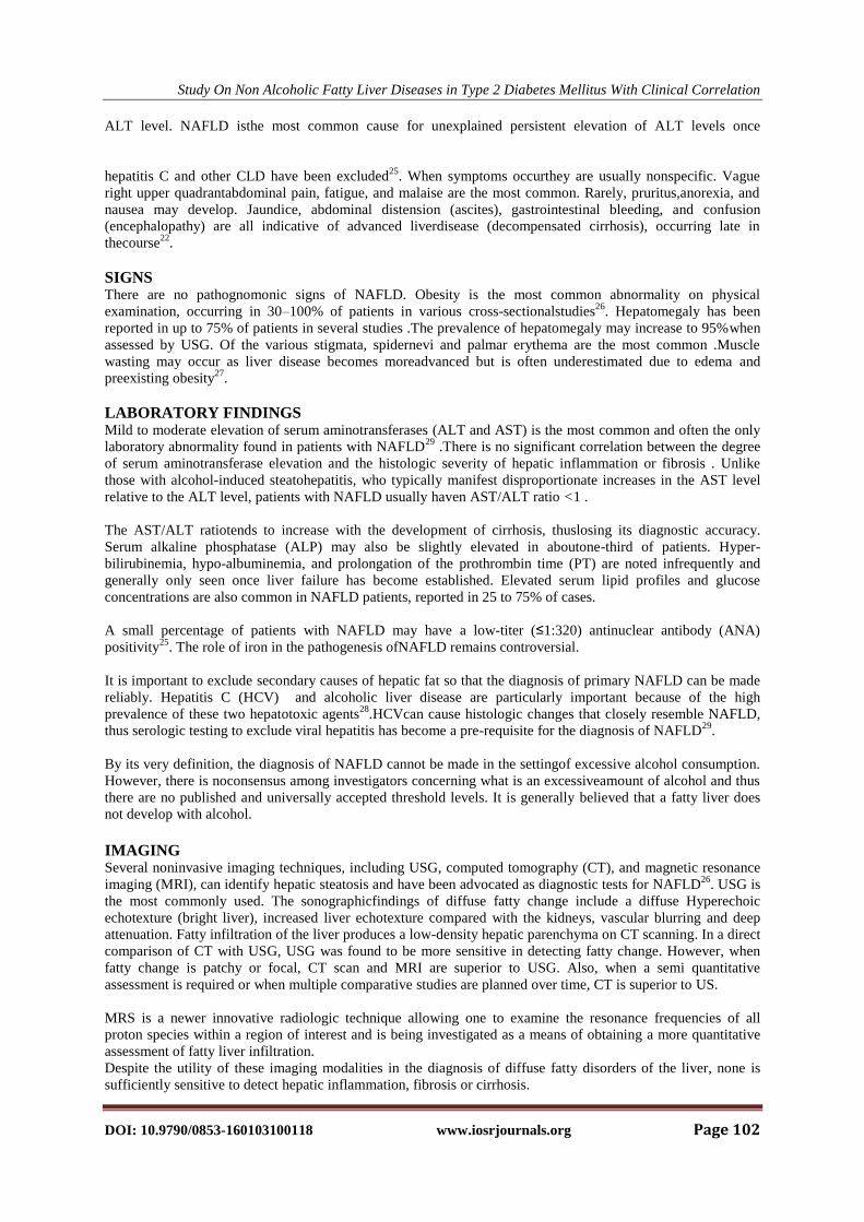
USG-FATTY LIVER
Frequency Percent
N
74 74.0
Y 26 26.0
Total 100 100.0
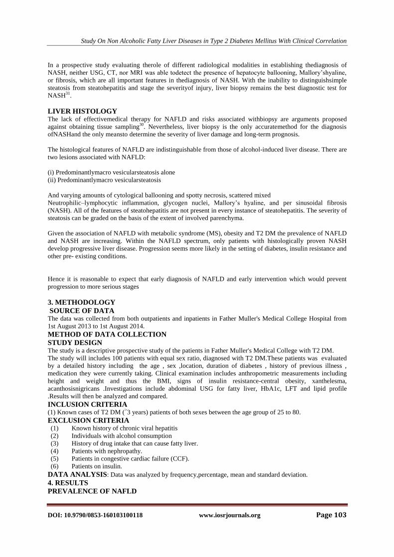
The prevalence of NAFLD among the total number of cases included in the study was found to be 26%
AGE DISTIBUTION
AGE * USG-FATTY LIVER Cross tabulation
74%
26%
USG-FATTY LIVER
N
Y
Study On Non Alcoholic Fatty Liver Diseases in Type 2 Diabetes Mellitus With Clinical Correlation
DOI: 10.9790/0853-160103100118 www.iosrjournals.org Page 105
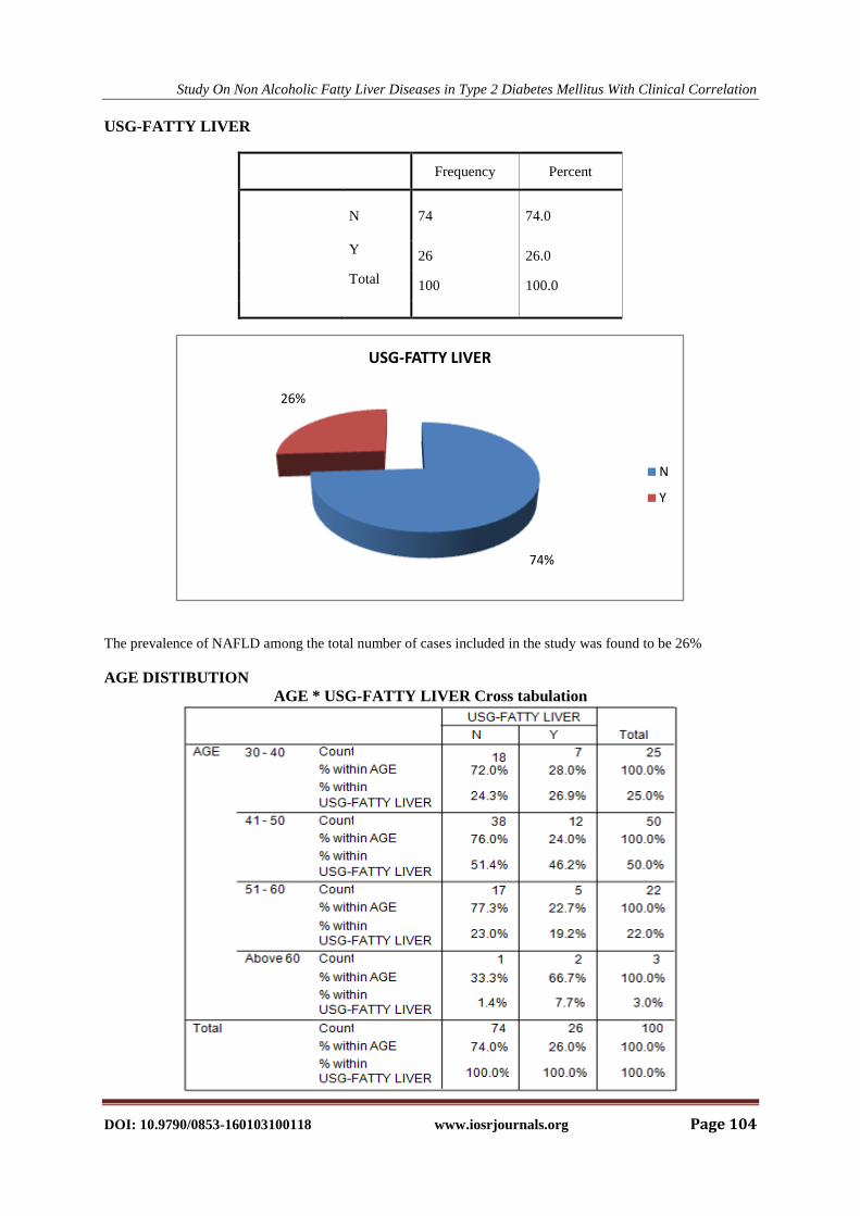
The prevalence of NAFLD was found to be higher in the fourth decade
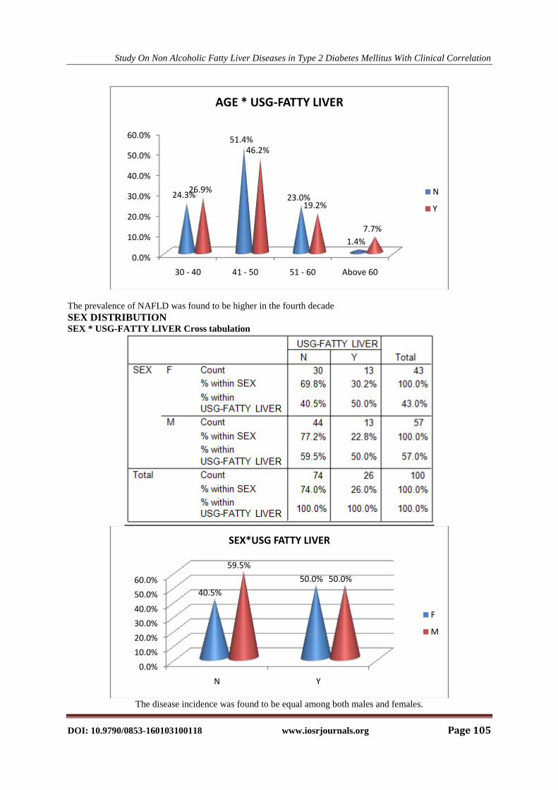
SEX DISTRIBUTION SEX * USG-FATTY LIVER Cross tabulation
The disease incidence was found to be equal among both males and females.
0.0%
10.0%
20.0%
30.0%
40.0%
50.0%
60.0%
30 - 40 41 - 50 51 - 60 Above 60
24.3%
51.4%
23.0%
1.4%
26.9%
46.2%
19.2%
7.7%
AGE * USG-FATTY LIVER
N
Y
0.0%
10.0%
20.0%
30.0%
40.0%
50.0%
60.0%
N Y
40.5%
50.0%
59.5%
50.0%
SEX*USG FATTY LIVER
F
M
Study On Non Alcoholic Fatty Liver Diseases in Type 2 Diabetes Mellitus With Clinical Correlation
DOI: 10.9790/0853-160103100118 www.iosrjournals.org Page 106
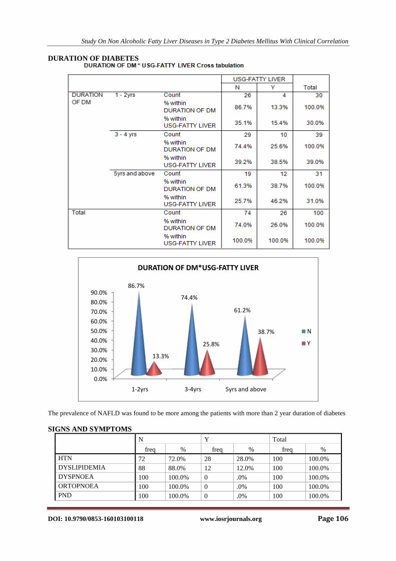
DURATION OF DIABETES
The prevalence of NAFLD was found to be more among the patients with more than 2 year duration of diabetes
SIGNS AND SYMPTOMS
N Y Total
freq % freq % freq %
HTN 72 72.0% 28 28.0% 100 100.0%
DYSLIPIDEMIA 88 88.0% 12 12.0% 100 100.0%
DYSPNOEA 100 100.0% 0 .0% 100 100.0%
ORTOPNOEA 100 100.0% 0 .0% 100 100.0%
PND 100 100.0% 0 .0% 100 100.0%
0.0%
10.0%
20.0%
30.0%
40.0%
50.0%
60.0%
70.0%
80.0%
90.0%
1-2yrs 3-4yrs 5yrs and above
86.7%
74.4%
61.2%
13.3%
25.8%
38.7%
DURATION OF DM*USG-FATTY LIVER
N
Y
Study On Non Alcoholic Fatty Liver Diseases in Type 2 Diabetes Mellitus With Clinical Correlation
DOI: 10.9790/0853-160103100118 www.iosrjournals.org Page 107
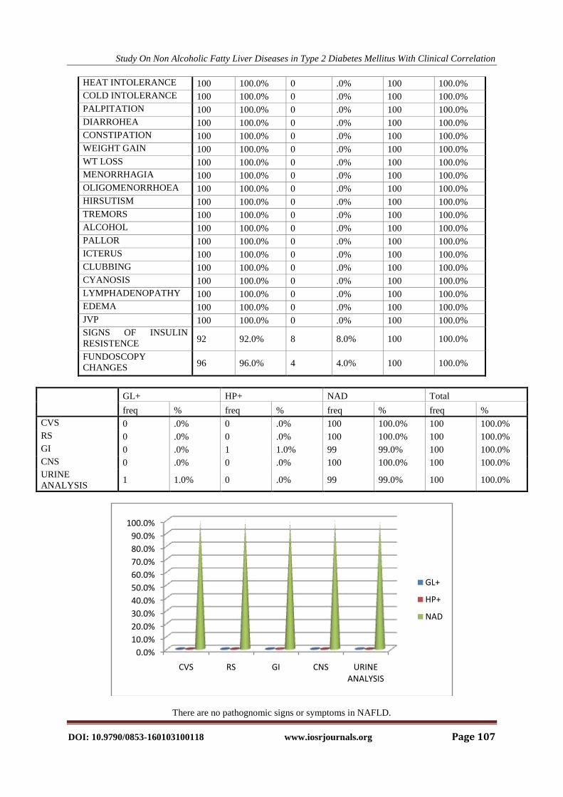
HEAT INTOLERANCE 100 100.0% 0 .0% 100 100.0%
COLD INTOLERANCE 100 100.0% 0 .0% 100 100.0%
PALPITATION 100 100.0% 0 .0% 100 100.0%
DIARROHEA 100 100.0% 0 .0% 100 100.0%
CONSTIPATION 100 100.0% 0 .0% 100 100.0%
WEIGHT GAIN 100 100.0% 0 .0% 100 100.0%
WT LOSS 100 100.0% 0 .0% 100 100.0%
MENORRHAGIA 100 100.0% 0 .0% 100 100.0%
OLIGOMENORRHOEA 100 100.0% 0 .0% 100 100.0%
HIRSUTISM 100 100.0% 0 .0% 100 100.0%
TREMORS 100 100.0% 0 .0% 100 100.0%
ALCOHOL 100 100.0% 0 .0% 100 100.0%
PALLOR 100 100.0% 0 .0% 100 100.0%
ICTERUS 100 100.0% 0 .0% 100 100.0%
CLUBBING 100 100.0% 0 .0% 100 100.0%
CYANOSIS 100 100.0% 0 .0% 100 100.0%
LYMPHADENOPATHY 100 100.0% 0 .0% 100 100.0%
EDEMA 100 100.0% 0 .0% 100 100.0%
JVP 100 100.0% 0 .0% 100 100.0%
SIGNS OF INSULIN
RESISTENCE 92 92.0% 8 8.0% 100 100.0%
FUNDOSCOPY
CHANGES 96 96.0% 4 4.0% 100 100.0%
There are no pathognomic signs or symptoms in NAFLD.
0.0%
10.0%
20.0%
30.0%
40.0%
50.0%
60.0%
70.0%
80.0%
90.0%
100.0%
CVS RS GI CNS URINE ANALYSIS
GL+
HP+
NAD
GL+ HP+ NAD Total
freq % freq % freq % freq %
CVS 0 .0% 0 .0% 100 100.0% 100 100.0%
RS 0 .0% 0 .0% 100 100.0% 100 100.0%
GI 0 .0% 1 1.0% 99 99.0% 100 100.0%
CNS 0 .0% 0 .0% 100 100.0% 100 100.0%
URINE
ANALYSIS 1 1.0% 0 .0% 99 99.0% 100 100.0%
Study On Non Alcoholic Fatty Liver Diseases in Type 2 Diabetes Mellitus With Clinical Correlation
DOI: 10.9790/0853-160103100118 www.iosrjournals.org Page 108
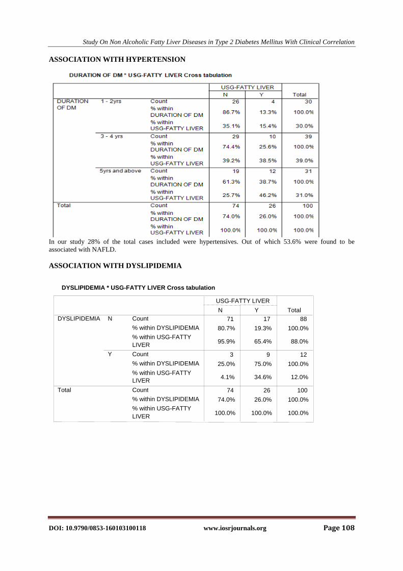
ASSOCIATION WITH HYPERTENSION
In our study 28% of the total cases included were hypertensives. Out of which 53.6% were found to be
associated with NAFLD.
ASSOCIATION WITH DYSLIPIDEMIA
DYSLIPIDEMIA * USG-FATTY LIVER Cross tabulation
71 17 88 80.7% 19.3% 100.0%
95.9% 65.4% 88.0%
3 9 12 25.0% 75.0% 100.0%
4.1% 34.6% 12.0%
74 26 100 74.0% 26.0% 100.0%
100.0% 100.0% 100.0%
Count % within DYSLIPIDEMIA % within USG-FATTY LIVER Count % within DYSLIPIDEMIA
% within USG-FATTY LIVER
Count % within DYSLIPIDEMIA % within USG-FATTY LIVER
N
Y
DYSLIPIDEMIA
Total
N Y USG-FATTY LIVER
Total
Study On Non Alcoholic Fatty Liver Diseases in Type 2 Diabetes Mellitus With Clinical Correlation
DOI: 10.9790/0853-160103100118 www.iosrjournals.org Page 109
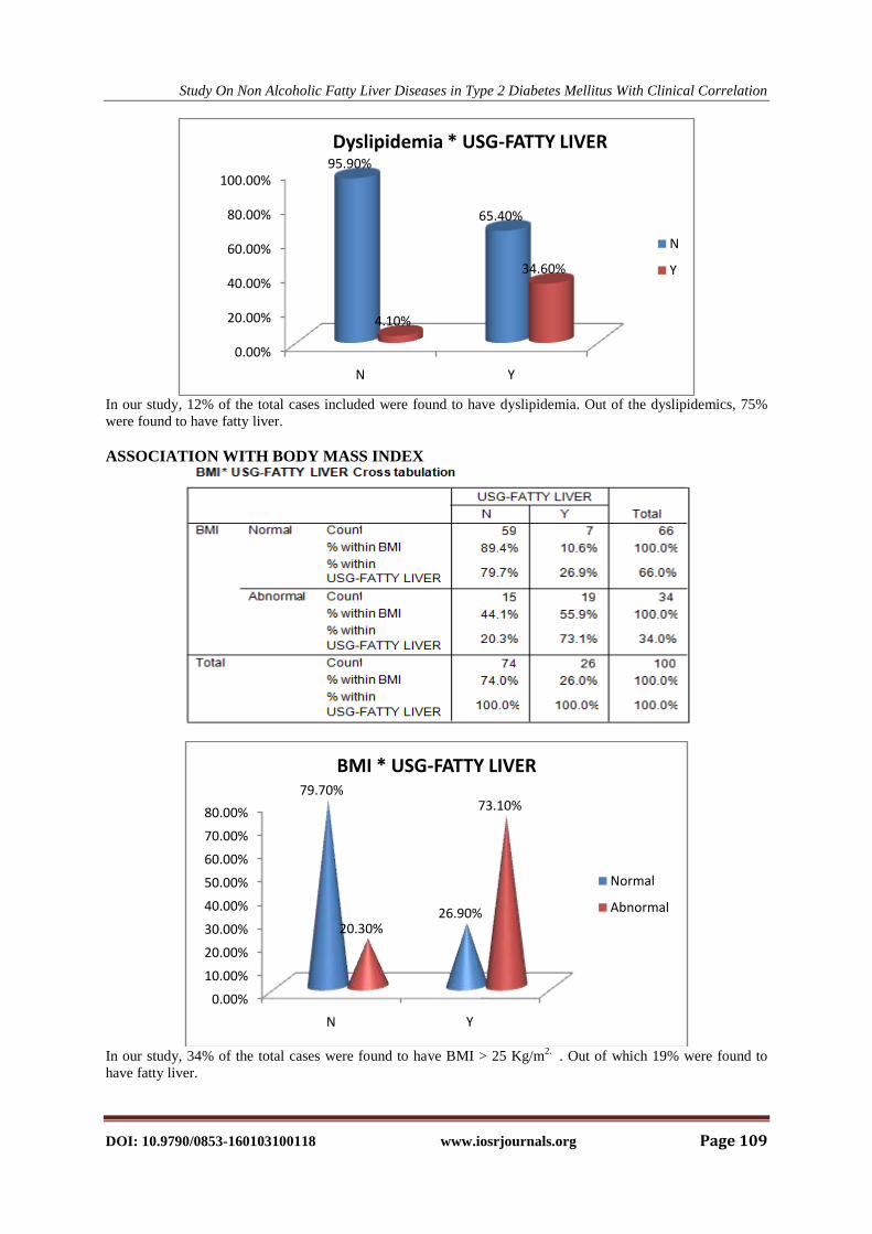
In our study, 12% of the total cases included were found to have dyslipidemia. Out of the dyslipidemics, 75%
were found to have fatty liver.
ASSOCIATION WITH BODY MASS INDEX
In our study, 34% of the total cases were found to have BMI > 25 Kg/m
2. . Out of which 19% were found to
have fatty liver.
0.00%
20.00%
40.00%
60.00%
80.00%
100.00%
N Y
95.90%
65.40%
4.10%
34.60%
Dyslipidemia * USG-FATTY LIVER
N
Y
0.00%
10.00%
20.00%
30.00%
40.00%
50.00%
60.00%
70.00%
80.00%
N Y
79.70%
26.90%20.30%
73.10%
BMI * USG-FATTY LIVER
Normal
Abnormal
Study On Non Alcoholic Fatty Liver Diseases in Type 2 Diabetes Mellitus With Clinical Correlation
DOI: 10.9790/0853-160103100118 www.iosrjournals.org Page 110
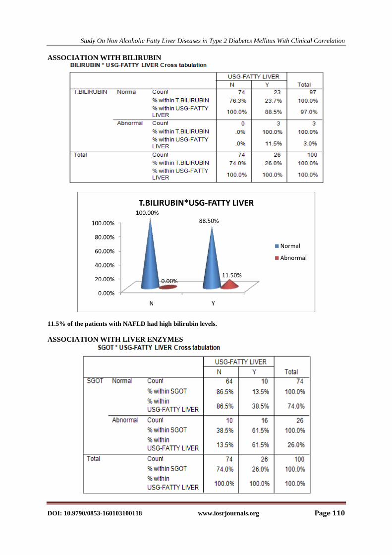
ASSOCIATION WITH BILIRUBIN
11.5% of the patients with NAFLD had high bilirubin levels.
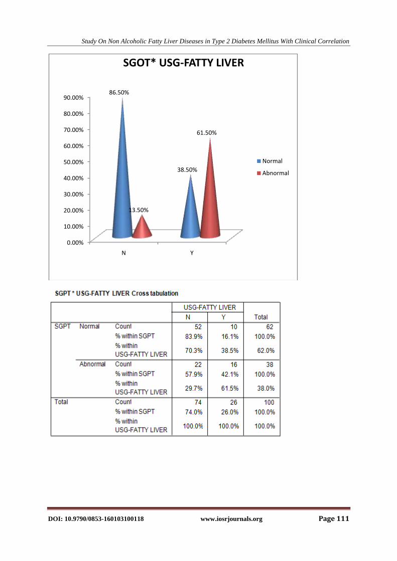
ASSOCIATION WITH LIVER ENZYMES
0.00%
20.00%
40.00%
60.00%
80.00%
100.00%
N Y
100.00%88.50%
0.00%11.50%
T.BILIRUBIN*USG-FATTY LIVER
Normal
Abnormal
Study On Non Alcoholic Fatty Liver Diseases in Type 2 Diabetes Mellitus With Clinical Correlation
DOI: 10.9790/0853-160103100118 www.iosrjournals.org Page 111
0.00%
10.00%
20.00%
30.00%
40.00%
50.00%
60.00%
70.00%
80.00%
90.00%
N Y
86.50%
38.50%
13.50%
61.50%
SGOT* USG-FATTY LIVER
Normal
Abnormal
Study On Non Alcoholic Fatty Liver Diseases in Type 2 Diabetes Mellitus With Clinical Correlation
DOI: 10.9790/0853-160103100118 www.iosrjournals.org Page 112
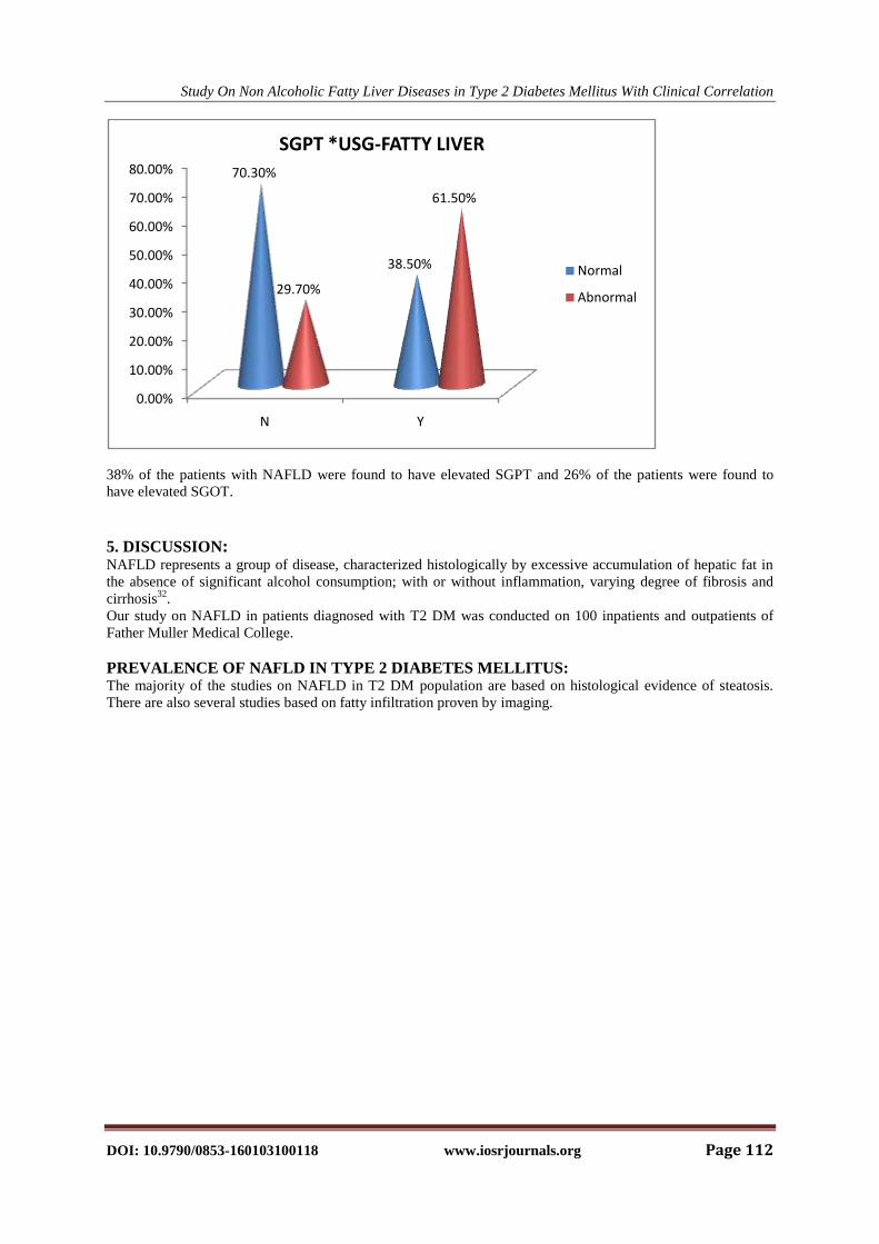
38% of the patients with NAFLD were found to have elevated SGPT and 26% of the patients were found to
have elevated SGOT.
5. DISCUSSION: NAFLD represents a group of disease, characterized histologically by excessive accumulation of hepatic fat in
the absence of significant alcohol consumption; with or without inflammation, varying degree of fibrosis and
cirrhosis32
.
Our study on NAFLD in patients diagnosed with T2 DM was conducted on 100 inpatients and outpatients of
Father Muller Medical College.
PREVALENCE OF NAFLD IN TYPE 2 DIABETES MELLITUS: The majority of the studies on NAFLD in T2 DM population are based on histological evidence of steatosis.
There are also several studies based on fatty infiltration proven by imaging.
0.00%
10.00%
20.00%
30.00%
40.00%
50.00%
60.00%
70.00%
80.00%
N Y
70.30%
38.50%
29.70%
61.50%
SGPT *USG-FATTY LIVER
Normal
Abnormal
Study On Non Alcoholic Fatty Liver Diseases in Type 2 Diabetes Mellitus With Clinical Correlation
DOI: 10.9790/0853-160103100118 www.iosrjournals.org Page 113
However there are only very few studies involving the clinical correlation with NAFLD with biochemical as
well as sonological evidence.
The overall prevalence of NAFLD in T2DM in this study was found to be 26%, which is lower than the
prevalence rates in different studies conducted in India- Kalra S et al which was 56.5% and Mohan et al which
was 54.5%41
.
However the prevalence rates were found to be higher than the prevalence rates of 12.5% and 20 %described by
AgalS etal44
.
One of the studies of NAFLD in T2 DM based on histological evidence by Banerjee S et al showed a much
higher prevalence rate of 87%.
Based on the type of study, the prevalence rates were much higher in those which had histological evidence for
NAFLD in comparison to those which were conducted based on biochemical and sonologicalevidence .
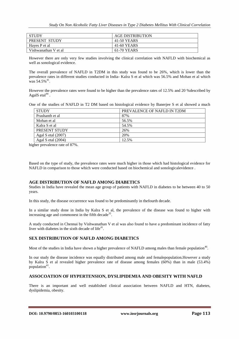
AGE DISTRIBUTION OF NAFLD AMONG DIABETICS Studies in India have revealed the mean age group of patients with NAFLD in diabetes to be between 40 to 50
years.
In this study, the disease occurrence was found to be predominantly in thefourth decade.
In a similar study done in India by Kalra S et al, the prevalence of the disease was found to higher with
increasing age and commonest in the fifth decade32
.
A study conducted in Chennai by Vishwanathan V et al was also found to have a predominant incidence of fatty
liver with diabetes in the sixth decade of life41
.
SEX DISTRIBUTION OF NAFLD AMONG DIABETICS
Most of the studies in India have shown a higher prevalence of NAFLD among males than female population
46.
In our study the disease incidence was equally distributed among male and femalepopulation.However a study
by Kalra S et al revealed higher prevalence rate of disease among females (60%) than in male (53.4%)
population41
.
ASSOCOATION OF HYPERTENSION, DYSLIPIDEMIA AND OBESITY WITH NAFLD
There is an important and well established clinical association between NAFLD and HTN, diabetes,
dyslipidemia, obesity.
STUDY PREVALENCE OF NAFLD IN T2DM
Prashanth et al 87%
Mohan et al 56.5%
Kalra S et al 54.5%
PRESENT STUDY 26%
Agal S etal (2007) 20%
Agal S etal (2004) 12.5%
STUDY AGE DISTRIBUTION
PRESENT STUDY 41-50 YEARS
Hayes P et al 41-60 YEARS
Vishwanathan V et al 61-70 YEARS
Study On Non Alcoholic Fatty Liver Diseases in Type 2 Diabetes Mellitus With Clinical Correlation
DOI: 10.9790/0853-160103100118 www.iosrjournals.org Page 114
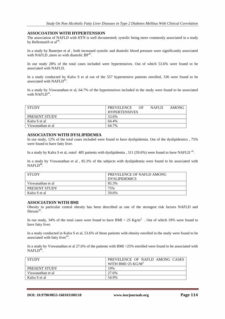
ASSOCOATION WITH HYPERTENSION The association of NAFLD with HTN is well documented; systolic being more commonly associated in a study
by BellentaniS et al44
.
In a study by Banerjee et al , both increased systolic and diastolic blood pressure were significantly associated
with NAFLD ,more so with diastolic BP43
.
In our study 28% of the total cases included were hypertensives. Out of which 53.6% were found to be
associated with NAFLD.
In a study conducted by Kalra S et al out of the 557 hypertensive patients enrolled, 336 were found to be
associated with NAFLD45
.
In a study by Viswanathan et al, 64.7% of the hypertensives included in the study were found to be associated
with NAFLD41
.
STUDY PREVELENCE OF NAFLD AMONG
HYPERTENSIVES
PRESENT STUDY 53.6%
Kalra S et al 64.4%
Viswanathan et al 64.7%
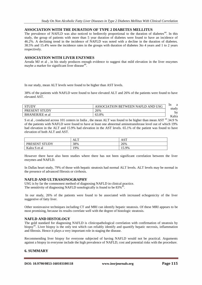
ASSOCIATION WITH DYSLIPIDEMIA In our study, 12% of the total cases included were found to have dyslipidemia. Out of the dyslipidemics , 75%
were found to have fatty liver.
In a study by Kalra S et al, outof 485 patients with dyslipidemia , 311 (59.6%) were found to have NAFLD 41
.
In a study by Viswanathan et al , 85.3% of the subjects with dyslipidemia were found to be associated with
NAFLD46
.
STUDY PREVELENCE OF NAFLD AMONG
DYSLIPIDEMICS
Viswanathan et al 85.3%
PRESENT STUDY 75%
Kalra S et al 59.6%
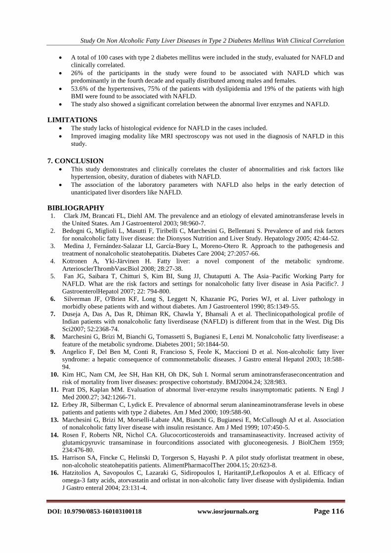
ASSOCIATION WITH BMI Obesity in particular central obesity has been described as one of the strongest risk factors NAFLD and
fibrosis42
.
In our study, 34% of the total cases were found to have BMI > 25 Kg/m2.
. Out of which 19% were found to
have fatty liver.
In a study conducted in Kalra S et al, 53.6% of those patients with obesity enrolled in the study were found to be
associated with fatty liver42
.
In a study by Viswanathan et al 27.6% of the patients with BMI >25% enrolled were found to be associated with
NAFLD46
.
STUDY PREVELENCE OF NAFLD AMONG CASES
WITH BMI>25 KG/M2
PRESENT STUDY 19%
Viswanathan et al 27.6%
Kalra S et al 54.9%
Study On Non Alcoholic Fatty Liver Diseases in Type 2 Diabetes Mellitus With Clinical Correlation
DOI: 10.9790/0853-160103100118 www.iosrjournals.org Page 115
ASSOCIATION WITH THE DURATION OF TYPE 2 DIABETES MELLITUS The prevalence of NAFLD was also noticed to bedirectly proportional to the duration of diabetes
44. In this
study, the group of patients with more than 5 year duration of diabetes were found to have an incidence of
46.2%. A declining trend in the incidence of NAFLD was noted with a decline in the duration of diabetes.
38.5% and 15.4% were the incidence rates in the groups with duration of diabetes 3to 4 years and 1 to 2 years
respectively.
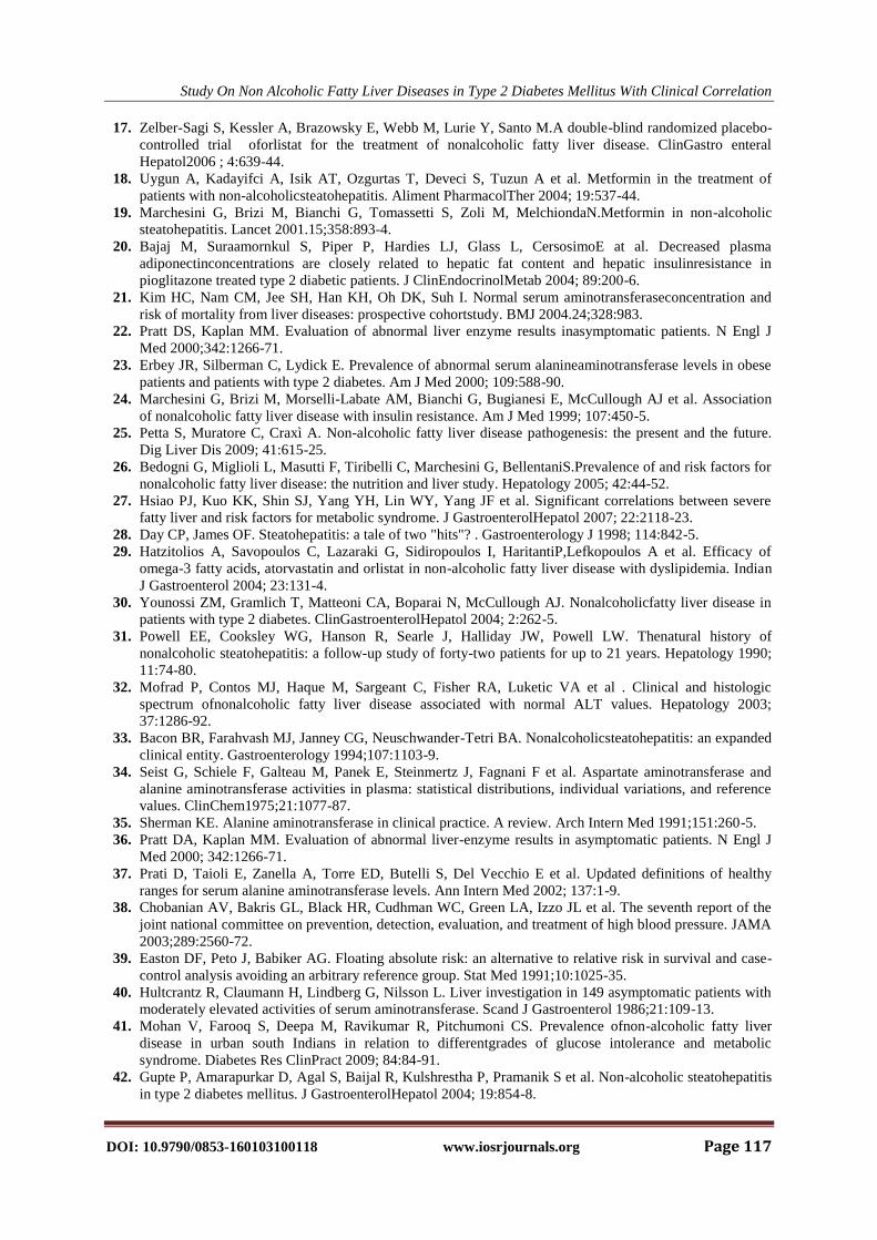
ASSOCIATION WITH LIVER ENZYMES Arruda MJ et al , in his study produces enough evidence to suggest that mild elevation in the liver enzymes
maybe a marker for significant liver disease40
.
In our study, mean ALT levels were found to be higher than AST levels.
38% of the patients with NAFLD were found to have elevated ALT and 26% of the patients were found to have
elevated AST.
In a
study
by
Kalra
S et al , conducted across 101 centers in India , the mean ALT was found to be higher than mean AST 41
.34.9 %
of the patients with NAFLD were found to have at least one abnormal aminotransferase level out of which 19%
had elevation in the ALT and 15.9% had elevation in the AST levels. 65.1% of the patient was found to have
elevation of both ALT and AST.
ALT AST
PRESENT STUDY 38% 26%
Kalra S et al 19% 15.9%
However there have also been studies where there has not been significant correlation between the liver
enzymes and NAFLD.
In Dallas heart study, 79% of those with hepatic steatosis had normal ALT levels. ALT levels may be normal in
the presence of advanced fibrosis or cirrhosis.
NAFLD AND ULTRASONOGRAPHY USG is by far the commonest method of diagnosing NAFLD in clinical practice.
The sensitivity of diagnosing NAFLD sonologically is found to be 83%45
.
In our study, 26% of the patients were found to be associated with increased echogenicity of the liver
suggestive of fatty liver.
Other noninvasive techniques including CT and MRI can identify hepatic steatosis. Of these MRI appears to be
most promising, because its results correlate well with the degree of histologic steatosis.
NAFLD AND HISTOLOGY The gold standard for diagnosing NAFLD is clinicopathological correlation with confirmation of steatosis by
biopsy42
. Liver biopsy is the only test which can reliably identify and quantify hepatic necrosis, inflammation
and fibrosis. Hence it plays a very important role in staging the disease.
Recommending liver biopsy for everyone subjected of having NAFLD would not be practical. Arguments
against a biopsy in everyone include the high prevalence of NAFLD, cost and potential risks with the procedure.
6. SUMMARY
STUDY ASSOCIATION BETWEEN NAFLD AND USG
PRESENT STUDY 26%
BHANERJEE et al 63.8%
Study On Non Alcoholic Fatty Liver Diseases in Type 2 Diabetes Mellitus With Clinical Correlation
DOI: 10.9790/0853-160103100118 www.iosrjournals.org Page 116
A total of 100 cases with type 2 diabetes mellitus were included in the study, evaluated for NAFLD and
clinically correlated.
26% of the participants in the study were found to be associated with NAFLD which was
predominantly in the fourth decade and equally distributed among males and females.
53.6% of the hypertensives, 75% of the patients with dyslipidemia and 19% of the patients with high
BMI were found to be associated with NAFLD.
The study also showed a significant correlation between the abnormal liver enzymes and NAFLD.
LIMITATIONS The study lacks of histological evidence for NAFLD in the cases included.
Improved imaging modality like MRI spectroscopy was not used in the diagnosis of NAFLD in this
study.
7. CONCLUSION
This study demonstrates and clinically correlates the cluster of abnormalities and risk factors like
hypertension, obesity, duration of diabetes with NAFLD.
The association of the laboratory parameters with NAFLD also helps in the early detection of
unanticipated liver disorders like NAFLD.
BIBLIOGRAPHY 1. Clark JM, Brancati FL, Diehl AM. The prevalence and an etiology of elevated aminotransferase levels in
the United States. Am J Gastroenterol 2003; 98:960-7.
2. Bedogni G, Miglioli L, Masutti F, Tiribelli C, Marchesini G, Bellentani S. Prevalence of and risk factors
for nonalcoholic fatty liver disease: the Dionysos Nutrition and Liver Study. Hepatology 2005; 42:44-52.
3. Medina J, Fernández-Salazar LI, García-Buey L, Moreno-Otero R. Approach to the pathogenesis and
treatment of nonalcoholic steatohepatitis. Diabetes Care 2004; 27:2057-66.
4. Kotronen A, Yki-Järvinen H. Fatty liver: a novel component of the metabolic syndrome.
ArteriosclerThrombVascBiol 2008; 28:27-38.
5. Fan JG, Saibara T, Chitturi S, Kim BI, Sung JJ, Chutaputti A. The Asia–Pacific Working Party for
NAFLD. What are the risk factors and settings for nonalcoholic fatty liver disease in Asia Pacific?. J
GastroenterolHepatol 2007; 22: 794-800.
6. Silverman JF, O'Brien KF, Long S, Leggett N, Khazanie PG, Pories WJ, et al. Liver pathology in
morbidly obese patients with and without diabetes. Am J Gastroenterol 1990; 85:1349-55.
7. Duseja A, Das A, Das R, Dhiman RK, Chawla Y, Bhansali A et al. Theclinicopathological profile of
Indian patients with nonalcoholic fatty liverdisease (NAFLD) is different from that in the West. Dig Dis
Sci2007; 52:2368-74.
8. Marchesini G, Brizi M, Bianchi G, Tomassetti S, Bugianesi E, Lenzi M. Nonalcoholic fatty liverdisease: a
feature of the metabolic syndrome. Diabetes 2001; 50:1844-50.
9. Angelico F, Del Ben M, Conti R, Francioso S, Feole K, Maccioni D et al. Non-alcoholic fatty liver
syndrome: a hepatic consequence of commonmetabolic diseases. J Gastro enteral Hepatol 2003; 18:588-
94.
10. Kim HC, Nam CM, Jee SH, Han KH, Oh DK, Suh I. Normal serum aminotransferaseconcentration and
risk of mortality from liver diseases: prospective cohortstudy. BMJ2004.24; 328:983.
11. Pratt DS, Kaplan MM. Evaluation of abnormal liver-enzyme results inasymptomatic patients. N Engl J
Med 2000.27; 342:1266-71.
12. Erbey JR, Silberman C, Lydick E. Prevalence of abnormal serum alanineaminotransferase levels in obese
patients and patients with type 2 diabetes. Am J Med 2000; 109:588-90.
13. Marchesini G, Brizi M, Morselli-Labate AM, Bianchi G, Bugianesi E, McCullough AJ et al. Association
of nonalcoholic fatty liver disease with insulin resistance. Am J Med 1999; 107:450-5.
14. Rosen F, Roberts NR, Nichol CA. Glucocorticosteroids and transaminaseactivity. Increased activity of
glutamicpyruvic transaminase in fourconditions associated with gluconeogenesis. J BiolChem 1959;
234:476-80.
15. Harrison SA, Fincke C, Helinski D, Torgerson S, Hayashi P. A pilot study oforlistat treatment in obese,
non-alcoholic steatohepatitis patients. AlimentPharmacolTher 2004.15; 20:623-8.
16. Hatzitolios A, Savopoulos C, Lazaraki G, Sidiropoulos I, HaritantiP,Lefkopoulos A et al. Efficacy of
omega-3 fatty acids, atorvastatin and orlistat in non-alcoholic fatty liver disease with dyslipidemia. Indian
J Gastro enteral 2004; 23:131-4.
Study On Non Alcoholic Fatty Liver Diseases in Type 2 Diabetes Mellitus With Clinical Correlation
DOI: 10.9790/0853-160103100118 www.iosrjournals.org Page 117
17. Zelber-Sagi S, Kessler A, Brazowsky E, Webb M, Lurie Y, Santo M.A double-blind randomized placebo-
controlled trial oforlistat for the treatment of nonalcoholic fatty liver disease. ClinGastro enteral
Hepatol2006 ; 4:639-44.
18. Uygun A, Kadayifci A, Isik AT, Ozgurtas T, Deveci S, Tuzun A et al. Metformin in the treatment of
patients with non-alcoholicsteatohepatitis. Aliment PharmacolTher 2004; 19:537-44.
19. Marchesini G, Brizi M, Bianchi G, Tomassetti S, Zoli M, MelchiondaN.Metformin in non-alcoholic
steatohepatitis. Lancet 2001.15;358:893-4.
20. Bajaj M, Suraamornkul S, Piper P, Hardies LJ, Glass L, CersosimoE at al. Decreased plasma
adiponectinconcentrations are closely related to hepatic fat content and hepatic insulinresistance in
pioglitazone treated type 2 diabetic patients. J ClinEndocrinolMetab 2004; 89:200-6.
21. Kim HC, Nam CM, Jee SH, Han KH, Oh DK, Suh I. Normal serum aminotransferaseconcentration and
risk of mortality from liver diseases: prospective cohortstudy. BMJ 2004.24;328:983.
22. Pratt DS, Kaplan MM. Evaluation of abnormal liver enzyme results inasymptomatic patients. N Engl J
Med 2000;342:1266-71.
23. Erbey JR, Silberman C, Lydick E. Prevalence of abnormal serum alanineaminotransferase levels in obese
patients and patients with type 2 diabetes. Am J Med 2000; 109:588-90.
24. Marchesini G, Brizi M, Morselli-Labate AM, Bianchi G, Bugianesi E, McCullough AJ et al. Association
of nonalcoholic fatty liver disease with insulin resistance. Am J Med 1999; 107:450-5.
25. Petta S, Muratore C, Craxì A. Non-alcoholic fatty liver disease pathogenesis: the present and the future.
Dig Liver Dis 2009; 41:615-25.
26. Bedogni G, Miglioli L, Masutti F, Tiribelli C, Marchesini G, BellentaniS.Prevalence of and risk factors for
nonalcoholic fatty liver disease: the nutrition and liver study. Hepatology 2005; 42:44-52.
27. Hsiao PJ, Kuo KK, Shin SJ, Yang YH, Lin WY, Yang JF et al. Significant correlations between severe
fatty liver and risk factors for metabolic syndrome. J GastroenterolHepatol 2007; 22:2118-23.
28. Day CP, James OF. Steatohepatitis: a tale of two "hits"? . Gastroenterology J 1998; 114:842-5.
29. Hatzitolios A, Savopoulos C, Lazaraki G, Sidiropoulos I, HaritantiP,Lefkopoulos A et al. Efficacy of
omega-3 fatty acids, atorvastatin and orlistat in non-alcoholic fatty liver disease with dyslipidemia. Indian
J Gastroenterol 2004; 23:131-4.
30. Younossi ZM, Gramlich T, Matteoni CA, Boparai N, McCullough AJ. Nonalcoholicfatty liver disease in
patients with type 2 diabetes. ClinGastroenterolHepatol 2004; 2:262-5.
31. Powell EE, Cooksley WG, Hanson R, Searle J, Halliday JW, Powell LW. Thenatural history of
nonalcoholic steatohepatitis: a follow-up study of forty-two patients for up to 21 years. Hepatology 1990;
11:74-80.
32. Mofrad P, Contos MJ, Haque M, Sargeant C, Fisher RA, Luketic VA et al . Clinical and histologic
spectrum ofnonalcoholic fatty liver disease associated with normal ALT values. Hepatology 2003;
37:1286-92.
33. Bacon BR, Farahvash MJ, Janney CG, Neuschwander-Tetri BA. Nonalcoholicsteatohepatitis: an expanded
clinical entity. Gastroenterology 1994;107:1103-9.
34. Seist G, Schiele F, Galteau M, Panek E, Steinmertz J, Fagnani F et al. Aspartate aminotransferase and
alanine aminotransferase activities in plasma: statistical distributions, individual variations, and reference
values. ClinChem1975;21:1077-87.
35. Sherman KE. Alanine aminotransferase in clinical practice. A review. Arch Intern Med 1991;151:260-5.
36. Pratt DA, Kaplan MM. Evaluation of abnormal liver-enzyme results in asymptomatic patients. N Engl J
Med 2000; 342:1266-71.
37. Prati D, Taioli E, Zanella A, Torre ED, Butelli S, Del Vecchio E et al. Updated definitions of healthy
ranges for serum alanine aminotransferase levels. Ann Intern Med 2002; 137:1-9.
38. Chobanian AV, Bakris GL, Black HR, Cudhman WC, Green LA, Izzo JL et al. The seventh report of the
joint national committee on prevention, detection, evaluation, and treatment of high blood pressure. JAMA
2003;289:2560-72.
39. Easton DF, Peto J, Babiker AG. Floating absolute risk: an alternative to relative risk in survival and case-
control analysis avoiding an arbitrary reference group. Stat Med 1991;10:1025-35.
40. Hultcrantz R, Claumann H, Lindberg G, Nilsson L. Liver investigation in 149 asymptomatic patients with
moderately elevated activities of serum aminotransferase. Scand J Gastroenterol 1986;21:109-13.
41. Mohan V, Farooq S, Deepa M, Ravikumar R, Pitchumoni CS. Prevalence ofnon-alcoholic fatty liver
disease in urban south Indians in relation to differentgrades of glucose intolerance and metabolic
syndrome. Diabetes Res ClinPract 2009; 84:84-91.
42. Gupte P, Amarapurkar D, Agal S, Baijal R, Kulshrestha P, Pramanik S et al. Non-alcoholic steatohepatitis
in type 2 diabetes mellitus. J GastroenterolHepatol 2004; 19:854-8.
Study On Non Alcoholic Fatty Liver Diseases in Type 2 Diabetes Mellitus With Clinical Correlation
DOI: 10.9790/0853-160103100118 www.iosrjournals.org Page 118
43. Prashanth M, Ganesh HK, Vima MV, John M, Bandgar T, Joshi SR et al. Prevalence of nonalcoholic
fattyliver disease in patients with type 2 diabetes mellitus. J Assoc PhysiciansIndia 2009 ;57:205-10.
44. Singh SP, Nayak S, Swain M, Rout N, Mallik RN, Agrawal O et al.Prevalence of nonalcoholic fatty liver
disease in coastal eastern India: aPreliminaryultrasonographic survey. Trop Gastro enteral 2004;25:76-9.
45. Uchil D, Pipalia D, Chawla M, Patel R, Maniar S, Narayani et al.Non-alcoholic fatty liver disease
(NAFLD)-the hepatic component of metabolicsyndrome. J Assoc Physicians India 2009;57:201-4.
46. Amarapurkar DN, Amarapurkar AD. Nonalcoholic steatohepatitis:clinicopathological profile. J Assoc
Physicians India 2000; 48:311-3.