Embed Size (px)
Citation preview
Study on antibacterial activity against acne-involved bacteria and stability of
naphthoquinone rich Plumbago indica root extract
Sermwut Kaewbumrung
A Thesis Submitted in Partial Fulfillment of the Requirements for the Degree of
Master of Pharmacy in Pharmaceutical Sciences Prince of Songkla University
2010 Copyright of Prince of Songkla University
Thesis Title Study on antibacterial activity against acne-involved bacteria and stability of naphthoquinone rich Plumbago indica root extract
Author Mr. Sermwut Kaewbumrung Major Program Pharmaceutical Sciences
The Graduate School, Prince of Songkla University, has approved this thesis as partial fulfillment of the requirements for the Master of Pharmacy Degree in Pharmaceutical Sciences
…............................................................ (Assoc. Prof. Dr. Krerkchai Thongnoo) Dean of Graduate School
Major Advisor .............................................................. (Assoc. Prof. Dr. Pharkphoom Panichayupakaranant)
Examining Committee: .............................................Chairperson (Assoc. Prof. Dr. Sunibhond Pummangura) ..............................................Committee (Assoc. Prof. Dr. Pharkphoom Panichayupakaranant) ..............................................Committee (Assist. Prof. Dr. Chatchai Wattanapiromsakul) ..............................................Committee (Assist. Prof. Dr. Sanae Kaewnopparat)
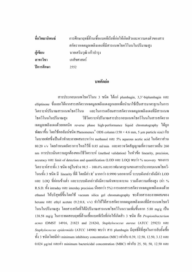
ชื่อวิทยานิพนธ การศึกษาฤทธิต์านเชื้อแบคทเีรียที่กอใหเกดิสิวและความคงตัวของสารสกัดรากเจตมลูเพลิงแดงที่มสีารแนพโธควิโนนในปริมาณสูง
ผูเขียน นายเสริมวุฒิ แกวบํารุง สาขาวิชา เภสัชศาสตร ปการศึกษา 2552
บทคัดยอ
สารประกอบแนพโธควิโนน 3 ชนิด ไดแก plumbagin, 3,3’-biplumbagin และ elliptinone ซ่ึงแยกไดจากสารสกัดรากเจตมูลเพลิงแดงถูกแยกเพื่อนํามาใชเปนสารมาตรฐานในการวิเคราะหปริมาณสารแนพโธควิโนน และในการเตรยีมสารสกัดรากเจตมูลเพลิงแดงที่มีสารแนพโธควิโนนในปริมาณสูง วิธีวิเคราะหปริมาณสารประกอบแนพโธควิโนนในสารสกดัรากเจตมูลเพลิงแดงดวยเทคนิค reverse phase high-performance liquid chromatography ไดถูกพัฒนาขึ้น โดยใชคอลัมนชนิด Phenomenex® ODS column (150 × 4.6 mm, 5 µm particle size) กับ โมบายเฟสซึ่งเปนตัวทําละลายผสมระหวาง methanol และ 5% aqueous acetic acid ในอัตราสวน 80:20 v/v โดยกําหนดอัตราการไหลไวที่ 0.85 ml/min และตรวจวดัสัญญาณที่ความยาวคลื่น 260 nm การประเมินความถูกตองของวิธีวิเคราะห (method validation) ในหัวขอ linearity, precision, accuracy และ limit of detection and quantification (LOD และ LOQ) พบวา % recovery ของการวิเคราะหสารทั้ง 3 ชนิด อยูในชวง 98.5 – 100.6% และกราฟมาตรฐานของสารประกอบแนพโธควิ-โนนทั้ง 3 ชนิด มี linearity ที่ดี โดยมีคา R2 มากกวา 0.9990 นอกจากนี้ ระบบดังกลาวยังมีคา LOD และ LOQ ที่คอนขางต่ํา และระบบดังกลาวยังมีความจําเพาะเจาะจง รวมถึงความเที่ยงสูง (คา % R.S.D. ทั้ง intraday และ interday precision นอยกวา 5%) การแยกสารสกัดรากเจตมูลเพลิงแดงดวย ethanol ใหบริสุทธิ์ขึ้นโดยใช vacuum silica gel chromatography ชะดวยสารละลายผสมของ hexane และ ethyl acetate (9.2:0.8, v/v) ทําใหไดสารสกัดรากเจตมูลเพลิงแดงทีม่ีสารแนพโธควิโนนในปริมาณสูง โดยสารสกัดที่ไดมีปริมาณสารแนพโธควิโนนรวมเพิ่มขึ้นจาก 5.80 mg/g เปน 138.58 mg/g ในการทดสอบฤทธิ์ตานเชื้อแบคทีเรียที่กอใหเกิดสิว 3 ชนิด คือ Propionibacterium acnes (DMST 14916, 21823 and 21824), Staphylococcus aureus (ATCC 25923) และ Staphylococcus epidermidis (ATCC 14990) พบวา สาร plumbagin มีฤทธิ์ดีที่สุดในการยับยั้งเชือ้ทั้ง 3 ชนิดโดยมีคา minimum inhibitory concentration (MIC) เทากับ 0.39, 12.50, 12.50, 3.12 และ 0.024 µg/ml และคา minimum bactericidal concentration (MBC) เทากับ 25, 50, 50, 12.50 และ
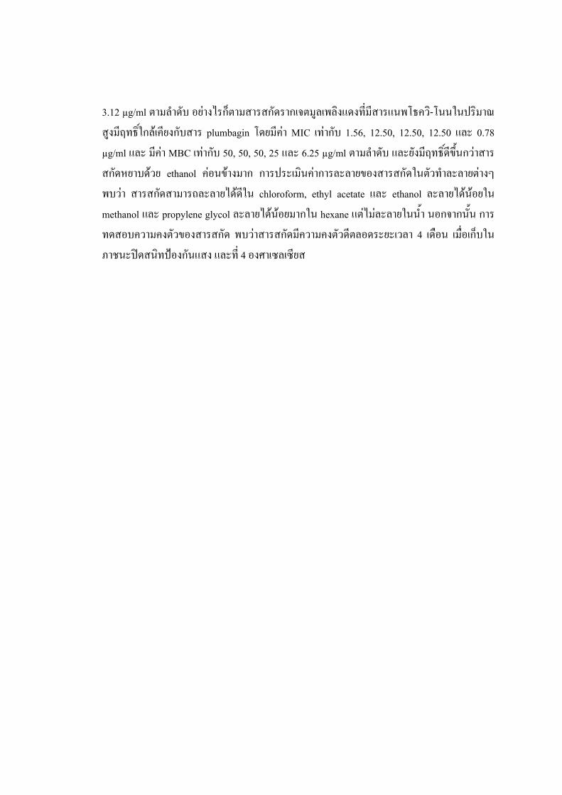
3.12 µg/ml ตามลําดับ อยางไรก็ตามสารสกัดรากเจตมูลเพลิงแดงที่มีสารแนพโธคว-ิโนนในปริมาณสูงมีฤทธิ์ใกลเคียงกับสาร plumbagin โดยมีคา MIC เทากับ 1.56, 12.50, 12.50, 12.50 และ 0.78 µg/ml และ มีคา MBC เทากบั 50, 50, 50, 25 และ 6.25 µg/ml ตามลําดับ และยังมีฤทธิ์ดีขึ้นกวาสารสกัดหยาบดวย ethanol คอนขางมาก การประเมินคาการละลายของสารสกัดในตวัทําละลายตางๆ พบวา สารสกดัสามารถละลายไดดีใน chloroform, ethyl acetate และ ethanol ละลายไดนอยใน methanol และ propylene glycol ละลายไดนอยมากใน hexane แตไมละลายในน้ํา นอกจากนั้น การทดสอบความคงตัวของสารสกัด พบวาสารสกัดมีความคงตัวดีตลอดระยะเวลา 4 เดอืน เมื่อเก็บในภาชนะปดสนทิปองกันแสง และที่ 4 องศาเซลเซียส
Thesis Title Study on antibacterial activity against acne-involved bacteria and stability of naphthoquinone rich Plumbago indica root extract
Author Mr. Sermwut Kaewbumrung Major Program Pharmaceutical Sciences Academic Year 2009
ABSTRACT
Three naphthoquinones including plumbagin, 3,3’-biplumbagin and elliptinone were isolated from the roots of Plumbago indica L. and used as standard naphthoquinones for determination and preparation of the naphthoquinone-rich P. indica root extract. The reversed-phase high-performance liquid chromatographic method was developed for quantification of plumbagin, 3,3’-biplumbagin and elliptinone in the root extract. The method involved the use of a Phenomenex® ODS column (150 × 4.6 mm, 5 µm particle size) with the mixture of methanol and 5% v/v aqueous acetic acid (80:20, v/v) as the mobile phase, flow rate 0.85 ml/min, and peaks were detected at 260 nm. The parameters of linearity, precision, accuracy and limit of detection and quantification of the method were evaluated. The recovery of the method was 98.5 – 100.6% and good linearity (R2 > 0.9990) was obtained for all naphthoquinones. The low limit of detection and quantification, high degree of specificity as well as intraday and interday precision (% R.S.D. was less than 5%) were also achieved. Fractionation of ethanol extract using a vacuum silica gel chromatography eluted with a mixture of hexane and ethyl acetate (9.2:0.8, v/v) afforded a naphthoquinone-rich P. indica root extract. The total content of naphthoquinones was increase from 0.58 mg/g to 138.58 mg/g of the extract. For antibacterial activity against acne-involved bacteria, including Propionibacterium acnes (DMST 14916, 21823 and 21824), Staphylococcus aureus (ATCC 25923) and Staphylococcus epidermidis (ATCC 14990), plumbagin exhibited the strongest antibacterial activity with the minimum inhibitory concentration (MIC) values of 0.39, 12.50, 12.50, 3.12 and 0.024 µg/ml and the minimum bactericidal concentration (MBC) values of 25, 50, 50, 12.50 and 3.12 µg/ml, respectively. However, the naphthoquinone-rich P. indica root extract showed closely activity to plumbagin which MIC values of 1.56, 12.50, 12.50, 12.50 and 0.78 µg/ml and MBC values of 50, 50, 50, 25 and 6.25 µg/ml, respectively. In addition, the
antimicrobial activity of the naphthoquinone-rich extract was also much more potent than the P. indica ethanol extract. Solubility evaluation of the naphthoquinone-rich extract in various solvents found that the extract was freely soluble in chloroform, ethyl acetate and ethanol, slightly soluble in methanol and propylene glycol, very slightly soluble in hexane and practically insoluble in water. The naphthoquinone-rich extract exhibited good stability when kept in well-sealed closed containers protected from light and stored in cool place (4°C).
CONTENT
Page บทคัดยอ iii ABSTRACT v ACKNOWLEDGEMENTS vii CONTENT viii LIST OF TABLES xi LIST OF FIGURES xiii LIST OF ABBREVIATIONS AND SYMBOLS xv CHAPTER 1. INTRODUCTION 1.1 Background 1 1.2 Objectives 3 1.3 Literature review 4 1.3.1 Plumbago indica L. 5 1.3.2 Plumbagin 11 1.3.3 Propionibacterium acnes 16 2. MATERIALS AND METHOD
2.1 Material 18 2.1.1 Plant material 18 2.1.2 Microorganism 19 2.1.3 Chemical and reagent 19 2.1.4 Instruments 20
2.2 Methods 2.2.1 Preparation of ethyl acetate extract of P. indica root 21 2.2.2 Isolation of naphthoquinones from P. indica. root 21 2.2.3 HPLC method 23
2.2.3.1 Standard solution 23
CONTENT (continued)
Page 2.2.3.2 Sample preparation 23
2.2.4 Validation of analytical method 24 2.2.4.1 Specificity 24 2.2.4.2 Linearity 24 2.2.4.3 Accuracy 24 2.2.4.4 Precision 25 2.2.4.5 LOD and LOQ 25
2.2.5 Determination of solvent for extraction 25 2.2.6 Determination of fractionation method
2.2.6.1 Preparation of P. indica root extract 25 2.2.6.2 Fractionation by anion exchange chromatography 26 2.2.6.3 Fractionation by silica gel vacuum chromatography 26 2.2.6.4 Partition by liquid-liquid extraction 26
2.2.7 Antibacterial activity against acne-involved bacteria 27 2.2.7.1 Determination of MIC 27 2.2.7.2 Determination of MBC 28 2.2.8 Determination of solubility 29 2.2.9 Stability of naphthoquinones rich P. indica L. root extract 30 2.2.9.1 Effect of light on stability of the extract 30 2.2.9.2 Effect of temperature on stability of the extract 30 2.2.9.3 Effect of pH on stability of the extract 31 2.2.9.4 Effect of accelerated condition on stability of the extract 31 2.2.10 Statistical analysis 31
3. RESULTS AND DISCUSSION
3.1 Isolation of naphthoquinones from P. indica root 32
CONTENT (continued)
Page 3.2 Structure identification 33
3.2.1 Identification of NQ 1 33 3.2.2 Identification of NQ 2 37 3.2.3 Identification of NQ 3 41
3.3 HPLC quantitative determination of naphthoquinones in 46 P. indica root extracts
3.4 Validation of analytical method 47 3.4.1 Specificity 47 3.4.2 Linearity 49 3.4.3 Accuracy 50 3.4.4 Precision 51 3.4.5 LOD and LOQ 51
3.5 Determination of solvent for extraction 52 3.6 Simple purification method to improved naphthoquinone content 53 3.7 Antibacterial activity against acne-involved bacteria 55 3.8 Solubility of naphthoquinone-rich P. indica root extract 57 3.9 Stability of naphthoquinones rich P. indica root extract 58 3.9.1 Effect of light on stability of the extract 58 3.9.2 Effect of temperature on stability of the extract 60 3.9.3 Effect of pH on stability of the extract 62 3.9.4 Effect of accelerated condition on stability of the extract 67
4. CONCLUSIONS 69 APPENDIX 71 BIBLIOGRAPHY 73 VITAE 80
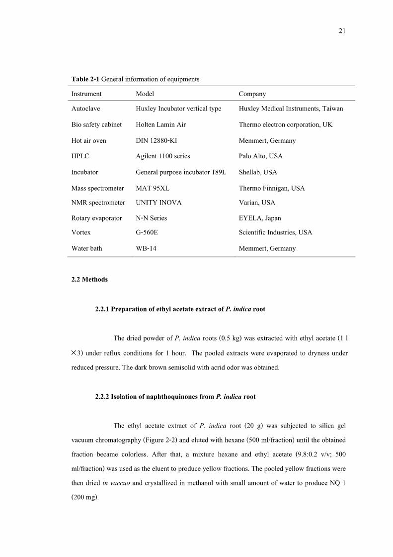
LIST OF TABLES Table Page 2-1 General information of equipments 21
2-2 The solubility term 30 3-1 1H NMR spectral data of NQ 1 and plumbagin 35 3-2 1H NMR and 13C NMR spectral data of NQ 2 37 3-3 1H NMR and 13C NMR spectral data of NQ 3 45 3-4 Retention time, linear ranges and correlation coefficients of calibration curves 50 3-5 Recoveries of naphthoquinones from P. indica root extract 50 3-6 Intraday and interday precision data of P. indica root extract 51 3-7 LOD and LOQ of P. indica root extract 52 3-8 Yield and naphthoquinone content in P. indica root extract 52 3-9 The naphthoquinone content in purified P. indica root extract from 54 various methods 3-10 The naphthoquinone content in naphthoquinone-rich P. indica root extract 54 3-11 The minimum inhibitory concentration against acne-involved bacteria 56 3-12 The minimum bactericidal concentration against acne-involved bacteria 57 3-13 Solubility of naphthoquinone-rich P. indica root extract in various solvents 58 3-14 Naphthoquinone content of naphthoquinone-rich P. indica root extract stored 59
under light and protected from light conditions 3-15 Naphthoquinone content of naphthoquinone-rich P. indica root extract stored 61 under 4°C and 30°C 3-16 Plumbagin content of naphthoquinone-rich P. indica root extract in solution 64
at pH 5.5, 7.0 and 8.0 3-17 3,3'-biplumbagin content of naphthoquinone-rich P. indica root extract in 64
solution at pH 5.5, 7.0 and 8.0 3-18 Elliptinone content of naphthoquinone-rich P. indica root extract in solution 65
at pH 5.5, 7.0 and 8.0

LIST OF TABLES (continued) Table Page
3-19 Naphthoquinone content of naphthoquinone-rich P. indica root extract store 67 under accelerate condition
LIST OF FIGURES Figure Page
1-1 Plumbago indica L. 5 1-2 Chemical structures of naphthoquinone from P. indica 7 1-3 Chemical structures of plumbaginol 8 1-4 Chemical structures of leucodelphinidin 8 1-5 Chemical structures of steroids from P. indica 9 1-6 Propionibacterium acnes 16 2-1 Dried roots P. indica 18 2-2 Vacuum silica gel chromatography (A) packed column before elution 22 (B) after elution with a mixture of hexane and ethyl acetate 2-3 Sterilized 96-well plates for determination of MIC before added Alamar blue 28 2-4 Determination of MIC using 96-well plates after added Alamar blue 29 3-1 Crude extract of P. indica 32 3-2 TLC chromatogram of (1) NQ 1, (2) NQ 2 and (3) NQ 3 developed in hexane 33
and ethyl acetate (9.6:0.4 v/v), (A) before spray with 10% KOH and (B) after spray with 10%
3-3 HPLC chromatogram of NQ 1 (A) compare to plumbagin (B) 34 3-4 Chemical structure of plumbagin 35 3-5 1H-NMR (CDCl3; 500 MHz) of NQ 1 36 3-6 1H-NMR (CDCl3; 500 MHz) of NQ 2 38 3-7 13C-NMR (CDCl3; 125 MHz) of NQ 2 39 3-8 Mass spectroscopy of NQ 2 40 3-9 Chemical structure of 3,3’-biplumbagin 41 3-10 1H-NMR (CDCl3; 500 MHz) of NQ 3 42 3-11 13C-NMR (CDCl3; 125 MHz) of NQ 3 43 3-12 Mass spectroscopy of NQ 3 44 3-13 Chemical structure of elliptinone 45
LIST OF FIGURES (Continued) Figure Page
3-14 HPLC-chromatograms of standard naphthoquinones (A) and naphthoquinone-rich 47 P. indica root extract (B) 3-15 UV absorption spectra of plumbagin in P. indica root extract 48 3-16 UV absorption spectra of elliptinone in P. indica root extract 48 3-17 UV absorption spectra of 3,3’-biplumbagin in P. indica root extract 49 3-18 Determination of MBC after steak on agar and incubated at properly conditions 56 3-19 Stability profile of naphthoquinones content at light condition 59 3-20 Stability profile of naphthoquinones content at dark condition 60 3-21 Stability profile of naphthoquinones content at 4 ± 2°C condition 61 3-22 Stability profile of naphthoquinones content at 30 ± 2°C condition 62 3-23 Extracts for stability under pH variation test at week 0 63 3-24 Extracts for stability under pH variation test after 16 weeks storage 63 3-25 Stability profile of plumbagin content at various pH conditions 65 3-26 Stability profile of 3,3’-biplumbagin content at various pH conditions 66 3-27 Stability profile of elliptinone content at various pH conditions 66 3-28 Stability profile of naphthoquinones content under accelerate condition 68
1
CHAPTER 1
INTRODUCTION
1.1 Background
Acne vulgaris is a skin disorder of the pilosebaceous units which most numerous on the face and upper back. It is characterized by open and closed comedones (blackheads and whiteheads) and inflammatory lesions including papules, pustules and nodules (Strauss et al., 2007). Although, acnes belong a common disease in adolescents and does not harmful for human life but it may cause of permanent scar and lead to significant emotional distress. (National Institute of Arthritis and Musculoskeletal and Skin Diseases, 2006)
Although mechanism of acnes has never been clearly proven, believe adrenal
androgens stimulate the lipid production of sebaceous glands and produce a more rapid turnover of follicular epithelium. Later, it becomes more cohesive. Thus the cells adhere to one another and form the follicular impaction known as the microcomedo, the precursor of all other acne lesions (Shalita, 2004).
The follicles can then be colonized by some skin normal flora bacteria include
Propionibacterium acnes, Staphylococcus aureus and Staphylococcus epidermidis (Baron, 1996). P. acnes has been described as the most important organism for development of inflammatory acne, which produce a variety of enzyme, include lipase and protease to hydrolysis sebum and release inflammatory compounds to stimulate immunological response (e.g., mast cells degranulation and neutrophil chemotaxis) resulting in inflammatory acne (Serena et al., 2006).
The current therapy to treat acne vulgaris includes topical comedolytic (retinoid
and derivative, benzoyl peroxide, azaleic acid) and antibiotics (doxycycline, tetracycline, minocycline, clindamycin and erythromycin) for both oral and topical (Krautheim, 2004).
2
Focusing on topical therapy retinoid and derivatives including tretinoid, isotretinoin, adapalene, tazarotene, motretinide, retinoyl �-glucuronide and retinaldehyde present as effective therapy with their mechanism to suppress the development of new microcomedones, inhibit inflammatory reactions via inhibition of the release of prostaglandins, leukotrienes and proinflammatory cytokines such as interferon gamma and IL-1�. Benzoyl peroxide has strong antimicrobial, slight anti-inflammatory and anticomedogenic effects. Azaleic acid has effect on chemotaxis suppression, anti-inflammation, modifies epidermal keratinization and exhibits antibacterial properties against both aerobic and anaerobic bacteria (Krautheim, 2004).
While the risk of antibiotic resistant was increased in prevalence within the
dermatologic setting (Swanson, 2003), the topical comedolytic drugs also have several side effects such as skin irritation, abnormal skin pigmentation, skin burning, skin dryness, peeling and photo sensitivity (Russell, 2000). Thus the new sources of anti acne drugs have been investigated.
For many year, medicinal plants became an extensively sources for study and
research on active compounds against several bacterial strains (Chomnawong et al., 2005). We recently studied on antibacterial activity of 20 medicinal plants including Allium sativum L. (Alliaceae), Arcangelisia flava (L.) Merr. (Menispermaceae), Azadirachta indica A. Juss. (Meliaceae), Cassia fistula L. (Fabaceae), Cassia siamea Lam. (Fabaceae), Eugenia cumini (L.) Druce. (Myrtaceae), Eupatorium odoratum L. (Asteraceae), Gynura pseudochina (L.) DC. (Compositae), Impatiens balsamina L. (Balsaminaceae), Mimusops elengi L. (Sapotaceae), Morinda citrifolia L. (Rubiaceae), Muntingia calabura L. (Muntingiaceae), Nelumbo nucifera Gaertn. (Nelumbonaceae), Phyllanthus emblica L. (Phyllanthaceae), Plumbago indica L. (Plumbaginaceae), Psidium guajava L. (Myrtaceae), Punica granatum L. (Lythraceae), Quercus infectoria Oliv. (Fagaceae), Rhinacanthus nasutus L. (Acanthaceae) and Uncaria gambia Roxb. (Rubiacea) against acne involved bacteria, P. acnes, S. aureus and S. epidermidis. The result showed that ethyl acetate extract of P. indica root exhibited the strongest antibacterial activity against P. acnes, S. aureus and S. epidermidis with minimum inhibitory concentration (MIC) values of 4.9, 312.5 and 2.4 µg/ml, respectively, and minimum bactericidal concentration (MBC)
3
values of 39.1, 312.5 and 78.1 µg/ml, respectively (Kaewbumrung and Panichayupakaranant, 2008). P. indica was therefore selected for this study.
Plumbagin, the naphthoquinone from the root part of P. indica, was well known as
the most active compound against several bacterial strains (Mallavadhani et al., 2002). So, plumbagin and its derived derivatives were used as a standard marker to prepare the naphthoquinone-rich P. indica root extract. In addition, the quantitative analytical method was necessary for monitoring naphthoquinone contents in the extract. The simple, rapid and high degree of sensitivity, precision and accuracy than would be developed and used as a valuable informative tool for naphthoquinones determination. Moreover, the solubility and stability of the extract were then studied at various solvents and conditions which useful as the necessary information for anti acne preparations from P. indica root extract in the future.
1.2 Objectives
1) To isolate naphthoquinones from Plumbago indica roots and evaluate their antibacterial activity against Propionibacterium acnes, Staphylococcus aureus and Staphylococcus epidermidis
2) To develop an HPLC analytical method for simultaneous determination of
naphthoquinones in P. indica root extracts
3) To prepare naphthoquinone-rich P. indica root extracts and investigate their properties
4
1.3 Literature review
Medicinal plants had been studied for a long time to evaluate their pharmaceutical activities and finding of active compounds. Especially in Thailand, which is rich in ethnobotanical knowledge and many plants have been used for traditional medicines. But, there are a few researches, which study on antibacterial activity against P. acnes.
There is a study on anti P. acnes activity of 19 medicinal plants including,
Andrographis paniculata Nees. (Acanthaceae), Azadirachta indica A. Juss. (Meliaceae), Barleria lupulina Lindl. (Acanthaceae), Carthamus tinctorius L. (Asteraceae), Centella asiatica (L.) Urban. (Mackinlayaceae), Clinacanthus nutans (Burm. f.) Lindau. (Acanthaceae), Cymbopogon citratus (DC.) Stapf. (Graminae), Eupatorium odoratum L. (Asteraceae), Garcinia mangostana L. (Clusiaceae), Hibiscus sabdariffa L. (Malvaceae), Houttuynia cordata Thunb. (Saururaceae), Lawsonia inermis L. (Lythraceae), Lycopersicon esculentum L. (Solanaceae), Murdannia loriformis Hassk. (Commelinaceae), Psidium guajava L. (Myrtaceae), Senna alata (L.) Roxb. (Fabaceae), Senna occidentalis L. (Fabaceae), Senna siamea (Lam.) Irwin&Barneby (Fabaceae) and Tagetes erecta L. (Compositae). It was found that G. mangostana fruit peel extract exhibited the strongest antibacterial activity against P. acnes with the same MIC and MBC values of 39 µg/ml and the active compound was mangostin (Chomnawang et al., 2005).
After that, Niyomkam (2006) has reported on a screening of antibacterial activity
against P. acnes of 18 Thai medicinal plants, including Alpinia galangal (L.) Willd. (Zingiberaceae), Andrographis paniculata Nees. (Acanthaceae), Azadirachta indica A. Juss. (Meliaceae), Boesenbergia pandurata (Roxb.) Holtt. (Zingiberaceae), Centella asiatica (L.) Urban. (Mackinlayaceae), Cinnamomum verum J. Presl. (Lauraceae), Cymbopogon citratus (DC.) Stapf. (Graminae), Dioscorea membranacea Pierre. (Dioscoreaceae), Morus alba L. (Moraceae), Ocimum americanum L. (Lamiaceae), Ocimum sanctum L. (Lamiaceae), Piper betle L. (Piperaceae), Plumbago zeylanica L. (Plumbaginaceae), Punica granatum L. (Lythraceae), Rhinacanthus nasutus L. (Acanthaceae), Syzygium aromaticum (L.) Merrill & Perry (Myrtaceae), Senna alata (L.) Roxb. (Fabaceae) and Zingiber officinalis Roscoe. (Zingiberaceae). The result
5
demonstrated that an ethyl acetate extract of A. galanga rhizome exhibited the strongest activity against P. acnes with MIC and MBC values of 156 and 312 µg/ml, respectively with the active compound was 1’-acetoxychavicol acetate (ปริศนา นิยมคํา, 2549).
Plumbago is a genus in Plumbaginaceae family. It’s characterized by herbs
perennial or rarely annual, rarely shrubs. Stems are usually branched and growing to 0.5-2 m tall. The leaves are spirally arranged, simple, entire, 0.5-12 cm long, with a tapered base and often with a hairy margin. The flowers are white, blue, purple, red, or pink, with a tubular corolla with five petal-like lobes; they are produced in racemes. The flower calyx has glandular hairs, which secrete sticky mucilage that is capable of trapping and killing insects. The ovary is ellipsoid, ovoid or pyriform. There are about 25 species around the world, native to warm temperate to tropical regions, but 2 species including Plumbago indica L. and Plumbago zeylanica L. were found in Thailand (Schlauer, 1997; Schmelzer and Gurib-Fakim, 2008).
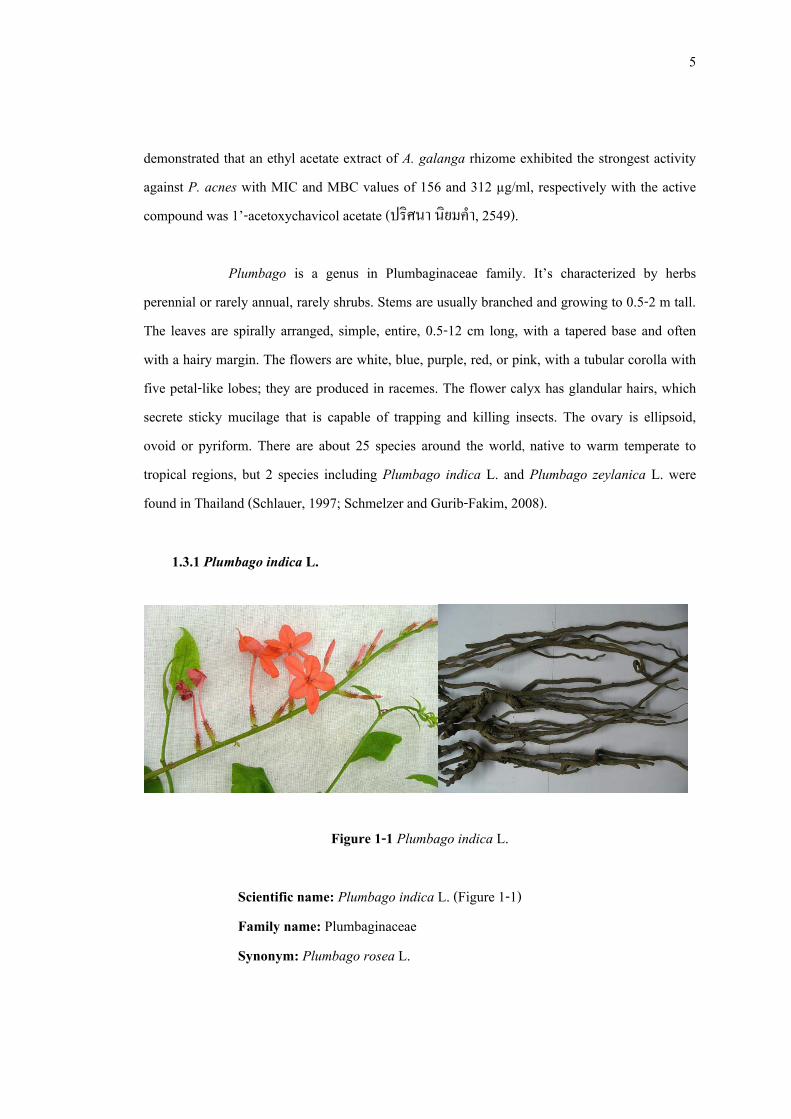
1.3.1 Plumbago indica L.
Figure 1-1 Plumbago indica L.
Scientific name: Plumbago indica L. (Figure 1-1) Family name: Plumbaginaceae Synonym: Plumbago rosea L.
6
Common name: ปดปวแดง (northern-east, northern), ไฟใตดนิ (southern), คุยวู (Khanchanaburi), Rose colored leadwort, Indian leadwort, Fire plant, Official leadwort, Kan-gyok-ni (Burmese)
P. indica is a shrubby and evergreen plant, which frequently grows to the height
of 0.5 – 2 m. Petiole base with-out auricles; leaf blade narrowly ovate to elliptic-ovate, papery, base rounded to obtuse, apex acute. Inflorescences 35–90 flowered; peduncle 1–3 cm, not glandular; rachis 10–40cm, not glandular; bracts ovate, 2–3 × 1.5–2 mm, apex acuminate; bractlets obovate-elliptic to ovate, 2–2.5 × 1.5–2 mm, apex acute. Flowers are heterostylous. Calyx 7.5–9.5 mm, glandular almost throughout, tube is 2 mm in diameter at middle. Corolla red to dark red, tube is 2–2.5 cm, apex rounded and mucronate. Anthers blue, 1.5–2 mm. Ovary ellipsoid-ovoid, indistinctly angular. Style basally pilose; short-styled forms with style arms partly exserted, stigmatic glands without enlarged apex; long-styled form with style arms completely exserted from corolla throat, stigmatic glands capitates (คณะอนกุรรมการจัดทําตําราอางอิงยาสมุนไพรไทย, 2551; Schmelzer and Gurib-Fakim, 2008).
1.2.1.1 Medicinal properties of Plumbago indica In Thai traditional medicine, P. indica roots were used for gastric stimulant,
flatulence, hemorrhoid, appetizer and adaptation of uterus after delivered. In large doses, it is acro-narcotic poison. Locally, it is used for wound healing, tinea versicolor and ringworm (คณะอนุกรรมการจัดทําตําราอางอิงยาสมุนไพรไทย, 2551).
In eastern Africa and India P. indica was traditionally used for gastric stimulant,
abortifacient and oral contraceptive. An infusion of roots is taken to treat dyspepsia, colic, cough and bronchitis. A liniment made from bruised root mixed with a little vegetable oil was used as a rubefacient to treat rheumatism and headache (Schmelzer and Gurib-Fakim, 2008).
7
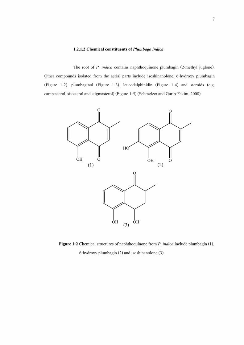
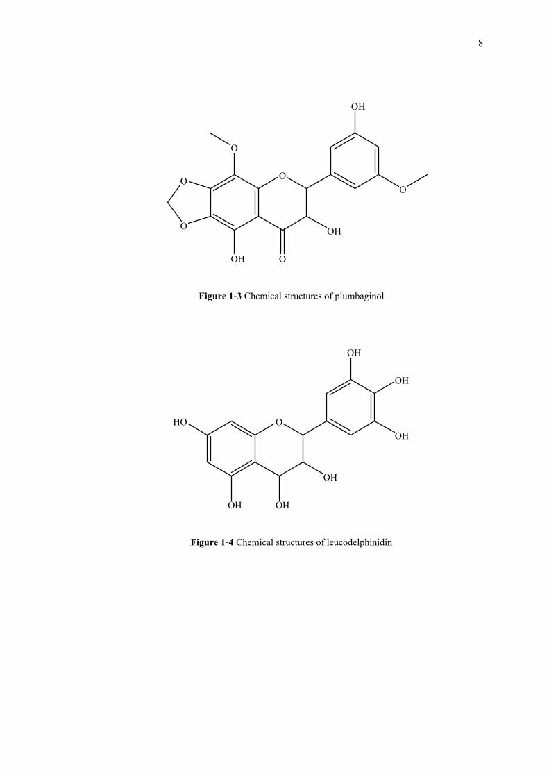
1.2.1.2 Chemical constituents of Plumbago indica The root of P. indica contains naphthoquinone plumbagin (2-methyl juglone).
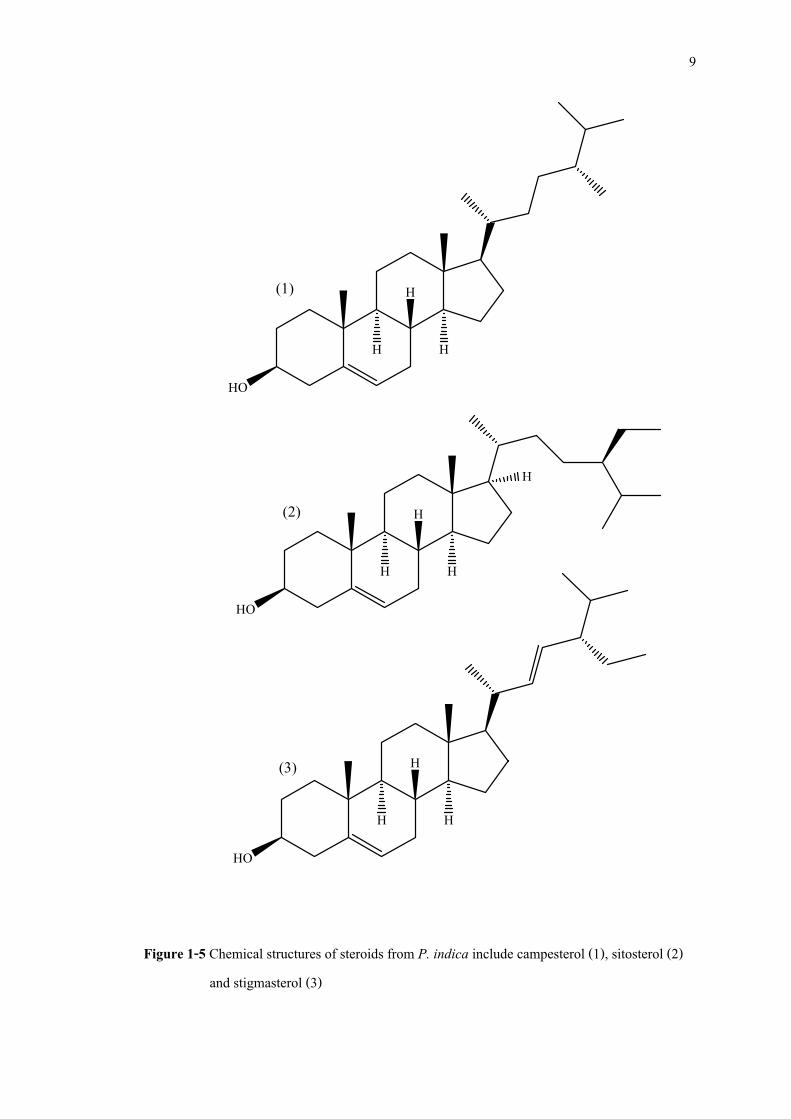
Other compounds isolated from the aerial parts include isoshinanolone, 6-hydroxy plumbagin (Figure 1-2), plumbaginol (Figure 1-3), leucodelphinidin (Figure 1-4) and steroids (e.g. campesterol, sitosterol and stigmasterol) (Figure 1-5) (Schmelzer and Gurib-Fakim, 2008).
O
OOH(1)
O
OOH
HO
(2) O
OHOH (3) Figure 1-2 Chemical structures of naphthoquinone from P. indica include plumbagin (1),
6-hydroxy plumbagin (2) and isoshinanolone (3)
8
O
O
O
O O
O
OH
OH
OH
Figure 1-3 Chemical structures of plumbaginol
OHO
OH OH
OH
OH
OH
OH
Figure 1-4 Chemical structures of leucodelphinidin
9
Figure 1-5 Chemical structures of steroids from P. indica include campesterol (1), sitosterol (2) and stigmasterol (3)
HO
H
H
H
(1)
HO
H
H
H
(3)
HO
H
H
H
H
(2)
10
1.2.1.3 Pharmacological properties of Plumbago indica Antifungal activity Hydroalcoholic (80% ethanol) extract of P. indica roots possessed potent
antifungal activity against Aspergillus niger and Candida albicans (Valsaraj et al., 1997). In addition, plumbagin had been reported as the active compound against C. albicans with MIC and MFC (Minimum fungicidal concentration) values of 0.78 and 1.56 µg/ml, respectively (Figueiredo et al., 2003).
Antibacterial activity Hydroalcoholic (80% ethanol) extract of P. indica roots exhibited antibacterial
activity against Bacillus subtilis, Pseudomonas aeruginosa, Escherichia coli and S. aureus with MIC value of 6.25 mg/ml for B. subtilis and 12.5 mg/ml for P. aeruginosa, E. coli and S. aureus (Valsaraj et al., 1997). Moreover, plumbagin had been reported as the active compound against S. aureus with MIC and MBC values of 1.56 and 25.0 µg/ml, respectively (Figueiredo et al., 2003).
Antiparasite activity P. indica roots extract showed a macrofilaricidal property against Setaria
digitata, a filarial parasite of cattle. Complete inhibition of motility was observed at concentrations range between 0.02 and 0.05 mg/ml. Fractionation of the crude extract resulted in the isolation of the active molecule plumbagin (Paiva et al., 2003).
Antifertility activity Acetone extract of P. indica stems exhibited activity in interrupting the normal
estrous cycle of female Albino rats at two dose levels, 200 and 400 mg/kg. The rats exhibited prolonged diestrous stage of the estrous cycle with consequent temporary inhibition of ovulation.
11
The anti-ovulatory activity was reversible on withdrawal of the extract. The effective acetone extract was further studied on estrogenic functionality in rats. The acetone extract showed significant estrogenic and antiestrogenic activity. Histological studies of the uteri further confirmed the estrogenic activity of the acetone extract (Sheeja et al., 2009).
1.3.2 Plumbagin
Plumbagin, the most active naphthoquinone derived from the species of Plumbago, Drosera and Diospyros, had been wildly studied on pharmacological activities. In small doses, it is a sudorific and stimulates the central nervous system, while in large doses may cause death from respiratory failure and paralysis. The pharmacological activities of plumbagin have been reported as follows:
Antitumor activity Plumbagin exhibited anticancer activity against melanoma cells line (Bowes cell)
and breast cancer cells line (MCF7) with IC50 values of 1.39 and 1.28 µM, respectively (Nguyen et al., 2004).
For breast cancer cells, plumbagin exhibited cell proliferation inhibition by
inducing cells to undergo G2-M arrest and autophagic cell death. Blockade of the cell cycle was associated with increased p21/WAF1 expression and Chk2 activation, and reduced amounts of cyclin B1, cyclin A, Cdc2, and Cdc25C. Plumbagin also reduced Cdc2 function by increasing the association of p21/WAF1/Cdc2 complex and the levels of inactivated phospho-Cdc2 and phospho-Cdc25C by Chk2 activation (Kuo et al., 2006).
Anticancer effect of plumbagin had been reported against human non-small cell
lung cancer cells A549 with IC50 value of 11.69 µM. It exhibited effective cell growth inhibition by inducing cancer cells to undergo G2-M phase arrest and apoptosis. Blockade of cell cycle was associated with increased levels of p21 and reduced amounts of cyclinB1, Cdc2, and Cdc25C.
12
Plumbagin treatment also enhanced the levels of inactivated phosphorylated Cdc2 and Cdc25C. Blockade of p53 activity by dominant-negative p53 transfection partially decreased plumbagin-induced apoptosis and G2-M arrest, suggesting it might be operated by p53- dependent and independent pathway. Plumbagin treatment triggered the mitochondrial apoptotic pathway indicated by a change in Bax/Bcl-2 ratios, resulting in mitochondrial membrane potential loss, cytochrome c release, and caspase-9 activation (Hsu et al., 2006).
Anti-inflammatory activity Plumbagin exhibited an immunomodulatory effects by inhibition of T cell
proliferation in response to polyclonal mitogen Concanavalin A (Con A) by blocking cell cycle progression (IC50 value of 50 nM). It also suppressed expression of early and late activation markers CD69 and CD25, respectively in activated T cells. The inhibition of T cell proliferation by plumbagin was accompanied by a decrease in the levels of Con A induced IL-2, IL-4, IL-6 and IFN-γ cytokines (Checker et al., 2009).
Antimalarial activity It has been reported that plumbagin exhibited anti-Plasmodium falciparum
activity by inhibition of isolated P. falciparum enzyme, succinate dehydrogenase (SDH), with IC50 value of 5 mM. It also inhibited in vitro growth of P. falciparum with IC50 value of 0.27 mM (Paiva et al., 2003).
Antibacterial activity Plumbagin has been reported as an Anti-Helicobacter pylori agent with MIC
value of 4.0 µg/ml, which more potent than that of metronidazole (MIC value of 32 µg/ml) (Park et al., 2006).
13
Farr and coworker (1985) reported on an antibacterial activity of plumbagin against wild-type E. coli strain AB1157 with 99.9% killed by exposure to 1.0 mM plumbagin for 1 hour at 37°C. Antibacterial mechanism of plumbagin may be due to its toxicity by generated active oxygen species and may damage DNA besides a pathway via H2O2.
In contrast, Jamieson and coworkers (1994) conducted tests in wild-type strain
Saccharomyces cerevisiae S150-2B and mutated strains using disruption mutations in the genes encoding of two superoxide dismutases, Cu/ZnSOD (SOD1) and mitochondrial MnSOD (SOD2), and showed that the SOD1 mutant was 100-fold more sensitive to plumbagin than its parent, while the sensitivity of the SOD2 strain to plumbagin was indistinguisable from that of the wild-type strain. Thus, Cu/ZnSOD was the prinicipal superoxide dismutating genes target.
Kamal and coworker (1995) conducted in vivo anti-S. aureus test in female mice
and showed that plumbagin was noticed to increase in the activity up to 8 weeks with 25 µg/kg body weight, due to its ability to stimulate the response on oxygen radical release by macrophages. While at high dose (50 µg/kg body weight), it has direct inhibitory activity against S. aureus.
Mutagenic activity Plumbagin was reported as an antimutagenic activity against Salmonella
typhimurium TA98 when induced by 2-nitrofluorene (2NF), 3-nitrofluoranthene (3-NFA) and 1-nitropyrene (1-NP) (Edenharder and Tang, 1997). Moreover, for Escherichia coli WP2s (uvrA trpE), plumbagin was not mutagenic when presence of plasmid pKM101 (Kato et al., 1994).
Antifertility activity Plumbagin containing albumin microspheres were implanted to 20 days pregnant
albino rats and found that their ovaries showed clear inhibition of growth of graffian follicules and degeneration of the mature follicles, and corpus luteum were observed and result to failed to
14
conceive, the antifertility action of plumbagin seemed to be related to its antiovulatory action (Kini et al., 1997).
Abortifacient activity Plumbagin administered by intubation to albino female rats at 10 mg/kg for 15
days significantly inhibited mating and prolonged duration of estrus cycle and diestrus phase. Plumbagin showed a dose-related abortifacient activity in rats administered 5-20 mg/kg orally from Day 5 to 11 of pregnancy. At doses 10-20 mg/kg from days 1 to 5 of pregnancy, plumbagin caused a significant anti-implantation effect. No gross teratogenic effects were noticed in pups born to female rats that had received 5 or 10 mg/kg plumbagin from days 1 to 5 of pregnancy (Premakumari et al., 1977).
Reproductive toxicity Plumbagin has demonstrated reproductive toxicity in male and female animals.
Teratogenic effects were not seen in limited studies. Only one of 12 female Long-Evans rats intubated with plumbagin at 10 mg/kg for 10 days conceived, bearing a litter of five pups. All 12 control animals conceived, producing an average litter size of six pups. One animal in the plumbagin group died of hemorrhage that the authors suspected was caused by competitive inhibition of vitamin K activity, needed for the synthesis of clotting factors (Azad Chowdhury et al., 1982).
Plumbagin given orally at 10 mg/kg for 10 days to adult female rats of the
Holtzman strain caused a highly significant decrease in the weight of ovaries as compared with the controls (Santhakumari and Suganthan, 1980).
Plumbagin administered intra-peritoneal at a dose of 10 mg/kg for 60 days
caused selective testicular lesions in dogs. The wet weights of testes and epididymides were
15
decreased. In addition, the seminiferous tubule and Leydig cell nuclei diameter were significantly decreased and cellular heights of epididymides were drastically curtailed (Bhargava, 1984).
Oral administration of plumbagin to male gerbils at 10 mg/day for 20 days
caused a decrease in the wet weight of seminal vesicle and prostate glands. The cell height of the secretory epithelium was also decreased, and little secretion in the lumen of these glands was observed (Bhargava, 1984).
Plumbagin caused a decrease in the number of spermatids, resting and pachytene
spermatocytes, and a significant reduction in seminiferous tubule and Leydig cell nuclei diameter when given orally to immature Wistar rats at 10 mg/kg for 32 days (Bhargava, 1986).
Cardiotonic action Plumbagin produced a triphasic inotropic response in guinea-pig papillary
muscle. Plumbagin did not cause any positive inotropy under anoxic conditions, and the positive inotropic effect was markedly inhibited by oxidative phosphorylation uncouplers (Itoigawa et al., 1991).
Hypolipidemic and antiatherosclerotic effects When administered to hyperlipidaemic rabbits, plumbagin reduced serum
cholesterol and LDL cholesterol by 53 to 86 percent and 61 to 91 percent, respectively. Furthermore, plumbagin treatment prevented the accumulation of cholesterol and triglycerides in liver and aorta and regress atheromatous plaques of the thoracic and abdominal aortas (Sharma et al., 1991).
Effects on microsomal enzymes Plumbagin exhibited a potent, dose dependent inhibitory activity against
aromatase cytochrome P450 in human placental microsomes. However, plumbagin showed
16
relatively weak reducing effects in the presence of microsomal membranes, suggesting that the inhibitory effects on monooxygenase reactions were not due to the formation of superoxide radicals (Muto et al., 1987).
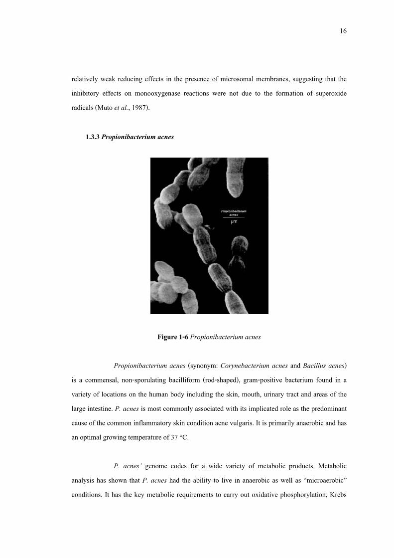
1.3.3 Propionibacterium acnes
Figure 1-6 Propionibacterium acnes
Propionibacterium acnes (synonym: Corynebacterium acnes and Bacillus acnes) is a commensal, non-sporulating bacilliform (rod-shaped), gram-positive bacterium found in a variety of locations on the human body including the skin, mouth, urinary tract and areas of the large intestine. P. acnes is most commonly associated with its implicated role as the predominant cause of the common inflammatory skin condition acne vulgaris. It is primarily anaerobic and has an optimal growing temperature of 37 °C.
P. acnes’ genome codes for a wide variety of metabolic products. Metabolic
analysis has shown that P. acnes had the ability to live in anaerobic as well as “microaerobic” conditions. It has the key metabolic requirements to carry out oxidative phosphorylation, Krebs
17
cycle, Embden-Meyerhof pathway and the pentose phosphate pathway. Under in vitro anaerobic conditions, P. acnes can grow permissively on media such as glucose, glycerol, ribose, fructose, mannose and N-acetylglucosamine. In vivo, P. acnes produce various lipases to digest excess skin oil and sebum in the pilosebaceous units (regions that contains the hair follicle and sebaceous gland) of adolescent and adult human skin. For energy, P. acnes can employ a fermentative process yielding byproducts like short-chain fatty acids and propionic acid from which it gets its name. In addition to fermentation, P. acnes can utilize various other anaerobic pathways deriving energy with the help of enzymes such as nitrate reductase, dimethyl sulfoxide reductase and fumarate reductase (Rosenberg, 1969)
18
CHAPTER 2
METERIALS AND METHODS
2.1 Materials
2.1.1 Plant material
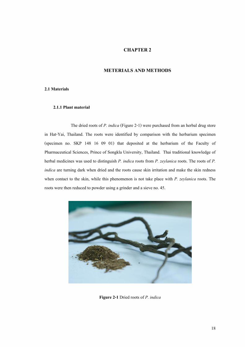
The dried roots of P. indica (Figure 2-1) were purchased from an herbal drug store in Hat-Yai, Thailand. The roots were identified by comparison with the herbarium specimen (specimen no. SKP 148 16 09 01) that deposited at the herbarium of the Faculty of Pharmaceutical Sciences, Prince of Songkla University, Thailand. Thai traditional knowledge of herbal medicines was used to distinguish P. indica roots from P. zeylanica roots. The roots of P. indica are turning dark when dried and the roots cause skin irritation and make the skin redness when contact to the skin, while this phenomenon is not take place with P. zeylanica roots. The roots were then reduced to powder using a grinder and a sieve no. 45.
Figure 2-1 Dried roots of P. indica
19
2.1.2 Microorganism
Three strains of Propionibacterium acnes (DMST 14916, DMST 21823 and DMST 21824) were obtained from Department of Medical Science Center, Thailand. They were cultured on Brain Heart Infusion agar (Becton Dickinson, USA) and incubated in anaerobic conditions using Anaerocult® A (Merck, Germany Germany) and anaerobic jar (Merck, Germany) at 37°C for 72 hours. Staphylococcus aureus (ATCC 25923) and Staphylococcus epidermidis (ATCC 14990) were obtained from Department of Microbiology, Faculty of Medicine, Prince of Songkla University, Thailand and Thailand Institute of Scientific and Technological research, respectively. They were cultured on Mueller-Hinton and incubated in aerobic conditions at 37°C for 24 hours. All tested bacteria were stored in glycerol broth at -20°C and subcultured twice in properly conditions for each bacterium before used.
2.1.3 Chemicals
2.1.3.1 For extraction and purification
- Amberlite® IRA-67 (Sigma, USA) - Chloroform, commercial grade (Lab scan Asia, Thailand) - Dichloromethane, analytical grade (Lab scan Asia, Thailand) - Diethylether, analytical grade (Lab scan Asia, Thailand) - Ethanol, commercial grade (Lab scan Asia, Thailand) - Ethyl acetate, commercial grade (Lab scan Asia, Thailand) - Hexane, commercial grade (Lab scan Asia, Thailand) - Isopropanol, analytical grade (Lab scan Asia, Thailand) - Methanol, commercial grade (Lab scan Asia, Thailand) - Methanol, HPLC grade (Lab scan Asia, Thailand) - Silica gel 60 (SiO2 60, 230-400 mesh) (Merck, Germany)
*All commercial grade organic solvents were distilled before use.
20
2.1.3.2 For antibacterial activity test
- 0.85 % w/v Sodium chloride solution - Bacto agar (Merck, Germany) - Brain heart infusion broth (Becton Dickinson, USA) - McFarland solution - Mueller-Hinton broth (Merck, Germany) - Resazurin sodium, Alama blue (Sigma, Switzerland) - Standard tetracycline disc (6 mm 30 mg/disc) (Oxoid, UK) - Tetracyclin HCl standard (Fluka, Switzerland)
2.1.3.3 For HPLC analysis
- Milli-Q grade water was purified in a Milli-Q system (Millipore, Bedford, USA)
- Methanol, HPLC grade (Lab scan Asia, Thailand) - Acetic acid, glacial AR grade (Lab scan Asia, Thailand)
2.1.4 Instruments
The equipments used in this study were listed in Table 2-1.
21
Table 2-1 General information of equipments Instrument Model Company Autoclave Huxley Incubator vertical type Huxley Medical Instruments, Taiwan Bio safety cabinet Holten Lamin Air Thermo electron corporation, UK Hot air oven DIN 12880-KI Memmert, Germany HPLC Agilent 1100 series Palo Alto, USA Incubator General purpose incubator 189L Shellab, USA Mass spectrometer MAT 95XL Thermo Finnigan, USA NMR spectrometer UNITY INOVA Varian, USA Rotary evaporator N-N Series EYELA, Japan Vortex G-560E Scientific Industries, USA Water bath WB-14 Memmert, Germany 2.2 Methods
2.2.1 Preparation of ethyl acetate extract of P. indica root
The dried powder of P. indica roots (0.5 kg) was extracted with ethyl acetate (1 l ×3) under reflux conditions for 1 hour. The pooled extracts were evaporated to dryness under reduced pressure. The dark brown semisolid with acrid odor was obtained.
2.2.2 Isolation of naphthoquinones from P. indica root
The ethyl acetate extract of P. indica root (20 g) was subjected to silica gel vacuum chromatography (Figure 2-2) and eluted with hexane (500 ml/fraction) until the obtained fraction became colorless. After that, a mixture hexane and ethyl acetate (9.8:0.2 v/v; 500 ml/fraction) was used as the eluent to produce yellow fractions. The pooled yellow fractions were then dried in vaccuo and crystallized in methanol with small amount of water to produce NQ 1 (200 mg).
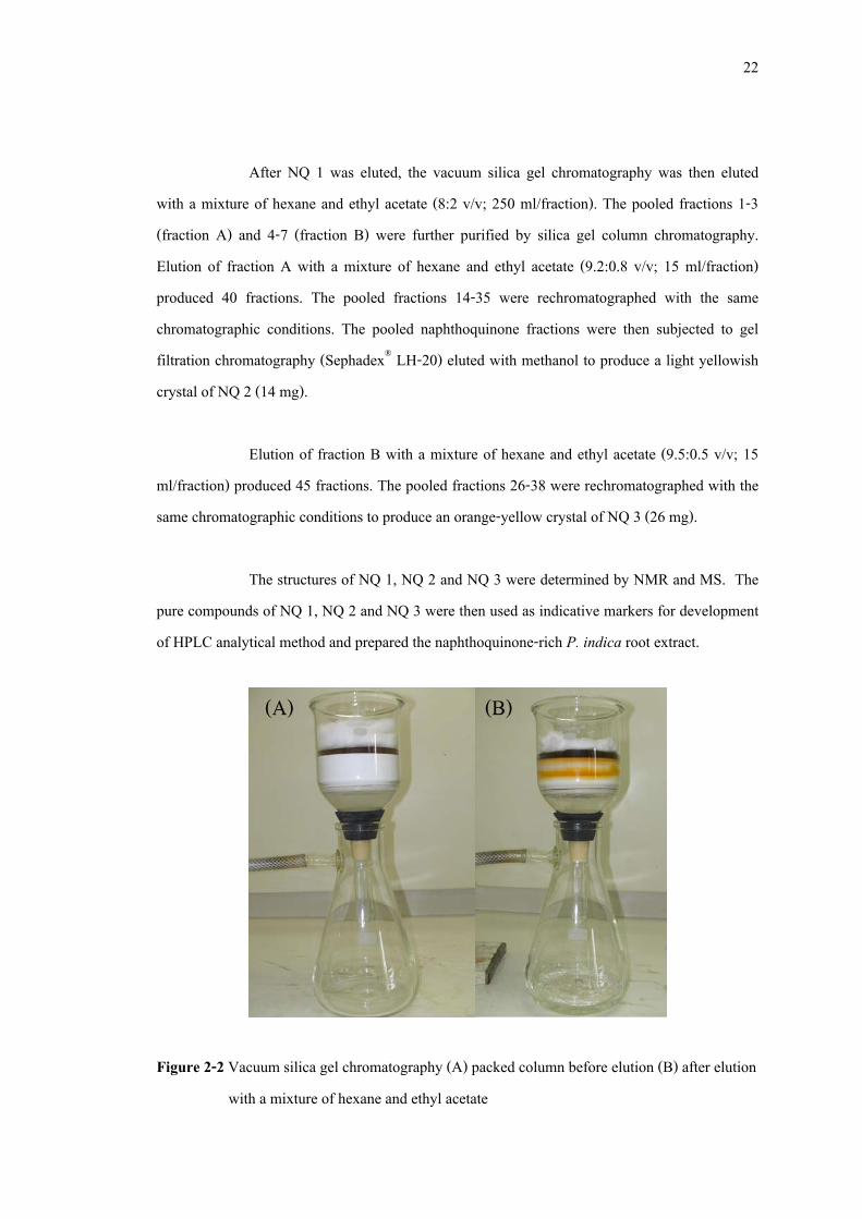
22
After NQ 1 was eluted, the vacuum silica gel chromatography was then eluted with a mixture of hexane and ethyl acetate (8:2 v/v; 250 ml/fraction). The pooled fractions 1-3 (fraction A) and 4-7 (fraction B) were further purified by silica gel column chromatography. Elution of fraction A with a mixture of hexane and ethyl acetate (9.2:0.8 v/v; 15 ml/fraction) produced 40 fractions. The pooled fractions 14-35 were rechromatographed with the same chromatographic conditions. The pooled naphthoquinone fractions were then subjected to gel filtration chromatography (Sephadex® LH-20) eluted with methanol to produce a light yellowish crystal of NQ 2 (14 mg).
Elution of fraction B with a mixture of hexane and ethyl acetate (9.5:0.5 v/v; 15
ml/fraction) produced 45 fractions. The pooled fractions 26-38 were rechromatographed with the same chromatographic conditions to produce an orange-yellow crystal of NQ 3 (26 mg).
The structures of NQ 1, NQ 2 and NQ 3 were determined by NMR and MS. The
pure compounds of NQ 1, NQ 2 and NQ 3 were then used as indicative markers for development of HPLC analytical method and prepared the naphthoquinone-rich P. indica root extract.
Figure 2-2 Vacuum silica gel chromatography (A) packed column before elution (B) after elution with a mixture of hexane and ethyl acetate
23
2.2.3 HPLC method
The HPLC analytical method was developed from the method used to quantitative analysis of 4 naphthoquinones, 1,4-naphthoquinone, lawsone, juglone and plumbagin in Dionaea muscipula Ell. crude extract (Babula et al., 2006).
HPLC analysis was carried out using Agilent 1100 liquid chromatographic system
(Palo Alto, USA) equipped with isocratic pump (G1312A), auto sampler (G1313A) and photodiode array (PDA) detector (model G1315B). Data analysis was performed using Agilent ChemStation for LC 3D software (Agilent, USA). The wavelength used for quantitative determination of the naphthoquinones was set as 260 nm. Separation was achieved isocratically at 25ºC on a Phenomenex® ODS column (150 × 4.6 mm, 5 µm particle size). The mobile phase consisted of methanol and 5% acetic acid in Milli-Q grade water (80:20 v/v) and was pumped at a flow rate of 0.85 ml/min. The injection volume was 10 µl.
2.2.3.1 Standard solution Separate stock solutions of the reference standards, plumbagin, 3,3’-biplumbagin
and elliptinone were made in methanol. A working solution of the standards was subsequently prepared in methanol and diluted to provide a series of the standard ranging from 0.03 to 10 µg/ml for 3,3’-biplumbagin and elliptinone and 0.01 to 30 µg/ml for plumbagin. The calibration curves were constructed for each of the target analyzes.
2.2.3.2 Sample preparation P. indica dried powder (500 mg) was extracted with ethanol (25 ml) under reflux
conditions for an hour (×3). The extract was then filtered and the solvent was evaporated to dryness in vacuo. The sample was then accurately weighted and dissolved in methanol to prepared stock sample solution at concentration of 10 mg/ml. The samples were filtered through
24
0.45 �m membrane filter and analyzed immediately after extraction in order to avoid possible chemical degradation.
2.2.4 Validation of analytical method
The analytical method validation for naphthoquinones derived from P. indica was examined for specificity, linearity, accuracy, precision, LOD and LOQ.
2.2.4.1 Specificity Peak identification was carried out using the standard and photodiode-array
detector. The UV spectra were taken at various points of the peaks to check peak homogeneity. 2.2.4.2 Linearity Calibration curves were constructed on three consecutive days by analysis of a
mixture containing each of the standard compounds at six concentrations. Plumbagin was performed at 0.01, 0.1, 5, 10, 20 and 30 µg/ml, 3,3’-biplumbagin and elliptinone were performed at 0.0313, 0.125, 0.5, 1, 5 and 10 µg/ml. The standard curve was analyzed using the linear least-squares regression equation derived from the peak area.
2.2.4.3 Accuracy Plumbagin at concentrations of 0.1, 5 and 20 µg/ml, 3,3’-biplumbagin and
elliptinone at concentrations of 0.125, 0.5 and 5 µg/ml were prepared and added into P. indica root extract at a ratio of 1:1 (v/v). Prior to analyze fortification, the background levels of plumbagin, 3,3’-biplumbagin and elliptinone in the extract of P. indica roots were determined so as to calculated actual recoveries. The amount of each analyte was determined in triplicate and percentage recoveries were then calculated.
25
2.2.4.4 Precision Precision experiments were conducted for intraday and interday. The solution of
one sample was used to achieve repeatability testing. The data of repeatability was the content of six injections separately in the same day. The data used to calculate % RSD of interday precision was the content of three samples analyzed in three days (three injections in succession each day).
2.2.4.5 Limit of detection (LOD) and quantification (LOQ) Serial dilutions of sample solution standards were made with methanol and
analyzed with the HPLC method. LOD and LOQ were determined by means of signal to noise ratio of 3:1 and 10:1, respectively.
2.2.5 Determination of solvent for extraction
P. indica dried powder (500 mg) was separately extracted with ethyl acetate, ethanol, isopropanol, dichloromethane and diethyl ether (25 ml) under reflux conditions for 1 hour (×3). After filtration, the pooled extracts of the same solvent were evaporated to dryness under reduced pressure, adjusted to 10 ml with methanol and subjected to HPLC analysis. The experiments were performed in triplicate.
2.2.6 Determination of fractionation method
2.2.6.1 Preparation of P. indica root extract
P. indica dried powder (1 kg) was successively extracted with the suitable solvent from 2.2.5 (5 l ×3) under reflux conditions for 1 hour. The pooled extracts were dried in vacuo and subsequently divided to purify by different technique.
26
2.2.6.2 Fractionation by anion exchange chromatography The anion exchange resin (Amberlite® IRA-67, Sigma, USA) was treated with
methanol and loaded into a column (10 × 126 cm). The column was washed twice with water and methanol, respectively. The P. indica extract (20 g) was dissolved in methanol and loaded into the column with a flow rate of 5 ml/min. The column was eluted with methanol until other impurity bands were completely washed out. The naphthoquinones were then eluted with 10% acetic acid in methanol. The pooled naphthoquinone fractions were dried in vacuo.
2.2.6.3 Fractionation by silica gel vacuum chromatography A sintered glass column (13 cm in diameter) was packed with silica gel
approximately 5 cm high. The P. indica extract (20 g), which pre-adsorbed on the silica gel, was loaded as a thin layer on the surface of column. The column was eluted with a mixture of hexane and ethyl acetate (9.2:0.8 v/v; 500 ml) with the aid of a vacuum pump. The pooled fractions of naphthoquinones were then dried in vacuo.
2.2.6.4 Partition by liquid-liquid extraction Partition with purified water The P. indica extract (20 g) was dissolved in ethyl acetate (150 ml) and then
repeatedly partitioned with water (150 ml). The pooled ethyl acetate phases were then dried in vacuo.
Partition with 5% acetic acid in water The P. indica extract (20 g) was dissolved in ethyl acetate (150 ml) and then
repeatedly partitioned with 5% acetic acid in water (150 ml). The pooled ethyl acetate phases were then dried in vacuo.
27
2.2.7 Antibacterial activity against acne-involved bacteria
The standard naphthoquinones and naphthoquinone-rich extract were evaluated for antibacterial activity against acne-involved bacteria include Propionibacterium acnes (DMST 14916, DMST 21823 and DMST 21824), Staphylococcus aureus (ATCC 25923) and Staphylococcus epidermidis (ATCC 14990). Three strains of P. acnes were grown in Brain heart infusion agar (BHI) and incubate at 37°C for 72 hours in anaerobic conditions (Anaerocult® A and Anaerobic jar, Merck, Germany). S. aureus and S. epidermidis were grown in Mueller-Hinton agar (MH agar) and incubate at 37°C for 24 hours in aerobic conditions.
2.2.7.1 Determination of MIC MIC value of the standard naphthoquinones and naphthoquinone-rich P. indica
root extract were determined using broth dilution method (Wiegan et al., 2008). The test compounds were dissolved in DMSO at the concentration of 100 µg/ml (stock solution) and then it was serial diluted with BHI or MH broth to gave the final concentrations between 0.025 and 100 µg/ml. Tetracycline hydrochloride and DMSO were used as a positive and negative control, respectively. The test was performed in 96-well plates. Two-fold dilutions were prepared directly in 96-well plate (NUNC, Denmark), as follows: 100 µl of the working solution of compounds or extract was added to well 1 of the dilution series. To each remaining well, 50 µl of BHI or MH broth were added. With a sterile pipette tip, 50 µl of the mixture was transferred from well 1 to well 2. After thorough mixing, 50 µl of the mixture was transferred to well 3. This process was continued until the last final concentration was obtained. The last well received no antimicrobial agent and served as a growth control.
The inoculum was prepared in sterile physiological saline solution and adjusted
turbidity to 0.5 McFarland standard (1.5 ×10⁸ cfu/ml). It was then further diluted 1:100 in sterile broth to contain 1.5 ×10⁶ cfu/ml and 50 µl of the adjusted inoculum was added to each well then incubated at properly conditions as described in 2.2.7.
28
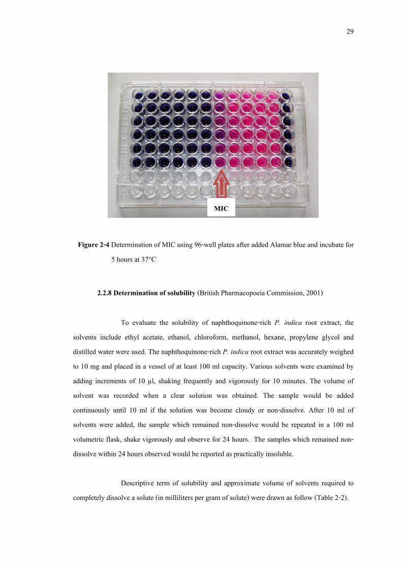
After incubation period, all wells were added with 50 µl Alamar blue (10 µg/ml) and incubated for 5 hours before determined MIC value. The blue color wells mean bacteria were inhibited by test sample and cannot growth in these medium, while pink wells mean test samples cannot inhibit bacteria growth. The lowest concentration that did not show any growth of bacteria was taken as the MIC.
2.2.7.2 Determination of MBC The incubation mixtures that showed positive result of inhibitory effect were
streaked on BHI or MH agar. The MBC was detected as the lowest concentration which no colonies of test bacteria formed on the cultivation medium.
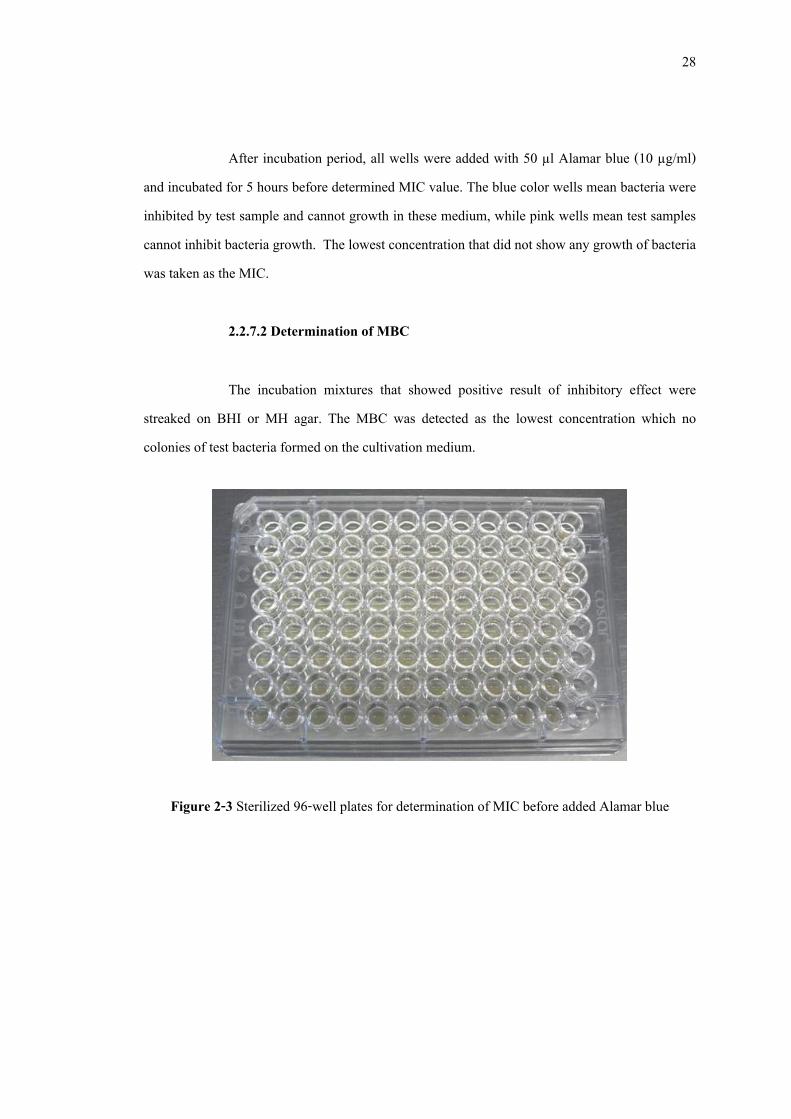
Figure 2-3 Sterilized 96-well plates for determination of MIC before added Alamar blue
29
Figure 2-4 Determination of MIC using 96-well plates after added Alamar blue and incubate for
5 hours at 37°C
2.2.8 Determination of solubility (British Pharmacopoeia Commission, 2001)
To evaluate the solubility of naphthoquinone-rich P. indica root extract, the solvents include ethyl acetate, ethanol, chloroform, methanol, hexane, propylene glycol and distilled water were used. The naphthoquinone-rich P. indica root extract was accurately weighed to 10 mg and placed in a vessel of at least 100 ml capacity. Various solvents were examined by adding increments of 10 µl, shaking frequently and vigorously for 10 minutes. The volume of solvent was recorded when a clear solution was obtained. The sample would be added continuously until 10 ml if the solution was become cloudy or non-dissolve. After 10 ml of solvents were added, the sample which remained non-dissolve would be repeated in a 100 ml volumetric flask, shake vigorously and observe for 24 hours. The samples which remained non-dissolve within 24 hours observed would be reported as practically insoluble.
Descriptive term of solubility and approximate volume of solvents required to
completely dissolve a solute (in milliliters per gram of solute) were drawn as follow (Table 2-2).
MIC
30
Table 2-2 The solubility term used to evaluated naphthoquinones rich P. indica root extract solubility
Solubility term Volume of solvent (ml) to dissolve 1 g of sample Very soluble less than 1 Freely soluble from 1 to 10 Soluble From 10 to 30 Sparingly soluble from 30 to 100 Slightly soluble from 100 to 1000 Very slightly soluble from 1000 to 10,000 Practically insoluble more than 10,000
2.2.9 Stability evaluation of naphthoquinone- rich P. indica root extract
(Sakunpak, et al., 2009)
2.2.9.1 Effect of light on stability of the extract The naphthoquinone-rich P. indica root extracts were accurately weighed to 2 mg
and kept in sealed microtubes (Axygen Scientific, USA). The extracts were then stored at 25 ± 2°C either protected from light or exposed to 36 Watts fluorescent light (40 cm distance) for 4 months. An aliquot of each sample was taken at 0, 1, 2, 3, 4, 6, 8, 12 and 16 weeks and subjected to quantitative analysis of naphthoquinone content using HPLC. The experiments were performed in triplicate.
2.2.9.2 Effect of temperature on stability of the extract The naphthoquinone-rich P. indica root extracts were accurately weighed to 2 mg
and kept in sealed microtubes and protected from light. The extracts were then stored at 4 ± 2°C and room temperature (30 ± 2°C) for 4 months. An aliquot of each sample was taken at 0, 1, 2, 3,
31
4, 6, 8, 12 and 16 weeks and subjected to quantitative analysis of naphthoquinone content using HPLC. The experiments were performed in triplicate.
2.2.9.3 Effect of pH on stability of the extract The naphthoquinone-rich P. indica root extracts were accurately weighed to 4 mg
and dissolved in phosphate buffer solution to achieve pH values of 5.5, 7.0 and 8.0. The sample solutions were then kept in well-closed containers, protected from light and stored at 25 ± 2°C for 4 months. An aliquot of each sample was taken at 0, 1, 2, 3, 4, 6, 8, 12 and 16 weeks and subjected to quantitative analysis of naphthoquinone content using HPLC. The experiments were performed in triplicate.
2.2.9.4 Effect of accelerated condition on stability of the extract The naphthoquinone-rich P. indica root extracts were accurately weighed to 2 mg
and kept in sealed microtubes and protected from light. The extracts were then stored at 45 ± 2°C and 75% humidity for 4 months. An aliquot of each sample was taken at 0, 1, 2, 3, 4, 6, 8, 12 and 16 weeks and subjected to quantitative analysis of naphthoquinone content using HPLC. The experiments were performed in triplicate.
2.2.10 Statistical analysis
Values are expressed as mean ± standard deviation (SD). Data were analyzed by Student’s t-test. The level of statistical significance was taken at p<0.05.
32
CHAPTER 3
RESULTS AND DISCUSSION 3.1 Isolation of naphthoquinones from P. indica root
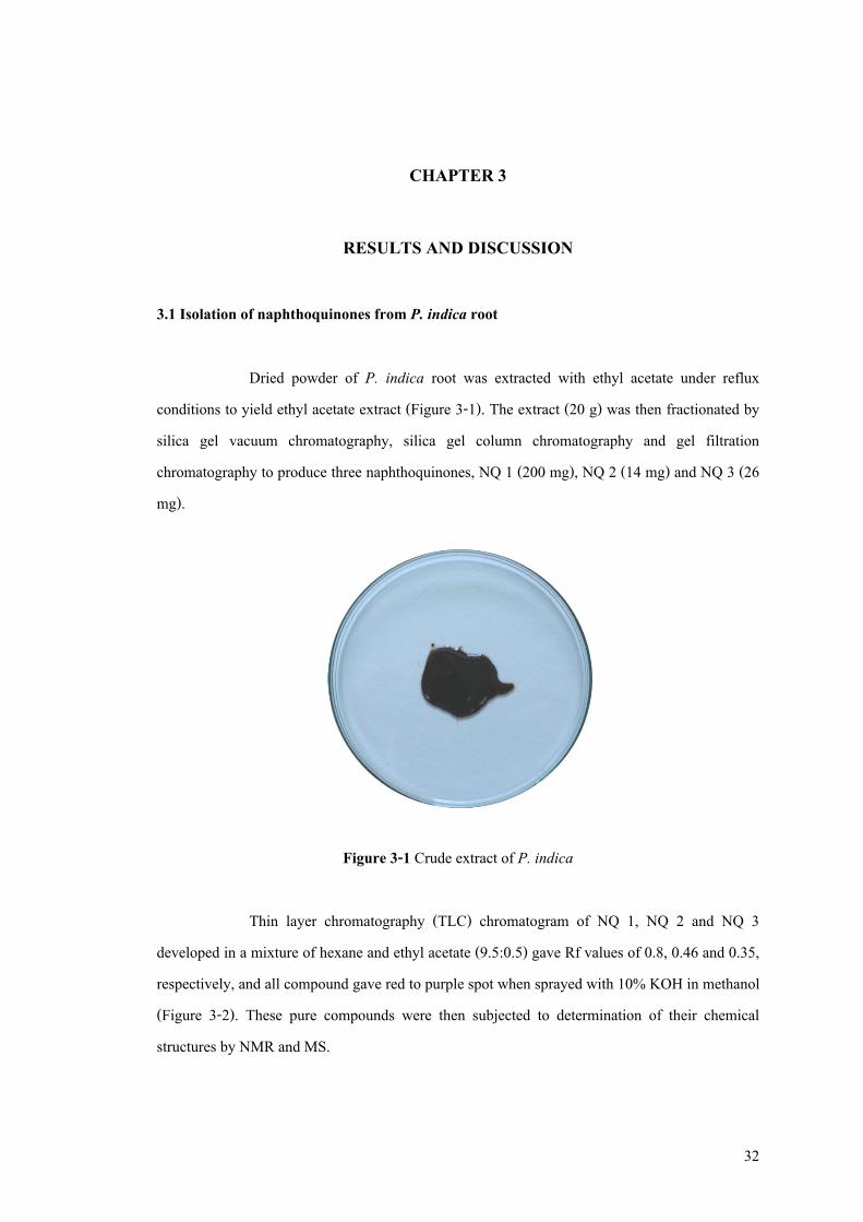
Dried powder of P. indica root was extracted with ethyl acetate under reflux conditions to yield ethyl acetate extract (Figure 3-1). The extract (20 g) was then fractionated by silica gel vacuum chromatography, silica gel column chromatography and gel filtration chromatography to produce three naphthoquinones, NQ 1 (200 mg), NQ 2 (14 mg) and NQ 3 (26 mg).
Figure 3-1 Crude extract of P. indica Thin layer chromatography (TLC) chromatogram of NQ 1, NQ 2 and NQ 3
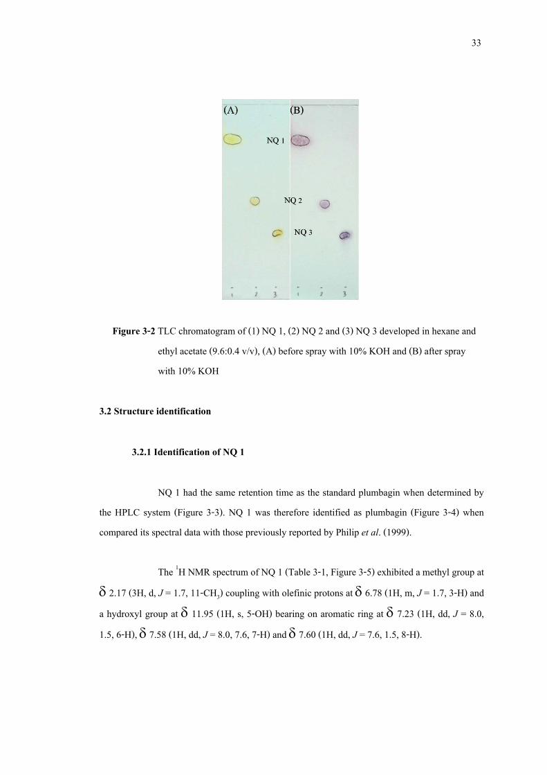
developed in a mixture of hexane and ethyl acetate (9.5:0.5) gave Rf values of 0.8, 0.46 and 0.35, respectively, and all compound gave red to purple spot when sprayed with 10% KOH in methanol (Figure 3-2). These pure compounds were then subjected to determination of their chemical structures by NMR and MS.
33
Figure 3-2 TLC chromatogram of (1) NQ 1, (2) NQ 2 and (3) NQ 3 developed in hexane and ethyl acetate (9.6:0.4 v/v), (A) before spray with 10% KOH and (B) after spray with 10% KOH
3.2 Structure identification
3.2.1 Identification of NQ 1
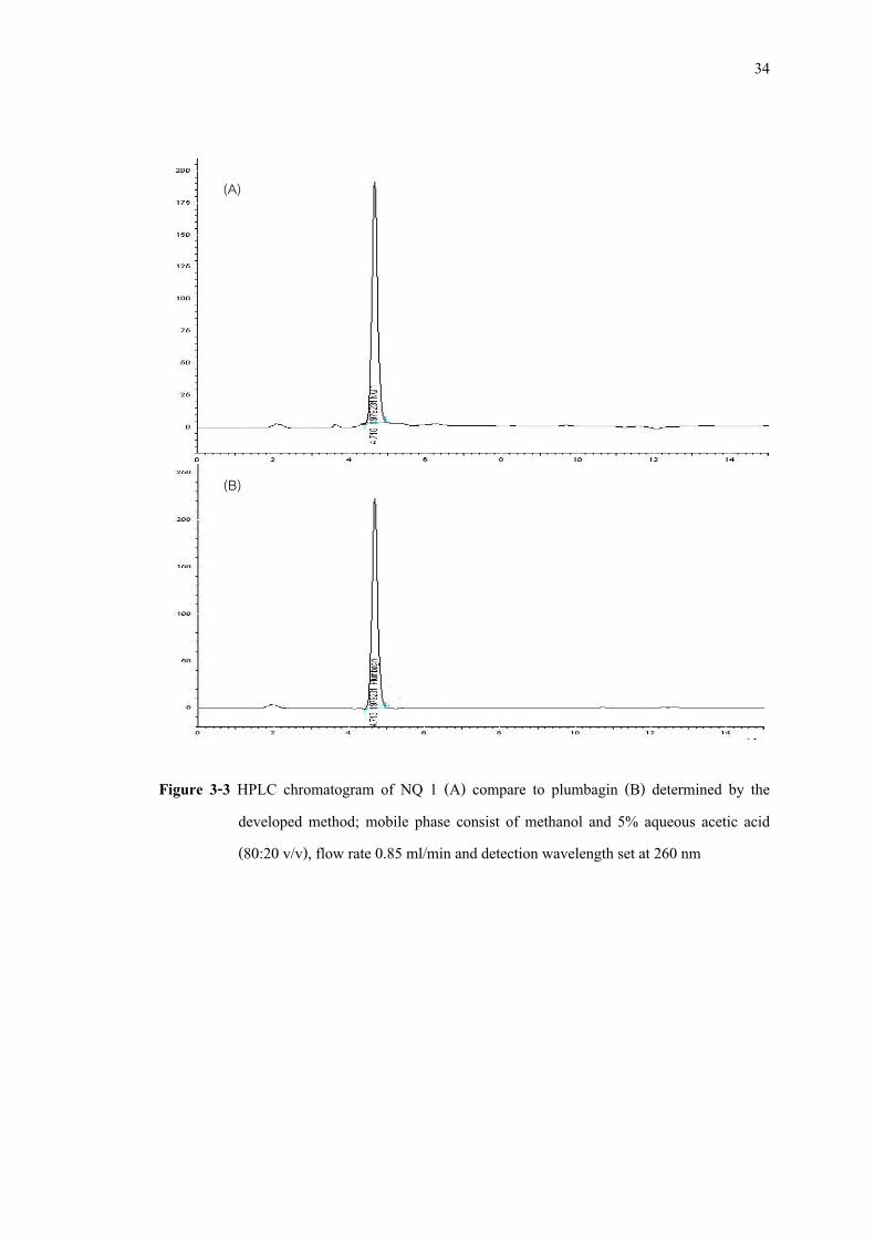
NQ 1 had the same retention time as the standard plumbagin when determined by the HPLC system (Figure 3-3). NQ 1 was therefore identified as plumbagin (Figure 3-4) when compared its spectral data with those previously reported by Philip et al. (1999).
The 1H NMR spectrum of NQ 1 (Table 3-1, Figure 3-5) exhibited a methyl group at
δ 2.17 (3H, d, J = 1.7, 11-CH3) coupling with olefinic protons at δ 6.78 (1H, m, J = 1.7, 3-H) and a hydroxyl group at δ 11.95 (1H, s, 5-OH) bearing on aromatic ring at δ 7.23 (1H, dd, J = 8.0, 1.5, 6-H), δ 7.58 (1H, dd, J = 8.0, 7.6, 7-H) and δ 7.60 (1H, dd, J = 7.6, 1.5, 8-H).
34
Figure 3-3 HPLC chromatogram of NQ 1 (A) compare to plumbagin (B) determined by the developed method; mobile phase consist of methanol and 5% aqueous acetic acid (80:20 v/v), flow rate 0.85 ml/min and detection wavelength set at 260 nm
(A)
(B)
35
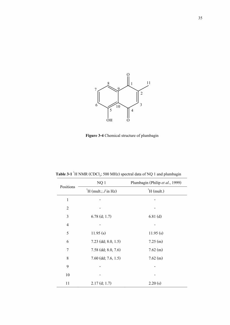
O
O
12
345
6
78
OH
9
10
11
Figure 3-4 Chemical structure of plumbagin
Table 3-1 1H NMR (CDCl3; 500 MHz) spectral data of NQ 1 and plumbagin NQ 1 Plumbagin (Philip et al., 1999) Positions 1H (mult.; J in Hz) 1H (mult.)
1 - - 2 - - 3 6.78 (d; 1.7) 6.81 (d) 4 - - 5 11.95 (s) 11.95 (s) 6 7.23 (dd; 8.0, 1.5) 7.25 (m) 7 7.58 (dd; 8.0, 7.6) 7.62 (m) 8 7.60 (dd; 7.6, 1.5) 7.62 (m) 9 - - 10 - - 11 2.17 (d; 1.7) 2.20 (s)
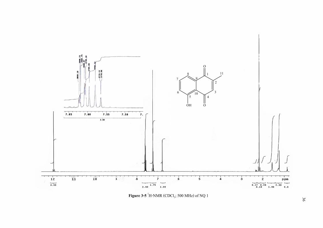
Figure 3-5 1H-NMR (CDCl3; 500 MHz) of NQ 1 36
O
O
12
345
6
78
OH
9
10
11
37
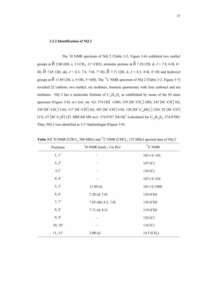
3.2.2 Identification of NQ 2
The 1H NMR spectrum of NQ 2 (Table 3-2, Figure 3-6) exhibited two methyl groups at δ 2.00 (6H, s, 11-CH3, 11’-CH3), aromatic protons at δ 7.28 (2H, d, J = 7.8, 6-H, 6’-H), δ 7.65 (2H, dd, J = 8.3, 7.8, 7-H, 7’-H), δ 7.71 (2H, d, J = 8.3, 8-H, 8’-H) and hydroxyl groups at δ 11.89 (2H, s, 5-OH, 5’-OH). The 13C NMR spectrum of NQ 2 (Table 3-2, Figure 3-7) revealed 22 carbons; two methyl, six methines, fourteen quarternary with four carbonyl and ten methines. NQ 2 has a molecular formula of C22H14O6 as established by mean of the EI mass spectrum (Figure 3-8), m/z (rel. int. %): 374 [M]+ (100), 359 [M+-CH3
•] (80), 345 [M+-CH•] (6), 330 [M+-CH3
•] (16), 317 [M+-CH•] (6), 303 [M+-CH•] (10), 120 [M+-C10HO4•] (10), 92 [M+-CO•]
(13), 67 [M+-C2H•] (5). HRFAB MS m/z: 374.0787 [M+H]+ (calculated for C22H1406, 374.0790). Thus, NQ 2 was identified as 3,3’-biplumbagin (Figure 3-9)
Table 3-2 1H NMR (CDCl3; 500 MHz) and 13C NMR (CDCl3; 125 MHz) spectral data of NQ 2
Positions 1H NMR (mult.; J in Hz) 13C NMR 1, 1’ - 183 (-C=O) 2, 2’ - 147 (C) 3,3’ - 139 (C) 4, 4’ - 187 (-C=O) 5, 5’ 11.89 (s) 161 (-C-OH) 6, 6’ 7.28 (d; 7.8) 124 (CH) 7, 7’ 7.65 (dd; 8.3, 7.8) 136 (CH) 8, 8’ 7.71 (d; 8.3) 119 (CH) 9, 9’ - 132 (C)
10, 10’ - 116 (C) 11, 11’ 2.00 (s) 14.5 (CH3)
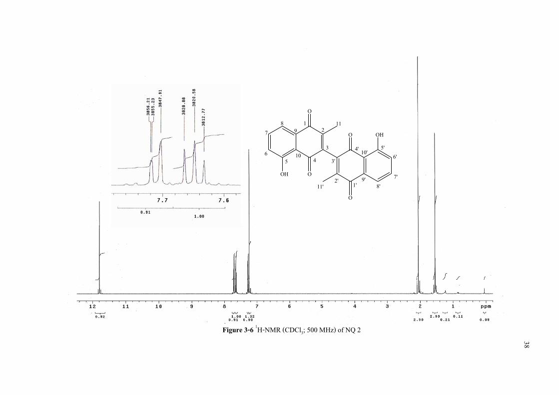
Figure 3-6 1H-NMR (CDCl3; 500 MHz) of NQ 2
38
12
345
6
78
O
O
O
O
OH
OH
1'2'
3'4' 5'
6'
7'8'
9
10
9'
10'
11'
11

Figure 3-7 13C-NMR (CDCl3; 125 MHz) of NQ 2
39
12
345
6
78
O
O
O
O
OH
OH
1'2'
3'4' 5'
6'
7'8'
9
10
9'
10'
11'
11
Figure 3-8 Mass spectroscopy of NQ 2
40
41
12
345
6
78
O
O
O
O
OH
OH
1'2'
3'4' 5'
6'
7'8'
9
10
9'
10'
11'
11
Figure 3-9 Chemical structure of 3,3’-biplumbagin
3.2.3 Identification of NQ 3
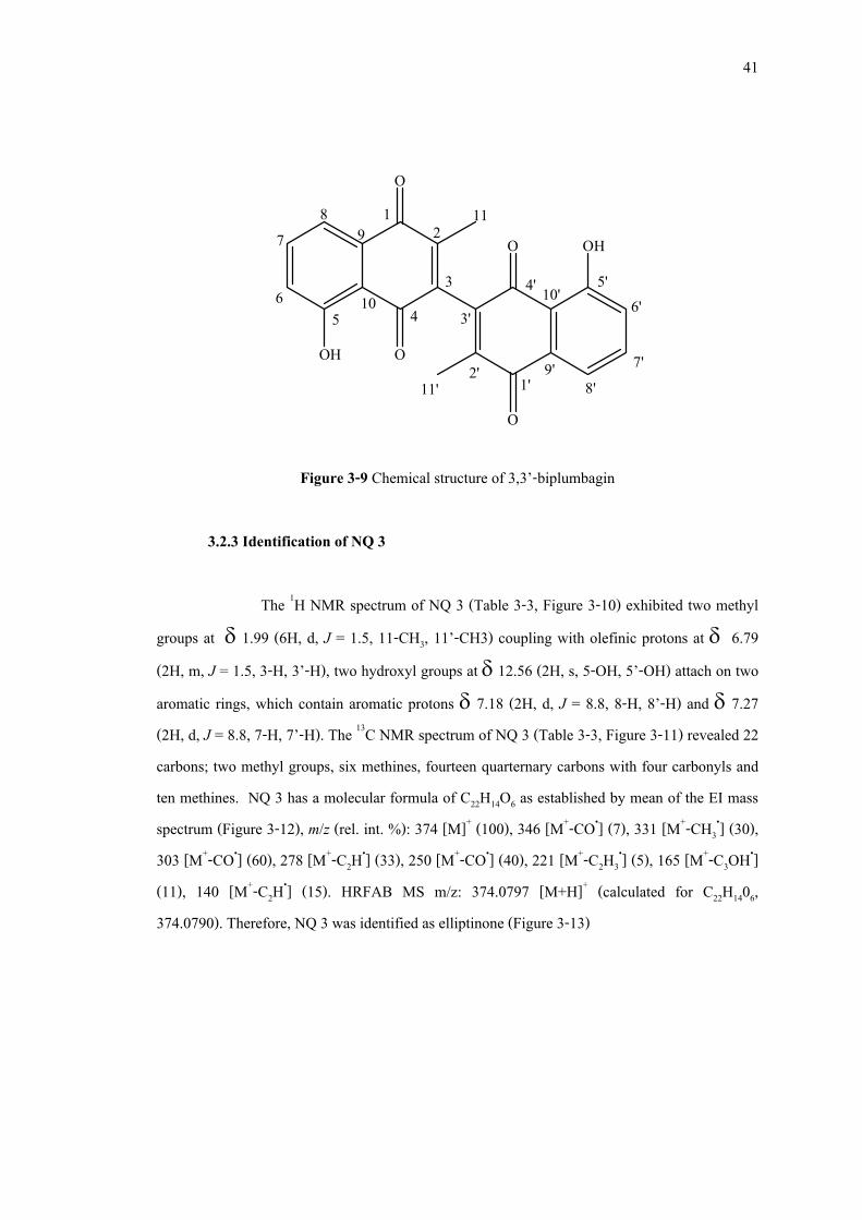
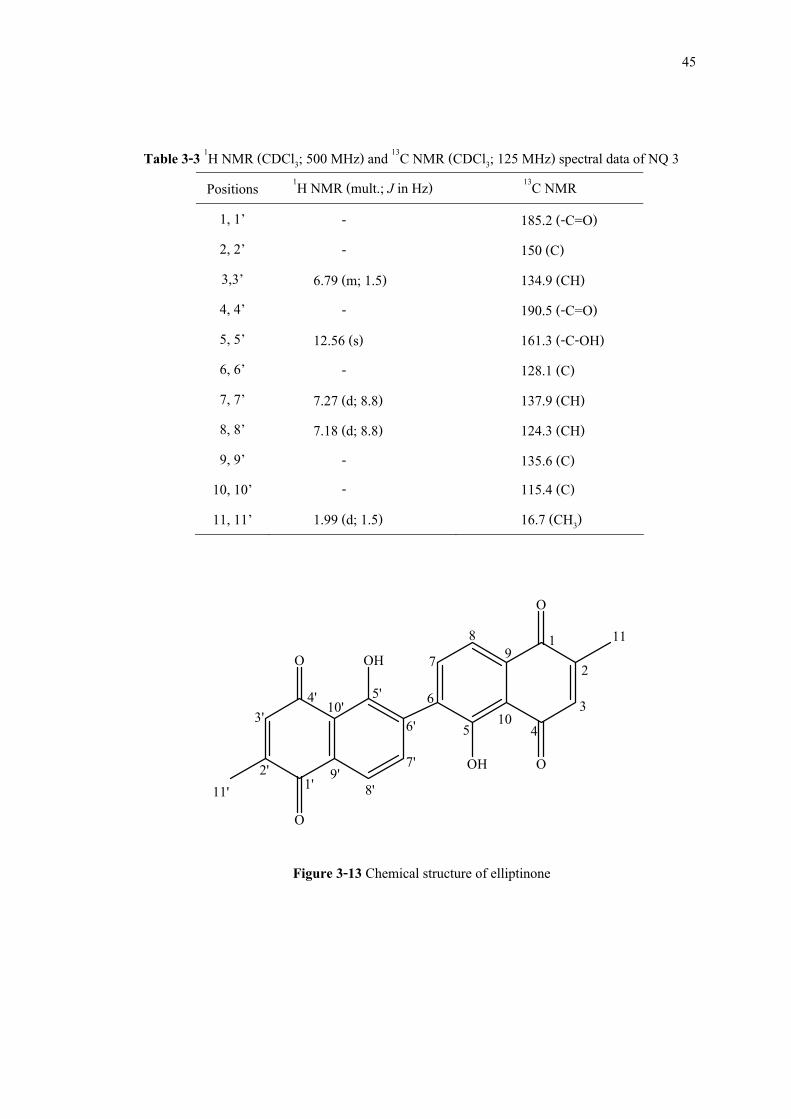
The 1H NMR spectrum of NQ 3 (Table 3-3, Figure 3-10) exhibited two methyl groups at δ 1.99 (6H, d, J = 1.5, 11-CH3, 11’-CH3) coupling with olefinic protons at δ 6.79 (2H, m, J = 1.5, 3-H, 3’-H), two hydroxyl groups at δ 12.56 (2H, s, 5-OH, 5’-OH) attach on two aromatic rings, which contain aromatic protons δ 7.18 (2H, d, J = 8.8, 8-H, 8’-H) and δ 7.27 (2H, d, J = 8.8, 7-H, 7’-H). The 13C NMR spectrum of NQ 3 (Table 3-3, Figure 3-11) revealed 22 carbons; two methyl groups, six methines, fourteen quarternary carbons with four carbonyls and ten methines. NQ 3 has a molecular formula of C22H14O6 as established by mean of the EI mass spectrum (Figure 3-12), m/z (rel. int. %): 374 [M]+ (100), 346 [M+-CO•] (7), 331 [M+-CH3
•] (30), 303 [M+-CO•] (60), 278 [M+-C2H•] (33), 250 [M+-CO•] (40), 221 [M+-C2H3
•] (5), 165 [M+-C3OH•] (11), 140 [M+-C2H•] (15). HRFAB MS m/z: 374.0797 [M+H]+ (calculated for C22H1406, 374.0790). Therefore, NQ 3 was identified as elliptinone (Figure 3-13)
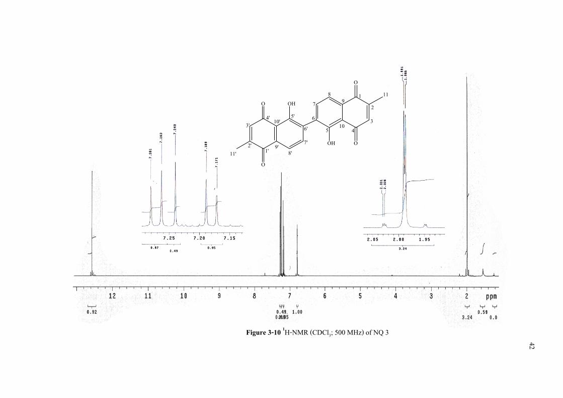
Figure 3-10 1H-NMR (CDCl3; 500 MHz) of NQ 3
42
12
345
6
78
1'2'
3'4' 5'
6'
7'8'
O
O
O
OHO
OH9'
10
11
10'
9
11'

Figure 3-11 13C-NMR (CDCl3; 125 MHz) of NQ 3
43
12
345
6
78
1'2'
3'4' 5'
6'
7'8'
O
O
O
OHO
OH9'
10
11
10'
9
11'
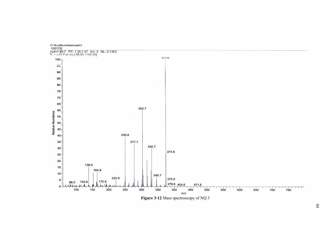
Figure 3-12 Mass spectroscopy of NQ 3
44
45
Table 3-3 1H NMR (CDCl3; 500 MHz) and 13C NMR (CDCl3; 125 MHz) spectral data of NQ 3 Positions 1H NMR (mult.; J in Hz) 13C NMR
1, 1’ - 185.2 (-C=O) 2, 2’ - 150 (C) 3,3’ 6.79 (m; 1.5) 134.9 (CH) 4, 4’ - 190.5 (-C=O) 5, 5’ 12.56 (s) 161.3 (-C-OH) 6, 6’ - 128.1 (C) 7, 7’ 7.27 (d; 8.8) 137.9 (CH) 8, 8’ 7.18 (d; 8.8) 124.3 (CH) 9, 9’ - 135.6 (C)
10, 10’ - 115.4 (C) 11, 11’ 1.99 (d; 1.5) 16.7 (CH3)
12
345
6
78
1'2'
3'4' 5'
6'
7'8'
O
O
O
OHO
OH9'
10
11
10'
9
11'
Figure 3-13 Chemical structure of elliptinone
46
3.3 HPLC quantitative determination of naphthoquinones in P. indica root extracts
The optimal conditions for simultaneous quantitative determination of naphthoquinones in P. indica root extracts were performed using reverse phase HPLC system on Agilent HPLC 1100 series with Agilent ChemStation for LC 3D software. The three naphthoquinones were used as the indicative markers for quantitative analysis of P. indica root extracts. As these compounds have maximum absorption at 260 nm, this wavelength was then used for quantification. Samples were injected at 10 µl through a Phenomenex® ODS column (150 × 4.6 mm 5 µm particle size) and isocratically eluted with a mixtures of methanol and 5% aqueous acetic acid in the ratio of 80 : 20. The flow rate was used at 0.85 ml/min. The retention times of all naphthoquinones were within 15 minutes (Figure 3-14) and separated with satisfactory resolution. On the basis of the reverse phase HPLC analysis, plumbagin, the most polar compound would be firstly eluted at the retention time about 4 min followed by elliptinone and 3,3’-biplumbagin with the retention times about 6 and 10 min, respectively.
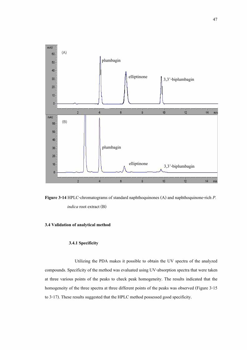
47
Figure 3-14 HPLC-chromatograms of standard naphthoquinones (A) and naphthoquinone-rich P. indica root extract (B)
3.4 Validation of analytical method
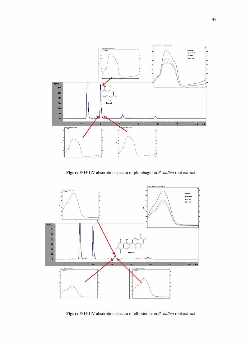
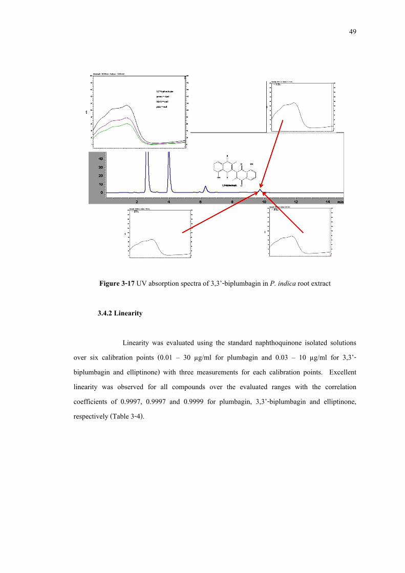
3.4.1 Specificity Utilizing the PDA makes it possible to obtain the UV spectra of the analyzed compounds. Specificity of the method was evaluated using UV-absorption spectra that were taken at three various points of the peaks to check peak homogeneity. The results indicated that the homogeneity of the three spectra at three different points of the peaks was observed (Figure 3-15 to 3-17). These results suggested that the HPLC method possessed good specificity.
(A) plumbagin
elliptinone 3,3’-biplumbagin
(B)
plumbagin
elliptinone 3,3’-biplumbagin
48
Figure 3-15 UV absorption spectra of plumbagin in P. indica root extract
Figure 3-16 UV absorption spectra of elliptinone in P. indica root extract
49
Figure 3-17 UV absorption spectra of 3,3’-biplumbagin in P. indica root extract
3.4.2 Linearity Linearity was evaluated using the standard naphthoquinone isolated solutions
over six calibration points (0.01 – 30 µg/ml for plumbagin and 0.03 – 10 µg/ml for 3,3’-biplumbagin and elliptinone) with three measurements for each calibration points. Excellent linearity was observed for all compounds over the evaluated ranges with the correlation coefficients of 0.9997, 0.9997 and 0.9999 for plumbagin, 3,3’-biplumbagin and elliptinone, respectively (Table 3-4).
50
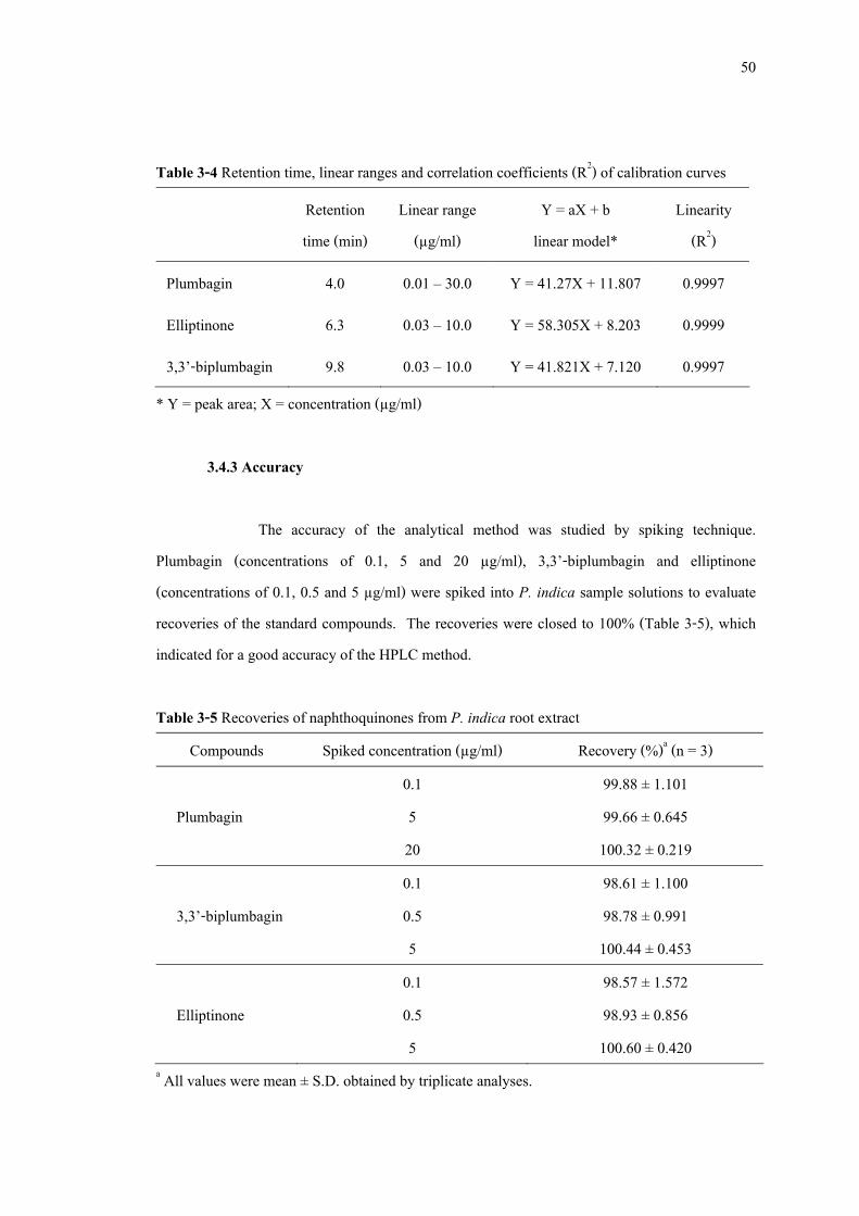
Table 3-4 Retention time, linear ranges and correlation coefficients (R2) of calibration curves
Retention time (min)
Linear range (µg/ml)
Y = aX + b linear model*
Linearity (R2)
Plumbagin 4.0 0.01 – 30.0 Y = 41.27X + 11.807 0.9997
Elliptinone 6.3 0.03 – 10.0 Y = 58.305X + 8.203 0.9999
3,3’-biplumbagin 9.8 0.03 – 10.0 Y = 41.821X + 7.120 0.9997 * Y = peak area; X = concentration (µg/ml)
3.4.3 Accuracy The accuracy of the analytical method was studied by spiking technique.
Plumbagin (concentrations of 0.1, 5 and 20 µg/ml), 3,3’-biplumbagin and elliptinone (concentrations of 0.1, 0.5 and 5 µg/ml) were spiked into P. indica sample solutions to evaluate recoveries of the standard compounds. The recoveries were closed to 100% (Table 3-5), which indicated for a good accuracy of the HPLC method. Table 3-5 Recoveries of naphthoquinones from P. indica root extract
Compounds Spiked concentration (µg/ml) Recovery (%)a (n = 3) 0.1 99.88 ± 1.101
Plumbagin 5 99.66 ± 0.645 20 100.32 ± 0.219 0.1 98.61 ± 1.100 3,3’-biplumbagin 0.5 98.78 ± 0.991 5 100.44 ± 0.453 0.1 98.57 ± 1.572 Elliptinone 0.5 98.93 ± 0.856
5 100.60 ± 0.420 a All values were mean ± S.D. obtained by triplicate analyses.
51
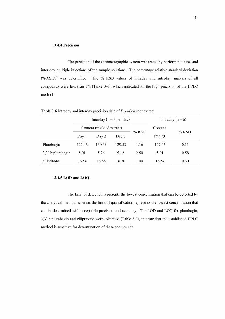
3.4.4 Precision
The precision of the chromatographic system was tested by performing intra- and inter-day multiple injections of the sample solutions. The percentage relative standard deviation (%R.S.D.) was determined. The % RSD values of intraday and interday analysis of all compounds were less than 5% (Table 3-6), which indicated for the high precision of the HPLC method. Table 3-6 Intraday and interday precision data of P. indica root extract
Interday (n = 3 per day) Intraday (n = 6) Content (mg/g of extract)
Day 1 Day 2 Day 3 % RSD Content
(mg/g) % RSD
Plumbagin 127.46 130.36 129.53 1.16 127.46 0.11 3,3’-biplumbagin 5.01 5.26 5.12 2.50 5.01 0.58 elliptinone 16.54 16.88 16.70 1.00 16.54 0.30
3.4.5 LOD and LOQ
The limit of detection represents the lowest concentration that can be detected by
the analytical method, whereas the limit of quantification represents the lowest concentration that can be determined with acceptable precision and accuracy. The LOD and LOQ for plumbagin, 3,3’-biplumbagin and elliptinone were exhibited (Table 3-7), indicate that the established HPLC method is sensitive for determination of these compounds

52
Table 3-7 LOD and LOQ of P. indica root extract Compounds LOD a (µg/ml) LOQb (µg/ml) Plumbagin 0.001 0.010 3,3’-biplumbagin 1.00 5.00 elliptinone 0.10 1.00
a Limit of detection (LOD): signal to noise ratio = 3 b Limit of quantification (LOQ): signal to noise ratio = 10 3.5 Determination of solvent for extraction
A few different extraction solvents include ethyl acetate, ethanol, isopropanol, dichloromethane and diethyl ether were examined to produce the highest content of naphthoquinones in P. indica root extracts. The result showed that although isopropanol produced the highest yield of the root extract, ethanol produced the highest content of total naphthoquinones (Table 3-8). Thus, ethanol was appropriately used for the extraction. Table 3-8 Yield and naphthoquinones content in P. indica root extracts
Contenta (mg/g of extract; Mean ± S.D.)
Solvents Yield (% w/w)
Plumbagin 3,3’-biplumbagin Elliptinone Total
naphthoquinones EtOH 11.5 4.76 ± 0.067 0.34 ± 0.044 0.69 ± 0.035 5.80 EtOAc 10.9 4.63 ± 0.266 0.25 ± 0.021* 0.51 ± 0.070* 5.39 C3H7OH 11.8 3.50 ± 0.606* 0.23 ± 0.051* 0.49 ± 0.108* 4.22 CH2Cl2 9.1 3.85 ± 0.910 0.19 ± 0.030* 0.34 ± 0.054* 4.38 C2H5O C2H5 8.8 4.26 ± 0.576 0.16 ± 0.075* 0.47 ± 0.088* 4.89
a All values were mean ± S.D. obtained by triplicate analyses. * Significant difference (P<0.05) when compared with the ethanol extract
EtOH = ethanol; EtOAc = ethyl acetate; C3H7OH = isopropanol; CH2Cl2 = dichloromethane; C2H5O C2H5 = diethyl ether
53
3.6 Simple purification method to improved naphthoquinone content
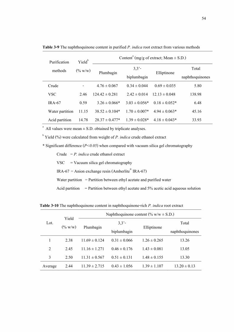
The ethanol extract was used for further study on determination of an appropriate fractionation method to produce naphthoquinone-rich P. indica root extract. The result exhibited that although anion exchange chromatographic method (Amberlite® IRA-67) produced the highest content of 3,3’-biplumbagin, it was difficult to eluted plumbagin and elliptinone from the column. Thus, this method produced a low content of the total naphthoquinones (Table 3-9). Moreover, the liquid-liquid extraction methods produced the highest yielding of extracts but the total naphthoquinone content also lower than silica gel vacuum chromatography. In addition, acetic acid presence in the extraction not able to improved the content of naphthoquinones in the extract. Thus, silica gel vacuum chromatography was used to prepare naphthoquinone-rich P. indica root extract, which increase the total naphthoquinones content from 5.80 mg/g, in the crude ethanol extract, to 138.98 mg/g of extract (Table 3-9).
54
Table 3-9 The naphthoquinone content in purified P. indica root extract from various methods
Contenta (mg/g of extract; Mean ± S.D.) Purification methods
Yieldb (% w/w) Plumbagin 3,3’-
biplumbagin Elliptinone Total naphthoquinones
Crude - 4.76 ± 0.067 0.34 ± 0.044 0.69 ± 0.035 5.80 VSC 2.46 124.42 ± 0.281 2.42 ± 0.014 12.13 ± 0.048 138.98 IRA-67 0.59 3.26 ± 0.066* 3.03 ± 0.056* 0.18 ± 0.052* 6.48 Water partition 11.15 38.52 ± 0.104* 1.70 ± 0.007* 4.94 ± 0.063* 45.16 Acid partition 14.78 28.37 ± 0.477* 1.39 ± 0.028* 4.18 ± 0.043* 33.93
a All values were mean ± S.D. obtained by triplicate analyses. b Yield (%) were calculated from weight of P. indica crude ethanol extract * Significant difference (P<0.05) when compared with vacuum silica gel chromatography
Crude = P. indica crude ethanol extract VSC = Vacuum silica gel chromatography IRA-67 = Anion exchange resin (Amberlite® IRA-67) Water partition = Partition between ethyl acetate and purified water Acid partition = Partition between ethyl acetate and 5% acetic acid aqueous solution
Table 3-10 The naphthoquinone content in naphthoquinone-rich P. indica root extract Naphthoquinone content (% w/w ± S.D.)
Lot. Yield (% w/w) Plumbagin 3,3’-
biplumbagin Elliptinone Total
naphthoquinones 1 2.38 11.69 ± 0.124 0.31 ± 0.066 1.26 ± 0.265 13.26 2 2.45 11.16 ± 1.271 0.46 ± 0.176 1.43 ± 0.081 13.05 3 2.50 11.31 ± 0.567 0.51 ± 0.131 1.48 ± 0.155 13.30
Average 2.44 11.39 ± 2.715 0.43 ± 1.056 1.39 ± 1.107 13.20 ± 0.13
55
3.7 Antibacterial activity against acne-involved bacteria
Antibacterial activity of naphthoquinone-rich P. indica root extract, plumbagin, 3,3’-biplumbagin and elliptinone were evaluated against acne-involved bacteria including P. acnes, S. aureus and S. epidermidis. The result demonstrated that, all tested bacteria were inhibited by plumbagin, 3,3’-biplumbagin and elliptinone with MIC value between 0.024 - 12.50 µg/ml, 0.024 - 100 µg/ml and 0.78 - 100 µg/ml, respectively. Naphthoquinone-rich P. indica root extract exhibited antibacterial activity against P. acnes and S. aureus close to that of plumbagin and more potent than both 3,3’-biplumbagin and elliptinone. Moreover, naphthoquinone-rich P. indica root extract gave much more potent antibacterial activity than P. indica crude ethanol (Table 3-11).
The MBC values of the naphthoquinone-rich P. indica root extract against all tested
bacterial were found between 6.25 – 50 µg/ml (Table 3-12 and Figure 3-18). Although all tested bacteria were killed by plumbagin, 3,3’-biplumbagin and elliptinone with MBC value between 3.12 – 100 µg/ml, only S. aureus that survived within the concentration of 3,3’-biplumbagin and elliptinone below 100 µg/ml.
These results indicated that naphthoquinone-rich P. indica root extract possessed
antibacterial activity against acne-involved bacteria more potent than 3,3’-biplumbagin and elliptinone, and its antibacterial activity was close to plumbagin. In addition, the total naphthoquinone content of naphthoquinone-rich P. indica root extract was 13.20 ± 0.13 % w/w. Thus, the naphthoquinone-rich P. indica root extract should contain total naphthoquinone not less than 13% w/w to give the acceptable antibacterial activity against acne-involved bacteria.
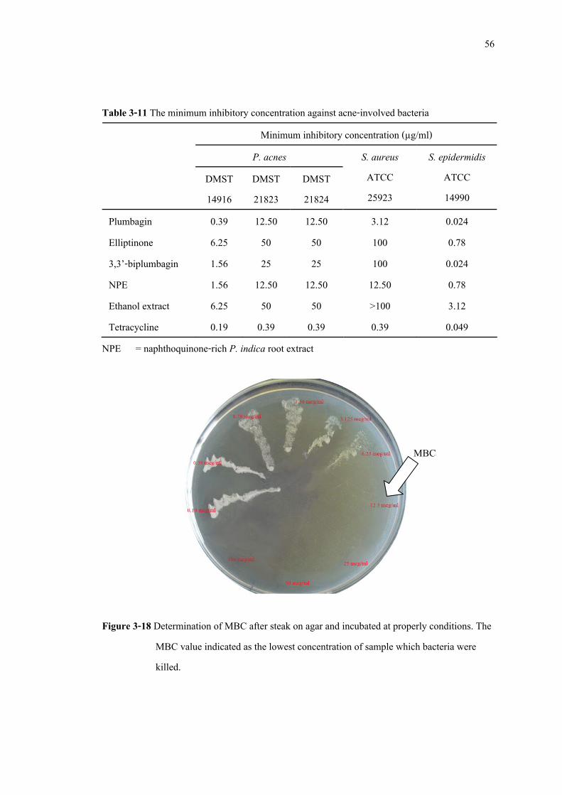
56
Table 3-11 The minimum inhibitory concentration against acne-involved bacteria Minimum inhibitory concentration (µg/ml)
P. acnes DMST 14916
DMST 21823
DMST 21824
S. aureus ATCC 25923
S. epidermidis ATCC 14990
Plumbagin 0.39 12.50 12.50 3.12 0.024 Elliptinone 6.25 50 50 100 0.78 3,3’-biplumbagin 1.56 25 25 100 0.024 NPE 1.56 12.50 12.50 12.50 0.78 Ethanol extract 6.25 50 50 >100 3.12 Tetracycline 0.19 0.39 0.39 0.39 0.049
NPE = naphthoquinone-rich P. indica root extract
Figure 3-18 Determination of MBC after steak on agar and incubated at properly conditions. The MBC value indicated as the lowest concentration of sample which bacteria were killed.
MBC
57
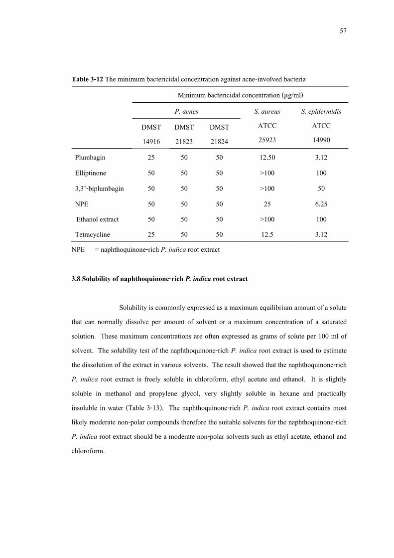
Table 3-12 The minimum bactericidal concentration against acne-involved bacteria Minimum bactericidal concentration (µg/ml)
P. acnes DMST 14916
DMST 21823
DMST 21824
S. aureus ATCC 25923
S. epidermidis ATCC 14990
Plumbagin 25 50 50 12.50 3.12 Elliptinone 50 50 50 >100 100 3,3’-biplumbagin 50 50 50 >100 50 NPE 50 50 50 25 6.25 Ethanol extract 50 50 50 >100 100 Tetracycline 25 50 50 12.5 3.12
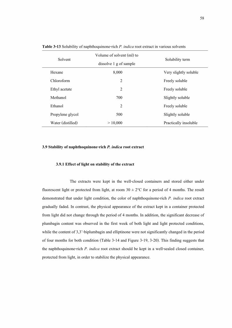
NPE = naphthoquinone-rich P. indica root extract 3.8 Solubility of naphthoquinone-rich P. indica root extract
Solubility is commonly expressed as a maximum equilibrium amount of a solute that can normally dissolve per amount of solvent or a maximum concentration of a saturated solution. These maximum concentrations are often expressed as grams of solute per 100 ml of solvent. The solubility test of the naphthoquinone-rich P. indica root extract is used to estimate the dissolution of the extract in various solvents. The result showed that the naphthoquinone-rich P. indica root extract is freely soluble in chloroform, ethyl acetate and ethanol. It is slightly soluble in methanol and propylene glycol, very slightly soluble in hexane and practically insoluble in water (Table 3-13). The naphthoquinone-rich P. indica root extract contains most likely moderate non-polar compounds therefore the suitable solvents for the naphthoquinone-rich P. indica root extract should be a moderate non-polar solvents such as ethyl acetate, ethanol and chloroform.
58
Table 3-13 Solubility of naphthoquinone-rich P. indica root extract in various solvents
Solvent Volume of solvent (ml) to dissolve 1 g of sample
Solubility term
Hexane 8,000 Very slightly soluble Chloroform 2 Freely soluble Ethyl acetate 2 Freely soluble Methanol 700 Slightly soluble Ethanol 2 Freely soluble Propylene glycol 500 Slightly soluble Water (distilled) > 10,000 Practically insoluble
3.9 Stability of naphthoquinone-rich P. indica root extract 3.9.1 Effect of light on stability of the extract
The extracts were kept in the well-closed containers and stored either under fluorescent light or protected from light, at room 30 ± 2°C for a period of 4 months. The result demonstrated that under light condition, the color of naphthoquinone-rich P. indica root extract gradually faded. In contrast, the physical appearance of the extract kept in a container protected from light did not change through the period of 4 months. In addition, the significant decrease of plumbagin content was observed in the first week of both light and light protected conditions, while the content of 3,3’-biplumbagin and elliptinone were not significantly changed in the period of four months for both condition (Table 3-14 and Figure 3-19, 3-20). This finding suggests that the naphthoquinone-rich P. indica root extract should be kept in a well-sealed closed container, protected from light, in order to stabilize the physical appearance.
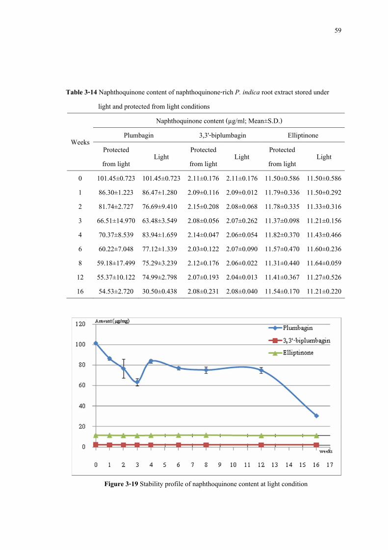
59
Table 3-14 Naphthoquinone content of naphthoquinone-rich P. indica root extract stored under
light and protected from light conditions Naphthoquinone content (µg/ml; Mean±S.D.)
Plumbagin 3,3'-biplumbagin Elliptinone Weeks Protected from light Light Protected
from light Light Protected from light Light
0 1 2 3 4 6 8 12 16
101.45±0.723 86.30±1.223 81.74±2.727 66.51±14.970 70.37±8.539 60.22±7.048 59.18±17.499 55.37±10.122 54.53±2.720
101.45±0.723 86.47±1.280 76.69±9.410 63.48±3.549 83.94±1.659 77.12±1.339 75.29±3.239 74.99±2.798 30.50±0.438
2.11±0.176 2.09±0.116 2.15±0.208 2.08±0.056 2.14±0.047 2.03±0.122 2.12±0.176 2.07±0.193 2.08±0.231
2.11±0.176 2.09±0.012 2.08±0.068 2.07±0.262 2.06±0.054 2.07±0.090 2.06±0.022 2.04±0.013 2.08±0.040
11.50±0.586 11.79±0.336 11.78±0.335 11.37±0.098 11.82±0.370 11.57±0.470 11.31±0.440 11.41±0.367 11.54±0.170
11.50±0.586 11.50±0.292 11.33±0.316 11.21±0.156 11.43±0.466 11.60±0.236 11.64±0.059 11.27±0.526 11.21±0.220
Figure 3-19 Stability profile of naphthoquinone content at light condition
60
Figure 3-20 Stability profile of naphthoquinone content at dark condition
3.9.2 Effect of temperature on stability of the extract
The effect of temperature on the stability of naphthoquinone-rich P. indica root extract was examined under two temperatures, 4 ± 2°C and 30 ± 2°C, protected from light. The result found that the significant decrease of plumbagin content was observed in the first week for 30±2°C storage but it was not significantly changed for 4 ± 2°C condition, while the content of 3,3’-biplumbagin and elliptinone were not significantly changed in the period of four months for both conditions through the period of 4 months (Table 3-15 and Figure 3-21, 3-22). This finding suggests that the naphthoquinone-rich P. indica root extract should be kept at 4 ± 2°C in order to stabilize the naphthoquinone content.
61
Table 3-15 Naphthoquinone content of naphthoquinone-rich P. indica root extract stored under 4°C and 30°C
Naphthoquinone content (µg/ml; Mean±S.D.) Plumbagin 3,3'-biplumbagin Elliptinone Weeks
4°C 30°C 4°C 30°C 4°C 30°C 0 1 2 3 4 6 8 12 16
101.45±0.723 107.76±5.947 105.48±2.757 104.11±7.782 104.27±9.168 109.03±0.121 105.18±11.168 105.62±11.119 107.14±5.139
101.45±0.723 96.79±1.639 73.14±14.683 69.02±14.016 86.93±10.545 55.96±13.303 53.10±5.586 53.10±5.586 49.11±6.893
2.11±0.176 2.18±0.097 2.13±0.110 2.20±0.150 2.12±0.124 2.09±0.191 2.17±0.148 2.13±0.076 2.26±0.247
2.11±0.176 2.07±0.102 2.03±0.054 2.12±0.022 2.12±0.144 2.10±0.030 2.11±0.040 2.12±0.012 2.11±0.089
11.50±0.586 11.75±0.074 11.62±0.304 11.66±1.191 11.70±0.272 11.69±0.477 11.48±0.668 11.63±0.724 11.49±0.488
11.50±0.586 11.94±0.284 11.83±0.497 11.39±0.777 11.66±0.316 11.45±0.633 11.73±1.405 11.68±1.452 11.39±0.169
Figure 3-21 Stability profile of naphthoquinone content at 4 ± 2°C condition
62
Figure 3-22 Stability profile of naphthoquinone content at 30 ± 2°C condition
3.9.3 Effect of pH on stability of the extract The acid-base stability evaluation of naphthoquinone-rich P. indica root extract
in solution was determined at three different pH values; 5.5, 7.0 and 8.0. It was found that at pH 5.5 was not change in physical appearance but pH 7.0 and 8.0 the color of sample changed from yellow to orange (Figure 3-23, 3-24). In addition, the naphthoquinone content was decrease under all tested pH. At pH 5.5, the content of elliptinone was significantly decreases after 4 weeks and at pH 7.0 and 8.0, it significantly decreases from the first week (Table 16 to 18; Figure 3-25 to 3-27). It implied that all naphthoquinones are not stable when they are in solution. However, naphthoquinones were more stable in the solution at pH 5.5 than 7.0 and 8.0. Thus, the preparation of naphthoquinone-rich P. indica root extract in an aqueous solution should be performed carefully.
63
Figure 3-23 Extracts for stability under pH variation test at week 0
Figure 3-24 Extracts for stability under pH variation test after 16 weeks storage
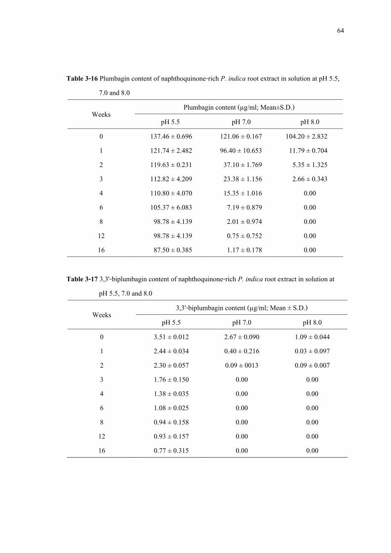
64
Table 3-16 Plumbagin content of naphthoquinone-rich P. indica root extract in solution at pH 5.5, 7.0 and 8.0
Plumbagin content (µg/ml; Mean±S.D.) Weeks pH 5.5 pH 7.0 pH 8.0
0 1 2 3 4 6 8 12 16
137.46 ± 0.696 121.74 ± 2.482 119.63 ± 0.231 112.82 ± 4.209 110.80 ± 4.070 105.37 ± 6.083 98.78 ± 4.139 98.78 ± 4.139 87.50 ± 0.385
121.06 ± 0.167 96.40 ± 10.653 37.10 ± 1.769 23.38 ± 1.156 15.35 ± 1.016 7.19 ± 0.879 2.01 ± 0.974 0.75 ± 0.752 1.17 ± 0.178
104.20 ± 2.832 11.79 ± 0.704 5.35 ± 1.325 2.66 ± 0.343
0.00 0.00 0.00 0.00 0.00
Table 3-17 3,3'-biplumbagin content of naphthoquinone-rich P. indica root extract in solution at
pH 5.5, 7.0 and 8.0 3,3'-biplumbagin content (µg/ml; Mean ± S.D.) Weeks
pH 5.5 pH 7.0 pH 8.0 0 1 2 3 4 6 8 12 16
3.51 ± 0.012 2.44 ± 0.034 2.30 ± 0.057 1.76 ± 0.150 1.38 ± 0.035 1.08 ± 0.025 0.94 ± 0.158 0.93 ± 0.157 0.77 ± 0.315
2.67 ± 0.090 0.40 ± 0.216 0.09 ± 0013
0.00 0.00 0.00 0.00 0.00 0.00
1.09 ± 0.044 0.03 ± 0.097 0.09 ± 0.007
0.00 0.00 0.00 0.00 0.00 0.00
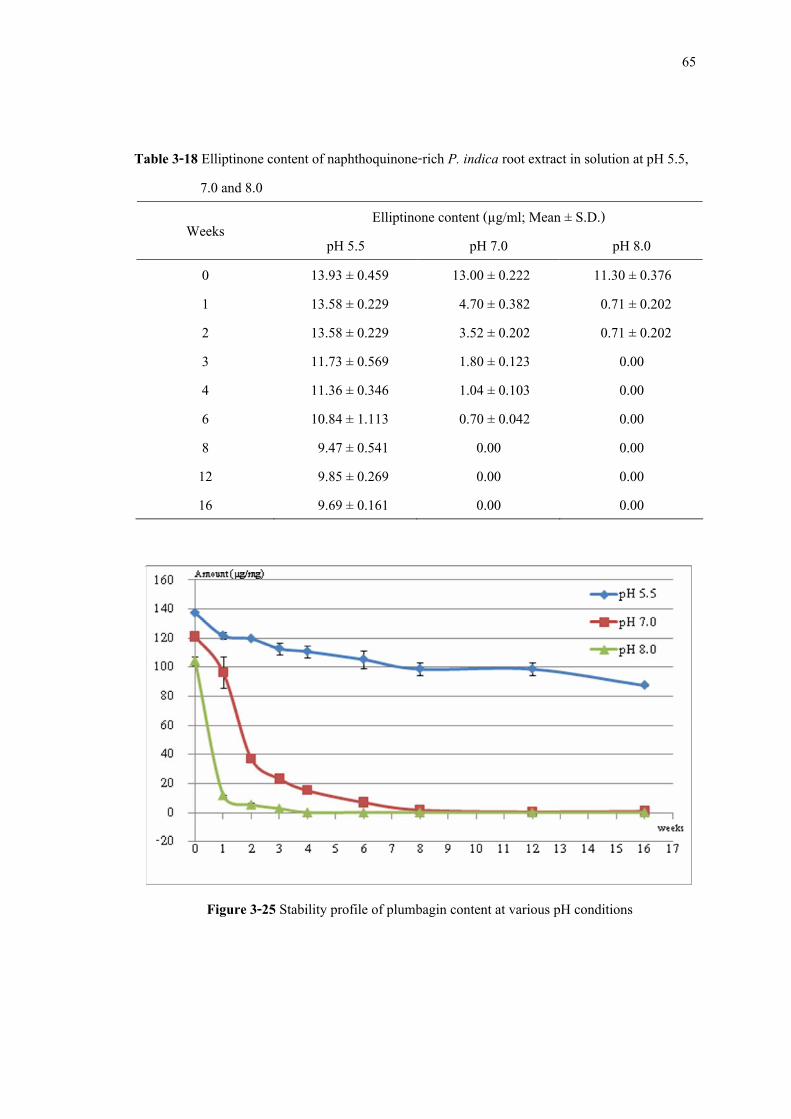
65
Table 3-18 Elliptinone content of naphthoquinone-rich P. indica root extract in solution at pH 5.5, 7.0 and 8.0
Elliptinone content (µg/ml; Mean ± S.D.) Weeks pH 5.5 pH 7.0 pH 8.0
0 1 2 3 4 6 8 12 16
13.93 ± 0.459 13.58 ± 0.229 13.58 ± 0.229 11.73 ± 0.569 11.36 ± 0.346 10.84 ± 1.113 9.47 ± 0.541 9.85 ± 0.269 9.69 ± 0.161
13.00 ± 0.222 4.70 ± 0.382 3.52 ± 0.202 1.80 ± 0.123 1.04 ± 0.103 0.70 ± 0.042
0.00 0.00 0.00
11.30 ± 0.376 0.71 ± 0.202 0.71 ± 0.202
0.00 0.00 0.00 0.00 0.00 0.00
Figure 3-25 Stability profile of plumbagin content at various pH conditions
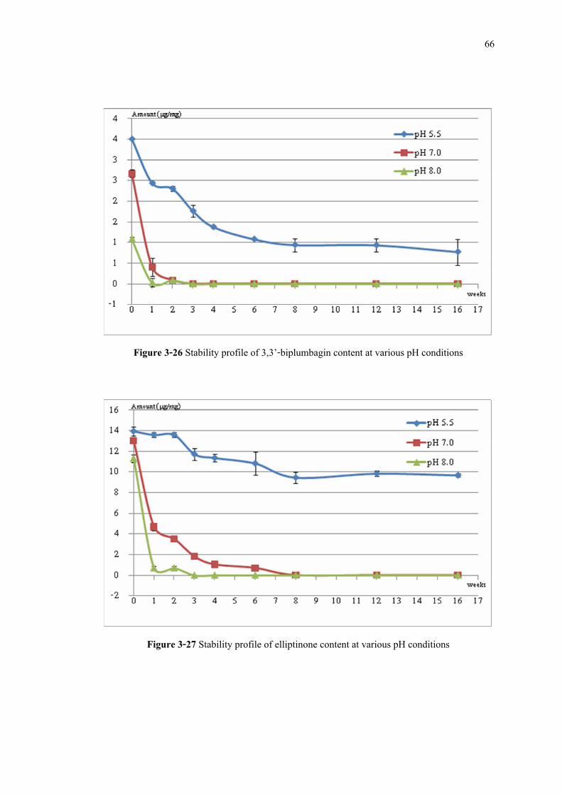
66
Figure 3-26 Stability profile of 3,3’-biplumbagin content at various pH conditions
Figure 3-27 Stability profile of elliptinone content at various pH conditions
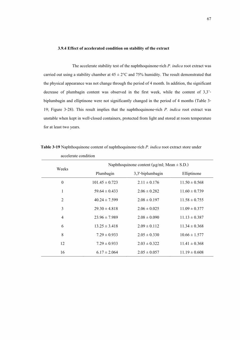
67
3.9.4 Effect of accelerated condition on stability of the extract The accelerate stability test of the naphthoquinone-rich P. indica root extract was
carried out using a stability chamber at 45 ± 2°C and 75% humidity. The result demonstrated that the physical appearance was not change through the period of 4 month. In addition, the significant decrease of plumbagin content was observed in the first week, while the content of 3,3’-biplumbagin and elliptinone were not significantly changed in the period of 4 months (Table 3-19; Figure 3-28). This result implies that the naphthoquinone-rich P. indica root extract was unstable when kept in well-closed containers, protected from light and stored at room temperature for at least two years.
Table 3-19 Naphthoquinone content of naphthoquinone-rich P. indica root extract store under
accelerate condition Naphthoquinone content (µg/ml; Mean ± S.D.) Weeks
Plumbagin 3,3'-biplumbagin Elliptinone 0 1 2 3 4 6 8 12 16
101.45 ± 0.723 59.64 ± 0.433 40.24 ± 7.599 29.30 ± 4.818 23.96 ± 7.989 13.25 ± 3.418 7.29 ± 0.933 7.29 ± 0.933 6.17 ± 2.064
2.11 ± 0.176 2.06 ± 0.282 2.08 ± 0.197 2.06 ± 0.025 2.08 ± 0.090 2.09 ± 0.112 2.05 ± 0.330 2.03 ± 0.322 2.05 ± 0.057
11.50 ± 0.568 11.60 ± 0.739 11.58 ± 0.755 11.09 ± 0.377 11.13 ± 0.387 11.34 ± 0.368 10.66 ± 1.577 11.41 ± 0.368 11.19 ± 0.608
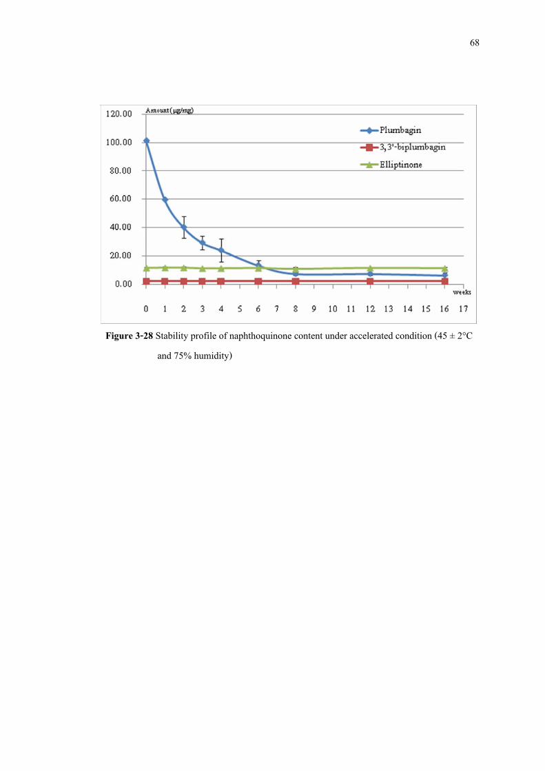
68
Figure 3-28 Stability profile of naphthoquinone content under accelerated condition (45 ± 2°C
and 75% humidity)

69
CHAPTER 4
CONCLUSION
1. Plumbagin, 3,3’-biplumbagin and elliptinone were isolated from P. indica roots and used as the indicative markers for quality control of P. indica root extracts. This is the first of isolation of 3,3’-biplumbagin and elliptinone from P. indica roots.
2. A simple, specific, precise, accurate, rapid and reproducible HPLC method has been
developed and validated for simultaneous quantitative determination of naphthoquinones, plumbagin, 3,3’-biplumbagin and elliptinone in P. indica root extracts.
3. Ethanol is a suitable solvent for extraction of naphthoquinones from P. indica roots. 4. Fractionation of the ethanol extract of P. indica roots using silica gel vacuum
chromatography eluted with a mixture of hexane and ethyl acetate (9.2:0.8 v/v; 500 ml) afforded the naphthoquinone-rich P. indica root extract, which contained the total naphthoquinone content up to 138.58 mg/g of extract.
5. The naphthoquinone-rich P. indica root extract possessed antibacterial activity against all
tested bacteria with the MIC values between 0.78 and 12.50 µg/ml. The extract showed satisfactory antibacterial activity against P. acnes and S. aureus with MIC values closed to those of plumbagin.
6. The standard specification of naphthoquinone content in naphthoquinone-rich P. indica
root extract was established as the extract should contain total naphthoquinone content not less than 13% w/w, and the major active compound plumbagin should be not less than 11% w/w.
70
7. The naphthoquinones-rich P. indica root extract contains most likely moderate non-polar naphthoquinones therefore the suitable solvents to dissolve the extract should be moderate non-polar solvents such as ethyl acetate, ethanol and chloroform.
8. Stability evaluations of the naphthoquinones-rich P. indica root extracts in several
conditions in the period of 4 months found that the extract possessed a satisfactory stability at 4°C condition. Thus, the extract should be stored in well-sealed closed containers, protected from light and cooled place (4°C) to stabilized total naphthoquinone content. The aqueous solutions of the extract are not stable either in acid or base conditions.
71
APPENDIX
Preparation of Brain Heart Infusion agar (BHI agar) Brain Heart Infusion 37 g Bacto agar 15 g Distill water 1000 ml
Preparation of Brain Heart Infusion broth (BHI broth) Brain Heart Infusion 37 g Distill water 1000 ml
Preparation of Mueller-Hinton agar Mueller-Hinton agar 38 g Distill water 1000 ml
Preparation of Mueller-Hinton broth Muelle- Hinton broth 21 g Distill water 1000 ml
Preparation of Glycerol broth Tryptose 10 g NaCl 5 g Beef extract (Protose BE) 3 g Yeast extract 5 g Cysteine HCl 0.4 g Glucose 1 g Na2HPO4 4 g Glycerol 150 ml Distill water 850 ml

72
Preparation of 0.5 McFarland standard 1 % v/v H2SO4 99.5 ml 1.175 % w/v BaCl2 0.5 ml
73
Bibliography คณะอนกุรรมการจัดทําตําราอางอิงยาสมุนไพรไทย (2551), “เจตมูลเพลิงแดง”, วารสารการแพทย
แผนไทยและการแพทยทางเลือก, ปที่ 5, ฉบับที่ 2, หนา 190-194. จุไรรัตน รักวาทิน (2538), การวิเคราะหขอมูลการศึกษาความคงสภาพของยาแบบเรงและแบบระยะ
ยาว, สํานักพิมพนิยมวิทยา, กรุงเทพฯ. จุไรรัตน รักวาทิน (2539), แนวทางการทดสอบความคงสภาพของยาตอแสง, สํานักพิมพนิยมวิทยา,
กรุงเทพฯ. นันทวนั บุณยะประภัทร และ อรนุช โชคชัยเจริญพร (2539), สมุนไพร ไมพื้นบาน เลม 1, พิมพคร้ัง
ที่ 1, โรงพิมพบริษัทประชาชนจํากัด, กรุงเทพฯ. ปริศนา นิยมคาํ (2549), การคัดเลือกสมุนไพรไทยที่มีฤทธิ์ตานเชื้อ Propionibacterium acnes และ
การศึกษาการตั้งตํารับเบื้องตน, วิทยานพินธปริญญาเภสัชศาสตรมหาบัณฑติ สาขาวิชาเภสัชศาสตร, บัณฑิตวิทยาลยั, มหาวิทยาลยัสงขลานครินทร.
Arina, Z.B. and Iqbal, A. (2000), “Effect of Plumbago zeylanica extract and certain curing agents
on multidrug resistant bacteria of clinical origin”, World J. Microb. Biot., vol. 16, pp. 841-844.
Association of Official Analytical Chemists (2002), Validation of chemical methods, AOAC
requirements for single laboratory, Gaithersburg, United State of America. Azad Chowdhury, A.K., Sushanta, K.C. and Azad Khan, A.K. (1982), “Antifertility activity of
Plumbago zeylanica Linn. Root”, Ind. J. Med. Res., vol. 76 (Suppl), pp. 99-101. Babula, P., Mikelov, R., Adam, V., Kizek, R., Havel, L. and Sladky, Z. (2006), “Using of liquid
chromatography coupled with diode array detector for determination of naphthoquinones
74
in plants and for investigation of influence of pH of cultivation medium on content of plumbagin in Dionaea muscipula”, J. Chromatogr. B, vol. 842, pp. 28-35.
Baron, S., Medical Microbiology, 4ed. University of Texas Medical Branch at Galveston,
Galveston, Texas, 1996. Bhargava, S.K. (1984), “Effects of plumbagin on reproductive function of male dog”, Ind. J. Exp.
Biol., vol. 22, pp. 153-156. Bhargava, S.K. (1986), “Effect of testosterone replacement therapy on quantitative
spermatogenesis following plumbagin treatment in immature rats”, Acta. Eur. Fertil., vol. 17, no. 3, pp. 217-219.
Bopaiah C.P. and Pradhan N. (2001), “Central nervous system stimulatory action from the root
extract of Plumbago zeylanica in rats”. Phytother. Res., vol.15, no. 2, pp. 153-6. British Pharmacopoeia commission (2001), British Pharmacopoeia, vol. 1, London. Checker, R., Sharma, D., Sandur, S.K., Khanam, S. and Poduval, T.B (2009), “Anti-inflammatory
effects of plumbagin are mediated by inhibition of NF-kappaB activation in lymphocytes”, Int. Immunopharmacol., vol.9, pp 949–958.
Chomnawang, M., Surassmo, S., Nukoolkarn, V.S. and Gritsanapan, W. (2005), “Antimicrobial
effects of Thai medicinal plants against acne-inducing bacteria”, J. Ethnopharmacol., vol. 101, pp. 330-333.
Daya, S., Walker, R.B., Glass B.D. and Anoopkumar-Dukie, S. (2001), “The effect of variation in
pH and temperature on stability of melatonin in aqueous solution”, J. Pineal. Res., vol. 31, pp. 155-158.
75
Edwin, S., Joshi, S.B. and Jain, D.C. (2009), “Antifertility activity of leaves of Plumbago zeylanica Linn. in female albino rats”, Eur. J. Contracept. Reprod. Health Care, vol. 14, no. 3, pp. 233-239.
Farr, S.B., Natvig, D.O. and Kogoma, T. (1985), “Toxicity and mutagenicity of plumbagin and
the induction of a possible new DNA repair pathway in Escherichia coli”, J. Bacteriol., vol. 164, no. 3, pp. 1309-1316.
Figueiredo, M.R., Paiva, S.R., Aragão, T.V. and Kaplan, M.A.C. (2003), “Antimicrobial activity
in vitro of plumbagin isolated from Plumbago species”, Mem. Inst. Oswaldo. Cruz., vol. 98, no. 7, pp. 959-961.
Hsu,Y.L., Cho, C.Y., Kuo, P.L., Huang, Y.T. and Lin, C.C. (2006), “Plumbagin (5-hydroxy-2-
methyl-1,4-naphthoquinone) induces apoptosis and cell cycle arrest in a549 cells through p53 accumulation via c-jun NH2-terminal kinase-mediated phosphorylation at serine 15 in vitro and in vivo”, J. Pharmacol. Exp. Ther., vol. 318, no. 2, pp. 484-494.
Itoigawa, M., Takeya, K. and Furukawa, H. (1991), “Cardiotonic action of plumbagin on guinea-
pig papillary muscle”, Planta. Med., vol. 57, no. 4, pp. 317-319. Jamieson, D.J., Rivers, S.L. and Stephen, D.W.S. (1994), “Analysis of Saccharomyces cerevisiae
proteins induced by peroxide and superoxide stress”, Microbiol., vol. 140, no. 12, pp. 3277-3283.
Kaewbumrung, S. and Panichayupakaranant, P. (2008), “In vitro antibacterial activity of selected
Thai medicinal plants used to treat acne vulgaris”, Proc. 22nd Federation of Asian Pharmaceutical Associations Congress, Singapore, pp. 348-349.
76
Kamal, M.A. and Ramchender, R.P. (1995), “Modulatory effect of plumbagin (5-hydroxy-2-methyl-l,4-naphthoquinone) on macrophage functions in BALB/c mice. Potentiation of macrophage bactericidal activity”, J. Immunopharmacol., vol. 30, pp. 231-236.
Kato, T., Watanabe, M. and Ohta, T. (1994), “Induction of the SOS response and mutations by
reactive oxygen-generating compounds in various Escherichia coli mutants defective in the mutM, mutY or soxRS loci”, Mutagenesis, vol. 9, no. 3, pp. 245-251.
Krautheim, A. and Gollnick, H.P.M. (2004), “Acne: Topical treatment”, Clin. Dermatol., vol. 22,
pp. 398-407. Kini, D.P., Pandey, S., Shenoy, B.D., Singh, U.V., Udupa, N., Umadevi, P., Kamath, R.,
Nagarajkumari, K. and Ramanarayan, K. (1997), “Antitumor and antifertility activities of plumbagin controlled release formulations”, Ind. J. Exp. Biol., vol. 35, no. 4, pp. 374-379.
Kuo, P.L., Hsu, Y.L. and Cho, C.Y. (2006), “Plumbagin induces G2-M arrest and autophagy by
inhibiting the AKT/mammalian target of rapamycin pathway in breast cancer cells”, Mol. Cancer Ther., vol. 5, no. 12, pp. 3209–21.
Mallavadhani, U.V., Sahu, G. and Muralidhar, J. (2002), “Screening of Plumbago species for the
bio-active marker plumbagin”, Pharm. Biol., vol. 40, no. 7, pp.508-511. Muto, N., Inouye, K., Inada, A., Nakanishi, T. and Tan, L. (1987), “Inhibition of cytochrome
P-450 linked monooxygenase systems by naphthoquinones”, Biochem. Biophys. Res. Commun., vol. 146, no. 2, pp. 487-494.
National Institute of Arthritis and Musculoskeletal and Skin Diseases (2006), Questions and
answers about acne, National Institutes of Health, Bethesda, United State of America. Nguyen, A.T., Malonne, H., Duez, P., Fastre, R.V., Vanhaelen, M. and Fontaine, J. (2004),
“Cytotoxic constituents from Plumbago zeylanica”, Fitoterapia, vol. 75, pp. 500-504.
77
Ono, T., Nunoshiba, T. and Nishioka, H. (1991), “Sensitivity and adaptive response of E. coli mutants lacking active oxygen defense systems against different active oxygen species”, Mutat. Res., vol. 253, no. 3, pp. 271.
Orser, C.S., Foong, F. C.F., Capaldi, S.R., Nalezny, J., Mackay, W., Benjamin, M. and Farr, S.B.
(1995), “Use of prokaryotic stress promoters as indicators of the mechanisms of chemical toxicity”, In Vitro Toxicol., vol. 8, no. 1, pp. 71-85.
Paiva, S.R., Marques, S.S., Figueiredo, M.R. and Kaplan, M.A.C. (2003), “Plumbaginales: a
pharmacological approach”, Floresta e Ambiente, vol. 10, no. 1, pp. 98-105. Park, B.S., Lee, H.K., Lee, S.E., Piao, X.L., Takeoka, G.R., Wong, R.Y., AHN, Y.J. and Kim,
J.H. (2006), “Antibacterial activity of Tabebuia impetiginosa Martius ex DC (Taheebo) against Helicobacter pylori”, J. Ethnopharmacol., vol. 105, pp. 255-262.
Philip, H.E., William, S.B., Marc, L. and Thierry S. (1999), “Short Note: Plumbagin
from Diospyros olen”, Molecules, vol. 4, no. 4, pp. M93. Philpott, M.P. (2003), “Defensins and acne”, Mol. Immunol., vol. 40, pp. 457-462. Premakumari, P., Rathinam, K. and Santhakumari, G. (1977), “Antifertility activity of
plumbagin”, Ind. J. Med. Res., vol. 65, pp. 829-838. Rahman ,S.M. and Anwar, N.M. (2007), “Antimicrobial Activity of Crude Extract Obtained from
the Root of Plumbago zeylanica”, Bangladesh J. Microbiol., vol. 24, No. 1, pp 73-75. Rosenberg, E.W. (1969), “Bacteriology of Acne”, Annual Reviews, vol. 20, pp. 201-206. Russell, JJ. (2000), “Topical therapy for acne”, Am. Fam. Physician, vol. 61, no. 2, pp. 357-66.
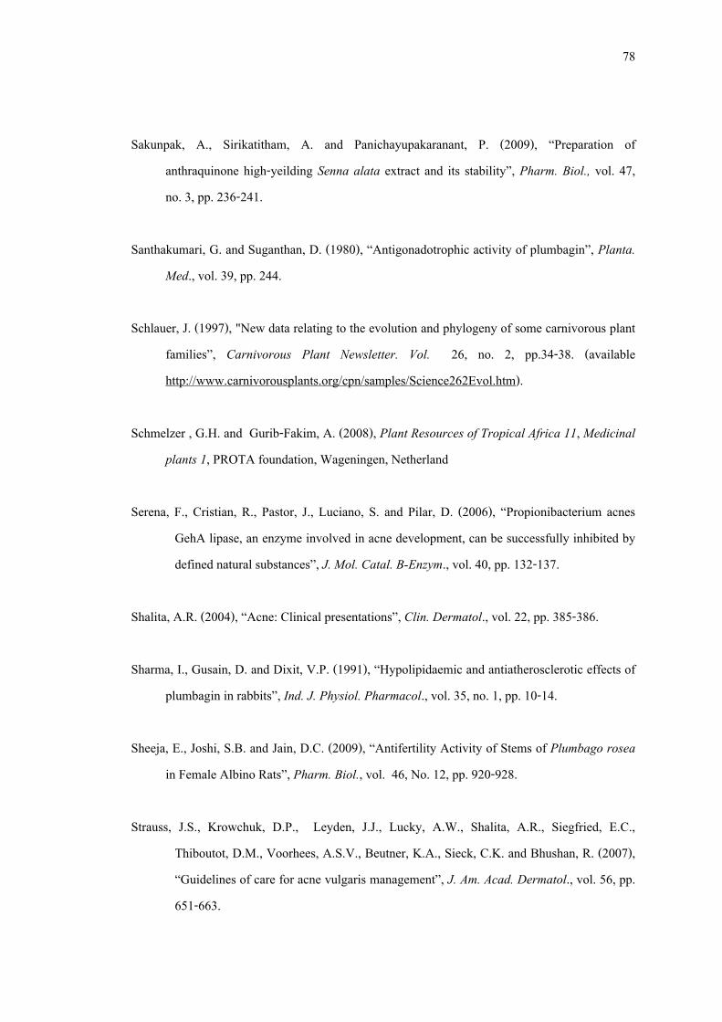
78
Sakunpak, A., Sirikatitham, A. and Panichayupakaranant, P. (2009), “Preparation of anthraquinone high-yeilding Senna alata extract and its stability”, Pharm. Biol., vol. 47, no. 3, pp. 236-241.
Santhakumari, G. and Suganthan, D. (1980), “Antigonadotrophic activity of plumbagin”, Planta.
Med., vol. 39, pp. 244. Schlauer, J. (1997), "New data relating to the evolution and phylogeny of some carnivorous plant
families”, Carnivorous Plant Newsletter. Vol. 26, no. 2, pp.34-38. (available http://www.carnivorousplants.org/cpn/samples/Science262Evol.htm).
Schmelzer , G.H. and Gurib-Fakim, A. (2008), Plant Resources of Tropical Africa 11, Medicinal
plants 1, PROTA foundation, Wageningen, Netherland Serena, F., Cristian, R., Pastor, J., Luciano, S. and Pilar, D. (2006), “Propionibacterium acnes
GehA lipase, an enzyme involved in acne development, can be successfully inhibited by defined natural substances”, J. Mol. Catal. B-Enzym., vol. 40, pp. 132-137.
Shalita, A.R. (2004), “Acne: Clinical presentations”, Clin. Dermatol., vol. 22, pp. 385-386. Sharma, I., Gusain, D. and Dixit, V.P. (1991), “Hypolipidaemic and antiatherosclerotic effects of
plumbagin in rabbits”, Ind. J. Physiol. Pharmacol., vol. 35, no. 1, pp. 10-14. Sheeja, E., Joshi, S.B. and Jain, D.C. (2009), “Antifertility Activity of Stems of Plumbago rosea
in Female Albino Rats”, Pharm. Biol., vol. 46, No. 12, pp. 920-928. Strauss, J.S., Krowchuk, D.P., Leyden, J.J., Lucky, A.W., Shalita, A.R., Siegfried, E.C.,
Thiboutot, D.M., Voorhees, A.S.V., Beutner, K.A., Sieck, C.K. and Bhushan, R. (2007), “Guidelines of care for acne vulgaris management”, J. Am. Acad. Dermatol., vol. 56, pp. 651-663.
79
Swanson, I.K. (2003), “Antibiotic resistance of Propionibacterium acnes in acnes vulgaris”, Dermatol. Nurs., vol. 5, pp. 359–361.
Valsaraj, R., Pushpangadan, P., Smitt, U.W., Adsersen, A. and Nyman, A. (1997), “Antimicrobial
screening of selected medicinal plants from India”, J. Ethnopharmacol., vol. 58, pp. 75-83.
Wang Y.C and Huang T.L. (2005), “Anti-Helicobacter pylori activity of Plumbago zeylanica L.”,
FEMS Immunol. Med. Microbiol., vol. 43, no. 3, pp. 407-412. Wiegand, I., Hilpert, K. and Hancock, R.E.W. (2008), “Agar and broth dilution methods to
determine the minimal inhibitory concentration (MIC) of antimicrobial substances”, Nat. Protoc., vol. 3, no. 2, pp. 163-175.