Embed Size (px)
Citation preview

FULL PAPER
DOI: 10.1002/ejic.200600215
Study of the In Situ Postintercalative Polymerization of Metanilic AnionsIntercalated in NiAl-Layered Double Hydroxides under a Nitrogen Atmosphere
Min Wei,[a] Xiaofei Tian,[a] Jing He,[a] Min Pu,[a] Guoying Rao,[a] Heli Yang,[a] Lan Yang,[a]
Tao Liu,[b] David G. Evans,[a] and Xue Duan*[a]
Keywords: In situ polymerization / Polyaniline / Layered double hydroxides / Nitrogen atmosphere
A new route has been developed for preparing polyaniline(PANI) layered double hydroxide (LDH) nanocompositesthrough in situ chemical oxidative polymerization of metan-ilic anions (m-NH2C6H4SO3
–) intercalated in NiAl LDHs un-der a nitrogen atmosphere by using pre-intercalated nitrateas an oxidizing agent. The interlayer space of NiAl LDHs hasbeen used as an original nanoreactor for the in situ polymeri-zation of the intercalated monomer. The whole process in-volves the synthesis of the precursor LDH [Ni2Al (OH)6(NO3)·nH2O], the intercalation of the monomer metanilic anionsinto the LDH and its in situ polymerization between the lay-ers by thermal treatment under a nitrogen atmosphere. Theinterlayer polymerization reaction was monitored by thermo-gravimetric analysis (TG), differential thermal analysis(DTA), mass spectrometry (MS), UV/Vis absorption spec-
Introduction
In recent years, considerable interest has been devoted tonanocomposites prepared from the assembly of an organicpolymer and an inorganic layered material. Polymer inter-calation nanocomposites prepared by using layered materi-als are expected to consist of a high degree of polymericordering and to exhibit advanced physicochemical proper-ties compared with the individual parts.[1–5]
Among the organic polymers, polyaniline (PANI) hasbeen and continues to be extensively studied as an organiccomponent in such systems. Polyaniline is a promising con-jugated polymer because of its simple synthesis, high con-ductivity, and excellent environmental stability, although itis associated with rather poor properties with respect toprocessing. Consequently, there are many reports focusingon the preparation of novel nanocomposites consisting ofPANI with various layered materials such as V2O5 xero-gel,[6] MoO3 bronze,[7] graphitic oxide,[8] α-RuCl3,[9] mont-morillonite,[10] and layered metal phosphates.[11]
[a] State Key Laboratory of Chemical Resource Engineering, Beij-ing University of Chemical Technology,Beijing 100029, P. R. China
[b] Institute of High Energy Physics, Chinese Academy of Science,Beijing 100039, P. R. ChinaE-mail: [email protected]
© 2006 Wiley-VCH Verlag GmbH & Co. KGaA, Weinheim Eur. J. Inorg. Chem. 2006, 3442–34503442
troscopy, in situ X-ray absorption near edge structure(XANES) spectroscopy, in situ high-temperature X-ray dif-fraction (HT-XRD) and in situ Fourier transform infrared(FTIR) spectroscopy. The UV/Vis spectra provide evidencefor the polymerization of the intercalated metanilic anions,and an increase in the interlayer distance from 16.0 to 17.2 Åis observed by HT-XRD. It has been found by the in situ tech-niques that the pre-intercalated nitrate anions act as the oxi-dizing agent that induces the polymerization of the interlayermonomer under a nitrogen atmosphere upon heating at300 °C. The orientation of the interlayer polymerization prod-uct has also been proposed.
(© Wiley-VCH Verlag GmbH & Co. KGaA, 69451 Weinheim,Germany, 2006)
As for the layered materials, LDHs have received con-siderable attention because of their special intercalationproperties. LDHs, which are widely known as a class ofanionic clays, can be represented by the general formula[M1–x
IIMxIII(OH)2]x+(An–)x/n·mH2O, where MII and MIII
are divalent and trivalent metal cations, respectively. An– isan exchangeable inorganic or organic anion and the x value,i.e. the charge density, is equal to the molar ratio MIII/(MIII
+ MII). LDHs have positively charged layers, and a widevariety of charge-balancing anionic species have been in-tercalated into the gallery region.[12] As a result, LDHs arenow well established as excellent anion-exchange materialsand their extensive intercalation chemistry has widespreadapplications in the area of organic/inorganic nanocompos-ites.[13]
PANI/LDH nanocomposites were first reported byChallier and Slade.[14] Prior to the incorporation, the LDHhost structure was exchanged with terephthalate or hexacy-anoferrate anions in order to expand the basal spacing. Thepre-swollen LDH materials were then heated at reflux withpure aniline. Analysis of the XRD patterns of the productsconfirmed the presence of multiple phases. More recently,interlayer polymerization methods have concentrated onheat treatment after polymeric monomer intercalation intothe LDH. This process is the so called “soft thermal treat-ment”, which is a two-step soft chemistry route that in-

In Situ Postintercalative Polymerization of Intercalated Metanilic Anions FULL PAPERcludes the intercalation of aniline sulfonic acid between thesheets of the LDHs and its subsequent in situ polymeriza-tion at a temperature below 200 °C in air. This approachcan avoid the ion-exchange reaction competing with theinterlayer polymerization as well as the destruction of thelayered structure by external oxidizing agents. As in the caseof other lamellar nanocomposites, atmospheric oxygen wasfound to play a major role in the polymerization pro-cess.[15–17]
In the present study, we report a new route for preparingPANI/LDH nanocomposites by using pre-intercalated ni-trates as the oxidizing agent. In situ chemical oxidative po-lymerization of metanilic anions (m-NH2C6H4SO3
–) in-tercalated in NiAl LDHs under a nitrogen atmosphere hasbeen performed for the first time. The whole process in-volves the synthesis of the precursor LDH [Ni2Al(OH)6-(NO3)·nH2O], the intercalation of the monomer metanilicanions into the LDH and its in situ polymerization betweenthe layers by thermal treatment under nitrogen. Advantagesof this method are: (1) the restricted interlayer region of theLDH makes it easier to obtain nanosized oligomers withuniform size; (2) the pre-intercalated NO3
– acting as an oxi-dizing agent can prevent the influence of mass transfer anddiffusion on the polymerization reaction of the interlayermonomer. A combination of techniques, including TG–DTA–MS, UV/Vis, in situ XANES, in situ HT-XRD, andin situ FTIR spectroscopy, was used for the characteriza-tion of intercalated metanilic anions and their in situ inter-layer polymerization. UV/Vis spectroscopy provides evi-dence for the polymerization of the intercalated monomer,and an increase in the interlayer distance from 16.0 to17.3 Å is observed by HT-XRD. It has been found by thein situ techniques that the pre-intercalated nitrate anionsact as the oxidizing agent, which induces the polymerizationof the interlayer monomer upon heating at 300 °C undernitrogen. The orientation of the interlayer polymerizationproduct has also been proposed.
Results and Discussion
Structure of NiAl-Metanilic LDHs
The X-ray diffraction patterns of the precursor NiAl-NO3 LDH and the metanilic anion intercalated LDH areshown in Figure 1. In each case, the reflections can be in-dexed to a hexagonal lattice with R-3m rhombohedral sym-metry, commonly used for the description of the LDHstructures.[19] The main diffraction peaks for NiAl-NO3
LDH appear at 9.9° (003), 19.9° (006), 29.5° (009), and62.1° (110) (Figure 1a), while the corresponding peaks forNiAl-metanilic LDH are observed at 5.4°, 11.1°, 16.8°, and61.7° (Figure 1b), respectively. Apparently, the XRDpattern for NiAl-metanilic LDH exhibits the characteristicreflections of LDH materials with a series of (00l) peaks,which are evidence for the layered character. The interlayerdistance for the NiAl-NO3 LDH is 8.9 Å which is largerthan that reported by Prinetto et al. (8.3 Å, for the existenceof a large amount of CO3
2–).[20] After the intercalation of
Eur. J. Inorg. Chem. 2006, 3442–3450 © 2006 Wiley-VCH Verlag GmbH & Co. KGaA, Weinheim www.eurjic.org 3443
metanilic acid, the value of the interlayer distance increasedto 16.3 Å, which is 7.4 Å larger than that of NiAl-NO3
LDH. The expansion of the basal spacing was due to theintercalation of metanilic anions into the LDH lamellar.Thus, the interaction of NiAl-NO3 LDH with an aqueoussolution of metanilic acid at pH 7.0 led to the anion ex-change of NO3
– for the metanilic anions with conservationof the layered structure. As for the (110) reflection, no obvi-ous shift was observed after intercalation, indicating thatno significant change occurred in the LDH layer.
Figure 1. XRD patterns for (a) NiAl-NO3 LDH and (b) NiAl-metanilic LDH.
The size of the gallery height of 11.5 Å, obtained by sub-tracting the thickness of the aluminium hydroxide layer(4.8 Å) from the interlayer distance (16.3 Å),[21] was muchlonger than a single perpendicular anion height (6.4 Å, cal-culated with Chemwin 6.0) but shorter than its doublevalue. This may indicate that the guest anions are accom-modated with an interpenetrating arrangement, leaving in-terstitial gaps between the monomers and the hydroxidesheets. Water molecules and nitrate anions occupy these in-terstitial spaces.
Compared with the NiAl-NO3 LDH precursor (Fig-ure 2a), three strong bands at 1599, 1484, and 1454 cm–1
and two weak bands at 1257 and 1315 cm–1 are observedin the FTIR spectrum of NiAl-metanilic LDH (Figure 2b),which can be attributed to the characteristic absorptionsof metanilic anions.[22] The remarkable absorption band at1384 cm–1 resulting from the stretching vibration of NO3
–
is also observed. It can be seen that not all of the NO3–
anions have been exchanged by metanilic anions, and sim-ilar results have also been reported by other researchers instudies of the intercalation of large anions into NO3-LDHprecursors.[23,24] Furthermore, elemental analysis data showthat the chemical composition of the NiAl-metanilic LDHis Ni0.66Al0.34(OH)2(C6H4NH2SO3)0.23(NO3)0.11·0.78H2O,which is consistent with the results of XRD and IR spec-troscopy.
The structure of an LDH is based on brucite-like layersin which octahedrally coordinated metal ions share edgesto form infinite sheets.[25] The area of each [Ni1–xAlx-(OH)2]x+ octahedral unit is related to the unit cell param-eter a by the formula: S = �3(a2/2).[26] The value of a can be

X. Duan et al.FULL PAPER
Figure 2. FT IR spectra for (a) NiAl-NO3 LDH and (b) NiAl-metanilic LDH.
calculated from the XRD pattern in Figure 1b (a = 2d110 =2.99 Å). The area of each octahedral unit is therefore7.74 Å2, giving one positive charge per 23 Å2 in the case ofx = 0.34 (based on the chemical composition of NiAl-met-anilic LDH given above). The maximum dimensions of theanion are calculated as 6–7 Å (calculated with Chemwin6.0), and the corresponding cross-sectional area of the com-plex anions can thus be estimated to be in the range of 28–38 Å2. Consequently, it is impossible for metanilic anionsalone to balance the positive charge of the host layer,thereby accounting for the co-intercalation of NO3
–.On the basis of the discussion above, the structure of
NiAl-metanilic LDHs can be represented as alternatingNiAl-OH hydroxide layers and layers containing metanilicanions, NO3
–, and water molecules. The schematic modelof NiAl-metanilic LDHs is proposed in Figure 3.
Figure 3. A schematic representation of the possible arrangementfor NiAl-metanilic LDHs.
UV/Vis Spectroscopy
Results obtained by UV/Vis spectroscopy give evidencefor the polymerization of metanilic anions intercalated inNiAl LDHs by heat treatment under nitrogen. Figure 4shows the optical absorption spectra, in the wavelengthrange 230–800 nm, of the synthesized NiAl-metanilic LDHand the resultant products after heat treatment at differenttemperatures.
www.eurjic.org © 2006 Wiley-VCH Verlag GmbH & Co. KGaA, Weinheim Eur. J. Inorg. Chem. 2006, 3442–34503444
Figure 4. UV/Vis spectra of NiAl-metanilic LDH after heat treat-ment at different temperatures under nitrogen.
It can be seen from Figure 4 that two absorption bandsat 262 and 269 nm at room temperature are observed, whichare associated with the phenyl group of the metanilic mono-mer. Along with the heat treatment, a strong absorptionband (centered at 296 nm) corresponding to the πB–πB*transition appears when the temperature reaches 300 °C,[27]
indicating that the polymerization of the intercalated mono-mer occurrs and the polymeric product possesses alternativebenzenoid units (see structure A). The absorption band cen-tered at 550 nm, attributable to charge transfer from thebenzoid to the quinoid segments (πB–πQ*), becomes moreobvious when the temperature increases to 320 °C (seestructures B and C).[28] The πB–πQ* transition provides di-rect evidence that quinoid units are produced and the inter-layer polyaniline sulfonic (PANIS) is present in its highlyoxidized form. This result is consistent with reports on thestudy, by optical absorption spectroscopy, of the intercon-version of polyaniline oxidation states.[27,29,30]
In Situ XANES
Previous studies on PANI/CuCr LDH nanocompositesshow that oxidative Cu2+ has been used to induce the reac-tion of the oxidative polymerization of aniline in the inter-layer galleries of pillared hosts.[14] In order to study whetherNi2+ in the host layer participates in the interlayer polyme-rization in this system, in situ X-ray absorption near edgestructure (XANES) measurements were carried out at theNi K-edge. The features in the absorption edge are sensitive
In Situ Postintercalative Polymerization of Intercalated Metanilic Anions FULL PAPERto the immediate Ni environment and local geometry. Forthe K edge, the existence of a pre-edge and its intensity areclosely related to the degree of d–p orbital mixing, and lossof centrosymmetry can cause a large intensity of this peak.The dominant jump at the edge arises from the dipolar-allowed transition related to final electronic 4p states.Therefore, it is sensitive to the variation of the oxidationstate of Ni.[31]
Figure 5 shows the normalized XANES spectra at the NiK-edge of NiAl-metanilic LDH heated under nitrogen. Thepre-edge peak (A) can be observed and this arises from thequadrupolar 1s�3d transition, which is directly related tothe occupation of the 3d orbital and to the local symmetry.Peak C reflects the long-range ordering of the Ni–O or Ni–Ni shell.[32] During the heat treatment, a broadening ofboth peaks B and C was observed as a result of the thermaldisorder. However, no significant shifts in the three peakscould be observed, implying that no dramatic redox processinvolving Ni occurred during the heat treatment. The re-sults therefore demonstrate that Ni2+ in the host layer doesnot serve as the oxidant for the interlayer oxidative polyme-rization and thus maintains the same valence in the tem-perature range 25–320 °C.
Figure 5. In situ XANES spectra at the Ni K-edge of NiAl-metan-ilic LDH heated from 25 to 320 °C under nitrogen.
TG–DTA–MS Analyses
Simultaneous thermogravimetric (TG) and–differentialthermal analyses (DTA) combined with analysis by massspectrometry was found to be useful for following the in situthermal behavior of the organic/inorganic nanocompositesover a wide temperature range.[33]
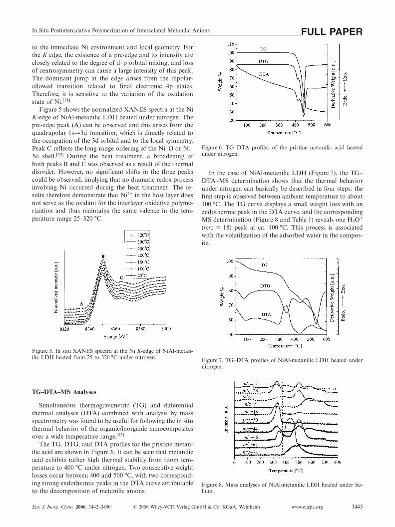
The TG, DTG, and DTA profiles for the pristine metan-ilic acid are shown in Figure 6. It can be seen that metanilicacid exhibits rather high thermal stability from room tem-perature to 400 °C under nitrogen. Two consecutive weightlosses occur between 400 and 500 °C, with two correspond-ing strong endothermic peaks in the DTA curve attributableto the decomposition of metanilic anions.
Eur. J. Inorg. Chem. 2006, 3442–3450 © 2006 Wiley-VCH Verlag GmbH & Co. KGaA, Weinheim www.eurjic.org 3445
Figure 6. TG–DTA profiles of the pristine metanilic acid heatedunder nitrogen.
In the case of NiAl-metanilic LDH (Figure 7), the TG–DTA–MS determination shows that the thermal behaviorunder nitrogen can basically be described in four steps: thefirst step is observed between ambient temperature to about100 °C. The TG curve displays a small weight loss with anendothermic peak in the DTA curve, and the correspondingMS determination (Figure 8 and Table 1) reveals one H2O+
(m/z = 18) peak at ca. 100 °C. This process is associatedwith the volatilization of the adsorbed water in the compos-ite.
Figure 7. TG–DTA profiles of NiAl-metanilic LDH heated undernitrogen.
Figure 8. Mass analyses of NiAl-metanilic LDH heated under he-lium.

X. Duan et al.FULL PAPERTable 1. Results for MS analyses of NiAl-metanilic LDH.
ProposedH2
+ N+ O+ H2O+ N2+ NO+ N2O+ NO2
+ SO2+ C6H4
+
positive ion
m/z 2 14 16 18 28 30 44 46 64 76Intensity [A] 10–11 10–11 10–11 10–10 10–10 10–11 10–11 10–12 10–12 10–13
The second step takes place in the temperature range300–350 °C. The TG curve shows a big weight loss ac-companied by a strong exothermic peak in the DTA curve.Several fragments assigned to N2
+ (m/z = 28), NO+ (m/z =30), N2O+ (m/z = 44), H2O+ (m/z = 18), and NO2
+ (m/z =46) appear in the corresponding MS curves. For the nitrate-containing LDHs, the release of NO2 and NO was generallyattributable to the pyrolysis of nitrates, whereas the inter-layer nitrate would be evolved as NO and N2O only duringthe TPR experiment under hydrogen.[34] In this work, therelease of N2, NO, and N2O at the same time provides evi-dence that the co-intercalated nitrate anions are involved inan interlayer redox reaction. The NO2
+ peak lags behindthe N2
+, NO+, N2O+, and H2O+ peaks, indicating that py-rolysis of the pre-intercalated nitrates occurs after re-duction. The DTA curve in this step displays a strong exo-thermic peak at about 320 °C, which was not found in theDTA curve of the pristine metanilic acid. Roland-Swansonet al. have reported DTA traces with such an exothermicpeak assigned to the polymerization of the 3-sulfopropylmethacrylate potassium salt (SPMA) intercalated in ZnAlLDHs by thermal treatment.[35] Because the reaction wascomplete without the use of any external chemical agent,the polymerization process was interpreted as an oxidativereaction induced by atmospheric oxygen. In the presentstudy, this strong exothermic peak and the reduction of theco-intercalated nitrates correspond to the emergence of thenew absorption bands attributable to the πB–πB* and πB–πQ* transitions in UV/Vis spectra, which demonstrates thepolymerization of metanilic anions intercalated NiAl LDHsin the same temperature range. On the basis of the dis-cussion above and of the comparison with other studies onthe thermal behavior of the polymeric monomer/LDH nano-composites, the strong exothermic process under an inertatmosphere in this work can be assigned to the chemicaloxidative polymerization of the interlayer monomer withthe co-intercalated nitrates acting as the oxidizing agent.This will be further discussed in the next sections.
The third weight loss (350–450 °C) is due to the pyrolysisof the hydroxide layers and the interlayer organic materials,which results in an enhanced signal of H2O+ (m/z = 18) andSO2
+ (m/z = 64). The corresponding endothermic peak isobserved at ca. 430 °C. The complete thermal decomposi-tion of the nanocomposite occurs in the fourth step (450–600 °C).
In Situ FTIR
Figure 9 shows the in situ FTIR spectra for NiAl-metan-ilic LDH heated to different scheduled temperatures undernitrogen. Table 2 summarizes the assignments of the IR ab-
www.eurjic.org © 2006 Wiley-VCH Verlag GmbH & Co. KGaA, Weinheim Eur. J. Inorg. Chem. 2006, 3442–34503446
sorption bands for metanilic-intercalated LDH and its ther-mal products.[36,37]
Figure 9. In situ FTIR spectra for the thermal polymerization ofNiAl-metanilic LDH.
Table 2. Assignments for IR absorption bands for metanilic-in-tercalated LDH and its thermal products.[a]
Frequency [cm–1] Assignment
1599 disorder-induced C–C ring stretching1581 stretching of N=Q=N1484 ring stretching of C=C1468 stretching of N=B=N1453 stretching of benzene ring1315 N–H in or out of plane bending1275 C–H bending or C–N stretching1170 C–H bending in plane1110 C–S bending in plane1035 S=O bending991 C–H bending out of plane
[a] Abbreviations: B, benzenoid unit; Q, quinonoid unit.
It can be seen that very little change is observed for thethree strong bands at 1599, 1484, and 1454 cm–1 and theweak band at 1315 cm–1, which are characteristic absorp-tions of metanilic anions from room temperature to 250 °C.When the temperature increases to 300 °C, the former fourcharacteristic bands weaken remarkably because of the in-crease in the conjugated degree of the polymerization prod-ucts upon thermal treatment. Moreover, the emergence ofthe bands at 1581 and 1468 cm–1 in the spectrum of NiAl-metanilic LDH at 300 °C indicates that the polymerizationproduct is in a high oxidation state.[38] The comparison ofthe IR spectrum of the original metanilic-intercalated LDHand those of its oxidation products after treatment at300 °C reveals a sharp decrease in the intensity of the bandsat 1170 (υC–H in plane), 1275, and 1315 cm–1, which pointsto a substantial decrease in the number of amino groups(–NH2). This further confirms the formation of an inter-layer polyconjugated system. Moreover, the intensity of theband at 1384 cm–1 attributable to cointercalated NO3
– de-creases sharply as the temperature reaches 300 °C, therebygiving evidence that NO3
– is engaged in the interlayer redoxreaction. This is in agreement with the results of UV/Visspectroscopy and TG–MS.

In Situ Postintercalative Polymerization of Intercalated Metanilic Anions FULL PAPERIn Situ HT-XRD
In order to get further insight into the thermal polymeri-zation process of the monomer intercalated NiAl LDH, insitu X-ray measurements were carried out. The tempera-tures at which the X-ray data were collected were definedon the basis of TG–DTA analysis. The in situ HT-XRDpatterns of the intercalation product NiAl-metanilic LDHin the temperature range 25–320 °C under nitrogen areshown in Figure 10. The variation in the d003 basal spacingof NiAl-metanilic LDH with temperature is displayed inFigure 11.
Figure 10. In situ HT-XRD patterns of NiAl-metanilic LDH in thetemperature range 25–320 °C.
Figure 11. The relationship between the d003 basal spacing and tem-perature.
Upon increasing the temperature, the (003), (006), and(009) reflections of NiAl-metanilic LDH move to a slightlyhigher 2θ angle (as shown in Figure 10), and the (003) basalspacing decreases from 16.3 to 16.0 Å (Figure 11) when thematerial was heated from 25 °C to 150 °C. This contractionmay result from the loss of absorbed water and parts ofgallery water.[39] However, the value of d003 slowly increasesfrom 16.0 Å at 150 °C to 16.2 Å at 250 °C. This might bedue to the reorientation of the interlayer monomer ac-companied by the loss of the gallery water.
Eur. J. Inorg. Chem. 2006, 3442–3450 © 2006 Wiley-VCH Verlag GmbH & Co. KGaA, Weinheim www.eurjic.org 3447
When the temperature reaches 300 °C, the (003) reflec-tion of the nanocomposite becomes broader and moves to aremarkably lower 2θ angle. The interlayer distance increasesdrastically from 16.2 Å at 250 °C to 17.2 Å at 300 °C, indi-cating that interlayer polymerization of the interlamellarspecies occurs. This phenomenon was also observed in thecase of post-intercalative polymerization of aniline and itsderivatives in layered metal phosphates after thermal treat-ment at 130 °C in air.[11] In addition, the color of the nano-composite turns from bright green to dark brown after theheat treatment. The broadening of the (003) reflection andthe decrease in intensity of other diffraction peaks of thenanocomposite at 300 °C indicate the decrease in orderedstacking sequences and in crystallinity during the polymeri-zation process. Nevertheless, the diffraction maximum be-tween the 2θ values of 62 and 63° (the spectrum at 300 °Cin Figure 10) can still be observed, indicating the conser-vation of the lamellar structure with no substantial struc-tural collapse during the interlayer polymerization reaction.
The coupling between the monomers and the arrange-ment of their subsequent dimerization or polymerizationhave been discussed in term of the gap sizes, which can becalculated by using the peak fit program of XRD-6000 Ver-sion 4.1.[17] The geometry and atomic charges of the pos-sible dimers have been optimized by using the MOPACsemi-empirical method, employing the PM3 Hamilto-nian.[40]
Figure 12 shows the peak fit results of d003 at 300 °C, atwhich the interlayer polymerization occurs. Three differentd003 values, i.e. 15.77, 17.48, and 20.07 Å were obtained inthis case, and the corresponding gallery heights were calcu-lated to be 10.97, 12.68, and 15.27 Å, respectively, by sub-tracting the inorganic layer thickness of 4.8 Å. The polyme-rization between monomers located on different layers (op-posite inner side) may proceed iso- or syndiotactically, andthe coupling between monomers may occur by a linkage(α,β), (β,β), or (N,N). The calculated lengths of the threepossible dimers are 10.43, 11.63, and 12.42 Å (as shown inScheme 1), respectively. It can be concluded that the coup-ling between monomers most likely occurs by the linkages(α,β) and (N,N) since the calculated lengths of the two pos-sible dimers (10.43 and 12.42 Å) correspond well with thetwo gallery heights of 10.97 and 12.68 Å. As a result, thecoupling between monomers on different layers hereinbe-fore should give rise to the quasi-monolayer arrangementby the linkage (α,β) (Figure 13a), whereas the gallery heightof 15.27 Å may result from the bilayer arrangement whenpolymerization occurs between the two monomers on thesame layer (Figure 13b).[17] The linkage of N and N charac-
Figure 12. The peak fit results of d003 of NiAl-metanilic LDH at300 °C.

X. Duan et al.FULL PAPER
Scheme 1. The structure for the three possible dimers of metanilic anions formed by different linkages: (a) (α,β), (b) (β,β), and (c) (N,N).
Figure 13. Schematic representations of the interlayer polymerization products.
teristics, which results from the “head-to-head” coupling(Figure 13c), was observed only in the spectra of productsprepared under neutral or basic conditions. For instance,the formation of azobenzene derivatives in basic or in anacetonitrile/pyridine medium has also been reported byWawzonek and MacIntyre.[41] On the basis of the discussionabove, it can be concluded that the coupling between mono-mers most likely occurs by the (α,β) and (N,N) linkages,and the main coupling likely occurs by an (α,β) linkage.This is in agreement with the much earlier work ofMohilner et al., in which aniline oxidation products ob-tained in neutral or weakly acidic media have a predomi-nantly “head-to-tail” arrangement.[42]
TEM
The TEM micrographs of the prepared materials are il-lustrated in Figure 14. It can be seen that NiAl-metanilicLDH exhibits the characteristic LDH platelet, an ill-definedshape with a uniform size from 200 to 300 Å (Figure 14a).This morphological result is consistent with that from HT-XRD analysis. The polymerization product PANIS shows
www.eurjic.org © 2006 Wiley-VCH Verlag GmbH & Co. KGaA, Weinheim Eur. J. Inorg. Chem. 2006, 3442–34503448
a similar platelike morphology (Figure 14b) with averageparticle size of 50–100 Å. Such a small size is possibly dueto the restriction on the chain growth of PANIS affordedby the layers of LDH.
Figure 14. TEM images of (a) NiAl-metanilic LDH and (b) thepolymerization product (PANIS).

In Situ Postintercalative Polymerization of Intercalated Metanilic Anions FULL PAPER
Conclusions
The in situ chemical oxidative polymerization of metan-ilic anions (m-NH2C6H4SO3
–) intercalated in NiAl LDHhas been performed for the first time by the use of pre-intercalated nitrate as the oxidizing agent. From the charac-terization by XRD, IR spectroscopy, elemental analysis,and geometrical considerations, the schematic model ofNiAl-metanilic LDH has been proposed in which the struc-ture can be represented as alternating NiAl-OH hydroxidelayers and layers containing metanilic anions, NO3
– ions,and water molecules.
Results obtained by UV/Vis spectroscopy give evidencefor the polymerization of the metanilic anions intercalatedNiAl LDH when the temperature reaches 300 °C. The par-ticle sizes of 50–100 Å of the interlayer polymerizationproduct determined by TEM microscopy indicates the lowconjugation extent of the interlayer PANIS.
In situ XANES demonstrates that Ni2+ in the host layerdoes not serve as the oxidant for interlayer oxidative poly-merization. According to the results of TG–DTA–MS forthe pristine metanilic acid and the NiAl-metanilic LDH un-der nitrogen, we propose that the pre-intercalated nitrateanions act as the oxidant, which induces the polymerizationof the interlayer monomer under nitrogen upon heating at300 °C.
In situ FTIR and in situ HT-XRD provide further in-sight into the thermal polymerization process. The basalspacing increases from 16.0 to 17.2 Å, and the characteristicvibrational absorptions of polyaniline emerge at 300 °C un-der nitrogen. It can be concluded, on the basis of the peakfit program and the MOPAC semi-empirical method, thatthe coupling between monomers most likely occurs by the(α,β) and (N,N) linkages. The possible schematic arrange-ment for the interlayer products has also been proposed.
Experimental SectionReagents: All chemicals used in this synthesis including Ni(NO3)2·6H2O, Al(NO3)3·9H2O, NaOH, and metanilic acid (m-NH2C6H4SO3
–) were of analytical grade and were purchased fromAldrich and used without any further purification. High purity ni-trogen gas (O2 � 1 ppm) and high purity helium gas (O2 � 1 ppm)were purchased from the Beijing Chemical Plant Limited. All solu-tions were prepared with distilled and decarbonated water.
Synthesis: The precursor LDH [Ni2Al(OH)6(NO3)]·nH2O was pre-pared following a standard aqueous coprecipitation and thermalcrystallization method.[18] A solution of NaOH (8.0 g, 0.20 mol) inwater (100 mL) was added dropwise over 2 h to a solution (100 mL)containing Ni(NO3)2·6H2O (19.4 g, 0.066 mol) and Al(NO3)3·9H2O (12.5 g, 0.033 mol), with vigorous stirring under nitrogen.The pH value of the solution at the end of addition was 7.0. Theresultant gelatinous precipitate was maintained at 100 °C for 72 h,centrifuged, and washed thoroughly with water before drying at70 °C for 24 h under vacuum.
The metanilic intercalated NiAl-LDH was prepared following theion-exchange method. A solution (100 mL) of metanilic acid(10.4 g, 6.0 mmol) with a pH value adjusted to 7.0 using NaOH(2.0 ) was added to a suspension of NiAl-NO3 LDH (5.0 g, ca.
Eur. J. Inorg. Chem. 2006, 3442–3450 © 2006 Wiley-VCH Verlag GmbH & Co. KGaA, Weinheim www.eurjic.org 3449
1.5 mmol) in water (100 mL). The mixture was held at 30 °C undernitrogen for 72 h. The product was washed extensively with water,centrifuged, and dried under vacuum for 24 h.
For the thermal treatment, the hybrid phase was placed in a tubefurnace heated at 25, 100, 150, 200, 250, 300, or 320 °C for 0.5 hunder nitrogen.
Characterization Techniques: Powder X-ray diffraction (XRD) mea-surements were performed on a Rigaku XRD-6000 diffractometer,using Cu-Kα radiation (λ = 1.5418 Å) at 40 kV and 30 mA. Datawere collected over the angular range of 2θ from 3° to 70° at ascanning rate of 0.02° per second at room temperature. The UV/Vis spectra were collected in reflectance mode using a ShimadzuUV-2401 spectrophotometer in the 230–800 nm region (the sampleswere dissolved in 0.1 hydrochloric acid). Fourier transform infra-red (FTIR) spectra were recorded using a Vector 22 (Bruker) spec-trophotometer in the range 4000 to 400 cm–1 with 2 cm–1 resolu-tion. The standard KBr disk (1 mg of sample in 100 mg of KBr)was used. In situ XANES spectra were collected at the XAFS sta-tion (beam line 4W1B) of the Beijing Synchrotron Radiation Facil-ity. The storage ring energy was 2.2 GeV and the current was 50–60 mA. The X-rays were monochromated by using a Si (111) singlecrystal. Samples were shaped into ingots with a thickness of about1 mm. X-ray absorption spectra of the Ni K-edge were collected atambient temperature up to 320 °C with a heating rate of 5 °Cmin–1
under nitrogen in the transmission mode. The incident and trans-mission X-ray intensities were detected using ion chambers whichwere installed in front of and behind the sample, respectively. Insitu HT-XRD measurements were performed on a Rigaku D/max2500VB2+/PC diffractometer in the temperature range 25–320 °Cunder nitrogen by using Cu-Kα radiation (λ = 1.5418 Å) at 40 kVand 30 mA. The samples as disoriented powders were scanned insteps of 0.02° in the 2θ range 3°–70° by using a count time of 4 sper step. An α-Al2O3 substrate was used. In situ FTIR spectra wererecorded with a Nicolet 60sxb spectrometer in the range 4000 to400 cm–1 with 2 cm–1 resolution. The spectra were obtained every10° with a heating rate of 5 °Cmin–1 under nitrogen. The standardKBr disk method (1 mg of sample in 100 mg of KBr) was used.TG–DTA analyses were carried out under nitrogen (flux of100 mLmin–1) with a Seiko 6300 simultaneous DTA–TGA appara-tus from Seiko Instruments, at a heating rate of 5 °Cmin–1, withAl2O3 as a reference. Simultaneous TG–MS analyses were per-formed with a Pyris Diamond TG–DTA instrument coupled to aThermoStarTM QM220 mass spectrometer by a quartz capillarytransfer line at 180 °C. The heating rate was 5 °Cmin–1, with ahelium flow of 100 mLmin–1. The scanning speed of mass was1 a.m.us–1, with a filtering time 0.03 s. The TGA apparatus oper-ated at atmospheric pressure, and the mass spectrometer at a work-ing pressure of 3×10⁻⁶ mPa and an electron energy of 70 eV. Theproposed mass number, the ion current intensity, and the amplify-ing rate are listed in Table 1. Elemental analyses were performedwith a Shimadzu ICPS-7500 instrument. C, H, and N content weredetermined by using an Elementarvario elemental analysis instru-ment. TEM images were obtained by using a JEOL JEM-1200 in-strument operating at an acceleration voltage of 80 kV. The TEMsamples were ultrasonically dispersed in water, and then a suspen-sion was deposited onto a holey carbon film deposited on a Cugrid.
Acknowledgments
This project was supported by the National Natural Science Foun-dation Key Project of China (Project No.: 20531010), the National

X. Duan et al.FULL PAPERNatural Science Foundation Major International Joint ResearchProgram (Project No.: 20620130108), the Beijing Nova Program(No.: 2004A13), and the Program for Changjiang Scholars and theInnovative Research Team at the University (PCSIRT). We alsoacknowledge the Beijing Synchrotron Radiation Facility (BSRF)for provision of synchrotron radiation facilities and thank Dr. TaoLiu and Yaning Xie for assistance in using beamline 4W1B.
[1] A. I. Khan, D. O’Hare, J. Mater. Chem. 2002, 12, 3191–3198.[2] F. Leroux, J. P. Besse, Chem. Mater. 2001, 13, 3507–3515.[3] R. Schollhorn, Chem. Mater. 1996, 8, 1747–1757.[4] L. Vieille, E. M. Moujahid, C. Taviot-Guého, J. Cellier, J. P.
Besse, F. Leroux, J. Phys. Chem. Solids 2004, 65, 385–393.[5] E. M. Moujahid, F. Leroux, M. Dubois, J. P. Besse, C. R. Chim.
2003, 6, 259–264.[6] M. G. Kanatzidis, C. G. Wu, H. O. Marcy, C. R. Kannewurf,
J. Am. Chem. Soc. 1989, 111, 4139–4141.[7] T. A. Kerr, H. Wu, L. F. Nazar, Chem. Mater. 1996, 8, 2005–
2015.[8] P. Liu, K. Gong, Carbon 1999, 37, 706–707.[9] L. Wang, P. Brazis, M. Rocci, C. R. Kannewurf, M. G. Kanatz-
idis, Chem. Mater. 1998, 10, 3298–3300.[10] H. Inoue, H. J. Yoneyama, J. Electroanal. Chem. 1987, 233,
291–294.[11] Y. J. Liu, M. G. Kanatzidis, Chem. Mater. 1995, 7, 1525–1533.[12] C. B. Koch, Hyperfine Interact. 1998, 117, 131–157.[13] F. Cavani, F. Trifiro, A. Vaccari, Catal. Today 1991, 11, 173–
301.[14] T. Challier, R. C. T. Slade, J. Mater. Chem. 1994, 4, 367–371.[15] E. M. Moujahid, M. Dubois, J. P. Besse, F. Leroux, Chem. Ma-
ter. 2002, 14, 3799–3807.[16] V. P. Isupov, L. E. Chupakhina, M. A. Ozerova, V. G. Kostrov-
sky, V. A. Poluboyarov, Solid State Ionics 2001, 141–142, 231–236.
[17] E. M. Moujahid, M. Dubois, J. P. Besse, F. Leroux, Chem. Ma-ter. 2005, 17, 373–382.
[18] J. Wilson, T. Olorunyolemi, A. Jaworski, L. Borum, D. Young,A. Siriwat, E. Dickens, Appl. Clay Sci. 1999, 15, 265–279.
[19] N. T. Whilton, P. J. Vickers, S. Mann, J. Mater. Chem. 1997, 7,1623–1629.
[20] F. Prinetto, D. Tichit, R. Teissier, B. Coq, Catal. Today 2000,55, 103–116.
www.eurjic.org © 2006 Wiley-VCH Verlag GmbH & Co. KGaA, Weinheim Eur. J. Inorg. Chem. 2006, 3442–34503450
[21] J. Zhang, F. Z. Zhang, L. L. Ren, D. G. Evans, X. Duan, Ma-ter. Chem. Phys. 2004, 85, 207–214.
[22] T. A. Huber, A Literature Survey of Polyaniline, Defence R&DCanada – Atlantic, 2003, p. 014.
[23] N. T. Whilton, P. J. Vickers, S. Mann, J. Mater. Chem. 1997, 7,1623–1629.
[24] A. Fudala, I. Palinko, I. Kiricsi, Inorg. Chem. 1999, 38, 4653–4658.
[25] G. W. Brindley, S. Kikkawa, Am. Mineral. 1979, 64, 836–843.[26] S. K. Yun, T. J. Pinnavaia, Chem. Mater. 1995, 7, 348–354.[27] J. E. de Albuquerque, L. H. C. Mattoso, M. Faria, J. G. Mas-
ters, A. G. MacDiarmid, Synth. Met. 2004, 146, 1–10.[28] B. A. Deore, I. Yu, M. S. Freund, J. Am. Chem. Soc. 2004, 126,
52–53.[29] V. Luca, S. Thomson, J. Mater. Chem. 2000, 10, 2121–2126.[30] L. W. Shacklette, J. F. Wolf, S. Gould, R. H. Baughman, J.
Chem. Phys. 1988, 88, 3955–3961.[31] F. Leroux, P. J. Dewar, M. Intissar, G. Ouvrard, L. F. Nazar, J.
Mater. Chem. 2002, 12, 3245–3253.[32] M. Jiménez-Ruiz, C. Prieto, J. L. Martínez, J. M. Alonso, J.
Solid State Chem. 1998, 140, 278–284.[33] C. W. Lu, Z. He, T. G. Xi, Y. X. Chen, L. Luo, Thermochim.
Acta 1999, 334, 149–155.[34] D. Tichit, F. Medina, B. Coq, R. Dutartre, Appl. Catal. A 1997,
159, 241–258.[35] C. Roland-Swanson, J. P. Besse, F. Leroux, Chem. Mater. 2004,
16, 5512–5517.[36] M. C. Miras, C. Barbero, O. Haas, Synth. Met. 1991, 43, 3081–
3084.[37] Y. Furukawa, F. Ueda, Y. Hyodo, I. Harada, T. Nakajima, T.
Kawagoe, Macromolecules 1988, 21, 1297–1305.[38] E. T. Kang, K. G. Neoh, K. L. Tan, Prog. Polym. Sci. 1998, 23,
277–324.[39] M. Wei, J. Wang, J. He, D. G. Evans, X. Duan, Microporous
Mesoporous Mater. 2005, 78, 53–61.[40] A. M. Aicken, I. S. Bell, P. V. Coveney, W. Jones, Adv. Mater.
1997, 9, 496–500.[41] S. Wawzonek, T. W. MacIntyre, J. Electrochem. Soc. 1972, 119,
1350–1357.[42] D. M. Mohilner, R. N. Adams, W. J. Argersinger, J. Am. Chem.
Soc. 1962, 84, 3618–3622.Received: March 9, 2006
Published Online: July 11, 2006