Embed Size (px)
Citation preview

STUDY OF SERUM MAGNESIUM LEVEL
IN TYPE 2 DIABETES MELLITUS
Dissertation submitted in partial fulfilment of requirements for
M.D.DEGREE IN GENERAL MEDICINE
BRANCH I
Of
THE TAMILNADU Dr. M.G.R. MEDICAL UNIVERSITY,
CHENNAI, INDIA.
THANJAVUR MEDICAL COLLEGE ,
THANJAVUR - 613004.
THE TAMILNADU Dr. M.G.R. MEDICAL UNIVERSITY,
CHENNAI-600032.
APRIL 2013

CERTIFICATE
This is to certify that the dissertation entitled “STUDY OF SERUM
MAGNESIUM LEVEL IN TYPE 2 DIABETES MELLITUS” is a bonafide
work done by Dr.SUBASH CHANDRABOSE G. at Thanjavur Medical
College,Thanjavur in partial fulfilment of the university rules and regulations
for award of M.D., Degree in General Medicine (Branch-I) under my guidance
and supervision during the academic year 2010-2013.
Prof.Dr.C.GUNASEKARAN M.D.D.C.H,
The Dean I/C,
Thanjavur Medical College,
Thanjavur–613004.
Prof.Dr.S.MUTHUKUMARAN,M.D.,
Head of the Department,
Department of Internal Medicine,
Thanjavur Medical College,
Thanjavur–613004.
Prof.Dr.K.NAGARAJAN,M.D.,
Unit Chief M-III,
Department of Internal Medicine,
Thanjavur Medical College,
Thanjavur–613004.

DECLARATION
I solemnly declare that the dissertation titled “STUDY OF SERUM
MAGNESIUM LEVEL IN TYPE 2 DIABETES MELLITUS” is done by
me at Thanjavur Medical College, Thanjavur during 2010-2013 under the
guidance and supervision of Prof. Dr.K.NAGARAJAN,M.D., The dissertation
is submitted to The Tamilnadu Dr.M.G.R. Medical University towards the
partial fulfilment of requirements for the award of M.D. degree in General
Medicine (Branch Ι).
Place: Thanjavur Signature of candidate
Date: ( DR.G.SUBASH CHANDRABOSE )

ACKNOWLEDGEMENT
At the outset, I thank Prof. Dr.C.GUNASEKARAN M.D.D.C.H,
The Dean I/C, Thanjavur Medical College and Hospital, Thanjavur for granting
me permission to do my dissertation and for allowing me to use institutional
facilities.
I am grateful to Prof. Dr.S.MUTHUKUMARAN M.D., Professor and
Head, Department of Internal Medicine, Thanjavur medical college and
Hospital for his constant support and guidance throughout my Post graduate
period and during the course of this study.
I am indebted to Prof. Dr.K.NAGARAJAN M.D., Professor of
Medicine and my unit chief, who constantly helped me and guided me in this
study and during my post-graduate period.
I profoundly express my heartiest thanks to my respected Professors
Prof. Dr.P.G.SANKARANARAYANAN M.D, Prof. Dr.S.MANOHARAN
M.D, Prof. Dr C.GANESAN M.D, and Prof. Dr.D.NEHRU M.D. D.M.R.D,
for their advice, guidance and valuable criticism which enabled me to do this
work effectively.
I would also like to thank Prof. Dr.M.THANGARAJ M.D.D.M, Head
of Department of Neurology, Department of Neurology, Thanjavur Medical
College and Hospital, Thanjavur who supported and helped me during this
study.

I would also like to thank Assistant Professors Dr.S.ELANGOVAN
M.D.D.M, Dr. K.BALAMURALI M.D.D.M, Department of Neurology,
Thanjavur Medical College and Hospital, Thanjavur for their encouragement
during this study.
I would also like to thank Dr. C. PARANTHAGAN M.D. Registrar,
Department of Internal Medicine for his support and guidance.
I would also like to thank Dr.J.VIJAYBABU M.D.D.M.,.
Dr.S.VETRIVEL M.D., and Dr. THIRUMURUGAN M.D., Assistant
Professors of my unit, Department of Internal Medicine for their constant help,
encouragement and support throughout this study and during my post graduate
period.
I would also like to thank J.S.JESUS RAJA M.E, for his excellent
support in statistical analysis.
I express my sincere gratitude to all the patients who participated in the
study.
Lastly, I thank all my professional colleagues for their support and
valuable criticism.


CONTENTS
SL.NO TITLE PAGE NO
1 INTRODUCTION 1
2 AIMS AND OBJECTIVES 3
3 REVIEW OF LITERATURE 4
4 METHODOLOGY 53
5 RESULTS 58
6 DISCUSSION 80
7 CONCLUSION 86
8 ANNEXURES
BIBLIOGRAPHY
ANNEXURE I -ABBREVIATIONS
ANNEXURE II- PROFORMA
ANNEXURE III-MASTER CHART
ANNEXURE IV- KEYS TO MASTER CHART
ANNEXURE V- CONSENT FORM

STUDY OF SERUM MAGNESIUM LEVEL IN TYPE 2
DIABETES MELLITUS
ABSTRACT
BACKGROUND
A high prevalence of magnesium deficiency is reported in diabetics.
Magnesium depletion has a negative impact on glucose homeostasis and insulin
sensitivity in type 2 diabetic patients as well as on the evolution of
complications such as retinopathy, nephropathy, neuropathy and arterial
atherosclerosis. The aim of this study is to estimate the prevalence of
hypomagnesemia in patients with type 2 diabetes mellitus and its correlations
with microvascular complications of diabetes like retinopathy, nephropathy,
neuropathy.
MATERIALS AND METHODS:
Patients with type 2 diabetes admitted in Thanjavur Medical College and
Hospital over a period of one year between October 2011 to October 2012
formed the study population. The sample size was 100 patients. Serum
magnesium concentration was measured by calmagite dye method.
RESULTS:
The study revealed that prevalence of hypomagnesemia in study subjects
was 35%. Sex, age and duration of diabetes were not significant predictors of

serum magnesium. Significant association was found between hypomagnesemia
and diabetic retinopathy, nephropathy, neuropathy. Significant correlations were
not found with co morbidities such as ischemic heart disease and hypertension.
Low serum magnesium concentrations are common in type 2 diabetics.
Magnesium deficiency is conclusively associated with diabetic retinopathy,
nephropathy, neuropathy.
KEY WORDS:
Magnesium ,Diabetes Mellitus, Retinopathy, Nephropathy, Neuropathy
1
INTRODUCTION
India is frequently referred to as the diabetic capital of the world as it has
the highest number of cases in the world.
In worldwide the last 2 decades, incidence is suddenly increased from 30
million cases in 1985 to 171 million in 2000. Recent data suggests that
prevalence of DM by the year 2030 could be 360 million. DM is worldwide in
distribution and the incidence of both types is rising.1, 2
The distribution of both T1 DM and T2DM varies worldwide, due to
relative difference in genetic and environmental factors in different parts of the
world. Recent data shows it is associated with 10-30% reduction of life
expectancy, most common cause of blindness in the age group of 20 to 65 years,
25 fold increased risk of non traumatic lower limb amputations and increase
incidence of end stage renal disease approximately 1000 patients per year2.
India had around 31.7 million cases in year 2000 which is expected to
rise alarmingly to around 79.4 million in 2030 by which time every fifth
diabetic subject in the world would be an Indian3.
In Tamilnadu the prevalence in 2008 is 18.6% in urban areas and 9.1% in
rural areas3.
Although DM can cause hypomagnesemia , low serum magnesium level
is a risk factor for DM. Magnesium is an essential element for several enzymes

2
that play important role in glucose metabolism. Animal studies found that low
magnesium has a negative effect on post receptor signalling of insulin. Some
studies have also found that magnesium supplementation improves insulin
action and glucose metabolism in diabetics
4.
Magnesium is involved in multiple levels of insulin secretion.
Magnesium deficiency can modify the Na+ K
+ ATPase channel that maintains
sodium, potassium and glucose transport5.
In DM there is a direct correlation between serum magnesium
concentration in blood and cellular glucose disposal that is independent of
insulin secretion.6
Low serum magnesium level has direct correlation with microvascular
complications (retinopathy7, neuropathy. nephropathy) and macrovascular
complications (IHD and cerebrovascular disease)8.
In elderly type2 DM Paolisso hypothesized that oral magnesium
supplementation for 4 weeks results in decreased fasting blood sugar level
increased plasma and RBC magnesium level.9
In this study serum magnesium concentration in type2 DM patients is
correlated with occurrence of microvascular complications.

3
OBJECTIVES
The aims of this study are
1. Measurement of serum magnesium concentration in diabetes mellitus .
2. To estimate the prevalence of hypomagnesemia in patients with type 2
diabetes mellitus
3. Its correlation with microvascular complications like retinopathy,
nephropathy, neuropathy.

4
REVIEW OF LITERATURE
DIABETES MELLITUS
Diabetes Mellitus is a clinical syndrome characterized by high blood
sugar level (hyperglycemia) and glycosuria due to relative or absolute
deficiency of insulin secretion or its action, or insulin resistance that leads to
disturbances in carbohydrate ,protein, fat metabolism , water and electrolyte
homeostasis.
THE HISTORICAL ASPECTS
Its history has been characterized by numerous cycles of discovery,
neglect and rediscovery. Its history may be divided into four major periods. The
„ANCIENT‟ period showed the first clinical features of DM and its
complications. The 16 th
to 18th
centuries have been termed the „DIAGNOSTIC‟
period, as DM was then identified as a separate disease entity. The mid to late
19th centuries may be consider as the first „EXPERIMENTAL‟ period. During
this period role of the pancreas became clear and the molecular level of diabetes
were initially identified10
. Finally, in the 20th
century knowledge about diabetes
is well known.

5
The word Diabetes in Greek means – “I run through Siphon”. Indian
name for Diabetes is Madhumeha – Honey in rain. In 16th century, Susrutha in
the Sanskirit book of surgery, and Charaka in the Sanskirit book of medicine
have mentioned about Diabetes. The first person -Vaidys – tested the urine of
diabetic patients.
SPECTRUM OF DIABETES MELLITUS & GLUCOSE HOMEOSTASIS
Types
Normal
glucose
tolerance
(mg/dl)
HYPERGLYCEMIA
PREDIABETES
IFG
&
IGT
(mg/dl)
DIABETES MELLITUS
NOT
INSULIN
REQUIRED
INSULIN
REQUIRED
FOR
CONTROL
INSULIN
REQUIRED
FOR
SURVIVAL
T1DM
T2DM
Other types
GDM
Time
(years)
FBG
(mg/dl)
2-h PPBG
(mg/dl)
< 110
< 140
110-125
140 – 199
≥ 126
≥ 200
ETIOLOGICAL CLASSIFICATION OF DIABETES
1. Type 1 Diabetes
2. Type 2 Diabetes
3. Specific types
4. Gestational Diabetes Mellitus (GDM)

6
SPECIFIC TYPES OF DIABETES
A. Genetic mutation in the β-cell function:
1. MODY - maturity onset diabetes of the young
Type1- Hepatocyte nuclear transcription factor 4α.
Type2 -Glucokinase.
Type 3 -HNF 1α .
Type 4- Insulin promoter factor 1.
Type5- HNF 1β.
Type 6- Neuro D1.
2. Mitochondrial DNA
3. Proinsulin or insulin conversion
B. Genetic mutations in insulin action:
1. Lipodystrophy syndromes
2. Leprechaunism syndrome
3. Rabson-Mendenhall syndrome
4. Type A insulin resistance

7
C. Diseases of the pancreas:
It includes pancreatitis, pancreatectomy, pancreatic malignancy, cystic
fibrosis, etc.
D. Endocrine disorders:
Pheochromocytoma, somatostatinoma, aldosteronoma, etc.
E. Drugs:
Glucocorticoids, diazoxide, beta agonists, thiazide diuretics.
F. Infectious causes:
Rubella, cytomegalovirus, coxsackie virus.
G. Immune-mediated Diabetes:
Stiff-man syndrome and anti-insulin receptor antibodies.
H. Genetic syndromes
Myotonic dystrophy, porphyria, Prader-Willi syndrome. , Klinefelter‟s, Turner‟s
and Down‟s syndrome.
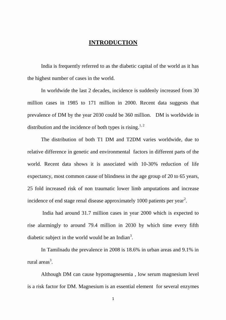
GESTATIONAL DIABETES MELLITUS (GDM)11
It is the occurrence of reduced glucose tolerance during gestation.
Incidence: 7% (range 2–10%) of pregnancies.
Most women revert to normal euglycemia in postpartum period, but have a
subsequent risk (35–60%) of developing diabetes mellitus in the next 10–20
years.
8
Effects of diabetes on pregnancy:
Hydraminos
Toxaemia of pregnancy
Maternal infections
Difficult labour
Recurrent abortions
Postpartum haemorrhage
Puerperal sepsis.
Effects of diabetes on the foetus:
Prematurity
Still birth
Macrosomia
Postpartum hypoglycaemia
Respiratory distress syndrome
Hyperbilirubinemia
Congenital heart disease
Neural tube defects.

9
TYPE 1 DIABETES MELLITUS
Pathogenesis Of Type 1 Diabetes Mellitus:
Normal islet
?Viral infection in pancreatic beta cell
Secretion of IFN-α by pancreatic beta cell
Hyperexpression of MHC class1 antigen within beta cell
Insulitis
Insulin deficient islets
Immune Mediated Diabetes (Type 1A)
Its incidence is around 5-10% all diabetes. It occurs due to immune
mediated destruction of pancreas.
Pathogenesis:
1. Genetic predisposition: Diabetogenic genes are located in short arm of
chromosome 6
2. Environmental triggers
a) Viral infections like rubella virus, CMV, mumps virus, etc.
b) Toxins like pentamidine, streptozocin

10
Viruses and above agents directly act on beta cells and initiate autoimmune
processes against these cells.
3. Immune mechanism
DM can occur with other autoimmune disorders. It is due to presence of
diabetogenic peptide which triggers the immune system. .Antibodies against β-
cells include
1. Islet cell auto antibodies,
2. Autoantibodies to insulin,
3. Autoantibodies to glutamic acid decarboxylase (GAD65) and
4. Autoantibodies to tyrosine phosphatases IA-2 and IA-2B.
Anyone of above auto antibodies present in 85-90% of patients.
Age of onset is usually less than 30 years. Usually associated with
prominent muscle wasting. It has an abrupt onset with rapid progression course.
Classical symptoms of diabetes like polyuria, polyphagia, polydipsia are present.
Family history of diabetes mellitus is usually absent.
HLA DR3 or DR4 seen in>90% of patients. Pancreatic islet cells (of
langerhans) almost destroyed and plasma insulin is low to absent. Plasma C
peptide level is low. It is usually associated with other autoimmune diseases.
Acute complications like diabetic ketoacidosis are very common. Insulin
therapy is needed for survival and mortality is higher in untreated patients.

11
Idiopathic Diabetes (Type 1B)
These persons have absolute deficiency of insulin and are at risk of
developing ketoacidosis. It is strongly inherited and there is no evidence for
antibodies to beta cells. It is not associated with HLA.

12
TYPE2 DIABETES MELLITUS
90-95% of diabetics are Type 2 DM. Patients have insulin resistance and
have relative deficiency of insulin. Specific aetiologies are not known. It usually
starts after the age of 30 years. It has insidious onset, with gradual progressive
course. Polyuria, polyphagia, polydipsia are not so classically seen as in type 1
DM. Family history of diabetes mellitus is usually present. No HLA links are
seen. 50% concordance is seen in identical twins. Pancreatic islet cells are not
totally destroyed .Plasma insulin in serum normal to high. Complications like
hyperosmolar hyperglycaemic non ketotic coma are very often seen.
Risk of developing T2DM:
Age of onset of T2DM in proband
(years)
Age corrected risk of T2DM for
Siblings(%)
25-44 53
45-54 37
55-64 38
65-80 31
Pathogenesis of Type 2 DM:
1) Insulin Resistance
2) Pancreatic Beta Cell Failure

13
Insulin resistance:
Exact cause of insulin resistance is unknown. Possible mechanism is
increase release of free fatty acids from adipose tissue that induce insulin
resistance. In addition there is increased release of adipokines which act on
receptors of insulin and dampen the action of insulin. There is a higher
prevalence of insulin resistance is seen in metabolic syndrome.
Pancreatic Beta cell failure:
In the earlier stage of diabetes mellitus only moderate reduction of insulin
secreting beta cells lost. At the time of diagnosis of type 2 DM more than 50%
reduction of beta cell is seen. Most important pathological feature is the
deposition of amyloid in pancreatic cells. There is no change in alpha cells but
progressive destruction of beta cells leads to development of hyperglycemia.

14
Differences between Type 1 and Type 2 Diabetes Mellitus:
T1DM T2DM
Age of onset(years) Less than 40 More than 50
Duration of symptoms Usually weeks Months to years
weight Normal or decrease Obese
Ketone bodies in urine Present Absent
Insulin requirement for
survival
Needed Not needed
Auto antibodies Present Not present
Diabetic complications at
the time of presentation
Not present Present
Family history Uncommon Seen in 25%
Other autoimmune
disorders
common Uncommon
Clinical features of DM
Polyuria
Increased thirst
Dry mouth
Nocturia
Tiredness, lethargy, fatigability
Visual defect

15
Excessive weight loss
Nausea, vomiting, headache
Polyphagia
Genital candidiasis
Mood changes, irritability
Diagnostic criteria for Diabetes mellitus
It includes
A. Symptoms of diabetes plus random blood sugar ≥ 200 mg/dl
B. Fasting plasma sugar ≥ 126 mg/dl
C.2 hour postload plasma sugar ≥ 200 mg/dl during an oral GTT
D.HbA1C >6.5%
The new Diagnostic criteria for pre-diabetes and diabetes mellitus
FBS > 100 mg/dl = normal fasting glucose
FBS ≥100 mg/dl and <126 mg/dl =impaired fasting glucose (IFG)
FBS ≥ 126 mg/dl = provisional diagnosis of diabetes (on more than one
occasion)
The corresponding categories when the oral GTT is used ,are as follows
2 h BS < 140 mg/dl = normal glucose tolerance
2 h BS ≥ 140 mg/dl and < 200 mg/dl = impaired glucose tolerance (IGT)
2 h BS ≥ 200 mg/dl = provisional diagnosis of diabetes (must be confirmed on
subsequent day)

16
FBS-fasting blood sugar, 2 h BS- 2 hour post load blood sugar.
The prognostic significance and outcome are same whether it is the FBS
>126 mg/dl or 2 hour postprandial blood sugar >200 mg/dl(in diabetes).The
FBG test is now mostly performed because of ease of administration ,
convenience, acceptability to patients and its lower cost.
2-hour post load glucose is done by taking 75 g of glucose dissolved in
300 ml of water.
Fasting is defined as no food intake (i.e. overnight) for at least 8 hours.
Random is defined as any time of day without regard to time since the last meal.
Screening for diabetes:
Diabetes is one of the diseases diagnosed late when multiple
complications have appeared. Nearly 1/3rd
of group remain undiagnosed. But no
studies have supported screening of asymptomatic persons. But use of fasting
blood sugar as a screening test for Type 2 DM is justified in individuals at high
risk group.
Glycosylated haemoglobin C is also recommended for screening.
Adverse factors for Type 2 Diabetes Mellitus
1. Obesity
2. Family history of DM
3. History of diabetes mellitus during pregnancy (GDM) or Birth of baby
weight > 4kg
4. HDL cholesterol level ≤ 35mg/dL or triglyceride level ≥ 250mg/dL

17
5. Race/ethnicity
6. Type A personality
7. Evidence of vascular disease features
Standards of medical care in DM
It is a chronic progressive disease that required frequent medical care for
to decrease acute and chronic complications.
Initial Evaluation includes
Detailed Medical history
Previous HbA1C reports.
Detailed history of dietary pattern, and weight history and any
developmental delay in earlier age.
Detailed history of prior treatment and present treatment of DM.
Exercise history.
Previous or present infections, particularly skin, foot, oral GIT and
urinary tract infections.
2) Physical examination:
Height, weight and body mass index
Sexual maturation staging.
BP measurement, including orthostatic measurements
Examination of pulses
Oral examination.

18
Thyroid gland examination.
Cardiac evaluation.
Eye-fundoscopic examination
Abdominal examination
Hand examination.
Foot examination.
Dermatological examination (for tinea, acanthosis nigricans and insulin-
injection sites).
CNS examination.
Examination for secondary causes of diabetes
(e.g.hemochromatosis,pancreatic disease).
3) Laboratory evaluation
HbA1C measurement
Fasting serum lipid profile levels, which includes total cholesterol, HDL,
Triglycerides, LDL, VLDL.
Examination of micro albumin in urine in T1DM who have had duration
of > 5 years and in all cases with T2 DM .
Serum creatinine.
Thyroid stimulating hormone (TSH) level in all T1DM, in T2DM with
clinical features.
ECG in adults, if clinically needed..

19
Examination of urine for ketone bodies, protein and sediments.
4) Referrals:
• Ocular examination.
• Behavioural specialist.
• Podiatrist.
Goals to be achieved in Diabetes mellitus:
HbA1C < 7.0%
Fasting plasma sugar: 90 – 130 mg/dl
Post prandial plasma sugar: < 180 mg/dl
Blood Pressure :< 130/80 mmHg
LDL < 100 mg/dl
Triglycerides < 150 mg/dl
HDL> 40 mg/dl
Blood Sugar Control Goals- An Approach :
•Goals vary from person to person.
•Following persons (Children, Women who have conceived and older age) need
special care.
•Less severe control is required in patients with recurrent episodes of
hypoglycaemia.
•More adequate control of blood glucose (i.e. a normal HbA1c < 6%) may
further reduce complications.

20
•Postprandial sugar may be targeted if glycosylated haemoglobin C levels are
not met despite reaching fasting sugar level.
Complications of Diabetes Mellitus:
Diabetes has both acute and chronic complications.
Acute complications:
It includes
1.Diabetic ketoacidosis (DKA):
It is an acute medical emergency, characterised by
Increase blood sugar
Ketone bodies in serum and urine
Metabolic acidosis
Complications:
Cerebral edema
Acute respiratory distress syndrome
Disseminated intra vascular coagulation
Thromboembolism
Circulatory failure
2. Hyperglycemic Hyperosmolar state (HHS):
It is characterised by
hyperglycemia without metabolic acidosis or ketone bodies
thromboembolic manifestations are more common

21
3. Hypoglycemia:
It is more common in insulin treated patients.
4. Lactic acidosis
Chronic Complications:
1.Diabetic Retinopathy
2.Diabetic Neuropathy
3.Diabetic Nephropathy
4.IHD
5. Cerebrovascular accident
Transient ischemic attack, stroke
6.Peripheral vascular disease
claudication, ischaemia
7. HT
8.Infection like Tuberculosis, Candidaisis, Mucormycosis, Necrotising fasciitis,
Periodontitis
9.Dupuytren's contracture
10.Pseudogout

22
DIABETIC RETINOPATHY12
It is one of the commonest cause of blindness in adults in the age group30
to 65 years in developed countries. During the 1st two decades of disease, nearly
all patients with T1DM and >60% with T2DM have retinopathy. 21 percent of
T2DM patients have presented with retinopathy at first visit.
Classification:
Nonproliferative Diabetic Retinopathy (NPDR):
1. Mild type:
Presence of 1 micro aneurysm with one or more of the following :
Retinal haemorrhage,
Hard and soft exudates.
2. Moderate type:
Presence of Haemorrhage/ micro aneurysms or
Presence of both in at least one quadrant with one or more of the
following:
Soft exudates, venous beading and intra retinal microvascular
abnormalities.
3. Severe type:
Haemorrhage or micro aneurysms or
Both in all quadrants,

23
Venous beading in two or more quadrants , intra retinal microvascular
abnormalities in at least one quadrant.
Proliferative Diabetic Retinopathy (PDR):
1. Early: One or more of the following:
a) NVE
b) NVD
c) Vitreous or preretinal haemorrhage
d) NVE< ½ disc area.
2. High risk: One or more of the following.
a) NVD > ¼- 1
/3 disc area
b) NVD with vitreous or preretinal haemorrhage
c) NVE > ½ disc area. Preretinal or vitreous haemorrhage.
3. Advanced PDR:
High risk PDR, traction retinal detachment involving macula or
Vitreous haemorrhage obscuring ability to grade NVD or NVE.
IRMA – Intraretinal micro vascular abnormalities.
NVE – Neovascularisation elsewhere.
NVD – Neovascularisation over the disc
Clinical features of Retinopathy
Micro aneurysms
Retinal haemorrhage

24
Exudates
Cotton wool spots
Neovascularisation of retina and iris
Subhyaloid haemorrhage
Vitreous haemorrhage and fibrosis
Macular edema can occur at any stage of diabetic retinopathy. Non
proliferative diabetic retinopathy usually appear at end of first decade or early
second decade in cases of type2 diabetes mellitus. Proliferative diabetic
retinopathy usually appears within 5 years of non proliferative diabetic
retinopathy. Pregnancy, uncontrolled diabetes mellitus, uncontrolled HT can
accelerate these changes.
UKPDS study showed that strict sugar control(i.e. for every percentage of
reduction of HbA1C) associated with a 35% reduction in risk of retinopathy13
,
and strict BP control (to < 150/85 mmHg) results in 34% reduction14

25
Pathophysiology of microvascular complications:
Increase glucose level
Activation of protein kinase c, Endothelial nitric oxide synthase uncoupling,
Increase production of advanced glycation products, Activation of polyol
pathway
Activation of reactive oxygen species
Increase oxidative stress
1.Altered gene expression
2.Decrease nitric oxide synthesis
3.Activation of protein kinase C
4.Increased formation of advanced glycation
products
5.Induction of DNA damage

26
Other ocular complications :
Cataract
Glaucoma
Retinal detachment
Macular edema
Investigations for retinopathy:
Visual acuity
Fundus examination
Fundus fluorescein angiography
Slit lamp examination
Treatment options for retinopathy:
Laser photocoagulation
Injection of steroids
Anti vascular endothelial growth factor

27
DIABETIC NEPHROPATHY
This is most common cause of stage five chronic kidney disease in world
wide. Compared to type2 diabetes progression to chronic kidney disease is
higher in type1 DM.
The diabetic nephropathy progresses from stage of microalbuminuria to
stage of macroalbuminuria / clinical albuminuria to end stage renal disease.
Progression from micro to macroalbuminuria usually taken upto 10-15 years.
ESRD develops in half of T1DM patients with clinical nephropathy
within ten years and 3/4of patients by twenty years. But in type 2 DM, even
after twenty years of overt nephropathy only 20% progress to ESRD.
Screening for Microalbuminuria:
A screening test should be done for urine microalbumin at the time of
diagnosis in persons with T2 DM at the time of first visit , repeated after five
years of disease duration. For patients with T1DM, test should be repeated
yearly.
Screening for microalbuminuria can be performed by 3 ways.
1. Calculation of the albumin to creatinine ratio in a spot urine sample.
2. 24 hour Urine sample and measurement of albumin excretion.
3. Timed (e.g. 4 hr or overnight) collection.
24 hour collection urine sample is most reliable.

28
CATEGORY
Spot collection
(μg/mg creatinine)
24 Hr collection
(mg/24 hrs)
Timed collection
(μg/min)
Normal <30 <30 <20
Microalbuminuria 30 – 299 30-299 20-199
Macroalbuminuria ≥300 ≥300 ≥200
In addition microalbuminuria is the earliest feature of renal involvement,
it is also as a independent marker of CAD.

29
DIABETIC NEUROPATHY
It occurs in almost half of cases with long duration of diabetes. The
development of neuropathy is directly in correlation with the duration and
degree of glycemic control2,15
.
Classification:
Somatic
Visceral or autonomic
Somatic:
1) Polyneuropathy
Symmetrical mainly sensory and motor
Asymmetrical mainly motor and proximal including amyotrophy
2) Mononeuropathy including mononeuritis multiflex
Visceral:
Cardiovascular
GIT
Pupillary
Genitourinary
Sudomotor
Vasomotor
Possible causes of neuropathy in DM includes
Microangiopathy

30
Formation of advanced glycation end products
Increased level of protein kinase c
Activation of polyol pathway leads to accumulation of sorbitol
Histopathology:
Axonal degeneration of both myelinated and unmyelinated fibres
Schwann cell basal lamina hypertrophy
Patchy, segmental demyelination
Basement membrane thickening and presence of microthrombi in neural
vessels
The most common type reported is distal symmetrical sensory poly
neuropathy. Most cases frequently presented with distal sensory loss,
hyperesthesia, paresthesia and dysesthesia.
Painful neuropathies may also occur. It can be an acute (lasting < 12
months) and a chronic (lasting > 12 months).
Individuals with long standing T1 or T2 DM are prone to develop
autonomic diabetic neuropathy.
Clinical features of Autonomic Neuropathy:
Cardiovascular features
Orthostatic hypotension
Fixed heart rate

31
Resting tachycardia
Gastrointestinal features
Dysphagia
Nausea, vomiting, abdominal fullness
Constipation
Nocturnal diarrhoea
Genitourinary features
Erectile dysfunction
Urinary incontinence
Recurrent urinary tract infections
Retrograde ejaculation
Pupillary features
Small pupil size
Delayed response to light reflex
Sudomotor features
Gustatory sweating
Increased sweating in the night
Anhidrosis and fissures in the feet
Vasomotor features
Dependent edema
Bullous formation
Feet feel cold due to loss of vasomotor response

32
Tests for cardiovascular autonomic functions
1) Simple reflex test
A) Heart rate responses
to valsalva manoeuvre ( fifteen seconds): ratio of longest to shortest R- R
interval Normal ≥1.21 ,abnormal≤1.20
to deep breathing :6 breaths over 1 minute Normal ≥15 abnormal≤10
to standing after lying :Ratio of R- R interval of 30th to 15
th beats Normal
≥1.04 ,abnormal≤1
B) Blood pressure response to standing -Systolic BP fall Normal ≤10
abnormal≥30
C) Special Tests
heart rate and BP in response to handgrip
heart rate and BP variability using domain analysis of ambulatory
monitoring
MIBG scan of heart
heart rate and BP variability using power spectral analysis of ECG
monitoring
Diagnosis:
Examination of the feet
Look for ulceration,
Ankle reflexes,

33
Tuning fork using 128-Hz shows decreased vibration perception
Semmes-Weinstein monofilament for pressure sensation
Normal results on vibration testing (Likelihood ratio range, 0.33–0.51) or
monofilament (Likelihood ratio range, 0.09–0.54) make large fiber
peripheral neuropathy from diabetes less likely.
Nerve conduction studies may show decreased conduction of the
peripheral nerves.

34
MAGNESIUM
Magnesium is the 4th most abundant cation in the body. It is also the 2
nd
largest amount cation intracellularly, next to potassium. A normal adult has 21-
28gms (approximately 2000mEq) of magnesium. Nearly 60% of total body
magnesium is present in bone, 38% is in soft tissues with slightly higher
concentrations in liver and skeletal muscle. (15-20 mEq/Kg). and less than 2%
is present in extracellular compartment16
.
Serum concentration of magnesium varies from 1.7 to 2.4 mg/dL. The
plasma concentration in healthy adults remain remarkably constant with very
less fluctuations due to sensitive control mechanisms that are not fully
understood
The mean daily oral intake of magnesium is roughly 25 mEq (140-360
mg/day). About 40% of the dietary magnesium is absorbed in the small intestine
mainly the ileum. Elimination is mainly through kidneys and is about
100mg/day and it is well regulated. So when blood levels rise more than 2.4
mg/dl, magnesium excretion increases many folds. In event of magnesium loss
absorption is increased dramatically, the main site being the thick ascending
loop of Henle. Many factors inhibit reabsorption, like increased ECF volume,
hypercalcemia, and use of thiazides.17

35
Biochemical importance of Magnesium:
Magnesium is an activator of critical enzyme systems that maintain
cellular metabolism. The most important being the activation of enzymes that
hydrolyze and transfer phosphate groups in the processes involving adenosine
triphosphate (ATP). This ATP is essential for glucose catabolism as well as ,
lipid, amino acids, etc .
Magnesium is a cofactor for oxidative phosphorylation inside the
mitochondria.
The macromolecular structure of DNA, RNA and ribosomes is
maintained by magnesium.18,19
It is also involved in protein synthesis by
regulating the attachment of mRNA to the 70s ribosome.20
Regulation of Serum Magnesium
A) Renal regulation:
Renal regulation of serum magnesium concentration is mainly by altering
its reabsorption at the thick ascending loop of Henle. Magnesium reabsorption
is increased by parathyroid hormone and is reduced by hypercalcemia and
hypermagnesemia

36
B) Intestinal absorption :
Intestinal magnesium absorption mainly at jejunum and ileum is
increased by 1,25 (OH)2 Vitamin D
C) Hormonal factors23
Increasing serum magnesium,
Parathyroid hormone
Glucagon
1, 25(OH)2 Calcitriol.
Decreasing serum magnesium,
Aldosterone
Vasopressin (ADH)
Thyroxine
Calcitonin
Selected Food Sources of Magnesium22
-Magnesium content in mg/100gm)
Nuts
Almonds – 315
Cashews – 260
Peanuts – 175

37
Legumes
Split Beans – 50
Soyabean – 86
Fruits
Dates – 35
Banana – 30
Oranges – 10
Apple – 5
Dairy Products
Milk – 24
Butter – 20
Yoghurt – 12
Cereals
Shredded Wheat -110
Rice – 40
Meat and fish
Pork – 22
Chicken – 21

38
Beef – 18
Fish – 22
Hypermagnesemia :
Hypermagnesemia is very uncommon in the absence of renal
insufficiency, kidneys can excrete huge quantities of magnesium when needed
(up to 250 mmol/d).24
Causes of hypermagnesemia
A) Impaired magnesium excretion
Renal failure
Familial hypocalciuric hypercalcemia
B) Excessive magnesium intake
Cathartics
Antacid preparations
Parentral magnesium administration (eg.magnesium sulfate in PIH)
C) Rapid magnesium mobilisation from soft tissues
Trauma
Extensive burns
Shock, sepsis
Post-cardiac arrest

39
D) Other disorders
Adrenal insufficiency
Hypothyroidism
Hypothermia
Clinical features:
The most important clinical presentation of hypermagnesemia are
vasodilation and neuromuscular blockade, occurring at concentrations > 4.8
mg/dL (>2mmol/L). Hypotension usually does not respond to intravenous fluids
and vasopressors.
Lethargy and weakness may progress to respiratory failure, paralysis and
coma with depressed deep tendon reflexes occurs at serum magnesium levels >
4 mmol/L). Paralytic ileus may occur.
Prolongation of PR, QRS intervals, heart blocks and asystole occurs at
serum magnesium levels around 10 mmol/L.
Treatment of Hypermagnesemia:
Generally involves recognising and removing the magnesium source
Vigorous intravenous fluids and hemodialysis are necessary at times.
Intravenous calcium in doses of 100-200 mg over 1 to 2 hrs provides a short
term improvement

40
Hypomagnesemia :
Hypomagnesemia means a significant decrease in body magnesium stores
(0.5 to 1 mmol/Kg). . Dietary magnesium deficiency is uncommon except in
persons consuming alcohol.
Causes of Hypomagensemia24
I.Impaired intestinal absorption
A.Primary infantile hypomagnesemia
B.Malabsorption syndromes
C.Vitamin D deficiency.
II.Increased intestinal losses
A.Protracted vomiting / diarrhoea
B.Intestinal drainage, fistulae
III.Impaired renal tubular reabsorption
A.Genetic magnesium wasting syndromes.
1.Gitelman syndrome
2.Bartter syndrome
3.Na-K ATPase g-subunit mutations
B.Acquired renal disease
1.Tubulointerstitial disease
2.Post obstruction /ATN (diuretic phase)
3.Renal transplantation.

41
C.Drugs
1.Ethanol
2.Diuretics (loop, thiazide and osmotic)
3.Cisplatin, cyclosporine
4.Aminoglycosides, Amphotericin B
IV.Metabolic causes
1.Hyperaldosteronism
2.SIADH
3.Diabetes mellitus
4.Metabolic acidosis
5.Hypercalcemia
6.Hyperthyroidism
V.Others
1.Pancreatitis
2.Excessive sweating
3.Osteoblastic metastases
Many genetic/hereditary magnesium losing conditions are described , but
are exceedingly rare. Prolonged nasogastric aspiration, intravenous fluids,
infectious diarrhoea, fat losing enteropathies and inflammatory bowel disease
may produce hypomagnesemia.23
Magnesium deficiency is quite frequent in
patients receiving loop diuretics like Furosemide.24
42
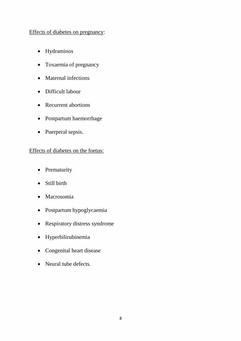
Incidence :
Hypomagnesemia is a relatively common electrolyte imbalance seen in
around 12% of in- patients.25
.In intensive care settings incidence is very high
(60%).Parentral nutrition, diuretic usage, decreased serum Albumin levels , and
aminoglycosides may be responsible for this high frequency26
Risk of incidence:27
2% in common people.
10 - 20% in hospitalized patients.
60-70% in ICU settings.
30 - 80% in Alcoholics.
25% in diabetic patients.
Hypomagnsemia occurs equally in males and females.
Clinical features:28
The clinical features appear only when serum magnesium concentrations
are <1.2 mg/dL (0.5 mmol/L). It is presented as irritability, Central nervous
system hyper excitability, and arrhythmias.29

43
History :
History concerned with hypomagensemia are usually nonspecific. They
present with weakness, muscular cramps / palpitations. Sometimes abnormal
behaviour in the form of irritability, apathy, psychosis, may be seen in severe
cases. In few vertigo, incoordination, depression, or fits may occur.
Physical signs :
Exaggerated deep tendon reflexes.
Trousseau and Chvostek sign
Difficulty in swallowing due to esophageal hypomotility
Altered mental status
Ataxia, nystagmus or seizures (at levels <0.8 mg/dl)
Pulses may be irregular due to VPCs
ECG :
Hpomagnesemia can produce non specific alterations in the
electrocardiogram. Mild to moderate hypomagnesemia (1.2 to 1.7 mg/dl)
producing widened QRS complexes with tall T-waves. Severe
Hypomagnesemia (<1.2 mg/dl) produces increased PR interval, broad QRS
complex, flattening / T wave inversion and prominent U waves.30
44
Cardiac arrhythmias including sinus tachycardia, other supraventricular
tachycardia and ventricular tachyarrhythmia also can occur.
Investigations:
Serum magnesium levels are estimated by several methods.
Neutron activation analysis
Atomic absorption spectrometry
Ion selective electrodes (ISE)
Equilibrium dialysis
Calmagite dye method.
Calcium, potassium and phosphorous levels must be assessed. BUN and
creatinine levels And Blood glucose levels should also be measured.
Treatment of Hypomagnesemia:
The route of magnesium replacement is according to severity of the
clinical manifestations. Moderate to severe manifestations like patients with
tetany or ventricular arrhythmias require 50 mEq of intravenous route slowly
over 8 to 24 hours and repeated as and when needed to maintain concentration
above 1.0 mg/dl.31

45
Oral replacement ( magnesium chloride & Magensium lactate) should be
given in patients with milder presentations. The background cause should be
corrected.
Hypomagnesemia and other diseases:
A) Magnesium and cardiovascular diseases32
Arrhythmias can be precipitated in hypomagnesemia especially in acute
coronary syndrome, cardiac failure and associated hypokalemia.
Torsade De pointes is a fatal arrhythmia precipitated by drugs that
increase QT interval, electrolyte imbalance (low potassium and magnesium), or
a decrease heart rate. Intravenous magnesium is recommended for this fatal
condition.
Trials show significant correlation with ischemic heart disease. decreased
level of serum magnesium is associated with higher incidence of ischemic heart
disease.39
The underlying mechanism for the increased risk is not fully
understood.
Magnesium supplementation may improve the exercise tolerance,
antithrombotic effect with aspirin and thus improves outcome in individuals
with ischemic heart disease.40

46
Acute MI(myocardial infarction) is often associated with significant
reduction in serum magnesium levels.41
Hypomagnesemia associated with acute
MI is probably responsible for increase in the frequency of fatal arrhythmias in
the acute period.33
A positive correlation is seen in patients with hypomagnesaemia and
CCF .It occurs due to use of diuretics. Whether this is associated with increased
mortality is not known.41
B) Hypertension
Magnesium has significant role in controlling BP.35
Hypomagnesemia
leads to increased intracellular potassium and calcium levels which leads to
vasoconstriction and increased peripheral vascular resistance. Magnesium may
also have some direct effect on vascular smooth muscle.36
Studies have also found that requirement of anti hypertensive dosage is
more in patients with low serum magnesium levels.
As per JNC recommendation high magnesium rich diet is necessary for
prophylaxis and treatment of hypertension.37

47
C) Stroke
Studies have also found that increased magnesium intake decreases
incidence of stroke.42
D) Osteoporosis
There is a significant correlation between serum magnesium levels and
bone mineral density.43
E) Asthma:
Normal functioning of lung requires magnesium rich diet. There is a
significant correlation between hypomagnesemia and incidence of bronchial
asthma.
F) Dyslipidemia
Hypomagnesemia is associated with increased plasma LDL,
cholesterol and triglyceride and reduced HDL.44

48
MAGNESIUM AND DIABETES MELLITUS
Magnesium is a essential element for metabolism of carbohydrate . 25 to
39% diabetes mellitus patients have hypomagnesemia
The clinical implications of magnesium deficiency in diabetes are many.
Hypomagnesemia can be a result of hyperglycemia and can produce or increase
insulin resistance. Hypomagnemia is implicated in the development of diabetic
retinopathy.
most often higher incidence occur if associated with hypertension,38
thrombotic tendency,45
insulin resistance46
and the Reaven – Modan syndrome, a
unique clinical entity that connects diabetes mellitus, hyperinsulinemia,
hypertension and increased thrombotic tendency – all producing adverse
cardiovascular outcomes.47
Causes of Hypomagnesemia in Diabetes Mellitus :
Initially, the cause of hypomagnesemia in diabetes was supposed to be due to
(1) Osmotic renal loss from glycosuria.
(2) Impaired absorption from GIT .
(3) Insulin mediated transfer of magnesium to RBC.
Recently a specific tubular defect has been suggested which results in
hypermagnesuria. The exact site of this defect is not yet been identified. Insulin

49
treatment has been shown to reduce magnesium losses through kidneys.
Garland48
suggested that delay in insulin treatment is less effective in correcting
the renal magnesium losses, probably due to certain irreversible changes.
The role of magnesium in Insulin Action :
Magnesium is a essential cofactor for insulin secretion, and activity.
Hypomagnesemia decreases insulin secretion by the pancreas.49
Multiple studies have shown hypomagnesemia to increase insulin
resistance. This insulin resistance is a post receptor defect and may be
associated with calcium mediating the signal for insulin action.46
Number of
studies have shown that tissue response to insulin is more in the presence of
magnesium.
In a recent study, the cellular uptake of magnesium, was shown to be
reduced in diabetics.50
there is also some proof that low magnesium levels
directly produces insulin resistance. Nadler et al.56
analysed 16 non diabetic
individuals and found decreased insulin sensitivity after production of
magnesium deficiency.
Similarly elderly non diabetic individuals had improved glucose tolerance,
when subjected to magnesium supplementations for 4 weeks.51
In non diabetic

50
obese subjects, insulin resistance was found to be associated with low
magnesium levels, compared to non obese individuals.52
Tonyai, et al.53
suggested that a low erythrocyte magnesium content can
change membrane viscosity, and impair the binding of insulin with its
membrane. Paolisso, et al.51
were able to rectify the altered erythrocyte
microviscosity with long-term magnesium supplementation.
Role of magnesium deficiency in diabetic end organ damage:
Magnesium deficiency is associated with diabetic microvascular disease.
Hypomagnesemia has been shown in patients with diabetic retinopathy, with
still lower magnesium levels in severe diabetic retinopathy.
Magnesium
depletion is also related to development of diabetic polyneuropathy. Corsonello,
et al have suggested a relationship between diabetic microalbuminuria and
serum ionized magnesium levels. Magnesium depletion also has been associated
with multiple macrovascular complications.43,50
Grafton, et al57
have suggested on the inositol transport theory for the
development and progression of diabetic complications. According to this
theory hyperglycemia leads to excessive stimulation of aldose reductase causing
high cellular level of sorbitol. This sorbitol inhibits transport of inositol, thus
decreasing intracellular inositol. The data of Grafton, et al show that

51
hypomagnesaemia causes a decrease in the affinity of the inositol transport
protein for inositol, leading to a two fold reduction in rate of inositol transport
and accelerated development of diabetic complications.
Resnick proposed that the „primary‟ defect is the abnormal cellular ion
handling leading to complications47
There is a significant correlation between magnesium and antioxidant
levels. Weglicki, et al have hypothesized that a fall in magnesium level leads to
loss of cellular activity against oxidative substances. Magnesium deficiency has
also been shown to lower the efficiency of substances acting againt oxidants
such as glutathione, vitamin c and Tocopherol.
Evidence for efficacy of Magnesium Supplementation in Diabetes Mellitus:
There is strong evidence that repletion of magnesium can reduce the
insulin resistance, platelet reactivity and other cardiovascular risk factors .54
In a study involving 16 diabetics and 30 healthy controls, oral
supplementation with magnesium hydroxide (250mg twice daily) resulted in
reduced insulin requirements in the diabetic patients. Paolisso,51
et al
demonstrated magnesium supplementation given resulted in lower fasting
plasma glucose levels, increased plasma magnesium levels and a significant
improvement in β-cell response to glucose.

52
In type 2 diabetics, oral supplementation of magnesium decreases platelet
reactivity, and decreases the incidence of hypertension and improves lipid
profile.44
Clinical approach to Magensium Supplementation in Diabetes Mellitus
Diabetic patients at risk for hypomagnesemia such as ACS, DKA, chronic
alcohol intake, prolonged duration of parentral therapy, chronic diarrhoea ,
diuretic therapy should have their magnesium levels measured.
Overt serum hypomagnesaemia should always be corrected. If
hypomagnesemia is clinically suspected, but not supported by serum levels, a
further test with erythrocyte or platelet magnesium concentration is mandatory
Magnesium chloride is the preparation of choice. Doses vary from 100 to
600mg/day .54
Diarrhoea is the most important and the dose limiting side effect.
Renal insufficiency with Glomerular filtration rate of less than 30 ml/min is the
only factor that requires withholding dietary supplementation.

53
METHODOLOGY
SOURCE OF DATA
Patients with type 2 diabetes admitted in THANJAVUR MEDICAL
COLLEGE & HOSPITAL, THANJAVUR, who satisfied the inclusion criteria
and consented to participate in the study were included.
PERIOD OF STUDY
October 2011-October 2012
TYPE OF STUDY
Cross sectional study
100 patients were randomly selected of which 50 were males and 50 were
females. Cases with renal failure, Acute Coronary Syndromes, patients on
diuretics, alcoholics or with malabsorption were excluded. None were taking
magnesium supplements or magnesium containing antacids. Informed consent
was obtained.
INCLUSION CRITERIA
All patients both males and females above 13 years of age group with
type 2 diabetes mellitus admitted in Internal medicine units of Thanjavur
Medical College & Hospital, Thanjavur.

54
EXCLUSION CRITERIA
1. Patients with chronic renal failure.
2. Acute myocardial infarction in last 6 months.
3. Patients on diuretics.
4. Patients with history of alcohol abuse.
5. Patients receiving magnesium supplements or magnesium containing
antacids.
6. Malabsorption or chronic diarrhea.
7.Age<13years.
DATA COLLECTION
The 100 diabetics (50 Men & 50 Women) were included in the study.
Detailed history – including duration of diabetes, treatment mode, symptoms
suggestive of diabetic neuropathy, associated diseases such as hypertension and
ischemic heart disease was obtained as per the proforma.
Detailed physical and neurological examination was done. Retinopathy
was assessed by direct opthalmoscopy.
Samples were collected for estimation of fasting blood glucose and
magnesium level. Postprandial blood sugar was measured two hours after a
standard meal. Nerve conduction study was done on selected patients by

55
experience neurologist with symptoms & signs suggestive of neuropathy. Blood
urea, serum creatinine and 24 hour urinary albumin were estimated. Serum
magnesium was estimated by Calmagite dye method. HbA1C measurement
done by a modified calorimetric method.
CALMAGITE DYE METHOD – TEST PRINCIPLE
Under alkaline conditions, magnesium combines with calmagite dye to
form a red colour which is read spectrophotometrically at 530 nm. Formation of
colour is depends on magnesium levels. To eliminate the interference of
calcium during estimation, EDTA is included in the reagent. Cyanide reduces
heavy metal interference. Surfactant reduces protein interference.
TEST PROCEDURE
Three test tubes labeled Blank, Standard and Test are prepared as in table.
In test tubes Blank Standard Test
Calmagite 1 ml 1 ml 1ml
Standard
sample
- 10 ml -
Patient‟s
sample
- - 10 ml
Distilled
water
10 ml - -

56
This test tubes are incubated at room temperature (22-28ºC). The
absorbance of Test (AT), Standard (A
S) and Blank (A
B) are read at 530nm in
spectrophotometer. Magnesium concentration is calculated by the following
formula.
Magnesium concentration (mEq/L) = (AT-A
B / A
S-A
B) x 2
Serum magnesium concentration is expressed in mg/dl by linearity of 1
mEq/L = 1.2 mg/dl.
According to magnesium levels patients were classified into:
1) Normal, 1.7 to 2.4 mg/dl,
2) Low <1.7mg/dl,
3) High >2.4 mg/dl.
Patients were also categorized on the basis of duration of diabetes,
presence of ischemic heart disease or hypertension, mode of treatment,
presence/absence of retinopathy, neuropathy and nephropathy, and glycemic
control (FBS and HbA1C).
Cases with diabetic retinopathy were further divided into
a) Nonproliferative diabetic retinopathy.
b) Proliferative diabetic retinopathy.
57
Diabetic nephropathy was graded depending on 24 hour urinary excretion of
albumin as follows:
No nephropathy, < 30mg/24hour.
Microalbuminuria 30 – 299mg/24hour
Macroalbuminuria (clinical proteinuria) >= 300 mg/24hour.
STATISTICAL ANALYSIS
The statistical analysis was done by SPSS 15 software.MS Word and
Excel were used to generate tables and charts. Following tests were used:
1. Chi square test
2. Student T test
3. Oneway ANOVA test
Statistical results were considered significant at P <.05

58
RESULTS
100 cases of type 2 DM (50 males, 50 females, mean age 56.87 years)
comprised the study group.
Table 1: Age Distribution
Age (in years)
No. of Patients
(n=100)
Below 50yrs 30
51 to 60yrs 41
61 to 70yrs 22
71yrs & above 7
.
Patients were distributed across the age spectrum of 42 to 78 years. Mean
age 56.87 years. Most patients (n=41) were present in 51-60 group. Youngest
patient was 42 years old.
0
20
40
60
Below 50yrs
51 to 60yrs 61 to 70yrs 71yrs & above
30
41
22
7
No
. o
f P
ati
ents
Age
Graph 1: Age Distribution

59
Table 2: Characteristics of study population
Characteristics
No. of subjects 100
Age (Years) 56.87 (42 – 78)
Men 50
Women 50
Duration of diabetes
(years)
8.89(2-23)
Medication
67 Oral hypoglycemics
Insulin and oral
hypoglycemics
33
Diet only 0
Comorbidities
30 Hypertension
Ischemic heart disease 15
Diabetic retinopathy
NPDR
33
PDR 2
Diabetic neuropathy 15
Diabetic nephropathy
Microalbumineria
29
Macroalbumineria 4
Poor glycemic control 39

60
.
The average duration of diabetes in study population was 8.89 years and
range was 2 year to 23 years. 67 patients received only oral hypoglycemic
agents and 33 patients received both. 30 patients had hypertension and 15
patients had ischemic heart disease and 55 patients had no comorbidities. Total
35 patients had diabetic retinopathy. Total of 15 patients had diabetic
neuropathy. 33 patients had Nephropathy.
0
5
10
15
20
25
30
35
40
No
.of
Pa
tien
ts
Characteristics
Graph 2: Characteristics of study Population

61
Table 3: Prevalence of Hypomagnesemia
Sex
Magnesium
Statistical inference Normomagnesemia
(n=65)
Hypomagnesemia
(n=35)
Male 34 (52.3%) 16 (45.7%) X2=.396
Df=1
.529>0.05
Not Significant
Female 31 (47.7%) 19 (54.3%)
Sl.no MG Mean S.D Statistical inference
1 Male (n=50) .4920 .72530 T=-.622 df=98
.536>0.05
Not Significant 2 Female (n=50) .5840 .75468

62
Hypomagnesemia was found in 35 patients. 65 patients had
normomagnesemia No patient had hypermagnesemia. No correlation was found
between hypomagnesemia in men and women (45.7% and 54.3%%
respectively).
Graph 3: Prevalence of Hypomagnesemia
Hypomagnesemia 35%
Normomagnesemia 65%

63
Table 4: Prevalence of Hypomagnesemia and duration of diabetes
Oneway ANOVA
Sl.no MG Mean S.D SS Df MS
Statistical
inference
1
Between
Groups
32.917 4 8.229
F=13.265
.060<0.05
Not Significant
2
Below 5yrs
(n=45)
.0000 .00000
3
6 to 10yrs
(n=25)
.8040 .78977
4
11 to 15yrs
(n=15)
1.4333 .40119
5
16 to 20yrs
(n=10)
1.2200 .64601
6
21yrs & above
(n=5)
.0000 .00000
7 Within Groups 20.979 95 .221

64
Sl.
no
Duration(years)
Magnesium
Statistical
inference
Normomagnesemia
(n=65)
Hypomagnesemia
(n=35)
1 Below 5yrs 46 (70.8%) 2 (5.7%) X2=4.568
Df=4
.067>0.05
Not
Significant
2 6 to 10yrs 12 (18.5%) 13 (37.1%)
3 11 to 15yrs 1 (1.5%) 11 (31.4%)
4 16 to 20yrs 2 (3.1%) 8 (22.9%)
5 21yrs & above 4 (6.2%) 1 (2.9%)
chi-square (χ2) value is 4.568.So the correlation is insignificant p value. So, the
duration of diabetes not significantly predict serum magnesium concentration.
Oneway ANOVA F=13.265 in between groups also shows no significant
correlation. The mean duration was 8.89 years (2-23).
0
10
20
30
40
50
Below 5yrs
6 to 10yrs
11 to 15yrs
16 to 20yrs
21yrs & above
No
.of
Pa
tien
ts
Age in Years
Graph 4: Prevalance of Hypomagnesemia and
Duration of Diabetes
Normomagnesemia
hypomagnesemia

65
Table 5: Prevalence of Hypomagnesemia and Mode of Diabetic treatment
Treatment mode
Magnesium Statistical
inference Normomagnesemia
(n=65)
Hypomagnesemia
(n=35)
OHA 45 (69.2%) 22 (62.9%) X2=.418
Df=1
.518>0.05
Not
Significant
OHA+insulin 20 (30.8%) 13 (37.1%)
20
45
13
22
0
5
10
15
20
25
30
35
40
45
50
Insulin treated Non insulin treated
Graph 5: Prevalence of Hypomagnesemia
Normomagnesemia
Hypomagnesemia

66
Table 6: Prevalence of Hypomagnesemia and Fasting blood sugar
Sl.No FBS
Magnesium
Statistical
inference
Normomagnesemia
(n=65)
Hypomagnesemia
(n=35)
1 Below 90 8 (12.3%) 2 (5.7%)
X2=45.582
Df=5
.000<0.05
Significant
2 91 to 100 11 (16.9%) 0
3
101 to
110
10 (15.4%) 9 (25.7%)
4
111 to
120
19 (29.2%) 1 (2.9%)
5
121 to
130
13 (20%) 2 (5.7%)
6
131 &
above
4 (6.2%) 21 (60%)

67
.
Prevalence of hypomagnesemia is high when fasting blood sugar
>131(60%). The chi-square (χ2) value is 45.582. So the correlation is significant.
So, fasting blood sugar can significantly predict serum magnesium
concentration.
0
5
10
15
20
25
Below 90 91 to 100 101 to 110 111 to 120 121 to 130 131 & above
No
. o
f p
ati
ents
Treatment
Graph 6: Prevalence of Hypomagnesemia and Fasting
Blood Sugar
Normomagnesemia
Hypomagnesemia

68
Table 7: Prevalence of Hypomagnesemia and HbA1c
Sl.no HbA1C
Magnesium
Statistical
inference
Normomagnesemia
(n=65)
Hypomagnesemia
(n=35)
1 Below 6 8 (12.3%) 2 (5.7%)
X2=10.408
Df=5
.064>0.05
Not Significant
2 6 to 7 18 (27.7%) 5 (14.3%)
3 7 to 8 15 (23.1%) 5 (14.3%)
4 8 to 9 5 (7.7%) 9 (25.7%)
5 9 to 10 8 (12.3%) 4 (11.4%)
6
10 &
above
11 (16.9%) 10 (28.6%)
Serum magnesium concentration showed no significant association with
HbA1C. Chi square X2=10.408,Df=5 P value .064 . Higher prevalance of
hypomagnesemia observed in HbA1c > 10 (28.6%).

69
.
0
2
4
6
8
10
12
14
16
18
Below 6 6 to 7 7 to 8 8 to 9 9 to 10 10 & above
No
of
pa
tien
ts
HbA1C
Graph 7: Prevalence of Hypomagnesemia and HbA1C
Normomagnesemia
Hypomagnesemia
70
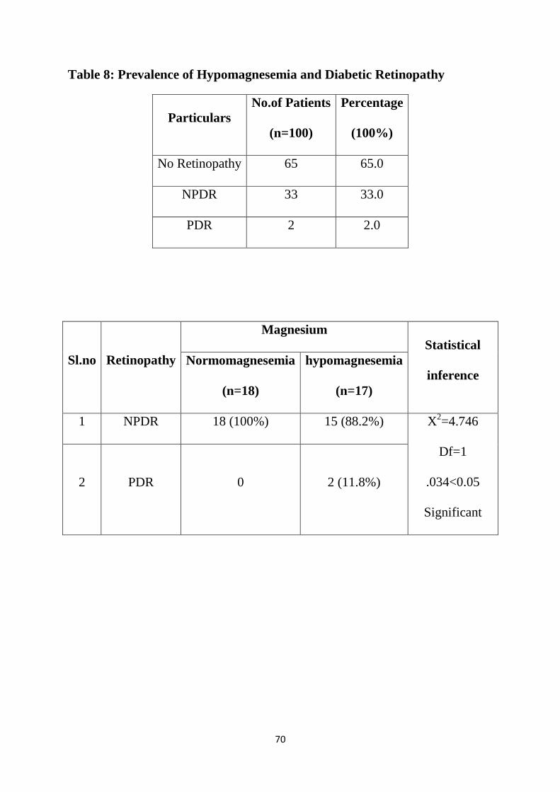
Table 8: Prevalence of Hypomagnesemia and Diabetic Retinopathy
Particulars
No.of Patients
(n=100)
Percentage
(100%)
No Retinopathy 65 65.0
NPDR 33 33.0
PDR 2 2.0
Sl.no Retinopathy
Magnesium
Statistical
inference
Normomagnesemia
(n=18)
hypomagnesemia
(n=17)
1 NPDR 18 (100%) 15 (88.2%) X2=4.746
Df=1
.034<0.05
Significant
2 PDR 0 2 (11.8%)

71
Observations revealed a definite correlation between hypomagnesemia
and diabetic retinopathy. The chi-square (χ2) value is 4.746. Df=1 .p value is
0.034.
0
10
20
30
40
50
60
70
Retinopathy No Retinopathy
No
Of
Pa
tien
ts
Graph 8: Prevalence of Hypomagnesemia and
Diabetic Retinopathy
No of patients
Hypomagnesemia

72
TABLE.9: Prevalence of Hypomagnesemia and Diabetic Neuropathy
Particulars
No. of patients
(n=100)
Percentage
(100%)
No neuropathy 85 85.0
neuropathy 15 15.0
Sl.no Neuropathy
Magnesium
Statistical
inference
Normomagnesemia
(n=65)
Hypomagnesemia
(n=35)
1 Negative 60 (92.3%) 25 (71.4%) X2=7.778
Df=1
.005<0.05
Significant
2 Positive 5 (7.7%) 10 (28.6%)
Observations revealed a definite correlation between hypomagnesemia
and diabetic neuropathy. The chi-square (χ2) value is 7.778. Df=1 .p value is
0.005.

73
0
10
20
30
40
50
60
70
80
90
Neuropathy No Neuropathy
No
of
pa
tien
ts
Graph 9: Prevalence of Hypomagnesemia and
Diabetic Neuropathy
No of patients
hypomagnesemia
74
Table 10: Prevalence of Hypomagnesemia and Diabetic Nephropathy
Particulars
No.of persons
(n=100)
Percentage
(100%)
No albumminuria 67 67.0
Macroalbuminuria 4 4.0
Microalbuminuria 29 29.0
Sl.
no
Nephropathy
Magnesium
Statistical
inference
Normomagnesemia
(n=65)
Hypomagnesemia
(n=35)
1 No albuminuria 65 (100%) 2 (5.7%) X2=91.471
Df=2
.000<0.05
Significant
2 Macroalbuminuria 0 4 (11.4%)
3 Microalbuminuria 0 29 (82.9%)

75
.
Observations revealed a definite correlation between hypomagnesemia
and diabetic nephropathy. The chi-square (χ2) value is 91.471. Df=2 .
0
10
20
30
40
50
60
70
No
. o
f p
ati
ents
Graph 10: Prevalence of Hypomagnesemia
and Diabetic Nephropathy
No Of Patients
Hypomagnesemia

76
Table 11: Prevalence of Hypomagnesemia and Ischemic Heart Disease
Sl.no IHD
Magnesium
Statistical
inference
Normomagnesemia
(n=65)
Hypomagnesemia
(n=35)
1 Absent 54 (83.1%) 31 (88.6%) X2=.539
Df=1
.463>0.05
Not Significant
2 Present 11 (16.9%) 4 (11.4%)
Serum magnesium concentration showed no significant relation with ischemic
heart disease, X2=.539,Df=1.P value is insignificant.
.
.
0
10
20
30
40
50
60
70
80
90
IHD No IHD
No
of
pa
tien
ts
Graph 11: Prevalence of Hypomagnesemia and
Ischemic Heart Disease
No of patients
Hypomagnesemia

77
Table 12: Prevalence of Hypomagnesemia and Hypertension
Sl.no SHT
Magnesium Statistical
inference Normomagnesemia
(n=65)
Hypomagnesemia
(n=35)
1 Absent 43 (66.2%) 27 (77.1%) X2=1.308
Df=1
.253>0.05
Not Significant 2 Present 22 (33.8%) 8 (22.9%)
Serum magnesium concentration showed no significant with systemic
hypertension, X2=1.308,Df=1.P value is insignificant
.
0
10
20
30
40
50
60
70
HTN No HTN
No
of
pa
tien
ts
Graph 12: Prevalence of Hypomagnesemia and
Hypertension
NO of patients
Hypomagnesemia

78
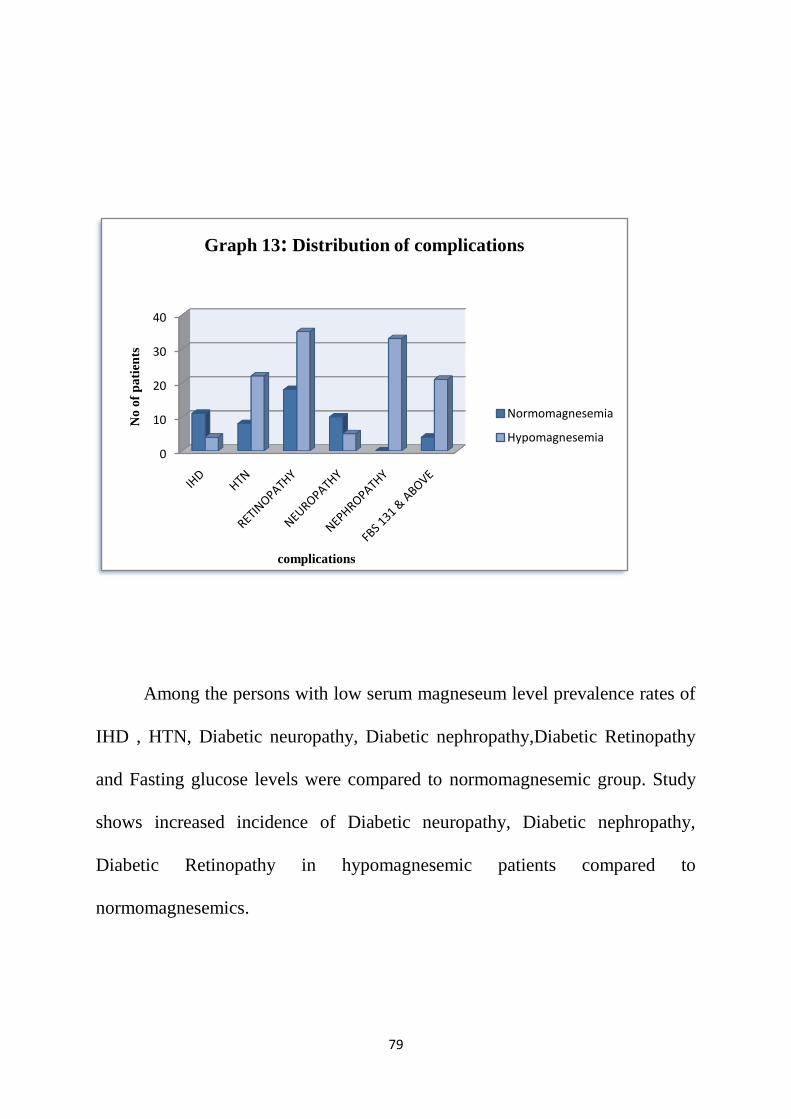
Table 13: Distribution of Complications
COMPLICATIONS
Hypomagnesemia
(no=35)
Normomagnesemia
(no=65)
IHD
4(11.4%) 11(16.9%)
HTN
8(22.9%) 22(33.8%)
RETINOPATHY
35(48.6%) 18(27.7%)
NEUROPATHY
10(28.6%) 5(7.7%)
NEPHROPATHY
33(94.3%) 0
FBS 131 & ABOVE
21(60%) 4(6.2%)
.
79
Among the persons with low serum magneseum level prevalence rates of
IHD , HTN, Diabetic neuropathy, Diabetic nephropathy,Diabetic Retinopathy
and Fasting glucose levels were compared to normomagnesemic group. Study
shows increased incidence of Diabetic neuropathy, Diabetic nephropathy,
Diabetic Retinopathy in hypomagnesemic patients compared to
normomagnesemics.
0
10
20
30
40
No
of
pa
tien
ts
complications
Graph 13: Distribution of complications
Normomagnesemia
Hypomagnesemia

80
DISCUSSION
Reports of increase prevalence of low plasma magnesium concentrations
among diabetic patients and possible association of hypomagnesemia with
diabetic complications prompted this study.
Marked magnesium deficiency has been reported in the previous case
series of type-2 DM.
However, some workers have also reported normal and
even high levels.58
The present study, shows serum magnesium level of 35 cases
with type 2 DM, below the reference range. This confirms to the reported
prevalence of low plasma magnesium status in T2DM in several studies, which
ranged from 13.5% to 47.7%. Prevalence of hypomagnesemia in T2 DM in our
study was similar to that reported by Nadler et al.56
Walti MK et al.60
also
reported a 37.6% prevalence of hypomagnesemia in T2DM versus 10.9% in
nondiabetic controls in Zurich, Switzerland.
Serum magnesium concentration though less sensitive, is a highly
specific indicator of low magnesium status.
Low dietary intake is an unlikely cause of impaired magnesium status in
diabetes. A dietary assessment conducted in Europe showed that only 5.4
percentage of the diabetics and 9.1perccentage of the control group had
magnesium intake less than reference range .
In addition, recently it has been
shown that controlled diabetics have absorption of magnesium to a similar as

81
healthy controls. Increased urinary magnesium excretion due to hyperglycemia
and osmotic diuresis may contribute to hypomagnesemia in diabetes.
Serum levels of magnesium have been found by several investigators to
correlate inversely with fasting blood glucose concentration
and the percentage
of HbA1C.
Schlienger et al.59
hypothesized that patients with uncontrolled
diabetics showed low serum magnesium concentration. The present study also
revealed statistically significant correlation between serum magnesium levels
and fasting blood sugar ( Prevalence of hypomagnesemia high in fasting blood
sugar value above 131 mgs% is 60% ) but there is no significant correlation
with HbA1C. However a higher prevalance of hypomagnesemia is observed in
HbA1c > 10 (28.6%) in the present study.
Hypomagnesemia is reported to be both a cause and result of poor
glycemic control. Magnesium is necessary for glucose entry into the cell and act
as a cofactor for enzymes of carbohydrate metabolism. In addition, magnesium
deficiency has been shown to promote insulin resistance in multiple studies.
Nadler et al.56
have reported that insulin sensitivity decreases even in
nondiabetics individuals after induction of magnesium deficiency. Likewise,
elderly subjects were shown to have improved glucose tolerance when they
received magnesium supplements. Thus hypomagnesemia by itself results in
poor glycemic control.

82
Conversely, hyperglycemia and osmotic diuresis may lead to increased
urinary magnesium excretion and hypomagnesemia in diabetics. However, high
prevalence of hypomagnesemia is reported in type – 2 diabetics with good
glycemic control.
So, although poor glycemic control is associated with
magnesium deficiency, it is not simply induced by hyperglycemia and is not
corrected by improvement in metabolic control alone.
Sex, age duration of diabetes were not the significant predictors of serum
magnesium levels. Yajnick et al.58
in 1984 reported that among patients with
DM serum magnesium levels depends on age and males had higher serum
magnesium levels than females. The increasing magnesium levels with age
were probably due to impaired renal function and the sample size, (87 diabetics,
30 non diabetics) was relatively small to confirm male preponderance. In our
study, patients with impaired renal functions were excluded. Our results confirm
to the recent reports that have not shown any significant associations between
sex, age and duration of diabetes with serum magnesium levels.61
Significant differences, in serum magnesium concentrations have been
reported between the insulin treated and non-insulin treated diabetics. In our
study revealed prevalence of hypomagnesemia is low in insulin treated patients
compared to non insulin treated patients. ((37.1% v/s 62.9%) . Yajnik et al.58
reported that insulin treated diabetics have significantly lower serum
magnesium levels compared to non insulin treated ones. However, the

83
difference was statistically not significant. Walti MK et al61
have reported that
diabetes treatment (insulin or OHA) did not significantly predict
hypomagnesemia. Insulin mediates entry of magnesium from plasma to RBC. In
a recent study Alzaida et al.50
have found that cellular uptake of magnesium is
normally stimulated by insulin. So insulin treatment may enhance cellular
magnesium uptake and result in increased prevalence of hypomagnesemia.
In our study, no association was found between incidence of ischemic
heart disease and hypomagnesemia. However, several observational studies
have reported hypomagnesemia is associated with higher risk of ischemic heart
disease. As part of Atherosclerosis risk in communities study, a cohort of
15,792 subjects were studied over 7 years and an increasing relative risk of
coronary artery disease with decreasing serum magnesium was reported.39
How
a low serum magnesium predisposes to coronary artery diseases is not identified
yet. However, in our study, no difference in prevalence of hypomagnesemia was
found between those with ischemic heart disease and others. Similarly, no
difference in prevalence of hypomagnesemia was found between the
hypertensive and non hypertensive subjects.
Trails have shown that magnesium deficiency has been associated with
diabetic microvascular disease. In our study increased prevalence of
hypomagnesemia was observed in diabetics with microvascular complications

84
and mean serum concentration of magnesium in diabetics with microvascular
complications was comparatively lower than in diabetics with no microvascular
complications.
Hypomagnesemia has been reported in patients with diabetic retinopathy,
with lower magnesium levels predicting a greater risk of severe diabetic
retinopathy.
Our observations revealed a definite association between diabetic
retinopathy and lower serum magnesium levels. There was a significant
difference is prevalence of hypomagnesemia in diabetics with retinopathy and
without retinopathy (48.6% Vs 27.7%; P< 0.005). These observations are
similar to other reports. Grafton et al.57
have proposed the inositol transport
theory to explain this association. But the exact reason remains obscure.
Hypomagnesemia is seen in cases with diabetic neuropathy, with lower
magnesium levels predicting a greater risk of severe diabetic neuropathy. The
present study revealed patients with diabetic neuropathy had a slightly higher
prevalence of hypomagnesemia compared to those without neuropathy (28.6%
v/s 7.7%). Rodriguez- Moran and Guerrero-Romero hypothesized that low
serum magnesium level causes high incidence of diabetic foot ulcers. .
Hypomagnesemia is seen in cases with diabetic nephropathy. Decreased
magnesium levels predicts a increased risk of severe diabetic nephropathy. The
present study shows patients with diabetic nephropathy had a slightly increased
incidence of hypomagnesemia than in those without nephropathy(94.3% v/s

85
0%). Corsonello, et al demonstrated decreased serum magnesium in type 2DM
with nephropathy compared to normal patients. Recent study, shows that
decreased magnesium concentration is associated with rapid loss of renal
function in patients with type 2DM.
In summary, the present study has demonstrated that hypomagnesemia is
common in type2 diabetics and magnesium deficiency is conclusively
associated with diabetic retinopathy, neuropathy, nephropathy.
Diabetic patients need regular monitoring of serum magnesium
concentration. If low level is reported magnesium supplementation is necessary.

86
CONCLUSION
1) Prevalence of hypomagnesemia in type2 Diabetics is 35%.
2) Prevalence of hypomagnesemia is higher in Patients with microvascular
diabetic complications than in those without microvascular
complications.
3) Hypomagnesemia is significantly associated with retinopathy,
neuropathy and nephropathy.
4) No significant association was seen with ischemic heart disease and
Hypomagnesemia.
5) No significant association was seen with Hypertension and
Hypomagnesemia.
6) Prevalence of hypomagnesemia was high in patients with fasting blood
sugar >131mg/dl.
7) No significant differences were seen among both sexes.
8) No significant association of hypomagnesemia were seen with duration
of diabetes and HbA1c levels.

87
SUMMARY
Present study was conducted to estimate the prevalence of
hypomagnesaemia in type 2 diabetics and to study the possible association of
hypomagnesaemia with diabetic complications and comorbidities.
The study included 100 type 2 diabetic cases, with no factors significantly
altering the serum magnesium levels. Fasting serum magnesium levels were
estimated and correlation was done with variable parameters.
The results found that prevalence of low magnesium levels(<1.7 mg/dl)
in type 2 diabetic cases were 35% and magnesium deficiency was significantly
associated with diabetic retinopathy, neuropathy and nephropathy . No
significant correlation was identified with other parameters – IHD, hypertension,
duration of diabetes and treatment modality. Causes of hypomagnesemia are
multifactorial. Because available data suggests that low magnesium levels
associated with adverse clinical outcomes. So it is prudent in clinical practice to
periodically monitor plasma magnesium concentrations in diabetic patients and
the condition corrected whenever possible.

88

BIBLIOGRAPHY
1.Wild S, Roiglic G, Grren A, Sicree R, King H. Global Prevalence of Diabetes.
Diabetes Care, 2009, 27:1047–1053.
2.Nicki R.Colledge,Brian R.Walker, Stuart H.Raiston; Davidson' Principles &
practice of medicine 21st
Edition P 795-834.
3. Ramachandran A, Mary S, Yamuna A, Murugesan N, Snehalatha C: High
Prevalence of Diabetes and Cardiovascular Risk Factors Associated with
urbanization in India: Diabetes Care, 2008, 31:893–898.
4.Riduara RL,Stamfer MJ,Willet WC,et al.Magnesium intake and risk of type 2
diabetes mellitus in men and women .Diabetes care 2004:27:134-140
5.Huerta MG, Holmes V F, Romenich J N ,et al .magnesium deficiency is
associated with insulin resistance in obese children. Diabetes care
2005:28:1175-1181.
6.Yajnik CS ,smith RF ,Hockaday TD, Ward NI.Fasting plasma magnesium
concentration and glucose disposal in diabetes BMJ 1984: 288:1032-1034
7. Hatwal A,Gujral AS,Bhatia RP, Agarwal JK,Bajpai HS.association of
hypomagnesemia with diabetic retinopathy Acta Ophthalmol 1989:67:714-716.
8.Sasaki S, Oshima T ,Matsuura H,et al .Abnormal magnesium status in patients
with cardiovascular disease Clin sci(Colch). 2000; 175-181
9. Paolisso G, Passariello N, Pizza G ,et al. Dietary magnesium replacement B
cell response to glucose and arginine in elderly non insulin dependent diabetic
subjects .Acta endocrinol 1989;121:16-20

10. Von MJ, Minowski O, Diabetes Mellitus and pancreas extirpation. Arch.
Exper. Path Pharm. 1980; 26: 371-87.
11. Carpenter MW, Coustan OR. Criteria for screening tests for gestational
diabetes. Am J Obstet Gynecol. 1982; 144: 768-73.
12. Aiello LP. Diabetic retinopathy (Technical Review). Diabetes Care. 1998;
21: 143-56.
13. UK Prospective Diabetes Study (UKPDS) Group: Effect of intensive blood
glucose control with metformin on complications in overweight patients with
type 2 diabetes (UKPDS 34). Lancet. 1998: 352: 854-65.
14. UK Prospective Diabetes Study Group. Tight blood pressure control and
risk of macrovascular and microvascular complications in type 2 diabetes
(UKPDS 38). BMJ. 1998; 317; 708-13.
15. Pickup J., Williams G. Text book of Diabetes: Clinical features of diabetic
neuropathy. 2nd
Ed. Oxford: Black Well Science; 1997.
16. Widdowson EM, McCance RA, Spray CM. Chemical composition of
human body. Clin Sc. 1951; 10: 113-25.
17. Bringhurst FR, Demay MB, Krane SM, Kronenberg HM. Harrison’s
Principles of Internal Medicine: Magnesium metabolism. 18th
Ed. New Delhi:
Mc Graw Hill Medical Publishing Division.
18. Dove WF, Davidson N. Cation effects on denaturation of DNA. J Molec
Biol. 1962; 5: 467-78.

19. Venner H, Zimmer C. Studies on nucleic acids – changes in stability of
DNA secondary structure by interaction with divalent metal ions. Biopolymers
1966; 4:321-35.
20. Edelman IS, Ts’o POP, Vinograd J. Binding of magnesium to microsomal
nucleoprotein and ribonucleic acid. Biochem et biophys acta. 1960; 43: 393-403.
21. Haenni A, Ohrvall M, Lithell H. Magnesium homeostasis. Metabolism.
2001; 50:1147-51.
22. US Department of Agriculture. Agricultural Research Service: USDA
National Nutrient Database for standard reference, Release 16, 2003: Nutrient
Data Laboratory Home Page http://www.nal.usda.gov/fnic/food comp.
23. Whang R, Oei JO. Predictors of clinical hypomagnesemia. Arch Intern Med.
1984; 144: 1794 – 96.
24. Swales JD. Magnesium deficiency and diuretics. BMJ. 1982; 285: 1377-8.
25. Wong ET, Rude RK, Singer FR. A high prevalence of hypomagnesemia in
hospitalized patients. Am J Clin Pathol. 1983; 79: 348-52.
26. Chernow B, Bamberger S, Stoiko M. Hypomagnesemia in patients in
postoperative intensive care. Chest. 1989; 95: 391-97.
27. Whang R, Ryder KW. Frequency of hypomagnesemia and
hypermagnesemia-Requested versus routine. JAMA. 1990; 263: 3063-4.
28. Monico EP, Bachman D, Anthony RG. Hypomagnesemia. Am J Emerg
Med. 1997; 15(4): 441-2.

29. Vallee B, Wacker WE, Ulmer DD. The magnesium deficiency tetany
syndrome in man. N Engl J Med. 1960; 262: 155-61.
30. Tsuzi H, Venditti FJ Jr., Evans JC. The associations of levels of serum
potassium and magnesium with ventricular premature complexes. The
Framingham Heart Study. Am J cardiol. 1994; 74: 232-35.
31.Zipes DP. Braunwald’s Heart Disease - A Textbook of Cardiovascular
Medicine: Specific arrhythmias – Diagnosis and treatment. 7th
Ed. Philadelphia:
Saunders; 2005.
32. Dyckner T. Serum magnesium in acute myocardial infarction – Relation to
arrhythmias. Acta Med Scand. 1980; 207: 59-66.
33. Kafka H. Langevin L. Armstrong PW. Serum magnesium and potassium in
acute myocardial infarction. Arch Intern Med. 1987; 147: 465-69.
34. Grillo JA. Gonzalez ER. Changes in pharmacotherapy of CPR. Heart Lung.
1993; 22: 548-53.
35. Yamori Y, Nara Y, Mizushima S. Nutritional factors for stroke and major
cardiovascular diseases – International Epidemiological Comparison of Dietary
Prevention. Health Rep. 1994; 6(1): 22-7.
36. Ascherio A, Rimm EB, Giovannucci EL, Coldifz GA, Rosner B, Willett
WC, et al. A prospective study of nutritional factors and hypertension among
US men. Circulation 1992; 86: 1475-84.

37. National Heart, Lung and Blood Institute. Joint National Committee for
Prevention, Detection, Evaluation and Treatment of high blood pressure-The
sixth report. Arch Intern Med 1997; 157: 2413-46.
38. Dyckner T, Wester PO. Effect of magnesium on blood pressure. BMJ. 1983;
286: 1847-49.
39. Liao F, Folsom AR, Brancati FL. Is low magnesium concentration a risk
factor for coronary heart disease? The ARIC study. Am Heart J. 1998; 136:
480-90.
40. Ford ES. Serum magnesium and ischemic heart disease – Findings from a
national sample of US adults. Intl J of Epidem. 1999; 28: 645-51.
41. Babel S, Bhatnagar NS, Bhatnagar LK. Serum magnesium levels in acute
myocardial infarction and its prognostic significance. JAPI. 1983; 31: 12-4.
42. Ascherio A, Rimm EB, Hernan MA, Giovannucci EL, Kawachi J Stampfer
MJ. Intake of potassium, magnesium, calcium and fibre and risk of stroke
among US men. Circulation. 1998; 98: 1198-204.
43. Rude RK, Olerich M. Magnesium deficiency – Possible role in osteoporosis.
Osteoporos Int. 1996; 6: 453-61.
44. Lal J, Vasudev K, Kela AK, Jain SK. Effect of oral magnesium
supplementation on lipid profile and blood glucose of patients with type 2
diabetes mellitus. JAPI. 2003; 51: 37-42.
45. Nadler JC, Malayan S, Luong H, Shaw S, Natarajan R, Rude RK.
Intracellular free magnesium deficiency plays a key role in increased platelet
reactivity in type 2 diabetes mellitus. Diabetes care 1992; 15: 835-41.
46. Dzurik R, Stetikova K, Spustova V. The role of magnesium deficiency in
insulin resistance- an invitro study. J Hypertens 1991; 9(6): S312-3.
47. Reaven GM. Role of insulin resistance in human disease. Diabetes 1988; 37:
1575–607.
48. Garland HO. New experimental data on the relationship between diabetes
mellitus and magnesium. Magnes Res. 1992; 5: 193-202.
49. Durlach J, Altura B, Altura BM. Highlights and summary of the 10th
Annual
French Colloquium on magnesium. Magnesium 1983; 2: 330-6.
50. Alzaida A, Dinneen SF, Moyer TP, Rizza RA. Effects of insulin on plasma
magnesium in noninsulin dependant diabetes mellitus – evidence for insulin
ressitance. J Clin Endocrinol Metab. 1995; 80: 1376-81.
51. Paolisso G, Sgambato S. Daily magnesium supplements improve glucose
handling in elderly subjects. Am J Clin Nutr. 1992; 55: 1161-7.
52. Paolisso G, DeRiu S. Impaired insulin mediated erythrocyte magnesium
accumulation in nondiabetic obese patients. Diabetes metab. 1990: 16: 328-33.
53. Tonyai S, Motto C. Erythrocyte membrane in magnesium deficiency. Am J
Nutr. 1985: 4: 399.
54. Tosiello L. Hypomagnesemia and Diabetes mellitus: A review of clinical
implications. Arch. Intem. Med. 1996; 156: 1143-8.

55. Eichhorn EJ, Tandon PK, DiBianco R. Clinical and prognostic significance
of serum magnesium concentration in patients with severe chronic congestive
heart failure. The PROMISE study. J Am Coll Cardiol. 1993; 21: 634-40.
56. Nadler JL, Buchnan T, Natarajan R, Antonipillai I, Bergman R, Rude RK.
Magnesium deficiency produces insulin resistance and increased thromboxane
synthesis. Hypertension. 1993; 21: 1024-9.
57.Grafton G, Baxter MA, Sheppard MC. Effects of magnesium on sodium
dependant inositol transport. Diabetes. 1992; 41: 35-9.
58. Yajnick CS, Smith RF, Hockaday TDR, Ward NI. Fasting plasma
magnesium concentration and glucose disposal in diabetes. BMJ 1984; 288:
1032-4.
59.Schlinger JL, Grunenberger F, Maier EA, Simon C, Chabrier G, Leroy MJF.
Disturbances of plasma trace elements in diabetes – relations with glycemic
control. Presse Med. 1988; 17: 1076-9.
60.Walti MK, Zimmermann MB, Hurrell RF. Low plasma magnesium in type-2
diabetes. Swiss Med Wkly. 2003; 133: 289-92.
61.Walti MK, Zimmermann MB, Hurrell RF. Low plasma magnesium in type-2
diabetes. Swiss Med Wkly. 2003; 133: 289-92.

ANNEXURE I- ABBREVIATIONS
T1 and T2DM ─Typy1 and type2 Diabetes Mellitus
HTN ─ Systemic Hypertension
CAD/IHD ─ Coronary Artery Disease/
Ischemic Heart Disease
CVA ─ Cerebrovascular Disease
FPG/FBS ─ Fasting Plasma Glucose/
fasting blood sugar
PPPG/PPBS ─ Post Prandial Plasma Glucose/
Post Prandial Blood sugar.
HDL ─ High Density Lipoprotein
VLDL ─Very low Density Lipoprotein
ECG ─ Electrocardiogram
IGT ─ Impaired Glucose Tolerance
HbA1C ─ Glycosylated Haemoglobin
IFG ─ Impaired Fasting Glucose
OGTT ─ Oral Glucose Tolerance Test

ANNEXURE II – PROFORMA
Serial No. :
Patient Details
Name :
Hospital No. :
Age :
Sex :
Occupation :
Diabetic History
Age of onset :
Total Duration :
Mode of Treatment
1. Oral hypoglycemic agents :
2. Insulin (Type) :
3. Diabetic Diet :
Symptoms Related to Complications
A) Symptoms of Neuropathy
• Postural dizziness :
• Weakness :
• Numbness/parasthesia :
• Pain/hyperaesthesia :

• Bladder incontinence :
• Impotence :
B) Symptoms of Nephropathy
• Oliguria :
• Oedema :
C) Symptoms of Retinopathy
• Dimness of vision :
• Blindness :
Past History
• IHD : • HTN :
Family History
• DM :
• IHD :
• HTN :
Examination
Height : Weight :
General Examination:
Pulse Rate: Icterus : Cyanosis: Clubbing: Lymph Nodes:
Edema:
BP: Supine: Standing:
A. Sensory motor Neuropathy
• Loss of pain and temperature sense :

• Loss of touch :
• Loss of position and vibration sense :
• Rhomberg test :
B. Proximal Muscle Neuropathy
• Wasting :
• Power :
• Tone :
• Knee jerk :
• Ankle jerk :
C. Eye Signs
• Diabetic retinopathy :
o Non Proliferative :
o Proliferative :
D. Signs of nephropathy
• Oedema :
• Facial puffiness :
E. Cardiovascular system
F. Respiratory system
G. Abdomen
Investigations
1. FBS: PPBS: HbA1C :
2. Serum Magnesium:

3. 24 hour albuminuria:
4. Urea: Creatinine:
5. Routine Urine:
Sugar: Protein : Microscopy:
6 ECG:
7 Nerve conduction study:
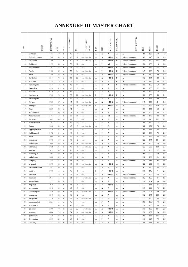
ANNEXURE III-MASTER CHART S
.NO
NA
ME
HO
SP
ITA
L N
O
AG
E
SE
X
AG
E O
F O
NS
ET
DU
RA
TIO
N(Y
EA
R
S)
TR
EA
TM
EN
T
MO
DE
IHD
SH
T
RE
TIN
OP
AT
HY
NE
UR
OP
AT
HY
NE
PH
RO
PA
TH
Y
FB
S
PP
BS
HbA
1C
Mg
1 Sundarraj 2153 50 m 46 4 oha A A A A A 90 130 5.8 2
2 Balasubramanian 2168 58 m 48 10 oha+insulin A A NPDR A Microalbuminuria 102 145 6.6 1.5
3 Rajendran 2169 56 m 46 10 oha+insulin A P NPDR P Microalbuminuria 155 160 11.1 1.6
4 Jothiraman 2175 65 m 53 12 oha A P pdr A Microalbuminuria 120 180 8 1.5
5 Rajamanikam 2201 65 m 54 11 oha P P NPDR P Microalbuminuria 105 145 5.4 1.4
6 Samivel 2198 60 m 56 14 oha+insulin A P NPDR P Microalbuminuria 145 166 7.8 1.6
7 Sekar 2196 55 m 45 10 oha A A NPDR A Microalbuminuria 102 170 9.6 1.5
8 Govindaraj 2211 78 m 56 22 oha+insulin A A NPDR P A 112 184 8.8 2.1
9 Singaram 2214 75 m 54 21 oha A A A P A 120 175 5.8 2.2
10 Ramalingam 2217 62 m 48 14 oha A A A P Microalbuminuria 155 196 6.6 1.6
11 Devendran 26234 42 m 40 2 oha A A A A A 100 185 8.5 2.4
12 Johnson 26239 45 m 42 3 oha A A A A A 80 130 6.8 2.2
13 Kandasamy 2756 78 m 56 22 oha+insulin P P NPDR P A 120 155 9.8 2.1
14 Visvalingam 2757 53 m 49 4 oha A A A A A 102 154 8.6 2.4
15 Selvaraj 2750 57 m 47 10 oha+insulin A A NPDR A Microalbuminuria 145 190 5.4 1.6
16 Pandiyan 2754 70 m 52 18 oha+insulin P P NPDR A A 112 165 10.6 2.2
17 Ravi 2357 45 m 41 4 oha A A A A A 100 155 6.8 2.1
18 Mahalingam 2359 55 m 51 4 oha A A A A A 140 200 9.8 2.2
19 Narayanasamy 2461 63 m 53 10 oha A A pdr A Microalbuminuria 106 170 8.5 1.5
20 Ramasamy 2462 45 m 42 3 oha A A A A A 112 146 10.5 2.2
21 Subramanyam 2463 51 m 47 4 oha A A A A A 80 130 5.4 2.4
22 Paulraj 2468 50 m 45 5 oha+insulin A P A A A 102 172 7.8 2.4
23 Ayyamperumal 2470 45 m 42 3 oha A A A A A 90 165 5.5 2.2
24 Kalilahamed 2472 51 m 48 3 oha A P A A A 120 188 7.6 2.4
25 Sekar 2664 50 m 46 4 oha A P A A A 95 165 6.4 2.4
26 murugesan 2665 50 m 48 2 oha A A A A A 122 174 10.4 2.2
27 mahalingam 2668 65 m 51 14 oha+insulin A A A P Microalbuminuria 106 166 7.6 1.6
28 srinivasan 2659 45 m 40 5 oha+insulin A A A A A 122 200 10.8 2.4
29 valathan 2892 50 m 46 4 oha A P A A A 96 160 6.5 2.4
30 ramalingam 2894 60 m 46 14 oha A A A A A 160 200 9.6 1.5
31 mahalingam 2888 42 m 40 2 oha A A A A A 112 180 6.4 2.3
32 thangaraj 2885 71 m 53 18 oha A A A A Microalbuminuria 140 186 8.5 1.6
33 jayamani 2877 55 m 45 10 oha+insulin A A NPDR A A 122 202 11 2.3
34 dachianamoorthi 2881 50 m 47 3 oha A A A A A 102 160 6.4 2.3
35 manivel 2878 55 m 46 9 oha P P NPDR A A 118 146 7.4 2.4
36 nagarajan 2921 70 m 52 18 oha A A NPDR A Microalbuminuria 90 166 8.8 1.5
37 natarajan 2911 65 m 51 14 oha+insulin A A A A Microalbuminuria 152 190 9.4 1.5
38 kumarasamy 2919 55 m 50 5 oha A A A A A 110 164 9.4 2.4
39 nagarajan 2910 57 m 48 9 oha P P NPDR A A 122 153 6.4 2.2
40 saminathan 2912 50 m 47 3 oha A A A A A 115 142 7.6 2.2
41 subramanyam 2840 60 m 45 15 oha+insulin A A NPDR A Microalbuminuria 140 204 6.6 1.6
42 murugesan 2317 48 m 46 2 oha A A A A A 112 174 10.8 2.2
43 narayanan 2320 65 m 55 10 oha+insulin A P A A A 122 190 6.8 2.4
44 ponnaiyapillai 2321 51 m 46 4 oha A P A A A 105 168 7.6 2.4
45 sarangabani 2327 60 m 45 15 oha A A A A A 150 190 9.8 1.5
46 govindan 2500 78 m 56 22 oha+insulin A A NPDR A A 115 155 11.4 2.3
47 govindaraj 4084 70 m 54 16 oha+insulin A A NPDR A A 136 162 6.4 2.3
48 gunasekaran 4730 48 m 45 3 oha A A A A A 105 156 11.1 2.4
49 devendaran 3993 42 m 40 2 oha A P A A A 118 146 7.5 2.2
50 sundarraj 2347 52 m 47 5 oha A A A A A 80 155 5.4 2.3

S.N
O
NA
ME
HO
SP
ITA
L N
O
AG
E
SE
X
AG
E O
F O
NS
ET
DU
RA
TIO
N(Y
EA
RS
)
TR
EA
TM
EN
T M
OD
E
IHD
SH
T
RE
TIN
OP
AT
HY
NE
UR
OP
AT
HY
NE
PH
RO
PA
TH
Y
FB
S
PP
BS
HbA
1C
Mg
51 banumathi 2156 52 f 49 3 oha A A A A A 126 170 7.6 2.2
52 vijayanthimala 2163 45 f 43 2 oha A A A A A 95 130 6.8 2.4
53 neelavathi 2165 60 f 51 9 oha+insulin P P NPDR A A 126 176 5.6 2.4
54 mariammal 2166 55 f 46 9 oha+insulin A A NPDR A A 110 146 7.4 2.4
55 pitchaiammal 2173 60 f 46 14 oha+insulin A A A A Microalbuminuria 140 190 8.4 1.5
56 rengammal 2184 50 f 46 4 oha A A A A A 155 174 6.8 2.3
57 mariammal 2185 60 f 46 14 oha A A A A Microalbuminuria 90 145 10.6 1.6
58 ramamirtham 2190 61 f 46 15 oha P P NPDR A A 130 180 6.6 2.3
59 visalatchi 2219 64 f 49 15 oha+insulin A A NPDR A Microalbuminuria 155 210 10.4 1.6
60 panchavarnam 2230 65 f 50 15 oha A A NPDR A Macroalbuminuria 126 186 7.4 1.5
61 muthulakshmi 2238 55 f 47 8 oha+insulin P P NPDR A A 105 146 9.5 2.3
62 jayashanthi 2240 48 f 45 3 oha A A A A A 130 174 11 2.4
63 anjalai 2262 55 f 50 5 oha A A A A A 115 146 6.2 2.2
64 sundaravadhini 2263 52 f 48 4 oha A A A A A 95 156 10.5 2.4
65 rathinammal 2278 60 f 51 9 oha+insulin P P NPDR A A 126 172 7.8 2.3
66 tharasa 2268 50 f 46 4 oha A A A A A 106 165 9.4 2.4
67 gowri 2307 70 f 60 10 oha+insulin A P A P Microalbuminuria 146 189 10.5 1.6
68 meri 2335 63 f 46 17 oha A P A P Macroalbuminuria 155 208 8.8 1.5
69 muniammal 2338 68 f 52 16 oha+insulin P P A P Microalbuminuria 105 145 10.8 1.6
70 ambiga 2342 63 f 53 10 oha A P A P Microalbuminuria 150 204 6.6 1.6
71 manoranjitham 2354 50 f 47 3 oha A A A A A 118 165 7.5 2.2
72 mamta beevi 2360 62 f 43 19 oha A A A A Microalbuminuria 140 175 11.6 1.5
73 shanta 2473 55 f 52 3 oha A A A A A 96 155 6.4 2.2
74 indirani 2507 50 f 46 4 oha A P A A A 135 160 7.4 2.2
75 chinnamani 2521 70 f 54 16 oha A A A A Microalbuminuria 110 170 11.4 1.6
76 saroja 2530 65 f 47 18 oha P A NPDR A Microalbuminuria 145 190 8.6 1.4
77 vairam 2540 65 f 49 16 oha P A A A Macroalbuminuria 126 176 10.8 1.5
78 shanhakumari 2550 45 f 41 4 oha A A A A A 98 140 10.6 2.2
79 rani 2555 45 f 43 2 oha A P A A A 130 184 6.4 2.3
80 amaravathi 2554 60 f 50 10 oha+insulin A A A A Microalbuminuria 160 205 8.4 1.6
81 tamilselvi 2867 55 f 47 8 oha+insulin A A NPDR A A 130 166 9.6 2.4
82 rajathi 2572 44 f 42 2 oha A P A A A 90 145 7.7 2.4
83 mariyayi 2595 43 f 41 2 oha A A A A A 98 160 8.6 2.4
84
dhanalakshmi 2624
3
73 f 50 23 oha+insulin P A NPDR P A 130 174 6.5 2.4
85 anjalai 2624
5
55 f 47 8 oha+insulin P A NPDR P A 118 186 9.4 2.3
86 arokiyameri 2623
7
60 f 50 10 oha+insulin A A NPDR A Microalbuminuria 110 185 10.6 1.5
87 jayaseeli 2624
0
75 f 65 10 oha A A NPDR P Microalbuminuria 138 202 6.6 1.6
88 malarvizhi 2645 52 f 47 5 Oha A A A A A 115 165 7.4 2.3
89 saroja 2667 58 f 48 10 oha+insulin P A NPDR A A 106 144 6.4 2.4
90 pappa 2666 59 f 49 10 Oha A A A A Macroalbuminuria 145 180 8.4 1.5
91 sebastiammal 2694 50 f 46 4 Oha A A A A A 90 145 7.6 2.4

S
.NO
NA
ME
HO
SP
ITA
L N
O
AG
E
SE
X
AG
E O
F O
NS
ET
DU
RA
TIO
N(Y
EA
RS
)
TR
EA
TM
EN
T M
OD
E
IHD
SH
T
RE
TIN
OP
AT
HY
NE
UR
OP
AT
HY
NE
PH
RO
PA
TH
Y
FB
S+
PP
BS
HbA
1C
Mg
92 chellammal 2696 55 f 50 5 oha+insulin A A NPDR A A 118 147 6.8 2.3
93 ranjitham 2691 60 f 50 10 oha A A NPDR A Microalbuminuria 155 180 11.2 1.4
94 dhanalakshmi 2700 47 f 44 3 oha A P A A A 95 165 5.6 2.3
95 chellammal 3902 55 f 50 5 oha+insulin A P A A A 115 145 8.6 2.3
96 vatchala 4223 62 f 52 10 oha A A NPDR A Microalbuminuria 140 180 7.4 1.6
97 ambigabathi 2707 58 f 50 8 oha A P A A A 120 176 9.6 2.3
98 malarkodi 2708 60 f 45 15 oha A A A A Microalbuminuria 110 190 8.8 1.5
99 saratha 2709 50 f 46 4 oha A A A A A 100 173 7.6 2.4
10
0
padmavathi 2713 55 f 50 5 oha+insulin A P A A A 90 155 5.4 2.4

ANNEXURE IV – KEYS TO MASTER CHART
A- Absent
F - Female
FBS - Fasting Blood Sugar
HbA1C - Glycosylated Hemoglobin
HTN - Hypertension
IHD - Ischemic Heart Disease
M - Male
Mg - Serum magnesium concentration
NPDR - Non Proliferative diabetic retinopathy
OHA - Oral Hypoglycemic Agent
PDR - Proliferative diabetic retinopathy
PPBS - Post Prandial Blood Sugar
P - Present

ANNEXUREV-PATIENT CONSENT FORM
Study detail : “STUDY OF SERUM MAGNESIUM LEVEL IN TYPE 2
DIABETES MELLITUS”
Study centre : THANJAVUR MEDICAL COLLEGE & HOSPITAL
Patients Name :
Patients Age :
Identification Number:
Patient may check ( ) these boxes
I confirm that I have understood the purpose of procedure for the above study.
I have the opportunity to ask question and all my questions and doubts have
been answered to my complete satisfaction.
I understand that my participation in the study is voluntary and that I am free
to withdraw at any time without giving reason, without my legal rights being
affected.
I understand that sponsor of the clinical study, others working on the sponsor’s
behalf, the ethical committee and the regulatory authorities will not need
my permission to look at my health records, both in respect of current study and any
further research that may be conducted in relation to it, even if I withdraw from the study I
agree to this access. However, I understand that my identity will not be revealed in any
information released to third parties or published, unless as required under the law. I agree
not to restrict the use of any data or results that arise from this study.
I agree to take part in the above study and to comply with the instructions
given during the study and faithfully cooperate with the study team and
to immediately inform the study staff if I suffer from any deterioration in my health or
well being or any unexpected or unusual symptoms.
I hereby consent to participate in this study.
I hereby give permission to undergo complete clinical examination and
diagnostic tests including haematological, biochemical, radiological tests.
Signature/thumb impression:
Patients Name and Address: Place Date
Signature of investigator :
Study investigator’s Name : Place Date


Your digital receiptThis receipt acknowledges that Turnitin received your paper. Below you will find the receipt informationregarding your submission.
Paper ID 291102789
Paper title STUDY OF SERUM MAGNESIUM LEVEL IN TYPE 2 DIABETESMELLITUS
Assignment title MedicalAuthor Subash Chandra Bose 20101182 M.D. General MedicineE-mail [email protected]
Submissiontime 18-Dec-2012 06:13PM
Total words 8827
First 100 words of your submission
INTRODUCTION India is frequently referred to as the diabetic capital of the world as it has the highestnumber of cases in the world. In worldwide the last 2 decades, incidence is suddenly increased from30 million cases in 1985 to 171 million in 2000. Recent data suggests that prevalence of DM by theyear 2030 could be 360 million. DM is worldwide in distribution and the incidence of both types isrising. 1, 2 The distribution of both T1 DM and T2DM varies worldwide, due to relative difference ingenetic and environmental factors in different parts of the world. Recent data shows it is associatedwith 10-30% reduction of life expectancy, most common cause of blindness in the age group of 20...
Copyright 2012 Turnitin. All rights reserved.