Embed Size (px)
Citation preview

i
STUDY OF HYDROXYAPATITE AND
HYDROXYAPATITE-CHITOSAN COMPOSITE
COATINGS ON STAINLESS STEEL BY
ELECTROPHORETIC DEPOSITION METHOD
A THESIS SUBMITTED IN PARTIAL FULFILMENT OF THE
REQUIREMENT FOR THE DEGREE OF
Bachelor in Technology
in
Biomedical Engineering
by
AJAY SAXENA (107BM003)
and
ABHINANDAN ROUT (107BM009)
Department of Biotechnology and Medical Engineering
National Institute of Technology
Rourkela

ii
STUDY OF HYDROXYAPATITE AND
HYDROXYAPATITE-CHITOSAN COMPOSITE
COATINGS ON STAINLESS STEEL BY
ELECTROPHORETIC DEPOSITION METHOD
A THESIS SUBMITTED IN PARTIAL FULFILMENT OF THE
REQUIREMENT FOR THE DEGREE OF
Bachelor in Technology
in
Biomedical Engineering
by
AJAY SAXENA (107BM003)
and
ABHINANDAN ROUT (107BM009)
under the guidance of
Dr. AMIT BISWAS
Department of Biotechnology and Medical Engineering
National Institute of Technology
Rourkela

iii
CERTIFICATE
This is to certify that the thesis entitled „The study of hydroxyapatite and hydroxyapatite-
chitosan composite coatings on stainless steel by electrophoretic deposition method‟
submitted by Ajay Saxena and Abhinandan Rout in partial fulfilment of the requirements
for the award of Bachelor of Technology from the Department of Biotechnology and Medical
Engineering with specialization in “Biomedical Engineering” at National Institute of
Technology, Rourkela (Deemed University) is an authentic work carried out by them under
my supervision and guidance. To the best of my knowledge, the matter embodied in the
thesis has not been submitted to any other University/Institute for the award of any degree or
diploma.
Place:
Date:
Supervisor
Dr. Amit Biswas
Department of Biotechnology and Medical Engineering
National Institute of Technology, Rourkela- 769008

iv
ACKNOWLEDGEMENT
With deep regards and profound respect, we avail this opportunity to express our gratitude
and indebtedness to Prof. Amit Biswas, Dept. of Biotechnology and Medical Engineering,
NIT Rourkela, for introducing the present research topic and for his inspiring guidance,
constructive criticism and valuable suggestions throughout in this research work. It would
have not been possible for us to bring out this thesis without his help and constant
encouragement.
We are sincerely thankful to Prof. Subhankar Paul, HOD, Dept. of Biotechnology and
Medical Engineering for his advice and inputs, and for providing the necessary facilities for
our work. We are also thankful to Prof. B.B. Verma, Head of the Dept. of Metallurgical and
Materials Engineering for providing the necessary facilities for our work.
Last but not the least; we are highly grateful to Subrat Sahu, M.Tech. scholar of Biomedical
Engineering, and laboratory members of the departments of Biotechnology and Medical
Engineering and Metallurgical and Materials Engineering, especially Mr. Shyamu Hembram
for their help during the preparation of samples and execution of experiments.
Ajay Saxena Abhinandan Rout
Date:
Place:

v
ABSTRACT
Hydroxyapatite is a salt of calcium and phosphorus having the Ca/P ratio as 1.6.
Hydroxyapatite is one of the major mineral components of the bone due to which it shows
excellent biocompatibility. Even though it is biocompatible, hydroxyapatite cannot be used
directly in load bearing structures because of its poor mechanical properties. An alternative
approach to utilize the biocompatibility of hydroxyapatite is by using it as a coating material.
The biocompatibility of hydroxyapatite needs to be complemented by coating it on a
substance having desirable mechanical properties. A suitable material for this purpose is
medical grade stainless steel, which has been used to make prosthesis, bone fixation screws,
guide wires etc. It has been shown that coating medical grade stainless steel (SS 316L)
implants with hydroxyapatite leads to better biocompatibility. For the coating process various
techniques can be used. Electrophoretic deposition is a procedure of mild nature not
involving extreme temperatures and has the advantage of producing a stoichiometric, uniform
coating of the desired thickness. In this study a practical approach to coating was undertaken
and a HA and composite HA-Chitosan coating obtained by varying the processing parameters
was studied.
A coating of HA and composite coating of stoichiometric hydroxyapatite and chitosan were
produced on medical grade stainless steel (SS 316L). Industrially pure chitosan was bought
for the coating purposes and hydroxyapatite nanoparticles were synthesized by a chemical
reaction of calcium nitrate and ammonium phosphate. In the synthesis process of HA

vi
nanoparticles the pH was controlled by the addition of ammonium hydroxide. The coating
was carried out stainless steel as the cathode and graphite rod as the counter electrode, the
distance between electrodes being 2 cm at a current density of 0.11 Ampere per metre square.
The SEM analysis showed that at a constant voltage the coating deposition increases with an
increase in the coating time, whereas when the coating time is kept constant, the fineness of
the coating increases with an increase in the voltage. From the XRD analysis it was
confirmed that the phase purity of chitosan and HA is maintained even after the coating.

vii
CONTENTS
Cover Page i
Certificate iii
Acknowledgement iv
Abstract v
Contents vii
Chapter 1 - Introduction 1
Chapter 2 - Literature Review 4
2.1 Stainless Steel 5
2.2 Hydroxyapatite 5
2.3 Chitosan 6
2.4 Techniques of coating 7
2.5 Electrophoretic Deposition 9
2.5.1 Mechanism of Electrophoretic Deposition 9
2.5.2 Effect of processing parameters 10
Chapter 3 – Experimental Procedure 12
3.1 Sample and Solution preparation for HA coating 13
3.2 Sample and Solution preparation for HA-Chitosan composite coating 13
3.3 Experimental procedure for electrophoretic deposition 14
3.4. Characterisation of coating 14
3.4 Flowchart depicting the experimental procedure 15
Chapter 4 – Results and Discussion 16
4.1 Introduction 17
4.2 Electrophoretic deposition of hydroxyapatite (HA) on 316L Stainless steel 17
4.2.1 Characteristics of the HA layer on 316L Stainless steel 17
4.2.1.1 Microstructural analysis 17
4.2.1.2. Analysis of pore structure 21
4.2.2 Phase Analysis 22
4.3 Electrophoretic deposition of HA-chitosan composite on 316L Stainless steel 23
4.3.1 Characteristics of the HA-chitosan layer on 316L Stainless steel 23
4.3.1.1 Microstructural analysis 23
4.3.1.2. Analysis of pore structure 26
4.3.2 Phase Analysis 27
Chapter 5 - Conclusion 28
References 29

1
CHAPTER 1
2
INTRODUCTION
A wide variety of materials are used for medical purposes. These include but are not limited
to ceramic, glasses, metallic and polymeric biomaterials. Among these materials, metallic
biomaterials find application where load bearing structures are required such as in
orthopaedics and dentistry. The advantages of using metallic biomaterials include the
relatively easy fabrication of both simple and complex shapes by the help of well-established
fabrication techniques such as casting, forging, machining. The various biomaterials that are
used include stainless steel, titanium, tantalum, nickel titanium alloys. Medical grade
stainless steel (SS 316L) is widely used in fracture repair devices and joint replacement
components. Its wide usage is due to the fact that it is a more economical alternative as
compared to other metallic biomaterials. However 316L SS has a high susceptibility to
corrosion and to overcome this shortcoming surface modification of stainless steel is done
before its usage in the body. The common biocompatible coatings are of thermally grown
oxides, electrolytically grown oxides and hydroxyapatite, the various processes used for the
coatings are thermal exposure (poly crystalline and amorphous oxide), proprietary processes
and electropolishing (in a phosphoric acid glycerine solution). HA coated SS 316L shows a
positive shift in the OCP which is an indication of stable coating/insulation behaviour, along
with an improvement in the pitting corrosion. The coating processes have the advantage of
changing the properties of the surface of the material without affecting the properties of bulk.
The alternative to coating process is the use of highly corrosion resistant alloy, but using
highly corrosion resistant alloys will increase the cost of the implants making them
economically infeasible. Along with the conventional techniques of thermal oxidation and
electrochemical oxidation, advanced techniques plasma ion implantation, plasma source ion

3
implantation, laser melting, laser alloying, ion beam and physical vapour deposition have
been widely applied to biomaterials. Electrophoretic deposition can be used to produce
stoichiometric coating. It has the advantage of being able to produce uniform deposits with
high microstructural homogeneity; it also provides adequate control of the deposit thickness.
Electrophoretic deposition can be used to coat a wide range of shapes and 3D complex and
porous structures.
Hydroxyapatite is the major mineral constituent of the bone and hence as a biomaterial
hydroxyapatite shows good biocompatibility and bone adhesion. However hydroxyapatite
suffers from poor mechanical properties and implants made entirely from Hydroxyapatite
will suffer from a mechanical failure. To utilize the excellent biocompatibility of
hydroxyapatite it can be used as a coating material for stainless steel, by which it will
complement the mechanical strength of stainless steel. Such an implant made of stainless
steel coated with hydroxyapatite can be used for bone fixation as it will show both a high
strength and high biocompatibility. In the present study a coating of HA and a composite
coating of hydroxyapatite chitosan was applied on stainless steel substrate by electrophoretic
deposition method. Chitosan is a naturally occurring cationic polysaccharide. It is
biocompatible, antimicrobial and biodegradable.

4
CHAPTER 2

5
LITERATURE SURVEY
2.1. Stainless steel
With improved medical facilities worldwide, more and more people are living to advanced
ages in today‟s world. As a result, the chances of body parts (especially those subjected to
stress and pressure on a regular basis like bones and joints) wearing out and stopping
functioning are much greater over this extended period of time. So, implants are being
increasingly used in today‟s world to replace the functioning of bones and joints.
Since these parts are subjected to various stresses, the implants need to be strong enough to
absorb all of the stress that will be inflicted upon them by the body. The implants should have
good mechanical properties, high corrosion resistance, be cheaply available, have good
fatigue life, and undergo no wear and tear while functioning. Generally, metals and metallic
alloys have been used as implant materials because they fulfil most of these requirements.
Stainless steel (technically 316L stainless steel) has been increasingly used as implant
materials because of its similarities to human bone in terms of young‟s modulus of elasticity,
hardness and other mechanical properties. It is considered as one of the attractive metallic
materials for biomedical applications also due to its mechanical properties, biocompatibility,
and corrosion resistance. This material is a popular metal for use as acetabular cup (one half
of an artificial hip joint) applications [14].
2.2. Hydroxyapatite
Hydroxyapatite (HA) is a bioactive ceramic material with high bioaffinity, biocompatibility
and osseoconductivity which is the main constituent of bones and teeth. Natural HA has the
advantage that it inherits some properties of the raw material such as composition and

6
structure. Properties of HA have found useful application in low-load bearing porous
implants and coatings of metallic implants. Bioactivity and acting as a template for forming
and growing of the surrounding bone tissues make HA an excellent choice for coating of the
metallic implants. After implantation of prostheses, a close surface contact between the
metallic prosthesis and the surrounding bone tissue is needed for subsequent bone ingrowths.
Coating of the metallic implants with bioactive HA leads to a rapid bonding between
hydroxyapatite and surrounding bone tissue. Application of HA as a coating of the metallic
implants combines the strength and toughness of the substrate with bioactive characteristic of
HA which can induce the surrounding bone tissue ingrowths and future formation of
chemical bonding. Further, the presence of HA coating can improve corrosion resistance of
the coated implant in human body which can reduce the metallic ion release and also
promotes fixation via chemical bonding [15].
2.3. Chitosan
Chitosan is a cationic polysaccharide and one of the most promising natural biopolymers for
tissue engineering, biocompatible coatings and drug delivery. Due to its unique properties
including biodegradability, biocompatibility, non-toxicity and bio-functionality, Chitosan has
attracted much attention for a wide variety of applications ranging from skin, bone, cartilage
and vascular grafts to substrates for cell culture. Previous studies have shown the feasibility
of cationic electrophoretic deposition (EPD) of chitosan. The deposition mechanism is related
to the pH variation caused by electrochemical decomposition of water. A recent progress has
been the chitosan-mediated electrosynthesis of composites based on bioactive glass,
hydroxyapatite (HA), bioactive glass–HA, HA–wollastonite, HA–carbon nanotubes and HA–
CaSiO3. More recently, Mayer et al. have used electrodeposition of chitosan on gold wires to

7
fabricate a biosensing platform, showing the high potential of the method to develop novel
chitosan based devices. It is evident that knowledge of the electromigration of chitosan
molecules is necessary in order to control the deposition rate and to achieve flexibility in
microstructural manipulation of chitosan coatings. Aider et al. have shown that the
electromigration of chitosan D-glucosamine and oligomers in aqueous solutions depends on
the pH. To the best knowledge of the authors, the growth rate of chitosan film
macromolecules as a function of pH and biopolymer concentration has never been reported.
Understanding the electrophoretic mobility and deposition kinetics of chitosan will not only
be useful for controlling the deposition of neat chitosan films but also to describe the
interaction between the polymer and ceramic particles during co-deposition in order to
control the EPD process of inorganic–organic composite coatings, which are of high
relevance for biomedical applications [16].
2.4. Techniques of coating
There are several coating methods used for surface modifications of materials. The following
techniques are a few of them used for applying coatings on metals:
Electroplating – Electroplating is a process of coating a deposition on a cathode part
immersed into an electrolyte solution, where the anode is made of the depositing
material, which is dissolved into the solution in the form of the metal ions, that travel
through the solution and deposit on the cathode surface.
Electroless plating - The process of deposition of metal ions from electrolyte solution
onto the substrate when no electric current is involved and the plating is a result of

8
chemical reactions occurring on the surface of the substrate.
Conversion coating - The process in which the coating is formed as a result of
chemical or electrochemical reaction on the substrate surface. These are non-metallic
coatings obtained on the metal surface in the form of compounds of the substrate
metals.
Hot dipping - Immersing the part into a molten metal followed by removal of the
substrate from the metal bath, which results in formation of the metal coating on the
substrate surface.
Physical Vapour Deposition (PVD) - the process involving vaporization of the coating
material in vacuum, transportation of the vapour to the substrate and condensation of
the vapour on the substrate surface.
Chemical Vapour Deposition (CVD) – The process, in which the coating is formed on
the hot substrate surface placed in an atmosphere of a mixture of gases, as a result of
chemical reaction or decomposition of the gases on the substrate material.
Thermal spraying – Deposition of the atomized at high temperature metal, delivered to
the substrate surface in a high velocity gas stream

9
2.5 Electrophoretic Deposition
Electrophoretic deposition is a conventional technique. But it is used vastly due to its certain
advantages over other coating techniques like that of low cost, low energy requirement,
capability to handle complex geometry, simple scale-up with easily maintainable equipment
and good chemical stability. One of its important advantages is that a very large number of
pure metals, alloys, composites and ceramics can be electrodeposited with grain size less than
100 nm. Metals, alloys and biopolymers can also be deposited by this process. Multilayer
deposition is possible in this process. Materials can be coated for use in industrial
applications such as coatings of engine cylinders, high pressure valves, musical instruments,
car accessories, small aircraft microelectronics, aerospace, medical devices, marine,
agriculture and nuclear fields [17].
2.5.1 Mechanism of Electrophoretic Deposition
Electrophoretic deposition is a process by which charged ions in the solution are attracted and
deposited onto an electrode of the opposite charge by the application of an electric field.
Electrophoretic deposition is essentially a two-step process. In the first step the charged
particles migrate under the electric field to the electrode having the opposite charge. In the
second step, the particles deposit on the electrode forming a relatively dense, compact and
homogenous film. There can be two types of electrophoretic deposition processes on the basis
of the electrode on which the coating is produced. When the particles are positively charged
the deposition happens on the cathode the process is called cathodic electrophoretic
deposition whereas when the particles are negatively charged and the deposition occurs on
the anode the process is known as anodic electrophoretic deposition. The process leads to
creation of stoichiometric films whose composition depends on the stoichiometry of the

10
powder used. The principal driving force for electrophoretic deposition is the charge on the
particle and its electrophoretic mobility in the solvent under the presence of the applied
electric field. According to Sarkar and Nicholson, particle/electrode reactions are not
involved in EPD and ceramic particles do not lose their charge on being deposited [18].
Fig 1: Electrophoretic deposition apparatus
2.5.2 Effect of processing parameters on deposition
The parameters related to the EPD can be categorized into solution related and process
related parameters. The solution related parameters are particle size, dielectric constant of the
liquid, conductivity and viscosity of the suspension, zeta potential and stability of the
suspension. The process related parameters are deposition time, applied voltage,
concentration of the solid in suspension and conductivity of the substrate,

11
Effect of deposition time
Basu et al have found that for a fixed electric field when the deposition time increases the
deposition rate decreases, similar observations were made by Chen and Liu. This is attributed
to the decrease in the electric field influencing electrophoresis due to the formation of an
insulating layer of ceramic particles on the electrode surface (Zhitomirsky and L Gal).
Effect of Applied Voltage
It is observed that the deposit increases with an increase in voltage. When stronger electric
fields are used, the deposition rate increases but the quality of the deposit gets affected. Basu
et al found that more uniform films are deposited at moderate applied fields (25-100 V/cm)
whereas film quality deteriorates if relatively higher applied fields are used. The stronger
electric fields cause turbulence in the medium which affects the developing coating; also the
nature of the coating that is produced depends on the accumulation rate which influences the
packing behaviour of the particles in the coating. In case of a stronger electric field, the
accumulation rate is higher and the particles move faster; so they don‟t have time to sit in
their positions in close packed structure. The lateral motion of the particles is restricted on the
surface of the already deposited layer as the higher field exerts more pressure on particle flux
and movement.
Effect of concentration of solid in suspension
The rate at which the particles deposit on the electrodes depends on their electrophoretic
mobilities; it becomes an important factor when the volume fraction of the solids is low.
When the volume fraction of the solids is high, they deposit at an equal rate. However, when
the volume fraction of the solids is low the particles deposit at rate proportional to their
individual electrophoretic mobility (Vandeperre, Biest and Clegg).

12
CHAPTER 3

13
EXPERIMENTAL PROCEDURE
3.1 Sample and solution preparation for HA coating
1. A steel rod having diameter 1 cm was cut into cylindrical samples of length 1 cm.
2. Samples were polished by belt grinder and emery paper of standard 1/0, 2/0, 3/0 and 4/0
successively. After this, cloth polishing was done by using aluminium oxide. On completion
of cloth polishing diamond polishing was done.
3. HA obtained from chemical synthesis was dissolved in ethanol with the help of magnetic
stirrer and 150 ml of this solution was taken.
3.2 Sample and solution preparation for HA-Chitosan composite coating
1. A steel rod having diameter 1 cm was cut into cylindrical samples of length 1 cm.
2. Sample was polished by belt grinder and emery paper of standard 1/0, 2/0, 3/0 and 4/0
successively. After this, cloth polishing was done by using aluminium oxide. On completion
of cloth polishing diamond polishing was done.
3. HA obtained from chemical synthesis was dissolved in ethanol with the help of magnetic
stirrer and 150 ml of this solution was taken.
4. A solution was formed by dissolving 0.4 gm chitosan in 500 ml acetic acid and water
solution, where the pH was maintained between 3.5 and 3.9 and 150 ml of this solution was
taken.
5. 300 ml of the final solution was prepared by mixing the solutions obtained in the above two
steps.

14
3.3 Experimental procedure for electrophoretic deposition
1. The electrodes were washed with acetone and dried.
2. The deposition was carried out at room temperature from 150 ml of the solution (as per the
coating desired) in 250 ml beaker. The solution was stirred using a magnetic stirrer.
3. A graphite rod was used as an anode and the sample was used at cathode.
4. The distance between the electrodes was 2 cm.
5. Deposition was carried out on a substrate surface area of 0.785 cm2.
6. HA and HA-chitosan composite was deposited on the stainless steel by electrophoretic
deposition.
7. For deposition of HA, voltages were varied from 15 V, 20V, 25 V for time periods of 10 min,
15 min, 20 min.
8. For deposition of HA-chitosan composite coatings voltages were varied from 20 V, 25 V, 30
V for time periods of 15 min, 20 min, 30 min.
3.4 Characterization
The SEM analysis of the sample was carried out on JEOL JSM 6480 lv, to study the microstructure of
the coating. The phase purity was tested by X Ray diffraction on Philips Xpert PANalytical XRD
machine.

15
3.4 FLOWCHART DEPICTING THE EXPERIMENTAL PROCEDURE
Sample and solution Preparation
Cutting of the sample
Polishing of the sample
Hydroxyapatite
Synthesis
Formation of chitosan
solution
Electrophoretic Deposition
Hydroxyapatite coating Hydroxyapatite Chitosan
composite coating
Characterization of the
coating
XRD analysis FTIR Analysis SEM Analysis

16
CHAPTER 4

17
RESULTS AND DISCUSSION
4.1 Introduction
This project is aimed at a detailed investigation of the electrophoretic deposition of HA and HA-
chitosan layer on 316L stainless steel. The surface characterization using scanning electron
microscope (SEM) and X-ray diffraction (XRD) of the coated layer have been done.
4.2 Electrophoretic deposition of hydroxyapatite (HA) on 316L Stainless
steel
4.2.1 Characteristics of the HA layer on 316L Stainless steel
4.2.1.1 Microstructural analysis
Figure 4.1 shows the effectiveness of the HA coating on 316L SS with time. From the figures
4.1 (a), (b) and (c), it is observed that HA is unformly deposited on the surface.when seen
under low magnification. But in case of Fig 4.1 (c), it is observed that microcracks are
formed on the coated surface. This is due to formation of a thicker layer which undergoes
rearrangement upon contraction leading to separation. If the layer were thinner, formation of
cracks would be difficult because the metallic substrate would have adhered better to the
thinner HA layer.

18
Fig 4.1: Scanning electron micrograph (500x magnification) of electrophoretically deposited
HA on 316L stainless steel at (a) 30 V - 10 mins (b) 30 V - 15 mins (c) 30 V - 20 mins
(a)
(b)
(c)

19
Fig 4.2: Scanning electron micrograph (3000x magnification) of electrophoretically deposited
HA on 316L stainless steel at (a) 30 V - 10 mins, (b) 30 V - 15 mins (c) 30 V - 20 mins
(a)
(b)
(c)

20
Figures 4.2 (a), (b) and (c) show the scanning electron micrograph of HA coated 316L
stainless steel sample for three different time durations (10, 15 and 20 mins respectively) at
30 Volts. From the figures, it is observed that HA is deposited in a feathery form with good
coverage over the entire surface. The coating thus formed on both the samples is quite
uniform. The growth of the HA crystals is more pronounced in the 20 mins sample as
compared to the 10 and 15 mins samples, which leads to better coating on the surface as
observed from the figures. This better coverage of the coating in the 20 mins treated sample
is due to the fact that a proper growth of the HA crystals has taken place with more time,
which also results in giving a good thick layer on the surface.
4.2.1.2. Analysis of pore structure
Figure 4.3 shows the distribution of pores of the HA coating on 316L SS with time. Fig 4.3
(a) and (b) show that the pore size decreases with an increase in the coating time. The average
pore size (calculated on the basis of simple mathematical averages) decreases from 316 nm to
253 nm.
This is because of the fact that growth of the HA crystals occurs with greater deposition times
and the crystals cover up maximum area on the implant surface leaving very small holes.

21
Fig 4.3: Scanning electron micrograph of electrophoretically deposited HA on 316L stainless
steel at (a) 30 V - 10 mins (b) 30 V - 15 mins
(a)
(b)

22
4.2.2. Phase Analysis
Fig 4.4: Scanning electron micrograph of electrophoretically deposited HA on 316L stainless
steel at (a) 30 V - 10 mins (b) 30 V - 15 mins
Fig 4.4: shows the X ray diffraction profile of electrophoretically deposited HA on 316L SS
at 30 Volts and 10 mins. From the analysis of the XRD data obtained from the scrape of the
coating on the sample surface, it was found that that the peaks obtained are a match with the
standard peaks of HA in the JCPDS data book. The matching of the peaks are done in
PHILIPS X pert high score software. Hence we can conclude that HA has not lost its phase
purity after deposition.

23
The coating thus formed consists of mainly HA as verified from the X Ray Diffraction
pattern. No other lower calcium compounds are detected in the data, which confirms that the
purity of the coating in terms of HA is almost 99 %.
4.3 Electrophoretic deposition of hydroxyapatite-chitosan composite on
316L stainless steel
4.3.1 Characteristics of the HA-chitosan layer on 316L Stainless steel
4.3.1.1 Microstructural analysis
Figures 4.5 (a), (b) and (c) show the scanning electron micrograph of HA-chitosan composite
coated on 316L stainless steel sample for three different applied voltages (20, 25 and 30 volts
respectively) for 25 mins. From the figures, it is observed that HA-chitosan is deposited with
good coverage over the entire surface without any cracks on it. The crystals of HA look like
facets with random orientation on the surface. The microstructure of the coating is finer at
higher voltages due to higher driving force at 30 volt compared to 20 volt.

24
Fig 4.5: Scanning electron micrograph (500x magnification) of electrophoretically deposited
HA-chitosan composite on 316L stainless steel at (a) 20 V - 25 mins (b) 25 V - 25 mins (c)
30 V - 25 mins
(a)
(b)
(c)

25
Fig 4.6: Scanning electron micrograph (5000x magnification) of electrophoretically deposited
HA-chitosan composite on 316L stainless steel at (a) 20 V - 25 mins (b) 25 V - 25 mins (c)
30 V - 25 mins
(b)
(c)
(a)
26
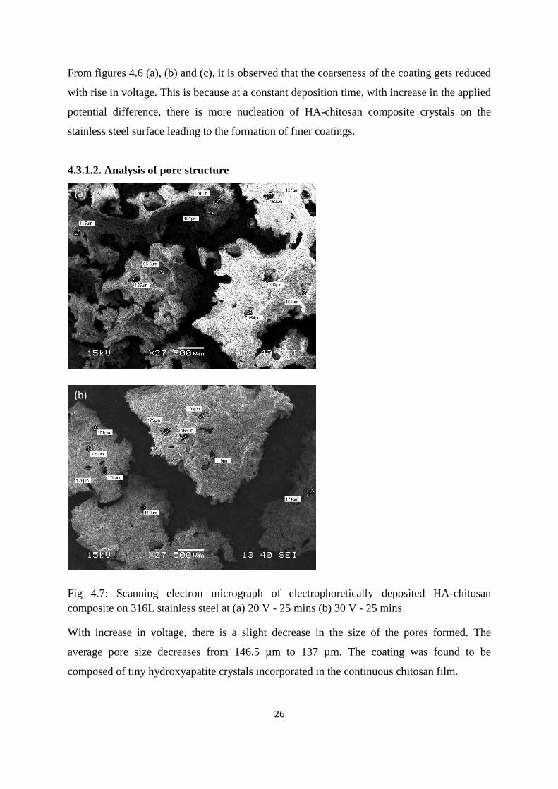
From figures 4.6 (a), (b) and (c), it is observed that the coarseness of the coating gets reduced
with rise in voltage. This is because at a constant deposition time, with increase in the applied
potential difference, there is more nucleation of HA-chitosan composite crystals on the
stainless steel surface leading to the formation of finer coatings.
4.3.1.2. Analysis of pore structure
Fig 4.7: Scanning electron micrograph of electrophoretically deposited HA-chitosan
composite on 316L stainless steel at (a) 20 V - 25 mins (b) 30 V - 25 mins
With increase in voltage, there is a slight decrease in the size of the pores formed. The
average pore size decreases from 146.5 µm to 137 µm. The coating was found to be
composed of tiny hydroxyapatite crystals incorporated in the continuous chitosan film.
(a)
(b)

27
4.3.2. Phase Analysis
Fig 4.8: Scanning electron micrograph of electrophoretically deposited HA-chitosan
composite on 316L stainless steel at (a) 30 V - 10 mins (b) 30 V - 15 mins
From the analysis of the XRD data obtained from the sample surface, it was found that that
the peaks obtained match with the standard peaks of HA and chitosan in the JCPDS data
book. This is done by PHILIPS X pert high score software.
Hence we can conclude that neither HA nor chitosan lose their phase purity upon deposition
and the presence of both in the coating is verified from the XRD plot.
.

28
CONCLUSION
1. HA and HA-chitosan were successfully coated onto 316L SS at lower voltages.
2. Electrophoretic deposition gives proper coating thickness, and the phase purity of HA
and HA-chitosan was maintained after deposition.
3. Since the pore sizes are in the range of 50-320 nm, the coatings are suitable for cell
attachment and proliferation.
4. With increase in voltage, there is more nucleation; with increase in time, there is
growth of crystals. So a proper combination of voltage and time can effectively coat
the steel surface with HA/ HA-Chitosan with desired pore size for bioimplant
applications.
5. For HA coating the best coating parameters are found to be 30 volt and 15 mins. With
increase in deposition time, there is an increase in both the density and coarseness of
the HA coating formed. This happens because with increase in deposition time, the
deposited HA crystals get more time to grow on the 316L SS surface.
6. Electrophoretic deposition method can effectively be used for HA and HA-chitosan
coating by using the solution having HA in ethanol and chitosan in acetic acid.
.

29
References
[1] J.R. Dees, J.E. Spruiell. Journal of Applied Polymer Science 18(1974)1053–78.
[2] J.M. Deitzel, N.C. Beck Tan, J.D. Kleinmeyer, J. Rehman, D. Tevault, D. Reneker, I.
Sendijarevic, A. McHugh. Army Research Laboratory Technical Report (1999) ARL-
TR-1989.
[3] P.W. Gibson, H.L. Shreuder-Gibson, D. Rivin. AICHE Journal 45(1999)190-4.
[4] P.W. Gibson, H.L. Shreuder-Gibson. US Army Soldier and Biological Chemical
Command Technical Report (1999) Natick/TR-99/016L
[5] C.J. Buchko, L.C. Chen, Y. Shen, D.C. Martin. Polymer 40(1999)7397–407
[6] M. Krumova, D. López, R. Benavente, C. Mijangos and J.M. Pereña. Polymer
(2000)9265.
[7] B. Ding, H. Kim, S. Lee, C. Shao, D. Lee, S. Park, G. Kwag and K. Choi. Journal of
Polymer Science. Part B, Polymer Physics (2002)1261.
[8] Y.T. Jia, Jian Gong, X. Hua Gu, H.Y. Kim, Dong and X.Y. Shen. Fabrication and
characterization of poly (vinyl alcohol)/chitosan blend nanofibers produced by
electrospinning method. Carbohydrate Polymers 67(2007)403-409
[9] S. Senel and S.J. McClure. Potential applications of chitosan in veterinary medicine,
Advanced Drug Delivery Reviews 10(2004)1467–1480.
[10] I. Yamaguchi, K. T. Preparation and microstructure analysis of
chitosan/hydroxyapatite nanocomposites. J Biomed Mater Res (2001)20–27.
[11] I. Zhitomirsky, L. G.-O.Electrophoretic deposition of Hydroxyapatite. Journal Of
Materials Science: Materials In Medicine 8(1997)213-219.
[12] I. Corni, M. P. Electrophoretic deposition: From traditional ceramics to
nanotechnology. Journal of the European Ceramic Society 28(2008)1353-1367.

30
[13] L. Besra, M. L. A review on fundamentals and applications of electrophoretic
deposition. Progress in Materials Science (2007)1–61.
[14] L. He, H. L. Fabrication of HAp/Ni biomedical coatings using an electro-codeposition
technique. Surface and Coatings Technology (2002)109-113.
[15] S. Nayar, P. A. Hydroxyapatite coating on stainless steel pre-coated with bovine
serum albumin at ambient conditions. Colloids and Surfaces B: Biointerfaces
48(2006)183-187.
[16] N. Ma, X. F. Ag–TiO2/HAp/Al2O3 bioceramic composite membrane: Fabrication,
characterization and bactericidal activity. Journal of Membrane Science ,
336(2009)109-117.
[17] Pilliar, R. M. Metallic Biomaterials. In R. Narayan, Biomedical Materials. Springer.
(2009)
[18] X. Pang, I. Z. Electrophoretic Deposition of composite hydroxyapatite chitosan
coatings. Materials Characterization (2007)339-348.
[19] I. Zhitomirsky. Cathodic electrodeposition of ceramic and organoceramic materials.
Fundamental aspects. Advances in Colloid and Interface Science (2002)279-317.