Embed Size (px)
Citation preview

1
Study of Hydrogel based Controlled Release
Drug Delivery System for Captopril and
its in-vitro, in-vivo Evaluation
A dissertation submitted in
partial fulfillment of the requirements for the degree of
DOCTOR OF PHILOSOPHY
(Pharmaceutics)
by
Furqan Muhammad Iqbal B.Pharm., M.Phil.
Department of Pharmacy
Faculty of Pharmacy and Alternative Medicine
The Islamia University of Bahawalpur PAKISTAN
(2012-2015)

2
In the name of Allah, the Most Merciful, the Most Kind

3
DEDICATION
Dedicated to my beloved
parents, wife and children

4
ACKNOWLEDGEMENT
Foremost, I should bow to ALLAH ALMIGHTY who made me able to work and
accomplish my work within time .All respects are for The Last Prophet, HAZRAT
MUHAMMAD (Peace Be Upon Him),who enable us to recognize our Creator.
I owe a profound debt of gratitude and heartful thanks to my research supervisor,
Prof. Dr. Mahmood Ahmad, the Dean of Pharmacy and alternative medicine, Islamia
University Bahawalpur. His unique welcoming, affectionate and encouraging style
played a vital role in harnessing my potentialities and capabilities to accomplish this
research project.
I am highly thankful to Prof. Dr. Naveed Akhtar (Chairman, Department of
Pharmacy) for his facilitation to complete the enterprising assignment.
I appreciate the cooperation and moral support of my research fellows Ume Ruqia
Tulain and Ayesha Rasheed. I am thankful to Dr. Malik Zubair, Dr. Raees Akhtar,
and Fahad Pervaiz for providing me the expertise for completion of this project. I am
also thankful to Dr. Usman Minhas and Ikrima Khalid for their cooperation.
I am thankful to all those who are exerting themselves for the advancement of
knowledge and specially devising and designing the new drug delivery systems to
serve the humanity.
Furqan Muhammad Iqbal

5
Abstract
A foremost step towards controlled and targeted administration of therapeutic agents is
development of new drug delivery systems. Oral administration is mostly preferred and
desired as a non-invasive mean of providing drug at controlled rate. In present research work,
hydrogels were prepared for controlled release of captopril, an angiotensin converting
enzyme (ACE) inhibitor, used for the treatment of hypertension. Three types of hydrogel
formulations were prepared by different proportions of polymers and monomers. A chemical
crosslinking method, free radical polymerization was selected for synthesis of polymeric
networks, involving use of thermostatic water bath as well as induction by microwave
radiations. A microwave assisted hydrogel synthesis, was used for preparation of
hydroxypropyl methylcellulose-graft-poly(vinyl alcohol)-co-poly(acrylic acid) copolymeric
network. N,N-methylenebisacrylamide and potassium persulfate (KPS) were used as
crosslinking agent and initiator, respectively. Formulations with same combinations of
polymers and monomers were also prepared by utilizing conventional thermostatic water
bath. The hydrogels obtained by these techniques were compared with each other in terms of
morphological properties, swelling ratios, drug loading and drug release behavior.
The hydrogel formulations were also prepared by crosslinking of 2-acrylamido-2-methyl-1-
propanesulfonic acid (AMPS) and acrylic acid with hydroxypropyl methylcellulose (HPMC).
These hydrogels had shown higher ability to absorb and retain aqueous solutions and solute
particles. Another type of polymeric network was synthesized under influence of microwaves
radiations, with lower initiator concentration, by crosslinking of poly(vinyl alcohol) (PVA)
with 2-acrylamido-2-methyl-1-propanesulfonic acid (AMPS). They have ability to exhibit
relatively higher swelling behavior at pH 2 in comparison to pH 7.4 and have gastro retentive
characteristics. Due to their massive swelling tendencies, these could be retained in stomach
and unable to pass through next segment of gastrointestinal tract. Thus, after oral
administration of captopril loaded hydrogels, they could have ability to release drug
continuously at acidic pH of stomach, in a control manner for longer time periods. The
results of drug release are according to swelling powers of formed copolymeric hydrogels.

6
All types of hydrogel formulations prepared were evaluated by in-vitro and in-vivo analytical
procedures. The in-vitro characterization was done by Fourier Transform Infrared
Spectroscopy (FT-IR), scanning electron microscopy (SEM), X-ray diffraction (XRD),
thermogravimetric analysis (TGA), differential scanning calorimetry (DSC), swelling
properties, drug loading and release. The drug release was evaluated by the application of
zero order kinetics, first order kinetics, Higuchi model, Korsmayer-Peppas model and
Weibull model.
The hydrogels selected on the basis of their in-vitro evaluation were subjected to in-vivo
characterization. High performance liquid chromatography (HPLC) method, with UV
detector was utilized for in-vivo characterization. The study was performed on twenty four
rabbits and liquid-liquid extraction procedure was used for separation of captopril from
plasma samples. The bioavailability and pharmacokinetic parameters were determined by
kinetica (version 5.0). The maximum concentration (Cmax) of captopril was reduced while
time to reach maximum concentration (Tmax) was increased by hydrogels in comparison to
control (free drug enclosed in hard gelatin capsules). The values of area under curve AUC
(calculated by trapezoidal rule) and elimination half-life were higher for controlled release
hydrogel formulations than control. The drug could be available for longer periods of time
after administration of captopril loaded hydrogels, maintaining optimum concentration in
blood, exerting its efficacious effects as an antihypertensive therapeutic agent.

7
Study of Hydrogel based Controlled Release
Drug Delivery System for Captopril and
its in-vitro, in-vivo Evaluation

8
List of Contents
Description Page
no.
Title I
Bismillah II
Dedication III
Acknowledgement IV

9
2.2.1.4 Crosslinking using enzymes 15 2.2.2 Physical crosslinking methods 15
2.2.2.1 Crosslinking by ionic interactions 16
2.2.2.2 Crosslinking by crystallization 16 2.2.2.3 Crosslinking by hydrogen bonds 17
2.2.2.4 Crosslinking by protein interactions 18
2.3 Characterization of hydrogels 18 2.3.1 Scanning electron micrography (SEM) 20
2.3.2 X-ray diffraction (XRD) 20
2.3.3 Magnetic Resonance Imaging (MRI) 20
2.3.4 Fourier transform infrared (FTIR) 21 2.3.5 Thermal Analysis 22
2.3.6 Swelling behavior 22
2.3.7 Gel Fraction 23
2.3.8 Porosity 24
2.3.9 Rheology 24
2.3.10 In-vitro Release Studies 25
2.3.11 In-vivo Evaluation 25
2.4 Hydrogels for pharmaceutical and biomedical applications 26
2.4.1 Transdermal drug delivery 26
2.4.2 Orally administered hydrogels 27 2.4.3 Ocular delivery 28
2.4.4 Subcutaneous delivery 28
List of Contents V
Abstract X
Chapter- 1. Introduction 1
Chapter- 2. Literature Review 5
2.1 Types of hydrogels 6 2.1.1 pH sensitive or ionic hydrogels 6
2.1.2 Temperature sensitive hydrogels 7
2.1.3 Glucose sensitive hydrogels 8 2.1.4 Other stimuli sensitive hydrogels 9
2.1.4.1 Electro-sensitive hydrogels 9
2.1.4.2 Light-sensitive hydrogels 9 2.1.4.3 Pressure-sensitive hydrogels 10
2.1.4.4 Protein-sensitive hydrogels 10
2.1.4.5 Microgels and nanogels 11
2.2 Methods of hydrogel preparation 12 2.2.1 Chemical Crosslinking methods 13
2.2.1.2 Crosslinking by chemical reaction of functional groups 13
2.2.1.3 Crosslinking by high-energy irradiation 14

10
2.4.5 Rectal and vaginal delivery 30
2.4.6 Hydrogels for tissue engineering 30
2.5 Drug 31
2.5.1 Physical properties 34
2.5.2 Captopril Stability 34
2.5.3 Pharmacokinetics 35
2.5.4 Clinical Uses 35 2.5.5 Adverse Drug Reaction 36
2.6 Excepients and Formulations 36
2.6.1 Polymers and monomers 37
2.6.1.1 Hydroxypropyl methylcellose (HPMC) 38
2.6.1.2 Poly vinyl alcohol (PVA) 41
2.6.1.3 Acrylic acid (AA) 43
2.6.1.4 2-Acrylamido-2-methylpropane sulfonic acid (AMPS) 45 2.6.1.5 Potassium persulfate (KPS) 47
2.6.1.6 N,N'-Methylenebisacrylamide (MBAm or MBAA) 48
Chapter-3. Synthesis of Hydroxypropyl methylcellulose-graft-poly(vinyl alcohol)-co-poly(acrylic acid) hydrogels for the Controlled Release of captopril and its in-vitro Evaluation
50
3.1 Introduction 52
3.2 Materials & Methods 54
3.2.1 Chemicals 54
3.2.2 Preparation of Hydrogel 54
3.2.2.1 Method using Thermostatic Water Bath 54
3.2.2.1.1 Hydrogel Formulations prepared using different concentration of Acrylic acid and crosslinking agent
55
3.2.2.1.2 Hydrogel Formulations using different proportions and concentrations of Polymers
56
3.2.2.2 Hydrogel formulation prepared by Microwave Radiation 57
3.3 In vitro Evaluation 59
3.3.1 Fourier Transform Infrared Spectroscopy (FT-IR) 59
3.3.2 Scanning Electron Microscopy (SEM) 59
3.3.3 X-Ray Diffraction (XRD) 60
3.3.4 Thermal analysis 60

11
3.3.5 Swelling Study 60
3.3.6 Drug loading 61
3.3.7 Determination of Gel Fraction 61
3.3.8 Drug Release 62
3.3.9 Drug release kinetics 62
3.4 Results and Discussions 63
3.4.1 FTIR 63
3.4.2 SEM 65
3.4.3 XRD 67
3.4.4 Thermogravimetric analysis (TGA) and Differential scanning calorimetry (DSC) 69
3.4.5 Swelling Study 72
3.4.6 Comparative Swelling of F and R hydrogels 74
3.4.7 Gel Fraction 77
3.4.8 Drug loading and release studies 79
Chapter-4. Synthesis and in-vitro characterization of Hydroxy propyl methyl-cellulose-g-
poly(acrylic acid-co-2-Acrylamido-2-methylpropane sulfonic acid) polymeric
network for controlled release of captopril
84
4.1 Introduction 86
4.2 Materials and Methods 87
4.2.1 Chemicals 87
4.2.2 Preparation of hydrogel 87
4.3 In-vitro Evaluation 90
4.3.1 Fourier Transform Infrared Spectroscopy (FT-IR) 90
4.3.2 Scanning Electron Microscopy (SEM) 90
4.3.3 X-Ray Diffraction (XRD) 90

12
4.3.4 Thermal analysis 90
4.3.5 Swelling Study 91
4.3.6 Drug loading 91
4.3.7 Drug release 92
4.3.8 Drug release kinetics 92
4.4 Results and Discussions 94
4.4.1 FT-IR Spectroscopy 94
4.4.2 SEM 96
4.4.3 XRD 97
4.4.4 Thermal analysis 98
4.4.5 Swelling Study 100
4.4.6 Drug loading and release studies 105
Chapter-5. Poly(vinyl alcohol)-co-poly (2-acrylamido-2-methyl-1-propane-
sulfonic acid) gastro-retentive hydrogel by microwave radiation
109
5.1 Introduction 111
5.2 Materials & Methods 112
5.2.1 Chemicals 112
5.2.2 Hydrogel Synthesis 112
5.3 In vitro Evaluation 114
5.3.1 Fourier Transform Infrared Spectroscopy (FT-IR) 114
5.3.2 Scanning Electron Microscopy (SEM) 114
5.3.3 X-Ray Diffraction (XRD) 115
5.3.4 Thermal analysis 115
5.3.5 Swelling Study 115

13
5.3.6 Drug loading 116
5.3.7 Drug Release 116
5.4 Results and Discussion 117
5.4.1 FTIR Spectroscopy 117
5.4.2 SEM 119
5.4.3 XRD 121
5.4.4 Thermal analysis 122
5.4.5 Swelling Study 126
5.4.6 In-vitro drug release studies 133
Chapter-6. In-vivo Evaluation of hydrogel formulations for Controlled Release
Drug Delivery of Captopril
137
6.1 Introduction 139
6.2 Experimental Methods 140
6.2.1 Instrumentation and analytical conditions 140
6.2.2 Materials 140
6.2.3 Preparation of the Mobile Phase 140
6.2.4 Stock and working solutions 141
6.2.5 Drug-plasma solution 141
6.2.6 Chromatographic Analytical Conditions 141
6.2.7 Sample Extraction 141
6.3 Method validation 142
6.3.1 Specificity 142
6.3.2 Linearity and Standard Curve Preparation 142
6.3.3 Lowest Limit of detection (LLOD) and quantitation (LLOQ) 142

14
Research Article accepted in HEC Recognized/ Impact factor Journal
1) Furqan Muhammad Iqbal, Mahmood Ahmad and Aysha Rashid.
Synthesis and in-vitro characterization of Hydroxypropyl
methylcellulose-graft-poly(acrylic acid/2-Acrylamido-2-methylpropane
sulfonic acid) polymeric network for controlled release of captopril.
6.3.4 Precision and accuracy 143
6.3.5 Extraction efficacy 143
6.4 Results and Discussions 143
6.4.1 Specificity 143
6.4.2 Lowest Limit of detection (LLOD) and quantification (LLOQ) 147
6.4.3 Linearity and Calibration Curve 147
6.4.4 Precision and accuracy 149
6.4.5 Extraction efficacy 151
6.5 Application of the Method 152
6.5.1 Operating conditions 153
6.5.2 Plasma Concentrations Profile and Pharmacokinetic parameters of Captopril 153
6.5.2.1 GROUP 1 154
6.5.2.2 GROUP 2 156
6.5.2.3 GROUP 3 158
6.5.2.4 GROUP4 160
Overall conclusion 165
References 168

15
Accepted for publication in Acta Poloniae Pharmaceutica – Drug
Research. vol. 73 (2016), issue no. 1.
2) Furqan Muhammad Iqbal, Mahmood Ahmad, Malik Muhammad Zubair,
Ume Ruqia Tulain and Aysha Rashid. Determination of captopril in
plasma by high-performance liquid chromatography: Application in an
in-vivo Evaluation of drug release from hydrogel. Accepted for
publication in Latin American Journal of Pharmacy. 34 (5): (2015)
3) Microwave radiation induced synthesis of hydrogel for the Controlled
Release of captopril and its in-vitro Evaluation. Submitted in AAPS
Pharm SciTech.
4) Synthesis and in-vitro evaluation of Polvinyl alcohol- co- 2-acrylamido-2-
methyl-1-propanesulfonic acid gastro-retentive hydrogel by microwave
radiation. Submitted in Journal of Pharmaceutical innovations.
5) Synthesis, Characterizations, Current and Future Potential Applications
of Hydrogel: A Review. Under preparation.
6) In vitro and In vivo correlation study of Captopril loaded hydrogels.
Under preparation.

16
Chapter no.1
INTRODUCTION
Introduction

17
A remarkable advancement has been made in dosage form design; more progress is yet to be
made for treating a number of clinical diseases. The drug administration should be in a
manner that its concentration achieved matches the physiological needs at predefined periods
of time and at proper site for desired therapeutic action. An optimum concentration of drug
will overcome the side effects related to conventional dosage forms. This can ultimately lead
to cost effective treatment by minimizing the overall expenses. Evolution of an existing drug
molecule from a conventional form to a novel delivery system can significantly improve its
performance in terms of patient compliance, safety, and efficacy. These days, drug
manufacturing companies are engaged in the development of multiple platform technologies
to get competitive advantage, extend patent life, and increase market share of their products.
Various controlled release drug delivery systems have been formulated and are under
progress, e.g. matrix systems, hydrogels, microcapsules, microspheres, liposomes,
nanoparticles and many more. Developing new drug delivery technologies and utilizing them
in product development is critical for pharmaceutical companies to survive. Advances in
drug delivery are occurring at a rapid pace, and it is important to keep up with innovations
and applications of these technologies. Considerable progress has been made in hydrogels
syntheses and applications, that is playing a key role in controlled drug delivery technology.
The polymers coming from natural, renewable sources, nontoxicity and biocompatibility,
hydrogels also have economic advantages over other drug delivery systems. They are easy
and economical to synthesize requiring simple preparation methods to crosslink the
polymers. The Hydrogels have been extensively used in the development of smart drug
delivery systems. Synthesis of new polymers, polymer combinations in different ratios,
crosslinking agents with more biocompatibility and better biodegradability would be
essential for successful applications.
With ever-growing advancement of research in the field of pharmaceutical technology,
hydrogels have received considerable attention as convenient, biocompatible and stable
carrier for a wide range of drugs, such as NSAIDs (non-steroidal anti-inflammatory drugs),
antihypertensives, pharmaceutical proteins and peptides. Hydrogels protect the degradation
of drugs from unfavorable conditions and control the drug release by changing the gel
structure in response to environmental stimuli such as temperature, pH, ionic strength, solute

18
concentration, electric field, magnetic field, light, sound etc. This ensures acceptable drug
stability conforming official standards. Hydrogels are known to reduce the problems of both
conventional and novel drug delivery systems.1,2 They are extensively used in the area of
pharmaceutical and medical applications such as for controlled drug release and delivery,
tissue engineering and regenerative medicine. They have been designed for drug targeting by
using biocompatible polymers along with drug in micronized form and attaching “homing
devices” like antibodies. It protects the normal cells and targets the diseased ones.3,4
Hydrogels are generally defined as two- or multicomponent systems consisting of a three-
dimensional network of hydrophilic polymers bound by crosslinking or other cohesive
forces, and can absorb large quantities of water while maintaining the structure. The
crosslinking of the hydrophilic polymer chains prevent their dissolution. Depending on the
properties of the polymer(s) used, as well as on the nature and density of the network joints,
such structures in equilibrium can swell and retain a significant portion of water when placed
in an aqueous solution. In the swollen state, the mass fraction of water in a hydrogel is much
higher than the mass fraction of polymer. Their affinity to absorb water is due to the
presence of hydrophilic groups such as –OH, -CONH, -CONH2, -SO3H etc. in the polymers
forming hydrogel structures.5-7 Depending upon the nature of aqueous environment and
polymer composition, the polymer can be hydrated up to more than 90% due to the
contribution of these groups and domains in the polymer’s network.8 Hydrogels can be
formed by physical or chemical crosslinking of homopolymers or copolymers. Two general
classes of hydrogels can be defined - physical gels (pseudogels), as well as chemical gels
(true, permanent). In physical hydrogels, the networks are connected by non-covalent
interactions, such as electrostatic forces, hydrogen bonds, protein interactions, hydrophobic
interactions or chain entanglements (such gels are non-permanent and usually they can be
converted to polymer solutions by stress/heating). The other is chemical hydrogels with
covalent bonds (replacing hydrogen bond by a stronger and stable covalent bonds) linking
the chains. Chemical crosslinking methods include radical polymerization, chemical
reactions of functional groups, high-energy radiations and enzyme usage. They attain an
equilibrium swelling state which is dependent upon interaction polymer with water of and the
crosslink density.9-11

19
A broad range of synthetic and also natural polymers have been used in the synthesis of
hydrogels. Usually the materials applied for general-purpose hydrogels are poly (ethylene
oxide), poly(vinyl alcohol), polyvinylpyrrolidone, poly(hydroxyethyl methacrylate) and
cellulose derivatives such as Hydroxypropyl methylcellulose (HPMC), Methylcellulose
(MC), Carboxymethylcellulose (CMC) etc. Owing to this, a new class of hydrogels known
as environment sensitive hydrogels, capable of reacting to various physical and chemical
stimuli such as temperature, pH, ionic strength, solute concentration, electric field, magnetic
field, light, sound etc., have been tested for use in the so-called "intelligent biomaterials".12-15
In Pharmaceutical technology, the research study has objectives related to betterment of the
health care system to improve the quality of life. This research project is concerned with the
formulation and characterization of hydrogels. Hydrogels are known to reduce the problems
of both conventional and novel drug delivery systems. They are extensively used in the area
of pharmaceutical and medical applications such as for controlled drug release and delivery,
tissue engineering and regenerative medicine.
This work was aimed to develop an orally administered controlled release hydrogel
formation loading an antihypertensive drug “Captopril”. Different polymers, monomers were
used in various combinations and subjected to In-vitro and In-vivo characterizations. Oral
controlled release dosage forms have been developed over the past three decades due to their
considerable therapeutic advantages such as ease of administration, patient compliance, cost
effective manufacturing process and flexibility in formulation. Extensive studies are required
to examine the factors that play role in development of controlled release formulations.
Despite of the already existing research work, there are likely to be no well-established
captopril controlled release formulations reported in the market. Development of a once daily
captopril oral formulation would be a significant advantage for patient compliance
accompanied by minimization of the drug side effects as a result of reduction in the drug
blood concentration fluctuations, especially in long-term therapy. The hydrogel based dosage
system that provides sustained release without the need to use special coatings or structures,
both of which also add to the cost of manufacturing. Hence, a cost effective treatment will be
provided in hypertension management.

20
Chapter no.2
Literature Review

21
Hydrogels are crosslinked polymeric networks with the ability to swell in an aqueous
medium. Crosslinking in hydrogels occurs by chemical or physical means depending on the
properties of various polymers, monomers, crosslinking agents and experimental conditions
adopted for their synthesis. Due to different types of chemical structure and variety of
crosslinking methods, a wide range of hydrogels have been prepared for various applications
in pharmaceutical and biomedical fields. This chapter describes hydrogel classification, their
methods of preparation, characterizations and applications.
The swelling behavior of hydrogel formulations depends upon various factors, such as the
nature of the polymer, the polymer-solvent compatibility and the degree of crosslinking. The
polymeric network becomes more hydrophilic as the degree of ionization increases and the
drug loading as well as release is dependent upon the swellability of the polymer. Depending
upon the polymer’s structure, the hydrogels can undergo significant volume changes in
response to slight changes in environment which can involve pH, temperature, the
composition of the surrounding liquid etc. The hydrogels are usually classified according to
their response to their response to environmental stimuli as given below:
2.1 Types of hydrogels
2.1.1 pH sensitive or ionic hydrogels
The ionic hydrogels respond to changes in pH of the external environment. They can be
anionic or cationic, due to the presence of certain ionic groups. Some of the pH sensitive
polymers used in hydrogels’ preparations are polymethyl methacrylate (PMMA),
polyacrylamide (PAAm), methacrylic acid (MAA), polyacrylic acid (PAA), polyethylene
glycol and poly dimethlaminoethylmethacrylate (PDEAEMA). Acrylic acid (AA) and
methacrylic acid (MAA) are the most commonly used monomer to fabricate anionic
hydrogels.16-18 The copolymer of bacterial cellulose and acrylic acid, which are anionic
copolymers, swell high in neutral or high pH but do not swell in acidic medium.19 On the
other hand, poly-dimethyl-amino-ethylmethacrylate (PDEAEMA)20 and some cellulose
derivatives have been used in cationic hydrogel formation. Two cationic
hydroxyethylcelluloses of different hydroxyethyl and ammonium group contents were
crosslinked and loaded with diclofenac sodium, with which they interacted through ionic and

22
hydrophobic bonding at acidic pH. As the pH is increased up to 8 the interactions break and
release process was sustained for more than four hours.21
The pH- sensitive hydrogels have mainly been used to encapsulate proteins and peptides for
oral administration. Other drugs have been delivered such as ketoprofen, caffeine, diclofenac
sodium and anticancer drugs. The composite hydrogel, based on a methacrylated and
succinic derivatives due to its pH-sensitive swelling and enzymatic degradability, together
with mucoadhesion and cell compatibility, could be potentially useful as system for the oral
treatment of colonic cancer, choosing 2-methoxyestradiol as a model of anticancer drug.22
2.1.2 Temperature sensitive hydrogels
These environment sensitive hydrogels have ability to swell or/and deswell as a result of
changes in temperature. Thermoresponsive hydrogels have led to dramatic advances in the
bioengineering and biotechnological fields.23,24 They have gained considerable attention for
delivering large number of temperature sensitive drugs. The release and mechanical
characteristics of both drug and hydrogels are altered with the change in the temperature of
external environment.25 The hydrogel of polymers bearing N-isopropylacrylamide
(NIPAAm) and acrylamide (AAm) so synthesized showed variable physical appearance from
transparent solution to translucent gel depending upon temperature and was utilized to entrap
insulin for prolonged release.26,27 Another thermo-sensitive hydrogel comprising of
polyorganophosphazene with amino- omegamethylpolyethylene glycol was formulated for
delivering human growth hormone.28, 29
Many polymers exhibit a temperature-responsive phase transition property. The common
characteristics of these hydrogels are the presence of hydrophobic groups such as methyl,
ethyl and propyl groups. Most commonly used are poly N-isopropylacrylamide (PNIPAAm),
Poly (N, N-diethylacrylamide (PDEAAm), copolymers of NIPAAm can also be made using
other monomers, e.g. butyl methacrylate (BMA), to alter the lower critical solution
temperature (LCST). They can be sub-categorized into negatively thermosensitive and
positively thermosensitive gels. Negative thermo-sensitive hydrogels contract upon heating
above their low critical solution temperature. The poly N-isopropylacrylamide (PNIPAAm)
hydrogel is well-known thermosensitive hydrogels for biomedical applications, because of its

23
lower critical solution temperature (LCST) at around 32 ºC in aqueous solution. When
solution temperature is below LCST, the network expands; it is extremely soluble in water
and appears transparent. PNIPAAm chains contract and dehydrated when heated to a
temperature above its LCST. At this point, PNIPAAm precipitates out from the aqueous
solution, appearing opaque.30
Certain hydrogels swell at high temperature and shrink at low temperature are termed as
positive thermosensitivity. For example, inter penetrating polymer networks (IPNs) of poly
(acrylic acid) and polyacrylamide (PAAm) or Poly(AAm–co-BMA) exhibits positive
temperature dependence of swelling.31
2.1.3 Glucose sensitive hydrogels
Glucose-responsive hydrogels, exhibiting response to glucose concentration, are widely
applicable in biosensing, microfluidics and bio-microelectromechanical systems, as well as
implantable drug delivery systems for diabetes management applications.32-34 Four types of
glucose-sensitive hydrogels have been intensively investigated, which are on the basis of
glucose oxidase,35 concanavalin A,36 phenylboronic acid37 and glucose binding protein.38
The development of modulated insulin delivery systems is one of the challenging problems
in controlled drug delivery area, as insulin has to be delivered in exact amount and time. Due
to outstanding mechanical swelling properties, the glucose-sensitive hydrogels are promising
biomaterials for development of smart insulin delivery systems. As the glucose concentration
increases, the crosslinking density of the gel decreases and the gel swells or erodes to release
the insulin.39, 40
These hydrogels are usually based on glucose biosensor, which is sensitized to glucose
concentration. A series of glucose-sensitive hydrogels based on glycidyl methacrylate
modified dextran (Dex-G), ethylene glycol acrylate methacrylate modified concanavalin A
(Con A–E) and poly (ethylene glycol) dimethacrylate (PEGDMA) were synthesized by
photopolymerization. The hydrogels were highly glucose sensitive and biocompatibile,
which could be prospectively applied as glucose biosensor and intelligent insulin delivery
carrier.41

24
Glucose-sensitive hydrogels (GSHs) responsive to both pH value and glucose concentration
have also been prepared by polymerizing solutions containing hydroxypropyl methacrylate,
(N, N-dimethylamino) ethyl methacrylate, and tetraethylene glycol dimethacrylate in the
mole ratio 70:30:2.42
2.1.4 Other stimuli sensitive hydrogels
Temperature, pH and glucose sensitive hydrogels, have gained considerable attention in the
field of drug delivery. However, other stimuli like light, electric field, pressure, protein
sensitive hydrogels have been utilized in formulation of responsive hydrogels, but these have
limited applications in this area.43,44
2.1.4.1 Electro-sensitive hydrogels
Electric current is another envoirnmental signal to induce responses in hydrogel. These
electro-sensitive hydrogels are usually synthesized from polyelectrolytes, which undergo
shrinking or swelling in the presence of an applied electric field. Various conditions affect
the swelling, shrinkage and bending of hydrogels. The hydrogels may show variation in
responses when placed in water (or acetone- water mixture) in contact with electrode to that
without touching the electrode. The presence or absence of electrolytes in the aqueous
solution can also influence the results.
Application of electric field causes the shrinkage of hydrogels, which recover their original
size as the electric field is turned off. This property of has been used for the modulated drug
delivery by ‘on–off’ of the electric field.45 Poly (2-acrylamido-2-methylpropane sulfonic
acid– co-n-butylmethacrylate). Hydrogels have ablity to release edrophonium chloride and
hydrocortisone in a pulsatile manner using electric current.46
2.1.4.2 Light-sensitive hydrogels
Light-sensitive hydrogels have potential applications in developing optical switches, display
units and ophthalmic drug delivery devices. Light-sensitive hydrogels can either be UV-
sensitive or visible light-sensitive hydrogels.47

25
The UV-sensitive hydrogels were synthesized by introducing a leuco derivative molecule,
bis(4-dimethylamino) phenylmethyl leucocyanide, into the polymeric network. The UV
light-induced swelling was due to an increase in osmotic pressure within the gel due to the
appearance of cyanide ions formed by UV irradiations.
Visible light-sensitive hydrogels were prepared by introducing a light-sensitive chromophore
(e.g. trisodium salt of copper chlorophyllin) to poly (N-isopropylacrylamide) hydrogels.48
The Visible light exposure (e.g. 488 nm), of Hydrogels causes light absorption in
chromophore, where it is dissipated as heat and raises the local temperature. It alters the
swelling behavior of poly (N-iso propylacrylamide) hydrogels, which are thermo sensitive
hydrogels.
2.1.4.3 Pressure-sensitive hydrogels
The pressure sensitivity appeared to be a common characteristic of temperature-sensitive
gels. It was concluded that the pressure sensitivity of the temperature-sensitive gels was due
to an increase in their LCST value with pressure.49
The degree of swelling of poly (N-isopropylacrylamide) hydrogels increased under hydro
static pressure when the temperature is close to its LCST. Other hydrogels, such as poly (N-
n-propylacrylamide), poly (N, N-diethylacrylamide) and poly (N-isopropylacrylamide), all
showed the pressure sensitivity near their LCSTs.50
2.1.4.4 Protein-sensitive hydrogels
Stimuli-sensitive hydrogels can sense environmental changes and induce structural changes
by themselves. They have attracted considerable attention as intelligent materials in the
biochemical and biomedical fields. In particular, biomolecule sensitive hydrogels that
respond to specific biomolecules have become increasingly important because of their
potential applications in the development of biomaterials and drug delivery systems. The
protein-sensitive hydrogels including enzymatically degradable hydrogels and antigen
sensitive hydrogels undergo swelling changes in response to larger biomolecules.51
Biodegradable polymers have high potential in biomedical fields because of their increasing
importance in genetic engineering and drug delivery systems. They can be digested by

26
specific enzymes, for this they are used in formulation of enzyme sensitive hydrogels.
Hovgaard et al.52 focused on the fact that microbial enzymes in the colon, such as
dextranases, can degrade the polysaccharide dextran. They prepared dextran hydrogels cross-
linked with diisocyanate for colon specific drug delivery.
An antibody has recognition sites to bind with a specific antigen through multiple
noncovalent bonds such as electrostatic interactions, hydrogen bonds, hydrophobic
interactions, and van der Waals interactions. Antigen-sensitive hydrogels were prepared by
using antigen–antibody bonds at cross-linking points in the hydrogels.53, 54
To investigate the possibility of an antigen-sensitive hydrogel as an intelligent system for
novel drug delivery applications, the permeation of a model drug through an antigen–
antibody semi-interpenetrating polymer network (semi-IPN) hydrogel membrane was
investigated in the presence and absence of rabbit IgG as a free antigen.54
2.1.4.5 Microgels and nanogels
Apart from the synthesis of macroscopic networks, the hydrogels can be confined into
smaller dimensions such as microgels. When the microgel particles are submicronized, they
are known as nanogels. They have unique advantage of tunable size from nanometers to
micrometers.55,56 They possess high water content, biocompatibility and adjustable
mechanical properties. The properties provide a unique mode for targeted delivery of
encapsulated drugs via blood circulation. Nanocarriers due to their size smaller than typical
blood cells can be administered intravenously. They can freely float in the bloodstream into
the smallest vessels/capillaries and achieve the target site- or tissue-specific delivery.57, 58
There are recent developments of microgel or nanogel particles as drug delivery carriers for
biological, biomedical and drug delivery applications. They have also received attention as
environmentally responsive systems and now are widely used as carriers for therapeutic
drugs and diagnostic agents. They release the entrapped drug by swelling caused by change
in the pH of the surrounding environment. For example, an anticancer drug adriamycin
delivered to tumor cells showed the highest release at pH below 6.8.59
27
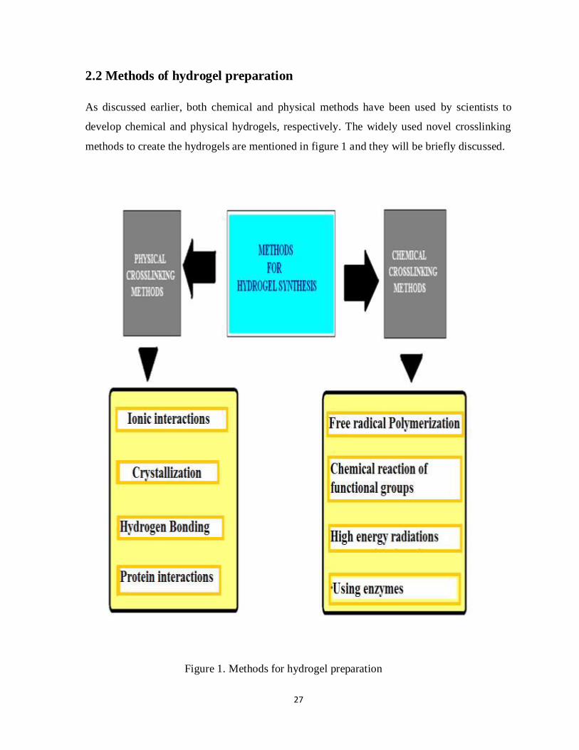
2.2 Methods of hydrogel preparation
As discussed earlier, both chemical and physical methods have been used by scientists to
develop chemical and physical hydrogels, respectively. The widely used novel crosslinking
methods to create the hydrogels are mentioned in figure 1 and they will be briefly discussed.
Figure 1. Methods for hydrogel preparation

28
2.2.1 Chemical Crosslinking methods
2.2.1.1 Crosslinking by radical polymerization
During polymerization process the monomer molecules are crosslinked chemically, resulting
in the formation of either linear chains or a three-dimensional network of polymer chains.
Radical polymerization is one of the commonly used methods to synthesize the hydrogels,
where low molecular weight monomers are crosslinked in the presence of crosslinking
agents. A variety of hydrogels can be designed by this procedure, for example, different
stimuli sensitive materials, hydrogels using water-soluble (synthetic, semi-synthetic and
natural) polymers.11
In an attempt to determine the optimum conditions for hydrogel synthesis by the free-radical
polymerization of sorbitan methacrylate (SMA), the hydrogel used in this study was well
polymerized under the following conditions: 50% (w/v) SMA as monomer, 1% (w/w), α, α’ -
azo-bis (isobutyro-nitrile) as thermal initiator, and 1% (w/w) ethylene glycol dimethacrylate
as cross-linking agent. Under these conditions, the moisture content of the polymerized SMA
hydrogel was higher than in the other conditions and also from poly (methyl methacrylate
[MMA]) hydrogels.60
Triblock copolymers prepared via consecutive atom transfer radical polymerizations using
monomers, N-isopropylacrylamide (NIPAAm), (2-dimethyl amino) ethyl methacrylate
(DMAEMA) and 2-hydroxyethyl methacrylate (HEMA), in the presence of ethylene glycol
di-2-bromoisobutyrate as initiator. The so-prepared hydrogels exhibited both temperature-
and pH-sensitive behavior.61
2.2.1.2 Crosslinking by chemical reaction of functional groups
The water solubility of the polymers is attributed to the presence of functional groups like -
OH, -COOH, -NH2, used to create hydrogels. The hydrogels are formed by the covalent
bonding between the polymer chains and functional groups, such as amine-carboxylic acid,
isocyanate-OH/NH2 or by Schiff base formation. Water-soluble polymers with hydroxyl
groups can be crosslinked with aldehydes e.g. crosslinking of poly (vinyl alcohol) can be
crosslinked using glutaraldehyde.

29
Crosslinking of water soluble polymers can also occur by the addition reactions, where the
hydrogels are formed using higher functional cross linking agents such as 1,6-
hexamethylene-diisocyanate, di-vinyl sulfone and many other reagents react with functional
groups of water-soluble polymers. Other frequently applied synthesis of hydrogels involves
condensation reactions between hydroxyl groups or amines with carboxylic acids or
derivatives to yield polyesters and polyamides, respectively. N, N-(3-dimethylaminopropyl)-
N-ethyl carbodiimide (EDC) is an efficient reagent to establish chemical crosslinking of
water-soluble polymers with amide bonds in the preparation of gelatin hydrogels.62, 63
2.2.1.3 Crosslinking by high-energy irradiation
Nevertheless, owing to unique advantages such as shorter reaction times, crosslinking under
mild conditions (room temperature and physiological pH), higher yields; limited generation
of by-products and relatively easy scale-up without detrimental effects, radiation-assisted
preparation of hydrogels have become an appealing synthetic tool. In addition, simultaneous
synthesis and sterilization of hydrogels are the unique advantages of radiation processing.64-69
The permeability and swelling characteristics of the formed gel are dependent upon the
amount of polymer and radiation intensity. Usually the crosslink density increases with
increasing polymer concentration and radiation dose.70
Particularly gamma radiations, electron beams as well as microwave radiations are used for
polymerization. Gamma radiation was used to crosslink the biodegradable hydrogels based
on an acryloylated poly-aspartamide.71 An environment sensitive bacterial cellulose and
acrylic acid composite was formed via electron beam. It exhibited higher swelling ability and
the degree of swelling increased as the pH of surrounding medium increased.19 Similarly
temperature-sensitive poly (N-isopropylacrylamide) PNIPAAm hydrogels were prepared by
microwave irradiation using Mars-5 microwave accelerator.72 However the domestic
microwave ovens are more convenient source of microwave radiation to create the chemical
crosslinking among a variety of monomers and polymers. A copolymer hydrogel of k-
carrageenan (kC) and acrylamide (AAm), has been synthesized in aqueous medium at pH 7
in the presence of the initiator potassium persulfate (KPS), by microwave irradiation using
LG make domestic microwave oven.73

30
2.2.1.4 Crosslinking using enzymes
An emerging and interesting approach for formation of hydrogels is based on enzyme-
catalyzed crosslinking reactions. The enzymes from various sources, such as microbial
Transglutaminase and mushroom tyrosinase provide a method for creating gels and may offer
interesting opportunities for in situ applications. The ability of these two enzymes to catalyze
the formation of gels from solutions of gelatin and chitosan was observed and compared.74
Similarly, hydrogels were synthesized from glutaminamide-functionalized poly (ethylene
glycol) (PEG) and poly (lysine-co-phenylalanine) using transglutaminase (TG) in the
presence of calcium ions as cofactors. The covalent crosslinking occurred by formation of an
amide linkage between the carboxamide groups of peptidyl glutamine residues and primary
amine groups of lysine residues.75
Enzyme-mediated redox chain initiation involving glucose oxidase (GOX) was employed in
a dip-coating technique to polymerize multiple, three-dimensional hydrogel layers using mild
aqueous conditions at ambient temperature and oxygen levels.76
Dextran hydrogels were formed in situ by enzymatic crosslinking of dextran-tyramine
conjugates and their mechanical; swelling and degradation properties were evaluated. These
results demonstrated that enzymatic crosslinking is an efficient way to obtain fast in situ
formation of hydrogels. These dextran-based hydrogels are promising for use as injectable
systems for biomedical applications including tissue engineering and protein delivery.77
2.2.2 Physical crosslinking methods
Most of the crosslinkers used for covalent crosslinking in hydrogel synthesis may induce
toxicity if found in free traces before administration. To overcome this problem purification
and verification step is needed. To avoid the use of crosslinking agents by physical
crosslinking techniques have been investigated for the designing of hydrogel networks.

31
2.2.2.1 Crosslinking by ionic interactions
Various polymers that can be crosslinked by ionic interactions hence form hydrogels using
this method. As covalent crosslinking requires multifunctional molecules as crosslinking
agents, the ionic crosslinking requires multivalent counter-ions as crosslinkers to link
polymeric chains. Chitosan based hydrogels were obtained by crosslinking of this chitosan
with glycerol-phosphate disodium salt.78
Alginate is a well-known example of a natural polymer that can be crosslinked by ionic
interactions. It is a polysaccharide that can be crosslinked by calcium ions at room
temperature and physiological pH. Alginate gels have been used in both drug delivery and
cell encapsulation applications in the beads form usually produced by dripping alginate
solution into a CaCl2 bath.79 For hydrogel preparation, the presence of ionic groups in
polymer is not compulsory for ionic crosslinking. For example, dextran, which lacks ionic
binding sites for cations, forms a hydrogel in the presence of potassium ions. However, this
dextran /potassium gel is unstable in water and therefore is less suitable for drug delivery
purposes.80
2.2.2.2 Crosslinking by crystallization
Apart from ionic interactions or hydrophobic interactions in physical cross-linking of
hydrophilic polymers in hydrogel formation, crystallites can act in physical cross-links in
block-copolymers and even in homopolymers. The dextran hydrogels were prepared by the
process of crystallization. Dextrans are soluble in water, but precipitation was observed in
concentrated aqueous solutions of low molecular weight dextran (dextran 6000). The kinetics
of the precipitation process showed that the rate of precipitation is accelerated by increase in
concentration of dextran solutions, stirring and the presence of salts. Depending on the
precipitation time, microspheres or gels were obtained. The precipitates were insoluble in
water at room temperature, but readily dissolved in boiling water. IR spectroscopy and
modulated differential scanning calorimetry (DSC) demonstrated that the precipitates were
crystalline.81
A novel hydrogel system in which crosslinking was established by stereocomplex formation
between lactic acid oligomers of opposite chirality has been developed. Poly L-Lactic acid

32
(PLLA) and Poly D-lactic acid (PDLA) are semi crystalline materials. Each stereoisomer has
a melting temperature of around 170 ºC. Interestingly, in blends of high molecular weight
PLLA and PDLA a phase of a higher melting point (around 230 ºC) was observed.82 This is
attributed to the formation of racemic crystallites, also called stereocomplexes and was first
described by Ikada et al.83
2.2.2.3 Crosslinking by hydrogen bonds
A spontaneous formation of hydrogel was observed by mixing of two water-soluble
phospholipids polymers, such as poly (2-methacryloyloxyethyl phosphorylcholine-co-
methacrylic acid) (PMA) and poly (2-methacryloyloxyethyl phosphorylcholine-co-n-butyl
methacrylate) (PMB), in aqueous medium at room temperature without any chemical
treatment. The gelation mechanism, effects of ions on gelation and dissolution behavior were
determined. The spectroscopic analysis and FT-IR analysis revealed that carboxyl groups in
methacrylic acid (MA) formed dimer when two polymer solutions were mixed, and the
results of the rheological study showed dissociation of carboxyl groups caused dissolution of
the hydrogel. The hydrogen bonds are only formed when carboxylic acid groups are
protonated. Thus, the gelation occurred due to the formation of dimers by hydrogen bonding
which acts as a physical cross-linking of polymer chains.84
Poly (vinyl alcohol) (PVA) hydrogels interacting with DNA mediated by hydrogen bonds
(PVA/DNA hydrogel) were developed using ultra-high pressure (UHP) technology. The goal
was to create a new method of gene delivery by controlled release of DNA.85
Poly (acrylic acid) and poly (methacrylic acid) forms complexes with poly(ethylene glycol).
These complexes are held together by hydrogen bonds between the oxygen of the
poly(ethylene glycol) and the carboxylic group of poly(meth)acrylic acid, whereas for
poly(methacrylic acid) hydrophobic interactions also play a role.86
Asymmetric bolaamphiphilic sugar-based crown ether hydrogel were synthesized and their
gelation ability with and without alkylammonium ions was investigated. Particularly, the
gelation was drastically enhanced by addition of alkylammonium ions, which could result in
stabilization due to the intermolecular hydrogen bonding and electrostatic interactions.87

33
2.2.2.4 Crosslinking by protein interactions
Another novel method in producing hydrogels involves the crosslinking by protein linkages.
It can be either by using genetically engineered proteins or crosslinking by antigen–antibody
interactions.
A hydrogel self-assembling method driven by the interaction between recombinant tax
interactive protein-1 (TIP1) with the PDZ domain [(PDZ is an acronym combining the first
letters of three proteins — post synaptic density protein (PSD95), Drosophila disc large
tumor suppressor (Dlg1), and zonula occludens-1 protein (zo-1)] in a molecule, which is
fused to each end of the triangular trimeric CutA protein (CutA-TIP1), and a PDZ domain-
recognizable peptide which is covalently bound to each terminus of four-armed
poly(ethylene glycol) (PDZ-peptide-PEG). Genetic manipulation based on molecular-
dynamic simulation generated a cell-adhesive RGD (Rat Genome Database) tripeptidyl
sequence in the CutA loop region [CutA(RGD)-TIP1]. In this way, an approach was
developed for in situ hydrogel formation enabling cell entrapment via biospecific interaction
between protein and peptide at physiological pH and temperature.88
An antigen sensitive hydrogel was prepared by Miyata et al. in which an antigen (rabbit IgG)
was grafted to chemically crosslinked polyacrylamide in the presence of antibody as an
additional crosslinker. The hydrogel had poor swelling characteristics in the presence of free
antigen due to the replacement of polymer-bound antigen, resulting in the release of the
antibodies and thereby decreasing crosslink density.89
2.3 Characterization of hydrogels
Hydrogels are usually characterized for their morphology, the crosslink density and the
structural integrity (porosity, pore size and its distribution), the ultimate capacity to absorb
liquids (swelling property) as well as their elasticity. Various techniques have been
investigated to investigate the crosslinking interactions among the polymers90 as could be
seen in figure 2.

34
Figure 2. Methods of Characterization of hydrogels

35
2.3.1 Scanning electron micrography (SEM)
SEM photomicrographs of the polymers are taken in order to investigate and compare their
surface morphology. The texture is analyzed by SEM to ensure that hydrogels, such as based
on starch, retain their granular structures.91
The morphology of the poly (methacrylic acid)/poly (N-isopropylacrylamide)
interpenetrating polymeric networks (IPN) was studied with both conventional SEM and
cryogenic SEM experiments. Cryogenic SEM was used as a new approach to visualize the
IPN morphological behavior in its swollen state. The pH and temperature influence on the
IPN morphology was studied. The results showed that a decrease in pH and increase in
temperature resulted in a drastic decrease in the pore size of the IPNs.92
2.3.2 X-ray diffraction (XRD)
This is another technique used to describe the retention or deformation of the crystalline
structure of polymers during the processing pressurization process. Characterization and drug
delivery behaviour of starch-based hydrogels prepared via isostatic ultrahigh pressure
(IUHP). Szepes et al.91 characterized and investigated the drug delivery behaviour of starch-
based hydrogels prepared by ultrahigh pressure. The changes in structure and morphology of
potato and maize starches were determined by X-ray diffraction examinations of the samples
using D4 Endeavour diffractometer. The crystalline structure of maize starch was sensitive to
UHP, so it was changed, while potato starch pressurized in aqueous medium remained stable
and retained its original X-ray pattern.
The X-ray diffraction patterns of a copolymer hydrogel of kC-graft-PAAm, k-carrageenan
(kC) and acrylamide (AAm) were observed. A considerable modification was noticed in the
polysaccharide, leading to a change in molecular association in the formed of hydrogel when
compared with kC and AAm.70

36
2.3.3 Magnetic Resonance Imaging (MRI)
MRI uses a powerful magnetic field, radio frequency pulses and a computer to produce
detailed pictures at cellular and molecular level. Proton Magnetic Resonance Imaging (MRI)
has been used to study the physical changes of hydroxypropyl methylcellulose (HPMC)
hydrogels due to microwave irradiation of the polymer. The proton one-dimensional images,
derived from relaxation, spin density and diffusion-weighted spin-echo experiments, provide
insights on the dynamics of water and the motional state of the polymer inside the hydrogels.
The obtained results indicated that the microwave irradiation causes the breaking of the
HPMC polymer–hydrogen bonding network in HPMC powder which influences the
dynamics of water and polymer chains within its hydrogels.93
Proton Magnetic Resonance (PMR) imaging in a thermo reversible gel using Bruker MSL-
300 FT-NMR spectrometer measured volume-phase-transition. This was demonstrated in the
lower critical solution temperature ( LCST) polymer poly (N- isopropylacrylamide) which is
swollen in water. The swelling ratios in the axial and radial directions were the same after the
thermal collapse.94
2.3.4 Fourier Transform Infrared Spectroscopy (FTIR)
Fourier transform infrared (FTIR) spectroscopy is an established tool for the structural
characterization; any change in the morphology of hydrogels changes their IR absorption
spectra. The structure and properties of the superabsorbent hydrogels synthesized by graft
copolymerization of acrylic acid (AA)/acrylamide (AM)/2-acrylamido-2-methyl-1
propanesulfonic acid (AMPS) onto sodium carboxymethylcellulose (CMC) and
montmorillonite (MMT) were evaluated where the intermolecular interaction and
morphological change of the hydrogels were characterized by Fourier Transform Infrared
(FTIR) spectroscope. It was shown that superabsorbent hydrogel product comprises a
crosslink structure of MMT and CMC with side chains that carry carboxylate, carboxamide
and sulfate.95
The fractions of dissociation of acrylic acid (AAc) units within hydrogel in response to
changes in pH and ionic strength of external aqueous solution were determined by FTIR-
ATR spectroscopy. The swelling response of hydrogels to the changes in external pH and

37
ionic strength was governed mainly by the ionic osmotic pressure due to the accumulation of
diffusible ions within hydrogels.96.
2.3.5 Thermal Analysis
DTA and DSC measure, respectively, the temperature difference and the heat flow difference
between a sample and a reference material (subjected to the same temperature variation in a
controlled atmosphere). DTA detects any change in all categories of materials, whereas DSC
determines the temperature and heat of transformation.
Differential Scanning Calorimetry (DSC) was used to monitor the reaction scheme of arylic
based superabsorbing polymers. The heat effects were studied during the polymer synthesis
in DSC pan as a micro-scale reactor. Two distinct observations, i.e. inhibition period (IP) and
onset of gel formation were recorded during polymerization. By this the effect of reaction
temperature and initiator concentration was assessed in the synthesis of superabsorbent
hydrogels.97
The DSC technique allows studying the drug release and diffusion from a polymeric device
to the site mimicking a biological membrane. The drug release from inulin-based hydrogel to
a biomembrane model was investigated at pH 4.0 and 7.4 by using DSC that appears to be a
suitable technique to follow the transfer kinetics of a drug from a controlled release system to
a biomembrane model.98
2.3.6 Swelling behavior
The hydrogels were allowed to immerse in aqueous medium or medium of specific pH to
know the swellability of these polymeric networks. These polymers showed increase in
dimensions related to swelling. A gelatin based pharmaceutical hydrogels, gelled in minutes
using oxidized konjac glucomannan (DAK) as a macromolecular cross-linker, and was
estimated for equilibrium swelling ratio. From the photographs of the hydrogel in both dry
and swollen state, it was determined that the hydrogels remain in the cylindrical form after
swelling. However marked volume differences were noticed where the diameter of swollen
hydrogel was about 4.0 cm, while the diameter of the dry state hydrogel was only 1.5 cm.99

38
The swelling and deswelling kinetics of poly (N-isopropylacrylamide) (PNIPAAm)
hydrogels separately synthesized by means of microwave irradiation and normal water-bath
heating. The swelling and deswelling kinetic curves of the PNIPAAm hydrogels were
measured in water below and above the lower critical solution temperature (LCST), and their
swelling and deswelling kinetic parameters were estimated. Results showed that in
comparison to the hydrogel synthesized by the conventional method, the hydrogel
synthesized by microwave irradiation had larger swelling and deswelling rate constants as
well as lower swelling/deswelling activation energy due to its higher surface area and larger
pore sizes, and thus it had faster response behavior.72
The swelling responses of pH sensitive psyllium and polyacrylamide based hydrogels were
measured in aqueous medium by gravimetric method. The equilibrium percent swelling (Ps)
of the polymeric network were calculated as follows:
Ps = × 100 (1)
Where Ws and Wd are weights of swollen polymers and dried polymers respectively.100,101
2.3.7 Gel Fraction
Gel fraction is mass fraction of the network material resulting from a network forming
polymerization or crosslinking process. The gel fraction assays are performed to determine
the level of crosslinking, greater the gel fraction, higher is the crosslinking density. If all
polymers is in gel fraction (no soluble fraction) and it is completely crosslinked. The degree
of cross linking is usually dependent on the molecular weight of the polymers. The polymers
with low molecular weight poorly form gel than one with higher molecular weight. Gel
fraction was determined in Poly (vinyl alcohol) hydrogels, where Cross-linking does not
occur entirely and certain PVA macromolecules remain in the network uncross-linked (sol).
The gel fraction G in hydrogels was estimated by the formula:
G (%) = × 100 (2)

39
Where, W1 is the weight of the dried cross-linked sample with sol and Wd is the weight of the
dried sample after the removing of sol by extraction in water.102-104
2.3.8 Porosity
During swelling, the pores located inside the network are rapidly filled with the solvent; atthe
same time, the polymer region takes up the solvent from the environment, whose extent
depends on the attractive force between the solvent molecules and the polymer segments.105
Solvent replacement method was used for porosity measurement. Weighed dried discs were
immersed in absolute ethanol overnight and weighed after excess ethanol on the surface was
blotted using blotting paper. Porosity was calculated using the following equation
Porosity (%) = × 100 (3)
where M1 and M2 are the weights of hydrogels before and after immersion in absolute
ethanol, respectively. ρis the density of absolute ethanol and V the volume of gel.106
The decrease in density can be attributed to the increase in porosity. The bulk density of
dried hydrogels can be determined using picnometer. Certain substances have influence on
the density and porosity of the hydrogels. Mahdavinia et al.107 studied the effect of CaCO3
content on the density of the hydrogels. Using the high content of CaCO3 to synthesize
hydrogel causes the high number of produced pores, and subsequently the density will be
decreased.
2.3.9 Rheology
Hydrogels were evaluated for viscosity under constant temperature of usually 4 °C by using
Cone Plate type viscometer when a small amount of material was available. For most of the
experiments a flat-plate measuring geometry (acrylic, 4 cm diameter; gap 1 mm) was used.
The mechanical strength of the swollen sample of acrylic-based Superabsorbent polymer
(SAP) hydrogel was measured by a rheological method. The characterization was conducted

40
by a controlled strain rheometer at 25ºC. Dependency of the rheological properties of the
sample on strain and frequency was investigated.108
The rheology measurements yield information of the nature of the water–polymer interaction.
The phase transition behavior of water within the chitosan/polyacrylate hydrogels was
investigated by means of oscillatory shear rheology. Changes in structure were determined
by comparing differences in the rheological measurements at temperatures above and below
freezing.109
2.3.10 In-vitro Release Studies
Since hydrogels are the swollen polymeric networks, interior of which is occupied by drug
molecules, therefore, release studies are carried out to understand the mechanism of release
over a period of application.110, 111 In-vitro drug release studies can be performed by using
diffusion cell method. The release rate of the timolol maleate from the stimuli sensitive
hydrogels was determined by the diffusion process. The samples withdrawn were analysed
spectrophotometrically at 294 nm for the timolol maleate using Shimazdu Double beam UV-
Visible spectrophotometer.112 First-order model, Higuchi square root time model, Hixson–
Crowell model, Weibull distribution, Korsmeyer–Peppas model, etc, are used to evaluate the
dissolution profiles of the samples.113,114
2.3.11 In-vivo Evaluation
In-vivo studies are conducted on prepared optimized hydrogel formulation (test) and on
marketed formulation (standard). A wide range of acute, sub-chronic and chronic toxicity
studies are conducted, using various routes of administrations in different species. The
animal treatment should be complied with the Principles of Laboratory Animal Care
formulated by the National Society for Medical Research. The biocompatibility and
degradation of the Dacron matrices impregnated with gelatin- chondroitin sulphate (ChS)
gels was studied after implantation in subcutaneous pockets in rats. Chemically cross-linked
gelatin-ChS gels showed a mild tissue reaction, and almost complete degradation within 18
weeks of implantation. Before in vivo implantation, the Dacron samples, as such or
impregnated with gelatin or gelatin and ChS, were sterilised by γ irradiation.115

41
In vivo evaluation on the long-term is now necessary to confirm their biocompatibility and
establish their life-time. Moreover, the inter-subject variation is more significant in the in
vivo study than in vitro skin permeation experiments. It was confirmed by study designed to
investigate the in vitro and in vivo skin absorption of capsaicin and nonivamide from
hydrogels.109
2.4 Hydrogels for pharmaceutical and biomedical applications
Hydrogels are promising candidates for controlled release devices, bioadhesive devices, or
targetable devices of therapeutic agents. The excellent hydrophilic properties, high swelling
ratio and biocompatibility, have led them widely applicable in biomedical / pharmaceutical
area as antibacterial materials, tissue engineering, biosensors and sorbents for the removal of
heavy metals.116-120 These water-swollen, crosslinked biomedical materials are efficient
carriers for the development of novel pharmaceutical formulations for the delivery of drugs
(peptides and proteins), as targeting agents for site specific delivery and as components for
the preparation of protein or enzyme conjugates. They have gained considerable existence in
drug delivery through parenteral, ocular, rectal, vaginal, dermal and nasal routes.121, 122
2.4.1 Transdermal drug delivery
Water-based polymeric gels offer several advantages over traditional oleaginous bases in
terms of ease of application, cosmetic acceptability (colorless and water-washable) and
desirable drug release characteristics.123 Drug delivery to the skin has been traditionally
conducted for topical use of dermatological drugs to treat skin diseases, or for disinfection of
the skin itself. In recent years, a transdermal route has been considered as a possible site for
the systemic delivery of drugs. The possible benefits of transdermal drug delivery include
that drugs can be delivered for a long duration at a constant rate, that drug delivery can be
easily interrupted on demand by simply removing the devices, and that drugs can bypass
hepatic first-pass metabolism. Furthermore, because of their high water content, swollen
hydrogels can provide a better feeling for the skin in comparison to conventional ointments
and patches. Versatile hydrogel-based devices for transdermal delivery have been proposed
so far.122

42
The hydrogel membranes mainly composed of three kinds of latex particles within
carboxymethyl cellulose (CMC) matrix were prepared for the purpose of transdermal drug
release. These microgels were poly (acrylic acid-cosodium acrylate), poly (acrylic acid-co-2-
ethylhexyl acrylate) and poly(N-isopropyl acrylamide), which were developed to give the
membrane a higher swelling ratio, a better adhesive property and a thermo-responsive
behavior, respectively. Gel particles of PNIPAAm or its copolymers underwent deswelling
above lower critical solution temperature (LCST), creating more space for the membrane to
absorb more water and in turn increasing the swelling ratio. Also, they simultaneously
expelled caffeine to the highly swollen CMC matrix, thus increasing the caffeine-release
rate.124
A transdermal system for delivering selegiline using a hydrogel-based drug reservoir and a
rate-controlling membrane (Solupor polyethylene membranes) was designed. Both the R-
and S-forms of selegiline were examined in this study to elucidate the stereoselectivity of
skin to selegiline. The experimental results suggested that Solupor can be used as a substrate
to control the permeation of selegiline. The amount of drug permeating across the skin can be
reduced by the membranes.125
Hydrogels are used for local drug delivery in the control of wound healing. Nanocomposite
hydrogel wound dressing was prepared using combination of polyvinyl alcohol hydogel and
organoclay, i.e. Na-montmorillonite. The results showed that the nanocomposite hydrogels
could meet the essential requirements for the reasonable wound dressing with some desirable
characteristics such as relatively good swelling, appreciated vapour transmission rate,
excellent barrier against microbe penetration and mechanical properties.126
2.4.2 Orally administered hydrogels
The orally administered hydrogels are used for peroral and oral drug delivery and drug
delivery. It leads the drug to mouth (oral cavity), stomach, small intestine, or colon. Drug
delivery to the oral cavity can have versatile applications in local treatment of diseases of the
mouth, such as periodontal disease, stomatitis, fungal and viral infections, and oral cavity
cancers. Long-term adhesion of the drug containing hydrogel against copious salivary flow,
which bathes the oral cavity mucosa, is required to achieve this local drug delivery. A

43
bioadhesive tablet or hydrogel-based ointment can also be utilized for the topical treatment of
certain diseases in the oral cavity. Chitosan-based hydrogels have been recognized as
excellent candidates for oral delivery due to their mucoadhesive properties. Chitosan
hydrogels have been developed for the local release of a number of other drugs in the oral
cavity. In addition to the released drugs, the chitosan polymer itself has shown antifungal
activity. For instance, chitosan hydrogels and films were able to limit adhesion of the
common pathogen Candida albicans to human buccal cells.127
The Chitosan based matrix has been used as a reliable colonic controlled-release system for
the release of 5-aminosalicylic acid (5-ASA) or diclofenac sodium (DS) and introduced into
enteric-coated capsules for controlled release to the colon.128 Alginate-N, O-carboxymethyl
chitosan hydrogels with calcium for oral delivery of protein drugs to different regions of the
intestinal tract e.g., for duodenal targeting, small intestine targeting, or colon targeting.129
Colon specific hydrogels of polysaccharides have been specifically designed because of
presence of high concentration of polysaccharidase enzymes in the colon region of GI
(gastrointestinal) tract. Drugs loaded in such hydrogels showed tissue specificity and changed
in the pH or enzymatic actions that cause liberation of drug.130
2.4.3 Ocular delivery
In comparison to other ophthalmic formulations such as suspensions and eye ointments, the
hydrogels may offer better feeling, with less gritty sensation to patients. In particular, in-situ-
forming hydrogels are attractive as an ocular drug delivery system because of their facility in
dosing as a liquid and their long-term retention property as a gel after dosing.
Fast forming hydrogels prepared by crosslinking a poly (ethylene glycol) (PEG)-based
copolymer containing multiple thiol (-SH) groups were evaluated for the controlled ocular
delivery of pilocarpine and subsequent pupillary constriction. A strong correlation between
pilocarpine release and pupillary response was observed. In conclusion, the current studies
demonstrate that in situ forming PEG hydrogels possess the viscoelastic, retention and
sustained delivery properties required for an efficient ocular drug delivery system.131

44
In situ forming poly (ethylene glycol) (PEG)-based doxycycline hydrogels were developed
and evaluated for their wound healing efficacy in rabbit corneas in organ culture. The
doxycycline-PEG hydrogels accelerated corneal wound healing after vesicant injury offering
a therapeutic option for ocular mustard injuries. Histology and immunofluorescence studies
showed a significant reduction of matrix metalloproteinase-9 (MMP-9) and improved the
healing of vesicant-exposed corneas by doxycycline hydrogels compared to a similar dose of
doxycycline delivered in phosphate buffered saline (PBS, pH 7.4).132
2.4.4 Subcutaneous delivery
Biocompatibility is a prerequisite requirement to overcome undesirable body responses, such
as inflammation, carcinogenicity and immunogenecity. Due to their high water content,
hydrogels are generally considered as biocompatible materials. They also cause minimal
mechanical irritation upon in-vivo implantation, due to their soft and elastic properties and
possess broad acceptability for individual drugs with different hydrophilicities and molecular
sizes; and unique possibilities (crosslinking density and swelling) to manipulate the release of
incorporated drugs.133,134
Some of these may offer an advantage for the delivery of certain delicate drugs, such as
peptides and proteins. The high-strength injectable Pluronic hydrogels were synthesized by
enzyme-mediated cross-linking for controlled drug delivery. They showed controlled
erosion, bio-adhesion, thermo-sensitivity, and injectable properties, ideal along with in situ
depot formation in the tissue. They can possibly be utilized for sustained delivery of
therapeutic proteins, genes, and chemical drugs.135 The poly (β-amino ester) (PAE) as a duo -
functional group for synthesis of the novel sensitive injectable hydrogel is used for controlled
drug/protein delivery. Furthermore, the cationic nature of PAE is utilized to make the ionic
complexes with anionic biomolecules loaded into the hydrogel such as insulin.136
The hydrogels could target drugs to specific body sites and control the release of drugs for
prolonged periods of time. For this they can be used as promising drug delivery systems for
treating various types of cancers. Injectable chitosan hydrogels have been synthesized for
localised cancer therapy using paclitaxel as a model drug.137

45
2.4.5 Rectal and vaginal delivery
The bioadhesive characteristics of hydrogels make them a valuable way to overcome the
shortcomings of conventional suppositories, such as uncontrolled drug release and
insufficient retention in the rectum. Once in the rectal cavity, the development of
mucoadhesiveness would help to immobilize the hydrogel for a required period of time thus
prolonging the drug release for local or systemic use.
A thermosensitive hydrogels was formulated which was based on poloxamer 407, a
thermosensitive polymer, and hydroxypropylmethylcellulose (HPMC), a bioadhesive
polymer, intended for the rectal delivery of quinine in children. In consideration to the
therapeutic applications, the formulations should be in the liquid state at ambient temperature
(generally 25 ºC), should gel at body temperature (37 ºC) and should be adhesive on the
rectal mucous membrane.138 The temperature sensitive and mucoadhesive pluronic-based
hydrogels were designed, which were capable of retaining their rheological and
mucoadhesive properties after dilution with vaginal fluids. It enables them as promising
formulations for the vaginal administration of drugs.139
2.4.6 Hydrogels for tissue engineering
When designing suitable biomaterials for tissue-engineering applications, biological and
chemical parameters are frequently taken into account. The hydrogels, due to their swelling
properties, can be made to resemble the physical characteristics of soft tissues. Hydrogel
materials also generally exhibit high permeability and good biocompatibility making these
materials attractive for use in cell encapsulation and tissue engineering applications. Due to
the biocompatibility, permeability and physical characteristics, hydrogels are good
candidates as biomaterials for use in many medical applications, including tissue
engineering. Hydrogels may be useful for manipulation of tissue function or for tissue
regeneration or replacement. The use of photopolymerization in the preparation of hydrogels
is advantageous in comparison with conventional crosslinking methods because liquid
hydrogel precursors can be delivered and crosslinked to form hydrogels in situ in a minimally
invasive manner such as by injection. This process also gives one spatial and temporal
control over the conversion of a liquid to a gel, so that complex shapes can be fabricated.140

46
Cellulose is a major extracellular matrix component of plant cells, composed of poly (1,4´ -
anhydro-b-β-D-glucopyranose). Being an abundant naturally occurring polymer, it is
renewable, as well as biodegradable and derivatizable for a large number of biomedical and
pharmaceutical applications. Cellulose based materials including bacterial cellulose are of
increasing interest in tissue engineering. They have been utilized in wound healing,141
artificial kidney, artificial blood vessels, bone, cartilage and cardiovascular tissue
engineering etc.142-146
2.5 DRUG
Captopril, (2S)-1-[(2S)-2-Methyl-3-sulphanylpropanoyl] pyrrolidine-2-carboxylic acid an
angiotensin-converting enzyme (ACE) inhibitor was chosen as a drug to be loaded into the
hydrogel formulation. Its chemical structure is shown in the figure.
Figure 3.Chemical structure of captopril
(C9H15NO3S, MW = 217.3)
It is widely used as an antihypertensive drug and for the management of congestive heart
failure because of its effectiveness and safety. It has low toxicity and commonly prescribed
patients suffering from chronic illness. Therefore, a long term therapy is needed for their
treatment. Commercially, it is available as immediate release tablets of 12.5- 50 mg. It is
available in market with following brand names.

47
Table 1. Commercially Available brands of Captopril
Brand names Manufacturers
Capoten Glaxosmithkline
Captil Werrick Pharmaceuticals
Capoten Bristol-Myers Squibb
Captolane Sanofi-Aventis
Captoril Benson
Acepril BMS (United Kingdom)
Acepress BMS (Italy)
Cesplon Esteve (Spain)
Dilabar Qualigen (Spain)
Captopril has been a drug of choice in hypertension management and also effectively used
for congestive heart failure. There are almost 25% people among the world’s population
suffering from hydpertension.147,148 Captopril is successfully used for hypertensive patients
but its antihypertensive effect remains for a period of 6–8 hours. For maintaining minimum
effective concentration three to four CAP administrations are required. For this reason,
usually 25-100 mg drug is needed for successful medication of hypertension and heart
disorders. In some conditions, for therapy of heart failure an initial dose of 6.25 mg is needed
three times a day. Therefore, development of a controlled release drug delivery system for
CAP would be beneficial; hence once daily formulation will be needed to achieve significant
effects, providing an optimum drug concentration, minimizing drug fluctuations and adverse
drug effects.
Captopril is a suitable candidate for controlled release drug delivery systems due to its short
elimination half-life of less than 2 hours.149 However, it is not easy to develop an oral
controlled release formulation of captopril, due to its in-vitro and in-vivo stability concerns.
The drug being freely soluble in water and physiological aqueous solutions may undergoes
the problems like dose dumping as well as burst phenomena from controlled/sustained
release drug delivery systems.150 Moreover, its bioavailability is also affected in presence of
food and higher intestinal pH.

48
Captopril has been formulated in both rapid and slow release drug delivery systems either for
its fast availability into blood circulation to attain peak plasma concentration in lesser time or
to be available for longer period of time. For its instant availability, sublingual tablets were
prepared, which was an effective way for lowering the arterial blood pressures in emergency
situations. Hence, there were rapidly available therapeutic levels of drug as compared to
other orally administered captopril tablets.151
In treating hypertension and heart failure long term management is required by
antihypertensive drugs. In this context, there is a need to develop controlled release dosage
form, where dissolution rate should be controlled. For this reason, various researchers have
prepared the controlled release formulations such as floating matrix tablets, gastroretentive
systems, microcapsules, and nanoparticles. Martinez et al prepared the in-vitro sustained
release floating tablets of captopril by using metalose SH 4000 SR/ Sodium bicarbonate. The
floating tablets were prepared to prolong the gastric residential time and to avoid the
degradation by higher pH of intestine.152 Polymeric matrices were formulated to control the
release rate of captopril using various polymers such as, Hydroxypropyl methyl cellulose
(HPMC), Sodium carboxy methyl cellulose (NaCMC) and ethyl cellulose (EC). This work
was concerned to study the effect of polymers and surfactants on release of captopril.153
In another work, the captopril was entrapped in albumin based biodegradable microparticles
formulated by Dandagi et al.154 by using emulsification- heat stabilization technique. Its in-
vitro analysis had shown the drug release upto 24 hours and in-vivo evaluation proved its
successful targeting to lungs, liver, kidneys and spleen. Similarly Cellulose propionate (CP)
microparticles were prepared by solvent evaporation technique for controlled release of
captopril. They were proven as useful products in gradually decreasing systolic blood
pressure throughout 24 hour time period in comparison to reference solution.155 Captopril-
loaded microspheres have also been prepared by solvent evaporation method, using Methocel
and Eudragit RS as release-controlling factors and to evaluate captopril release. Microspheres
and micropellets of captopril have also been prepared with different polymers (chitosan,
ethyl cellulose, sodium alginate and Hydroxy propyl methyl cellulose) through the
techniques of microencapsulation. Polyacrylamide-co-methylcellulose (PAAMA-co-MC)
hydrogels loaded with captopril were prepared and evaluated for their controlled release

49
characteristics. The sustained releases of captopril from cyclodextrin nanoparticles have also
been investigated.156
As already mentioned that the hydrogels are not only overcomes the shortcomings of
conventional dosage forms but also have advantages over other controlled release
formulations like microparticles and matrix tablets. Because of their cross-linked polymeric
network they have ability to entrap drugs and protect them from unfavorable physiological
environments. Captopril loaded into hydrogel formulations may remain stable and released
for longer periods of time.
2.5.1 Physical properties
Captopril is a white or almost white crystalline powder. These white crystals melt at about
88°C, which recodifies and melt again at 105°C or 106°C. It is freely soluble in water, in
methylene chloride and in methanol and also dissolves in dilute solutionsof alkali
hydroxides. It has a characteristic sulfide-like odour.157,158 Captopril is not hygroscopic under
ordinary conditions. Above 40°C, captopril shows extraordinarily high water solubility.159
2.5.2 Captopril Stability
In solution, this product undergoes an oxygen facilitated, first order, and free radical
oxidation at its thiol to yield captopril disulfide. Hydrolysis at the amide linkage occurs only
under forcing conditions. Oxidation is delayed using lower pH, chelating agents, higher
concentration, degassing, minimizing headspace, and incorporation of antioxidants. No
degradation is seen in methanol (40μg/ml) for up to 2 weeks at 5 °C. It shows maximum
stability below pH 4; however in the absence of oxygen it remains stable at relatively higher
pH values. 160 Captopril contains a sulphydryl group that results in its self-dimerization, rapid
formation of disulfide conjugates with endogenous thiol-containing compounds (cysteine,
glutathione) and plasma protein binding. Therefore, during in-vivo evaluation, the detection
or measurement of free captopril concentration needs molecule derivatization or an addition
of chemical stabilizer in biological or in vitro samples to prevent captopril disulphide
formation. Various Flouresence or UV active agent such as N-(1-pyrenyl) maleimide (NPM)
and p-bromophenacyl bromide (p-BPB), have been used as chemical stabilizers. The
formation of Captopril disulfide can be controlled by lowering the pH below 4, adding

50
chelating agents (EDTA) or antioxidants. Dithiothreitol (DTT) added to the plasma samples
successively reconstitutes captopril converted to disulfide, by increasing free thiol content
from human serum albumin.
2.5.3 Pharmacokinetics
Captopril, after oral administration, is rapidly absorbed from stomach and small intestine.
Approximately 75% of orally administered captopril is absorbed in fasting conditions. After
oral ingestion of a single dose the maximum blood level and therapeutic effect is observed
after 45–90 min. By presence of food in stomach Bioavailability is reduced to 25-30%,
therefore the drug should be administered one hour before meal. When enters into blood,
nearly 25% of captopril is binds to plasma protein. Captopril is partly undergoes metabolism
and about 95 % of the absorbed drug is excreted in urine, where 40-50 % is unchanged and
the remaining is captopril disulfide.161
2.5.4 Clinical Uses
Therapeutic benefits of Captopril are due to its vasodilatory effects as well as inhibition of
some renal function activities. The following are its main uses in various clinical conditions:
i) A widely acceptable and drug of choice for treating Hypertension.
ii) It has been approved by FDA for the treatment of cardiac disorders like
congestive heart failure, left ventricular dysfunction and post-myocardial
infarction.
iii) Captopril has an additional advantage to preserve the function of kidneys during
diabetic nephropathy. The effect of captopril on creatinine clearance, protein-urea
and metabolic controlled in diabetic neuropathy were determined. In addition of
controlling blood pressure alone, captopril provides protection against
deterioration of renal function in patients with insulin dependent diabetic
nephropathy.162
iv) Captopril also possesses mood elevating characteristics and has shown in some
patients. This is indicative of its antidepressant effect. However, it has not been
reported in formal clinical trials.163

51
v) Additionally, this compound has also been investigated to possess cytotoxic
activity for use in the treatment of certain types of cancer such as in treating lung
tumor. The impact of this drug on cell proliferation, apoptosis, and angiogenesis
has been evaluated.164
2.5.5 Adverse drug reaction (ADR)
1) The adverse drug reaction (ADR) profile of all ACE inhibitors is almost similar to
one another. Captopril like other ACE inhibitors commonly cause cough, which is
attributed to increase in bradykinen plasma level.165
2) Unlike most of the other ACE, captopril usually causes rash.
3) Due to oral administration of this drug, taste alterations have been noticed in many
cases. It may impart a characteristic metallic taste and in some situations it leads to
taste loss. This effect is associated with sulfhydryl group present in Captopril.166
4) Other adverse effects of captopril include proteinuria, angioedema, hyperkalemia,
tetragenicity, postural hypertension, agranulocytosis, leukopenia and in some cases it
may be responsible to acute renal failure.167
2.6 Excipients and Formulations
In this research work, two polymers and two monomers were used which are given as
following:
The polymers used were:
i. Hydroxypropyl methylcellulose (HPMC) and
ii. Polyvinyl Alcohal (PVA),
The monomers were:
i. Acrylic acid and
ii. 2-acrylamido-2-methylpropane-sulfonic acid (AMPS).
They were cross-linked in different combinations and proportions for preparing three types of
hydrogel formulations.
I. Hydroxypropylmethylcellulose-g-polyvinyl alcohol-co-poly(acrylic acid)

52
II. Polyvinyl alcohol-co-poly (\2-acrylamido-2-methylpropane-sulfonic acid)
III. Hydroxypropylmethylcellulose-g-poly (Acrylic acid-co-2-acrylamido-2-
methylpropane-sulfonic acid)
The method used for developing the above polymeric network was free radical
polymerization, using potassium persulfate (KPS) as initiator and N, N-Methylene-bis-
acrylamide was used as crosslinking agent in a hydrothermal water bath. The hydrogel
formulations were also synthesized under influence of microwave radiations. A pH- sensitive
hydrogel semi-IPN hydrogel formulation was prepared by crosslinking of HPMC with PVA
and acrylic acid. It was prepared to protect the drug from exposure to unfavourable
physiological conditions and enables drug release for longer periods of time. The other
formulation was a gastroretentive hydrogel comprising of PVA and AMPS to avoid any
instability of captopril at higher intestinal pH. Moreover, a superabsorbent hydrogel was
synthesized by using HPMC, acrylic acid and AMPS, to release the drug throughout its
passage from gastrointestinal tract. Hence, the drug would be available at both lower and
higher pH.
2.6.1 Polymers and monomers
Polymers are macromolecules with molecular weights, consisting of repeated units of
monomers. Monomers are polymerized by different techniques and results in formation of
polymers. A large number of polymers of different grades have been used and are being used
in the development of various pharmaceutical formulations, such as coated tablets, matrix
tablets, microcapsules and hydrogels. They can be natural polymer (cellulose, carrageenan,
collagen, guar gum, alginate etc) synthetic polymers (polyvinyl alcohal, polyvinyl
pyrollidone, polyethylene glycol, polyethylene oxide, polymethacrylate, polyacrylic acid etc)
as well as semisynthetic polymers (Hydroxypropyl methylcellose, Carboxymethylcellulose,
methyl cellulose, chitosan etc). Usually, the semi-synthetic polymers are derived from
natural sources. The polymers derived from natural origins are more biocompatible with
physiological envoirnment. Cellulose and its derivatives are widely accepted among research
groups because of their eco-friendly properties. Cellulose can be degraded by several
microorganisms (bacteria and fungi) in air, soil and water.168

53
Cellulose-based polymeric networks can be synthesized either by using chemical agents or
high energy radiations such as electron beam, gamma radiations or even microwave
radiations. The selection of chemical agents (Cross-linkers and initiators) depends upon the
characteristics of cellulose derivatives such as aldehydes, urea derivatives, multifunctional
carboxylic acids and epichlorhydrin. Usually the crosslinking occurs in water or aqueous
solutions but in some cases it also occurs in dry state such as crosslinking of cellulose by
polycarboxylic acids via condensation polymerization without water and requires reasonably
higher tempratures.169,170 In comparison to crosslinking reactions involving chemical agents,
use of high energy radiations have attracted the attention of researchers as it is easily
controlled and involves lesser amounts of chemical agents. Radiation induced crosslinking
has an additional advantage in biomedical research fields as they sterilize the product. In this
way, crosslinking and sterilization occur simultaneously; therefore, use of radiations is
beneficial with respect to health and environmental safety. High energy radiations cause
scission of polymer chains and many cellulose derivatives can be crosslinked under influence
of mild intensity of radiation. It can take place in both solid form as well as aqueous solution
of polymers.171-174 The crosslinking is dependent upon amount of polymer and radiation dose
that can either be intensity or exposure time. The microwave heating of many polymers has
influence on their properties such as cellulose derivative; Hydroxypropyl methylcellose
(HPMC) used in food production makes a question about effect of radiation on polymer.
2.6.1.1 Hydroxypropyl methylcellose (HPMC)
Hydroxypropyl-methylcellose (HPMC) or Hypromellose is a white or slightly off-white
tasteless, odourless powder. It is soluble in water and when dissolved, it forms a viscous
colloidal solution.175-177 It is semisynthetic, viscoelastic and inert non-ionic cellulose ether. In
solution, it does not react with salts or metals and there is no intake of ionic charges. Hence,
it does not exhibit any sort of reaction during and after preparation of Pharmaceutical
preparations. One of its other significant characteristic is its stability in both acids and
alkaline medium, even for long term storage. The aqueous solutions of HPMC are resistant to
enzymatic degradation and the formed products have better stability than material produced
by starch, dextrin and other natural polymers.178,179

54
HPMC is one of the most successfully used polymers in preparing different oral drug
delivery systems.180,181 In pharmaceutical industry, it is used in manufacturing of both coated
and controlled release matrix tablets. Depending on its grade, it can be either used as binder
or as a component with ability to prolong the release of medicinal agent in digestive tract.
Moreover, it is commonly used as lubricant in ophthalmic preparations and has numerous
applications in cosmetic industry and in many other commercial products. It also serves as
structural and storage material in plants.182-185
HPMC comprise of polymeric backbone of cellulose and during its synthesis cellulose fibers
are heated with a caustic solution, which are then reacted with methyl chloride and propylene
oxide. The obtained product is fibrous material, which is finally purified and ground to a fine,
uniform powder. HPMC is a linear polymer comprising of repeated units of glucose which
are joined together by β-1, 4 glucosidal bonds with the functional groups (–OH, -CH3, and -
OCH2CHOHCH3) attached to basic unit of HPMC. Additional networks of hydrogen bonds
link the polymer chains with one another.186
Hydroxypropyl methylcellose consists of functional groups in various ranges, 3-12% of
Hydroxypropyl groups and 19-30% of methoxyl group (-OCH3). Its chemical name is
Propylene glycol ether of methylcellulose Hydroxypropyl methylcellulose.187 Its structural
formula is presented in the figure 4.
Figure 4. Chemical structure of HPMC
Hydroxyproplymethylcellulose is propylene glycol ether of methylcellulose. As illustrated
from its chemical structure in figure, that substituent ‘R’ can either be –H or –CH3 as well
as –CH2CH(CH3) OH. Because of different proportions of methoxyl and hydroxypropyl

55
groups in the structure of polymer, it is available in numerous grades, which are categorized
according to USP in terms of measuring viscosity in 2% aqueous solution at 20°C by using a
Ubbelodhe type of viscometer. The other way to differentiate HPMC grades is on basis of
thermal gelation temperature by drying at 105°C for 2 hours. The determination of apparent
viscosity specifies the chain length of polymer and categorizes it into HPMC of lower or
higher viscosity grades.188
The HPMC polymer on exposure with water swells and forms a viscous, colorless and
transparent gel product. Due to its high swelling capability, it has an important effect on the
release of loaded/ incorporated drug. The water or biological fluids diffuse the formulation
that causes relaxation in polymer chain and consequently volume expansion occurs.189 This
resultantly leads to release of entrapped drug from polymeric device.
The mechanism of drug release from pharmaceutical drug delivery systems containing
HPMC is dependent upon their designing and particular characteristics. In dry systems such
as matrix tablets, there is low coefficient of diffusion. On the other hand, in highly swollen
systems i.e. hydrogels, diffusion coefficient is of high magnitude as in case of pure water.
Upon contact with water, the HPMC swells leading to changes in polymer and the drug
diffuses out of the hydrogel formulation.188
HPMC has been used by many research groups in preparing different controlled release
formulations such as matrix tablets, microspheres and hydrogels. Hydroxypropyl
Methylcellulose Matrices containing naproxen and naproxen sodium were prepared, their
structure and hydration characteristics were determined.190 In that work, the effect of
molecular weight of polymer on the drug release mechanism was measured. Carbamazepine
loaded HPMC and Chitosan microspheres were prepared by spray drying technique, where
different grades of low, medium and high molecular weight polymers were used to study
their effect on release profile.191 HPMC has been used in preparing alginate/hydroxypropyl-
methylcellulose gel beads, which were loaded withbovine serum albumin (BSA). HPMC was
successful in making a suitable carrier system for controlled release of albumin, by
improving its release rate in physiological saline solution.192 In another work HPMC was
grafted with acrylic acid with aim of obtaining copolymeric hydrogelprepared by in situ
emulsifier-free emulsion polymerization using benzyl peroxide as an initiator.193 Davaran et

56
al.194 prepared HPMC hydrogels with polyethylene glycol (PEG), which were found to be a
promising drug delivery system for delivering 5-amino salicylic acid to the colon for
treatment of ulcerative colitis. Thermo-Sensitive HPMC/PNIPAAm Hydrogel were prepared
for oral drug delivery of 5-fluorouracil. Being a thermo-responsive., the swelling behavior of
the formed hydrogels was dependent upon temperature changes, where swelling ratios
reduced with increasing temperature. It was also concluded that drug release was dependent
on the amount of HPMC in the prepared hydrogel.
2.6.1.2 Poly(vinyl alcohol) (PVA)
Polyvinl alcohal was discovered in 1924, and since that time it has been widely used in
variety of scientific researches due to its desireable characteristics such as its bio inertness
and non-carcinogenicity.196 It is a synthetic polymer composed of 1,3-diol linkages [-CH2-
CH(OH)-CH2-CH(OH)-] or 1,2-diols [-CH2-CH(OH)-CH(OH)-CH2-]. This variation in its
microstructure depends upon the polymerization conditions for vinyl ester precursor. Due to
stability concerns polymer is not simply formed by polymerization of vinyl alcohol; firstly,
polyvinyl acetate is prepared by polymerization of vinyl acetate and then converted to
polyvinyl alcohol.197 Its structural formula is given below in figure 5.
Figure 5. Chemical structure of PVA,
Molecular formula: (C2H4O) n
Polyvinyl alcohal is available in both powdered and granular form. It is white or creamy
white in color and possess no odour. Its melting point ranges from 220 °C - 240 °C. It is a
water soluble polymer, but practically insoluble in organic solvents. However, it is soluble in
water at room temprature, but its solubility in water is increased at higher temperatures. PVA
4% solution at room temperature has viscosity range of 35-50 cp. In FTIR spectrum

57
Polyvinyl alcohal shows characteristic peaks at wavenumber (cm-1) 3300, 2941, 1456, 1329,
1142, 1089, 921, 844 and 620.198, 199
Due to its flexibility and high tensile strength, it has numerous applications in pharmaceutical
industry and biomedical fields. It has an excellent adhesive, film forming and emulsifying
characteristics. It exhibit resistance to solvents, grease and oil as well as also to act as barrier
to oxygen and aroma. PVA has Poisson's ratio between 0.42 and 0.48, means it is close to
incompressible.200
Studies on the mechanism of dissolution and changes in crystallinity and swelling behaviour
of PVA and its physical gel-forming capabilities, have been carried out.201,202 PVA has
various applications such as in making controlled drug delivery systems, membrane
preparation, transdermal patches, tendon repair, contact lenses as well as stabilizer in
emulsions. PVA is used as viscosity enhancer in ophthalmic preparations for prolonging the
contact time of drug with eye.203-206 Due to biocompatibility of PVA, its hydrogels have been
reported to resist cell adhesion and adsorption of protein.207, 208 PVA hydrogels have been
formed by various techniques like physical crosslinking using freeze-thaw processes, using
chemical agents (initiator and crosslinker), or physical treatments by radiations. The most
common synthesis of PVA hydrogels is free radical crosslinking involing the use of various
crosslinking agents such as glutaraldehyde, N, N’- Methylene bis-acrylamide (MBAAm), etc.
However, PVA hydrogels crosslinked with acrylic acid have also been prepared by
microwave radiations. Rapid synthesis of three dimensional PAA/PVA network was
prepared in aqueous solution by this technique.109-211
The swelling behavior and drug release mechanism of poly(vinyl alcohol) hydrogel have
been investigated by Varshosaz et al. The kinetics of swelling and drug release depending on
amout of polymer as well as crosslinking density and percentage of drug loading were
studied. The effect of structural properties of polymeric network on drug release was also
determined.212 The use of photo-crosslinkable PVA hydrogels as tissue engineering scaffolds
has been studied bySchmedlen et al. because of their mechanical properties, elasticity and
tensile strength of PVA hydrogels they can be effectively utilized in soft tissue applications.
They are preferred over PEG photopolymerizable hydrogels due to availability of more

58
active sites for bioactive molecules. The mechanical characteristics of PVA are dependent
upon number of functional groups available for crosslinking.213 Cavalieri et al.214 prepared
hydrogel microparticles based on cross-linking of poly (vinyl alcohol) and methacrylate
PVA-MA, in presence of dextran T70 to evaluate the capability of formed microdevice for
drug delivery. Doxorubicin was loaded as model drug and its cytotoxic effect on colon
cancer cells was analyzed. Electro-responsive polyvinyl alcohol (PVA) hydrogels were
prepared by solution-casting using glutaraldehyde as crosslinking agent. They were loaded
with sulfosalicylic acid and studied the effect of electric field on the rate of drug release.215
2.6.1.3 Acrylic acid (AA)
Acrylic acid also commonly known aspropenoic acid is widely used monomer in hydrogel
preparations. It is synthesized from propylene, that is well known gaseous byproduct of oil
refineries, Althouth, it is not the basis of acrylic acid synthesis but it can be referred as
derivative of ethylene in which one of hydrogen atom has been substituted with carboxylic
group. There also exist natural sources of acrylic acid as it has been detected in rumen fluid
of sheep and also produced by some species of marine algae.216 The chemical structure and
formula of acrylic acid are presented in figure 6.
Figure 6. Chemical structure of Acrylic acid
Acrylic acid serves as a basic substance for its various derivatives such as acrylamine,
acrylonitrile and acrylic esters. At room temperature, glacial acetic acid is transparent and
colourless liquid miscible with water, possessing sharp penetrating odour resembling the
odour of acetic acid. It freezes to crystalline form at lower temperature; hence it should be
stored above its melting point i.e. 13.5 °C. It has a boiling point of 141°C at 101kPa and
density 1.045 g/ml at 25 °C. Acrylic acid can be explosive as it has tendency to polymerize
spontaneously. Therefore certain measures should be adopted during its storage, most

59
importantly is selection of suitable container of glass, ceramic or stainless steel and using a
polymerization inhibitor (hydroquinone monomethyl ether).217,218
Several precautionary measures should be adopted while working with acrylic acid monmer
because of its toxic and irritating effects. Acrylic acid causes irritation and has corrosive
action on skin and the respiratory tract. High and prolonged exposure results in pulmonary
edema. Eyes should be protected as it can cause severe and irreversible injury of eyes.
Acrylic acid, after oral administration in rats has LD50 value of 340 mg/kg.
Acrylic acid has number of industrial applications, where its primary use, accounting for is in
the production of resins and acrylic esters (about 67% of its total usage), that are utilized in
coatings and as adhesives. One of its most common and important application is in producing
superabsorbent polymers. Other uses are in oil treatment and water treatment chemicals.219
On other side, it has a demerit in creating environmental pollution. The acrylic acid released
to the environment usually enters the water resources. Due to its miscibility with water and
its vapor pressure, its removal is not possible remains in water. From atmosphere it can be
cleared by rain and if comes to contact with soil, it enters surface or ground waters.220
Acrylic acid monomers polymerize by reacting at their double bonds and form high
molecular weight homopolymer known as polyacrylic acid. Acrylic acid also combines with
its derivative monomers (acrylamides, vinyl, acrylonitrile, butadiene and styrene) and
resultantly produces copolymers. These homopolymers and copolymers are utilized
pharmaceutical industry and daily life such as, in manufacturing coatings, plastics, paints and
adhesives. Poly (acrylic acid) is brittle, hygroscopic and colorless solid having glass
transition temperature 106°C. At high temperature range of 200-250°C, it loose water and
become crosslinked water insoluble polymer anhydride. It finally decomposes at temperature
approximately 350°C.218 Poly(acrylic acid) (PAA) or Carbomer is a well-known ionic,
hydrophilic, water permselective polymer. It has higher affinity to physiological medium of
colonic mucosa than for mucosal tissue in stomach or small intestine.221,222 PAA is an
anionic polymer, hence at pH 7 or above its side chains will lose their protons and attain
negative charge. By this phenomenon, the polymer will be able to absorb water, many times
of its original weight.
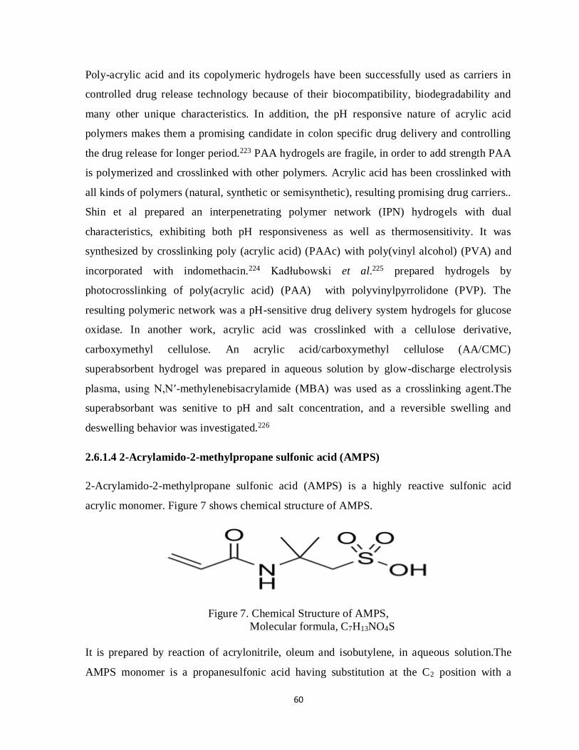
60
Poly-acrylic acid and its copolymeric hydrogels have been successfully used as carriers in
controlled drug release technology because of their biocompatibility, biodegradability and
many other unique characteristics. In addition, the pH responsive nature of acrylic acid
polymers makes them a promising candidate in colon specific drug delivery and controlling
the drug release for longer period.223 PAA hydrogels are fragile, in order to add strength PAA
is polymerized and crosslinked with other polymers. Acrylic acid has been crosslinked with
all kinds of polymers (natural, synthetic or semisynthetic), resulting promising drug carriers..
Shin et al prepared an interpenetrating polymer network (IPN) hydrogels with dual
characteristics, exhibiting both pH responsiveness as well as thermosensitivity. It was
synthesized by crosslinking poly (acrylic acid) (PAAc) with poly(vinyl alcohol) (PVA) and
incorporated with indomethacin.224 Kadłubowski et al.225 prepared hydrogels by
photocrosslinking of poly(acrylic acid) (PAA) with polyvinylpyrrolidone (PVP). The
resulting polymeric network was a pH-sensitive drug delivery system hydrogels for glucose
oxidase. In another work, acrylic acid was crosslinked with a cellulose derivative,
carboxymethyl cellulose. An acrylic acid/carboxymethyl cellulose (AA/CMC)
superabsorbent hydrogel was prepared in aqueous solution by glow-discharge electrolysis
plasma, using N,N′-methylenebisacrylamide (MBA) was used as a crosslinking agent.The
superabsorbant was senitive to pH and salt concentration, and a reversible swelling and
deswelling behavior was investigated.226
2.6.1.4 2-Acrylamido-2-methylpropane sulfonic acid (AMPS)
2-Acrylamido-2-methylpropane sulfonic acid (AMPS) is a highly reactive sulfonic acid
acrylic monomer. Figure 7 shows chemical structure of AMPS.
Figure 7. Chemical Structure of AMPS,
Molecular formula, C7H13NO4S
It is prepared by reaction of acrylonitrile, oleum and isobutylene, in aqueous solution.The
AMPS monomer is a propanesulfonic acid having substitution at the C2 position with a

61
methyl group and an acrylamido moiety. It contains an unsaturated vinyl group and a
terminal sulfonic acid, which are its reactive sites. AMPS is also available as its sodium and
ammonium salts which are formed by its neutralization with sodium hydroxide or ammonium
hydroxide, respectively.227
AMPS monomer appears as white crystalline powder or granular particles having water
possessing hygroscopic character. The melting point of this solid has been determined as
Melting point 195 °C (383 °F; 468 K). Its density is 1.1 g/cm3 (15.6 °C) and soluble in
water having solubility of about 1×10⁶ mg/L at 25°C.228, 229
In recent years, 2-Acrylamido-2-methylpropane sulfonic acid (AMPS) has attracted the
attention of pharmaceutical and other scientific researchers because of its thermal stability,
resistance to hydrolysis, water solubility and ability to impart a remarkable swelling behavior
in hydrogels. Its thermal and hydrolytic stability is due to the presence of sulfomethyl and
dimethyl groups which in combination create a steric hindrance to amide functionality. This
high swelling character is attributed to its highly ionizable sulfonate groups that make it a
strongly hydrophilic compound.230-234 Dissociation of AMPS is not dependent upon specific
pH; hence AMPS derived hydrogels are able to swell at all pH ranges.235,236 However, these
hydrogels exhibit higher swelling ratio at lower pH in comparison to neutral or alkaline pH.
Due to the presence of vinyl group in AMPS, it can be polymerized or crosslinked under the
influence of high energy radiations. Increasing the concentration of AMPS in reaction
medium provides more reactive vinyl groups, consequently, higher radiation assisted
polymerization could occur. Therefore, crosslinking during AMPS based hydrogel
formulation depends upon content of AMPS, which causes an increment of ionizeable
functional groups resulting higher electrostatic repulsion that leads to expansion of polymeric
network system.237 After hydrogel synthesis, unreacted residual monomer can be easily
removed by washing with water.238
It is synthetic monomer used in combination with number of polymers altering their
physicochemical characteristics. AMPS has been used in preparing hydrogels by crosslinking
with broad range of synthetic polymers, natural polymers, derivatives of natural polymers as
well as with other monomers such as acrylamide (AAm) and acrylic acid. These hydrogels

62
have applications in biomedical fields like controlled release drug delivery and
superabsorbants for removal of heavy metal ions from aqueous solutions. Sulfonic acid in
present in AMPS has ability to ionize completely in aqueous solutions. Due to this
characteristic, AMPS inhibits the undesirable precipitation of mineral salts including iron,
aluminum, calcium, magnesium, barium, zinc and chromium.AMPS/Polyvinyl alcohol
(PVA) hydrogels have been synthesized by gamma radiation.239 In another work,
Carboxymethyl cellulose (CMC) was crosslinked with AMPS along with other monomers.95
Durmaz et al.240 prepared and characterized 2-acrylamido-2-methyl-1-propane sulfonic acid -
co- acrylic acid (AMPS/AA) hydrogels. They were synthesized by radical polymerization in
the presence of N,N′-methylenebisacrylamide (MBA) as the crosslinking agent using
potassium persulfate (KPS) as initiator.
2.6.1.5 Potassium persulfate (KPS)
Potassium persulfate (KPS) also named as potassium peroxydisulfate is an inorganic
compound. This salt is an odorless, white solid powder with a molar mass 270.322 g/mol and
melting point 1067°C. Its density is 2.477 g/cm3 and is highly soluble in water with
solubility of 5 g/100 mL at 20 ºC. It is commonly used as initiator to initiate polymerization
reactions. It can be synthesized by electrolysis of cold solution of potassium hydrogen sulfate
in sulfuric acid under influence of high current density.241,242
Figure 8. Chemical Structure of Potassium persulfate , Molecular formula= K2S2O8
Potassium persulfate is a thermal initiator widely used in polymerization or crosslinking
process for preparing hydrogels. KPS and other thermal initiators possess high activation
energy (125-160 kJ•mol-1), therefore the polymerization reactions are usually occur at higher
temperatures ranging from 60ºC to 90ºC.243 The initiation of polymerization is first step of
hydrogel synthesis, during which there is formation of an active center that further starts
polymerization in monomer units or crosslinking of polymers. In this process, firstly the

63
initiator creates radicals, which are then transferred to monomer, resulting in generation of
polymer chain. All kinds of initiators cannot be used for every monomer and polymer. For
example, radical initiators are best suited for initiation on carbon-oxygen double bond in
aldehydes and ketones as well as carbon-carbon double bond of vinyl monomers.244 As
initiator KPS has been used in many polymerization processes such as in preparing
polytetrafluoroethylene and styrene-butadiene rubber, which are important commercially
available materials.245 It has been utilized in preparing hydrogels for drug delivery and other
useful scientific procedures. Acrylic acid, acrylamide, methacrlyic acid and many other
monomers as well as polymers have been polymerized and crosslinked, respectively.246-249
Being an oxidizing agent, KPS is used in oxidation of many products in organic chemistry,
for instance in oxidation of phenols.250
2.6.1.6 N,N'-Methylenebisacrylamide (MBAm or MBAA)
N,N'-Methylenebisacrylamide is a white crystalline powder, very slightly water soluble
with water solubility of 0.01-0.1 g/100 mL at 18 ºC. It is incompatible with strong acids,
strong bases and strong oxidizing agents. It is a cross-linking agent used for formation of
polymers and in crosslinking of polymers in hydrogel synthesis. Bisacrylamide is used for
SDS-PAGE in biochemistry because it is one of the compounds of polyacrylamide gel.251
The chemical structure of MBA is shown in figure 9.
Figure 9. N,N'-Methylenebisacrylamide (MBAm), Molecular formula C7H10N2O2
Bisacrylamide polymerizes with various monomers, among them one of the commonly used
is acrylamide. MBAm is capable of creating cross-linkages between polyacrylamide chains,
therefore; creating a strong polymeric network rather than unconnected linear chains of
polymer. An optimum concentration of MBA should be used for polymerization and
crosslinking due to its stability concerns. It has been investigated that by using very low
concentration of crosslinker, its methylene group by hydrolytic degradation is converted to

64
formaldehyde. By using crosslinker concentration above 0.3- 0.6 %, crosslinking density was
increased and degradation was controlled for several days (upto 10 days). Further increasing
the content to 1% or above, it remained stable and no degradation was observed for longer
periods.252,253 N,N-methylenebisacrylamide has been used as crosslinking agent for preparing
biomedical hydrogels with potential for utilization controlled drug release or enhancing
mechanical strength of bone substitutes.254 Ray et al.255 prepared pH sensitive poly (acrylic
acid-co-acrylamide)/MBA nanosized hydrogel colon-specific delivery of 5-fluorouracil. The
hydrogel drug delivery systems, as those based on N-isopropylacrylamide (NiPAAm) and
itaconic acid (IA), were prepared by using N,N-methylenebisacrylamide (MBA) as
crosslinking agent.The concentration of MBA used was ranging from 2.0- 4.0 wt.% with
respect to monomer concentration. The hydrogels were successfully synthesized by MBA,
loaded with Lipase and its controlled release was investigated under mild conditions. These
hydrogels were responsive to both temperature inside tract and pH of its media.256,257
Aims and Objectives
In Pharmaceutical technology, the research study has objectives related to betterment of the
health care system to improve the quality of life. The goals of this study are briefly discussed
as below:
1) The aim of this work is to detect the possibility of using mixtures of polymers as prolong
drug release systems using Captopril (CPT).
2) This will determine the compatibility and usefulness of the polymer combinations being
used in Hydrogel formulation.
3) The formulation will be prepared using a Radical graft polymerization technique and
evaluated by in vitro and in vivo studies.
4) This research project will contribute to the rational drug therapy.
5) This will be a promising study to ensure the safe and efficacious administration of the
drug providing more effective treatment. A suitable fraction of drug will be available for
treatment, thus minimizing the adverse drug reactions related to conventional drug
delivery systems.
6) This research study will also have a contribution towards cost effective treatment.

65
Chapter no. 3
Synthesis of Hydroxypropyl methylcellulose-
graft- poly (vinyl alcohol)-co-poly(acrylic acid)
hydrogels for the Controlled Release of
captopril and its in-vitro Evaluation

66
Summary
Background of the Study
A microwave induced irradiation synthesis, was proposed for the preparation of
Hydroxypropyl methylcellulose-graft-poly(vinyl alcohol)-co-poly (acrylic acid) hydrogels
for the controlled release of Captopril, an antihypertensive drug.
Methods
The hydrogels were separately synthesized by using microwave irradiation method and
conventional water-bath heating method. Chemical groups, thermal stability and surface
morphology of these hydrogels were characterized by FT-IR, DSC and SEM. Swelling ratios
of the gels were measured gravimetrically in the pH range from 1.2 to 7.4.
Results
Micrographs obtained from scanning electron microscopy (SEM), revealed that the gels
synthesized using microwave irradiation had more porous structure; therefore, they had
higher swelling ratios in comparison to hydrogels synthesized by water-bath method.
Thermal analysis (DCS and TGA), FTIR and XRD determination had confirmed the
formation as well as stability of the new polymeric network.
Conclusion
A stable network of Hydroxypropyl methylcellulose (HPMC), poly (vinyl alcohal) (PVA)
and acrylic acid was developed in shorter time period under influence of microwave
radiations. The formed hydrogels could be an efficient drug carrier for controlled delivery of
captopril for hypertension management.
Keywords: Hydroxypropyl methylcellulose, poly(vinylalcohal), acrylic acid, microwave
radiation, captopril

67
3.1 Introduction
The crosslinking of polymeric materials prevents the dissolution of hydrophilic polymer
chains / segments into the aqueous environment.258,259 These swellable polymeric materials
have been widely recognized as the stable carrier for drug delivery systems, because of their
ability to simulate biological tissues. Hydrogels being biocompatible materials have been
investigated to protect the drug, from in-vivo environment e.g, enzymatic degradation,
unfavorable pH or other hostile conditions.33
Development and designing of multi-component polymeric blends has become the subject of
great interests because such materials possess an efficient hybrid performance superior to
their individual components.260 In various hydrogel formulations more than one polymer are
crosslinked to form semi-interpenetrated polymer network (semi-IPN). Semi-IPN is a
technique of formulating a stable polymeric network of at least two polymers with an
additional non-covalent interaction.261 It is attractive in producing synergistic properties from
the component polymers to provide combined physical and mechanical properties to the
hydrogels for use in drug delivery. When a hydrophilic gelling polymer is interpenetrated
with a relatively hydrophobic gelling polymer, the resultant IPN hydrogel is expected to have
an improved capability of immobilizing a drug. Hydrogels based on natural polymers exhibit
poor mechanical strength, which limits their usefulness.262 Hence, blends of judicially
selected natural and synthetic polymers over comes this short coming by improving the
strength. It combines the biocompatibility of biological component with physical and
mechanical properties of the synthetic components. A combination of natural and synthetic
polymers has been found to be useful in enhancing the release of short half‐lived drugs under
physiological conditions. Grafting of vinyl monomers onto natural polymers such as
cellulose and its deivatives has been widely accepted.263-265
To improve the synthesis of hydrogel as well as their properties, many researchers tried to
use a variety of novel methods, including e-beam radiation, microwave radiation, photo-
initiated polymerization and plasma-induced grafting. The radiation crosslinking of polymers
avoid the use of additional chemical reagents as they have ability to crosslink water-soluble
polymers in their aqueous solution, without using initiators or using low quantity of
initiator.266-268 In cases of biomedical applications, it allows the simultaneous synthesis and

68
sterilization of the product. High-energy radiation causes the generation of radicals that leads
to scission on the polymer chain. This has been also investigated for cellulose and as well as
its derivatives.269-271 Microwave radiation possess special heating energy, having significant
advantages over the conventional thermal methods for preparing hydrogels. Due to the rapid
and uniform penetration properties, microwave energy can instantaneously absorbed and
directly heat the entire volume of a material.272 The microwave irradiation has been
successfully used in recent years for preparing useful polymeric devices for drug delivery. In
a research work, a guar-g-polyacrylamide hydrogel was prepared by microwave radiations,
without using the radical initiator.273 In another work, the temprature sensitive hydrogels
were prepared separately by microwave irradiation and conventional water bath heating
method. Both formulations were characterized by FT-IR, DSC and SEM and compared by
the results, which showed that microwave radiations shortened the preparation time,
improves the yield and swelling.72 Similarly, PVA/PAA hydrogels were prepared, where
acrylic acid was modified with PVA in the synthesis of polymeric matrix under the influence
of microwave radiations.211 The effects of varying power and exposure of microwave
radiations have been investigated in the grafting of methyl methacrylate on bamboo
cellulose.274
Hydroxypropyl methylcellulose (HPMC), one of the most widely used cellulose derivative as
hydrogel-forming polymer with numerous industrial applications such as, in the production
of coated and controlled release tablets, as a component of body lotions, ointments etc. It also
serves as structural and storage material in plants.275, 185
Polyvinyl Alcohol (PVA) possesses the strength, high water retaining ability, long-term
temperature and pH stability. It is polymerized and crosslinked with other monomers and
also with polymers to strengthen the hydrophilic systems.276
Acrylic acid (AA) has been extensively used monomer in hydrogel synthesis because AA is
relatively economical and easily polymerized to a higher molecular weight polymer277 by
various formulation techniques.
In this work, microwave irradiation method, is used for hydrogels synthesis. The hydrogels
were synthesized respectively by microwave irradiation method and conventional

69
hydrothermal method. The porous structures of the hydrogels synthesized using two different
methods were compared, and discussed the effects of hydrogel structures on its swelling.
3.2 MATERIALS & METHODS
3.2.1 Chemicals
Hydroxypropyl-methyl cellulose (2600-5600 cps), PVA, 99% hydrolyzed, Mw, 85,000 -
124,000 (Aldrich, product of USA), Acrylic Acid (Sigma Aldrich-Netherlands), N, N-
Methylene-bis-acrylamide, 98% (Fluka-Switzerland), Potassium persulphate (AnalaR, BDH-
England), Potassium dihydrogen phosphate (Merk- Germany), All the solvents used were of
analytical grades. Deionized distilled water was obtained from our laboratory.
3.2.2 Preparation of Hydrogel
The hydrogel was prepared by free radical polymerization, using thermostatic water bath and
microwave raditions
3.2.2.1 Method using Thermostatic Water Bath
Various quantities of polymers (HPMC and PVA) were added in distilled water and stirred at
80ºC for 1 h. Then the HPMC solution subjected to nitrogen purging for about 30 min and
potassium persulfate (0.5% W/W) was added to initiate the reaction by generating free
radicals. After that the reactants was cooled down to 30ºC and MBA as cross-linking agent
dissolved in acrylic acid (AA) was added under magnetic stirring. Final volume was adjusted
by addition of deionized distilled water. After that, the above mixture was poured in test
tubes and heated in water bath at 50°C, 55°C, 65°C and 75°C for 30min, 1h, 2h and 3h,
respectively. Then, the tubes were cooled to 25°C and hydrogels were taken out and cut in
the form of discs of nearly 8mm long. They were then thoroughly treated with ethanol and
distilled water mixture (50:50) for removing catalysts and uncross-linked monomer till the
pH of solutions after washing becomes nearly same as before being used. After washing
process, the hydrogel discs were air dried for overnight and then transferred to oven at 45°C
for 4 to 5 days until they attain a constant weight.

70
3.2.2.1.1 Hydrogel Formulations prepared using different concentrations of Acrylic acid
and crosslinking agent
The following were the suitable quantities of acrylic acid and crosslinker (MBA) to be
further used in the formulations as shown in table 1 and table 2, respectively. Among these
three M2 was selected on the basis of visual appearance and stability of discs during swelling.
After selecting the appropriate concentration of acrylic acid, the concentration of crosslinking
agent was varied. MBA, 1% of monomer concentration was considered to show better results
and stability.
Table 1. Hydrogel Formulations using different concentrations of Monomers
Formulation
Code
Polymer
(HPMC and PVA,
50:50)g/100g
Monomer
(Acrylic acid) g/100g
Crosslinker
MBA, mol% of Monomer
M1 2 20 1
M2 2 15 1
M3 2 10 1
Table 2. Hydrogel Formulations using different concentrations of Crosslinker
Formulation
Code
Polymer
(HPMC and PVA, 50:50) g/100g
Monomer
(Acrylic acid) g/100g
Crosslinker
MBA, mol% of
monomer
C1 2 15 0.5
C2 2 15 0.75
C3 2 15 1
C4 2 15 1.25
3.2.2.1.2 Hydrogel Formulations using different proportions and concentrations of
Polymers
Both polymers (HPMC and PVA) were used in various ratios. HPMC, a cellulose derivative
was crosslinked with PVA to form semi- IPN as shown in table 3. Among these

71
formulations, the ratio of these polymers used in P1 was consdidered more suitable on the
basis of swelling. Hydrogels with varying polymer concentration are presented in table 4.
Table 3. Hydrogel Formulations using different ratios of PVA and HPMC
Formulation
Code
Polymer (2g/100g)
(HPMC:PVA)
Monomer (g/100g)
(Acrylic acid)
Crosslinker(MBA),
mol% of Monomer
P1 3:1 15 1
P2 2:1 15 1
P3 1:1 15 1
P4 1:2 15 1
P5 1:3 15 1
Table 4. Hydrogel Formulations using different concentrations of Polymers
Formulations by Conventional Thermostatic water bath method
Formulation Code Polymers, g/100g
(HPMC and PVA, 3:1)
Monomers,
g/100g
Crosslinking agent,
mol % of monomer
concentration
F1 0.5 15 1
F2 1 15 1
F3 1.5 15 1
F4 2 15 1
F5 2.5 15 1
F6 3 15 1
3.2.2.2 Hydrogel formulation prepared by Microwave Radiation
The above mentioned traditional heating method was used that let the reaction to take place
in the thermo-stated water bath for longer periods of time. On the other hand, rapid synthesis

72
of Hydrogels was performed by using microwave radiations. In order to make comparison of
both methods, same proportions of polymers and monomers were taken. The whole mixture
was placed in Electrolux domestic microwave oven and it was first irradiated by electrical
power of 100 W for 5 minutes. Then, after an interval of one minute, it was further exposed
for 5 minutes at 180 W and finally, after one minute interval, the material was treated for
another 5 minutes with maximum power output set at 300 W. After that, the hydrogels
formed were cut into discs and treated with ethanol/ water mixture as previously described in
the above method.
Crosslinking reactions during hydrogel formation involve chemical or physical interaction
among functional groups of the components. In present work, crosslinking of both polymers
(HPMC and PVA) with acrylic acid in the presence of crosslinking agent (MBA) could be
presented as shown in figure 1.
Hydrogels prepared using different ratios of polymers and monomers by microwave
radiation) have been presented in table 5. The hydrogel formulations synthesized using
conventional water bath have been donated by ‘F’, while polymeric networks developed
under influence of microwave radiations are named as ‘R’. The ‘R’ hydrogels were prepared
with the same ratio of components as that for ‘F’ hydrogels, involving exposure to different
doses of radiations.

73
Figure 1. Crosslinked HPMC-g- PVA-co Poly (AA) Hydrogel

74
Table 5. Hydrogels using different concentrations of Polymers and radiation dose
Formulations by Microwave Radiations
Formulation
Code
Exposure time at
300W (min)
Polymers
(HPMC and
PVA) g/100g
Monomers,
g/100g
( Acrylic acid)
Crosslinking agent,
mol % of monomer
concentration
R1 5 0.5 15 1
R2 5 1 15 1
R3 5 1.5 15 1
R4 5 2 15 1
R5 2.5 1.5 15 1
R6 7.5 1.5 15 1
R7 10 1.5 15 1
3.3 In vitro Evaluation
3.3.1 Fourier Transform Infrared Spectroscopy (FT-IR)
FT-IR spectrophotometer (Bruker, Tensor 27) was used to record the spectra of hydrogel,
HPMC, acrylic acid and AMPS. The hydrogel samples were ground by the help of cutter as
well as pestle and mortar. The components and crushed hydrogel samples were then analysed
in wavelength range of 4000 to 500 cm-1.
3.3.2 Scanning Electron Microscopy (SEM)
SEM images were taken to investigate the surface morphology of super-absorbent hydrogels
using a scanning electron microscope (Quanta 250, FEI). Both drug free formulations and
drug loaded samples were ground and scanned at different magnifications to observe the
microscopic surface of dried hydrogels. It is therefore performed, to assess the capability to
adsorb and entrap the drug into their polymeric network.

75
3.3.3 X-Ray Diffraction (XRD)
X-Ray Diffraction analysis determines the crystallinaty and amorphous properties of the
substances. It investigates the interaction of components or polymers and drug. Xpert Pro
diffractometer (Panalytical) diffractometer used to record x-ray diffraction. The XRD
patterns of pure drug and drug loaded formulation were measured at room temperature by
scanning at angle 5-50° (2 Theta), scanning speed of 20/ min-1.
3.3.4 Thermal analysis
Thermal analysis was recorded by thermogravimetric analysis (TGA) and differential
scanning calorimetry (DSC) using Q5000 series (TGA instruments) and Q2000 series (TA
instruments), respectively. The hydrogel samples were crushed into powder form using
pestle and mortar and passed through a mesh no. 50. For measuring TGA, 1- 4 mg of ground
sample was placed in platinum pan connected to microbalance and heated upto 500°C at a
rate of 20°C/min in nitrogen atmosphere. To record DSC, hydrogel samples (1 to 3mg) along
with HPMC, AMPS and acrylic acid were placed in aluminum pan crimped with an
aluminum lid and heated from 0-500°C at the same rate used for TGA.
3.3.5 Swelling Study
The swelling of hydrogels was measured at different pH (1.2, 4.5 and 7.4) at room
temperature. Dried discs of hydrogels were accurately weighed and immersed in swelling
medium i.e. 0.1 M USP phosphate buffer solution. Hydrogel discs were weighed at regular
intervals of time and before weighing they were placed on filter paper to remove excess of
solution from the surface. The hydrogels were weighed for a period until they attain
equilibrium. The swelling ratio was calculated as:
S = (1)
Where, ws is the weight achieved after swelling and wd denotes the weight of dry hydrogel
discs. The percentage equilibrium swelling was determined by equation given below:
% ES = (2)

76
Where, weq is the equilibrium weight and wd is the initial weight of hydrogels before swelling
study.
3.3.6 Drug loading
Hydrogels were loaded with drug (captopril) using absorption method by immersing the dry
discs of hydrogels in 100ml captopril solution (1% w/v) comprising of phosphate buffer
solution and methanol (50:50). The dics were swollen till they achieved equilibrium, then
taken out and dried in oven at 40°C to their constant weights. The amount of drug loaded in
hydrogels was measured by extracting them with the methanol/ phosphate buffer solution in
the same ratio used for drug loading. The extraction was done repeatedly at regular intervals
and each time with freshly prepared solution until no drug remains in the extracting solution.
All samples of drug solutions used during extraction procedure were analyzed for drug
contents. The caliberation curve of captopril was drawn by preparing its various dilutions to
determine the drug concentration spectrometrically at λmax of 205nm. The amount of
captopril loaded in hydrogels was calculated by following relation.
Amount of drug = WD – Wd (3)
Where WD and Wd represents weights of dried hydrogel discs after and before immersion in
drug solution, respectively.17
3.3.7 Determination of Gel Fraction
Dried hydrogels were extracted at room temperature in distilled water for 7 days. The
extraction process was done to remove any sol that may be present in the hydrogel. The
extracted hydrogels were then dried again in the oven at 60ºC until a constant weight is
achieved. The percent gel fraction (%G) will be calculated using the equation as below:
%GF= (4)
where wi and we are the dry weight of the sample before and after extraction, respectively.

77
3.3.8 Drug Release
Drug release measurement was carried out by dissolution process using 0.1 M USP
phosphate buffer solutions of lower and higher pH values (pH 1.2 and pH 7.4). The dried
hydrogel discs loaded with captopril were placed in 500 ml buffer solution (dissolution
medium) maintained at 37°C, agitated by a paddle stirrer at a speed of 50 rpm. Then, the
samples were taken out at specific time intervals and drug released was measured by UV-
spectrophotometer at λ max of 205nm.
3.3.9 Drug release kinetics
Drug release models were used to determine the mechanism of drug release as given below:
Zero order kinetic models
It relates the drug delivery systems, where the rate of drug release does not exhibit
concentration dependency. It is represented as:
M0 - Mt = Kt (5)
Where, M0 isthe initial quantity of drug, Mt is the fraction of drug released at time t and K is
proportionality constant.
First order kinetic models
The first order kinetics describes the concentration dependent release of drug and is
represented by the following equation:
Log M0 − Log Mt = K1t/2.303 (6)
Where, M0 is the initial amount of drug, Mt is the drug concentration released at time t and
K1release constant.
Higuchi Model
Higuchi model can be presented by a simplified equation as:
Q = KH t1/2 (7)

78
Where, Q represents the fraction of drug released at time t and KH is Higuchi constant.
Korsmeyer- Peppas model
Korsemeyer-Peppas model is described by a simple empirical equation to describe the
Fickian and non-Fickian drug release from polymeric drug carriers, given as following:
Mt/M∞ = K tn (8)
Where, ‘K’ is kinetic constant that incorporates the geometric and structural properties of the
hydrogels and other drug carriers. Mt/ M∞ represents the drug fraction released at time t and
n is release exponent. When the value of n is 0.45, it indicates Fickian release order and for n
= 1, represents case II transport mechanism. On the other hand, n value between 0.45 and 1
corresponds to non- Fickian diffusion.
Weibull model
The dissolution and release process was described by an equation expressing the fraction of
drug accumulated ‘M’ in dissolution medium at time t given as:
Where, a defines the dependency on time, b denotes the shape parameter of dissolution curve
and the other parameter ‘Ti’ represents the lag time before dissolution process.
3.4 Results and Discussions
3.4.1 FT-IR
The structure and formation of crosslinkage among the polymers were investigated by
spectra recorded using FT-IR spectroscopy. In figure 2, spectrum of HPMC (a) shows an
absorption band at 3444.60 cm-1 is assigned to stretching frequency of the hydroxyl (-OH)
group. Another band at 1373.63 cm-1 is due to bending vibration of –OH. Other stretching
vibration bands related to C-H and C-O were observed at 2929 cm-1 and 1055.52 cm-1,
respectively. The noted peaks of pure HPMC were similar to observations of Wang et al.278

79
Figure 2. FT-IR Spectra of HPMC (a), PVA (b), Acrylic acid (c) HPMC/PVA-
co- AA hydrogels, F1 (d) and R1 (e)
In PVA’s FT-IR spectrum (b), at 2941.43 cm-1 stretching –CH2 groups in alkanes, 1089.38
cm-1 characteristic C–OH stretching, and a wide-strong absorption peak at 3286.82 cm-1 due
to O-H stretching was observed.

80
The spectra of HPMC-g-PVA-co-AA polymeric network suggests the formation of
intermolecular hydrogen bonding due to carboxylic acid groups of acrylic acid as observed
by the appearance of an absorption peak at 1716.99 cm-1 in (d) and 1706.39 cm-1 in (e) .
These peaks noted were similar to peaks observed by Debajyoti et al.279 and Pillai et al.280
In figure 2 the hydrogel spectrum (d) and (e) indicates the shifting of -OH vibration band of
HPMC from 3444.60 cm-1 to 3400.16 cm-1 and 3392.11 cm-1, respectively. This was
observed due to formation of hydrogen bonds confirming that -OH groups of HPMC and
PVA have reacted with -COOH groups of acrylic acid.
Hydrogels prepared by both methods, (conventional water bath and microwave radiation)
exhibited OH- stretching due to hydrogen bonding and carboxylic vibrations and CO-
because of intramolecular bonding in the same regions. At the same time, it could be clearly
seen that the FTIR spectra shapes of the hydrogels R and F were similar. It suggested that the
use of microwave irradiation method could successfully crosslink the HPMC/PVA-co-acrylic
acid polymeric network.
3.4.2 SEM
Scanning electron microscopy (SEM) was performed to study the morphology of hydrogels.
Figure 3 shows fractured surface morphology of drug loaded and drug free hydrogels. The
surface roughness and porosity of both types of hydrogels (F and R) could be compared from
their SEM images. From the SEM micrographs of surfaces of the air-dried gels R and F, it
can be seen that the surface of gel F was denser and rather smooth containing few cavities
and small spherically shaped protrusions. In comparison, the surface of R hydrogel
formulation is rough with some deep and interconnected pores. In comparison to F hydrogel,
the R hydrogel formulation had more uniformly porous network structures as shown in figure
3 (B). The uniformity in porosity was due to rapid and instantaneous penetration of
microwave energy throughout the surface.272 Hence, R hydrogels would facilitate the
diffusion more easily into hydrogel matrix during swelling process.

81
Figure 3. SEM Micrographs of Drug free (A and B) and Drug loaded (C and D)
HPMC/PVA-co-acrylic acid Hydrogels prepared by Microwave Radiation and
Conventional water bath method, with same ratios of Polymers
(Hydroxypropyl-methylcellulose and Poly(vinyl alcohol) and acrylic acid.
Figure 3 (C and D) shows hydrogels loaded with captopril as model drug, which is entrapped
and adhered on the surface of matrix. Higher quantity of drug could be loaded into the
formulations produced by microwave radiations as it shown figure 3 (C and D) showing drug
loaded R and F hydrogels, respectively.
From SEM images, it was observed that the application of microwave irradiation to prepare
semi-IPN created the hydrogels more porous than the gels synthesized by using water bath

82
heating method. These observations were relevant to the work done by Zhao et al.,72 who
prepared thermo-sensitive poly (N-isopropylacrylamide) (PNIPAAm) hydrogels using
microwave radiations in terms of morphological analysis.
3.4.3 XRD
The X-ray powder diffraction patterns of pure drug are compared with drug-loaded
hydrogels. X-ray diffractograms of Captopril, drug free Hydrogels and drug-loaded
Hydrogels are presented in figure 4. Figure 4 (B) and figure 4 (C) presents the diffraction
pattern of F3 and R3 formulations, respectively. Figure 4 (D) and 4 (E) show the
diffractograms of their respective drug loaded formulations.
The XRD scan of pure captopril had shown characteristic sharp and intense peaks between 0°
and 50° (2θ) due to its crystalline nature (Figure 4 A), The appearance of a sharp peak at 2θ=
27.79° is the characteristic of captopril. The diffractograms of hydrogels not containing the
drug was dense with only two marked peaks at nearly 2θ= 44° and 2θ= 49°, in both F3 and
R3 formulations. This similarity was due to same constitutions of F3 and R3 hydrogels.
It was observed from the diffractograms in both formulations (R3 and F3) that the drug free
(B and C) and hydrogels loaded with captopril (D and E) had a variation in diffraction pattern
due to entrapment of drug. However, in drug loaded hydrogels, the peaks were of less
intensity and diffraction pattern was dense in comparison to that of pure drug in figure (A).
Therefore, crystallinity of captopril was decreased after its entrapment into polymeric
networks, indicating the amorphous dispersion of drug into the hydrogels. The resultant
evaluation on the basis of XRD analysis was in relevance with observations made by
Manjanna et al.281 In that research work, Aceclofenac Sodium was loaded into
Polysaccharide hydrogel beads. Another related work was done by Giri et al.,282 where
diltiazem hydrochloride was loaded into cross-linked biodegradable IPN hydrogel beads of
pectin and modified xanthan gum. They had also noted similar diffraction patterns of pure
drug, drug free hydrogels and drug loaded hydrogels.

83
.
Figure 4. X- Ray Diffraction patterns of Captopril (A), Drug free Hydrogels ( B and C) and Drug
loaded Hydrogels ( D and E)
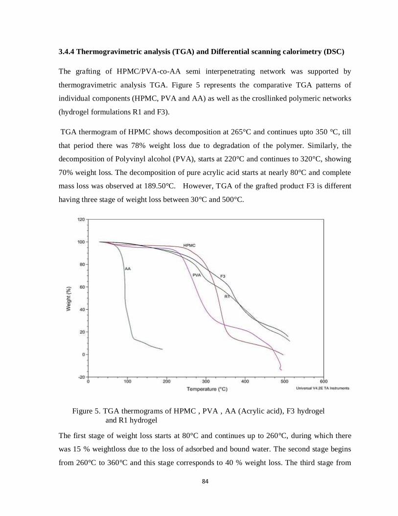
84
3.4.4 Thermogravimetric analysis (TGA) and Differential scanning calorimetry (DSC)
The grafting of HPMC/PVA-co-AA semi interpenetrating network was supported by
thermogravimetric analysis TGA. Figure 5 represents the comparative TGA patterns of
individual components (HPMC, PVA and AA) as well as the crosllinked polymeric networks
(hydrogel formulations R1 and F3).
TGA thermogram of HPMC shows decomposition at 265°C and continues upto 350 °C, till
that period there was 78% weight loss due to degradation of the polymer. Similarly, the
decomposition of Polyvinyl alcohol (PVA), starts at 220°C and continues to 320°C, showing
70% weight loss. The decomposition of pure acrylic acid starts at nearly 80°C and complete
mass loss was observed at 189.50°C. However, TGA of the grafted product F3 is different
having three stage of weight loss between 30°C and 500°C.
Figure 5. TGA thermograms of HPMC , PVA , AA (Acrylic acid), F3 hydrogel
and R1 hydrogel
The first stage of weight loss starts at 80°C and continues up to 260°C, during which there
was 15 % weightloss due to the loss of adsorbed and bound water. The second stage begins
from 260°C to 360°C and this stage corresponds to 40 % weight loss. The third stage from

85
350°C to 500°C may contribute to the decomposition of different structure of the graft
copolymer. At 490°C and 475°C there was complete weight loss of HPMC and PVA,
respectively, whereas their copolymer had 80% weight loss at that temperature. These
observations indicate the thermal stability of formed hydrogels in comparison to individual
polymers and monomers (PVA, HPMC and acrylic acid). TGA thermograms determined in
present work, indicate similarity to TGA patterns of Graft copolymer of chitosan with acrylic
acid under microwave irradiation by Huacai et al.,283 where thermal degradation was in three
stages. Similar observations on basis of thermal analysis were also evaluated by Bao et al.95
and Thakur et al.284
The DSC endothermic peaks of pure HPMC, PVA, AA and cross-linked polymeric network
were in accordance with TGA thermal patterns. DSC is a well-known and established method
often used as a qualitative technique for characterization of polymeric drug delivery systems.
It determines the physicochemical changes in either enthalpy or heat capacity of a crystalline
drug in the polymer matrix during the manufacturing process. The thermal behavior of pure
Captopril, drug loaded R1 (DR1) and F3 (DF3) Hydrogels characterized by DSC are
presented in figures 6a and 6b.
The thermogram of pure Captopril showed a sharp endothermic peak at 106°C followed by
corresponding melting point. However, in DSC thermogram of Drug loaded samples DR1
and DF3, peaks were observed at 288.28°C and 265.29°C, respectively. The appearance of
these peaks suggested that the drug loaded hydrogels showed an increase in the exothermic
peak temperature as presented in figures 6 a and 6 b. The extra obvious peak of drug (106°C)
was not observed in any type of the prepared hydrogels containing drug. The thermal
stability of hydrogels was maintained after the incorporation of captopril. No marked change
in thermal behavior of drug was indicated from the DSC thermogram of drug loaded
hydrogels. It is indicated that captopril is uniformly dispersed into the polymeric network of
HPMC, PVA and acrylic acid. This thermal evaluation of drug loaded hydrogels is in
relevance to that determined by Manjanna et al.281
The TGA and DSC thermograms of both hydrogel formulations synthesized by different
microwave radiations (R1) and conventional water bath (F3) were almost similar as could be
clearly seen in figures 5 and 6. Hence, it means that the grafting of these polymers by both

86
methods increases the thermal stability of the polymers. These results are corresponding with
the earlier findings of Zhao et al.72
Figure 6a. DSC thermograms of captopril, drug free F3 hydrogel and drug loaded F3
hydrogel (DF3)
Figure 6b. DSC thermograms of captopril, drug free R1 hydrogel and drug loaded R1
hydrogel (DR1)

87
3.4.5 Swelling Study
The effect of using different proportions of HPMC and PVA on swelling capacity of
hydrogels have been presented in figure 7 (a, b and c) at three levels of pH (1.2, 4.5 and
7.4), respectively. Similarly, the effect of crosslinking agent (MBA) on swelling at these
pH values are presented in (d, e and f) in figure 7. Alteration in swelling ratio observed
with different concentration of acrylic acid are shown in figure 7 (g). Higher swelling ratios
were observed at higher pH values.
Effect of Polymer’s proportions
The hydrogel P1 contains highest amount of HPMC among other hydrogel formulations
containing same concentrations of acrylic acid and crosslinker. The swelling ratio is
decreased by using higher amounts of PVA as it is lowest in P5 hydrogels with HPMC and
PVA content in a ratio of 1:3. On the other hand, hydrogel P1 had HPMC and PVA content
in a ratio of 3:1 (inverse of P5 content ratio). It indicated that HPMC has greater swelling
capacity in comparison to PVA.
Effect of crosslinker concentration
It can be observed from the figure 7 (d, e and f) that increasing concentration of
crosslinking agent leads to decrement in swelling power of hydrogels. This is because, it
increases the grafting of polymers and monomer crosslinking. Due to higher concentrations
of MBA, the crosslink density increases that makes the hydrogels lesser spaces for
incorporation of aqueous solutions, which results in lower swelling ratios. These results
were similar to earliaer observations of Huacai et al.,283 who determined an increment in
percent grafting of chitosan-g- acrylic acid hydrogels with increasing amount of MBA that
decreased the swelling behavior.

88
Figure 7. Effect of polymer’s proportion, MBA and acrylic acid concentrations on swelling
ratio

89
Effect of acrylic acid concentration
Figure 7(g) shows that by increasing the concentration of acrylic acid create an
enhancement in swelling ratio. Hydrogel formulations M1, M2 and M3 have the same
amounts of components except acrylic acid. M1 contains acrylic acid 20% w/w exhibit
higher swelling tendency in comparison to M2 and M3 containing lower amounts of
monomer. The noted results were according to swelling pattern observed by Ranjha et al.281
where, swelling tendency of pectin/acrylic acid hydrogels was higher in formulations
containing higher amounts of acrylic acid.
3.4.6 Comparative Swelling of F and R hydrogels
Swelling ratios of the synthesized gels were measured gravimetrically at the pH 1.2, 4.5
and 7.4 in phosphate buffer. The swelling of hydrogels is dependent upon the presence of
hydrophilic groups (ionizable functional groups). PVA and HPMC contain high hydroxyl
groups that make this polymer highly interactive with water. The carboxylic groups of
acrylic acid were mainly responsible for swelling tendency in hydrogels. When the pH is
increased, the COOH was ionized and deprotonated to a negatively charged COO--. As a
result, the electrostatic repulsion causes the swelling of hydrogel and greater expansion of
the network thus gives a high swelling ratio. The swelling ratios of hydrogels noted were
similar to swelling measurements performed by Gemeinhart et al.286 for poly (acrylamide-
co-acrylic acid) super porous hydrogels. The swelling study was also relevant to work done
by Shah et al.287 Therefore, the carboxylic groups associated with acrylic acid made the
coploymeric system a pH responsive that could be observed from swelling characteristics.
Effect of polymer concentration and radiation dose on swelling ratio at various pH were
studied to evaluate the swelling with respect to time.
As shown in figures 8 and 9, hydrogels containing higher amount of polymer show lesser
comparative swelling ratios. R1 and F1 comprising lower amount of polymer exhibited
higher swelling behaviour as compared to the other formulations. As the quantity of
polymer increases in the hydrogels, the value of their swelling ratio decreases. Generally, a
more porous matrix provides more space to accommodate larger quantities of water.288 As
shown in figures 3 (SEM images), the R hydrogels had more porous structure than the F
hydrogels, therefore, it would have higher water uptake. As a result, the hydrogels prepared

90
by microwave radiation method show better swelling characteristics in comparison to
conventional methods. Thus, microwave radiations develop uniform crosslinking and a
more porous polymeric system than simple water bath heating method. It could be seen
from comparative swelling ratios of R4 and F4 formulations in figure 10, which depicts
higher swelling of R4 formulation at pH 1.2 and pH 7.4.
Figure 8.Swelling ratios of the Semi IPN hydrogels synthesized by Microwave Radiations
Figure 9. Swelling ratios of the Semi IPN hydrogels synthesized by Conventional
Water bath Method
0
10
20
30
40
50
60
70
80
0 10 20 30 40 50 60 70 80
Swel
ling
Rat
io (q
)
Time (hrs)
R1 pH 1.2
R2 pH 1.2
R3 pH 1.2
R1 pH 4.5
R2 pH 4.5
R3 pH 4.5
R1 pH 7.4
R2 pH 7.4
R3 pH 7.4
0
10
20
30
40
50
60
70
0 10 20 30 40 50 60 70 80
Swel
ling
Rat
io (q
)
Time (hrs)
F1 pH 1.2
F2 pH 1.2
F3 pH 1.2
F1 pH 4.5
F2 pH 4.5
F3 pH 4.5
F1 pH 7.4
F2 pH 7.4
F3 pH 7.4
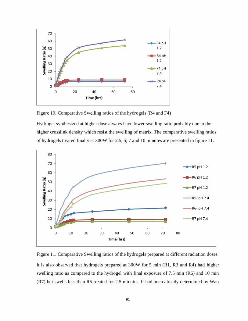
91
Figure 10. Comparative Swelling ratios of the hydrogels (R4 and F4)
Hydrogel synthesized at higher dose always have lower swelling ratio probably due to the
higher crosslink density which resist the swelling of matrix. The comparative swelling ratios
of hydrogels treated finally at 300W for 2.5, 5, 7 and 10 minutes are presented in figure 11.
Figure 11. Comparative Swelling ratios of the hydrogels prepared at different radiation doses
It is also observed that hydrogels prepared at 300W for 5 min (R1, R3 and R4) had higher
swelling ratio as compared to the hydrogel with final exposure of 7.5 min (R6) and 10 min
(R7) but swells less than R5 treated for 2.5 minutes. It had been already determined by Wan
0
10
20
30
40
50
60
70
0 20 40 60 80
Swe
llin
g R
atio
(q)
Time (hrs)
F4 pH1.2
R4 pH1.2
F4 pH7.4
R4 pH7.4
0
10
20
30
40
50
60
70
80
0 10 20 30 40 50 60 70 80
Swel
ling
Rat
io (q
)
Time (hrs)
R5 pH 1.2
R6 pH 1.2
R7 pH 1.2
R5- pH 7.4
R6- pH 7.4
R7 pH 7.4

92
et al.,274 that increasing the exposure time of microwave radiation increases the grafting of
polymers and ultimately higher crosslinking in polymeric network. This may be concluded
from the lower crosslink density of hydrogel which formed a porous structure and thus
allowed the water molecules to diffuse easier, hence faster response time.
3.4.7 Gel Fraction
The percent gel fraction was determined for formulations with different concentration of
polymers (HPMC and PVA), monomer (acrylic acid) and crosslinking agent (MBA). The
values of percent gel fraction are presented in table 6.
Table 6. Percent gel fraction of formulations with different amounts of components
Formulation code Wi (g) We (g) % gel fraction
F1 0.321 0.277 86.3
F3 0.328 0.289 88.1
F5 0.326 0.295 90.4
M3 0.313 0.269 85.9
M2 0.318 0.284 89.3
M1 0.324 0.301 92.9
C2 0.311 0.276 88.7
C3 0.315 0.286 90.7
C4 0.322 0.302 93.7
The amount of polymer, monomer and crosslinker are among the main factors affecting the
gel fraction of hydrogels. It could be observed from the figures 11, 12 and 13 depicting the
effect of concentration of polymer, monomer and crosslinker on per cent gel fraction.
Increasing the concentration of these components increases the value of %GF.

93
Figure 12.Effect of polymer concentration on %GF.
Figure 13.Effect of monomer concentration on %GF.
Figure 14.Effect of crosslinker concentration on %GF.
86
87
88
89
90
91
0 1 2 3
gel f
ract
ion
Polymer Concentration % w/w
Gel fraction
gel fraction
20, 92.9
84
86
88
90
92
94
0 5 10 15 20 25
gel f
ract
ion
concentration of acrylic acid % w/w
Gel fraction
Gel fraction
88
89
90
91
92
93
94
0 0.01 0.02 0.03 0.04
Ge
l fra
ctio
n
Crosslinker concentration (g/100g)
Gel Fraction
Gel Fraction

94
Figure 11 indicates that by increasing the concentration of polymer (HPMC and PVA, 3:1)
from 0.5% to 2.5%, an increment in value of %GF has been observed from 86.3% to 90.4%,
respectively. In comparison to polymer concentration, the monomer quantity used has more
influence on gel fraction due to its relatively higher concentrations used in hydrogel
formation. Figure 12 shows that by increasing the amount of acrylic acid from 10% to 20%,
percent gel fraction was increased from 85.9% to 92.9%. Moreover, crosslinker’s
concentration has similar effects on percent gel fraction because of enhancing crosslinking
interaction of acrylic acid with polyvinyl alcohol, which are then interacted with
hydroxypropyl methyl cellulose. Yoshii et al.289 and Roberta et al.290 had used the same
technique for determination of percent gel fraction and obtained similar results of higher %G
with increasing concentration of constituents.
3.4.8 Drug loading and release studies
The hydrogel discs exhibiting greater swelling accommodated higher amounts of drug. The
amount of captopril loaded in hydrogel formulation and percent drug release at acidic pH and
higher pH has been presented in table 7. The dissolution study of captopril was performed at
pH 1.2 and pH 7.4 as shown in figure 15 and figure 16. The drug release at both pH (1.2 and
7.4) was observed for a period of 24 hours, where USP phosphate buffer was used as
dissolution medium. Drug release measured is directly related to swelling studies where
relatively more amount of drug was loaded and ultimately released in formulations exhibiting
more swelling tendency.
Table 7. Amount of Captopril loaded and percentage of drug release at
pH 2 and pH 7.4
Formulation
code
Amount of captopril loaded
(mg) per 0.3 gram of dry
hydrogel discs
% release of captopril
(for 24 hour period)
pH 1.2 pH 7.4
F1 98.13 31.54 82.15
F2 95.08 29.49 80.06
F3 89.58 26.88 78.57
F4 83.50 24.72 75.79
R1 104.5 35.65 87.92
R2 97.81 32.84 85.01
R3 92.28 28.96 83.56
R4 86.8 26.45 78.54

95
It could be clearly observed from table 7, that hydrogels possessing higher swelling ratios
had entrapped more quantity of drug. R1 and F1 had same ratios of polymers (HPMC and
PVA), acrylic acid and crosslinking agent, but they were prepared using different techniques.
The captopril loaded in R1 formulation was 104.5 mg in 0.3 gram dry hydrogel disc, whereas
it was 98.13 mg in F1 formulation, hence, relatively higher amount of captopril was loaded.
This difference could also be noticed in other formulations prepared using microwave
radiations (R2, R3 and R4) and conventional water bath (F2, F3 and F4).
These variations are may be due to uniformity in porous structure and presence of larger
voids in R hydrogel formulations, leading to higher and more uniform dispersion of drug in
polymeric network. Moreover, factors such as polymer concentration, monomer
concentration and other factors affecting swelling behavior of hydrogels directly affect drug
loading and release.
The cumulative percent drug release from hydrogel formulation F2 and R2 have been
presented in figure 15 and figure 16. The drug release pattern at both lower pH 1.2 and
higher pH 7.4, could be seen from the figures 15 and 16.
A similarity in release pattern was observed, as both formulations were releasing more drugs
at higher pH values. At pH 1.2, the cumulative percent drug release was 29.49% in F2 and
32.84% in R2 hydrogels, while at pH 7.4, drug release were 80.06% and 85.01% for F2 and
R2 formulations, respectively. These values of percent drug release are also mentioned in
table 7. Therefore, the drug release is dependent upon the pH sensitivity imparted due to
presence of acrylic acid in hydrogels. The cumulative percent drug release is in
correspondence to the results obtained by swelling study.
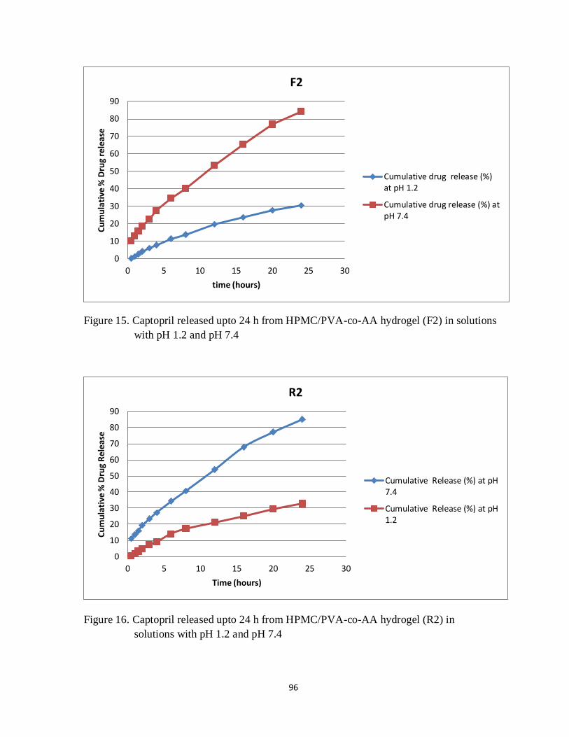
96
Figure 15. Captopril released upto 24 h from HPMC/PVA-co-AA hydrogel (F2) in solutions
with pH 1.2 and pH 7.4
Figure 16. Captopril released upto 24 h from HPMC/PVA-co-AA hydrogel (R2) in
solutions with pH 1.2 and pH 7.4
0
10
20
30
40
50
60
70
80
90
0 5 10 15 20 25 30
Cu
mu
lati
ve %
Dru
g re
leas
e
time (hours)
F2
Cumulative drug release (%)at pH 1.2
Cumulative drug release (%) atpH 7.4
0
10
20
30
40
50
60
70
80
90
0 5 10 15 20 25 30
Cu
mu
lati
ve %
Dru
g R
elea
se
Time (hours)
R2
Cumulative Release (%) at pH7.4
Cumulative Release (%) at pH1.2

97
The drug release was evaluated by the application of zero order kinetics, first order kinetics,
Higuchi model, Korsmayer-Peppas model and Weibull model. The values of release coefficient R2
and K calculated by these kinetic models are presented in table 8. The captopril released from F and
R hydrogel formulations were best fitted into the kinetic model having value of R2 close to 1.
Table 8: Determination of coefficient (R2), K and release exponent of various drug release kinetic
models
Sample
code
pH Zero order
kinetics
First order
kinetics
Higuchi model Korsmeyer Peppas
model
Weibull
model
R2 K R2 K R2 K R2 K n R2
F1 1.2 0.9719 1.84 0.7215 0.165 0.9969 4.83 0.9913 3.23 0.703 0.7012
7.4 0.9874 3.96 0.5248 0.263 0.9876 14.81 0.9967 13.57 0.5488 0.8955
F2 1.2 0.9767 1.69 0.702 0.156 0.9963 4.33 0.996 3.54 0.598 0.669
7.4 0.989 3.94 0.5274 0.261 0.9901 14.586 0.9979 12.022 0.609 0.8905
F3 1.2 0.9811 1.45 0.7674 0.134 0.9917 3.54 0.9968 3.37 0.5221 0.7395
7.4 0.9843 3.76 0.539 0.2557 0.9902 13.793 0.9971 13.88 0.496 0.8835
F4 1.2 0.9736 1.42 0.7598 0.158 0.9955 3.06 0.9864 1.952 0.7042 0.7775
7.4 0.99 3.59 0.6252 0.238 0.9844 11.56 0.9966 9.93 0.579 0.8774
R1 1.2 0.9815 1.83 0.7362 0.169 0.9928 5.12 0.9954 4.2945 0.587 0.7544
7.4 0.9812 4.179 0.5054 0.2668 0.9921 15.92 0.9968 15.58 0.5118 0.8804
R2 1.2 0.9632 1.86 0.7083 0.1674 0.9975 4.93 0.9881 3.59 0.659 0.6939
7.4 0.9896 3.85 0.5239 0.262 0.9882 14.814 0.9973 13.4 0.556 0.8983
R3 1.2 0.9805 1.61 0.7532 0.1513 0.9929 4.03 0.9963 3.54 0.56 0.5894
7.4 0.9817 4.02 0.5267 0.26 0.9912 14.472 0.9956 15.02 0.479 0.8771
R4 1.2 0.9761 1.48 0.7024 0.135 0.9949 3.52 0.9965 3.33 0.524 0.7314
7.4 0.9877 3.64 0.5642 0.25 0.9876 13.194 0.9983 12.6 0.525 0.8896
As shown in table 8, the value of drug release coefficient (R2) calculated by zero order was ranging
from 0.97 to 0.99. Similarly, the values of R2 calculated by Higuchi model and Korsmeyer Peppas
model were in that range. Hence, it could be observed that all captopril loaded hydrogel formulations
prepared were following zero order kinetics, Higuchi model and Korsmeyer Peppas model in terms
of drug release. The mechanism of drug release was indicated by the values of n i.e. release
exponent. By fitting of recorded data to Peppas model, it was investigated that approximately all
hydrogel formulations in spite of having different concentrations of polymer and monomers were
following non- Fickian mechanism of drug release as presented in Table 8. The value of n in all
cases were more than 0.45 but lesser than 1.

98
Conclusions
The above discussion concludes that use of microwave irradiation is more efficient technique
for hydrogel synthesis for the components used in this study. It can remarkably shorten the
reaction time required for polymer crosslinking from several hours to few minutes in
comparison to conventional hydrothermal method. The polymeric network HPMC-g-PVA-
co-poly (acrylic acid) was prepared successfully by induction of microwave radiations. The
hydrogels formed by using this method had comparatively higher swelling tendency and drug
loading. Therefore, microwave assisted hydrogel synthesis developed a promising drug
carrier that could be effectively utilized for delivering captopril for longer time period in a
controlled manner. It could be a suitable candidate to prove its worth in the treatment of
hypertension and certain other heart disorders.

99
Chapter no. 4
Synthesis and in-vitro characterization of
Hydroxy propyl methylcellulose-g-poly(acrylic
acid-co-2-Acrylamido-2-methylpropane
sulfonic acid) polymeric network for
controlled release of captopril

100
Summary
Background of the Study
A super-absorbent hydrogel was developed by crosslinking of 2-acrylamido-2-methyl-1-
propanesulfonic acid (AMPS) and acrylic acid with hydroxypropyl methylcellulose(HPMC)
for controlled release drug delivery of captopril, a well-known antihypertensive drug.
Methods
Acrylic acid and AMPS were polymerized and crosslinked with HPMC by free radical
polymerization, a widely used chemical crosslinking method. N,N'-methylenebisacrylamide
(MBA) and potassium persulfate (KPS) were added as cross-linker and initiator, respectively.
The hydrogel formulation was loaded with captopril. The concentration of captopril was
monitored at 205 nm using UV spectrophotometer. Equilibrium swelling ratio was
determined at pH 2, 4.5 and 7.4 to evaluate the pH- responsiveness of the formed hydrogel.
The hydrogels were evaluated by FTIR, SEM, XRD, and thermal analysis (DSC and TGA).
Results
The formation of new copolymeric network was determined by FTIR, XRD, TGA and DSC
analysis. The hydrogel formulations with acrylic acid and AMPS ratio of 4:1 and lower
amounts of crosslinker had shown maximum swelling. Moreover, higher release rate of
captopril was observed at pH 7.4 than at pH 2, because of more swelling capacity of
copolymer with increasing pH of the aqueous medium.
Conclusions
The present research work confirms the development of a stable hydrogel comprising of
HPMC with acrylic acid and AMPS. The prepared hydrogels exhibited pH- sensitive
behavior. This superabsorbent composite prepared could be a successful drug carrier for
treating hypertension.
Keywords: Composite, Superabsorbent, Polymerization, Acrylic acid, 2-Acrylamido-2-
methyl-1-propanesulfonic acid, Hydroxypropylmethylcellulose, Initiator, Cross linker,
Hydrogel.

101
4.1 Introduction
Drug delivery systems have been known for enhancing therapeutic efficacy, minimizing side
effects, and improving patient compliance. Among drug delivery systems, hydrogels have
attracted the interest of biomaterial scientists due to their hydrophilic character and
biocompatibility.291 Due to their softness and pliable nature, they have tendency to absorb
higher amounts of water and physiological solutions, these absorbed aqueous solutions are
capable of retaining water even if subjected to pressure. They have numerous applications as
drug carriers, water-absorbents and food additives.292
Superabsorbent hydrogels have an ability to absorb water from 10% to thousands times of
their dry weight.293 Due to special characteristics, these materials have gained attention in the
fields of agriculture, waste water treatment,294-296 pharmaceutical and biomedical297,298 and
biotechnology.299,300 Polysaccharide-based hydrogels are currently attracting much interest
fortheir unique properties, which are their better biocompatibility, biodegradability,
renewability, and nontoxicity. Various polysaccharides, such as starch,301 chitosan,302
carrageenan,303 alginate,304 cellulose and cellulose derivatives305−307 have been used for
superabsorbent hydrogel formulation.
In this work, a super-absorbent hydrogel was synthesized by copolymerization of two
monomers (acrylic acid and AMPS) and their crosslinking with HPMC using potassium
persulfate to initiate the reaction and MBA as crosslinking agent.
Hydroxypropylmethylcellulose (HPMC), the cellulose derivative, is a semisynthetic, inert,
and viscoelastic polymer found in a variety of commercial products. Depending on the grade,
HPMC is widely utilized in oral solid dosage forms (tablets and capsules) as well as an
ophthalmic lubricant.308,309 HPMC comprises of repeated units of glucose, linked with one
another by 1 4-glycosidic bonding, while the polymer chains are attached together by
hydrogen bonding.310
Acrylic acid (AA) has been extensively used as monomer in hydrogel synthesis due to
relatively economical advantage, crosslinking ability as well as rapid polymerization by
various formulation techniques. It possesses a pH and electrically responsive behavior due to
ionic repulsion between anionic charged carboxylate groups. It polymerizes to polyacrylic

102
acid (PAA) an established vehicle in controlled release drug delivery.277,311 2-Acrylamido-2-
methyl-1-propanesulfonic acid (AMPS) it has attracted an attention in hydrogel formation
possess due to presence of sulfonate groups. Strongly ionizable sulfonate groups increase the
hydrophilicity and ultimately swelling capability of hydrogels. The polymeric network
comprising of AMPS have ability to swell at all pH ranges, therefore it does not impart a pH-
sensitive behavior to its hydrogel formulation. As stated in literature, it swells rapidly in
acidic medium and relatively slower at pH higher than 5. AMPS contain both nonionic and
anionic groups; whereas, AA is an anionic monomer.312 Due to these characteristics acrylic
acid was combined with AMPS, to release the drug loaded in a controlled manner. The
hydrogels prepared were observed for their swelling behavior different pH (2, 4.5 and 7.4).
They were loaded with captopril and its release in was studied by dissolution process at pH 2
and pH 7.4. Moreover, FTIR, SEM, XRD, DSC and TGA analysis were performed for in-
vitro characterization of super-absorbent composite.
4.2 MATERIALS AND METHODS
4.2.1 Chemicals
Hydroxypropylmethylcellulose (2600−5600 cps, Sigma Aldrich, Netherlands), acrylic acid
(Sigma Aldrich-Netherlands), AMPS, (99%, Aldrich-product of Germany) N,N-Methylene-
bis-acrylamide (98%, Fluka-Switzerland), potassium persulphate (Analar, BDH-England),
and potassium dihydrogen phosphate (Merck, Germany) were purchased through local
commercial sources. Distilled water from laboratory and solvents of analytical grades were
used.
4.2.2 Preparation of hydrogel
The hydrogel was prepared by free radical polymerization, where the polymer (HPMC) in
varying quantities was added in distilled water and stirred at 80ºC for 1 h. Then the HPMC
solution subjected to nitrogen purging for about 30 min and potassium persulfate (0.5%
W/W) was added to initiate the reaction by generating free radicals. After that the reactants
was cooled down to 30ºC and MBA as cross-linking agent dissolved in acrylic acid (AA) was
added under magnetic stirring. Then, AMPS previously dissolved in small quantity of water
was added to above mixture and final volume was adjusted by addition of deionized distilled

103
water. After that, the above mixture was poured in test tubes and heated in water bath at
50°C, 55°C, 65°C and 75°C for 30 minutes, 1 hour, 2 hour and 3 hour, respectively. Then,
the glass tubes were cooled to 25°C and hydrogels were taken out and cut in the form of
discs of nearly 8mm long. They were then thoroughly treated with ethanol and distilled water
mixture (50:50) for removing catalysts and uncross-linked monomer till the pH of solutions
after washing becomes nearly same as before being used. After washing process, the
hydrogel discs were air dried for overnight and then transferred to oven at 45°C for 4 to 5
days until they attain a constant weight. The whole crosslinking reaction initiated by
potassium persulfate (KPS), involving interaction among polymer and monomers are
presented in figure 1. Table 1 shows hydrogels prepared using different concentration of
components.
Table 1. Hydrogels formulations using different amounts of HPMC, AMPS and MBA
Formulation
code
Polymer
(HPMC)
g/100g
Monomers, g/100g Crosslinking agent,
mol % of each
monomer’s
concentration AA AMPS AA/AMPS ratio
S1 1.0 15 2. 5 6:1 0.6
S2 1.0 15 3 5:1 0.6
S3 1.0 15 3.75 4:1 0.6
S4 1.0 15 5 3:1 0.6
S5 1.0 15 7.5 2:1 0.6
S6 1.5 15 3.75 4:1 0.6
S7 2.0 15 3.75 4:1 0.6
S8 1.0 15 3.75 4:1 0.8
S9 1.0 15 3.75 4:1 1.0
S10 1.0 15 3.75 4:1 0.4

104
Figure 1. Crosslinked HPMC-g-poly (AA-co-AMPS) Hydrogel

105
4.3 In vitro Evaluation
4.3.1 Fourier Transform Infrared Spectroscopy (FT-IR)
FT-IR spectrophotometer (Bruker, Tensor 27) was used to record the spectra of hydrogel,
HPMC, acrylic acid and AMPS. The hydrogel samples were ground by the help of cutter as
well as pestle and mortar. The components and crushed hydrogel samples were then analysed
in wavelength range of 4000 to 500 cm-1.
4.3.2 Scanning Electron Microscopy (SEM)
SEM images were taken to investigate the surface morphology of super-absorbent hydrogels
using a scanning electron microscope (Quanta 250, FEI). Both drug free formulations and
drug loaded samples were ground and scanned at different magnifications to observe the
microscopic surface of dried hydrogels. It is therefore to assess the capability to adsorb and
entrap the drug into their polymeric network.
4.3.3 X-Ray Diffraction (XRD)
X-Ray Diffraction analysis determines the crystalline and amorphous properties of the
substances. It investigates the interaction of components or polymers and drug. Xpert Pro
diffractometer (Panalytical) diffractometer used to record x-ray diffraction. The XRD
patterns of pure drug and drug loaded formulation were measured at room temperature by
scanning at angle 5-50° (2 Theta), scanning speed of 20/ min-1.
4.3.4 Thermal analysis
Thermal analysis was recorded by thermogravimetric analysis (TGA) and differential
scanning calorimetry (DSC) using Q5000 series (TA instruments) and Q2000 series (TA
instruments), respectively. The hydrogel samples were crushed into powder form using
pestle and mortar and passed through a mesh no. 50.
TGA
For measuring TGA, 1- 4 mg of ground sample was placed in platinum pan connected to
microbalance and heated till 500°C at a rate of 20°C/min in nitrogen atmosphere.
106
DSC
To record DSC, hydrogel samples (1 to 3mg) along with HPMC, AMPS and acrylic acid
were placed in aluminum pan crimped with an aluminum lid and heated from 0-500°C at the
same rate used for TGA.
4.3.5 Swelling Study
The swelling of hydrogels was measured at different pH (2, 4.5 and 7.4) at room temperature.
Dried discs of hydrogels were accurately weighed and immersed in swelling medium i.e. 0.1
M USP phosphate buffer solution. Hydrogel discs were weighed at regular intervals of time
and before weighing they were placed on filter paper to remove excess of solution from the
surface. The hydrogels were weighed for a period until they attain equilibrium. The swelling
ratio was calculated as:
Where, ws is the weight achieved after swelling and wd denotes the weight of dry hydrogel
discs. The percentage equilibrium swelling was determined by equation given below:
Where, weq is the equilibrium weight and wd is the initial weight of hydrogels before swelling
study.
4.3.6 Drug loading
Hydrogels were loaded with drug (captopril) using absorption method by immersing the dry
discs of hydrogels in 100mL captopril solution (1% w/v) comprising of phosphate buffer
solution and methanol (50:50). The hydrogels were swollen till they achieved equilibrium,
then taken out and dried in oven at 40°C to their constant weights. The amount of drug
loaded in hydrogels was measured by extracting them with the methanol/ phosphate buffer
solution in the same ratio used for drug loading. The extraction was done repeatedly at
regular intervals and each time with freshly prepared solution until no drug remains in the

107
extracting solution. All samples of drug solutions used during extraction procedure were
analyzed for drug contents. The calibration curve of captopril was drawn by preparing its
various dilutions to determine the drug concentration spectrometrically at λmax of 205nm.
4.3.7 Drug release
Drug release measurement was carried out by dissolution process using 0.1 M USP
phosphate buffer solutions of lower and higher pH values (pH 2 and pH 7.4). The dried
hydrogel discs loaded with captopril were placed in 500 ml buffer solution (dissolution
medium) maintained at 37°C, agitated by a paddle stirrer at a speed of 50 rpm. Then, the
samples were taken at specific time intervals and drug released was measured by UV-
spectrophotometer at λ max of 205nm.
4.3.8 Drug release kinetics
Various drug release models were used to determine the mechanism of drug release as given
below:
Zero order kinetic models
It relates the drug delivery systems, where the rate of drug release does not exhibit
concentration dependency. It is represented as:
M0 - Mt = Kt (3)
Where, M0isthe initial quantity of drug, Mt is the fraction of drug released at time t and K is
proportionality constant.
First order kinetic models
The first order kinetics describes the concentration dependent release of drug and is
represented by the following equation:
Log M0 − Log Mt = K1t/2.303 (4)
Where, M0 is the initial amount of drug, Mt is the drug concentration released at time t and
K1release constant.

108
Higuchi Model
Higuchi model can be presented by a simplified equation as:
Q = KHt1/2 (5)
Where, Q represents the fraction of drug released at time t and KH is Higuchi constant.
Weibull model
The dissolution and release process was described by an equation expressing the fraction of
drug accumulated ‘M’ in dissolution medium at time t given as:
Where,adefines the dependency on time, b denotes the shape parameter of dissolution curve
and the other parameter ‘Ti’represents the lag time before dissolution process.
Korsmeyer- Peppas model
Korsemeyer-Peppas model is described by a simple empirical equation to describe the
Fickian and non-Fickian drug release from polymeric drug carriers, given as following:
Mt/M∞ = K tn (7)
Where, ‘K’ is kinetic constant that incorporates the geometric and structural properties of the
hydrogels and other drug carriers. Mt/ M∞ represents the drug fraction released at timet and
nis release exponent. When the value of n is 0.45, it indicates Fickian release order and for n
= 1, represents case II transport mechanism. On the other hand, n value between 0.45 and 1
corresponds to non- Fickian diffusion.

109
4.4 Results and Discussions
4.4.1 FT-IR Spectroscopy
The structure and formation of cross-linkageamong the polymers were investigated by
spectra recorded using FT-IR spectroscopy as presented in figure 2. The spectrum(b) of
HPMC shows an absorption band at 3444.60 cm-1 is assigned to stretching frequency of the
hydroxyl (-OH) group. Another band at 1373.63 cm-1 is due to bending vibration of –OH.
Other stretching vibration bands related to C-H and C-O were observed at 2929 cm-1 and
1055.52 cm-1, respectively.
Spectrum (c) and spectrum (d) represents the FTIR pattern of acrylic acid and AMPS,
respectively. Acrylic acid shows a characteristic peak nearly 1700cm-1 due to presence of
carboxylic group. The spectrum of AMPS showed a band at 1666.03 cm-1 related to
stretching of amide link (–CONH), another band at 1550.03 cm-1 corresponds to bending
vibration of N-H and 1126.54 cm-1 was due to stretching vibration of -SO3H groups.
Another peak at 623.02 cm-1 in AMPS spectrum was also related to the -SO3H group.
In figure 2,the spectrum (a) of HPMC-g-poly(AA-co-AMPS) suggests the formation of
intermolecular hydrogen bonding due to carboxylic acid groups of acrylic acid as observed
by the appearance of an absorption peak at 1710.75 cm-1that was not present in individual
spectra of AMPS and HPMC. The hydrogel spectrum (a) indicates the shifting of –OH
vibration band of HPMC from 3444.60 cm-1 to 3292.58 cm-1 due to formation of hydrogen
bonds. Hence, it confirms the crosslinking of HPMC with acrylic acid involving the reaction
of –OH (of HPMC) with –COOH (of acrylic acid). In addition, characteristic bands at
1547.30 cm-1 (C=O stretching vibration of –CONH groups) and 1153.14 cm-1 were attributed
to stretching vibration of sulfonate (–SO3H) groups of AMPS monomer. The results
indicated successful grafting of monomers (acrylic acid and AMPS) onto HPMC polymer
chains. These peaks observed were according to the FTIR spectrum noticed by Wang et al.313
however slight variation was due to interaction of acrylic acid and AMPS with functional
groups of polymers.

110
Figure 2. FT-IR Spectra of HPMC-g-poly ( AA-co-AMPS) (a), HPMC (b),
Acrylic acid (c) and AMPS (d)

111
4.4.2 SEM
In order to determine the microstructure and surface morphology of hydrogel formulations,
SEM images were taken. Scanning electron microscopy is one of the preferred methods to
characterize the hydrogels in terms of porosity and water retention. The micrographs
recorded as shown in figure 3 (A and B) show the surface of the drug free and drug loaded
hydrogels.
Figure 3. SEM images of HPMC-g-poly (AA-co-AMPS) hydrogels,
without drug (A) and loaded with drug (B).
The SEM images in figure 3 clearly show that hydrogel surface was rough along withmicro-
spaces for water retention and entrapment ofsolutes. Figure 3 (A) presents drug free hydrogel
formulation with voids and spaces for accommodation of biological fluids as well as drug
particles. Theyhad tendencyto exhibit remarkable swelling because of their water absorption
capability. Moreover, due to the presence of these voids and roughness of surface of
hydrogels, the captoprilwasloaded into these regions as shown in figure 3 (B).It was
indicated from the prepared grafted polymeric network HPMC-g-poly (AA-co-AMPS) had
ability to act as suitable drug carriers for drug delivery.

112
4.4.3 XRD
X-ray diffractograms of pure captopril and captopril loaded hydrogel formulation are
presented in figure 4. The diffraction patterns of hydrogels loaded with drug were compared
with pure drug. The XRD scan of plain captopril had characteristic sharp and intense peaks
between 0° and 50° (2θ), which were appeared due to its crystalline nature as shown in figure
4, diffractionpattern (c).
Figure 4. XRD patterns of captopril (a) and Drug loaded HPMC-g-poly (AA-co-AMPS)
hydrogel (b) Drug free HPMC-g-poly (AA-co-AMPS) hydrogel
The appearance of a sharp peak at 2 θ= 27.79° is characteristic of captopril. It could be seen
in diffraction pattern a, that no peak was observed in cross-linked copolymer. However,
diffractogram of drug loaded hydrogel was dense like that of drug free hydrogelbutshowing
peaks with low intensity. Therefore, in comparison to pure captopril, the captopril loaded
formulations had low intensity and dense peaks suggesting the amorphous distribution of
drug into polymeric network, as it could be observed from figure 4 diffractogram (b). This
observation iscorresponding with XRD analysis studied by Rao et al.314, where drug was
molecularly dispersed in the polymeric matrix of poly(acryl amide-co-2-acrylamido-2-

113
methyl-1-propanesulfonic acid-co-acrylamidoglycolic acid) hydrogels. Similar results had
also beennoted by Kim et al.315
4.4.4 Thermal analysis
The grafting of HPMC-g-poly (AA-co-AMPS) polymeric network was also determined by
theromogravimetric analysis (TGA). The pure acrylic acid decomposition starts at nearly
80°C and complete mass loss was observed at 189.50°C as shown in figure5, thermogram
(A). Similarly, the degradation of other monomer AMPS had taken lesser time as can be seen
in thermogram (B), figure 5. The TGA thermogram (C) of HPMC shows decomposition at
265°C which then continued till 350°C, during that period 78% of loss in weight was
observedbecause of polymer degradation.
In comparison to the individual components, thermogram of the grafted product (D) was
recorded, where three stages of decomposition from 30°C -500°C were observed. In the first
stage of degradation, loss of weight started from 114°C to 25°C, there was about 12% weight
loss during this stage due to loss of absorbed and bound water. Then second stage of weight
loss started at 250°C and continued to 331.88°C, corresponding to 40% weight loss. Finally
the third stage beginning from 332°C till 500°C caused the degradation of hydrogel’s
structure. In case of HPMC, there was complete removal at 490°C, whereas the hydrogel had
67% weight loss and 33% still remaining at that temperature. A greater thermal stability of
the formed polymeric network was observed as compared to its individual polymer and
monomers (HPMC, acrylic acid and AMPS). Hence, the hydrogels prepared were more
stable and resistant to higher temperatures.

114
Figure 5. DSC and TGA thermograms of acrylic acid AA (A), AMPS (B), HPMC (C), drug
free hydrogel (D), captopril (E) and drug loaded hydrogel (F)
The alterations in heat capacity as well as enthalpy changes are measured using
Differential Scanning Calorimetry (DSC). It is a well-known and established technique
adopted for quantitative assessment of physicochemical variations in heat capacity of
crystalline drugs, when loaded into hydrogels. The DSC endothermic peaks of pure
HPMC, AA, AMPS and cross-linked polymeric network were in accordance with TGA
thermal patterns. The thermal behavior of the pure captopril, drug loaded S3 hydrogel was
characterized using DSC, as shown in Figure 5 (F).The disappearance of characteristic
peaks and appearance of other peaks suggests some sort of interaction of drug with
polymers. Thermogram (E) presenting DSC pattern of pure captopril, shows the
appearance of sharp peak at 106°C indicating melting point of captopril. This peak of drug

115
was not appeared in DSC thermogram of the drug loaded formulation, whereas two new
peaks were observed at 225°C and 292°C.
Figure 6. DSC thermograms of a) drug free and b) drug loaded and c) pure drug
It could be clearly suggested that the drug loaded Hydrogels showed an increase in the
exothermic peak temperature (Figure 6). The extra obvious peak of drug (106 °C) was not
observed in drug loaded hydrogels. It suggests that captopril was molecularly dispersed in
different hydrogel matrix without changing its thermal behavior, indicating the stability of
drug and drug loaded hydrogel formulation.The resultant DSC analysis isaccording to
observations of Kim et al.315and Manjanna et al.281
4.4.5 Swelling Study
Swelling ratios of the synthesized gels were measured gravimetrically at the pH from 1.2, 4.5
and 7.4 in phosphate buffer. The swelling of hydrogels is dependent upon the presence of
hydrophilic groups. Hydroxypropylmethylcellulose interact with water due to its –OH
groups. The hydrogel swelling is enhanced by increasing the amount of AMPS. This high
swelling property of AMPS is attributed to the presence of strongly ionizable sulfonate
groups that create charge repulsion among the grafted chains. The sulfonate groups present
have better hydrophilicity than carboxylate groups. The carboxylic groups associated with
acrylic acid impart a pH responsiveness that prevents the abrupt swelling of copolymeric

116
system due to AMPS. Increasing the pH of buffer solution made ionization of carboxylic
groups, which ultimately generates repulsive forces responsible for swelling behaviour of
hydrogels containing acrylic acid.
Effect of polymer concentration
Hydrogels containing higher amount of polymer show lesser comparative swelling ratios As
shown in figure7, S3 comprising lower amount of polymer swell more as compared to S6 and
S7 formulations. As the quantity of polymer increases in the hydrogels, the value of their
swelling ratio decreases.
Figure 7. Percent equilibrium swelling (%ES) of formulations at different
pH containing different concentrations of HPMC
Effect of Cross-linking agent
The graphic presentation of swelling ratios in figure 8 (a) and percent equilibrium swelling in
figure 8(b) elaborates the effect of different concentrations of MBA on swelling behavior. By
increasing the amount of crosslinking agent there was decrement in swelling ratio. Increasing
the crosslinker amount leads to increment of crosslinking density due to higher interaction
among the components and ultimately reduces the porosity. Hence, the structure of hydrogels
become dense having lesser spaces to accommodate aqueous solutions. On the other hand,
the hydrogels comprising less amounts of MBA were able to swell more due to lower
crosslinking density. The formulation S10 containing the lower quantity of crosslinker

117
exhibited highest swelling ratio among other formulations.Various quantities of MBA, 0.09%
w/w, 0.12% w/w and 0.15% w/w were added for S3, S8 and S9, respectively. S3 containing
lesser amount of crosslinking agent exhibit higher percent equilibrium swelling (% ES) as
shown in figure 8 (b).
Figure 8(a). Comparative Swelling ratios of the hydrogels with
different concentrations of crosslinking agent
Figure 8(b). Percent equilibrium swelling (%ES) of formulations at
different pH containing different concentrations of
MBA
S3 pH 2, 0, 1S3 pH 2, 0.16, 1.348837209
S3 pH 2, 0.33, 2.093023256
S3 pH 2, 0.5, 3.016611296
S3 pH 2, 1, 5.647840532
S3 pH 2, 2, 11.66112957
S3 pH 2, 3, 14.15282392
S3 pH 2, 4, 16.37873754
S3 pH 2, 5, 20.33222591
S3 pH 2, 6, 23.8538206
S3 pH 2, 8, 28.73754153
S3 pH 2, 10, 32.49169435
S3 pH 2, 12, 34.25249169
S3 pH 2, 24, 36.01328904
S3 pH 2, 48, 37.3089701
S3 pH 2, 72, 39.46843854
S8 pH 2, 0, 1S8 pH 2, 0.16, 1.272131148
S8 pH 2, 0.33, 1.901639344
S8 pH 2, 0.5, 2.66557377
S8 pH 2, 1, 5.442622951
S8 pH 2, 2, 8.393442623
S8 pH 2, 3, 13.40655738
S8 pH 2, 4, 15.31147541
S8 pH 2, 5, 18.85245902
S8 pH 2, 6, 21.67213115
S8 pH 2, 8, 25.80327869
S8 pH 2, 10, 28.91803279
S8 pH 2, 12, 30.45901639
S8 pH 2, 24, 32.55737705
S8 pH 2, 48, 33.47540984
S8 pH 2, 72, 34.36065574
S9 pH 2, 0, 1S9 pH 2, 0.16, 1.107142857S9 pH 2, 0.33, 1.282467532
S9 pH 2, 0.5, 1.558441558
S9 pH 2, 1, 3.506493506
S9 pH 2, 2, 5.324675325
S9 pH 2, 3, 8.701298701
S9 pH 2, 4, 10.29220779
S9 pH 2, 5, 13.73376623
S9 pH 2, 6, 17.95454545
S9 pH 2, 8, 21.42857143
S9 pH 2, 10, 25.12987013
S9 pH 2, 12, 27.17532468
S9 pH 2, 24, 29.25324675
S9 pH 2, 48, 29.80519481
S9 pH 2, 72, 30.0974026
S10 pH 2, 0, 1
S10 pH 2, 0.16,
1.872909699
S10 pH 2, 0.33,
2.200668896
S10 pH 2, 0.5, 3.117056856
S10 pH 2, 1, 6.123745819
S10 pH 2, 2, 12.80936455
S10 pH 2, 3, 15.48494983
S10 pH 2, 4, 18.02675585
S10 pH 2, 5, 21.47157191
S10 pH 2, 6, 24.46822742
S10 pH 2, 8, 29.27759197
S10 pH 2, 10, 33.01003344
S10 pH 2, 12, 36.65551839
S10 pH 2, 24, 43.04347826
S10 pH 2, 48, 44.24749164
S10 pH 2, 72, 45.08361204
S3 pH 7.4, 0, 1
S3 pH 7.4, 0.16,
2.319749216
S3 pH 7.4, 0.33,
3.573667712
S3 pH 7.4, 0.5,
5.705329154
S3 pH 7.4, 1, 8.213166144
S3 pH 7.4, 2, 14.67084639
S3 pH 7.4, 3, 19.65517241
S3 pH 7.4, 4, 24.79623824
S3 pH 7.4, 5, 30.78369906
S3 pH 7.4, 6, 35.20376176
S3 pH 7.4, 8, 43.00940439
S3 pH 7.4, 10, 47.64890282
S3 pH 7.4, 12, 56.17554859
S3 pH 7.4, 24, 79.05956113
S3 pH 7.4, 48, 88.15047022
S3 pH 7.4, 72, 92.57053292
S8 pH 7.4, 0, 1
S8 pH 7.4, 0.16,
1.823899371
S8 pH 7.4, 0.33,
3.081761006
S8 pH 7.4, 0.5,
5.314465409
S8 pH 7.4, 1, 7.264150943
S8 pH 7.4, 2, 13.30188679
S8 pH 7.4, 3, 17.83018868
S8 pH 7.4, 4, 21.72955975
S8 pH 7.4, 5, 25.31446541
S8 pH 7.4, 6, 31.10062893
S8 pH 7.4, 8, 35.75471698
S8 pH 7.4, 10, 43.64779874
S8 pH 7.4, 12, 49.87421384
S8 pH 7.4, 24, 73.80503145
S8 pH 7.4, 48, 83.42767296
S8 pH 7.4, 72, 87.54716981
S9 pH 7.4, 0, 1
S9 pH 7.4, 0.16,
1.733333333
S9 pH 7.4, 0.33,
2.825396825
S9 pH 7.4, 0.5,
4.507936508
S9 pH 7.4, 1, 6.793650794
S9 pH 7.4, 2, 11.58730159
S9 pH 7.4, 3, 16.73015873
S9 pH 7.4, 4, 19.65079365
S9 pH 7.4, 5, 26.06349206
S9 pH 7.4, 6, 32.50793651
S9 pH 7.4, 8, 40
S9 pH 7.4, 10, 43.84126984
S9 pH 7.4, 12, 47.33333333
S9 pH 7.4, 24, 66.53968254
S9 pH 7.4, 48, 77.52380952
S9 pH 7.4, 72, 82.31746032
S10 pH 7.4, 0, 1
S10 pH 7.4, 0.16,
2.147651007
S10 pH 7.4, 0.33,
3.758389262
S10 pH 7.4, 0.5,
5.939597315
S10 pH 7.4, 1, 8.422818792
S10 pH 7.4, 2, 15.03355705
S10 pH 7.4, 3, 20.5704698
S10 pH 7.4, 4, 25.30201342
S10 pH 7.4, 5, 32.48322148
S10 pH 7.4, 6, 37.2147651
S10 pH 7.4, 8, 45.46979866
S10 pH 7.4, 10,
50.40268456
S10 pH 7.4, 12,
59.36241611
S10 pH 7.4, 24,
84.26174497
S10 pH 7.4, 48,
94.09395973
S10 pH 7.4, 72,
98.38926174
Swe
llin
g R
atio
(q
)
Time (h)
S3 pH 2 S8 pH 2 S9 pH 2
S10 pH 2 S3 pH 7.4 S8 pH 7.4
S9 pH 7.4 S10 pH 7.4

118
Effect of AMPS concentration
The result of varying the ratio of acrylic acid to AMPS is demonstrated in Figure 9 (i) and 9
(ii). By increasing the amount of AMPS in formulation enhanced the swelling, which reached
maximum as the mass ratio of AA to AMPS was 4:1. Further increase of AMPS content led
to a decrement of swelling ratio. By increasing the amount of AMPS, number of hydrophilic
groups such as -CONH and -SO3H increased accordingly. As a result, the synergistic effect
produced by-CONH and -SO3H on AMPS, together with that of -COOH on AA increased,316
which brought about great enhancement of absorbency of aqueous solution.
However, when the mass ratio of AA to AMPS was below 4:1, which mean that AMPS
content is over one-fifth of total monomers, swelling decreased in respect that the
electrostatic repulsion between ions weakened and the three-dimensional network
compacted. In addition, as AMPS contained quaternary carbon atoms and -SO3H groups
which were enormous in size, stretching of polymer chains would be obstructed and
absorbency would decrease accordingly. The subsequent decrease in swelling ratio of
hydrogels can be ascribed to low reactivity of AMPS monomer.317,318This indicates that the
monomer grafting onto CMC chains decreases with increasing AMPS/AA molar ratio above
a certain limit.
The swelling of formed polymeric network with various ratios of acrylic acid and AMPS was
relevant to that observed by Zhu et al.,319 where the absorption of aqueous solution was
dependent upon optimum concentration of AMPS. Further increase in AMPS led to decrease
in water absorption characteristics and ultimately swelling of hydrogel. However, it was
varying from results obtained by Zhang et al.320 in terms of swelling behavior with higher
contents of AMPS than acrylic acid, which could be due to interaction of AMPS with
polymer i.e. Xylan. In our case the crosslinking was mainly due to interaction of functional
groups of AMPS and acrylic acid(sulfonic groupsand carboxylic groups) which were then
grafted on backbones of HPMC. Hence, the swelling characteristics were dependent upon the
optimum ratio of both monomers in polymeric network.

119
Figure 9(i). Increase in percent equilibrium swelling with increase in AMPS Concentration
Figure 9(ii). Decrease in percent equilibrium swelling(%ES)with increase in AMPS
concentration above 3.75 g (with AA/AMPS ratio 4:1)

120
4.4.6 Drug loading and release studies
The hydrogel discs exhibiting greater swelling accommodated higher amounts of drug. The
release study of captopril was performed at pH 2 and pH 7.4 as shown in figure 10. The drug
release at both pH (2 and 7.4) was observed for a period of 24 hrs, where USP phosphate
buffer was used as dissolution medium. Drug release measured was in correspondence to
swelling studies where relatively more amount of drug was loaded and ultimately released in
formulations exhibiting more swelling.
Table 2. Amount of Captopril loaded and percentage of drug released at pH 2 and pH 7.4
Formulation
code
Amount of captopril loaded (mg)
per 0.3 gram of dry hydrogel discs
% release of captopril
pH 2 pH 7.4
S1 113.5 32.83 82.57
S2 118.81 34.75 86.28
S3 124.28 38.22 88.50
S4 117.5 33.29 84.39
S5 109.7 30.84 81.56
S6 112.55 32.63 82.12
S7 104.15 28.65 77.68
S8 119.56 35.16 87.07
S9 114.31 33.08 83.16
S10 127.78 44.40 90.01
Figure 10. Captopril released up to 24 h from HPMC-g-poly (AA-co-AMPS)
hydrogels (S3, S7, and S9) in dissolution media of pH 2 and pH 7.4
S3 pH2, 0.5, 3.62
S3 pH2, 1, 6.32S3 pH2, 1.5,
8.90
S3 pH2, 2, 10.74
S3 pH2, 3, 13.97
S3 pH2, 4, 15.83
S3 pH2, 6, 18.59
S3 pH2, 8, 22.36
S3 pH2, 12, 25.75
S3 pH2, 16, 30.48
S3 pH2, 20, 34.47
S3 pH2, 24, 38.22
S3 pH7.4, 0.5, 10.15
S3 pH7.4, 1, 12.38
S3 pH7.4, 1.5, 13.92
S3 pH7.4, 2, 16.25
S3 pH7.4, 3, 19.54
S3 pH7.4, 4, 24.30
S3 pH7.4, 6, 33.24
S3 pH7.4, 8, 40.62
S3 pH7.4, 12, 56.22
S3 pH7.4, 16, 69.37
S3 pH7.4, 20, 80.86
S3 pH7.4, 24, 88.50
S7 pH2, 0.5, 0.6
S7 pH2, 1, 1.76S7 pH2, 1.5,
3.05S7 pH2, 2, 4.45S7 pH2, 3, 7.01S7 pH2, 4, 8.52
S7 pH2, 6, 13.23
S7 pH2, 8, 16.03
S7 pH2, 12, 19.99
S7 pH2, 16, 23.04
S7 pH2, 20, 25.37
S7 pH2, 24, 28.65
S7 pH7.4, 0.5, 9.69
S7 pH7.4, 1, 12.10
S7 pH7.4, 1.5, 14.07
S7 pH7.4, 2, 17.17
S7 pH7.4, 3, 20.67
S7 pH7.4, 4, 24.86
S7 pH7.4, 6, 30.82
S7 pH7.4, 8, 37.15
S7 pH7.4, 12, 46.33
S7 pH7.4, 16, 59.82
S7 pH7.4, 20, 71.68
S7 pH7.4, 24, 77.68
S9 pH2, 0.5, 1.44
S9 pH2, 1, 2.55S9 pH2, 1.5,
3.56S9 pH2, 2, 4.77S9 pH2, 3, 7.46S9 pH2, 4, 9.68
S9 pH2, 6, 12.06
S9 pH2, 8, 16.03
S9 pH2, 12, 21.37
S9 pH2, 16, 27.17
S9 pH2, 20, 30.41
S9 pH2, 24, 33.08
S9 pH7.4, 0.5, 8.47
S9 pH7.4, 1, 9.79
S9 pH7.4, 1.5, 11.58
S9 pH7.4, 2, 14.96
S9 pH7.4, 3, 19.29
S9 pH7.4, 4, 22.94
S9 pH7.4, 6, 28.41
S9 pH7.4, 8, 34.89
S9 pH7.4, 12, 43.25
S9 pH7.4, 16, 60.38
S9 pH7.4, 20, 73.6
S9 pH7.4, 24, 83.14
Dru
g re
leas
ed
(%
)
Time (h)
S3 pH2 S3 pH7.4
S7 pH2 S7 pH7.4

121
The drug release kinetics of prepared superabsorbent hydrogels was determined by different
kinetic models already mentioned. The values of release coefficient R2calculated by kinetic
models are presented in table 3. The drug release from hydrogel formulations were best fitted
into the kinetic model having value of R2 close to 1.
The release exponentnobtained byKorsemeyer-Peppas modelfor the formulations of HPMC-
g-poly (AA-co-AMPS) copolymer containing different components, polymer concentration,
AA/AMPS ratio and amount of crosslinker are presented in table 4.
Table 3. Determination coefficient (R2) of various drug release kinetic
models for the prepared superabsorbent hydrogel formulations
Sample
code
pH Zero order First order Higuchi Weibull
S1 2 0.9815 0.7443 0.9954 0.7489
7.4 0.9934 0.6024 0.9673 0.8986
S2 2 0.9629 0.6954 0.9959 0.7257
7.4 0.9956 0.5858 0.9659 0.9198
S3 2 0.9484 0.5329 0.9961 0.8081
7.4 0.991 0.5774 0.9822 0.8989
S4 2 0.9691 0.7125 0.9974 0.7001
7.4 0.9945 0.5842 0.9683 0.9133
S5 2 0.9717 0.7269 0.9978 0.7025
7.4 0.9855 0.5106 0.9931 0.8856
S6 2 0.9851 0.7503 0.9919 0.7142
7.4 0.9912 0.5766 0.9816 0.8894
S7 2 0.9434 0.6957 0.9947 0.6683
7.4 0.9917 0.5428 0.9842 0.901
S8 2 0.9633 0.6986 0.9941 0.7366
7.4 0.9958 0.5912 0.9637 0.9188
S9 2 0.9758 0.7307 0.9938 0.7743
7.4 0.9956 0.6003 0.9691 0.987
S10 2 0.9359 0.4794 0.9845 0.7975
7.4 0.994 0.5914 0.9775 0.9071

122
Table 4. Mechanism of drug release by determination of release
exponent ‘n’
Sample
code
pH N R
S1 2 0.59 0.9954
7.4 0.72 0.9855
S2 2 0.47 0.9914
7.4 0.73 0.9871
S3 2 0.56 0.9962
7.4 0.59 0.9989
S4 2 0.55 0.9957
7.4 0.69 0.987
S5 2 0.63 0.9957
7.4 0.58 0.9979
S6 2 0.79 0.9977
7.4 0.64 0.9869
S7 2 0.54 0.9795
7.4 0.63 09952
S8 2 0.46 0.991
7.4 0.78 0.9853
S9 2 0.48 0.9956
7.4 0.78 0.987
S10 2 0.61 0.9757
7.4 0.62 0.9991
The mechanism of drug release was indicated by the values of n i.e. release exponent. By
fitting of recorded data to Peppas model, it was investigated that approximately all hydrogel
formulations in spite of using different concentrations of polymer, monomers and
crosslinking agent were following non- Fickian mechanism of drug release as presented in
Table 4. The value of n in all cases were more than 0.45 but lesser than 0.85.

123
CONCLUSIONS
The results and discussion reveal that the developed superabsorbent hydrogel exhibit
remarkable swelling properties, reasonable stability and smart pH responsiveness. The
HPMC-g–poly (AA-co-AMPS) polymeric network was successfully crosslinked by a well-
established and widely used chemical crosslinking method, free radical polymerization. The
formulations prepared by varying amounts of components were then loaded with an
antihypertensive drug, captopril. The drug entrapped into these hydrogels remained stable
and was compatible with its components. The hydrogel network was also capable to release
relatively smaller fraction of drug in acidic medium and more quantity at higher pH. Thus,
after oral administration, the hydrogel formulation would be capable of exerting the effects
throughout its retention in the stomach and intestine. Therefore, this can prove its worth as a
successful and promising drug carrier for the controlled release of captopril, which can be
used for the treatment of hypertensive patients as well as for the management of cardiac
disorders.

124
Chapter no.5
Poly(vinyl alcohol)-co-poly(2-
acrylamido-2-methyl-1-propane sulfonic
acid) gastro-retentive hydrogel by
microwave radiation

125
Summary
Background of the Study
Hydrogel possessing highly swollen characteristics was prepared by crosslinking of
poly(vinyl alcohol) (PVA) with 2-acrylamido-2-methyl-1-propanesulfonic acid (AMPS) as a
drug carrier of Captopril.
Methods
Polyvinyl alcohal was crosslinked with AMPS by microwave radiation using N,N'-
methylenebisacrylamide (MBA) as crosslinker and very low quantities of potassium
persulfate (KPS) as initiator. The captopril was loaded as model drug and its concentration
was measured at wavelength of 205 nm using UV spectrophotometer. The swelling studies
were performed at pH 2 and pH 7.4 to determine variation in water retention at low and
higher pH. This super-absorbent polymeric system was evaluated by FT-IR, SEM, XRD, and
thermal analysis (DSC and TGA).
Results
The crosslinkage of components and synthesis of hydrogel were confirmed by FT-IR, XRD,
TGA and DSC analysis. The hydrogel formulations with higher contents of AMPS and
appropriate dose of radiation had shown maximum swelling. Drug release observed was
relatively higher at pH 2 than at pH 7.4, because of more swelling capacity of AMPS at
lower pH of the aqueous medium.
Conclusions
It was concluded from the current research work that a polymeric device containing
polyvinyl alcohol and AMPS was developed successfully under the effect of microwave
radiations. The prepared superporous hydrogel could be a potential candidate as drug carrier.
Keywords: Composite, Superabsorbent, Polymerization, 2-Acrylamido-2-methyl-1-propane
sulfonic acid, polyvinyl alcohol, Initiator, Cross linker, Hydrogel.

126
5.1 Introduction
In current era of science and technology, the swellable polymeric gels are the subject of keen
interest among the research groups due to diversity of their applications in various areas of
pharmaceutical and biomedical fields.321 Hydrogels based on natural polymers are often
preffered due to their better bioinertness, but they have a drawback of less mechanical
strength and higher costs.322-328 In comparison, using synthetic polymers overcomes these
shortcomings and imparts a reasonable strength to polymeric networks. Among them, PVA
is most commonly used polymer for preparing various hydrogels such as superabsorbants,
semi-interpreting networks and superporous hydrogels.329-331 It is a synthetic hydrophilic
polymer, which exhibit reasonable swelling capability, while maintaining stability of the
formulation. Other advantages are its non-toxicity, biocompatibility, physical and chemical
stability. Due to these properties they have been extensively used in controlled release drug
delivery as well as in medical devices such as implants, artificial pancreas, nanofilteration
and hemodialysis.332-336
Many hydrogels prepared containing polyvinyl alcohol in combination with acrylic acid or
methacrylic acid do not exhibit rapid swelling properties, thus, providing slow drug
release.337-339 In contrast, some situations desire a fast release of drug, by attaining higher
swelling in shorter time, for example, Super porous hydrogels. A gastro-retentive drug
delivery hydrogel was prepared by PVA and chitosan for controlled release of rosiglitazone
maleate.340 In the present work, a hydrogel with rapid and high swelling characteristics was
prepared to entrap an antihypertensive drug; Captopril, for the treatment of hypertension. To
develop gastroretentive system, PVA was crosslinked with 2-acrylamido-2-methyl-1-
propanesulfonic acid (AMPS), a hydrophilic monomer with strongly ionizable sulfonate
groups, having tendency to swell at all pH ranges.341-344 It imparts high swellability to
hydrogels at low pH that could be retained in stomach for longer periods of time, thus
releasing the drug in acidic medium. This will have a benefit regarding the stability of
captopril which remains more stable at acidic pH.
To fulfill the objectives of developing a porous hydrogel network, the PVA was crosslinked
with AMPS via microwave radiation method. Under the influence of microwaves, highly
porous structures are formed with uniform crosslinking among the polymeric chains.72

127
Microwaves have ability to initiate crosslinking among polymers, hence very low quantity of
initiator is required.267,274 The hydrogels were synthesized by exposure to different doses of
radiation and varying initiator concentration. They were characterized by FTIR, SEM, and
XRD, thermal analysis (DSC and TGA) as well as swelling and drug release were also
measured.
5.2 MATERIALS & METHODS
5.2.1 Chemicals
PVA, MW 72000 (Applicap), AMPS, (99%, Aldrich-product of Germany) N,N-Methylene-
bis-acrylamide (98%, Fluka-Switzerland), potassium persulphate (AnalaR, BDH-England),
and potassium dihydrogen phosphate (Merk- Germany) were purchased through local
commercial sources. Distilled water was used while produced in the labortary and solvents of
analytical grades were used.
5.2.2 Hydrogel Synthesis
Poly(vinyl alcohol) 1-3% W/W was dissolved in distilled water by continuous stirring at
80°C for 1 hour. The hot solution was brought to reduced temperature (60°C) and different
low concentrations (0.05- 0.25% W/W) of initiator were used and stirred for 15 minutes.
Then, the temperature was further decreased and cooled down to 30°C and finally crosslinker
(MBA 1 mol % of monomer concentration) was added along with monomer (AMPS)
solution. The whole mixture was transferred in a glass tube and placed on turntable of
Electrolux domestic microwave oven. Various batches of hydrogels were prepared by
exposing the material at different doses of microwave radiations at electric power of 100 W,
180 W and 300W. Then, the glass tubes were cooled to 25°C and hydrogels were taken out
and cut in the form of discs of nearly 8mm long. They were then thoroughly treated with
ethanol and distilled water mixture (50:50) for removing catalysts and uncross-linked
monomer till the pH of solutions (which was initially ranging from pH 3-3.5) after washing
becomes nearly same as pH of washing solution before being used i.e. pH 6.5 to 7. After
washing process, the hydrogel discs were air dried for overnight and then transferred to oven
at 45°C for 4 to 5 days until they attain a constant weight. Figure 1 shows the crosslinking
reaction of PVA and AMPS initiated by potassium persulfate (KPS) with assistance of

128
microwave radiation in the presence of crosslinker (MBA). Table 1 presents hydrogel
formulations prepared under exposure to different radiation dose and using.concentrations of
initiator, polymer (PVA), monomer (AMPS).
Figure 1. Crosslinking of PVA and AMPS using MBA as crosslinker under influence of
Microwave radiation

129
Table 1. Hydrogels formulations prepared by exposure to different radiation doses, initiator
concentration, amount of polymer and monomer
Sample code Electric power
(Watts)
Concentration of
initiator
(% W/W)
Monomer
concentration
(g/100g)
Polymer
Concentration
(% W/W)
SP1 100 0.25 25 1
SP2 180 0.25 25 1
SP3 300 0.25 25 1
SP4 300 0.15 25 1
SP5 180 0.15 25 1
SP6 180 0.05 25 1
SP7 180 0.15 20 1
SP8 180 0.15 30 1
SP9 180 0.15 25 2
SP10 180 0.15 25 3
5.3 In vitro Evaluation
5.3.1 Fourier Transform Infrared Spectroscopy (FT-IR)
FT-IR spectrophotometer (Bruker, Tensor 27) was used to record the spectra of hydrogel,
HPMC, acrylic acid and AMPS. The hydrogel samples were grounded by the help of cutter
as well as pestle and mortar. The components and crushed hydrogel samples were then
analysed in wavelength range of 4000 to 500 cm-1.
5.3.2 Scanning Electron Microscopy (SEM)
SEM images were taken to investigate the surface morphology of super-absorbent hydrogels
using a scanning electron microscope (Quanta 250, FEI). Both drug free formulations and
drug loaded samples were ground and scanned at different magnifications to observe the
microscopic surface of dried hydrogels.

130
5.3.3 X-Ray Diffraction (XRD)
X-Ray Diffraction analysis determines the crystallinaty and amorphous properties of
substances. It investigates the interaction of components or polymers and drug. Xpert Pro
diffractometer (Panalytical) diffractometer used to record x-ray diffraction. The XRD
patterns of pure drug and drug loaded formulation were measured at room temperature by
scanning at angle 5-50° (2 Theta) and scanning speed of 20/ min-1.
5.3.4 Thermal analysis
Thermal analysis was recorded by thermogravimetric analysis (TGA) and differential
scanning calorimetry (DSC) using Q5000 series (TA instruments) and Q2000 series (TA
instruments), respectively. The hydrogel samples were crushed into powder form using
pestle and mortar and passed through a mesh no. 50.
TGA: For measuring TGA, 1- 4 mg of ground sample was placed in platinum pan connected
to microbalance and heated upto 500°C at a rate of 20°C/min in nitrogen atmosphere.
DSC: To record DSC, hydrogel samples (1 to 3mg) along with HPMC, AMPS and acrylic
acid were placed in aluminum pan crimped with an aluminum lid and heated from 0-500°C
at the same rate used for TGA.
5.3.5 Swelling Study
The swelling of hydrogels was measured at different pH (2, 4.5 and 7.4) at room temperature.
Dried discs of hydrogels were accurately weighed and immersed in swelling medium i.e. 0.1
M USP phosphate buffer solution. Hydrogel discs were weighed at regular intervals of time
and before weighing they were placed on filter paper to remove excess of solution from the
surface. The hydrogels were weighed for a period until they attain equilibrium. The swelling
ratio was calculated as:
S = (1)

131
Where, ws is the weight achieved after swelling and wd denotes the weight of dry hydrogel
discs. The percentage equilibrium swelling was determined by equation given below:
% ES = (2)
Where, weq is the equilibrium weight and wd is the initial weight of hydrogels before swelling
study.
5.3.6 Drug loading
Hydrogels were loaded with drug (captopril) using absorption method by immersing the dry
discs of hydrogels in 100ml captopril solution (1% w/v) comprising of distilled water and
methanol (50:50). The dics were swollen till they achieved equilibrium, then taken out and
dried in oven at 40°C to their constant weights. The amount of drug loaded in hydrogels was
measured by extracting them with the methanol/ distilled water in the same ratio used for
drug loading. The extraction was done repeatedly at regular intervals and each time with
freshly prepared solution until no drug remains in the extracting solution. All samples of drug
solutions used during extraction procedure were analyzed for drug contents. The calibration
curve of captopril was drawn by preparing its various dilutions to determine the drug
concentration spectrometrically at λmax of 205nm.
5.3.7 Drug Release
Drug release measurement was carried out by dissolution process using 0.1 M USP
phosphate buffer solutions of lower and higher pH values (pH 2 and pH 7.4). The dried
hydrogel discs loaded with captopril were placed in 500 ml buffer solution (dissolution
medium) maintained at 37°C, agitated by a paddle stirrer at a speed of 50 rpm. Then, the
samples were taken at specific time intervals and drug released was measured by UV-
spectrophotometer at λ max of 205nm.

132
5.4 Results and Discussion
5.4.1 FT-IR Spectroscopy
FT-IR spectra of PVA, AMPS and grafted polymeric network were recorded as shown in
figure 2. In PVA spectrum a characteristic broad band at 3286.82 cm -1 illustrate the O–H
stretching vibration of hydroxyl group. A relatively smaller peak at 2941.43 cm-1 is attributed
to C-H stretching of methylene group and 1089.38 cm-1 for characteristic – C– O stretching.
The observed peaks for PVA were in accordance with that noted by Basak et al.345 where
3290 cm-1 indicated O-H stretching vibration, 2864 cm-1 for C-H stretching and 1069cm-1 for
-C-O stretching vibrations. In our work, a reduction in intensity of these stretching vibrations
was observed after crosslinking with AMPS in the presence of crosslinker (MBA) and
initiator. Due to reaction initiated by initiator, the double bonds in AMPS as well as
crosslinker was activated, hence C=C was not remained intact and contributed in bonding
during crosslinking reaction. In figure 2, the spectrum of AMPS showed a band at 1666.03
cm-1 related to stretching of amide link (–CONH), another band at 1550.03 cm-1 corresponds
to bending vibration of N-H and 1126.54 cm-1 is due to stretching vibration of -SO3H
groups. Another peak at 623.02 cm-1 in AMPS spectrum is also related to the -SO3H group.
The spectral analysis of PVA-co AMPS hydrogels shows modification in spectra of
individual components due to their crosslinking resulting in the formation of new bonds. The
characteristic vibrational peaks of PVA were shifted from 3286.82 cm-1, 2941.43 and
1089.38 cm-1 to 3214.94 cm-1, 2921.07 cm-1 and 1039.10 cm-1, respectively. Another peak at
844.49 cm-1 was shifted to 804 cm-1 indicated a reduction in bending vibrations of C – H. A
characteristic band in AMPS at 1666.03 cm-1 was reduced to 1646.27 cm-1 and 1550.03 was
shifted to 1544.29 cm-1. A peak at 1155.61cm-1 implies stretching vibration of -SO3H groups
of AMPS monomer. In addition, another band at 621.02 cm-1 was due to -SO3H as indicated
by Osorio-Fuente et al.,346 that the peaks in range 620 to 625 cm-1 confirms the presence of
sulfonic group (-SO3H). From these FT-IR spectra, it is indicated that AMPS is polymerized
and successfully crosslinked with polyvinyl alcohol.

133
Figure 2. FT-IR spectra of PVA, AMPS and PVA-co-AMPS hydrogel

134
5.4.2 SEM
For the determination of microstructure and surface morphologies of formed graft copolymer
scanning electron microscopy was performed. The SEM images of hydrogels were taken at
different magnifications of 800 X and 5000 X in order to visualize their surface roughness
and presence of pores. Figure 3 (A and B), describes that the hydrogel formulation SP5
prepared under influence of relatively lower radiation dose had a highly porous surface;
indicating its ability to absorb higher quantity of water and entrap more solute particles. In
comparison, the hydrogel SP3 comprising of same amounts of polymer, monomer and
crosslinking agent as that of SP5 but formed by higher doses of radiation had less porous
surface. The SP3 hydrogel formulations had cracked surface due to bursting effect at higher
electric power. Exposure to microwave radiations leads to formation of highly porous surface
because of their uniformly rapid and instantaneous penetration.272 It was determined by Zhao
et al.72 who compared the SEM images of hydrogels prepared by microwave radiations with
hydrogels prepared by conventional water bath method.
The presence of surface roughness and porous structure are directly related to swelling
tendency of polymeric network. It indicates the loose crosslinking among PVA and AMPS in
hydrogel formulation SP5. In hydrogel formulation SP5, there was lesser crosslinking among
the polymer and monomer due to its exposure to low intensity of radiation (180 W).
Moreover, the crosslinking reaction was initiated by low concentration of initiator (0.15%
W/W) under the effect of microwave radiations.
In comparison to SEM image of SP5 hydrogel, SP3 hydrogel formulation prepared by
receiving higher radiation doses at electric power of 300 W had dense and tight structure.
The surface roughness was reduced with lesser porous structure, having lower tendency to
absorb water and swell in aqueous fluids. This was due to greater crosslinking among the
components due to intense radiations and relatively higher amounts of potassium persulfate
(0.25% W/W) as initiator. Moreover, the exposure of hydrogels for same time period at
electric power 300 W led to formations of cracks as illustrated in figure 3 (C and D).
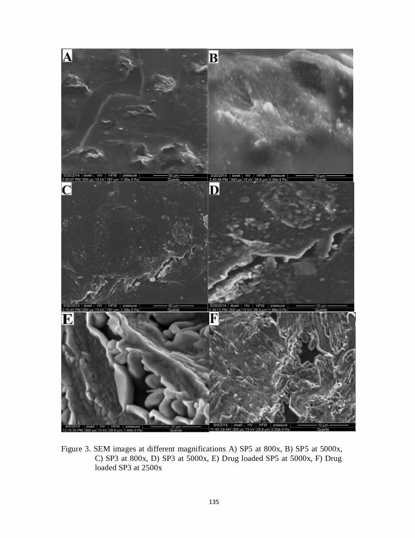
135
Figure 3. SEM images at different magnifications A) SP5 at 800x, B) SP5 at 5000x,
C) SP3 at 800x, D) SP3 at 5000x, E) Drug loaded SP5 at 5000x, F) Drug
loaded SP3 at 2500x

136
Hence, it was estimated that SP5 formulation had higher swelling tendency and drug loading
capacity as compared to SP3 formulation. These results were in accordance to determination
of grafting among the polymers by Huacai et al.283 and Wan et al.274 They prepared the
hydrogels were under different powers of microwaves and their moisture absorption was
evaluated, which was higher in hydrogels receiving lower doses of microwaves. The effect of
initiator on grafting determined by them was also indicative of porosity and swelling
behavior.
The entrapment of solute particles in hydrogels depends upon surface roughness as well as
porosity is related with quantity of drug loaded. It could be clearly seen from SEM images
shown in figure 3 E and 3 F for drug loaded hydrogel SP5 and SP3 respectively, that higher
amount of drug could be loaded in SP5 due to its more porous structure.
5.4.3 XRD
The diffraction patterns of hydrogels loaded with drug were compared with pure drug. X-ray
diffractograms of pure Captopril and drug-loaded Hydrogels are presented in Figure 4. The
XRD scan of plain Captopril had shown characteristic sharp and intense peaks between 0°
and 50° (2θ) due to its crystalline nature as shown in Figure 3 diffraction pattern (b). The
appearance of a sharp peak at 2 θ= 27.79° is the characteristic of captopril.
In comparison to diffractogram of pure captopril, the Captopril loaded formulations had low
intensity and dense peaks. The sharp peak of drug were significantly reduced suggesting the
amorphous distribution of drug into polymeric network, as it can be observed from figure 4
diffractogram (a).
Captopril is a crystalline drug and the presence of intense peaks in diffractrogram ‘b’ was
due to crystallinity of the Captopril. On the other hand, the hydrogels formed by crosslinking
of polymers usually develop an amorphous structure with denser peaks. After loading of drug
into PVA-co-AMPS hydrogel, the captopril was entrapped and dispersed in hydrogel
formulation. The characteristic sharp peak at 2 θ= 27.79° in pure captopril was much reduced
and broadened in drug loaded hydrogel as could be clearly observed from diffractograms a
and b.

137
Figure 4. X-Ray Diffraction patterns of Drug loaded hydrogel SP5 (a) and pure Captopril(b)
Therefore, the intensity and sharpness of peaks was reduced in captopril loaded hydrogel, only
indicated the presence of entrapped drug into polymeric networks of hydrogel formulation. The
resultant evaluation on basis of XRD analysis was in relevance with observations noticed by Giri
et al.,282 where diltiazem hydrochloride was loaded into cross-linked biodegradable IPN hydrogel
beads of pectin and modified xanthan gum. Similar diffraction patterns were noted for pure drug
and drug loaded hydrogels.
5.4.4 Thermal analysis
In order to determine the thermal stability and confirmation of crosslinking, thermal analysis was
performed using Thermogravimetric analysis (TGA) and differential scanning calorimetry (DSC).
Figure 5 illustrates the TGA thermograms of PVA, AMPS and formed polymeric network. Being
monomer, AMPS was less stable at higher tempratures, it was decomposed completely at 243°C.
PVA shows stability upto 227°C, till that temperature there was only 10% weight loss, which was

138
due to loss of water molecules and it was decomposed at about 300°C. At 325°C, weight loss
observed was 70% that degrades complete structure of the polymer. In comparison, thermogram
of hydrogel shows a gradual decrement in stability, indicating its enhanced thermal stability. At
300°C, there was only 25% weight loss and at 489°C still 34% was remaining mass, whereas
PVA was 0.07% at 477°C. Such different stages of thermal degradation in TGA analysis were
also evaluated by by Huacai et al.283 and Bao et al.95
Figure 5. TGA analysis of AMPS, PVA and hydrogel formulation SP5
From the TGA thermograms of individual components (PVA and AMPS) and formed
hydrogel, it could be clearly seen that the developed hydrogel formulation SP5 had
sufficiently strong crosslinked structure to retain itself for longer time periods. Therefore,
maintenance of stability in the polymeric network at high temperature in comparison to
uncrosslinked polymer and monomer was confirmed by TGA.
The alterations in heat capacity as well as enthalpy changes were measured using Differential
Scanning Calorimetry (DSC). Figure 6a and 6b show the DSC-TGA thermograms of
hydrogel and comparative DSC thermograms of drug, drug free hydrogel and drug loaded
hydrogel. The DSC endothermic peaks of cross-linked polymeric network were in
accordance with TGA thermal patterns as shown in figure 6a. Both TGA and DSC

139
thermograms indicates that the deterioration of PVA-co-AMPS started nearly at temperature
250°C.
Figure 6a. DSC-TGA thermogram of PVA-co-AMPS Hydrogel

140
Figure 6b. DSC analysis of SP5 without drug, captopril and Drug loaded SP5
Differential Scanning Calorimetry (DSC) is a well-known and established technique adopted
for quantitative assessment of physicochemical variations in heat capacity of crystalline
drugs, when loaded into hydrogels. The thermal behavior of pure captopril, drug free and
drug loaded SP5 hydrogel was characterized using DSC, as shown in Figure 6b. The
disappearance of characteristic peaks and appearance of other peaks suggests some sort of
interaction of drug with polymers. In figure 6b, the thermogram of pure captopril, the
appearance of sharp peak at 106°C indicated its melting point. This peak of drug was not
shown in DSC thermogram of the drug loaded formulation. A broad peak from 97°C to
160°C was appeared in thermogram of drug loaded hydrogel. It suggests that captopril is
molecularly dispersed in different hydrogel matrix without changing its thermal behavior.
Both formulations, without drug and drug loaded had shown major endothermic peak at
about 240°C, which illustrated the stability of formulation after drug loading without any
marked alteration in thermal stability pattern. These observations are relevant to DSC
analysis of aceclofenac sodium loaded hydrogels evaluated by Manjanna et al.281

141
5.4.5 Swelling Study
The swelling ratios ‘S’ and percent swelling equilibrium (%ES) were calculated by
performing the swelling studies at pH 2 and 7.4. The pH 2 represents the pH of gastric
medium in the stomach and pH 7.4, the intestinal pH. The current work was aimed to
develop a gastroretentive hydrogel formulation; therefore, it must exhibit higher swelling
rates at acidic pH. Greater swelling ratios were observed at pH 2 as compared to swelling at
pH 7.4 in a same manner as determined by Gupta et al.340 However, it was varying from their
work due to constitution of polymeric network. Various other factors were affecting the
swelling/water absorption into the prepared hydrogel formulations are discussed as given
below:
Effect of microwave radiation
In order to provide a proper swelling, the polymers must be sufficiently crosslinked, so that
the obtained material should not dissolve in aqueous medium. The crosslinking density in
hydrogels depends upon the power of microwaves received. Figure 7 illustrates the swelling
ratios of SP1, SP2, SP3 and SP4 hydrogels treated with different intensities of microwave
radiation. These observations are in correspondence to morphological characterizations
performed by scanning electron microscopy. The comparison of swelling capability of these
formulations was observed at lower pH 2 and higher pH 7.4.
Figure 7. Comparative swelling ratios of Formulations SP1, SP2, SP3 and
SP4 at pH 2 and 7.4
0
20
40
60
80
100
120
140
0 20 40 60 80
Swel
ling
Rat
io (q
)
Time (hrs)
SP1 pH 2
SP2 pH 2
SP3 pH 2
SP4 pH 2
SP1 pH7.4
SP2 pH7.4
SP3 pH7.4
SP4 pH7.4

142
The hydrogels treated at 100W were possessing higher swelling than one exposed at 180W,
both containing same concentration of crosslinker, PVA and AMPS. The hydrogels exposed
further to increased radiation dose i.e. at 300W exhibited low swelling, due to greater
crosslinking density among polymers. The high doses above 180W also cause the destruction
of crosslinked structure, that provide an irregular and lesser swelling than other treated at
lower frequency of microwaves.
Hydrogel formulation SP2 (prepared at 180 W) was considered more suitable due to
sufficient strength to maintain its disc integrity while swelling, so that it could remain stable
in the stomach for longer periods of time. However, formulation SP1 had shown more
swelling behavior but lesser mechanical strength due to low crosslinked density. SP3 and
SP4 hydrogel formulations treated at 300 W were showing lesser swelling ability. Figure 8
illustrates the effect of microwave radiation on percent equilibrium swelling.
Figure 8. Effect of microwave radiation dose on Percent Equilibrium Swelling
Figure 8 shows that by increasing the intensity of radiation exposure to hydrogel
formulations led to decrement in percent equilibrium swelling. Thus, it was observed that
microwave radiations due to their ability to initiate the crosslinking reactions effect
crosslinking density and ultimately the swelling of polymeric network. These observations
97.8
98
98.2
98.4
98.6
98.8
99
99.2
99.4
0 50 100 150 200 250 300 350
% E
qu
ilib
riu
m S
wel
lin
g
Watts
Effect of Differnt Radiation dose on %ES
pH 2
pH 7.4

143
were related to earlier findings of Singh et al.,267 and Wan et al.274 where hydrogels were
treated with different intensities of microwave radiations and there moisture absorption
properties were determined, which were inverse of percent grafting. Due to higher grafting,
the polymeric networks have more crosslinking density that ultimately reduces swelling ra tio.
Effect of Initiator concentration
Due to ability of microwave radiations to initiate the reaction for crosslinking of polymers,
lower concentrations of initiator were required. However, increasing the initiator
concentration increases the crosslinking among polymers and consequently reduces the
swelling characteristics as shown in figure 9 and 10. Figure 9 illustrates the comparative
swelling ratios of formulations SP2, SP5 and SP6 at pH 2 and 7.4. These formulations were
exposed to same radiation dose of 180W as well as comprising of same ratios of all
components except initiator concentration.
Figure 9. Comparative swelling ratios of Formulations SP2, SP5 and SP6 at pH 2 and 7.4
The hydrogel formulation SP2 containing higher initiator concentration (0.25% W/W) had
shown relatively low swelling ratio. Swelling of SP5 hydrogel was higher than SP2 as SP5
0
20
40
60
80
100
120
140
160
0 10 20 30 40 50 60 70 80
Swel
ling
Rat
io (q
)
Time (hrs)
SP2 pH 2
SP5 pH 2
SP6 pH-2
SP2 pH7.4
SP5 pH7.4
SP6 pH7.4

144
hydrogel had lesser concentration of initiator (0.15 % W/W). Moreover, highest swelling
ratio of SP6 hydrogel was observed among other (SP2 and SP5) as it had lesser amount of
initiator (0.05% W/W). Figure 10 is showing the effect of initiator concentration on Percent
Equilibrium Swelling. It illustrates that increasing the concentration of initiator decreases the
percent equilibrium swelling.
Figure 10. Effect of initiator concentration on Percent Equilibrium Swelling
The resultant reduction in swelling tendency with increasing initiator concentration were
confirmed earlier by Cheng et al.268 and Wan et al.274 They observed an increment in water
absorption capacity of hydrogels by using an optimum initiator concentration, under
influence of microwave radiations. Above, the optimum amounts of initiator the water
absortion characteristics were showing remarkably decreased.
Effect of AMPS Concentration
The effect of monomer concentration on swelling capability was determined that how the
quantity of AMPS affects the water absorption characteristics of superporous hydrogel
formulations. SP5, SP7 and SP8 formulations containing different amounts of AMPS but
same concentration of polymer (PVA- 1% W/W), crosslinker (MBA- 1 mol % of monomer
concentration), initiator (KPS- 0.15%W/W) and radiation dose were compared in terms of
swelling ratios as illustrated in figure 11.
98.2
98.4
98.6
98.8
99
99.2
99.4
0 0.05 0.1 0.15 0.2 0.25 0.3
% E
qu
ilib
riu
m S
wel
lin
g
Ratio (% w/w)
Effect of Differnt Ratio (%w/w)of Initiator on %ES
pH 2
pH 7.4

145
It was observed that by increasing the amount of monomer AMPS in formulation, there was
increment in swelling behavior. Variation in swelling ratio among these hydrogel
formulations (SP5, SP7 and SP8) was due to higher quantity of AMPS.
Figure 11. Comparative swelling ratios of Formulations SP5, SP7 and SP8 at pH 2 and 7.4
AMPS cause the availability of more sulfonate groups, which increase the hydrophilic
characteristics in polymeric network. At acidic pH, a remarkable ability of water absorption
was noticed because of high ionization of functional groups in AMPS. In addition to
ionization of sulfonic groups, the hydrogen bonds are also reduced that consequently
increased the swelling power of the formed superporous hydrogel. The hydrogel formulation
SP8 possess high quantity of AMPS (30 % W/W) had shown greater swellability as shown in
figure 11. In SP5 formulation, AMPS was lesser than SP8, therefore it had shown lesser
comparative swelling ratio, but it was higher in comparison to SP7 containing 20 % W/W of
hydrogel formulation. Increment in swelling tendency with increasing AMPS content is
corresponding to swelling study performed on AMPS based hydrogel by Qudah et al.239 On
the other hand it was varying from the observations made by Bao et al.,95 where AMPS was
cross-linked with a carboxymethylcellulose in the presence of acrylic acid and acrylamide,
0
20
40
60
80
100
120
140
160
0 10 20 30 40 50 60 70 80
Swel
ling
Rat
io (q
)
Time (hrs)
SP7 pH 2
SP5 pH 2
SP8-2
SP7 pH7.4
SP5 pH7.4
SP8 pH7.4

146
while in the present work, AMPS was only monomer grafted with polymer. Therefore, the
swelling behavior is dependent upon the amount of AMPS. Figure 12 is showing the effect
on AMPS concentration on percent equilibrium swelling.
Figure 12. Effect of AMPS concentrationon Percent Equilibrium Swelling
It could be observed from the figure 12 that by enhancing the amount of AMPS had made an
increment in percent equilibrium swelling of hydrogel formulations. AMPS created the
availability of ionizable sulfonate groups in the prepared superporous polymeric network,
which ultimately led to enhancement of their swelling characteristics.
Effect of Polymer concentration
The effect of polymer concentration on swelling behavior was confirmed by comparing three
hydrogel formulations containing same quantities of monomer, crosslinker, initiator and
radiation dose but different amounts of polymer. Hydrogel formulations SP5, SP9 and SP10
were evaluated for their swelling properties as shown in figure 13.
97.8
98
98.2
98.4
98.6
98.8
99
99.2
99.4
0 5 10 15 20 25 30 35
% E
qu
ilib
riu
m S
wel
lin
g
(% w/w) of AMPS
Effect of Differnt Ratio (%w/w)of AMPS on %ES
pH 2
pH 7.4

147
Figure 13. Comparative swelling ratios of Formulations SP5, SP7 and SP8 at pH 2 and 7.4
SP5 formulation had lower concentration of PVA (1% W/W) in comparison to SP9 and SP10
with PVA concentration 2% and 3%, respectively. A higher swelling ratio of SP5 hydrogel
was noted than other formulations (SP9 and SP10). SP10 formulation, because of high
polymer amount had lower swelling than both SP5 and SP9. Figure 14 depicts the effect of
polymer concentration on percent equilibrium swelling of hydrogels.
These observations are in correspondence to swelling study by Tyliszczak211 and Minhas et
al.339 where a decrement in swelling ability of hydrogels was observed by increasing the
amount of polyvinyl alcohol. However, the results of swelling evaluation were differing from
work of Hosseinzadeh,347 who observed increase in swelling by increasing PVA
concentration from 1.2 to 2.4 wt% in PVA-based hydrogels. By increasing further amount of
PVA created a decrement in absorption characteristics, due to increasing viscosity of reaction
medium that restricts the movements of PVA chains. In comparison, in our case, a decrease
in swelling ratio was noted by increasing the amount of PVA from 1 to 2 wt% in AMPS-
based hydrgels.
0
20
40
60
80
100
120
140
0 10 20 30 40 50 60 70 80
Swe
llin
g R
atio
(q)
Time (hrs)
SP5 pH 2
SP9 pH-2
SP10 pH-2
SP5 pH7.4
SP9 pH7.4
SP10 pH7.4

148
Figure 14. Effect of PVA concentrations on Percent Equilibrium Swelling
Polvinyl alcohol PVA imparts mechamical strength to the polymeric system and possesses
water absorption characteristics, but its swelling capability is low in comparison to AMPS.
Hence, by increasing the quantity of PVA in hydrogel formulation, causes a reduction in
swelling behavior as it could be observed from figure 13 and 14.
5.4.6 In-vitro drug release studies
After loading of captopril into the prepared superporous hydrogel formulations, the drug
release study was performed at pH 2 and pH 7.4 as shown in table 2. The drug release study
at mentioned pH was performed for a period of 24 hours, where USP phosphate buffer was
used as dissolution medium. All the formulations were subjected to drug loading and the
release of entrapped captopril was evaluated. The hydrogel discs possessing higher swelling
ratio were able to entrap higher quantity of drug. The amount of drug release is according to
swelling characteristics, where drug loading is directly related to water retention capability of
polymeric network. The hydrogels which exhibited higher swelling ratio entrapped more
amount of captopril.
98
98.2
98.4
98.6
98.8
99
99.2
99.4
0 1 2 3 4 5
% E
qu
ilib
riu
m S
well
ing
Ratio (% w/w)
Effect of Differnt Ratio (%w/w)of PVA on %ES
pH 2
pH 7.4

149
Table 2. Amount of Captopril loaded and percentage of drug released at pH 2 and pH 7.4
sample
code
Amount of captopril loaded (mg)
per 0.3 gram of dry hydrogel discs
% release of captopril
pH 2 pH 7.4
SP1 202.5 98.08 72.32
SP2 186.8 85.11 54.26
SP3 169.8 69 47.45
SP4 173.8 73.13 49.56
SP5 189.8 93.21 66.95
SP6 194.7 95.93 70.02
SP7 156.78 56.95 33.73
SP8 199.8 97.38 68.23
SP9 179.67 82.27 55.66
SP10 171.36 73.26 48.85
Hydrogel formulations SP1, SP6 and SP8 were loaded with higher quantity of drug and
ultimately had shown high amounts of drug release at both lower and higher pH (2 and &
7.4). Moreover, drug release was higher at acidic pH as compared to pH 7.4. This was due to
swelling characteristics of AMPS, which is not much dependent on pH of medium. However,
being an AMPS-based hydrogel it exhibited swelling to greater extent in low acidic pH as
compared to neutral or higher alkaline pH.
Figure 15 presents a comparison of three hydrogel formulations SP1, SP5 and SP10. Among
them, the SP1 hydrogel, the components were loosely crosslinked due to exposure at low
dose of microwave radiation, therefore it entrapped and released higher quantities of
captopril. On the other hand, formulation SP10 had shown lower swelling ratio due to higher
PVA concentration, hence lower drug loading and drug release was observed. Thus,
relatively more amount of drug was loaded and ultimately released in formulations exhibiting
more swelling power. The drug release kinetics of prepared superabsorbent hydrogels was
determined by different kinetic models mentioned in table 3. The kinetic models used were

150
Zero order kinetics, First order kinetics, Higuchi model, Korsmayer-Peppas model and
Wilbull model.
Figure 15. Captopril released up to 24 h from PVAco-AMPS hydrogels
(SP1, SP5, and SP10) in dissolution media of pH 2 and pH 7.4
Table 3. Determination of coefficient (R2), K and release exponent of various drug release kinetic models
Sample code
pH Zero order kinetics
First order kinetics
Higuchi model Korsmeyer-Peppas model Wilbull model
R2 K R2 K R2 K R2 K n R2
SP1 2 0.8745 5.6976 0.2887 0.3167 0.9716 26.424 0.9853 46.38 0.17 0.7897
7.4 0.9342 3.5779 0.4773 0.2569 0.9907 13.688 0.9751 17.0869 0.37 0.8113
SP2 2 0.9523 4.2344 0.3654 0.2920 0.9976 19.968 0.9978 26.9387 0.33 0.8703
7.4 0.959 2.9744 0.4552 0.2466 0.9914 11.024 0.9855 7.7882 0.7212 0.8429
SP3 2 0.7901 5.2216 0.2508 0.3005 0.9136 20.596 0.9864 28.0953 0.504 0.722
7.4 0.9057 2.7629 0.4807 0.2357 0.9834 10.309 0.9841 15.3971 0.644 0.7661
SP4 2 0.8196 5.3027 0.2702 0.3015 0.9388 21.272 0.9875 33.8937 0.2203 0.7465
7.4 0.888 2.8506 0.425 0.2471 0.9745 11.571 0.9837 16.3121 0.3081 0.7492
SP5 2 0.9027 4.8442 0.3018 0.3106 0.9833 24.761 0.9923 40.3292 0.2164 0.8114
7.4 0.9268 3.5394 0.4 0.2699 0.9908 15.37 0.9871 20.0108 0.35 0.829
SP6 2 0.8745 5.9259 0.2866 0.3198 0.9716 27.483 0.9853 48.2390 0.1749 0.7898
7.4 0.9342 3.7212 0.4733 0.2600 0.9907 14.237 0.9751 17.7714 0.3792 0.8115
SP7 2 0.9765 2.7670 0.5472 0.2335 0.9962 10.381 0.9978 9.2038 0.568 0.8643
7.4 0.9944 1.3716 0.6247 0.1916 0.9738 5.998 0.9861 4.6877 0.647 0.9277
SP8 2 0.8745 5.7746 0.288 0.3177 0.9716 28.781 0.9853 47.0076 0.1749 0.7898
7.4 0.9342 3.6262 0.4759 0.2579 0.9907 13.873 0.9751 17.3178 0.3792 0.8114
SP9 2 0.9681 4.0971 0.4882 0.2671 0.997 15.574 0.9954 15.8369 0.4907 0.8594
7.4 0.9917 1.9592 0.5609 0.2274 0.9716 9.843 0.991 7.3925 0.6717 0.9289
SP10 2 0.9692 3.2586 0.5306 0.2497 0.9865 13.233 0.9814 12.2555 0.54 0.8774
7.4 0.9879 1.9694 0.6073 0.2160 0.9826 8.474 0.9977 8.7381 0.48 0.8986
0.00
20.00
40.00
60.00
80.00
100.00
120.00
0 5 10 15 20 25 30
% c
um
ula
tive
dru
g re
leas
e
Time (hr)
Drug Release %
SP1 pH 2
SP1 pH 7.4
SP5 pH 2
SP5 pH 7.4
SP10 pH 2
SP10 pH 7.4

151
The results calculated in terms of values of release coefficient and release exponent from
PVA-co-AMPS copolymer containing different components, polymer concentration, initiator
concentration and radiation dose. The mechanism of drug release was indicated by the values
of n i.e. release exponent as presented in table 3.
The formulations were best fitted into the above mentioned kinetic models when values of R
were near to ‘1’. There was variation in value of ‘n’ among the formulations prepared. The
SP7 formulation containing lower monomer concentration, SP9 and SP10 hydrogel
formulations with higher amounts of polymer were exhibiting non-fickian diffusion
mechanism of drug release with value of n greater than or equal to 0.45 but lesser than 1.
Moreover, SP3 formulation receiving higher dose of radiation (300 W) and higher initiator
concentration were also following non-fickian diffusion. Other formulations with higher
amounts of monomer and lesser polymer concentration were following fickian diffusion with
value of ‘n’ less than 0.45.
Conclusion
It was concluded from the discussion, that super-porous hydrogels were prepared
successfully under influence of microwave radiations. Using low concentrations of initiator
could successfully develop polymeric network of polyvinyl alcohol and AMPS by
microwave radiations. The formed hydrogel had ability to swell excessively at acidic pH and
it could be retained in the stomach, releasing captopril at low pH for longer durations of time.
Therefore, the synthesized copolymer could be a promising candidate as carrier for captopril
in treating hypertension.

152
Chapter no.6
In-vivo Evaluation of hydrogel
formulations for Controlled Release
Drug Delivery of Captopril

153
Abstract:
A specific high performance liquid chromatography–ultravoilet spectrometric (HPLC-UV) assay
was developed for the determination of captopril in plasma. It was conducted on prepacked Hypersil
C8 column at room temperature using Phosphate buffer: acetonitrile (75:25 v/v) as a mobile phase,
pH adjusted at 2.8 with o-phosphoric acid and at a flow rate of 1.0 ml/min, while UV detection was
performed at 205 nm. The retention time was 6.5 min for captopril. The liquid-liquid extraction
method was used for the detection of captopril. Dithiothreitol was added in extraction medium in
order to stabilize the captopril extracted from plasma. Standard curve was linear in captopril
concentration ranging from 50 to 2000 ng/ml. The method had a suitable sensitivity to detect drug at
low concentrations due to its lower limit of quantification (LLOQ) value noted as 50ng/ml. Intra -
batch as well as inter-batch precision and accuracy measured had shown good results. The
extraction efficiency of captopril was ranging from 95 to 99.9 %. The method developed was
applied for the pharmacokinetic study in 24 rabbits for evaluation of pharmacokinetic parameters of
captopril. The method was simple, rapid, reliable, specific and sensitive with ability to determine
drug plasma concentrations from rabbits for longer duration.
154
6.1 Introduction
Angiotensin converting enzyme (ACE) inhibitors are commonly used for treatment of heart diseases
such as hypertension and heart failure. Among them, captopril, is an orally active potent ACE
inhibitor and widely accepted due to its antihypertensive action attained within 45 min to 1 hour
after oral administration.348-351 The in-vivo analysis of captopril is difficult because of its stability
concerns; the presence of sulphydrylgroup causes its self-dimerization, resulting in formation of
captopril disulfide. Moreover, captopril also binds to endogenous compounds such as cysteine,
glutathione as well as plasma proteins. The captopril disulfide is pharmacologically inactive but it
may serves as reservoir of active drug due to its reversible conversion to free captopril.352-354
To overcome the above mentioned problem, the detection or measurement of free captopril
concentration needs molecule derivatization or an addition of chemical stabilizer in biological
samples to prevent the formation of captopril disulphide.353 Various Fluorescence or UV active agent
such as N-(1-pyrenyl) maleimide(NPM) and p-bromophenacyl bromide(p-BPB), have been used as
chemical stabilizers. The formation of captopril disulfide can be controlled by lowering the pH
below 4, adding chelating agents (EDTA) or antioxidants.355-358 Dithiothreitol (DTT) added to the
plasma samples has ability to reconstitutes captopril from its disulfide, by increasing free thiol
content from serum albumin.359, 360
For the determination of captopril or its metabolites in blood or plasma, several analytical methods
have been reported. The most widely investigated are HPLC methods, including HPLC with
florescence and UV detector,361-365 gas chromatography (GC) or gas chromatography-mass
spectrometry (GC-MS) techniques, fast solid phase extraction (SFE) or liquid- liquid extraction
based on several evaporation and concentration steps. Other techniques like enzyme immunoassay
and radioimmunoassay (RIA) have also been investigated.366-369 The method should be successfully
applied to accurately measure total captopril concentration on a large number of plasma samples.
The present work describes a simple HPLC method for the determination of captopril using UV
detector. The values for LOD, precision of area and linearity show good performance of analysis.
The pharmacokinetics and the relative bioavailability of captopril were studied using blood samples
of 24 healthy rabbits. A written approval for required animal study was taken from Pharmacy
Research Ethics Committee (PREC), The Islamia university of Bahawalpur, Pakistan.

155
The objective of this work was to evaluate the bioavailability of captopril from three controlled
release hydrogel formulations (SP5, R3 and S3). The High Performance Liquid Chromatography
(HPLC) method with UV detector, using liquid-liquid extraction was developed and successfully
utilized for the determination of captopril in plasma.
6.2 EXPERIMENTAL METHODS
6.2.1 Instrumentation and analytical conditions
Agilent 1100 series HPLC system consisted of LC-10 AT VP pump, DGU-14 AM on-line degasser,
Rheodyne manual injector fitted with a 20 µL loop, and SPD-10 AVP UV–VIS detector and a
Hypersil BDS C 8 (250 X 4.6 mm) column was utilized for separation. Chromatographic system was
integrated via Shimadzu model CBM- 102 Communication Bus Module to P-IV computer loaded
with CLASS-GC software (Version 5.03) for data acquisition and mathematical
calculations.Centrifuge Machine (Model 4000-China), Vortex Mixer (Seouline BioScirnce-Korea),
pH Meter (WTW pH 300-Germany), Ultrasonic Bath (Fisher Scientific FS 28 H-Germany), Electric
Balance (Percia XB 120A), Membrane Filter (Sartorius, 0.45μm filters-Germany),Distillation Plant,
Micropipettes (Softpet- Finland), Filtration Assembly (Pyrex-France), Distillation Plant (WDA/4 R
& M England).
6.2.2 Materials
Captopril was a gift from Benson Pharmaceuticals, Industrial Area, Islamabad, Pakistan. All organic
solvents used for the mobile phase and extraction procedure were of HPLC grade. Methanol,
Acetonitrile, Orthophosphoric acid, Dichloromethane were purchased from Merck (Germany),
Diethyl-ethyl ether from AnalaR (England) and Dithiothreitol from Sigma (USA). Plasma samples
were obtained from healthy rabbits maintained under suitable conditions, not receiving any drug
substance.
6.2.3 Preparation of the Mobile Phase
The mobile phase prepared was comprising of 0.1 M Potassium dihydrogen phosphate buffer (75%)
and acetonitrile (25 %) in proportions. The pH of mobile phase was adjusted to pH 2.8 by
orthophosphoric acid and filtered through filtration assembly.

156
6.2.4 Stock and working solutions
Stock solution of 100 µg mL-1 of captopril was prepared by dissolving 10 mg of drugs in100 mL
volumetric flask using mobile phase as a diluent. Further dilutions were prepared in the range of 50-
2000 ng/ml captopril. Solutions were prepared once and subjected to intra-day and inter-day
variations of method and analyzed each time before drug analysis in biological samples, stored at
20°C. Then, 20 µL of these solutions were injected into HPLC system and chromatographed.
Dithiothreitol (200 mmol/l) solution was prepared at concentrations of 30.84 g/l. It was used for the
detection of captopril by reconstituting it from its disulfide dimer from plasma samples.
6.2.5 Drug-plasma solution
Standard curve was constructed to determine concentration range of captopril in rabbits. Blood
samples were collected from ear vein of healthy rabbits in heparin containing centrifuge tubes and
immediately centrifuged at 3500 rpm for 10 min. The supernatant obtained was stored at -20°C.
After thawing, the plasma was spiked with working solutions to obtain different concentrations of
captopril for construction of standard curve. All calibration curve samples (non-zero samples),
except blank plasma, were prepared by spiking blank plasma aliquots of 500 µl each, with 100 µl of
the intermediary captopril solutions, to yield final plasma concentrations of 50, 100, 200, 400, 800,
1200, 1600 and 2000 ng/ml.
6.2.6 Chromatographic Analytical Conditions
An Isocratic High performance Liquid Chromatography system (Agilent 1100 Series) comprising of
Quanta Pump and Degasser was used. Hypersil BDS C 8 (250 X 4.6 mm) column was utilized for
detection of analytes. The HPLC analysis was performed at ambient temperature, using a flow rate
1ml/min. The UV-detector was set at λ max of 205 nm.
6.2.7 Sample Extraction
Drug was extracted from plasma samples, using liquid- liquid extraction technique. To above formed
samples for calibration curve, ranging from 50 to 2000 ng/ml, 3mL diethyl ether/dichloromethane
(65/35) along with 0.04 mL of 200 mM dithiothreitol solution were added and the samples were
vortex-mixed for 30 to 40 seconds. The tubes were then centrifuged at 3000 rpm for 5min. The
upper organic layer was carefully removed, transferred to reacti-vials, and evaporated to dryness

157
with a gentle stream of nitrogen in a dry bath at 37ºC. A 100 μL aliquot of mobile phase, comprised
of phosphate buffer (75%), acetonitrile (25%) and orthophosphoric acid (0.1%), was added to the
tubes, which were then vortex-mixed for 15 seconds to reconstitute the residue. Then 20 μL were
injected into liquid chromatography system and the peak areas were noted for each concentration.
6.3 Method validation
6.3.1 Specificity
Plasma samples were collected randomly from three rabbits and three humans (drug free subjects).
Plasma samples from human and rabbit were taken to determine the extent to which endogenous
plasma components could affect the retention time of captopril. They were spiked with captopril
standard solution at three levels of drug concentrations 400 ng/ml, 800 ng/ml and 1400 ng/ml. The
spiked plasma samples were processed by liquid-liquid extraction procedure and chromatographed
for determination of peak area and retention time.
6.3.2 Linearity and Standard Curve Preparation
Standard curves were constructed using eleven non-zero calibration points ranging from 50 to 2000
ng/ml. The plasma samples were spiked with drug solutions to obtain final concentration of 50, 100,
200, 400, 600, 800, 1000, 1200, 1400, 1600 and 2000 ng/ml. These samples were prepared in
duplicate and subjected to liquid-liquid extraction. Five replicates of all drug concentration of each
plasma sample were injected into HPLC and Chromatographed. The average of their peak area was
calculated and standard calibration curves were constructed of both plasma samples (by plotting
peak area versus concentrations of the samples). Linear least-square regression analysis, with
weighing fact were of 1/x, was performed to assess the linearity, as well as to generate the standard
calibration equation: y = ax + b, where y is the peak–area ratio, x the concentration, a the slope and b
is intercept of regression line.
6.3.3 Lowest Limit of detection (LLOD) and quantitation (LLOQ)
The lowest limit of detection (LLOD) is a minimum amount of substance in a sample that could only
be detected but not necessarily quantitated, while the quantitation limit (LLOQ) is the lowest amount
of analyte in a sample which can be quantitatively determined properly with an accuracy and
precision.

158
6.3.4 Precision and accuracy
The calibration curve samples prepared were taken for assessment of precision and accuracy. The
intra-batch precision and accuracy was determined among five replicates from each batch (batch 1
and 2 batch). The average of all concentrations determined in both batches was calculated separately.
Then, batch 1 and batch 2 were subjected to inter-batch precision and accuracy. Accuracy is
percantage of mean of values to the reference value as given by equation:
Accuracy = % mean / Reference (i)
Whereas precision is percent coefficient variance (% CV) of standard deviation to mean of values:
Precision = S.D / mean (ii)
6.3.5 Extraction efficacy
Extraction efficacy (Recovery) was calculated by comparing peak-area of extracted sample of drug
to that of the unextracted pure drug solutions used for plasma spiking.Three concentrations (50-2000
ng/ml) of captopril were selected to determine extraction efficiency. The peak area of spiked plasma
samples were compared with standard diluents, using five replicates of each concentration.
6.4 Results and Discussions
The retention time observed by injecting 20µL standard captopril solution into HPLC system was
6.5 as shown in figure 2.
Figure 2. Retention time of captopril detected from
standard drug solution

159
6.4.1 Specificity
The detection of captopril from rabbit and human plasma samples were compared for their
respective peak area and retention time. Figure 3 and 4 represent the chromatograms of blank plasma
sample of rabbit and human, respectively. There was no marked variation observed in peak area and
retention time of captopril from chromatograms of spiked rabbit plasma and spiked human plasma as
shown in figure 5 and 6, respectively. The retention time remained in range of 6.4 to 6.5 min in both
plasma samples.
Figure 3. Blank plasma sample of rabbit
Figure 4. Blank Plasma samples of Human

160
Figure 5. Captopril detected from spiked rabbit plasma sample
Figure 6. Captopril detected from spiked human plasma sample

161
The peak areas noted from spiked plasma samples from both sources (rabbit plasma and human
plasma) with three levels of captopril concentrations 400ng/ml, 800ng/ml and 1400ng/ml are
presented in table 1 as given below.
Table 1. Peak area determined from Rabbit Plasma and Human Plasma
QC sample
(ng/ml) Peak area
( from Rabbit Plasma)
( mAU*)
Average
Peak area
( mAU*)
Peak area
(from Human Plasma)
( mAU*)
Average
Peak area
( mAU*)
400 362.36 371.27 359.72 364.45 366.23 358.34 359.72 361.43
800 761.66 770.24 760.29 764.06 756.34 759.45 767.29 761.02
1400 1359.56 1368.26 1362.35 1363.39 1370.34 1374.31 1353.55 1366.06
It was observed from the retention time and peak area determination (as illustrated by figure 5 and
figure 6) that there was no marked variation for detection of captopril from both plasma samples
(rabbit and human). Hence, this method could be specifically applied for detecting captopril in
plasma samples in human and animal (rabbit).
The determination of specificity of captopril was relevant to the evaluations made by Rezende et
al.361 in terms of using, randomly selected normal plasma, hyperlipemic and hemolyzed plasma
samples from distinct healthy human subjects. However, in the current method, plasma was taken
from healthy animal (rabbit) as well as human and compared, which resulted in no marked variation
in retention time of captopril in the presence of endogenous plasma components from both types of
plasma samples.

162
6.4.2 Lowest Limit of detection (LLOD) and quantification (LLOQ)
The sensitivity of this liquid-liquid extraction can be evaluated by determination of lowest limit of
detection (LLOD) and lowest limit of quantification (LLOQ). By this liquid- liquid extraction
method, the lowest detection limit of captopril was found as 20 ng/ml. This amount of drug in
standard solutions and spiked plasma samples could be detected only, but cannot be quantified. The
lower limit of detection was lower in comparison to the value of LLOD determined by Du et al.370
and Alves Soares et al.,360 where dithiothreitol in same quantity was added during liquid-liquid
extraction. The variation in detection was because in that case mass spectrometer was used for
detection, while in our method the drug was detected by UV detector. However, HPLC-UV
detection is less senseitive but more simple and could be conveniently performed.
The other parameter, LLOQ that is the lower limit of quantification, was determined as 50ng/ml.
Therefore, 50ng/ml was the lowest quantity of captopril that could be measured and effectively
quantified. For this reason it was considered as lowest concentration in the preparation of standard
curve and in plasma samples for determination of precision and accuracy.
6.4.3 Linearity and Calibration Curve
Figure 7a and 7b show the calibration curves (1 and 2) prepared from spiked plasma samples ranging
from 50- 2000 ng/ml. A good linear response to this method was observed in the concentrations
ranging from 50, 100, 200, 400, 600, 800, 1000, 1200, 1400, 1600 and 2000 ng/ml as shown in the
calibration curves.The drug concentrations used in preparing standard curve 1 and standard curve 2
were named as batch 1 and batch 2, respectively.

163
Figure 7a. Standard calibration curves prepared from spiked plasma samples of batch 1
Figure 7b. Standard calibration curves prepared from spiked plasma samples of batch 2
Five replicates of each concentration were injected into HPLC and chromatographed in both
batches (1 and 2). Then, mean of peak area of five replicates of each concentration in a batch was
calculated and standard curves were prepared for batch 1 and batch 2. The standard curves, their
slope, intercept and the correlation coefficient were determined, which could be seen in figure 7a
and 7b.
y = 0.9795x - 19.417R² = 0.9998
0
500
1000
1500
2000
2500
0 500 1000 1500 2000 2500
Pe
ak a
rea
(mA
U*)
Concentration (ng/ml)
Standard Curve 1
Peak Area
Linear (Peak Area)
y = 0.9802x - 24.425R² = 0.9998
0
500
1000
1500
2000
2500
0 500 1000 1500 2000 2500
pea
k ar
ea
(mA
U*)
concentration (ng/ml)
Standard curve 2
Series1
Linear (Series1)

164
6.4.4 Precision and accuracy
Intra-batch accuracy and precision evaluated are presented in table 2 (for batch 1 and for batch 2).
Accuracy and precision between two batches (batch 1 and batch 2) of plasma samples used for
calibration curves were determined as shown in table 3. Within run precision and accuracy of five
replicates of each concentration was calculated in each batch.
Table 2. Intra-batch precision and accuracy for captopril determination in spiked plasma samples
Analysis of Batch 1
Drug concentration (ng/ml)
Five replicates of captopril concentrations
determined in rabbit plasma
mean Standard
Deviation
Precision
(%CV)
Accuracy
1 2 3 4 5
50 49.223 51.843 46.373 46.893 48.533 48.573 2.1667 4.4608 97.146
100 88.66 94.43 93.47 90.763 98.153 93.097 3.62 3.8950 93.097
200 194.57 192.64 177.18 193.43 181.26 187.81 8.0079 4.2636 93.909
400 390.52 396.70 391.26 382.59 388.34 389.88 5.1073 1.3099 97.471
600 571.09 587.94 590.82 593.67 596.85 588.07 10.052 1.7093 98.013
800 763.83 774.18 807.09 786.09 802.05 786.65 18.245 2.3193 98.331
1000 991.29 998.64 989.48 994.66 986.18 992.05 4.7926 0.4831 99.205
1200 1174.1 1181.5 1198.0 1175.0 1189.8 1183.7 10.175 0.8595 98.644
1400 1391.8 1385.0 1395.3 1370.5 1398.0 1388.1 10.990 0.7916 99.155
1600 1563.0 1568.9 1561.0 1557.8 1570.8 1564.3 5.4272 0.3469 97.771
2000 1950.6 1946.0 1931.4 1932.3 1939.0 1939.9 8.3953 0.4327 96.996
Analysis of Batch 2 Drug
concentration
(ng/ml)
Five replicates of captopril concentrations
determined in rabbit plasma
mean Standard
Deviation
Precision
(%CV)
Accuracy
1 2 3 4 5
50 43.963 48.353 50.043 46.373 47.693 47.285 2.2793 4.8203 94.5705
100 85.263 89.073 90.093 95.083 93.093 90.521 3.7866 4.1831 90.5212
200 180.07 189.02 191.53 192.30 179.36 186.45 6.2770 3.3664 93.2296
400 380.82 388.77 394.35 392.59 386.11 388.53 5.3785 1.3843 97.1328
600 589.33 587.15 578.02 586.01 591.19 586.34 5.0608 0.8631 97.7238
800 784.63 781.59 787.89 783.19 798.93 787.24 6.20025 0.787587 98.406
1000 988.46 975.27 987.62 989.25 989.86 986.02 5.463 0.554 98.609
1200 1163.2 1169.9 1173.2 1178.1 1181.1 1173.1 7.0313 0.5993 97.7634
1400 1386.0 1387.3 1391.7 1388.2 1392.4 1389.1 2.8249 0.2033 99.2266
1600 1556.0 1560.4 1569.7 1575.1 1573.4 1566.9 8.3716 0.5342 97.9360
2000 1931.8 1936.4 1944.1 1937.7 1943.4 1938.7 5.1113 0.2636 96.9368

165
Table 3. Inter-batch precision and accuracy for captopril determination in spiked plasma samples
Drug concentration
(ng/ml)
Average of replicates in Batches (1 and 2) Mean Standard
Deviation
Precision
(%CV)
Accuracy Batch 1 Batch 2
50 48.573 47.285 47.929 0.910754 1.900214 95.858
100 93.097 90.521 91.809 1.821507 1.984018 91.809
200 187.81 186.45 187.13 0.961665 0.513902 93.565
400 389.88 388.53 389.205 0.954594 0.245268 97.30125
600 588.07 579.94 584.005 5.748778 0.984371 97.33417
800 786.65 771.44 779.045 10.75509 1.380549 97.38063
1000 992.05 982.09 987.07 7.042784 0.713504 98.707
1200 1183.7 1173.1 1178.4 7.495332 0.63606 98.2
1400 1388.1 1389.1 1388.6 0.707107 0.050922 99.18571
1600 1564.3 1566.9 1565.6 1.838478 0.11743 97.85
2000 1939.9 1938.7 1939.3 0.848528 0.043754 96.965
In batch 1, the values of precision are ranging from 0.34% to 4.4% and and accuracy determined is
in the range of 93.00% to 99.15%. Similarly, the values of precision and accuracy in batch 2 were
ranging from 0.26% to 4.8% and 90.53% to 99.22%, respectively.
The inter-batch precision and accuracy was in the range of 0.04 to 1.98 and 90.8 to 99.18,
respectively. The results obtained were within the acceptance criteria for precision and accuracy. It
was assessed from deviation values that were falling in ±15% of the authentic values.371 The
determination of intra- batch and inter-batch precision and accuracy were according to that
determined Rezende et al.,361 where intra-batch precision and accuracy (% CV) ranged from 2.49 to
5.66%, and 97.15 to 105.77%, respectively. Method inter-batch precision (% CV) and accuracy
ranged from 0.88 to 1.95%, and 98.85 to 104.22%, respectively. Similar findings were observed by
Alves Soares et al.,360 where liquid-liquid extraction was used for detection of captopril.
Moreover, the retention time of noted in all chromatograms of different concentrations and within
batch and among batches was ranging from 6.4 to 6.7 which was near to the retention time of
captopril determined by Jinsong et al.372

166
6.4.5 Extraction efficacy
Extraction efficacy (Recovery) was calculated by comparing the peak-area of extracted sample of
drug to that of the unextracted pure drug solutions used for plasma spiking as presented in table 4.
The pure Captopril solutions of concentrations (50, 100, 200, 400, 600, 800, 1000, 1200, 1400,
1600 and 2000 ng/ml) were considered as standard for comparison with spiked plasma samples.
The peak area of spiked plasma samples were compared with standard diluents, using five
replicates of each concentration (50, 100, 200, 400, 600, 800, 1000, 1200, 1400, 1600 and 2000
ng/ml). Their averages were calculated and then comparison was made to determine the
percentage of drug that could be possibly recovered from spiked plasma samples. The percent
recoveries of all samples with drug concentrations ranging from 50 to 2000 ng/ml are presented in
table 4.
Table 4. Percent Recoveries of different Captopril concentrations from spiked plasma
The values of percent recovery are varying from approximately 95.00% to 99.96%. The mean
percent recovery of captopril from the spiked plasma samples was 98.13%. These results indicated a
successful recovery of drug from biological samples, as more than 95% of drug could be detected
from plasma samples. Therefore, it evaluates the suitability of method for effective determination of
captopril from blood after its oral administration. The results of percent recovery were determined
in accordance to extraction efficiency (% recovery) measured by Rezende et al.361 and Sultan et
al.273 where percent recoveries of captopril were calculated in the similar manner.
Drug concentration
(ng/ml)
Peak area of spiked
plasma
(mAU*)
Peak area of standard
Solution
(mAU*)
% Recovery
50 28.76 29.62 97.09
100 73.28 77.37 94.71
200 168.01 176.43 95.22
400 370.07 374.29 98.87
600 568.26 572.39 99.27
800 766.84 767.13 99.96
1000 972.24 978.81 99.33
1200 1163.93 1169.42 99.53
1400 1368.36 1378.29 99.27
1600 1544.53 1576.45 97.97
2000 1920.13 1954.78 98.23

167
6.5 Application of the Method
The objective of this work was to evaluate the bioavailability of captopril from three controlled
release hydrogel formulations. The High Performance Liquid Chromatography (HPLC) method
with UV detector, using liquid- liquid extraction was developed and utilized for the determination
of captopril in plasma of rabbits.
Twenty four (24) rabbits weighing 2.0 to 2.5kg were used in this study. They were divided into four
groups (1, 2, 3 and 4) each group having six rabbits. Animals were starved for 24h prior to
administration of the drug. Each rabbit in group 1 (Control) was given a single dose of 25 mg pure
captopril with lactose enclosed in a hard gelatin capsule, which was ingested with 20 mL water
orally through a catheter. Rabbits in group 2, 3 and 4 were given hydrogel formulations SP5, R3 and
S3, respectively. Water was provided during starvation and throughout the experiment. Food was
withheld for 2 hours following drug administration to prevent drug food interaction, after which
they were fed at 4h and 10h after dosing.
Blood samples (3 mL each) were collected into heparin-containing centrifuge tubes at 0, 0.5, 1, 2, 4,
8, 12,16, 24 hours from the rabbit ear vein following drug administration. Plasma was separated
witha centrifuge (Heraeus Instrument, Biofuge Primo, Germany) at 3500 rpm for 10 min at room
temperature and the plasma recanted and stored at –20ºC until assayed for captopril content. Plasma
captopril concentrations were determined in duplicate by reversed phase liquid chromatography-
UV.
Briefly, 3mL diethyl ether/dichloromethane (65/35) was added to 0.5mL plasma with 0.04mL of
200mM dithiothreitol soltion, and the samples were vortex-mixed for 30 to 40 seconds. The tubes
were then centrifuged at 3500rpm for 5min at 4ºC. The upper organic layer was carefully removed,
transferred to reacti- vials, and evaporated to dryness with a gentle stream of nitrogen in a dry bath
at 37ºC. A 100 μL aliquot of mobile phase, comprised of phosphate buffer (75%), acetonitrile
(25%) and orthro phosphoric acid (0.1%), was added to the tubes, which were then vortex-mixed
for 15 seconds to reconstitute the residue. Then 20 μL were injected into liquid chromatography
system.

168
The drug was eluted with phosphate buffer and acetonitrile (75:25) using orthophosphoric acid to
adjust the pH to 3. It was filtered by a vacuum filter system equipped with a 0.8 mm filter and was
degassed by ultrasonic treatment.
6.5.1 Operating conditions
The flow rate was 1.0 mL/min at ambient temperature and UV detection was performed at 205 nm.
The total run time was 10 min and retention time was 6.5 min.
6.5.2 Plasma Concentrations Profile and Pharmacokinetic parameters of Captopril
Plasma concentrations of administered pure captopril and drug released from hydrogels
were calculated from peak area by using Microsoft excel 2010.
The pharmacokinetic parameter such as maximum concentration Cmax (ng/ml), time to reach
maximum concentration, Tmax (hr), elimination half-life ‘t’1/2 el, elinmination rate constant ke (hr-1)
Area under curve AUC (ng/ml*hr), area under the product of concentration and time AUMC(ng
/ml*h2), Clearance (L/h) , volume of steady state concentration Vss (L) volume of distribution Vd
(L), and other parameters mentioned in tables 4 to 27 were calculated using Kinetica 5.0.
The value of absorption half-life t1/2 (a) was calculated by method of inspection. From the value of
absorption half-life t1/2 (a), the absorption rate constant ka was estimated. Following equations were
used for the calculation of absorption half-life t1/2 (a) and absorption rate constant ka .
t1/2 (a) = (1)
ka= (2)

169
6.5.2.1 GROUP 1
The mean ± standard deviation concentrations of captopril determined in plasma of six rabbits
(group 1) after oral administration of control are presented in table 5. Figure 8 illustrates mean ±
standard deviation of plasma concentrations versus time profile of Captopril. The values of mean ±
standard deviation of Bioavailability and Pharmacokinetic parameters are given in table 6.
Table 5. Mean ± standard deviation of plasma concentration (ng/ml) of
Captopril (free drug) in 6 rabbits in Group 1
S. No. Time (Hrs) Concentration (ng/ml)
Captopril
1 0 0 ± 0
2 0.5 442.31 ± 21.70
3 1 782.18 ± 12.69
4 2 639.31 ± 21.19
5 4 363.55 ± 47.93
6 8 52.288 ± 5.35
7 12 0 ± 0
8 16 0 ± 0
9 24 0 ± 0
Figure 8. Mean ± standard deviation of plasma concentrations vs. time
profile of Captopril 25 mg plotted on rectangular co-ordinate
graph, in group 1.
-200
0
200
400
600
800
1000
0 5 10 15 20 25 30
Co
nce
ntr
ati
on
(ng/
ml)
Time (hours)
Group 1
Series1

170
Table 6. Mean ± standard deviation of Bioavailability
and Pharmacokinetic parameters of Captopril
administered in an oral dose of 25 mg (free
drug) in rabbits of Group 1.
Parameters Captopril (free drug)
Cmax (ng/ml) 782.198 ± 12.68
Tmax (hr) 1 ± 0
AUC (ng /ml*h) 3092.25 ± 176.03
AUMC (ng /ml*h2) 9144.63 ± 682.72
MRT (hr) 2.96 ± 0.081
ke (hr-1) 0.425 ± 0.017
ka (hr-1) 3.46 ± 0
Tlast (hr) 2.63 ± 0.0613
t1/2 el (hr) 1.61 ± 0.066
t1/2 (a)(hr) 0.2 ± 0
Vss (L) 23.94 ± 1.047
Vd (L) 18.98 ± 1.38
ClT (L/h) 8.106 ± 0.48
R2 0.987 ± 0.012
HVD (hr) 3.32 ± 0.317
From the results presented in table 5 and table 6, it can be concluded that maximum level of
drug concentration was attained in a sample taken after 1 hour of oral administration of
captopril powder enclosed in hard gelatin capsule, hence time to reach maximum
concentration tmax was 1 hour. Because of solubility of captopril in aqueous solution and
administration in powder form, it was immediately dissolved in biological fluids and reached
its maximum concentration in blood in shorter time. It was detected from the plasma samples
of rabbits taken upto a time period of 8 hours.
Similar results were also evaluated by Mallick et al.374 in assessment of bioavailability and
pharmacokinetic parameters of another drug nifedipine from controlled release micro
capsules in healthy rabbits. In their research work the control (reference) used was drug
enclosed in hard gelatin capsules and orally administered in a group of rabbits, as it was used
in our work for captopril.

171
6.5.2.2 GROUP 2
The mean ± standard deviation concentrations of captopril determined in plasma of six rabbits
(group 2) after oral administration of SP5 hydrogel formulation are presented in table 7.
Figure 9 illustrates mean ± standard deviation of plasma concentrations versus time profile of
Captopril. The values of mean ± standard deviation of Bioavailability and Pharmacokinetic
parameters are given in table 8.
Table 7. Mean ± standard deviation of plasma concentration (ng/ml)
of Captopril released from SP5 hydrogel formulation in 6
rabbits in Group 2
S. No. Time (Hrs) Concentration (ng/ml)
Captopril
1 0 0 ± 0
2 0.5 151.49 ± 8.48
3 1 361.19 ± 17.37
4 2 485.08 ± 23.36
5 4 661.85 ± 5.78
6 8 611.88 ± 11.58
7 12 378.87 ± 12.19
8 16 186.31 ± 22.92
9 24 50.570 ± 5.29
Figure 9. Mean ± standard deviation of plasma concentrations vs. time
profile of Captopril released from SP5 hydrogel formulation
plotted on rectangular co-ordinate graph, in group 2.
-200
0
200
400
600
800
0 5 10 15 20 25 30
Co
nce
ntr
ati
on
(ng/
ml)
Time (hours)
Group 2
Series1

172
Table 8. Mean ± standard deviation of Bioavailability and
Pharmacokinetic parameters of Captopril released
from SP5 hydrogel formulation in Group 2
Parameters Captopril (SP5 formulation)
Cmax (ng/ml) 661.853 ± 5.79
Tmax (hr) 4 ± 0
AUC (ng /ml*h) 8647.81 ± 133.05
AUMC (ng /ml*h2) 79382.36 ± 2619.9
MRT (hr) 9.17 ± 0.195
Ke (hr-1) 0.165 ± 0.009
ka (hr-1) 0.86 ± 0
Tlast (hr) 8.41 ± 0.127
t1/2 el (hr) 4.19 ± 0.28
t1/2 (a)(hr) 0.8 ± 0
Vss (L) 26.53 ± 0.46
Vd (L) 17.5 ± 1.13
ClT (L/h) 2.87 ± 0.058
R2 0.999 ± 0.0064
HVD (hr) 11.82 ± 0.146
It was shown from the data presented in table 7 and 8 that the maximum concentration of
drug was achieved in 4 hours after oral administration of SP5 hydrogel formulations. The
average Cmax was 661.85ng/ml, which was lesser than average Cmax (782.19ng/ml) noted after
oral administration of control. However, time to reach maximum concentration was 4 hours
that was higher in comparison control. Area under curve (AUC) and area under the product
of concentration and time (AUMC) were also increased.
The results indicated from the estimation of pharmacokinetic and bioavailability parameters
that the drug was released from the gastroretentive hydrogel formulation SP5 at controlled
rate and maintained its concentration in blood for longer periods of time. The drug released
by gastroretentive hydrogel was in correspondence with that observed by Nagarwal et al.,375
for determination of release of Cinnarizine Hydrochloride in rabbits blood from its
Gastroretentive Tablet. The pharmacokinetic parameters and bioavailability of Cinnarizine
Hydrochloride Gastroretentive Tablet were compared with reference (oral suspension) in
healthy rabbits in the similar manner.

173
6.5.2.3 GROUP 3
The mean ± standard deviation concentrations of captopril determined in plasma of six
rabbits (group 3) after oral administration of R3 hydrogel formulation are presented in table
9. Figure 10 illustrates mean ± standard deviation of plasma concentrations versus time
profile of Captopril. The mean ± standard deviation of Bioavailability and Pharmacokinetic
parameters are given in table 10.
Table 9. Mean ± standard deviation of plasma concentration (ng/ml) of
Captopril released from R3 hydrogel formulation in 6 rabbits in
Group 3
S. No. Time (Hrs) Concentration (ng/ml)
Captopril
1 0 0 ± 0
2 0.5 110.5 ± 11.99
3 1 221.78 ± 31.94
4 2 381.43 ± 17.1
5 4 491.09 ± 39.8
6 8 598.48 ± 15.33
7 12 512.11 ± 17.63
8 16 329.25 ± 13.68
9 24 61.15 ± 11.44
Figure 10. Mean ± standard deviation of plasma concentrations vs
time profile of Captopril released from R3 hydrogel
plotted on rectangular co-ordinate graph, in group 3
-200
0
200
400
600
800
0 5 10 15 20 25 30Co
nce
ntr
atio
n (n
g/m
l)
Time (hours)
Group 3
Series1

174
Table 10. Mean ± standard deviation of Bioavailability
and Pharmacokinetic parameters of Captopril
Captopril released from R3 hydrogel
formulation in 6 rabbits in Group 3
Parameters Captopril (R3 formulation)
Cmax (ng/ml) 598.48 ± 15.33
Tmax (hrs) 8 ± 0
AUC (ng /ml*h) 9320.255 ± 222.97
AUMC (ng /ml*h2) 11295.45 ± 964.66
MRT (hr) 10.88 ± 0.175
ke (hr-1) 0.173 ± 0.006
ka (hr-1) 0.43 ± 0
Tlast (hr) 10.03 ± 0.109
t1/2 el (hr) 3.95 ± 0.16
t1/2 (a)(hr) 1.6 ± 0
Vss (L) 29.21 ± 0.99
Vd (L) 15.31 ± 0.704
ClT (L/h) 2.678 ± 0.067
R2 0.9774 ± 0.013
HVD (hr) 14.94 ± 0.47
The results presented in table 8 and table 10 as well as depicted from figure 10, that time to
reach maximum concentration Tmax of captopril after oral administration captopril loaded R3
hydrogel was 8 hour. It was higher than one observed after administering captopril loaded
SP5 hydrogel in group 2. The maximum concentration Cmax attained (598.48 ng/ml) was
reduced in comparison to control. The values of mean AUC and mean AUMC were higher
even when compared to that in group 2. The captopril was released from R3 formulation and
detected in plasma samples upto 24 hours. It could be evaluated from the data obtained in
group 3, that captopril was released at slower rate from the controlled release hyfrogel
formulation R3 and remained available in blood for longer duration.

175
6.5.2.4 GROUP 4
The mean ± standard deviation concentrations of captopril determined in plasma of six rabbits
(group 4) after oral administration of S3 hydrogel formulation are presented in table 11.
Figure 10 illustrates mean ± standard deviation of plasma concentrations versus time profile
of Captopril. The values of mean ± standard deviation of Bioavailability and Pharmacokinetic
parameters are given in table 12.
Table 11. Mean ± standard deviation of plasma concentration (ng/ml)
of Captopril released from S3 hydrogel formulation in 6
rabbits in Group 4
S. No. Time (Hrs) Concentration (ng/ml)
Captopril
1 0 0 ± 0
2 0.5 131.4134 ± 29.31
3 1 277.95 ± 22.08
4 2 408.065 ± 14.02
5 4 557.54 ± 11.92
6 8 618.8584 ± 8.89
7 12 512.0184 ± 30.56
8 16 215.585 ± 29.69
9 24 0 ± 0
Figure 11. Mean ± standard deviation of plasma concentrations vs.
time profile of Captopril released from R3 hydrogel
formulation plotted on rectangular co-ordinate graph in
group 4.
-200
0
200
400
600
800
0 5 10 15 20 25 30
con
cen
trat
ion
(ng/
ml)
Time (hours)
Group 4
Series1

176
Table 12. Mean ± standard deviation of Bioavailability and
Pharmacokinetic parameters of Captopril released
from S3 hydrogel formulation in 6 rabbits in Group 4
Parameters Captopril (S3 formulation)
Cmax (ng/ml) 618.85 ± 8.896
Tmax (hr) 8 ± 0
AUC (ng /ml*h) 8878.77 ± 149.743
AUMC (ng /ml*h2) 91632.7 ± 2318.5
MRT (hr) 10.32 ± 0.188
Ke (hr-1) 0.145 ± 0.004
ka (hr-1) 0.43 ± 0
Tlast (hr) 7.83 ± 0.045
t1/2 el (hr) 4.707 ± 0.15
t1/2 (a)(hr) 1.6 ± 0
Vss (L) 29.057 ± 0.64
Vd (L) 19.13 ± 0.6
ClT (L/h) 2.82 ± 0.053
R2 0.876 ± 0.022
HVD (hr) 12.76 ± 0.18
The rabbits in group 4 were given captopril loaded S3 hydrogel formulation orally. The drug
was released at slower rate and attained maximum concentration in 8 hours similar to that in
group 3, hence Tmax was 8 hour as shown in table 12. The mean of maximum concentration of
captopril attained was 618.85 ng/ml. The drug remained detectable for duration of 16 hour
from rabbit blood.
The results presented from in vivo studies on rabbits had shown a difference in plasma
concentration of captopril attained after oral administration of free drug (control) and three
hydrogel formulations (SP5, R3 and S3) as shown in the figure 12. A clear variation in
maximum concentration of captopril achieved by control and controlled release hydrogel
formulations could be seen from the figure. The drug reached its maximum concentration in
short period of time in case of free drug administered in capsules. In comparison to control, a
lesser as well as slower drug release was observed from drug loaded hydrogels that was
dependent upon the swelling characteristics of crosslinked polymeric networks. Drug release

177
pattern from these hydrogels was also varying from one another due to their different
constitution.
Figure 12. Comparison of Mean Plasma concentrations vs. time profile of control (free
drug), Captopril released from SP5, R3 and S3 hydrogel formulation plotted
on rectangular co-ordinate graph.
Therefore, the maximum concentration Cmax was reduced while Tmax was increased by hydrogels.
The value of area under curve AUC (calculated by trapezoidal rule) and elimination half-life were
higher for controlled release hydrogel formulation. The drug could be available for longer periods
of time after administration of Captopril loaded hydrogels. Among the hydrogel formulations Tmax
was 4 hour for SP5 and 8 hr for R3 and S3 formulations, drug was available for 24 hours from
SP5 and R3 but in S3 formulation it was detectable for relatively shorter periods (16 hours). The
formulation SP5 was a super porous hydrogel and had gastro-retentive ability due to excessive
swelling at acidic pH. For this reason the time to reach maximum concentration levels (4 hours)
was lesser than other two formulations (S3 and R3) having maximum concentration noted at 8
hour sample. Hydrogels S and R resealed maximum drug concentrations at intestinal pH, hence
-100
0
100
200
300
400
500
600
700
800
900
0 5 10 15 20 25 30
Tim
e (h
ou
rs)
Concentration (ng/ml)
Comparative Plasma Concentrations
Free Drug
SP5
R3
S3

178
attaining maximum concentration of captopril in 8 hours. All the results obtained were subjected
to statistical analysis in terms of mean ± standard deviation as presented in the tables 5 to 12.
Similar patterns of captopril release in rabbits were observed by El-Shabouri et al.376 where
release of captopril was evaluated from prepared captopril buccoadhesive tablets, which were
compared with an immediate release captopril tablet as control. A decrement in maximum
concentration and increment in time to reach maximum drug concentration was observed in the
controlled release formulations. In the same manner release patterns of other drugs from their
controlled release formulation have been compared with controls using HPLC analysis. For
example, prepared sustained release leflunomide microcapsules were compared with
conventional tablets of leflunomide (control), which had shown similar comparative drug release
patterns from sustained release formulations.377 In another work, release of theophylline from
pH-Sensitive hydrogels of Carboxymethyl Chitosan had been evaluated in comparison to a
commercially available theophylline tablet as control.378 Similarly, an evaluation of the
pharmacological activity of glipizide was conducted by Abdelbary et al.379 on controlled release
microcapsules and compared with commercially available immediate release Minidiad® 5 mg in
normal healthy albino rabbits. Therefore, the controlled release drug delivery systems have
ability to maintain optimum levels of drug for longer durations of time.

179
CONCLUSION
In short, it was concluded that the above HPLC method is specific, sensitive, rapid and easy to
perform determination of captopril. The limit of quantification, small sample volume and
chromatographic time of this method makes it advantageous for adaptation to routine assay
requirements and enables simultaneous determination of captopril because of good separation of
the chromatographic peaks of captopril from plasma peaks. Results were accurate and precise and
confirmed by the statistical parameters. Reliability, rapidness, simplicity, sensitivity, economical
nature, good recovery and precision of this method give it advantage over the other reported
methods. Moreover, the developed method was successfully applied in determination of captopril
in plasma samples following the oral administration of captopril loaded hydrogels.

180
Overall Conclusion

181
From the whole research project, including methodologies, techniques, the results obtained and
entire discussion, it has been revealed that hydrogel formulations were successfully developed
for controlled release drug delivery of captopril. Swellable polymeric networks with varying
characteristics were prepared, by free radical polymerization, using different ratios of polymers,
monomers as well as techniques. A fast technique for hydrogel synthesis was utilized by the
assistance of microwave radiations, presenting better results in comparison to conventional
method (using thermostatic water bath). The prepared HPMC/PVA-co-poly(acrylic acid)
hydrogels had remarkable ability to entrap as well as protect drug and release it at a control rate,
hence providing an opportunity for drug to exert its required therapeutic effects for prolonged
durations. The other hydrogel formulations prepared, comprised of HPMC, acrylic acid along
with AMPS had better swelling capability as compared to HPMC/PVA-co-poly(acrylic acid)
hydrogels, due to the presence of highly ionize-able sulfonic groups.
Moreover, the gastro-retentive hydrogel formulations were prepared, which had additional
advantages of significantly high swelling power and ability to be retained in stomach and
releasing captopril for longer periods of time at low pH of stomach. Thus, captopril remains
more stable at acidic pH and relatively lesser quantity will be able to produce enhanced
therapeutic effect. These gastro-retentive polymeric networks had an optimum strength and
flexibility to maintain their shape and texture during swelling measurement, drug loading and
in-vitro drug release study, without any noticeable/apparent breaking. Other in-vitro
characterization measurements were presenting crosslinking among polymeric networks of
polymers and monomers as analyzed by FT-IR, porous structure was observed in morphological
analysis by scanning electron microscopy (SEM). The X-Ray Diffraction had shown amorphous
distribution or dispersion of crystalline drug in formed cross-linked structures. The thermal
analysis (TGA and DSC) evaluated the desirable stability of cross-linked polymeric networks as
well as stability of drug loaded into loaded into these hydrogels. The swelling properties were
significantly affected by the varying ratios of monomers, polymers and cross linker in each type
of hydrogel formulation. Radiation dose was an important factor affecting the swelling ratios of
hydrogels synthesized under their influence, due to their ability to initiate crosslinking
interactions. The drug loading and release from hydrogels were directly related to their water
absorption characteristics.

182
HPLC-UV analytical method, using liquid-liquid extraction procedure was developed for
determination of bioavailability and pharmacokinetic parameters of captopril in plasma samples
of rabbits. The liquid chromatography method, due to its specificity and sensitivity had ability
to detect even smaller concentrations of captopril. It was concluded from the in-vivo evaluation,
that captopril was released from hydrogel formulations at controlled rates and lesser amounts in
comparison to control. However, the drug remained available in blood for longer durations of
time hence providing a safe and efficacious amount for useful effects.
Therefore, the aim of preparing a hydrogel formulation was achieved fulfilling the criteria of
controlled release drug delivery system successfully by increasing time to reach maximum
concentration and overall bioavailability of drug. Thus, optimum levels of drug could be
maintained in blood for required therapeutic effects in the treatment of hypertension as well as
other heart disorders, reducing the dosing frequency, lower incidence of side effects and
ultimately improving patient compliance.