Embed Size (px)
Citation preview
Indian Journal of Pathology and Oncology 20196(4)627ndash635
Content available at iponlinejournalcom
Indian Journal of Pathology and Oncology
Journal homepage wwwinnovativepublicationcom
Original Research Article
Study of histopathological findings in gallbladder diseases
Cyrus Dara Jokhi1 Sujata R Kanetkar1 Nikita V Vohra1
1Dept of Pathology Krishna Institute of Medical Sciences Karad Maharashtra India
A R T I C L E I N F O
Article historyReceived 22-07-2019Accepted 19-10-2019Available online 22-11-2019
KeywordsUltrasonographyAdenocarcinomaIncidental carcinomaCholecystitis
A B S T R A C T
Introduction Cholecystectomy is one of the most frequently performed emergency abdominal operationas the gallstones are one of the major causes of morbidity and mortality all over the world affecting 10of adult populationAccording to consolidated report of population based registries of India 2009-2011 (by National centrefor disease informatics and research) India has shown high incidence of gall bladder cancer in femalepopulation of Kamrup (Assam) district which is highest among the worldClinical diagnosis of the cholecystitis is made based on history and physical examination along withlaboratory and radiological findings However histopathological study is the gold standard for the diagnosisof cholecystitis Histopathological examination many time reveals an unusual diagnosis bearing significantimplications on the treatment prognosis and outcome of the patientAim and Objectives To study various lesions encountered in cholecystectomy specimens onhistopathologyObservations and Results The most common histopathological diagnosis was chronic cholecystitis seenin 112 out of 130 cases (863) followed by acute cholecystitis 12 cases (92) There were 4 (30)cases of adenocarcinoma of gallbladder Total 925 chronic cholecystitis cases were calculous while 666 of acute cholecystitis and 25 (one out of four) of gall bladder carcinoma were associated with gallstones Out of total 113 (869) gallstones ndash 4 (35) were pure cholesterol stones 14(124) pigmentedstones and 95 (841) were mixed stones In the present study all 4 cases (100) of malignancy wereadenocarcinoma out of which 1 case was (25) papillary adenocarcinoma 1 case (25) was poorlydifferentiated and 2 cases (50) were moderately differentiated adenocarcinoma Amongst the four casesof adenocarcinoma 3 (75 ) cases showed serosal infiltration and liver infiltrationConclusions Clinical presentation of diseases of gall bladder is vague Even malignancy of gallbladderpresents late in the course and with nonspecific symptoms which can misguide the clinicians Findingsof malignancy were subtle on radiological examinations also Diagnosis of malignancy was made only byhistopathological examination (15 cases as incidental carcinoma)
copy 2019 Published by Innovative Publication This is an open access article under the CC BY-NC-NDlicense (httpscreativecommonsorglicensesby40)
1 Introduction
Cholecystectomy is one of the most frequently performedemergency abdominal operation as the gallstones are oneof the major causes of morbidity and mortality all over theworld affecting 10 of adult population1 Cholecystectomyspecimens are mostly associated with gall stone called ascalculous cholecystitis and because of westernization of life
Corresponding authorE-mail address cyrusdjokhigmailcom (C Dara Jokhi)
style incidence of gall stones is increasing in India Hencegallbladder is a frequent surgical specimen in most of thehistopathology laboratories
According to consolidated report of population basedregistries of India 2009-2011 (by National centre for diseaseinformatics and research) India has shown high incidenceof gall bladder cancer in female population of Kamrup(Assam) district which is highest among the world2
Clinical diagnosis of the cholecystitis is made basedon history and physical examination along with laboratory
httpsdoiorg1018231jijpo20191212394-6784copy 2019 Innovative Publication All rights reserved 627
628 Dara Jokhi R Kanetkar and V Vohra Indian Journal of Pathology and Oncology 20196(4)627ndash635
and radiological findings However histopathological studyis the gold standard for the diagnosis of cholecystitisHistopathological examination many time reveals anunusual diagnosis bearing significant implications onthe treatment prognosis and outcome of the patientHence present study was undertaken to emphasize therole of histopathological examination in cholecystectomyspecimens and its correlation with clinical presentation
2 Aim and Objectives
To study various lesions encountered in cholecystectomyspecimens on histopathology
3 Materials and Methods
31 Source of data
The present study includes prospective cases of two yearsfrom June 2015 to May 2017 and also includes cases fromretrospective archival of data of two and half years ie ndashJan 2013 to May 2015 Thus it includes 130 cases ofcholecystectomy specimens during Jan 2013 to June 2017
32 Inclusion criteria
All Cholecystectomy specimens received in the departmentof Pathology in our institute during Jan 2013 to June 2017
33 Exclusion criteria
There is no exclusion criterion in this study
34 Method of data collection
The specimens were collected in 10 formalin followingscrutiny of the patient details and identity The specimensof cholecystectomy were fixed in formalin for 12-24 hoursGross examination of all the specimens were done Bitsfrom one representative full-thickness section from thefundus one through the body one through neck of thegallbladder and one cross section of the cystic duct marginwere taken Additional sections were taken when focallesions were present These were followed by routineparaffin processing
4 Observations and Results
Out of these 130 cases in 129 (9923) cases gallbladders was surgically resected as a therapeutic measurefor clinically suspected cholecystitis and in remainingonly 1 case (077) gall bladder was removed withPancreaticoduodenectomy
Out of 130 cholecystectomy 111 were laproscopic and19 were open laprotomy cholecystectomy
Out of 19 open laprotomy procedures 2 procedures wereinitially started as laproscopic cholecystectomy and were
converted in to open procedures as gall bladders havingnecrotizing cholecystitis got ruptured during procedure
Maximum numbers of patients were in the age group of41-50 years (261) followed by 51-60 years (208) and61-70 (184) years
Mean age of the patient was 50 years Oldest patient was80 years and the youngest was 17 years of age
Females were common in age group of 31 to 60 yearsAfter 60 years male patients were more common
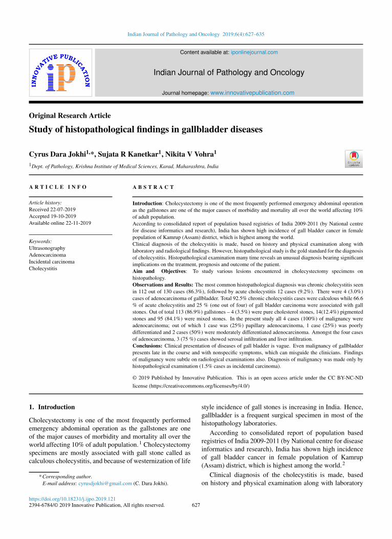
Fig 1 Photograph showing specimen of s hrunken gallbladderwith chronic cholecystitis Lumen is filled with bile and singlepure cholesterol stone
Fig 2 Photograph showing specimen of gallbladder withgangrenous cholecystitis case showing severely congested serosa
Out of 107 cases of chronic cholecystitis 99 (925)were calculous cholecystitis and 8 were acalculouscholecystitis
Out of 12 cases of acute cholecystitis 8(666) werecalculous cholecystitis and 4 were acalculous cholecystitis
Out of 4 cases of carcinoma specimens 1 showedcalculous
Dara Jokhi R Kanetkar and V Vohra Indian Journal of Pathology and Oncology 20196(4)627ndash635 629
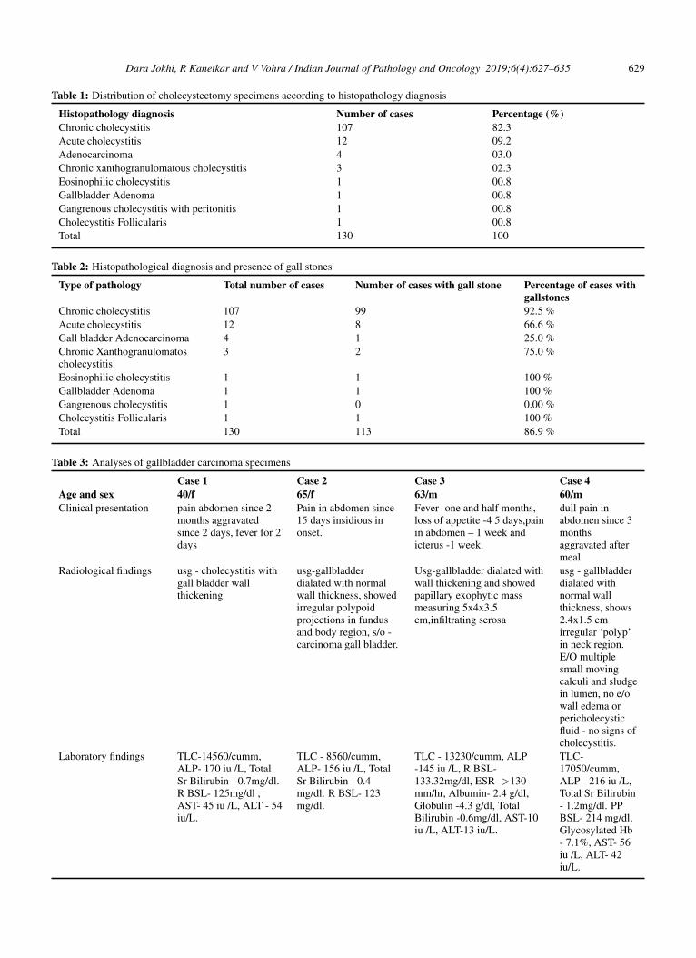
Table 1 Distribution of cholecystectomy specimens according to histopathology diagnosis
Histopathology diagnosis Number of cases Percentage ()Chronic cholecystitis 107 823Acute cholecystitis 12 092Adenocarcinoma 4 030Chronic xanthogranulomatous cholecystitis 3 023Eosinophilic cholecystitis 1 008Gallbladder Adenoma 1 008Gangrenous cholecystitis with peritonitis 1 008Cholecystitis Follicularis 1 008Total 130 100
Table 2 Histopathological diagnosis and presence of gall stones
Type of pathology Total number of cases Number of cases with gall stone Percentage of cases withgallstones
Chronic cholecystitis 107 99 925 Acute cholecystitis 12 8 666 Gall bladder Adenocarcinoma 4 1 250 Chronic Xanthogranulomatoscholecystitis
3 2 750
Eosinophilic cholecystitis 1 1 100 Gallbladder Adenoma 1 1 100 Gangrenous cholecystitis 1 0 000 Cholecystitis Follicularis 1 1 100 Total 130 113 869
Table 3 Analyses of gallbladder carcinoma specimens
Case 1 Case 2 Case 3 Case 4Age and sex 40f 65f 63m 60mClinical presentation pain abdomen since 2
months aggravatedsince 2 days fever for 2days
Pain in abdomen since15 days insidious inonset
Fever- one and half monthsloss of appetite -4 5 dayspainin abdomen ndash 1 week andicterus -1 week
dull pain inabdomen since 3monthsaggravated aftermeal
Radiological findings usg - cholecystitis withgall bladder wallthickening
usg-gallbladderdialated with normalwall thickness showedirregular polypoidprojections in fundusand body region so -carcinoma gall bladder
Usg-gallbladder dialated withwall thickening and showedpapillary exophytic massmeasuring 5x4x35cminfiltrating serosa
usg - gallbladderdialated withnormal wallthickness shows24x15 cmirregular lsquopolyprsquoin neck regionEO multiplesmall movingcalculi and sludgein lumen no eowall edema orpericholecysticfluid - no signs ofcholecystitis
Laboratory findings TLC-14560cummALP- 170 iu L TotalSr Bilirubin - 07mgdlR BSL- 125mgdl AST- 45 iu L ALT - 54iuL
TLC - 8560cummALP- 156 iu L TotalSr Bilirubin - 04mgdl R BSL- 123mgdl
TLC - 13230cumm ALP-145 iu L R BSL-13332mgdl ESR- gt130mmhr Albumin- 24 gdlGlobulin -43 gdl TotalBilirubin -06mgdl AST-10iu L ALT-13 iuL
TLC-17050cummALP - 216 iu LTotal Sr Bilirubin- 12mgdl PPBSL- 214 mgdlGlycosylated Hb- 71 AST- 56iu L ALT- 42iuL
630 Dara Jokhi R Kanetkar and V Vohra Indian Journal of Pathology and Oncology 20196(4)627ndash635
Table 4 Histopathological examination and outcome of gallbladder carcinoma patients
Case 1 Case 2 Case 3 Case 4Gross appearance ulceration of mucosa
mild thickening ofwall lumen filledwith mucus stoneand bile
gw soft to firmpapillary exophytictumor measuring53x45x42 cm bodyand fundus andshowing discretepapillary excrescencesin remaining part ofbody infiltrating serosa
gw nodular firm massreplacing body andneck of gallbladder
filled with mucusstones and bile at neck irregularulceroinfiltrative aswell as proliferativegrowth m 18x15x12cm
Presence and type of gallstone
no gall stone no gall stone no gall stone 5 ndash black pigmented
Histopathological type Well to moderatelydifferentiatedinvasiveadenocarcinoma
Well differentiatedpapillaryadenocarcinomainfiltrating up to serosawith evidence ofperitonitis
Poorly differentiatedadenocarcinoma withfocal mucin secretioninvading and fungatingthrough serosa
Well to moderatelydifferentiatedadenocarcinomareaching up to serosawith perineuralinvasion with chronicnonspecific calculouscholecystitis free fromtumor ndash cystic duct ofgall bladder
Reaching up to serosa butnot invading it
- Positive - -
Serosal invasion Present Absent Present PresentLiver parenchymainvasion
Present Absent Present Present
Outcome 10 days ndash expired 16 days- improved 15 days ndash expired 13 days - improved
Fig 3 Photograph showing specimen of gallbladder with acutecholecystitis showing swollen enlarged gallbladder with congestedserosa and thickened wall Lumen shows congested mucosa andmucosal ulceration
Out of total 130 cases 4 (3) patients of gall bladdercarcinoma-
1 Youngest patient was 40 years old and oldest was 65years old
2 On ultrasonography Case No1 showed diffuse wallthickening and Case No4 was diagnosed as polyp CaseNo 2 and 3 were diagnosed as gallbladder carcinoma butdiagnosis of malignancy was missed on USG in Caes No1and Case No4
3 Case No 2 was well differentiated papillarycarcinoma Case no 3 was of poorly differentiatedadenocarcinoma and Case No 1 and 4 were well tomoderately differentiated adenocarcinoma
Fig 4 Photograph showing specimen of gallbladder carcinomawith diffuse gallbladder wall thickening and ulceration of mucosa
41 Outcome and duration of hospital stay
Out of Total 130 Cases ndashImproved- 127 casesEspired due to gall bladder disease2 cases (both were carcinoma of gallbladder Out
of 4 cases of gall bladder carcinoma 2 cases expired inpostoperative period)
Expired due to other cause- 1 case (due to periampullaryadenocarcinoma)
Dara Jokhi R Kanetkar and V Vohra Indian Journal of Pathology and Oncology 20196(4)627ndash635 631
Fig 5 Photograph showing specimen of gallbladder carcinomawith growth replacing body and neck and infiltrating serosa
Fig 6 Photomicrograph of showing well differentiated papillaryadenocarcinoma (H amp E 40x)
Average days of hospital stay after laproscopic surgery ndash54 days
Average days of hospital stay after laprotomy (open)surgery - 1100 days
5 Discussion
Findings of age and sex distribution in this study are inconcordance with studies of Daniel Moslashnsted Shabanzadehet al3 Vikash Talreja et al4 Yen-Chun Chen et al5 TadashiTerada6
While most of the chronic cholecystitis were calculous(in present study 897) there was no significant differencenoted in gross as well as histological features of chroniccalculous cholecystitis and chronic acalculous cholecystitisAccording to literature also its doubtful to call stone asdirect etilogical factor for chronic cholecystitis rather theseboth processes formation of stone and development ofchronic inflammation are parallel processes due to chronicsupersaturation of bile and stasis7
In the present study acute cholecystitis comprises 92of total cholecystectomy cases This finding matches with
the study of Damor NT et al1 Vikash Talreja et al4
Incidence of acute cholecystitis varies round the globebut still it remains the second most common cause ofgallbladder pathology after chronic calculous cholecystitis
In present study out of total 130 cases 113 (869) hadgallstones this finding is in concordance with study of RThamil Selvi et al8 and Tadashi Terada6
Gallstones are directly responsible as a etiological factorin only acute calculous cholecystitis as the stone insidelumen in gallbladder neck or in cystic duct obstructs flowof bile That obstruction and accumulation of bile causesischemia to mucosa due to pressure effect as well as itcauses chemical injury to mucosa because of prolongedstasis of bile in lumen
Out of total 113 (869) gallstonesndash4 (35) were purecholesterol stones 14(124) pigmented stones 95 (841)were mixed stone Findings are in concordance withPradhan SB9 Dattal DS et al10
High serum bilirubin values with pigment stones arepredominant in children with hemolytic diseases but inpresent study no such case was observed
Most of the gallstones in north India are cholesterolstones (80) while in south India they are pigmented (60)
Appearance of cholesterol gall stones varies withcholesterol content of stone According to literature puregallstones account for 10 of total gallstones11 and theyare single large oval and yellowish Stones with lowercholesterol content are designated as mixed Dependingup on the proportion of calcium carbonate bilirubin andphosphate mixed stone may be gray yellow to gray blackin colour According to literature also 70-80 stones aremixed stones11 They are usually smaller multiple andfaceted11 In comparison pigmented stones are multiplesmall black or brown in colour8
In present study total 897 chronic cholecystitis werecalculous while 583 of acute cholecystitis and 25 of gallbladder carcinoma were associated with stone
According to literature 70-80 gallbladder cancerare associated with gallstones12 But in present studygallbladder (GB) stones were found more often in patientswith acute cholecystitis than in patients with gallbladdercarcinoma Patients with gallbladder carcinoma showedtypical masses or focal enhanced wall thickening whencompared to diffuse wall thickening in patients with acutecholecystitis Findings were in concordance with study ofKim SH13 et al
In present study out of total 130 cases 107 (823) werechronic cholecystitis Out of total 107 chronic cholecystitiscases 96(897) were calculous while 8(103) wereacalculous Findings are in concordance with study ofChiam H K et al14 Vikash Talreja et al4 R Thamil Selvi etal8
Jones-Monahcm K et al15 observed that change fromopen to laparoscopic cholecystectomy was accompanied by
632 Dara Jokhi R Kanetkar and V Vohra Indian Journal of Pathology and Oncology 20196(4)627ndash635
Table 5 Comparison of type of cholelithiasis among received cholecystectomy specimens with other studies
Study Year Cholesterol Pigment MixedPradhan SB 2009 1250 0875 7875 R Thamil Selvi et al 2011 1282 6025 1025 Tadashi Terada 2013 1900 4700 3400 Rajani Sharma et al 2014 2265 5185 2549 Samriddhi Sood et al 2016 3651 1830 4519 Dattal DS et al 2017 2290 1040 6670 Present study 2017 0350 1240 8410
more females undergoing operation for acalculous biliarydisease
Majority of gallbladder with chronic cholecystitis wereshrunken with unremarkable serosa On microscopy gall-bladders showed all typical features of chronic cholecystitislike chronic inflammatory infiltration of mucosa laminapropria Rokitansky Aschoff sinuses were present in40(37) cases of chronic cholecystitis One caes showeddiffuse calcification of gallbladder wall microscopically butgrossly specimen was unremarkableThese findings matcheswith study of R Thamil Selvi et al8 and Dattal DS et al10
There was no mortality reported in cases of chroniccholecystitis cholecystitis
Distribution of different types of pathologies showschronic cholecystitis (823) was most common findingfollowed by acute cholecystitis (920) followed bycarcinoma (3) which are in concordance with Amjad JAlmuslamani et al16 Ivy Sharma et al17 Vikash Talreja etal4 while Tadashi Terada6 found gall bladder carcinomaas more common lesion then acute cholecystitis Butin his study Tadashi Terada6 observed 94 cases aschronic cholecystitis while only 6 cases are having allother spectrum of gallbladder diseases which is not veryuncommon finding
Gall bladders with chronic cholecystitis were shrunkenwith unremarkable serosa where gallbladder with acutecholecystitis and acute on chronic cholecystitis wereswollen and showed exudate and congestion on serosaTotal 3 cases showed perforation all 3 cases had biliaryperitonitis
Total 113 (869) gallbladder had calculous In 84(684) cases gallbladder lumen was filled with bile In29(238) cases lumen was filled with mucous and bilethose gallbladders were swollen and congested and hadexudate on serosa where diagnosed as acute or acute onchronic cholecystitis In 4(76 ) cases there was presenceof growth in lumen and those 4 cases were diagnosed ascarcinoma
The case of Eosinophilic cholecystitis was characterizedby predominant infiltrate of eosinophils in the wall ofgallbladder Its etiology was unknown Conditions likeinfections drugs medicinal herbs autoimmune disordersallergy eosinophilic gastroenteritis parasitic infestationhypereosinophilic syndrome etc were excluded
The case of Cholecystitis follicularis was havinglymphoid follicles with hyperplastic germinal centersin all layers of gallbladder Grosly gallbladder hadfeatures of chronic cholecystitis Patient did not haveany lymphadenopathy organomegaly or peripheral bloodlymphocytosis As IHC study is necessary to ruleout lymphoma1819 may be needed in presence of suchsymptoms
There were 3 cases of Chronic Xanthogranulomatouscholecystitis Grossly the specimens were havingmarked thickening of wall and mucosal ulcerationsMicroscopically wall of gallbladders were infiltrated byfoamy macrophages in lamina propria and muscularislayers
Comparison of occurrence of gall bladder carcinoma invarious studies around the world shows incidence variesbetween 026 - 300 In the present study it was 300which was in concordance with study of Archana Tiwari etal20 Tadashi Terada6 and Romero Gonzalez21
Studies in western countries done by Taylor (1998) Dix(2003) Darmas (2007) Oommen (2007) Bazoua (2007)Byars (2012) of United Kingdom Romero-Gonzalez (2012)from Maxico Matthyssens (2006) from France andWolkomir (1991) from US believed that Histopathologicalexamination done post cholecystectomy should be selectiveand not routine for all cases As these studies believedon radiological examination as well as intraoperativeexamination of gallbladder specimen by surgeon couldexclude the need of histopathological examination if nosuspicion of malignancy on radiological and intraoperativeexamination
According to studies in India the highest incidence ofgallbladder cancer is in Kamrup district (Assam state) thatis 35 of all cancer in males and 82 in females (132 outof 3809 in male and 232 out of 2814 cases in females - totalcancer cases study done from year 2007 to 2011) The samestudy reported high mortality in patients with gall bladdercancer2
Incidental carcinoma is carcinoma which was missedon radiological examination (USG as well as CT) as wellas clinical examination of gall bladder intraoperatively andonly on Histopathological examination the case revealed tohave carcinoma which is expressed as percentage of totalcholecystectomy specimens studied with all different types
Dara Jokhi R Kanetkar and V Vohra Indian Journal of Pathology and Oncology 20196(4)627ndash635 633
Table 6 Comparison of histological type of gallbladder carcinomas with other studies
Parameters Joon JoobKhoo 2008
John PatrickByars 2012
K F Chin etal 2012
Dinesh Singh etal 2016
ArchanaTiwari et al2016
Present study2017
Type of tumor 78 adenocarcinoma 11squamouscarcinoma 11 adenosquamouscarcinoma
100 adenocarcinoma
100 adenocarcinoma
98 adenocarcinoma 2squamouscarcinoma
100 adeno-carcinoma
100 adeno-carcinoma
Grade oftumor
Grade notspecified in thisstudy
57moderatelydifferentiated28 poorlydifferentiated15 welldifferentiatedadenocarcinoma
14 welldifferentiated57moderatelydifferentiated14 poorlydifferentiated15 Gallbladdercarcinomawithcholangio-carcinoma
6 welldifferentiated28 moderatelydifferentiated58 poorlydifferentiated4 welldifferentiatedadenocarcinoma2 squamouscarcinoma 2mucin secretingadenocarcinoma
60 welldifferentiatedadeno-carcinoma20 papillaryadeno-carcinoma10 mucinsecretingadeno-carcinoma25 poorlydifferentiated
25 papillaryadeno-carcinoma50 moderatelydifferentiated25 poorlydifferentiated
Associationwith stone
55 - - 60 80 25
Carcinomalsquonotrsquodiagnosed onUltra- sonography
78 28 42 Not available 41 50
Incidentalcarcinoma
026 0 0 Not available 125 150
of pathologies
1 In the present study all 4 cases of (100) malignancywere adenocarcinoma which is in concordance withJohn Patrick Byars22 K F Chin et al23 ArchanaTiwari et al20 while Joon Joon Khoo obsereved78 adenocarcinoma 11 squamous and 11adenosquamous carcinoma in his study and DineshSingh et al24 observed 98 adenocarcinoma and 2squamous carcinoma
2 As shown in Table 6 almost all the studies showedmajority of carcinoma were lsquomoderately differentiatedor poorly differentiated adenocarcinomasrsquo
3 Association with gall stones was seen in 1 out of4 (25 ) cases of malignancy while Dinesh Singhet al24 Joon Joon Khoo25 studies showed 55-60 association with gall stone
4 Present study showed that diagnosis of 2 out of4 (50 ) cases of malignancy was missed onultrasonography Instead of reporting as malignancyultrasonography findings were reported as diffuse wallthickening and polyp
5 Study done by Joon Joon Khoo25 had missed 78carcinoma on ultrasonography John Patrick Byars22
missed 21 Archana Tiwari et al20 missed 41 andK F Chin et al23 missed 41 carcinoma cases onultrasonography
6 Present study showed occurrence of incidentalcarcinoma 15 Study done by Archana Tiwari et al20
showed 125 and study done by Joon Joon Khoo25
showed 026 incidental carcinoma in their studies
Study done by Dinesh Singh et al24 and Archana Tiwariet al20 showed positive association of malignancy withgallstones and female predominance Study done by Guptaet al26 also showed 80 association of carcinoma withstone and female predominance
In the present study there was no female predominanceand only 1 out of 4 cases of malignancy (25) wereassociated with stone which might be because of lowsample size (only 4 cases)
Studies done by Vikash Talreja4 Dinesh Singh et al24
showed occurrence of gall bladder cancer in countries likeIndia and Pakistan is relatively early that is before 60 yrsof age (47-57 years) which is in concordance with presentstudy
In the present study 2 out of 4 cases of malignancy (50)were male and 2 out of 4 cases of malignancy (50) were
634 Dara Jokhi R Kanetkar and V Vohra Indian Journal of Pathology and Oncology 20196(4)627ndash635
female but there is female predominance in gall bladdercancer according to other studies In fact in study of JohnPatrick Byars22 100 gallbladder carcinoma patients werefemale
Study done by RomeroGonzalez21 found infiltrationbeyond serosa in 100 cases while study done by Zhi-qiang Cai27 reported 5546 cases were positive for liverinfiltration In the present study 3 out of 4 cases ofmalignancy (75 cases) showed serosal as well as liverinfiltration
Study done by RomeroGonzalez21 found jaundice in66 cases while study done by Zhi-qiang Cai27 reportedjaundice in 228 cases In present study 1 case out of4 cases of malignancy (25) showed jaundice which washigh grade adenocarcinoma and patient expired in 15 daysof hospital stay
6 Conclusions
Results from this study show that variety of gallbladderlesions are observed in cholecystectomy specimens Themost common lesion observed was chronic cholecystitiswith cholelithiasis 99 cases (761) followed by acutecholecystitis 12 cases (92) Study showed predominanceof female patients between age group 41-50 years Clinicalpresentation of diseases of gall bladder is vague Evenmalignancy of gallbladder presents late in the course andwith nonspecific symptoms which can misguide the clini-cians Findings of malignancy were subtle on radiologicalexaminations also Diagnosis of malignancy was made onlyby histopathological examination (15 cases as incidentalcarcinoma) Hence each and every cholecystectomyspecimen must be sent for histopathological examinationand studied meticulously as some unusual findings bearingimplications on treatment and prognosis may be seenregardless of the reason for which cholecystectomy isperformed
7 Source of funding
None
8 Conflict of interest
None
References1 Damor NT Chauhan HM Hr J Histological study of human
gallbladder Int J Biomed Adv Res 201304(09)597ndash6012 Sharma JD Kalita I Das T Goswami P Krishnatreya M A
retrospective study of post-operative gall bladder pathology withspecial reference to incidental carcinoma of the gall bladder Int JRes Med Sci 201402(03)1050ndash1053
3 Shabanzadeh DM Srensen LT Jrgensen T Abdominal Symptomsand Incident Gallstones in a Population Unaware of Gallstone StatusCanadian J Gastroenterol Hepatol 2016Available from 101155201697306871-6
4 Talreja V Ali A Khawaja R Rani K Samnani SS Farid FNSurgically Resected Gall Bladder Is Histopathology Needed for All
Surg Res Pract 2016p 1ndash4 Available from DOI10115520169319147
5 Chen YC Chiou C Lin MN Lin CL The Prevalence and Risk Factorsfor Gallstone Disease in Taiwanese Vegetarians PLOS ONE 2014p1ndash11
6 Terada T Histopathologic features and frequency of gall bladderlesions in consecutive 540 cholecystectomies Int J Clin Exp Pathol20136(1)91ndash96
7 Theise ND Robbins and Cotran Pathologic Basis of Disease InKumar Abbas Aster editors Liver and Gallbladder vol 2 2015p 875ndash880
8 Selvi RT Sinha P Subramaniam PM Konapur PG Prabha CV Aclinicopathological study of cholecystitis with special reference toanalysis of cholelithiasis Int J Basic Med Sci 20112(3)68ndash72
9 Pradhan SB Joshi MR Vaidya A Prevalence of differenttypes of gallstone in the patients with cholelithiasis at KathmanduMedical College Nepal Kathmandu Univ Med Journal (KUMJ)20097(27)268ndash271
10 Dattal DS Kaushik R Gulati A Sharma VK Int J Res Med Sci20175(3)840ndash846
11 Adsay NV Carter D Reuter VE Greenson JK Gall bladder andExtrahepatic Biliary Tree and Ampulla In Stoler MH editorSternberglsquos diagnostic surgical pathology 5th edition PhiladelphiaLippincott Williums and Wilkins 2010 p 1600ndash1651
12 WHO Classification of Tumors of the Digestive system Lyon IARCPress 2010 p 204ndash217 4th edition
13 Kim SH Jung D Ahn JH Kim KS Differentiation betweengallbladder cancer with acute cholecystitis Considerations forsurgeons during emergency cholecystectomy a cohort study Int JSurg 2017451ndash7
14 Chiam HK Unni PN Hwang WS Cholelithiasis in Singapore AClinical study 1970p 148ndash152
15 Jones-Monahcm K Gruenberg JC Chronic Acalculous Cholecysti-tisChanges in Patient Demographics and Evaluation Since the Adventof Laparoscopy JSLS 19993221ndash224
16 Almuslamani AJ Alsoude M Histopathological examinationon suspicious gallbladder specimens at Royal Medical ServicesHospitals Rawal Med J 201136(2)1ndash7
17 Sharma I Chaudhari D Histopathological patterns of gallbladderdisease with special reference to incidental cases a hospital basedstudy Int J Res Med Sci 20153(12)3553ndash3557
18 Estrada RL Brown NM James CE Chronic follicular cholecystitisRadiological pathological and surgical aspects Br J Surg195848205ndash209
19 S R Jairajpuri ZS Khan S Hassan MJ Jetley S Gall bladderlymphoid hyperplasia Masquerading as lymphoma J Cancer ResTherapeutics 201410(03)749ndash751
20 Tiwari A Rai R Importance of Routine HistopathologicalExamination of Gallbladder Specimen in Detecting IncidentalMalignancies J Lumbini Med 20164(1)15ndash19
21 Gonzlez RJR Flores AGA Martnez-Prezmaldonado L Daz-ElizondoJA Muiz-Egua JJ Quintana AB Gallbladder selection forhistopathological analysis based on a simple method a prospectivecomparative study Ann R Coll Surg Engl 201294159ndash164
22 Byars JD Pursnani K An Alternative Approach to Sending AllGallbladders for Histology Following Cholecystectomy Surg Sci2012315ndash20
23 Chin KF Mohammad AA Khoo YY Krishnasamy T The impact ofroutine Histopathological examination on cholecystectomy specimensfrom an Asian demographic Ann R Coll Surg Engl 201294165ndash169
24 Singh D Poojary S Bhunia S Ahmad M Gupta S Shrivastav REpigenetic mutation of APC in molecular pathogenesis of gallbladdercancer Indian J of Med Res May 201614382ndash90
25 Misron NA A clinicopathological study of nine cases of gallbladdercarcinoma in 1122 cholecystectomies in Johor Malaysia MalaysianJ Pathol 200830(1)21ndash26
26 Gupta V Goel MM Chandra A Gupta P Kumar S Expression andclinicopathological significance of antiapoptotis protein survivin in
Dara Jokhi R Kanetkar and V Vohra Indian Journal of Pathology and Oncology 20196(4)627ndash635 635
gallbladder cancer Indian J Pathol Microbiol 201659143ndash14727 Cai ZQ Guo P Shu-Bin S Geng ZM Chen C Cong L Analysis
of prognostic factors for survival after surgery for gallbladder cancerbased on a Bayesian network Sci7 Available from 293|DOI101038s41598-017-00491-3
Author biography
Cyrus Dara Jokhi Assistant Lecturer
Sujata R Kanetkar Professor and HOD
Nikita V Vohra Assistant Professor
Cite this article Dara Jokhi C R Kanetkar S V Vohra N Study ofhistopathological findings in gallbladder diseases Indian J PatholOncol 20196(4)627-635
628 Dara Jokhi R Kanetkar and V Vohra Indian Journal of Pathology and Oncology 20196(4)627ndash635
and radiological findings However histopathological studyis the gold standard for the diagnosis of cholecystitisHistopathological examination many time reveals anunusual diagnosis bearing significant implications onthe treatment prognosis and outcome of the patientHence present study was undertaken to emphasize therole of histopathological examination in cholecystectomyspecimens and its correlation with clinical presentation
2 Aim and Objectives
To study various lesions encountered in cholecystectomyspecimens on histopathology
3 Materials and Methods
31 Source of data
The present study includes prospective cases of two yearsfrom June 2015 to May 2017 and also includes cases fromretrospective archival of data of two and half years ie ndashJan 2013 to May 2015 Thus it includes 130 cases ofcholecystectomy specimens during Jan 2013 to June 2017
32 Inclusion criteria
All Cholecystectomy specimens received in the departmentof Pathology in our institute during Jan 2013 to June 2017
33 Exclusion criteria
There is no exclusion criterion in this study
34 Method of data collection
The specimens were collected in 10 formalin followingscrutiny of the patient details and identity The specimensof cholecystectomy were fixed in formalin for 12-24 hoursGross examination of all the specimens were done Bitsfrom one representative full-thickness section from thefundus one through the body one through neck of thegallbladder and one cross section of the cystic duct marginwere taken Additional sections were taken when focallesions were present These were followed by routineparaffin processing
4 Observations and Results
Out of these 130 cases in 129 (9923) cases gallbladders was surgically resected as a therapeutic measurefor clinically suspected cholecystitis and in remainingonly 1 case (077) gall bladder was removed withPancreaticoduodenectomy
Out of 130 cholecystectomy 111 were laproscopic and19 were open laprotomy cholecystectomy
Out of 19 open laprotomy procedures 2 procedures wereinitially started as laproscopic cholecystectomy and were
converted in to open procedures as gall bladders havingnecrotizing cholecystitis got ruptured during procedure
Maximum numbers of patients were in the age group of41-50 years (261) followed by 51-60 years (208) and61-70 (184) years
Mean age of the patient was 50 years Oldest patient was80 years and the youngest was 17 years of age
Females were common in age group of 31 to 60 yearsAfter 60 years male patients were more common
Fig 1 Photograph showing specimen of s hrunken gallbladderwith chronic cholecystitis Lumen is filled with bile and singlepure cholesterol stone
Fig 2 Photograph showing specimen of gallbladder withgangrenous cholecystitis case showing severely congested serosa
Out of 107 cases of chronic cholecystitis 99 (925)were calculous cholecystitis and 8 were acalculouscholecystitis
Out of 12 cases of acute cholecystitis 8(666) werecalculous cholecystitis and 4 were acalculous cholecystitis
Out of 4 cases of carcinoma specimens 1 showedcalculous
Dara Jokhi R Kanetkar and V Vohra Indian Journal of Pathology and Oncology 20196(4)627ndash635 629
Table 1 Distribution of cholecystectomy specimens according to histopathology diagnosis
Histopathology diagnosis Number of cases Percentage ()Chronic cholecystitis 107 823Acute cholecystitis 12 092Adenocarcinoma 4 030Chronic xanthogranulomatous cholecystitis 3 023Eosinophilic cholecystitis 1 008Gallbladder Adenoma 1 008Gangrenous cholecystitis with peritonitis 1 008Cholecystitis Follicularis 1 008Total 130 100
Table 2 Histopathological diagnosis and presence of gall stones
Type of pathology Total number of cases Number of cases with gall stone Percentage of cases withgallstones
Chronic cholecystitis 107 99 925 Acute cholecystitis 12 8 666 Gall bladder Adenocarcinoma 4 1 250 Chronic Xanthogranulomatoscholecystitis
3 2 750
Eosinophilic cholecystitis 1 1 100 Gallbladder Adenoma 1 1 100 Gangrenous cholecystitis 1 0 000 Cholecystitis Follicularis 1 1 100 Total 130 113 869
Table 3 Analyses of gallbladder carcinoma specimens
Case 1 Case 2 Case 3 Case 4Age and sex 40f 65f 63m 60mClinical presentation pain abdomen since 2
months aggravatedsince 2 days fever for 2days
Pain in abdomen since15 days insidious inonset
Fever- one and half monthsloss of appetite -4 5 dayspainin abdomen ndash 1 week andicterus -1 week
dull pain inabdomen since 3monthsaggravated aftermeal
Radiological findings usg - cholecystitis withgall bladder wallthickening
usg-gallbladderdialated with normalwall thickness showedirregular polypoidprojections in fundusand body region so -carcinoma gall bladder
Usg-gallbladder dialated withwall thickening and showedpapillary exophytic massmeasuring 5x4x35cminfiltrating serosa
usg - gallbladderdialated withnormal wallthickness shows24x15 cmirregular lsquopolyprsquoin neck regionEO multiplesmall movingcalculi and sludgein lumen no eowall edema orpericholecysticfluid - no signs ofcholecystitis
Laboratory findings TLC-14560cummALP- 170 iu L TotalSr Bilirubin - 07mgdlR BSL- 125mgdl AST- 45 iu L ALT - 54iuL
TLC - 8560cummALP- 156 iu L TotalSr Bilirubin - 04mgdl R BSL- 123mgdl
TLC - 13230cumm ALP-145 iu L R BSL-13332mgdl ESR- gt130mmhr Albumin- 24 gdlGlobulin -43 gdl TotalBilirubin -06mgdl AST-10iu L ALT-13 iuL
TLC-17050cummALP - 216 iu LTotal Sr Bilirubin- 12mgdl PPBSL- 214 mgdlGlycosylated Hb- 71 AST- 56iu L ALT- 42iuL
630 Dara Jokhi R Kanetkar and V Vohra Indian Journal of Pathology and Oncology 20196(4)627ndash635
Table 4 Histopathological examination and outcome of gallbladder carcinoma patients
Case 1 Case 2 Case 3 Case 4Gross appearance ulceration of mucosa
mild thickening ofwall lumen filledwith mucus stoneand bile
gw soft to firmpapillary exophytictumor measuring53x45x42 cm bodyand fundus andshowing discretepapillary excrescencesin remaining part ofbody infiltrating serosa
gw nodular firm massreplacing body andneck of gallbladder
filled with mucusstones and bile at neck irregularulceroinfiltrative aswell as proliferativegrowth m 18x15x12cm
Presence and type of gallstone
no gall stone no gall stone no gall stone 5 ndash black pigmented
Histopathological type Well to moderatelydifferentiatedinvasiveadenocarcinoma
Well differentiatedpapillaryadenocarcinomainfiltrating up to serosawith evidence ofperitonitis
Poorly differentiatedadenocarcinoma withfocal mucin secretioninvading and fungatingthrough serosa
Well to moderatelydifferentiatedadenocarcinomareaching up to serosawith perineuralinvasion with chronicnonspecific calculouscholecystitis free fromtumor ndash cystic duct ofgall bladder
Reaching up to serosa butnot invading it
- Positive - -
Serosal invasion Present Absent Present PresentLiver parenchymainvasion
Present Absent Present Present
Outcome 10 days ndash expired 16 days- improved 15 days ndash expired 13 days - improved
Fig 3 Photograph showing specimen of gallbladder with acutecholecystitis showing swollen enlarged gallbladder with congestedserosa and thickened wall Lumen shows congested mucosa andmucosal ulceration
Out of total 130 cases 4 (3) patients of gall bladdercarcinoma-
1 Youngest patient was 40 years old and oldest was 65years old
2 On ultrasonography Case No1 showed diffuse wallthickening and Case No4 was diagnosed as polyp CaseNo 2 and 3 were diagnosed as gallbladder carcinoma butdiagnosis of malignancy was missed on USG in Caes No1and Case No4
3 Case No 2 was well differentiated papillarycarcinoma Case no 3 was of poorly differentiatedadenocarcinoma and Case No 1 and 4 were well tomoderately differentiated adenocarcinoma
Fig 4 Photograph showing specimen of gallbladder carcinomawith diffuse gallbladder wall thickening and ulceration of mucosa
41 Outcome and duration of hospital stay
Out of Total 130 Cases ndashImproved- 127 casesEspired due to gall bladder disease2 cases (both were carcinoma of gallbladder Out
of 4 cases of gall bladder carcinoma 2 cases expired inpostoperative period)
Expired due to other cause- 1 case (due to periampullaryadenocarcinoma)
Dara Jokhi R Kanetkar and V Vohra Indian Journal of Pathology and Oncology 20196(4)627ndash635 631
Fig 5 Photograph showing specimen of gallbladder carcinomawith growth replacing body and neck and infiltrating serosa
Fig 6 Photomicrograph of showing well differentiated papillaryadenocarcinoma (H amp E 40x)
Average days of hospital stay after laproscopic surgery ndash54 days
Average days of hospital stay after laprotomy (open)surgery - 1100 days
5 Discussion
Findings of age and sex distribution in this study are inconcordance with studies of Daniel Moslashnsted Shabanzadehet al3 Vikash Talreja et al4 Yen-Chun Chen et al5 TadashiTerada6
While most of the chronic cholecystitis were calculous(in present study 897) there was no significant differencenoted in gross as well as histological features of chroniccalculous cholecystitis and chronic acalculous cholecystitisAccording to literature also its doubtful to call stone asdirect etilogical factor for chronic cholecystitis rather theseboth processes formation of stone and development ofchronic inflammation are parallel processes due to chronicsupersaturation of bile and stasis7
In the present study acute cholecystitis comprises 92of total cholecystectomy cases This finding matches with
the study of Damor NT et al1 Vikash Talreja et al4
Incidence of acute cholecystitis varies round the globebut still it remains the second most common cause ofgallbladder pathology after chronic calculous cholecystitis
In present study out of total 130 cases 113 (869) hadgallstones this finding is in concordance with study of RThamil Selvi et al8 and Tadashi Terada6
Gallstones are directly responsible as a etiological factorin only acute calculous cholecystitis as the stone insidelumen in gallbladder neck or in cystic duct obstructs flowof bile That obstruction and accumulation of bile causesischemia to mucosa due to pressure effect as well as itcauses chemical injury to mucosa because of prolongedstasis of bile in lumen
Out of total 113 (869) gallstonesndash4 (35) were purecholesterol stones 14(124) pigmented stones 95 (841)were mixed stone Findings are in concordance withPradhan SB9 Dattal DS et al10
High serum bilirubin values with pigment stones arepredominant in children with hemolytic diseases but inpresent study no such case was observed
Most of the gallstones in north India are cholesterolstones (80) while in south India they are pigmented (60)
Appearance of cholesterol gall stones varies withcholesterol content of stone According to literature puregallstones account for 10 of total gallstones11 and theyare single large oval and yellowish Stones with lowercholesterol content are designated as mixed Dependingup on the proportion of calcium carbonate bilirubin andphosphate mixed stone may be gray yellow to gray blackin colour According to literature also 70-80 stones aremixed stones11 They are usually smaller multiple andfaceted11 In comparison pigmented stones are multiplesmall black or brown in colour8
In present study total 897 chronic cholecystitis werecalculous while 583 of acute cholecystitis and 25 of gallbladder carcinoma were associated with stone
According to literature 70-80 gallbladder cancerare associated with gallstones12 But in present studygallbladder (GB) stones were found more often in patientswith acute cholecystitis than in patients with gallbladdercarcinoma Patients with gallbladder carcinoma showedtypical masses or focal enhanced wall thickening whencompared to diffuse wall thickening in patients with acutecholecystitis Findings were in concordance with study ofKim SH13 et al
In present study out of total 130 cases 107 (823) werechronic cholecystitis Out of total 107 chronic cholecystitiscases 96(897) were calculous while 8(103) wereacalculous Findings are in concordance with study ofChiam H K et al14 Vikash Talreja et al4 R Thamil Selvi etal8
Jones-Monahcm K et al15 observed that change fromopen to laparoscopic cholecystectomy was accompanied by
632 Dara Jokhi R Kanetkar and V Vohra Indian Journal of Pathology and Oncology 20196(4)627ndash635
Table 5 Comparison of type of cholelithiasis among received cholecystectomy specimens with other studies
Study Year Cholesterol Pigment MixedPradhan SB 2009 1250 0875 7875 R Thamil Selvi et al 2011 1282 6025 1025 Tadashi Terada 2013 1900 4700 3400 Rajani Sharma et al 2014 2265 5185 2549 Samriddhi Sood et al 2016 3651 1830 4519 Dattal DS et al 2017 2290 1040 6670 Present study 2017 0350 1240 8410
more females undergoing operation for acalculous biliarydisease
Majority of gallbladder with chronic cholecystitis wereshrunken with unremarkable serosa On microscopy gall-bladders showed all typical features of chronic cholecystitislike chronic inflammatory infiltration of mucosa laminapropria Rokitansky Aschoff sinuses were present in40(37) cases of chronic cholecystitis One caes showeddiffuse calcification of gallbladder wall microscopically butgrossly specimen was unremarkableThese findings matcheswith study of R Thamil Selvi et al8 and Dattal DS et al10
There was no mortality reported in cases of chroniccholecystitis cholecystitis
Distribution of different types of pathologies showschronic cholecystitis (823) was most common findingfollowed by acute cholecystitis (920) followed bycarcinoma (3) which are in concordance with Amjad JAlmuslamani et al16 Ivy Sharma et al17 Vikash Talreja etal4 while Tadashi Terada6 found gall bladder carcinomaas more common lesion then acute cholecystitis Butin his study Tadashi Terada6 observed 94 cases aschronic cholecystitis while only 6 cases are having allother spectrum of gallbladder diseases which is not veryuncommon finding
Gall bladders with chronic cholecystitis were shrunkenwith unremarkable serosa where gallbladder with acutecholecystitis and acute on chronic cholecystitis wereswollen and showed exudate and congestion on serosaTotal 3 cases showed perforation all 3 cases had biliaryperitonitis
Total 113 (869) gallbladder had calculous In 84(684) cases gallbladder lumen was filled with bile In29(238) cases lumen was filled with mucous and bilethose gallbladders were swollen and congested and hadexudate on serosa where diagnosed as acute or acute onchronic cholecystitis In 4(76 ) cases there was presenceof growth in lumen and those 4 cases were diagnosed ascarcinoma
The case of Eosinophilic cholecystitis was characterizedby predominant infiltrate of eosinophils in the wall ofgallbladder Its etiology was unknown Conditions likeinfections drugs medicinal herbs autoimmune disordersallergy eosinophilic gastroenteritis parasitic infestationhypereosinophilic syndrome etc were excluded
The case of Cholecystitis follicularis was havinglymphoid follicles with hyperplastic germinal centersin all layers of gallbladder Grosly gallbladder hadfeatures of chronic cholecystitis Patient did not haveany lymphadenopathy organomegaly or peripheral bloodlymphocytosis As IHC study is necessary to ruleout lymphoma1819 may be needed in presence of suchsymptoms
There were 3 cases of Chronic Xanthogranulomatouscholecystitis Grossly the specimens were havingmarked thickening of wall and mucosal ulcerationsMicroscopically wall of gallbladders were infiltrated byfoamy macrophages in lamina propria and muscularislayers
Comparison of occurrence of gall bladder carcinoma invarious studies around the world shows incidence variesbetween 026 - 300 In the present study it was 300which was in concordance with study of Archana Tiwari etal20 Tadashi Terada6 and Romero Gonzalez21
Studies in western countries done by Taylor (1998) Dix(2003) Darmas (2007) Oommen (2007) Bazoua (2007)Byars (2012) of United Kingdom Romero-Gonzalez (2012)from Maxico Matthyssens (2006) from France andWolkomir (1991) from US believed that Histopathologicalexamination done post cholecystectomy should be selectiveand not routine for all cases As these studies believedon radiological examination as well as intraoperativeexamination of gallbladder specimen by surgeon couldexclude the need of histopathological examination if nosuspicion of malignancy on radiological and intraoperativeexamination
According to studies in India the highest incidence ofgallbladder cancer is in Kamrup district (Assam state) thatis 35 of all cancer in males and 82 in females (132 outof 3809 in male and 232 out of 2814 cases in females - totalcancer cases study done from year 2007 to 2011) The samestudy reported high mortality in patients with gall bladdercancer2
Incidental carcinoma is carcinoma which was missedon radiological examination (USG as well as CT) as wellas clinical examination of gall bladder intraoperatively andonly on Histopathological examination the case revealed tohave carcinoma which is expressed as percentage of totalcholecystectomy specimens studied with all different types
Dara Jokhi R Kanetkar and V Vohra Indian Journal of Pathology and Oncology 20196(4)627ndash635 633
Table 6 Comparison of histological type of gallbladder carcinomas with other studies
Parameters Joon JoobKhoo 2008
John PatrickByars 2012
K F Chin etal 2012
Dinesh Singh etal 2016
ArchanaTiwari et al2016
Present study2017
Type of tumor 78 adenocarcinoma 11squamouscarcinoma 11 adenosquamouscarcinoma
100 adenocarcinoma
100 adenocarcinoma
98 adenocarcinoma 2squamouscarcinoma
100 adeno-carcinoma
100 adeno-carcinoma
Grade oftumor
Grade notspecified in thisstudy
57moderatelydifferentiated28 poorlydifferentiated15 welldifferentiatedadenocarcinoma
14 welldifferentiated57moderatelydifferentiated14 poorlydifferentiated15 Gallbladdercarcinomawithcholangio-carcinoma
6 welldifferentiated28 moderatelydifferentiated58 poorlydifferentiated4 welldifferentiatedadenocarcinoma2 squamouscarcinoma 2mucin secretingadenocarcinoma
60 welldifferentiatedadeno-carcinoma20 papillaryadeno-carcinoma10 mucinsecretingadeno-carcinoma25 poorlydifferentiated
25 papillaryadeno-carcinoma50 moderatelydifferentiated25 poorlydifferentiated
Associationwith stone
55 - - 60 80 25
Carcinomalsquonotrsquodiagnosed onUltra- sonography
78 28 42 Not available 41 50
Incidentalcarcinoma
026 0 0 Not available 125 150
of pathologies
1 In the present study all 4 cases of (100) malignancywere adenocarcinoma which is in concordance withJohn Patrick Byars22 K F Chin et al23 ArchanaTiwari et al20 while Joon Joon Khoo obsereved78 adenocarcinoma 11 squamous and 11adenosquamous carcinoma in his study and DineshSingh et al24 observed 98 adenocarcinoma and 2squamous carcinoma
2 As shown in Table 6 almost all the studies showedmajority of carcinoma were lsquomoderately differentiatedor poorly differentiated adenocarcinomasrsquo
3 Association with gall stones was seen in 1 out of4 (25 ) cases of malignancy while Dinesh Singhet al24 Joon Joon Khoo25 studies showed 55-60 association with gall stone
4 Present study showed that diagnosis of 2 out of4 (50 ) cases of malignancy was missed onultrasonography Instead of reporting as malignancyultrasonography findings were reported as diffuse wallthickening and polyp
5 Study done by Joon Joon Khoo25 had missed 78carcinoma on ultrasonography John Patrick Byars22
missed 21 Archana Tiwari et al20 missed 41 andK F Chin et al23 missed 41 carcinoma cases onultrasonography
6 Present study showed occurrence of incidentalcarcinoma 15 Study done by Archana Tiwari et al20
showed 125 and study done by Joon Joon Khoo25
showed 026 incidental carcinoma in their studies
Study done by Dinesh Singh et al24 and Archana Tiwariet al20 showed positive association of malignancy withgallstones and female predominance Study done by Guptaet al26 also showed 80 association of carcinoma withstone and female predominance
In the present study there was no female predominanceand only 1 out of 4 cases of malignancy (25) wereassociated with stone which might be because of lowsample size (only 4 cases)
Studies done by Vikash Talreja4 Dinesh Singh et al24
showed occurrence of gall bladder cancer in countries likeIndia and Pakistan is relatively early that is before 60 yrsof age (47-57 years) which is in concordance with presentstudy
In the present study 2 out of 4 cases of malignancy (50)were male and 2 out of 4 cases of malignancy (50) were
634 Dara Jokhi R Kanetkar and V Vohra Indian Journal of Pathology and Oncology 20196(4)627ndash635
female but there is female predominance in gall bladdercancer according to other studies In fact in study of JohnPatrick Byars22 100 gallbladder carcinoma patients werefemale
Study done by RomeroGonzalez21 found infiltrationbeyond serosa in 100 cases while study done by Zhi-qiang Cai27 reported 5546 cases were positive for liverinfiltration In the present study 3 out of 4 cases ofmalignancy (75 cases) showed serosal as well as liverinfiltration
Study done by RomeroGonzalez21 found jaundice in66 cases while study done by Zhi-qiang Cai27 reportedjaundice in 228 cases In present study 1 case out of4 cases of malignancy (25) showed jaundice which washigh grade adenocarcinoma and patient expired in 15 daysof hospital stay
6 Conclusions
Results from this study show that variety of gallbladderlesions are observed in cholecystectomy specimens Themost common lesion observed was chronic cholecystitiswith cholelithiasis 99 cases (761) followed by acutecholecystitis 12 cases (92) Study showed predominanceof female patients between age group 41-50 years Clinicalpresentation of diseases of gall bladder is vague Evenmalignancy of gallbladder presents late in the course andwith nonspecific symptoms which can misguide the clini-cians Findings of malignancy were subtle on radiologicalexaminations also Diagnosis of malignancy was made onlyby histopathological examination (15 cases as incidentalcarcinoma) Hence each and every cholecystectomyspecimen must be sent for histopathological examinationand studied meticulously as some unusual findings bearingimplications on treatment and prognosis may be seenregardless of the reason for which cholecystectomy isperformed
7 Source of funding
None
8 Conflict of interest
None
References1 Damor NT Chauhan HM Hr J Histological study of human
gallbladder Int J Biomed Adv Res 201304(09)597ndash6012 Sharma JD Kalita I Das T Goswami P Krishnatreya M A
retrospective study of post-operative gall bladder pathology withspecial reference to incidental carcinoma of the gall bladder Int JRes Med Sci 201402(03)1050ndash1053
3 Shabanzadeh DM Srensen LT Jrgensen T Abdominal Symptomsand Incident Gallstones in a Population Unaware of Gallstone StatusCanadian J Gastroenterol Hepatol 2016Available from 101155201697306871-6
4 Talreja V Ali A Khawaja R Rani K Samnani SS Farid FNSurgically Resected Gall Bladder Is Histopathology Needed for All
Surg Res Pract 2016p 1ndash4 Available from DOI10115520169319147
5 Chen YC Chiou C Lin MN Lin CL The Prevalence and Risk Factorsfor Gallstone Disease in Taiwanese Vegetarians PLOS ONE 2014p1ndash11
6 Terada T Histopathologic features and frequency of gall bladderlesions in consecutive 540 cholecystectomies Int J Clin Exp Pathol20136(1)91ndash96
7 Theise ND Robbins and Cotran Pathologic Basis of Disease InKumar Abbas Aster editors Liver and Gallbladder vol 2 2015p 875ndash880
8 Selvi RT Sinha P Subramaniam PM Konapur PG Prabha CV Aclinicopathological study of cholecystitis with special reference toanalysis of cholelithiasis Int J Basic Med Sci 20112(3)68ndash72
9 Pradhan SB Joshi MR Vaidya A Prevalence of differenttypes of gallstone in the patients with cholelithiasis at KathmanduMedical College Nepal Kathmandu Univ Med Journal (KUMJ)20097(27)268ndash271
10 Dattal DS Kaushik R Gulati A Sharma VK Int J Res Med Sci20175(3)840ndash846
11 Adsay NV Carter D Reuter VE Greenson JK Gall bladder andExtrahepatic Biliary Tree and Ampulla In Stoler MH editorSternberglsquos diagnostic surgical pathology 5th edition PhiladelphiaLippincott Williums and Wilkins 2010 p 1600ndash1651
12 WHO Classification of Tumors of the Digestive system Lyon IARCPress 2010 p 204ndash217 4th edition
13 Kim SH Jung D Ahn JH Kim KS Differentiation betweengallbladder cancer with acute cholecystitis Considerations forsurgeons during emergency cholecystectomy a cohort study Int JSurg 2017451ndash7
14 Chiam HK Unni PN Hwang WS Cholelithiasis in Singapore AClinical study 1970p 148ndash152
15 Jones-Monahcm K Gruenberg JC Chronic Acalculous Cholecysti-tisChanges in Patient Demographics and Evaluation Since the Adventof Laparoscopy JSLS 19993221ndash224
16 Almuslamani AJ Alsoude M Histopathological examinationon suspicious gallbladder specimens at Royal Medical ServicesHospitals Rawal Med J 201136(2)1ndash7
17 Sharma I Chaudhari D Histopathological patterns of gallbladderdisease with special reference to incidental cases a hospital basedstudy Int J Res Med Sci 20153(12)3553ndash3557
18 Estrada RL Brown NM James CE Chronic follicular cholecystitisRadiological pathological and surgical aspects Br J Surg195848205ndash209
19 S R Jairajpuri ZS Khan S Hassan MJ Jetley S Gall bladderlymphoid hyperplasia Masquerading as lymphoma J Cancer ResTherapeutics 201410(03)749ndash751
20 Tiwari A Rai R Importance of Routine HistopathologicalExamination of Gallbladder Specimen in Detecting IncidentalMalignancies J Lumbini Med 20164(1)15ndash19
21 Gonzlez RJR Flores AGA Martnez-Prezmaldonado L Daz-ElizondoJA Muiz-Egua JJ Quintana AB Gallbladder selection forhistopathological analysis based on a simple method a prospectivecomparative study Ann R Coll Surg Engl 201294159ndash164
22 Byars JD Pursnani K An Alternative Approach to Sending AllGallbladders for Histology Following Cholecystectomy Surg Sci2012315ndash20
23 Chin KF Mohammad AA Khoo YY Krishnasamy T The impact ofroutine Histopathological examination on cholecystectomy specimensfrom an Asian demographic Ann R Coll Surg Engl 201294165ndash169
24 Singh D Poojary S Bhunia S Ahmad M Gupta S Shrivastav REpigenetic mutation of APC in molecular pathogenesis of gallbladdercancer Indian J of Med Res May 201614382ndash90
25 Misron NA A clinicopathological study of nine cases of gallbladdercarcinoma in 1122 cholecystectomies in Johor Malaysia MalaysianJ Pathol 200830(1)21ndash26
26 Gupta V Goel MM Chandra A Gupta P Kumar S Expression andclinicopathological significance of antiapoptotis protein survivin in
Dara Jokhi R Kanetkar and V Vohra Indian Journal of Pathology and Oncology 20196(4)627ndash635 635
gallbladder cancer Indian J Pathol Microbiol 201659143ndash14727 Cai ZQ Guo P Shu-Bin S Geng ZM Chen C Cong L Analysis
of prognostic factors for survival after surgery for gallbladder cancerbased on a Bayesian network Sci7 Available from 293|DOI101038s41598-017-00491-3
Author biography
Cyrus Dara Jokhi Assistant Lecturer
Sujata R Kanetkar Professor and HOD
Nikita V Vohra Assistant Professor
Cite this article Dara Jokhi C R Kanetkar S V Vohra N Study ofhistopathological findings in gallbladder diseases Indian J PatholOncol 20196(4)627-635
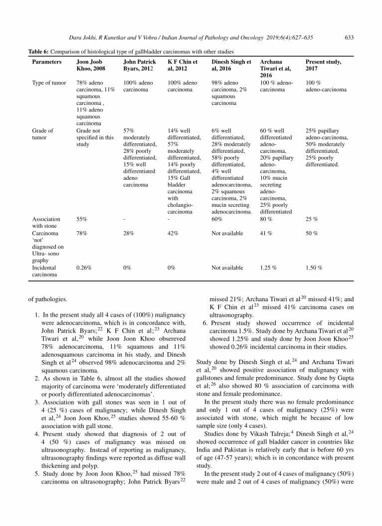
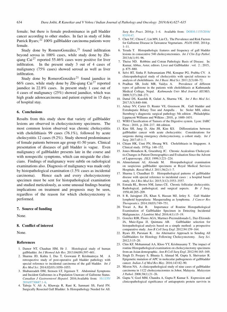