Embed Size (px)
Citation preview
STUDY OF ADHESIVE INTESTINAL OBSTRUCTION AND ITS
MANAGEMENT IN COIMBATORE MEDICAL COLLEGE HOSPITAL
Dissertation submitted in
Partial fulfilment of the regulations required for the award of
M.S. DEGREE
In
GENERAL SURGERY – BRANCH - I
THE TAMILNADU
DR. M.G.R. MEDICAL UNIVERSITY
CHENNAI
APRIL- 2013.
CERTIFICATE
This is to certify that the dissertation entitled “STUDY OF ADHESIVE INTESTINAL
OBSTRUCTION AND ITS MANAGEMENT IN COIMBATORE MEDICAL
COLLEGE HOSPITAL” is a bonafide work done by Dr.P.Murugadasan, Post
Graduate student in Department of General Surgery,Coimbatore Medical College,
under the supervision and guidance of Dr.P.Swaminathan, M.S., D.O., Professor of
Principles and Practice of Surgery, Department of General Surgery, Coimbatore
Medical College, Coimbatore, in partial fulfilment of the requirement of The
Tamilnadu Dr.M.G.R.Medical University for the award of M.S. Degree in General
Surgery.
Prof.Dr.P.Swaminathan, M.S.,D.O., Prof.Dr.P.V.Vasantha kumar, M.S.,
Professor of principles and practice Professor & Head of the Department,
of surgery, Department of General Surgery,
Department of General Surgery, Coimbatore Medical College,
Coimbatore Medical College, Coimbatore.
Coimbatore.
Prof.Dr.R.Vimala, M.D.,
The Dean,
Coimbatore Medical College,
Coimbatore.
DECLARATION
I, Dr.P.Murugadasan solemnly declare that dissertation titled, “STUDY OF
ADHESIVE INTESTINAL OBSTRUCTION AND ITS MANAGEMENT IN
COIMBATORE MEDICAL COLLEGE HOSPITAL” is a bonafide work done by me
at Coimbatore Medical College Hospital, during September 2011- November 2012
under the guidance and supervision of Prof. Dr.P. Swaminathan M.S.,D.O.,
Professor of Principles and Practice of Surgery, Department of General Surgery,
Coimbatore Medical College, Coimbatore.
The dissertation is submitted to The Tamilnadu Dr.M.G.R.Medical University,
towards partial fulfilment of requirement for the award of M.S.Degree in General
Surgery (BRANCH – I).
STATION: Coimbatore. Dr.P.MURUGADASAN
DATE:
ACKNOWLEDGEMENT
I thank the respectful Dean, Dr.R.VIMALA.M.D., Coimbatore
medical college hospital, Coimbatore for permitting access to all
hospital facilities in the course of this study.
I take this opportunity to express my profound gratitude to Professor
of surgery and head of department, Prof.Dr.P.V.VASANTHA KUMAR. M.S.,
for his support and encouragement throughout this study.
I am deeply indebted to my unit chief Dr.P. SWAMINATHAN.M. S.,
D.O., Professor of principles and practice of surgery, for his constant guidance
and motivation throughout the period of this study. Without his leadership and
motivation, this study would not have been possible.
I am extremely thankful to Prof.Dr.V.ELANGO.M.S., and all chiefs
for gracefully granting permission to include their respective unit patients in my
study.
I gratefully acknowledge the suggestions of Dr. UMASHANKAR. M.S.,
Dr. ANGELINE VINCENT.M.S., and rest of surgical faculty. Their combined
experience and constant queries helped me formulate and realize the goals of
the study.
CONTENTS
Topic
Page No.
1 Introduction 1
2 Objectives of the study 3
3 Historical highlights and pioneers in treatment 4
4 Surgical anatomy and applied physiology 7
5 Pathophysiology 15
6 Biochemical Changes in intestinal obstruction 34
7 Clinical features of adhesive intestinal obstruction 35
8 Investigations and diagnosis 38
9 Management of adhesive intestinal obstruction 44
10 Materials and methods of study 49
11 Observations in the study 51
12 Discussion 62
13 Summary 74
14 Conclusion 76
15 Bibliography
Annexure- 1 Master chart
Annexure- 2 Proforma

1
LIST OF TABLES
TABLE NO: TITLE PAGE NUMBER
1 AGE AND SEX INCIDENCE` 51
2 TYPE OF PRESENTATION 52
3 TYPE OF SURGERIES 53
4 PROCEDURE DONE 54
5 CONDITIONS OF THE BOWEL 55
6 SITE OF ADHESION 56
7 ORGAN INVOLVEMENT 57
8 CAUSE OF ADHESION 58
9 POST SURGICAL ADHESIVE
OBSTRUCTION
59
10 POST INFLAMMATORY
ADHESIVE OBSTRUCTION
60
11 COMPLICATIONS 61
2
LIST OF FIGURES
FIGURE NO: TITLE
1 PARTS OF THE BOWEL
2 BLOOD SUPPLY OF SMALL AND LARGE
BOWEL
3 INFERIOR MESENTRIC ARTERY
4 LAYERS OF SMALL INTESTINE
5 POST OPERATIVE PICTURE OF
ADHESIOLYSIS
6 ADHESIONS WITHIN THE LOOPS OF ILEUM
7 BOWEL TO BOWEL ADHESION
8 SMALL BOWEL ADHESION
9 LAPAROSCOPIC ADHESIOLYSIS
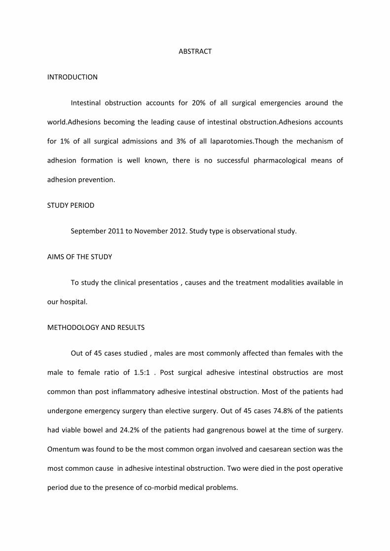
ABSTRACT
INTRODUCTION
Intestinal obstruction accounts for 20% of all surgical emergencies around the
world.Adhesions becoming the leading cause of intestinal obstruction.Adhesions accounts
for 1% of all surgical admissions and 3% of all laparotomies.Though the mechanism of
adhesion formation is well known, there is no successful pharmacological means of
adhesion prevention.
STUDY PERIOD
September 2011 to November 2012. Study type is observational study.
AIMS OF THE STUDY
To study the clinical presentatios , causes and the treatment modalities available in
our hospital.
METHODOLOGY AND RESULTS
Out of 45 cases studied , males are most commonly affected than females with the
male to female ratio of 1.5:1 . Post surgical adhesive intestinal obstructios are most
common than post inflammatory adhesive intestinal obstruction. Most of the patients had
undergone emergency surgery than elective surgery. Out of 45 cases 74.8% of the patients
had viable bowel and 24.2% of the patients had gangrenous bowel at the time of surgery.
Omentum was found to be the most common organ involved and caesarean section was the
most common cause in adhesive intestinal obstruction. Two were died in the post operative
period due to the presence of co-morbid medical problems.
CONCLUTION
Intestinal obstruction is a real emergency and adhesions becoming the leading cause
of intestinal obstruction. Post surgical adhesive intestinal obstructios are most common
than post inflammatory adhesive intestinal obstruction. Wound infection is the most
common complication. Mortality depends upon the co-morbid medical illness.
KEY WORDS
Intestinal obstruction, adhesions, post inflammatory adhesions.
3
INTRODUCTION
Intestinal obstruction accounts for 20 % of all surgical emergencies
around the world. Intestinal obstruction can result from a variety of causes.
Among them adhesions has become the leading cause of intestinal obstruction.
Though the diagnosis being straight forward, management of adhesive intestinal
obstruction possess a lot of problems due to the high incidence of recurrence.
The advent of laparoscopic surgery has altered the incidence of adhesions
to some extent. The incidence of post operative adhesive intestinal obstruction
has been increasing over the past few decades. Inflammatory adhesions and post
surgical adhesions are the two types of acquired adhesive intestinal obstruction.
Majority of the cases are post surgical adhesions.
Adhesions account for 1% of all surgical admissions and 3% of all
laparotomies. Handling of the viscera in the infracolic compartment is more
likely to produce adhesive intestinal obstruction. The incidence of adhesive
intestinal obstruction is more with major abdominal surgeries than minor
abdominal surgeries and still more with multiple abdominal surgeries. When the
patient presented with the feature of intestinal obstruction and with the history
of previous abdominal surgery, the most likely cause is postoperative adhesions.
Omentum plays a protective role in the formation of adhesions. Omentectomy
leaves adhesiogenic areas to the small bowel and this leads to higher incidence
of small bowel adhesions after total colectomy which involves omentectomy.
4
Simple adhesiolysis is usually employed in those patients who require surgery
for adhesive obstruction. Half of the surgeons prefer total adhesiolysis where as
half of them prefer adhesiolysis limited to adhesions causing obstructions.
Though the mechanism of adhesion formation is well known, yet there is no
successful pharmacological means of preventing the adhesion formation. In
spite of recent advances still adhesive intestinal obstruction holds a major share
of mortality due to many practical factors. Intestinal obstructions below the age
of twelve years are usually caused by congenital causes like bands. Post surgical
and post inflammatory adhesions are common in adults.
This study proposes to analyze the clinical course of the disease and
treatment modalities available for adhesive intestinal obstruction in our hospital.


5
OBJECTIVES OF THE STUDY
1) To study the clinical presentation of adhesive intestinal obstruction.
2) To find out the causes of adhesive intestinal obstruction.
3) To find out the most common site for adhesion.
4) To study the treatment modalities available for adhesive
intestinal obstruction in our hospital.
6
HISTORICAL HIGHLIGHTS AND PIONEERS IN TREATMENT
HIPPOCRATES AND CELSUS (460 BC):
Hippocrates, the father of medicine, and Celsus followed the pattern of
Egyptian treatment which was the administration of purgatives and enema for
three consecutive days once in a month to clear the bowel.
AMBROSE PARE (1510-1590)
Ambroise pare a French physician identified the bowel obstruction first
time and he reported a patient who died of twisted bowel.
LITTRE (1713)
Littre suggested the proximal decompression of the bowel by incision
PILLARE (1776)
He first made a successful caecostomy for a patient with carcinoma
rectum
DUCT (1793)
He performed the first successful sigmoidostomy.
DIFFEN BACK (1836)
He resected the small bowel and anastomosed. He did a major role in
suturing the bowels.
7
LAMCRANCES (1800)
He sutured the traumatic wounds of the colon
PAUL AND BLACK (1846 AND 1892)
He advocated the procedure of exteriorization of the colon.
MURPHY (1892)
First introduced the button method of anastomosis
PARKER AND KEEP (1908)
They introduced the principles of aseptic anastomosis.
WESTERMAN (1910)
He introduced the siphonage drainage of the stomach.
WANGENSTEIN (1913)
He described the duodenal tubes.
CANTOR (1946)
He introduced the ryle’s tube as gastric suction tube. Large food particles
could not be aspirated through ryle’s tube but can’t be done with stomach tube.
8
HUMER HULTL (1908)
He developed the surgical stapling method which was later modified by
Von Petz and Frederich in 1934.
NOBLE
He plicated the adjacent coils of small intestine to prevent further
recurrent adhesions.
CHILD PHILLIPS
He plicated the mesentery to prevent further adhesions.
WITZEL
Using witzel’s jejunostomy a stiff tube is passed through the jeunostomy
opening in the proximal bowel upto the entry into the large bowel, which acts as
intraluminal splinting.
ASSALIA et al
Gastrograffin was first used by Assalia et al in the 1980s.
FRIEDRICH TRENDELENBERG (1844-1924)
First tangential occlution clamp was developed by him.
AMYAND (1736)
He did the first appendicectomy.
9
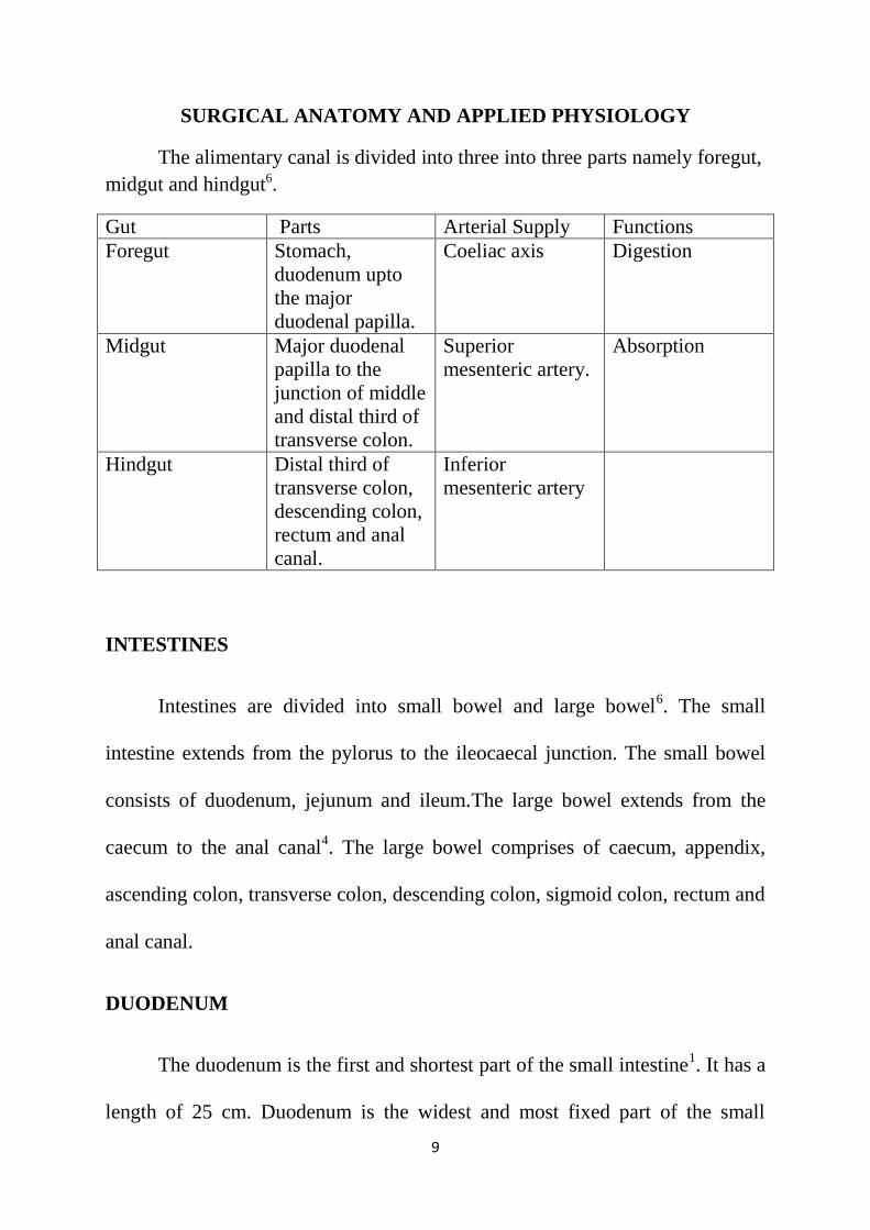
SURGICAL ANATOMY AND APPLIED PHYSIOLOGY
The alimentary canal is divided into three into three parts namely foregut,
midgut and hindgut6.
Gut Parts Arterial Supply Functions
Foregut Stomach,
duodenum upto
the major
duodenal papilla.
Coeliac axis Digestion
Midgut Major duodenal
papilla to the
junction of middle
and distal third of
transverse colon.
Superior
mesenteric artery.
Absorption
Hindgut Distal third of
transverse colon,
descending colon,
rectum and anal
canal.
Inferior
mesenteric artery
INTESTINES
Intestines are divided into small bowel and large bowel6. The small
intestine extends from the pylorus to the ileocaecal junction. The small bowel
consists of duodenum, jejunum and ileum.The large bowel extends from the
caecum to the anal canal4. The large bowel comprises of caecum, appendix,
ascending colon, transverse colon, descending colon, sigmoid colon, rectum and
anal canal.
DUODENUM
The duodenum is the first and shortest part of the small intestine1. It has a
length of 25 cm. Duodenum is the widest and most fixed part of the small

10
intestine. It has a C-shaped course around the head of pancreas6. Duodenum
starts at the pylorus and ends at the duodenojejunal flexure at the level of L2
vertebra. The first 2 cm of the small bowel has got mesentery and is mobile.
This part is called ampulla or duodenal cap. The arterial supply of duodenum
arises from celiac trunk and superior mesenteric artery3. The veins from
duodenum drain into hepatic and portal veins.
JEJUNUM
Jejunum is the second part of the small intestine. It begins at he
duodenojejunal flexure. Jejunum constitutes two fifths of the small intestine4.
Most of the jejunum lies in the left upper quadrant of the abdomen in the
infracolic compartment. Jejunum is deeper red in colour with a diameter of
about 2-4 cm. the vasa recta is long and fat in the mesentery is less. In jejunum
payer’s patches are few in number5.
ILEUM
Ileum is the third part of the small intestine. Ileum ends at the ileocaecal
junction. It constitutes three fifths of the small intestine7,8,9
. Most of the ileum
lies in the right upper quadrant of the abdomen in the infracolic compartment.
Ileum is paler pink in colour and 2-3 cm in diameter. Vascularity of ileum is
less than jejunum and vasa recta is short. Ileum has more fat in it’s mesentery.
Payer’s patches are more in ileum2.
11
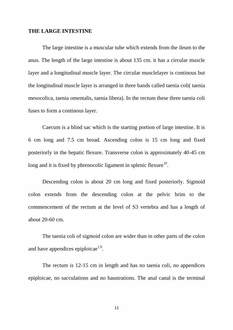
THE LARGE INTESTINE
The large intestine is a muscular tube which extends from the ileum to the
anus. The length of the large intestine is about 135 cm. it has a circular muscle
layer and a longitudinal muscle layer. The circular musclelayer is continous but
the longitudinal muscle layer is arranged in three bands called taenia coli( taenia
mesocolica, taenia omentalis, taenia libera). In the rectum these three taenia coli
fuses to form a continous layer.
Caecum is a blind sac which is the starting portion of large intestine. It is
6 cm long and 7.5 cm broad. Ascending colon is 15 cm long and fixed
posteriorly in the hepatic flexure. Transverse colon is approximately 40-45 cm
long and it is fixed by phrenocolic ligament in splenic flexure10
.
Descending colon is about 20 cm long and fixed posteriorly. Sigmoid
colon extends from the descending colon at the pelvic brim to the
commencement of the rectum at the level of S3 vertebra and has a length of
about 20-60 cm.
The taenia coli of sigmoid colon are wider than in other parts of the colon
and have appendices epiploicae1,9
.
The rectum is 12-15 cm in length and has no taenia coli, no appendices
epiploicae, no sacculations and no haustrations. The anal canal is the terminal
12
portion of the large intestineit is 3-8 cm in length and develops partly from
endoderm and partly from ectoderm.
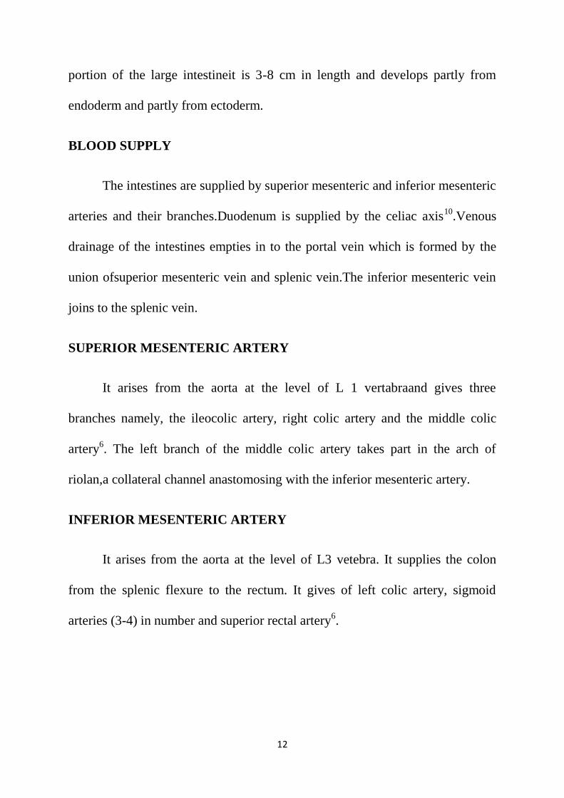
BLOOD SUPPLY
The intestines are supplied by superior mesenteric and inferior mesenteric
arteries and their branches.Duodenum is supplied by the celiac axis10
.Venous
drainage of the intestines empties in to the portal vein which is formed by the
union ofsuperior mesenteric vein and splenic vein.The inferior mesenteric vein
joins to the splenic vein.
SUPERIOR MESENTERIC ARTERY
It arises from the aorta at the level of L 1 vertabraand gives three
branches namely, the ileocolic artery, right colic artery and the middle colic
artery6. The left branch of the middle colic artery takes part in the arch of
riolan,a collateral channel anastomosing with the inferior mesenteric artery.
INFERIOR MESENTERIC ARTERY
It arises from the aorta at the level of L3 vetebra. It supplies the colon
from the splenic flexure to the rectum. It gives of left colic artery, sigmoid
arteries (3-4) in number and superior rectal artery6.
13
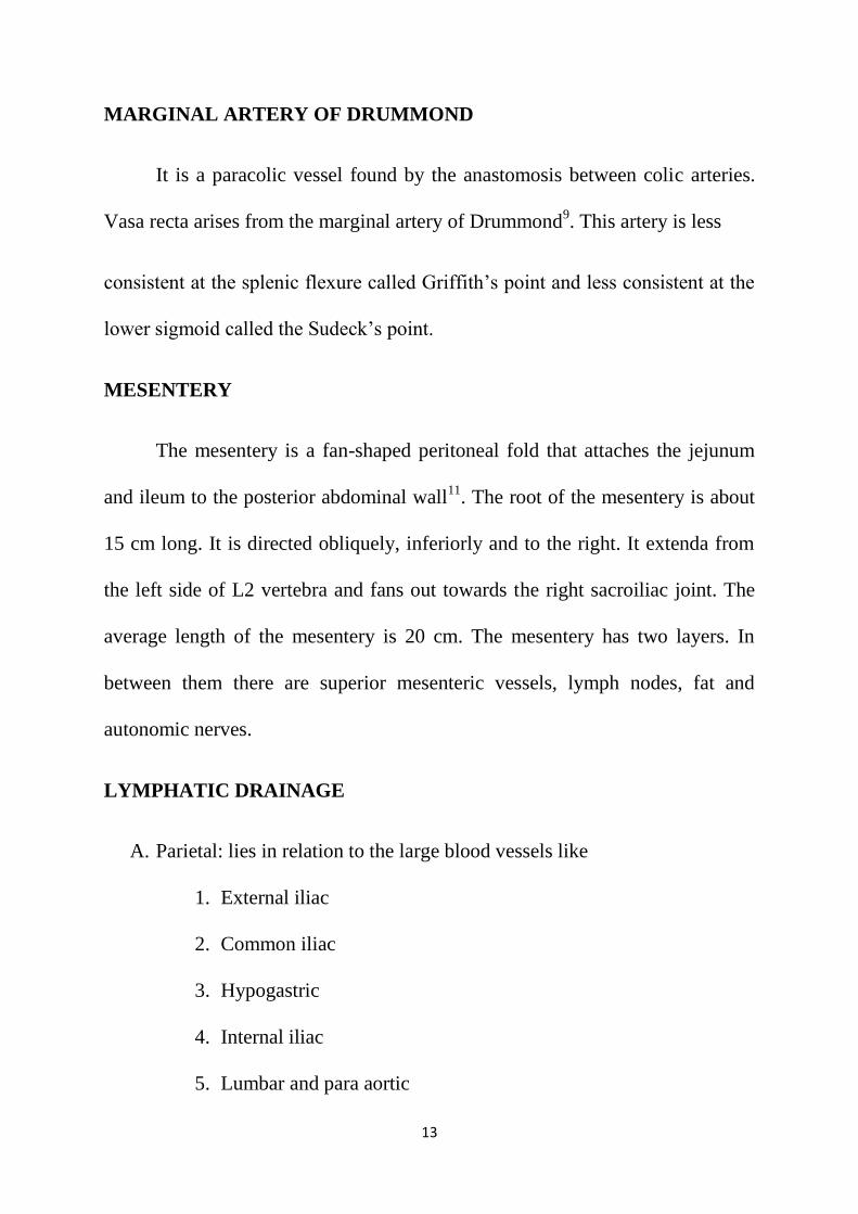
MARGINAL ARTERY OF DRUMMOND
It is a paracolic vessel found by the anastomosis between colic arteries.
Vasa recta arises from the marginal artery of Drummond9. This artery is less
consistent at the splenic flexure called Griffith’s point and less consistent at the
lower sigmoid called the Sudeck’s point.
MESENTERY
The mesentery is a fan-shaped peritoneal fold that attaches the jejunum
and ileum to the posterior abdominal wall11
. The root of the mesentery is about
15 cm long. It is directed obliquely, inferiorly and to the right. It extenda from
the left side of L2 vertebra and fans out towards the right sacroiliac joint. The
average length of the mesentery is 20 cm. The mesentery has two layers. In
between them there are superior mesenteric vessels, lymph nodes, fat and
autonomic nerves.
LYMPHATIC DRAINAGE
A. Parietal: lies in relation to the large blood vessels like
1. External iliac
2. Common iliac
3. Hypogastric
4. Internal iliac
5. Lumbar and para aortic
14
B. Visceral: lies along the superior and inferior mesenteric vessels11
.
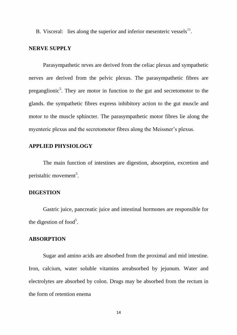
NERVE SUPPLY
Parasympathetic nrves are derived from the celiac plexus and sympathetic
nerves are derived from the pelvic plexus. The parasympathetic fibres are
preganglionic5. They are motor in function to the gut and secretomotor to the
glands. the sympathetic fibres express inhibitory action to the gut muscle and
motor to the muscle sphincter. The parasympathetic motor fibres lie along the
myenteric plexus and the secretomotor fibres along the Meissner’s plexus.
APPLIED PHYSIOLOGY
The main function of intestines are digestion, absorption, excretion and
peristaltic movement5.
DIGESTION
Gastric juice, pancreatic juice and intestinal hormones are responsible for
the digestion of food5.
ABSORPTION
Sugar and amino acids are absorbed from the proximal and mid intestine.
Iron, calcium, water soluble vitamins areabsorbed by jejunum. Water and
electrolytes are absorbed by colon. Drugs may be absorbed from the rectum in
the form of retention enema

15
PERISTALTIC MOVEMENTS
1. Rhythmic contractions or segmentations
2. True peristaltic movements
These are the two types of peristaltic movements of the intestine3,4
.
Rhythmic contraction or segmentation is myogenic in origin and this movement
helps in thourough mixing of the food. This movement is best developed in the
ileum, less in the jejunum and rare in the duodenum. The true peristaltic
movements occur in the whole length of the intestine. The amplitude and
propagatory distance vary with the phase of digestion over the loop proximal to
the obstruction. The amplitude and propagatory distance depends upon the
loaded condition of the bowel2. The rate of peristaltic waves remains constant
and not depend on the digestive phase and loaded condition of the colon.
In acute obstruction abdominal pain occurs as a first evidence which is due
to the vigorous contraction of the bowel musculature19
.The abdominal
distention is not so marked in higher level obstruction.
In the low level obstruction ,fluid accumulatesin the lumen very slowly
and hence the vomiting is delayed9.If the stomach and small bowel become
loaded with fluid,there will be considerable abdominal distention.The
abdominal distention is due to the accumulation of fluid and gases proximal to
the obstruction.The losses are water,sodium and potassium.RBC and plasma
16
may be lost from the strangulated bowel segment.General factors contribute to
the overall loss which is mainly from the extracellular compartment.The area of
absorptive mucosa is unavailable for the process of absorption distal to the
obstruction.
17
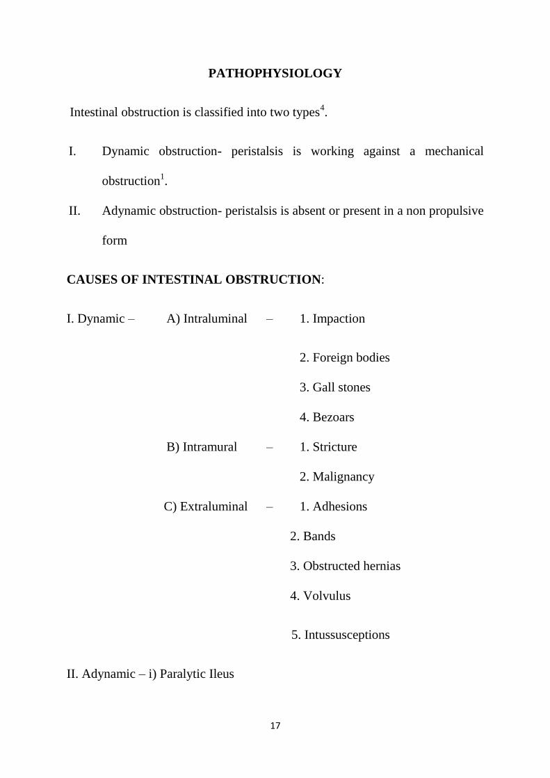
PATHOPHYSIOLOGY
Intestinal obstruction is classified into two types4.
I. Dynamic obstruction- peristalsis is working against a mechanical
obstruction1.
II. Adynamic obstruction- peristalsis is absent or present in a non propulsive
form
CAUSES OF INTESTINAL OBSTRUCTION:
I. Dynamic – A) Intraluminal – 1. Impaction
2. Foreign bodies
3. Gall stones
4. Bezoars
B) Intramural – 1. Stricture
2. Malignancy
C) Extraluminal – 1. Adhesions
2. Bands
3. Obstructed hernias
4. Volvulus
5. Intussusceptions
II. Adynamic – i) Paralytic Ileus
18
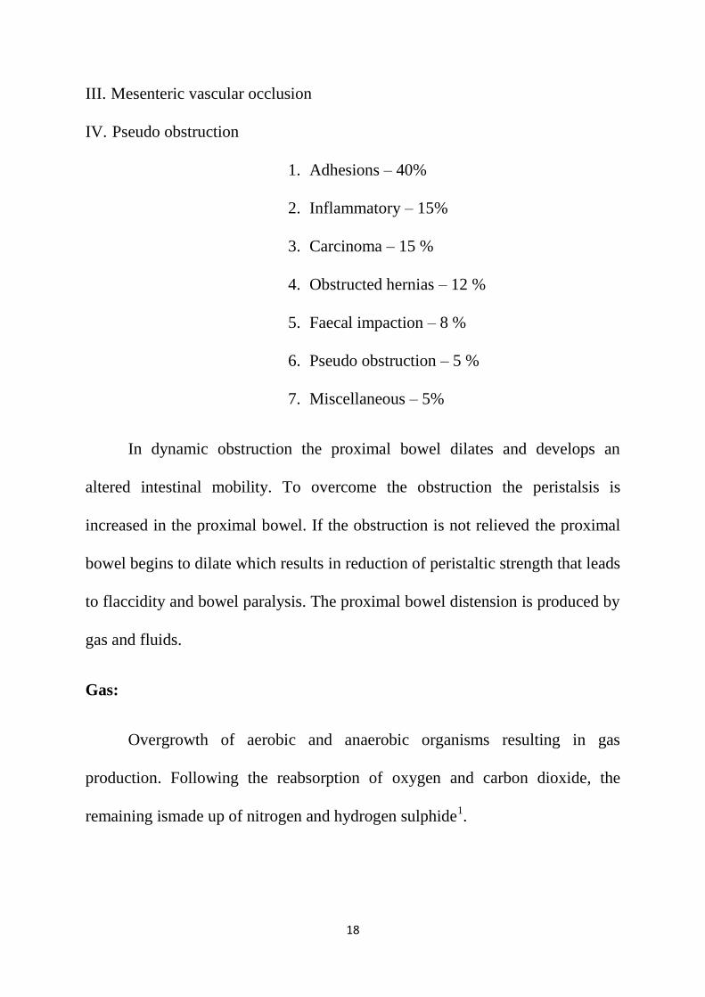
III. Mesenteric vascular occlusion
IV. Pseudo obstruction
1. Adhesions – 40%
2. Inflammatory – 15%
3. Carcinoma – 15 %
4. Obstructed hernias – 12 %
5. Faecal impaction – 8 %
6. Pseudo obstruction – 5 %
7. Miscellaneous – 5%
In dynamic obstruction the proximal bowel dilates and develops an
altered intestinal mobility. To overcome the obstruction the peristalsis is
increased in the proximal bowel. If the obstruction is not relieved the proximal
bowel begins to dilate which results in reduction of peristaltic strength that leads
to flaccidity and bowel paralysis. The proximal bowel distension is produced by
gas and fluids.
Gas:
Overgrowth of aerobic and anaerobic organisms resulting in gas
production. Following the reabsorption of oxygen and carbon dioxide, the
remaining ismade up of nitrogen and hydrogen sulphide1.
19
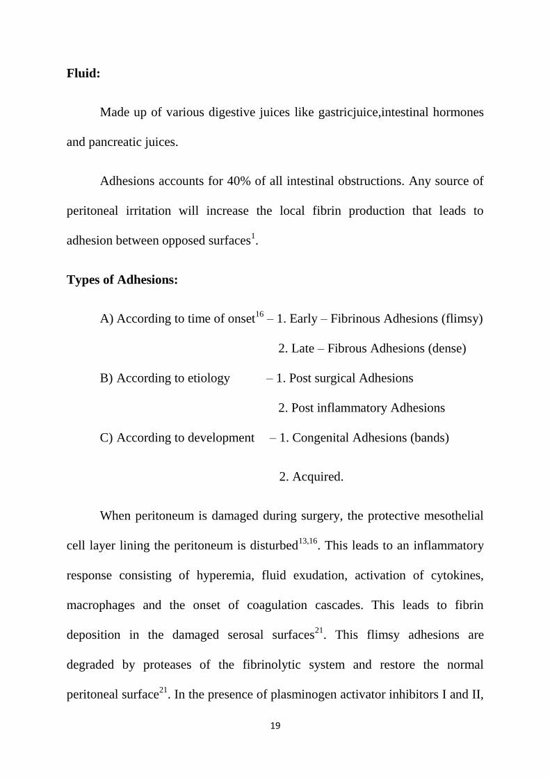
Fluid:
Made up of various digestive juices like gastricjuice,intestinal hormones
and pancreatic juices.
Adhesions accounts for 40% of all intestinal obstructions. Any source of
peritoneal irritation will increase the local fibrin production that leads to
adhesion between opposed surfaces1.
Types of Adhesions:
A) According to time of onset16
– 1. Early – Fibrinous Adhesions (flimsy)
2. Late – Fibrous Adhesions (dense)
B) According to etiology – 1. Post surgical Adhesions
2. Post inflammatory Adhesions
C) According to development – 1. Congenital Adhesions (bands)
2. Acquired.
When peritoneum is damaged during surgery, the protective mesothelial
cell layer lining the peritoneum is disturbed13,16
. This leads to an inflammatory
response consisting of hyperemia, fluid exudation, activation of cytokines,
macrophages and the onset of coagulation cascades. This leads to fibrin
deposition in the damaged serosal surfaces21
. This flimsy adhesions are
degraded by proteases of the fibrinolytic system and restore the normal
peritoneal surface21
. In the presence of plasminogen activator inhibitors I and II,
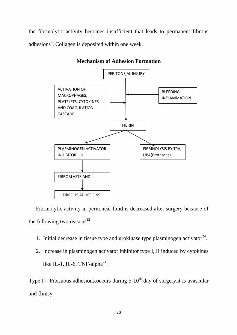
20
the fibrinolytic activity becomes insufficient that leads to permanent fibrous
adhesions4. Collagen is deposited within one week.
Mechanism of Adhesion Formation
Fibrinolytic activity in peritoneal fluid is decreased after surgery because of
the following two reasons12
.
1. Initial decrease in tissue type and urokinase type plasminogen activator24
.
2. Increase in plasminogen activator inhibitor type I, II induced by cytokines
like IL-1, IL-6, TNF-alpha24
.
Type I – Fibrinous adhesions.occurs during 5-10th
day of surgery.it is avascular
and flimsy.
PERITONEjAL INJURY
FIBRIN
FIBRINOLYSIS BY TPA,
UPA(Proteases)
PLASMINOGEN ACTIVATOR
INHIBITOR I, II
FIBROBLASTS AND
CAPILLARIES
CACAPILLARIES
FIBROUS ADHESIONS
BLEEDING,
INFLAMMATION
ACTIVATION OF
MACROPHAGES,
PLATELETS, CYTOKINES
AND COAGULATION
CASCADE
21
PERITONEAL REPAIR AND POST SURGICAL ADHESION
FORMATION:
Peritoneal healing differs from the skin healing mechanism. When a
diffect is made in the peritoneum the entire surface becomes epithelialized
simultaneously and not from the borders of the wound as in epidermalization of
skin wounds. Multiplication and migration of mesothelial cells from the borders
of the peritoneal wound play small part in the regenerative process. Surgically
traumatized tissues in apposition binds through fibrin bridges which become
organized by wound repair cells and neovascularization leading to adhesion
formation.
Healing occurs in 5 to 6 days after peritoneal injury. A single layer of
mesothelial cells resting on a continuous basement membrane is seen at tenth
day after surgery. Healing of parietal peritoneum is different from visceral
peritoneum in that, the perietal peritoneum contains basement membrane and
the mesothelium of visceral peritoneum does not contains basement membrane.
Peritoneal injury due to cauterization by electrical current leads to
delayed peritoneal healing because of extensive tissue damage. Even after 3
weeks of surgery the cauterized peritoneal site contains tissue necrosis,
polymorpho nuclear leucocytes, minimal fibroplasts and collagel. So peritoneal
healing is delayed in the site of peritoneal cauterization.
Fibrinolytic activity of peritoneum is present in all mesothelial surface
and is dependent primarily on the balance of the tissue plasminogen activator
22
and tissue plaminogen activator inhibitor. The tissue plaminogen activity is
decreased 4 to 6 hours after surgery and disappears after 24 to 48 hours. The
level of plasminogen activator inhibitor I,II are increased after surgical trauma
to peritoneum. This imbalance between the tissue plasminogen activator and
plasminogen activator inhibitor leads to adhesion formation.
Strategies for adhesion prevention
1. Minimize the surgical trauma
2. Minimal tissue handling
3. Avoiding dessication and ischaemia
4. Washing of peritoneal cavity with saline to remove cloth
5. Minimizing contact with gauze1.
6. Careful placing of drains
7. Laparoscopic procedures has got lesser chance of adhesions18
.
PREVENTION OF ADHESION FORMATION
Many substances have been instilled into the peritoneal cavity to prevent
adhesion formation. They are as follows:
1. Instillation of hyaluronidase into the peritoneal cavity
2. Hydrocortisone
3. Silicone
4. Dextran
23
5. Polyvinyl propylene
6. Streptomycin
7. Chondroitin
8. Anti coagulants
9. Anti histamins
10. Streptokinas
11. Ringer lactate
12. HMG-CO A reductase inhibitors22
.
13. Pentoxifylline- methyl xanthine derivatives
These substances increases the fibrinolytic activity in the damaged
peritoneum and thereby preventing adhesion formation12
. Dextran is used in
infertility surgeries to prevent adhesions.Povidone iodine has anti adhesive
effects but it is used for its antimicrobial action rather than its antiadhesive
effect.
CAUSES OF ADHESION FORMATION
1. Infections due to appendicitis,pancreatitis ,TB,peritonitis1.
2. Acute infective abdominal conditions.
3. Surgical materials like silk, mop, talk powder.
4. Bowel is chaemia.
5. Sepsis.
6. Inflammatory bowel disease.

24
7. Tuberculosis.
8. Malignancy,peritoneal injury.
ABDOMINAL TUBERCULOSIS
Abdominal tuberculosis is one of the important cause of post
inflammatory adhesive intestinal obstruction. Tuberculosis is common in India
& developing countries. tuberculous abdomen is the sixth most common type of
extrapulmonary tuberculosis. The incidence and the severity of abdominal
tuberculosis increases with increasing incidence of HIV infection.
TYPES OF ABDOMINAL TUBERCULOSIS
1. Intestinal.
2. Peritoneal Tuberculosis.
3. Tuberculosis of Mesentery and mesenteric nodes.
4. Ano-recto-sigmoid tuberculosis.
5. Miliary Tuberculosis.
6. Tuberculosis of the Omentum.
7. Retroperitoneal Tuberculosis.
Ileocecal region is most commonly affected in intestinal tuberculosis due
to presence of Peyer’s Patches and stasis of luminal contents aided by ileocecal
valve.Ulcerative, Hyperplastic, ulcerohyperplastic are the three types of
intestinal tuberculosis. Peritoneal tuberculosis may present as acute or chronic
25
illness. Chronic peritoneal tuberculosis may be associated withppericardial or
pleural effusion. Intestinal tuberculosis is called as Koenig’s syndrome.
Causative organisms of Abdominal Tuberculosis
1. Mycobacterium tuberculosis-acid fast and alcohol fast organism
2. Mycobacterium bovis-atypical mycobacterium
Mode of spread of abdominal tuberculosis
Ingestion of food materials contaminated with TB bacilli may cause
intestinal tuberculosis. Ingestion of tuberculous bacteria infective sputum from
primary focus may cause secondary tuberculosis. Abdominal TB can occur due
to haematogenous spread from lung tuberculosis or from neck lymph nodes
through lymphatic spread. Fallopian tube tuberculosis may retrogradely spread
to involve the peritoneum.
Presenting symptoms of abdominal tuberculosis
1. Abdominal pain.
2. Fever.
3. Night sweats.
4. Weight Loss.
5. Vomiting.
6. Constipation.
7. Diarrhea.
Computed Tomographic features of Abdominal tuberculosis
1. Peritoneal Involvement.
26
2. Ascites.
3. Mesenteric Fat Stranding.
4. Omental thickness.
5. Lymphadenopathy.
6. GI strictures.
7. Bowel wall thickening.
ILEOCECAL TUBERCULOSIS:
Ileocecal tuberculosis is the most common site of abdominal tuberculosis
due to the presence of Peyer’s patches. The ileocecal valve favours the stasis of
luminal contents and thus help in the development of ileocecal TB.Stricture is
most common in ileocecal region.Diffuse tuberculosis colitis is less
commonlyseen and mimics ulcerative colitis in colonoscopy.
Ulcerative,Hyperplastic and ulcerohyperplastic are the three types of
ileocecal TB. Ulcerative type of ileocecal TB occurs usually in old people
secondary to pulmonary TB. Diarrhea,Bleeding per rectum,loss of weight and
loss of appetite are the clinical features .Barium study may show ileal stricture
with hypermotility. Hyperplastic type is less common than ulcerative type of
ileocecal TB.It occurs as primary intestinal tuberculosis. Ulcerative type is
usually caused by Mycobacterium bovis bacilli.
Hyperplastic ileocecal tuberculosis may present as a mass in the RIF and
subacute intestinal obstruction .There is no primary lesion in the chest
27
Xray.Barium study shows pulled up cecum and obtuse ileocecal angle.The most
common complication of small bowel TB is obstruction due to narrowing of the
lumen by hyperplastic caecal tuberculosis.
Clinical Features of Ileocecal Tuberculosis:
1. Abdominal pain.
2. Anemia.
3. Loss of weight.
4. Loss of appetite.
5. Mass in RIF.
6. Fever.
7. Diarrhea.
8. Features of intestinal obstruction.
Ileocecal region is the common site of abdominal TB due to
1. Stasis.
2. Abundant Peyer’s patches.
3. More bacterial contact time with mucosa.
4. Liquid content of the stools.
5. Minimal digestive activity.
Tuberculous Mesenteric Lymphadenitis:
Mesenteric Tuberculous adenitis is most common in children and present
with anemia ,fever,loss of weight, loss of appetite and mass in the right iliac
fossa.Massive mesenteric lymph nodes enlargement due to tuberculosis is called
28
Tabes mesenterica.Right sided mesenteric nodes are more commonly involved
than left sided ones.Infection is usually through Peyer’s patches .Caseating
materials may be collected between the layers of mesentery to form cold
abscess.Usually the pain is felt in the umbilical region and the mass is felt in the
right iliac fossa.
TUBERCULOSIS OF THE OMENTUM
Rolled up omentum with thickening is characteristic of omental
tuberculosis.Cold abscess can develop in the omentum and can be dealt with
laparoscopy under cover of anti tubercular drugs.
PERITONEAL TUBERCULOSIS.
In peritoneal tuberculosis the parietal peritoneum is thickened with
multiple yellowish tubercles.
There is dense adhesions in the peritoneum and omentum which may
leads to intestinal adhesions. Multiple adhesions between bowel loops or
between bowel loops and peritoneum can develop. Peritoneal tuberculosis can
be dived in to acute or chronic types. Acute peritoneal tuberculosis mimics
acute abdomen. Chronic peritoneal tuberculosis can be further divided in to dry
type with adhesion,wet type with ascites and fibrous type with omental
thickening and loculated ascites. In ascitic type of abdominal tuberculosis, the
ascetic fluid is pale yellow, clear, rich in lymphocytes and the specificgravity is

29
high. Because of the fibrin deposition ascites may get loculated leading to
loculated ascites.
Plastic peritoneal tuberculosis
There are widespread adhesions between the coils of intestines,
abdominal wall and the omentum with bowel distention leading to blind loop,
ileus and intestinal obstruction. It may presents as colicky abdominalpain,
diarrhea, wasting, loss of weight, doughy abdomen and mass in the abdomen.
It responds well to antitubercular treatment and surgery is needed only
when there is intestinal obstruction and adhesions.
Paustian (1964) criteria for the diagnosis of abdominal tuberculosis
1. Histological evidence of tubercles with caseation necrosis
2. Typical operative findings with biopsy showing histological evidence of
tuberculosis.
3. Culture of suspected tissue resulting in growth of tuberculous bacilli.
4. Demonstration of acid fast bacilli in the lesion.
Purulent form of peritoneal tuberculosis occurs from tuberculous
salpingitis and may present as lower abdominal mass, abdominal wall abscess
or cold abscess. Prognosis is poor in purulent form of peritoneal tuberculosis.
Open or laparoscopic biopsy is very useful to diagnose the peritoneal
tuberculosis.

30
INVESTIGATIONS OF ABDOMINAL TUBERCULOSIS:
X-ray chest is very useful to find out the primary focus. Mantoux test,
ESR estimation or useful in the diagnosis of abdominal tuberculosis. Plain X-
ray of abdomen may shows clacified lesion in bowel, nod clacification in case
of intestinal obstruction. Barium study shows the following findings in
abdominal tuberculosis.
1. Pulled up ciecum.
2. Obtuse ileocaecal angle.
3. Narrow ileum with thickened ileocaecal valve.
4. Lack of barium in inflammed segment.
5. Ulcers and strictures in terminal ileum(napkin lesion).
6. Flocculation of barium.
7. Goose neck deformity of ileocaecal junction.
Colonoscopy is easiest and most direct method to diagnose the intestinal
tuberculosis. Capsule endoscopy is useful to see the small intestinal tuberculous
pathology.
Diagnostic laparoscopy is one of the most important investigation in the
diagnosis of abdominal tuberculosis. Adhesiolysis can be done using
laparoscopy. Ascites, multiple whitish tubercles, dens adhesions, bands,
hyperemic edematous bowel loops can be seen.

31
Ascitic fluid aspiration and analysis, bio chemical assay of ascitic fluid is
very useful to determine the abdominal tuberculosis. Presence of anticord
factors antibody differentiate the ascetic fluid from ascites due to crohn’s
disease. Adenosine deaminase activity is a sensitive and specific marker for
tuberculous ascites. Adenosine deaminase value more than 33 IU/L in
ascetic fluid and more than 42 IU/L in serum in significant.
Adenosine deaminase is an aminohydrolase. It coverts adenosine to
inosine and is thus involved in the purine bases catabolism. The enzyme activity
is more in T-lymphocytes than B-lymphocytes. Adenosine deaminase is
increased in tuberculous ascites due to stimulation of T-cells by mycobacterium
tuberculosis. Ascitic fluid to serum adenosine deaminase ratio more than 0.985
is suggestive of tuberculosis.
ASCITIC FLUID IN ABDOMINAL TUBERCULOSIS
1. Exudates with protein level >2.5 gm/dl.
2. Serum-ascitic fluid albumin gradient < 1.1
3. Lymphocytosis.
4. Adenosine deaminase >33IU/ l.
5. Specific gravity >1.016
6. Glucose <30mg.
7. LDH >90 U/ l.
32
ULTRASONAGRAM FEATURES IN ABDOMINAL TUBERCULOSIS:
Ultrasonagram of abdomen is useful in the diagnosis of abdominal
tuberculosis the following features can be seen in the ultrasonagram incase of
abdominal tuberculosis.
1. Thickend bowel wall, mesentery, omentum.
2. Loculated ascites with fine septae.
3. Interloop ascites with alternate echogenic and echo free areas.
4. Stellate sign-bowel loop radiates from its mesenteric root.
5. Pulled up caecum presenting with a mass-pseudokidnuy sign.
6. Concentric, uniform mural thickening.
7. Matted lymphnode enlargement.
8. Mesenteric thickness more than 15mm.
9. Hepatosplenomegaly.
CT SCAN IN ABDOMINAL TUBERCULOSIS:
CT scan is very useful and reliable investigation in the diagnosis of abdominal
tuberculosis it is done with plain or oral contrast medium. It is inexpensive and
non invasive method and gives more information about the abdominal
pathology. The following findings can be seen in CT Scan incase of abdominal
tuberculosis.
1. Thickend bowel wall.
33
2. Thickend peritoneum.
3. Ileo-caecal valve thickening.
4. Adhesions in the bowel.
5. Enlarged and matted mesenteric nodes.
6. Features of intestinal obstruction.
7. Loculated ascites.
8. Nodules in the peritoneum.
9. Solid organs visualization.
10. Strictures in bowel wall.
11. Dilatation of bowel wall.
CT guided FNAC, biopsy or ascitic fluid aspiration can be done. Tuberculosis
ascitic fluid has high attenuation value (25 to 45HU) due to its high protein
content.
COPLICATIONS OF ABDOMINAL TUBERCULOSIS:
1. Intestinal obstructions.
2. Malabsorption.
3. Blind loop syndrome.
4. Faecal fistula.
5. Adhesions.
6. Cold abscess formation.
7. Stricture formation.

34
TREATMENT OF ABDOMINAL TUBERCULOSIS:
Six to nine months of treatment with anti tubercular treatment is
mandatory for abdominal tuberculosis. Patients present with complications
should be treated for one year with anti tubercular drugs. Recurrent abdominal
tuberculosis is very difficult to manage and has got high mortality. Surgery is
indicated when there is intestinal obstruction, severe hemorrhage, perforation.
Adhesive obstruction may be released through laparoscopic adhesiolysis.
Sometimes dense adhesions may be present and is very difficult to release them
even by open method. Drainage of intra abdominal abscess is important to
control the disease.
TUBERCULOUS COCOON:
Abdominal cocoon is a rare cause of intestinal obstruction. It is
characterized by enlargement of small bowel by a thick fibrous membrane. It is
otherwise called as sclerosing encapsulated peritonitis or peritonitis chronica
fibrosa incapsulata. Abdominal cocoon primarily affect young females.
Tuberculous cocoon is very rare. Clinical manifestation of tuberculous cocoon
abdomen are non specific and include intestinal obstruction or abdominal mass.
Majority of the cases were incidentally diagnosed during laparotomy. Because
of redused awareness and atypical presentations pre operative diagnosis of
tuberculous cocoon is very difficult. Barium meal study and CT scan of
abdomen play an important role in the pre operative diagnosis of tuberculous

35
cocoon abdomen. The following findings are noted in the barium meal follow
through CT.
1. Features of small bowel obstruction.
2. Redused transit time.
3. Serpentine configuration.
4. Dilated bowel loops in a fixed ‘U’ shaped clusters.
5. Fibrous membrane encasing the bowel loops.
Usually the diagnosis of tuberculous cocoon is made at laparotomy where
the bowel loops are encased within a sac like cocoon. The lesion is primarily
involving the small bowel but can involves large intestine, liver and stomach.
There may be dense inter bowel adhesions which should be lysed. The
histological examination of cocoon membrane shows fibro connective tissue
and caseating epitheloid granulomas.
36
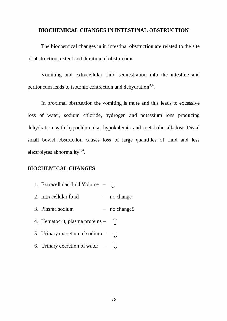
BIOCHEMICAL CHANGES IN INTESTINAL OBSTRUCTION
The biochemical changes in in intestinal obstruction are related to the site
of obstruction, extent and duration of obstruction.
Vomiting and extracellular fluid sequestration into the intestine and
peritoneum leads to isotonic contraction and dehydration3,4
.
In proximal obstruction the vomiting is more and this leads to excessive
loss of water, sodium chloride, hydrogen and potassium ions producing
dehydration with hypochloremia, hypokalemia and metabolic alkalosis.Distal
small bowel obstruction causes loss of large quantities of fluid and less
electrolytes abnormality1,9
.
BIOCHEMICAL CHANGES
1. Extracellular fluid Volume –
2. Intracellular fluid – no change
3. Plasma sodium – no change5.
4. Hematocrit, plasma proteins –
5. Urinary excretion of sodium –
6. Urinary excretion of water –

37
CLINICAL FEATURES OF ADHESIVE INTESTINAL OBSTRUCTION
The four cardinal features of adhesive intestinal obstruction are
1. Pain
2. Vomiting
3. Abdominal distension
4. Constipation
These features vary according to
1. The location of obstruction
2. The underlying pathology
3. The duration of obstruction
4. The presence or absence of intestinal ischaemia
Dehydration, fever, oliguria, septicaemia, hypovolumic shock are the late
manifestations.
PAIN
Pain is the first symptom. It occurs suddenly and is usually severe. The
pain is colicky in nature that coincides with increased peristaltic activity with
increasing distention.Development of severe pain indicates bowel
strangulation4.Pain never occurs in paralytic ileus.In case of strangulation
thereis always tenderness associated with rigidity.Generalised tenderness and
presence of rigidity are indicative of early laparotomy.
38
VOMITING
Vomiting is more in the proximal obstruction and less in the distal
obstruction. If the obstruction progresses the character of vomiting changes
from digested food to feculent material due to bacterial overgrowth.If the
obstruction is more distal, the time interval between theonset of symptoms and
the appearance of nausea and vomiting will be delayed.
DISTENSION
In small bowel obstruction the degree of distension depends upon the site
of obstruction. More distal the lesion greater is the distension9. There may be
visible peristalsis.
CONSTIPATION
Constipation may be absolute or relative. Absolute constipation is a
cardinal feature of complete intestinal obstruction.
DEHYDRATION
Dehydration is more in small bowel obstruction due to repeated vomiting
and fluid sequestration. This may result in dry tongue, dry skin, sunken eyes and
oliguria5.

39
FEVER
Fever in the presence of intestinal obstruction indicates
1. The onset of ischaemia
2. Intestinal perforation
HYPOTHERMIA
Hypothermia Indicates septicaemic shock.
ABDOMINAL TENDERNESS
Abdominal tenderness indicates bowel ischaemia or perforation.
HYPOKALEMIA
This is not a feature in simple mechanical intestinal obstruction.an
increase in serum potassium,amylase and lactate dehydrogenase may be
associated with bowel strangulation4.
40
INVESTIGATIONS AND DIAGNOSIS
A. Routine investigations - 1. Complete hemogram
2. Hemoglobin percentage
3. Blood sugar
4. Blood urea
5. Serum creatinine
6. Serum electrolytes
7. Urine analysis
8. Stool examination
9. Blood grouping
B. Special investigations – 1. X ray chest PA view
2. ECG
3. X Ray abdomen erect
4. Ultrasonogram of abdomen
5. CT scan of abdomen
6. Diagnostic laparoscopy
7. Gastrograffin - water soluble contrast
meal
COMPLETE HAEMOGRAM:
Complete hemogram was done for al cases. Five patients showed
leucocytosis about 13000 to 15000 per cubic millimeter.

41
HAEMOGLOBIN PERCENTAGE:
1. More than 90 % -- 9 cases
2. Hb between 80-90% -- 16 cases
3. Hb between 70-80% -- 14 cases
4. Hb between 50-70% -- 4 cases
5. Below 50% -- 2 cases
Patients with lesser than 50 % of haemoglobin had preoperative blood
transfusion.
BLOOD UREA:
Blood urea was done in all the 45 cases. In 44 cases blood urea was
within normal limits.
BLOOD SUGAR: Blood sugar testing is a basic investigation.
Blood sugar was tested in all the 45 cases out of which 40 patients had
normal blood sugar level and five patients had blood sugar levels more than 200
mg %.
URINE ANALYSIS:
Urine analysis was done in all the 45 patients out of which five were
found to have developed elevated urine sugar level. These five patients were
placed on antidiabetic medications after doing blood sugar estimation.
42
BLOOD GROUPING:
Blood grouping was done routinely for all patients.in our study B group
shows the highest incidence. AB group shows the lowest incidence.
SERUM CREATININE AND ELECTROLYTES
Routinely done for all cases. It is important for anaesthetic assessment .
X –RAY CHEST
X ray chest was taken in all the 45 patients for anaethesiological
assessment
ECG:
ECG was taken for all the 45 patients for the purpose of anaesthetic
assessment.
X-RAY ABDOMEN ERECT
Plain X ray abdomen was taken for all the 45 patients. Erect posture was
preferred for the following reasons.
1. The fluid levels are clearly seen in erect posture
2. Diaphragm can be better visualized and air under diaphragm can be clearly
seen4.
43
X ray findings in small bowel obstruction:
1. Dilated small bowel loops characterized by straight segments that are
central and lie transversely. Stepladder pattern of appearance can be seen.
2. The jejunum is characterized by it’s valvulae conniventes. They pass from
antimesenteric border to the mesenteric border9.
3. The distal ileum is featureless
4. Multiple air fluid levels are seen. Number of air fluid levels are directly
proportional to the degree of obstruction
X ray findings in large bowel obstruction:
The llowing findings are seen in x-ray. Dilated caecum shows haustral folds
which are spaced irregularly and the indentations are not placed opposite to one
another.
1. Dilated bowel loops are placed in the periphery
2. Multiple air fluid levels are seen but less in number when compared to small
bowel obstruction
3. Low colonic obstruction does not give rise to multiple fluid levels but in high
colonic obstruction there will be multiple air fluid levels.
4. Large amount of gas is seen in the caecum.
44
ULTRASONOGRAM:
Ultrasonogram of the abdomen was not done in all cases. Patients with
acute intestinal obstruction were taken up for surgery without doing
ultrasonogram. Patients with subacute intestinal obstruction were taken for
surgey after doing ultrasonogram of the abdomen. Ultrasonogram of the
abdomen was done in 18 patients. The following findings were noted in the
ultrasonogram in acute intestinal obstruction.
1. Dilated bowel loops
2. Adynamic bowel loops
3. Any fluid collection in the abdomen
CT SCAN ABDOMEN:
CT scan confirms the presence of complete obstruction. CT scan of the
abdomen was done routinely for all the cases. CT scan was done for all the
patients who were taken for elective surgery. Oral contrast with CT scan is very
useful for the diagnosis of intestinal obstruction. CT scan is useful to diagnose
the cause of small bowel obstruction and to exclude the non adhesional
pathology.
Diagnostic laparoscopy:
Diagnostic laparoscopy is vey useful to detect the adhesions in case of
intestinal adhesions18
. Dense adhesion, site of adhesions can be bettervisualized
with diagnostic laparoscopy.
45
Gastrograffin water soluble contrast study:
Gastrograffin is a hyperosmolar water soluble contrast medium. It has a
diagnostic and therapeautic role in adhesive intestinal obstruction which has
been evaluated recently. The use of gastrograffin water soluble medium in
adhesive small bowel obstruction is very safe and reduces the need for surgery
when conservative management fails20
..
History and Physical Examination:
Clinical history and thorough physical examination of the patient is very
important to diagnose the intestinal obstruction. History of previous surgery and
timing of previous surgery gives a clue to the possibility of adhesive intestinal
obstruction. If the patients presented with the features of intestinal obstruction
and previous abdominal scar the most likely diagnosis is adhesive intestinal
obstruction15
. History of constipation and per rectal examination is very
important to diagnose the obstruction.

46
MANAGEMENT OF ADHESIVE INTESTINAL OBSTRUCTION
The management of adhesive intestinal obstruction is very difficult
because surgery can induce new adhesions, whereas conservative treatment does
not remove the cause of obstruction.
1. Conservative management – for sub acute obstruction
2. Surgical management – for acute obstruction.
Conservative management
Conservative management involves nasogastric tube intubation,
intravenous fluid administration and clinical observation14
.
Indications for Conservative Management
1. Patient’s presented with sub acute pattern of obstruction.
2. Without signs of strangulation.
3. No persistant vomiting
4. CT scan findings – 1. No free fluid in the abdomen
2. No mesenteric oedema
3. No devascularised bowel
Contra indication For Conservative Management:
1. Patient who had surgery within 6 weeks
2. Patients with signs of peritonitis
47
3. Patients with signs of strangulation
4. Persistent vomiting
5. Acute intestinal obstruction
6. Patients with irreducible hernia
7. Patients who started to have signs of resolution at the time of admission
8. Metabolic acidosis
9. Continous abdominal pain
If the conservative management fails then the patient should be treated
with surgical management17
.In conservative management a nasogastric tube is
introduced to decompress the stomach and intestines. Early decompression with
nasogastric tube is beneficial for the patients.
When Conservative Management Should Be discontinued:
1. No resolution after 3 days, Onset of fever
2. Nasogastric drainage volume on day 3 is more than 500ml
3. Leucocytosis greater than 15000/cu.mm
Chance of Recurrence After Conservative Management:
The duration of nasogastric tube placement serves as a parameter for
predicting the recurrence after the conservative management 17
. Patients not
responding to the Ryle’s tube treatment within 72 hours have higher chance of
recurrent adhesive obstruction.
48
Surgical Management:
When the conservative treatment fails to relieve the obstruction, the
patients can be treated surgically. Open method and laparoscopic method are the
two surgical procedures available with their own advantages and disadvantages.
A. Surgical Options For Adhesive Intestinal Obstruction
1. Laparotomy and open adhesiolysis
2. Laparotomy and resection anastomosis with adhesiolysis
3. Laparoscopic adhesiolysis
B. Surgeries to prevent adhesive obstruction
1. Noble’s plication of intestines
2. childs Phillips mesenteric plication13,14
.
Indications for surgical management
1. Failure of conservative management.
2. Ileus persists for more than 3 days.
3. Persistant vomiting and abdominal pain
4. Nasogastric tube drainage volume on day 3 more than 500ml
5. Onset of fever, leucocytosis more than 15,000/cu.mm.
6. Signs of bowel strangulation
7. CT findings of intraperitoneal fluid,mesenteric oedema and lack of the
small bowel faeces sign
49
Preoperative management
1. I.V. line with wide bore cannula should be inserted to administer
intravenous fluids mostly with crystalloid solution1.
2. Gastric decompression with ryle,s tube
3. Foley’s catheter to be put todrain urine
4. All the basic blood investigations to be done
5. Maintaining the hourly abdominal girth chart, BP chart, pulse chart,
temperatue, respiration is important
6. X ray chest, X ray abdomen to be taken.
7. Infective endocarditis prophylaxis to be given.
8. Clinical examinations should be repeated to assess the progression of the
obstruction
Open Surgery
Laparotomy and open adhesiolysis is useful to relieve the obstruction
when conservative management fails. Open surgery is preferred method for the
surgical treatment of strangulating acute small bowel obstruction.
Laparoscopic Surgery
Laparoscopic adhesiolysis is better for first episode of adhesive
obstruction18
. In laparoscopic surgery the tissue trauma will be less and the
chance of recurrence is also less23
.

50
Advantages of laparoscopic adhesiolysis
1. Less tissue trauma, Earlier return to full activity
2. Less chance of recurrence, post operative pain
3. Early return of intestinal functions
4. Less number of hospital stay
Noble’s placation of intestines
Adjacent coils of small bowel are sutured to prevent further recurrence.
Childe – Phillips mesenteric placation
Plication of the intestinal mesentery will prevent crumpling of bowel and
adhesion formation.
51
MATERIALS AND METHODS OF THE STUDY
In Coimbatore medical college hospital in the year 2011-2012, 144 cases
of intestinal obstruction were admitted in both male and female wards of all the
six surgical units. Out of all the 154 cases 45 cases were diagnosed to have
intestinal obstruction due to adhesions. These 45 cases were included in our
study for analysis. The patients in the study group were proved to have adhesive
intestinal obstruction by means of surgery and relevant investigations.Patients
treated conservatively are excluded from the study. A separate proforma was
maintained. Whenever possible the histopathological examination of the
specimen was done to confirm the clinical diagnosis. All the patients were
looked for any complications during the post operative periods except the
patients who died post operatively.
Inclusion criteria
1. Patients above 18 years of age
2. Patients with confirmed intestinal obstruction due to adhesion
3. Recurrent cases of adhesive intestinal obstruction
Exclusion criteria
1. Intestinal obstruction due to other causes like congenital band,
mechanical bowel obstruction, growth, ileus and volvulus.
2. Patients with intestinal obstruction who were treated conservatively.
3. Pregnantwomen.
52
4. Psychiatric patients.
5. Patientsbelow the age of 18 years.
All the patients eligible by inclusion and criterias were included in the
study. All the patients eligible by exclution criterias were excluded from the
study. Datas are obtained from our study and analyzed using appropriate
statistical methods. Our study type is observational study.
53
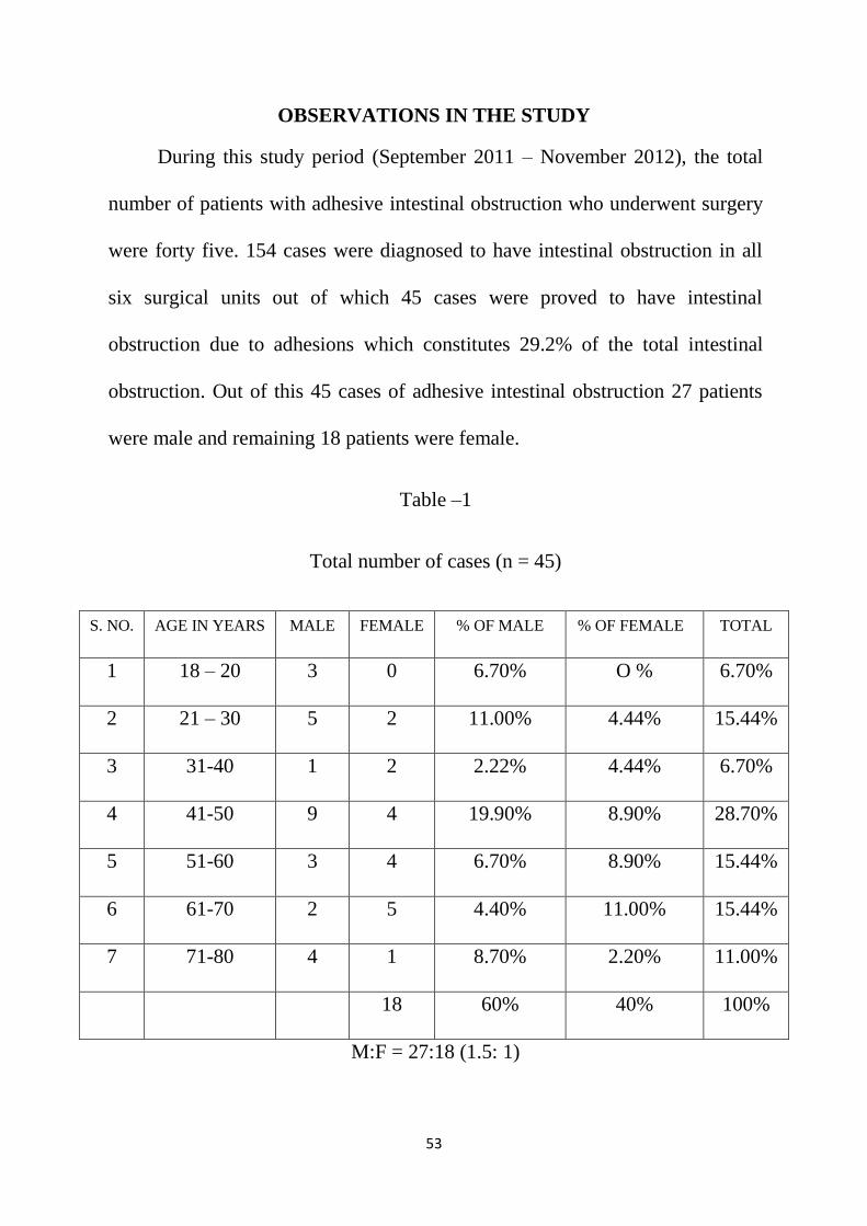
OBSERVATIONS IN THE STUDY
During this study period (September 2011 – November 2012), the total
number of patients with adhesive intestinal obstruction who underwent surgery
were forty five. 154 cases were diagnosed to have intestinal obstruction in all
six surgical units out of which 45 cases were proved to have intestinal
obstruction due to adhesions which constitutes 29.2% of the total intestinal
obstruction. Out of this 45 cases of adhesive intestinal obstruction 27 patients
were male and remaining 18 patients were female.
Table –1
Total number of cases (n = 45)
S. NO. AGE IN YEARS MALE FEMALE % OF MALE % OF FEMALE TOTAL
1 18 – 20 3 0 6.70% O % 6.70%
2 21 – 30 5 2 11.00% 4.44% 15.44%
3 31-40 1 2 2.22% 4.44% 6.70%
4 41-50 9 4 19.90% 8.90% 28.70%
5 51-60 3 4 6.70% 8.90% 15.44%
6 61-70 2 5 4.40% 11.00% 15.44%
7 71-80 4 1 8.70% 2.20% 11.00%
18 60% 40% 100%
M:F = 27:18 (1.5: 1)
54
Among the 45 cases of adhesive intestinal obstruction 60 % were male
patients and 40 % were females. This gives a male is to female ratio of 1.5 : 1
(28: 17). Table 1 shows the age and sex incidence of this study. In this study the
youngest patient was a 18 year old boy with adhesive intestinal obstruction. The
oldest patient was an eighty years old male with adhesive intestinal obstruction.
The maximum number of patients were in the age group of 41-50 years
representing 28.70 %. Majority of female patients were in the age group of 61-
70 years representing 11 % of total patients. Majority of the male patients were
in the age group of 41-50 years representing 19.90% 0f total patients.
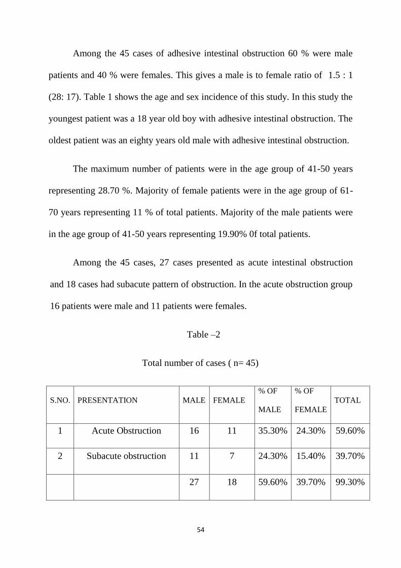
Among the 45 cases, 27 cases presented as acute intestinal obstruction
and 18 cases had subacute pattern of obstruction. In the acute obstruction group
16 patients were male and 11 patients were females.
Table –2
Total number of cases ( n= 45)
S.NO. PRESENTATION MALE FEMALE
% OF
MALE
% OF
FEMALE
TOTAL
1 Acute Obstruction 16 11 35.30% 24.30% 59.60%
2 Subacute obstruction 11 7 24.30% 15.40% 39.70%
27 18 59.60% 39.70% 99.30%
55
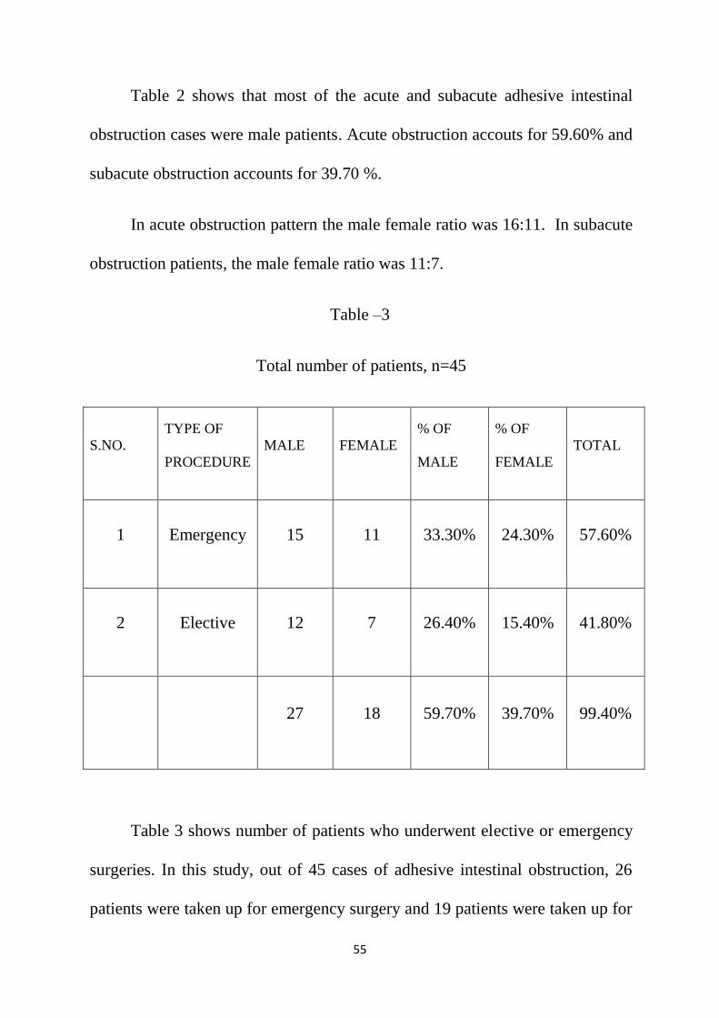
Table 2 shows that most of the acute and subacute adhesive intestinal
obstruction cases were male patients. Acute obstruction accouts for 59.60% and
subacute obstruction accounts for 39.70 %.
In acute obstruction pattern the male female ratio was 16:11. In subacute
obstruction patients, the male female ratio was 11:7.
Table –3
Total number of patients, n=45
S.NO.
TYPE OF
PROCEDURE
MALE FEMALE
% OF
MALE
% OF
FEMALE
TOTAL
1 Emergency 15 11 33.30% 24.30% 57.60%
2 Elective 12 7 26.40% 15.40% 41.80%
27 18 59.70% 39.70% 99.40%
Table 3 shows number of patients who underwent elective or emergency
surgeries. In this study, out of 45 cases of adhesive intestinal obstruction, 26
patients were taken up for emergency surgery and 19 patients were taken up for
56
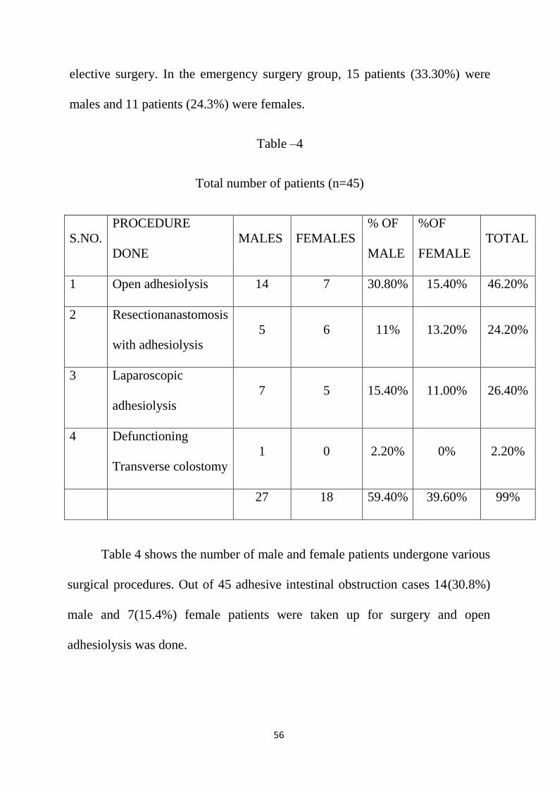
elective surgery. In the emergency surgery group, 15 patients (33.30%) were
males and 11 patients (24.3%) were females.
Table –4
Total number of patients (n=45)
S.NO.
PROCEDURE
DONE
MALES FEMALES
% OF
MALE
%OF
FEMALE
TOTAL
1 Open adhesiolysis 14 7 30.80% 15.40% 46.20%
2 Resectionanastomosis
with adhesiolysis
5 6 11% 13.20% 24.20%
3 Laparoscopic
adhesiolysis
7 5 15.40% 11.00% 26.40%
4 Defunctioning
Transverse colostomy
1 0 2.20% 0% 2.20%
27 18 59.40% 39.60% 99%
Table 4 shows the number of male and female patients undergone various
surgical procedures. Out of 45 adhesive intestinal obstruction cases 14(30.8%)
male and 7(15.4%) female patients were taken up for surgery and open
adhesiolysis was done.
57
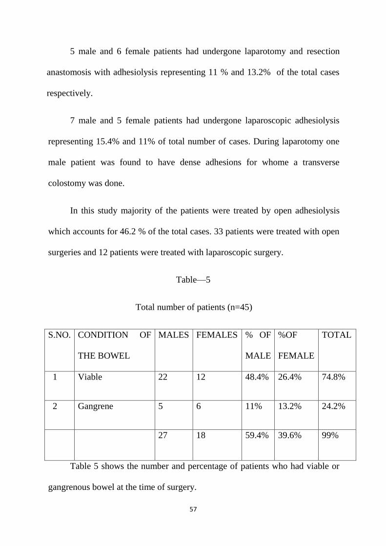
5 male and 6 female patients had undergone laparotomy and resection
anastomosis with adhesiolysis representing 11 % and 13.2% of the total cases
respectively.
7 male and 5 female patients had undergone laparoscopic adhesiolysis
representing 15.4% and 11% of total number of cases. During laparotomy one
male patient was found to have dense adhesions for whome a transverse
colostomy was done.
In this study majority of the patients were treated by open adhesiolysis
which accounts for 46.2 % of the total cases. 33 patients were treated with open
surgeries and 12 patients were treated with laparoscopic surgery.
Table—5
Total number of patients (n=45)
S.NO. CONDITION OF
THE BOWEL
MALES FEMALES % OF
MALE
%OF
FEMALE
TOTAL
1 Viable 22 12 48.4% 26.4% 74.8%
2 Gangrene 5 6 11% 13.2% 24.2%
27 18 59.4% 39.6% 99%
Table 5 shows the number and percentage of patients who had viable or
gangrenous bowel at the time of surgery.
58
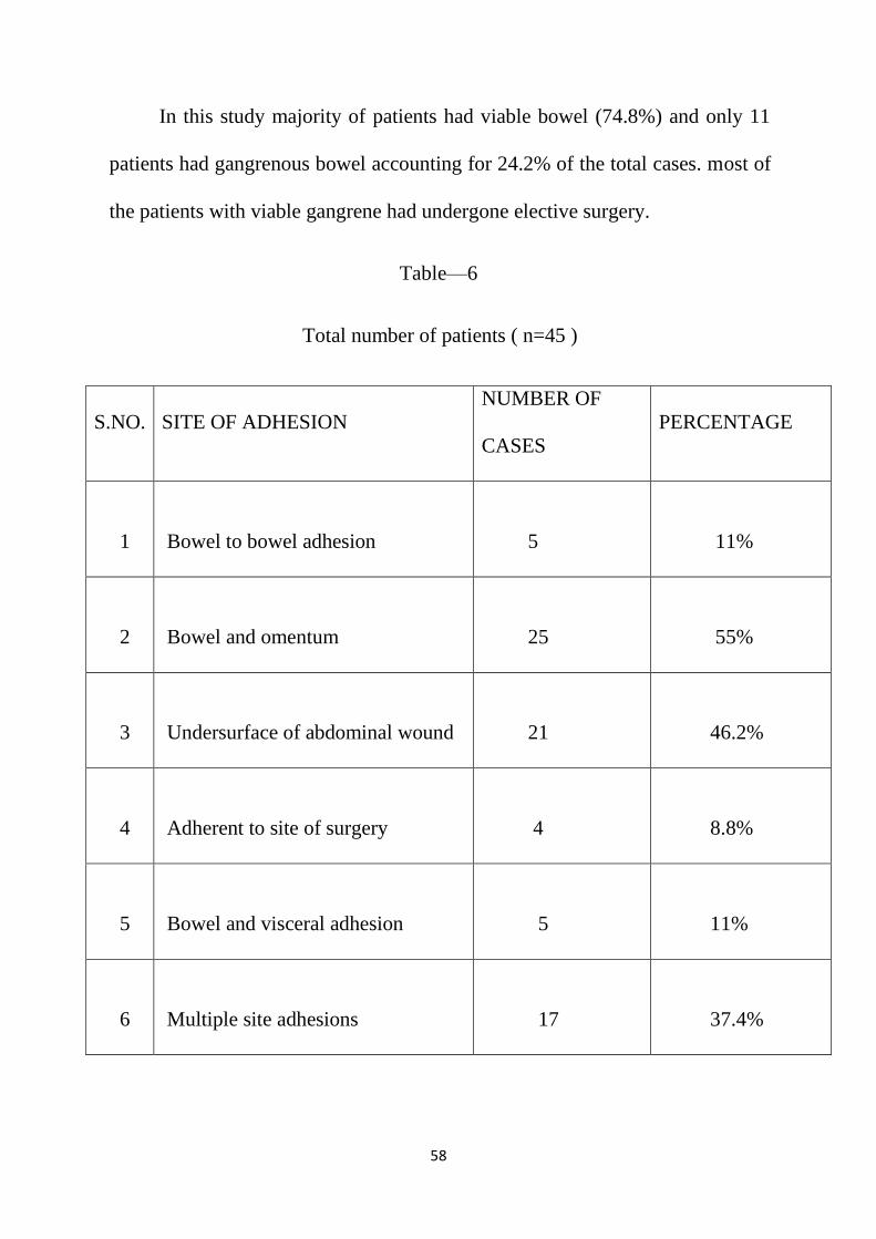
In this study majority of patients had viable bowel (74.8%) and only 11
patients had gangrenous bowel accounting for 24.2% of the total cases. most of
the patients with viable gangrene had undergone elective surgery.
Table—6
Total number of patients ( n=45 )
S.NO. SITE OF ADHESION
NUMBER OF
CASES
PERCENTAGE
1
Bowel to bowel adhesion
5
11%
2
Bowel and omentum
25
55%
3
Undersurface of abdominal wound
21
46.2%
4
Adherent to site of surgery
4
8.8%
5
Bowel and visceral adhesion
5
11%
6
Multiple site adhesions
17
37.4%
59
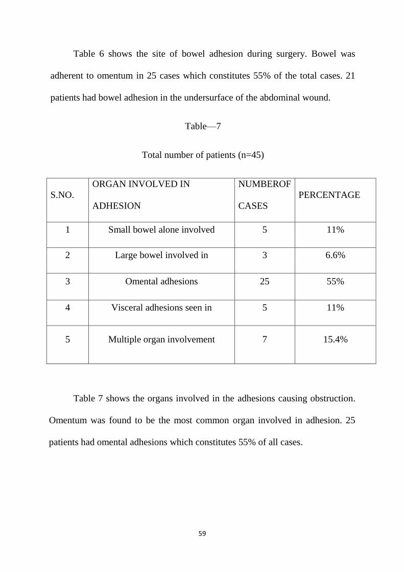
Table 6 shows the site of bowel adhesion during surgery. Bowel was
adherent to omentum in 25 cases which constitutes 55% of the total cases. 21
patients had bowel adhesion in the undersurface of the abdominal wound.
Table—7
Total number of patients (n=45)
S.NO.
ORGAN INVOLVED IN
ADHESION
NUMBEROF
CASES
PERCENTAGE
1 Small bowel alone involved 5 11%
2 Large bowel involved in 3 6.6%
3 Omental adhesions 25 55%
4 Visceral adhesions seen in 5 11%
5 Multiple organ involvement 7 15.4%
Table 7 shows the organs involved in the adhesions causing obstruction.
Omentum was found to be the most common organ involved in adhesion. 25
patients had omental adhesions which constitutes 55% of all cases.

60
Table—8
Total number of patients ( n=45 )
S.NO.
CAUSE OF
ADHESION
MALES
FEMALES
% OF
MALE
%OF
FEMALE
TOTAL
1
Post surgical
15
13
33.30%
28.60%
61.90%
2
Post
inflammatory
12
5
26.40%
11.00%
37.40%
TOTAL
27
18
59.70%
39.70%
99.40%
Table 8 shows the causes of adhesive intestinal obstruction in our study
group of patients. 15 male and 13 female patients had intestinal obstruction due
to post surgical adhesions and this constitutes 33.30% and 28.60% of all cases
respectively. On the whole post surgical adhesions was found to be the most
common cause of adhesive intestinal obstruction which constitutes 61.90% of
all cases. Post inflammatory adhesions found in 37.40% of all cases.
61
Table 9
Total number of post surgical adhesive obstructions (n=28)
S.NO. PERVIOUS SURGERY
CAUSING ADHESIONS
NUMBER OF
PATIENTS
PERCENTAGE
1 Appendicectomy 6 21.42%
2 Caesarean section 8 28.57%
3
Hollow viscous Perforation
closure
5 17.85%
4 Hernioplasty 3 10.71%
5 Hysterectomy 3 10.71%
6 Other surgeries 3 10.71%
28 99.97%
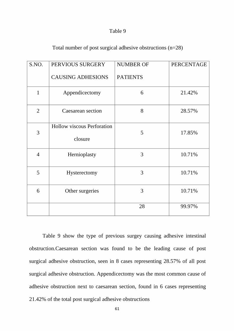
Table 9 show the type of previous surgey causing adhesive intestinal
obstruction.Caesarean section was found to be the leading cause of post
surgical adhesive obstruction, seen in 8 cases representing 28.57% of all post
surgical adhesive obstruction. Appendicectomy was the most common cause of
adhesive obstruction next to caesarean section, found in 6 cases representing
21.42% of the total post surgical adhesive obstructions
62
Table—10
Total number of post inflammatory adhesive obstruction (n=17)
S.NO.
POSTINFLAMMATORY
CONDITIONS CAUSING
ADHESIONS
NUMBER OF
CASES
PERCENTAGE
1 Appendicitis 6 35.29%
2 Peritonitis 3 17.65%
3 Pancreatitis 3 17.65%
4 Ileo caecal tuberculosis 5 29.41%
TOTAL 17 100%
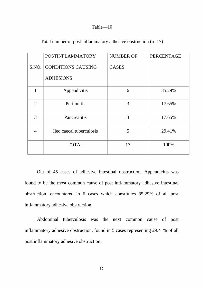
Out of 45 cases of adhesive intestinal obstruction, Appendicitis was
found to be the most common cause of post inflammatory adhesive intestinal
obstruction, encountered in 6 cases which constitutes 35.29% of all post
inflammatory adhesive obstruction.
Abdominal tuberculosis was the next common cause of post
inflammatory adhesive obstruction, found in 5 cases representing 29.41% of all
post inflammatory adhesive obstruction.
63
Table-11
Total number of patients (n=45)
S.NO COMPLICATONS
NO.OF
MALE
PATIENTS
NO.OF
FEMALE
PATIENTS
%OF
MALE
%OF
FEMALE
TOTAL
1 Wound infections 5 3 11.00% 6.60% 17.60%
2 Fecal fistula 2 0 4.40% 0% 4.40%
3 Post operative ileus 4 1 8.80% \2.20% 11.00%
4 Burst abdomen 1 0 2.20% 0% 2.20%
5 Death 0 2 0% 4.40% 4.40%
6 Seroma 2 1 4.40% 2.20% 6.60%
TOTAL 14 7 30.80% 15.40% 46.20%
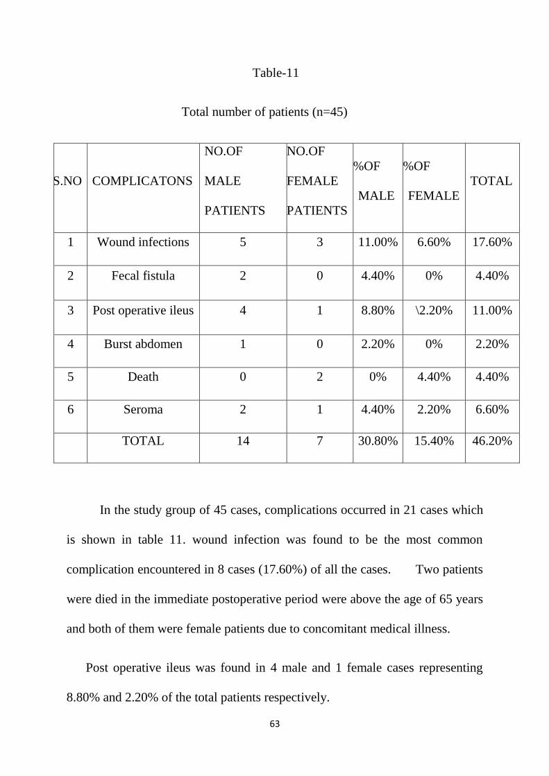
In the study group of 45 cases, complications occurred in 21 cases which
is shown in table 11. wound infection was found to be the most common
complication encountered in 8 cases (17.60%) of all the cases. Two patients
were died in the immediate postoperative period were above the age of 65 years
and both of them were female patients due to concomitant medical illness.
Post operative ileus was found in 4 male and 1 female cases representing
8.80% and 2.20% of the total patients respectively.
64
DISCUSSION
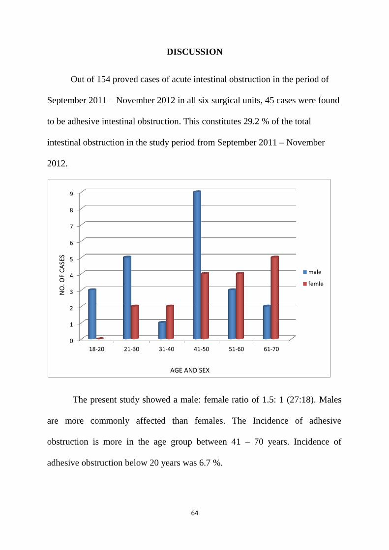
Out of 154 proved cases of acute intestinal obstruction in the period of
September 2011 – November 2012 in all six surgical units, 45 cases were found
to be adhesive intestinal obstruction. This constitutes 29.2 % of the total
intestinal obstruction in the study period from September 2011 – November
2012.
The present study showed a male: female ratio of 1.5: 1 (27:18). Males
are more commonly affected than females. The Incidence of adhesive
obstruction is more in the age group between 41 – 70 years. Incidence of
adhesive obstruction below 20 years was 6.7 %.
0
1
2
3
4
5
6
7
8
9
18-20 21-30 31-40 41-50 51-60 61-70
male
femle
AGE AND SEX
NO
. OF
CA
SES
65
In our study the incidence of adhesive intestinal obstruction is 29.2% of the
total intestinal obstruction cases.In a study conducted by Nemir ,Perry,Bevan and
MEntee,ADHESIONJOURNALS/birthofard3,2001, the incidence of adhesive
intestinal obstruction was 30% of all intestinal obstruction .Our results are similar
to the previous study results.
S. NO PARA METER Nemir,perry et al
STUDY
PRESENT STUDY
1
INCIDENCE OF
ADHESIVE
OBSTRUCTION IN
ALL OBSTRUCTION
30% 29.2%
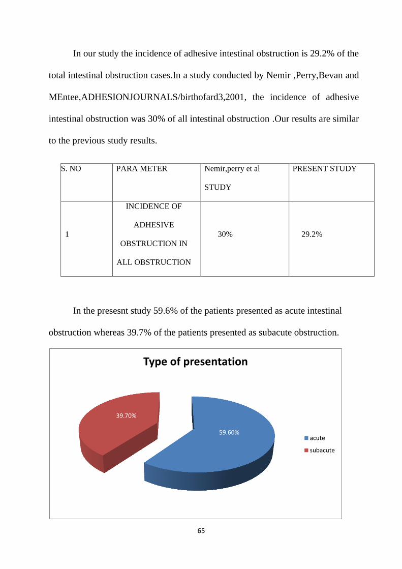
In the presesnt study 59.6% of the patients presented as acute intestinal
obstruction whereas 39.7% of the patients presented as subacute obstruction.
59.60%
39.70%
Type of presentation
acute
subacute
66
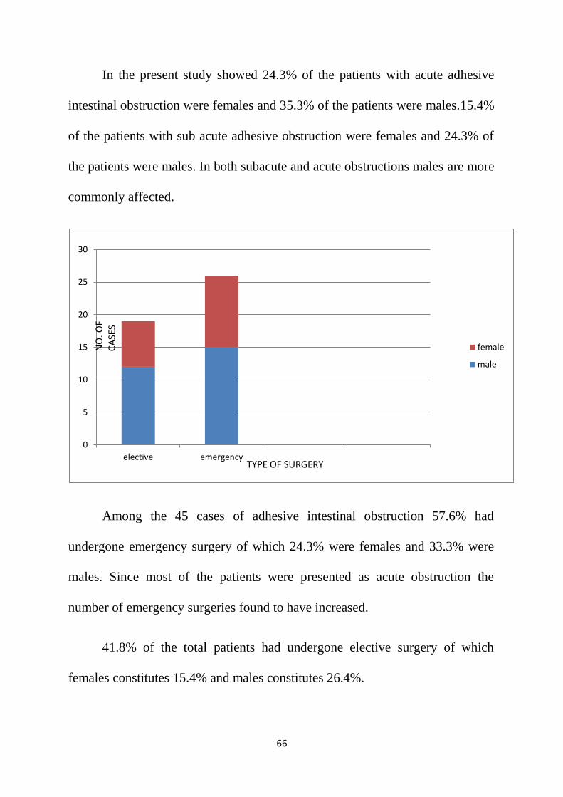
In the present study showed 24.3% of the patients with acute adhesive
intestinal obstruction were females and 35.3% of the patients were males.15.4%
of the patients with sub acute adhesive obstruction were females and 24.3% of
the patients were males. In both subacute and acute obstructions males are more
commonly affected.
Among the 45 cases of adhesive intestinal obstruction 57.6% had
undergone emergency surgery of which 24.3% were females and 33.3% were
males. Since most of the patients were presented as acute obstruction the
number of emergency surgeries found to have increased.
41.8% of the total patients had undergone elective surgery of which
females constitutes 15.4% and males constitutes 26.4%.
0
5
10
15
20
25
30
elective emergency
female
male
TYPE OF SURGERY
NO
. OF
CA
SES

67
In this study, 46.2% of the patients were treated with open adhesiolysis of
which males were 30.8% and females were 15.4%. laparoscopic adhesolysis
was done in 26.4% of cases out of which 11% were females and 15.4% were
males. Transverse colostomy was done in 1 male patient. Resection
anastomosis was done in 11 patients who had gangrenous bowel and this
constitutes 24.2% of all cases.
Out of 45 cases 22 male and 12 female patients had viable bowel. This
constitutes 74.8% of the total number of cases. 5 male and 6 female patients had
gangrenous bowel which accounts for 24.2% of the total cases. Majority of the
patients had viable bowel at the time of surgery
0.00%
5.00%
10.00%
15.00%
20.00%
25.00%
30.00%
35.00%
openadhesiolysis
resectionanastomosis
lap adhesiolysis colostomy
male
female
TYPE OF SURGERY
PER
CEN
TAG
E
68
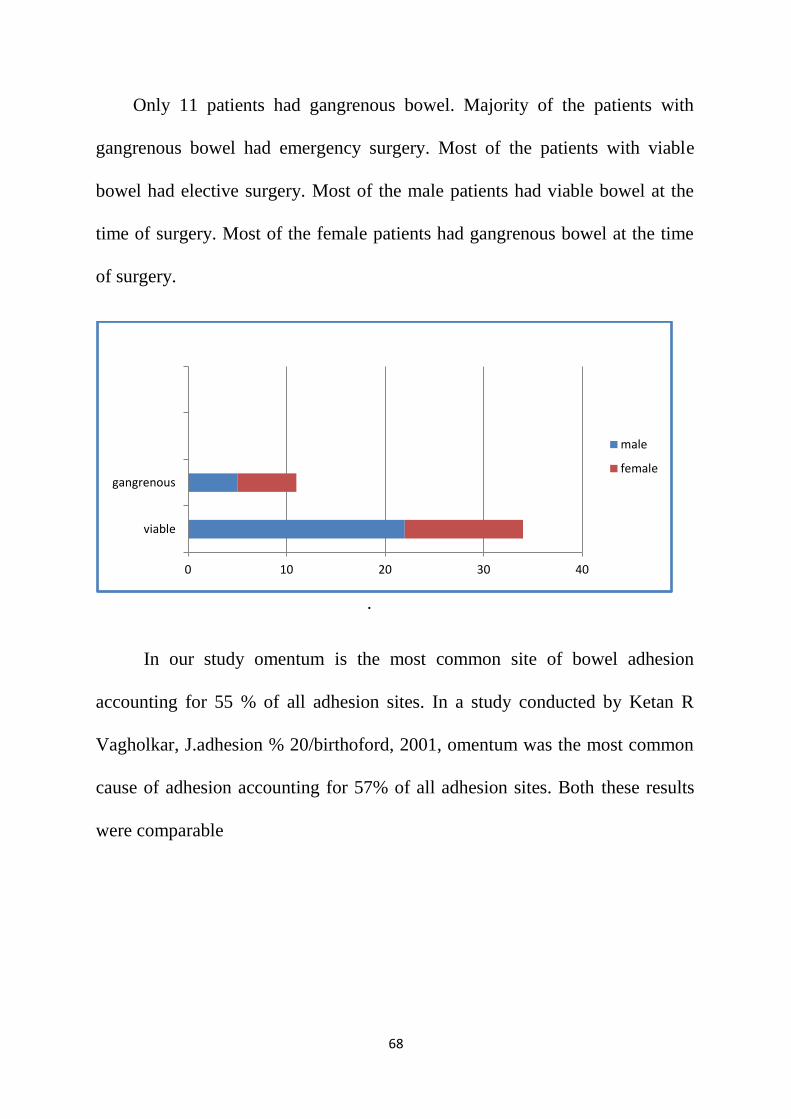
Only 11 patients had gangrenous bowel. Majority of the patients with
gangrenous bowel had emergency surgery. Most of the patients with viable
bowel had elective surgery. Most of the male patients had viable bowel at the
time of surgery. Most of the female patients had gangrenous bowel at the time
of surgery.
.
In our study omentum is the most common site of bowel adhesion
accounting for 55 % of all adhesion sites. In a study conducted by Ketan R
Vagholkar, J.adhesion % 20/birthoford, 2001, omentum was the most common
cause of adhesion accounting for 57% of all adhesion sites. Both these results
were comparable
0 10 20 30 40
viable
gangrenous
male
female
69
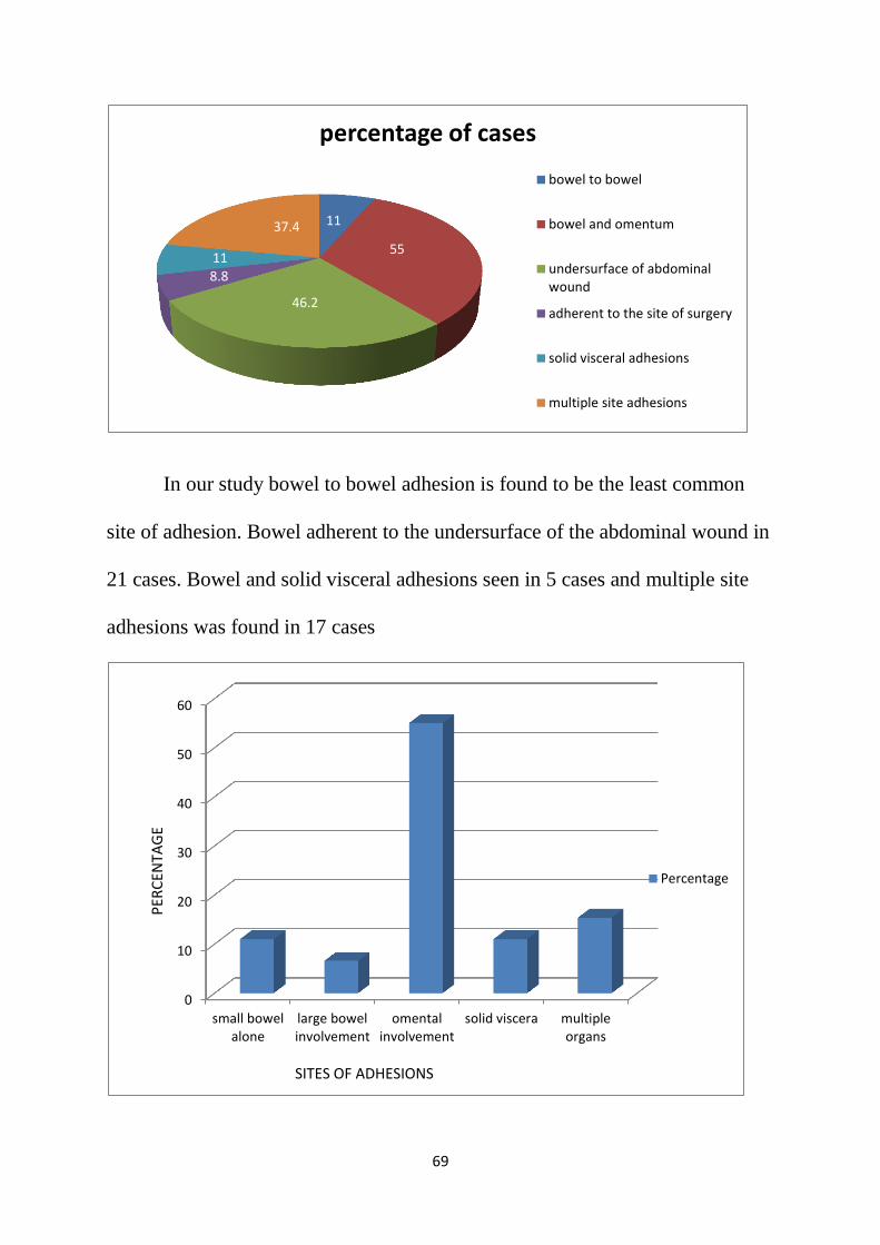
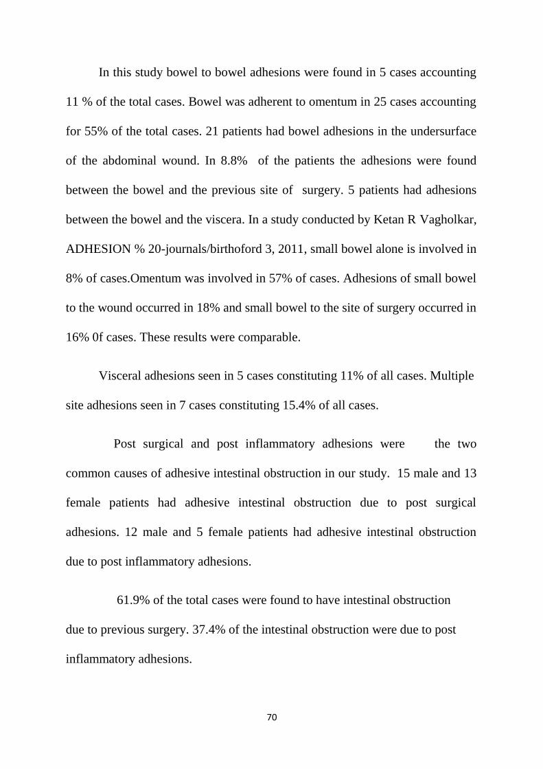
In our study bowel to bowel adhesion is found to be the least common
site of adhesion. Bowel adherent to the undersurface of the abdominal wound in
21 cases. Bowel and solid visceral adhesions seen in 5 cases and multiple site
adhesions was found in 17 cases
11
55
46.2
8.8 11
37.4
percentage of cases
bowel to bowel
bowel and omentum
undersurface of abdominalwound
adherent to the site of surgery
solid visceral adhesions
multiple site adhesions
0
10
20
30
40
50
60
small bowelalone
large bowelinvolvement
omentalinvolvement
solid viscera multipleorgans
Percentage
SITES OF ADHESIONS
PER
CEN
TAG
E
70
In this study bowel to bowel adhesions were found in 5 cases accounting
11 % of the total cases. Bowel was adherent to omentum in 25 cases accounting
for 55% of the total cases. 21 patients had bowel adhesions in the undersurface
of the abdominal wound. In 8.8% of the patients the adhesions were found
between the bowel and the previous site of surgery. 5 patients had adhesions
between the bowel and the viscera. In a study conducted by Ketan R Vagholkar,
ADHESION % 20-journals/birthoford 3, 2011, small bowel alone is involved in
8% of cases.Omentum was involved in 57% of cases. Adhesions of small bowel
to the wound occurred in 18% and small bowel to the site of surgery occurred in
16% 0f cases. These results were comparable.
Visceral adhesions seen in 5 cases constituting 11% of all cases. Multiple
site adhesions seen in 7 cases constituting 15.4% of all cases.
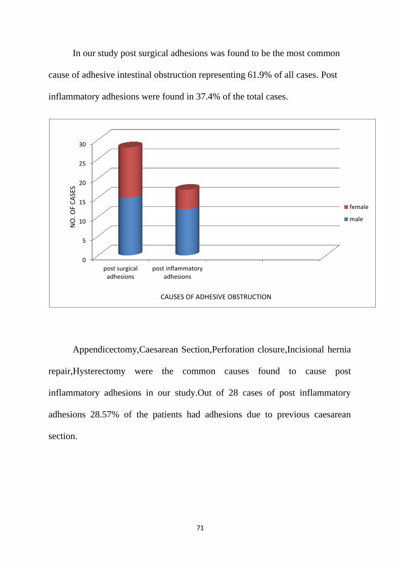
Post surgical and post inflammatory adhesions were the two
common causes of adhesive intestinal obstruction in our study. 15 male and 13
female patients had adhesive intestinal obstruction due to post surgical
adhesions. 12 male and 5 female patients had adhesive intestinal obstruction
due to post inflammatory adhesions.
61.9% of the total cases were found to have intestinal obstruction
due to previous surgery. 37.4% of the intestinal obstruction were due to post
inflammatory adhesions.
71
In our study post surgical adhesions was found to be the most common
cause of adhesive intestinal obstruction representing 61.9% of all cases. Post
inflammatory adhesions were found in 37.4% of the total cases.
Appendicectomy,Caesarean Section,Perforation closure,Incisional hernia
repair,Hysterectomy were the common causes found to cause post
inflammatory adhesions in our study.Out of 28 cases of post inflammatory
adhesions 28.57% of the patients had adhesions due to previous caesarean
section.
0
5
10
15
20
25
30
post surgicaladhesions
post inflammatoryadhesions
female
male
NO
. OF
CA
SES
CAUSES OF ADHESIVE OBSTRUCTION
72
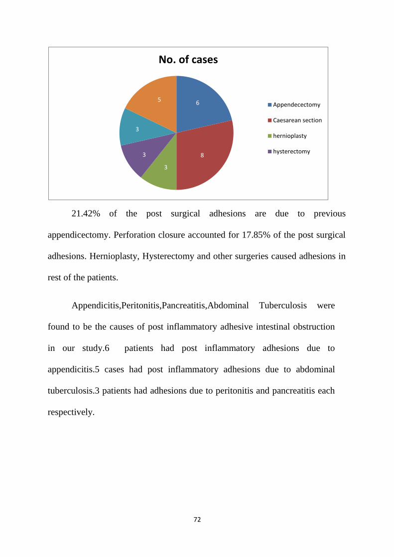
21.42% of the post surgical adhesions are due to previous
appendicectomy. Perforation closure accounted for 17.85% of the post surgical
adhesions. Hernioplasty, Hysterectomy and other surgeries caused adhesions in
rest of the patients.
Appendicitis,Peritonitis,Pancreatitis,Abdominal Tuberculosis were
found to be the causes of post inflammatory adhesive intestinal obstruction
in our study.6 patients had post inflammatory adhesions due to
appendicitis.5 cases had post inflammatory adhesions due to abdominal
tuberculosis.3 patients had adhesions due to peritonitis and pancreatitis each
respectively.
6
8
3
3
3
5
No. of cases
Appendecectomy
Caesarean section
hernioplasty
hysterectomy
73
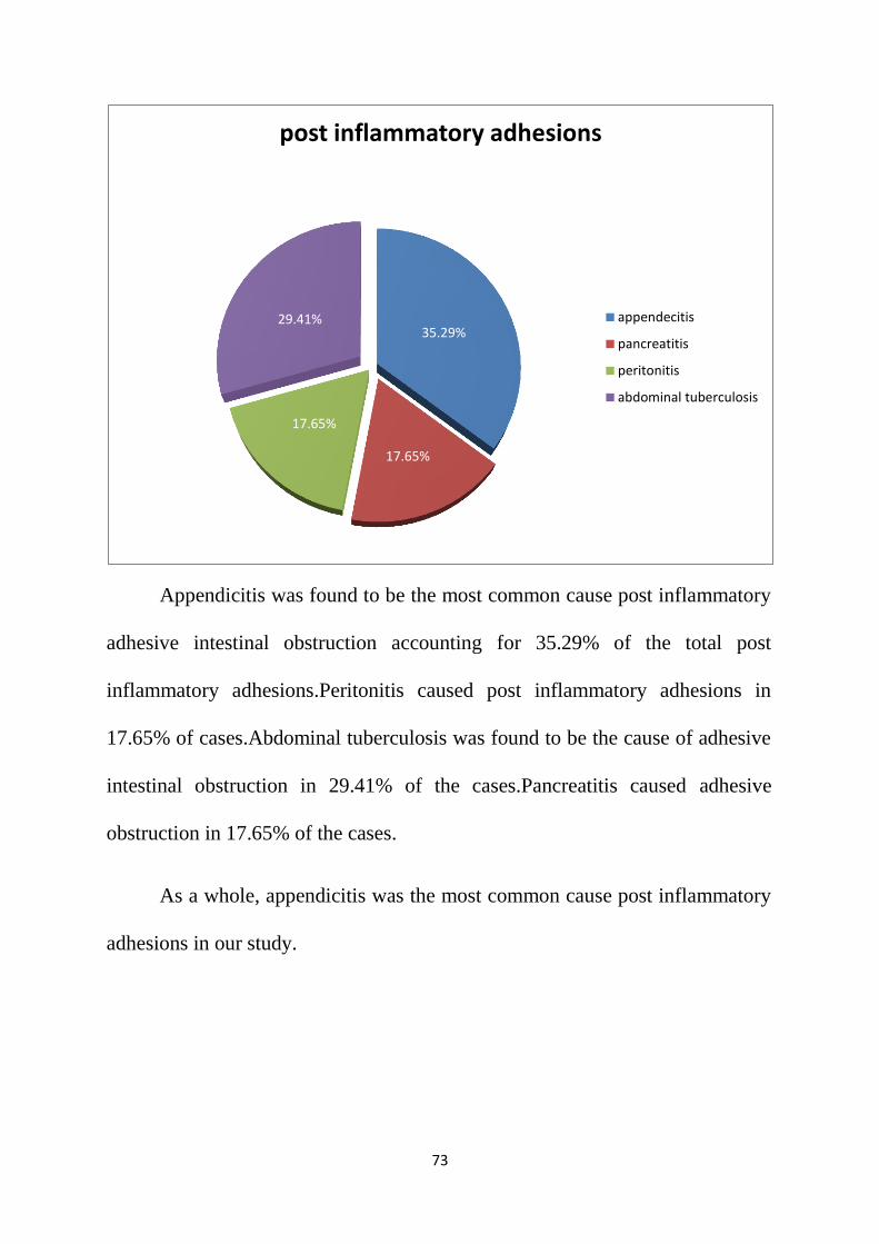
Appendicitis was found to be the most common cause post inflammatory
adhesive intestinal obstruction accounting for 35.29% of the total post
inflammatory adhesions.Peritonitis caused post inflammatory adhesions in
17.65% of cases.Abdominal tuberculosis was found to be the cause of adhesive
intestinal obstruction in 29.41% of the cases.Pancreatitis caused adhesive
obstruction in 17.65% of the cases.
As a whole, appendicitis was the most common cause post inflammatory
adhesions in our study.
35.29%
17.65%
17.65%
29.41%
post inflammatory adhesions
appendecitis
pancreatitis
peritonitis
abdominal tuberculosis
74
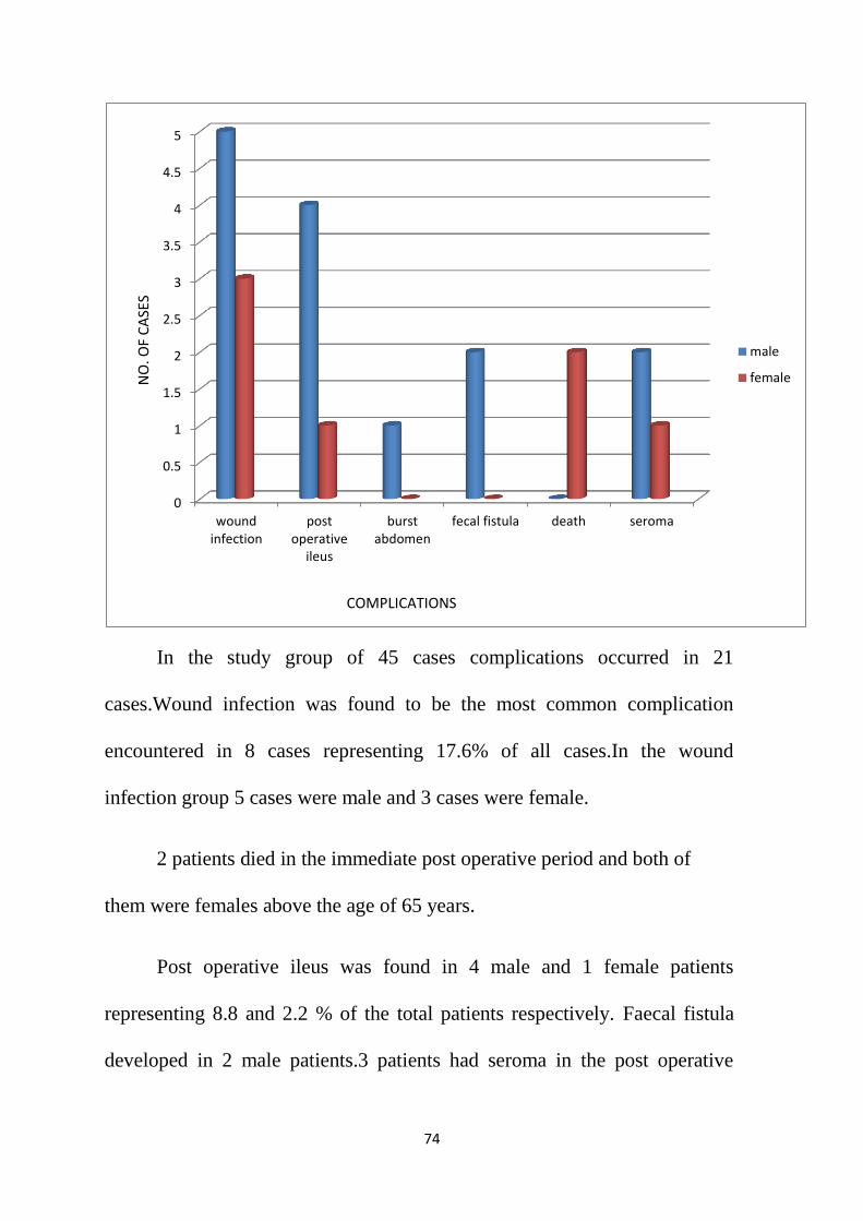
In the study group of 45 cases complications occurred in 21
cases.Wound infection was found to be the most common complication
encountered in 8 cases representing 17.6% of all cases.In the wound
infection group 5 cases were male and 3 cases were female.
2 patients died in the immediate post operative period and both of
them were females above the age of 65 years.
Post operative ileus was found in 4 male and 1 female patients
representing 8.8 and 2.2 % of the total patients respectively. Faecal fistula
developed in 2 male patients.3 patients had seroma in the post operative
0
0.5
1
1.5
2
2.5
3
3.5
4
4.5
5
woundinfection
postoperative
ileus
burstabdomen
fecal fistula death seroma
male
female
COMPLICATIONS
NO
. OF
CA
SES
75
period. Burst abdomen was encountered in 1 male patient which was later
treated by secondary suturing.
46.2% of the total patients had complications in the post operative
period.Among them,wound infection was found to be the leading
complication and burst abdomen was the least common complication in our
study.
76
SUMMARY
Adhesions are the leading cause of intestinal obstruction. Surgeons are
frequently facing the emergency of acute intestinal obstruction in most of their
duty days. Hence this study was focused on adhesive intestinal obstruction.
A collection of 45 cases of adhesive intestinal obstruction was done for
this study from all the 6 surgical units in the period between September 2011
and November 2012.
Adhesive intestinal obstruction accounted for 29.2% of total intestinal
obstructions in Coimbatore Medical College Hospital.
The incidence in sex is more in men than in women. Majority of the
adhesive intestinal obstruction patients were in the age group of 41-70 years of
age. In this study the male:female ratio is 1.5:1.
Majority of the patients presented as acute intestinal obstruction are males
.Acute adhesive intestinal obstruction representing 59.6% of the total adhesive
obstructions.
Most of the patients had undergone emergency surgery than elective
surgery, representing 57.6 % and 41.8% respectively.
Laparotomy with adhesiolysis, laparotomy with resection and
anastomosis, Laparoscopic surgeries and defunctioning colostomy are the
surgical procedures done in our hospital for adhesive intestinal obstruction.33
77
patients were treated with open surgeries and 12 patients were treated with
laparoscopic surgeries.
In this study out of 45 cases, 34 patients were found to have viable bowel
(74.8%) and 11 patients (24.2%) were found to have gangrenous bowel at the
time of surgery.
Omentum was found to be the most common site of adhesions and it
constituted 55% of all sites of adhesions.
Post surgical adhesive intestinal obstruction was more common than post
inflammatory adhesive intestinal obstruction,representing 61.9% and 37.4%
respectively.In this study caesarean section was found to be the most common
cause for adhesive obstruction which constituted 28.57% of all cases of post
surgical adhesions. 2 patients died in the immediate post operative period due to
concomitant medical illness which represented 4.4% of the total cases.
Adhesions due to tuberculous etiology was found in 5 cases which
constituted for 29.41% of all cases of post inflammatory adhesions.
Wound Infection was found to be the common complication.Wound
infection was encountered in 17.6% of the total cases.
78
CONCLUSION
Acute intestinal obstruction is a real emergency surgical case admitted in
our wards. Adhesions are the leading cause of intestinal obstruction.
Majority of the adhesive intestinal obstructions were post surgical than
post inflammatory. Ileocaecal tuberculosis is one of the important cause of post
inflammatory adhesive intestinal obstruction.
In subacute pattern of obstruction if the symptoms are not relieved with
conservative management, the patient should be taken up for surgery.
Gentle handling of tissues and avoiding unnecessary manipulation of the
bowel will reduce the incidence of intestinal obstructions due to post operative
adhesions.
Close monitoring of the abdominal girth,recording BP,pulse rate and
temperature and early recognition and early surgery will prevent gangrene of
bowel.
Wound infection being the most common complication in this study,
proper post operative wound care will prevent wound infections. Mortality
depends upon the age of the patient and co-morbid conditions
This study was conducted in Coimbatore Medical College Hospital and
the results are similar and comparable to other studies.
BIBLIOGRAPHY
1. Norman S Williams,Christopher, J K Bulstrode.Bailey and Love’s
Short Practice of Surgery, 25th
edition, pages 252,996, 1198-1203, 1217.
2. Fauci, Kasper, Hauser, Longo, Brown, Jameson, Braunwald. Harrison’s
Principles of Internal Medicine, 17th
edition, Volume II Pages 1912-
1914.
3. Josef E Fischer. Fischer’s Mastery of Surgery 6th
edition Volume II,
Pages 1542-1547.
4. Brunicardi, Dana K Andersen, Timothy R Billiar. Schwartz’s Principles
of Surgery 9th
edition, Pages 227,333,988-992, 1009-1011,1015-
1018,1504
5. Kim E Barrett, Susan M Barman, Scott Boitano. Ganong’s Review of
Medical Physiology 23rd
edition, Pages 429-449.
6. Keith L Moore,Arthur F Dalley, Anne M R .Clinically Oriented
Anatomy 6th
edition pages 242-253.
7. Farquharson’s Textbook of Operative Surgery
8. Hamilton Bailey A short practice of surgery
9. Rodney Maingot’s Abdominal Operations Volume II
10. Oxford Textbook of Surgery
11. Sabiston’s Textbook of Surgery

12. Holmdahi L E,Al-Jabreen M et al. Role of Fibrinolysis in the formation
of post operative adhesions. J Wound repair Regen 1994
Jul;2(3):171-6
13. Ketan Vagholkar. ADHESIVE INTESTINAL OBSTRUCTION J
ADHESION%20/birthofard3,2001
14. A Niyaf,R S Bhandari,K P Singh Management of Adhesive Intestinal
Obstruction;Journal of Institute of Medicine,August,2010;32:2,18-20.
15. Attard Maclean AR.AdhesiveSmallBowelObstruction:epidemiology,
biology and prevention;Can J Surg 2007 Aug;50(4):291-300
16. Arung W,Detry O et al. Pathophysiology and prevention of post
Operative peritoneal adhesions;World J Gastroenterol.2011Nov
7;17(41):4545-53
17. Bjorg-Tilde,Fevang,MD,Jonas Fevang MD PhD et al.Long term
prognosis after operation for adhesive small bowel obstruction; Ann Surg.
2004 August;240(2):193-201
18. Eriberto Farinella,Roberto Cirocchi et al .Feasibilty of Laparoscopy for
small bowel obstruction; World J Emerg Surg 2009; 4:3
19. Megan Fix,MD,Maine Medical Centre. Small Bowel Obstruction.
20. Hok –K Wok Choi,Kin wah Chu et al .Therapeutic Value of Gastrografin
In adhesive small bowel obstruction after unsuccessful conservative
treatment; Surg 2002 July;236(1):1-6
21. Ivarsson M L,Falk P,Holmdahl L .Response of visceral peritoneum to
abdominal surgery;Br J Surg 2001 Jan ;88(1):148-151
22. Cary B Aarons MD,Philip A Cohen et al.Statins (HMG-CoA Reductase
inhibitors) decrease post operative adhesions by increasing peritoneal
fibrinolytic activity;Ann surg 2007 February;245(12):176-184
23. Alpay Z,Saed G M,Diamond MP .Post operative adhesions: from
formation to prevention;Semin Reprod Med.2008 Jul;26(4):313-321
24. Alpay Z,Saed G M,Diamond MP. Pharmacologic Inhibition of adhesion
formation and peritoneal tissue type plasminogen activator activity;Semin
reprod 2008 Jul;26(4) 331-40.
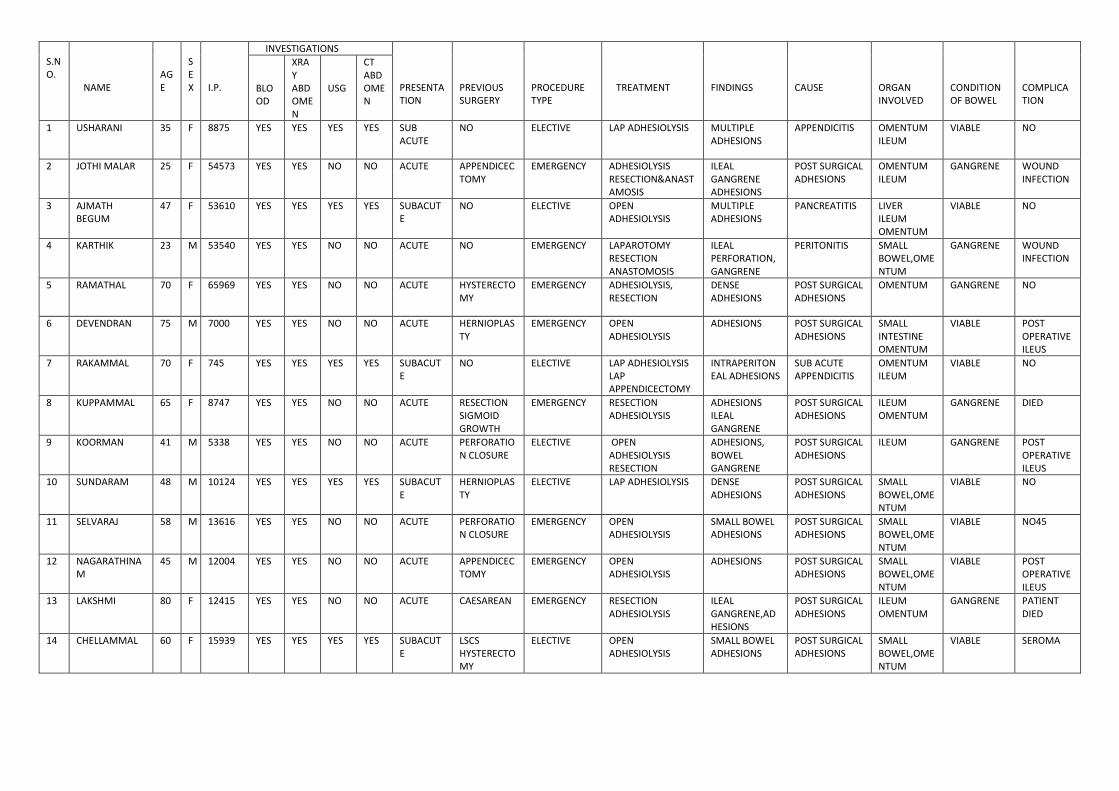
S.NO.
NAME
AGE
SEX
I.P.
INVESTIGATIONS PRESENTATION
PREVIOUS SURGERY
PROCEDURE TYPE
TREATMENT
FINDINGS
CAUSE
ORGAN INVOLVED
CONDITION OF BOWEL
COMPLICATION
BLOOD
XRAY ABDOMEN
USG
CT ABDOMEN
1 USHARANI 35 F 8875 YES YES YES YES SUB ACUTE
NO ELECTIVE LAP ADHESIOLYSIS MULTIPLE ADHESIONS
APPENDICITIS OMENTUM ILEUM
VIABLE NO
2 JOTHI MALAR 25 F 54573 YES YES NO NO ACUTE APPENDICECTOMY
EMERGENCY ADHESIOLYSIS RESECTION&ANASTAMOSIS
ILEAL GANGRENE ADHESIONS
POST SURGICAL ADHESIONS
OMENTUM ILEUM
GANGRENE WOUND INFECTION
3 AJMATH BEGUM
47 F 53610 YES YES YES YES SUBACUTE
NO ELECTIVE OPEN ADHESIOLYSIS
MULTIPLE ADHESIONS
PANCREATITIS LIVER ILEUM OMENTUM
VIABLE NO
4 KARTHIK 23 M 53540 YES YES NO NO ACUTE NO EMERGENCY LAPAROTOMY RESECTION ANASTOMOSIS
ILEAL PERFORATION,GANGRENE
PERITONITIS SMALL BOWEL,OMENTUM
GANGRENE WOUND INFECTION
5 RAMATHAL 70 F 65969 YES YES NO NO ACUTE HYSTERECTOMY
EMERGENCY ADHESIOLYSIS, RESECTION
DENSE ADHESIONS
POST SURGICAL ADHESIONS
OMENTUM GANGRENE NO
6 DEVENDRAN 75 M 7000 YES YES NO NO ACUTE HERNIOPLASTY
EMERGENCY OPEN ADHESIOLYSIS
ADHESIONS POST SURGICAL ADHESIONS
SMALL INTESTINE OMENTUM
VIABLE POST OPERATIVE ILEUS
7 RAKAMMAL 70 F 745 YES YES YES YES SUBACUTE
NO ELECTIVE LAP ADHESIOLYSIS LAP APPENDICECTOMY
INTRAPERITONEAL ADHESIONS
SUB ACUTE APPENDICITIS
OMENTUM ILEUM
VIABLE NO
8 KUPPAMMAL 65 F 8747 YES YES NO NO ACUTE RESECTION SIGMOID GROWTH
EMERGENCY RESECTION ADHESIOLYSIS
ADHESIONS ILEAL GANGRENE
POST SURGICAL ADHESIONS
ILEUM OMENTUM
GANGRENE DIED
9 KOORMAN 41 M 5338 YES YES NO NO ACUTE PERFORATION CLOSURE
ELECTIVE OPEN ADHESIOLYSIS RESECTION
ADHESIONS, BOWEL GANGRENE
POST SURGICAL ADHESIONS
ILEUM GANGRENE POST OPERATIVE ILEUS
10 SUNDARAM 48 M 10124 YES YES YES YES SUBACUTE
HERNIOPLASTY
ELECTIVE LAP ADHESIOLYSIS DENSE ADHESIONS
POST SURGICAL ADHESIONS
SMALL BOWEL,OMENTUM
VIABLE NO
11 SELVARAJ 58 M 13616 YES YES NO NO ACUTE PERFORATION CLOSURE
EMERGENCY OPEN ADHESIOLYSIS
SMALL BOWEL ADHESIONS
POST SURGICAL ADHESIONS
SMALL BOWEL,OMENTUM
VIABLE NO45
12 NAGARATHINAM
45 M 12004 YES YES NO NO ACUTE APPENDICECTOMY
EMERGENCY OPEN ADHESIOLYSIS
ADHESIONS POST SURGICAL ADHESIONS
SMALL BOWEL,OMENTUM
VIABLE POST OPERATIVE ILEUS
13 LAKSHMI 80 F 12415 YES YES NO NO ACUTE CAESAREAN EMERGENCY RESECTION ADHESIOLYSIS
ILEAL GANGRENE,ADHESIONS
POST SURGICAL ADHESIONS
ILEUM OMENTUM
GANGRENE PATIENT DIED
14 CHELLAMMAL 60 F 15939 YES YES YES YES SUBACUTE
LSCS HYSTERECTOMY
ELECTIVE OPEN ADHESIOLYSIS
SMALL BOWEL ADHESIONS
POST SURGICAL ADHESIONS
SMALL BOWEL,OMENTUM
VIABLE SEROMA
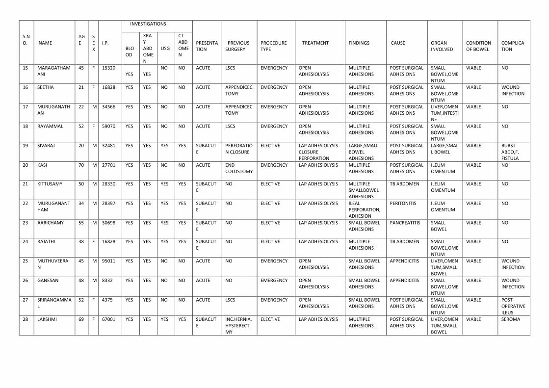
S.NO.
NAME
AGE
SEX
I.P.
INVESTIGATIONS PRESENTATION
PREVIOUS SURGERY
PROCEDURE TYPE
TREATMENT
FINDINGS
CAUSE
ORGAN INVOLVED
CONDITION OF BOWEL
COMPLICATION
BLOOD
XRAY ABDOMEN
USG
CT ABDOMEN
15 MARAGATHAMANI
45 F 15320 YES
YES
NO NO ACUTE LSCS EMERGENCY OPEN ADHESIOLYSIS
MULTIPLE ADHESIONS
POST SURGICAL ADHESIONS
SMALL BOWEL,OMENTUM
VIABLE NO
16 SEETHA 21 F 16828 YES YES NO NO ACUTE APPENDICECTOMY
EMERGENCY OPEN ADHESIOLYSIS
MULTIPLE ADHESIONS
POST SURGICAL ADHESIONS
SMALL BOWEL,OMENTUM
VIABLE WOUND INFECTION
17 MURUGANATHAN
22 M 34566 YES YES NO NO ACUTE APPENDICECTOMY
EMERGENCY OPEN ADHESIOLYSIS
MULTIPLE ADHESIONS
POST SURGICAL ADHESIONS
LIVER,OMENTUM,INTESTINE
VIABLE NO
18 RAYAMMAL 52 F 59070 YES YES NO NO ACUTE LSCS EMERGENCY OPEN ADHESIOLYSIS
MULTIPLE ADHESIONS
POST SURGICAL ADHESIONS
SMALL BOWEL,OMENTUM
VIABLE NO
19 SIVARAJ 20 M 32481 YES YES YES YES SUBACUTE
PERFORATION CLOSURE
ELECTIVE LAP ADHESIOLYSIS CLOSURE PERFORATION
LARGE,SMALL BOWEL ADHESIONS
POST SURGICAL ADHESIONS
LARGE,SMALL BOWEL
VIABLE BURST ABDO,F. FISTULA
20 KASI 70 M 27701 YES YES NO NO ACUTE END COLOSTOMY
EMERGENCY LAP ADHESIOLYSIS MULTIPLE ADHESIONS
POST SURGICAL ADHESIONS
ILEUM OMENTUM
VIABLE NO
21 KITTUSAMY 50 M 28330 YES YES YES YES SUBACUTE
NO ELECTIVE LAP ADHESIOLYSIS MULTIPLE SMALLBOWEL ADHESIONS
TB ABDOMEN ILEUM OMENTUM
VIABLE NO
22 MURUGANANTHAM
34 M 28397 YES YES YES YES SUBACUTE
NO ELECTIVE LAP ADHESIOLYSIS ILEAL PERFORATION,ADHESION
PERITONITIS ILEUM OMENTUM
VIABLE NO
23 AARICHAMY 55 M 30698 YES YES YES YES SUBACUTE
NO ELECTIVE LAP ADHESIOLYSIS SMALL BOWEL ADHESIONS
PANCREATITIS SMALL BOWEL
VIABLE NO
24 RAJATHI 38 F 16828 YES YES YES YES SUBACUTE
NO ELECTIVE LAP ADHESIOLYSIS MULTIPLE ADHESIONS
TB ABDOMEN SMALL BOWEL,OMENTUM
VIABLE NO
25 MUTHUVEERAN
45 M 95011 YES YES NO NO ACUTE NO EMERGENCY OPEN ADHESIOLYSIS
SMALL BOWEL ADHESIONS
APPENDICITIS LIVER,OMENTUM,SMALL BOWEL
VIABLE WOUND INFECTION
26 GANESAN 48 M 8332 YES YES NO NO ACUTE NO EMERGENCY OPEN ADHESIOLYSIS
SMALL BOWEL ADHESIONS
APPENDICITIS SMALL BOWEL,OMENTUM
VIABLE WOUND INFECTION
27 SRIRANGAMMAL
52 F 4375 YES YES NO NO ACUTE LSCS EMERGENCY OPEN ADHESIOLYSIS
SMALL BOWEL ADHESIONS
POST SURGICAL ADHESIONS
SMALL BOWEL,OMENTUM
VIABLE POST OPERATIVE ILEUS
28 LAKSHMI 69 F 67001 YES YES YES YES SUBACUTE
INC.HERNIA,HYSTERECTMY
ELECTIVE LAP ADHESIOLYSIS MULTIPLE ADHESIONS
POST SURGICAL ADHESIONS
LIVER,OMENTUM,SMALL BOWEL
VIABLE SEROMA
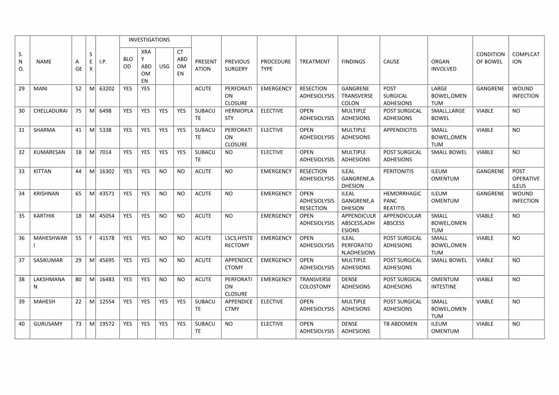
S.NO.
NAME
AGE
SEX
I.P.
INVESTIGATIONS PRESENTATION
PREVIOUS SURGERY
PROCEDURE TYPE
TREATMENT
FINDINGS
CAUSE
ORGAN INVOLVED
CONDITION OF BOWEL
COMPLCATION
BLOOD
XRAY ABDOMEN
USG
CT ABDOMEN
29 MANI 52 M 63202 YES YES ACUTE PERFORATION CLOSURE
EMERGENCY RESECTION ADHESIOLYSIS
GANGRENE TRANSVERSE COLON
POST SURGICAL ADHESIONS
LARGE BOWEL,OMENTUM
GANGRENE WOUND INFECTION
30 CHELLADURAI 75 M 6498 YES YES YES YES SUBACUTE
HERNIOPLASTY
ELECTIVE OPEN ADHESIOLYSIS
MULTIPLE ADHESIONS
POST SURGICAL ADHESIONS
SMALL,LARGE BOWEL
VIABLE NO
31 SHARMA 41 M 5338 YES YES YES YES SUBACUTE
PERFORATION CLOSURE
ELECTIVE OPEN ADHESIOLYSIS
MULTIPLE ADHESIONS
APPENDICITIS SMALL BOWEL,OMENTUM
VIABLE NO
32 KUMARESAN 18 M 7014 YES YES YES YES SUBACUTE
NO ELECTIVE OPEN ADHESIOLYSIS
MULTIPLE ADHESIONS
POST SURGICAL ADHESIONS
SMALL BOWEL VIABLE NO
33 KITTAN 44 M 16302 YES YES NO NO ACUTE NO EMERGENCY RESECTION ADHESIOLYSIS
ILEAL GANGRENE,ADHESION
PERITONITIS ILEUM OMENTUM
GANGRENE POST OPERATIVE ILEUS
34 KRISHNAN 65 M 43571 YES YES NO NO ACUTE NO EMERGENCY OPEN ADHESIOLYSIS RESECTION
ILEAL GANGRENE,ADHESION
HEMORRHAGIC PANC REATITIS
ILEUM OMENTUM
GANGRENE WOUND INFECTION
35 KARTHIK 18 M 45054 YES YES NO NO ACUTE NO EMERGENCY OPEN ADHESIOLYSIS
APPENDICULRABSCESS,ADHESIONS
APPENDICULAR ABSCESS
SMALL BOWEL,OMENTUM
VIABLE NO
36 MAHESHWARI
55 F 41578 YES YES NO NO ACUTE LSCS,HYSTERECTOMY
EMERGENCY OPEN ADHESIOLYSIS
ILEAL PERFORATION,ADHESIONS
POST SURGICAL ADHESIONS
SMALL BOWEL,OMENTUM
VIABLE NO
37 SASIKUMAR 29 M 45695 YES YES NO NO ACUTE APPENDICECTOMY
EMERGENCY OPEN ADHESIOLYSIS
MULTIPLE ADHESIONS
POST SURGICAL ADHESIONS
SMALL BOWEL VIABLE NO
38 LAKSHMANAN
80 M 16483 YES YES NO NO ACUTE PERFORATION CLOSURE
EMERGENCY TRANSVERSE COLOSTOMY
DENSE ADHESIONS
POST SURGICAL ADHESIONS
OMENTUM INTESTINE
VIABLE NO
39 MAHESH 22 M 12554 YES YES YES YES SUBACUTE
APPENDICECTMY
ELECTIVE OPEN ADHESIOLYSIS
MULTIPLE ADHESIONS
POST SURGICAL ADHESIONS
SMALL BOWEL,OMENTUM
VIABLE NO
40 GURUSAMY 73 M 19572 YES YES YES YES SUBACUTE
NO ELECTIVE OPEN ADHESIOLYSIS
DENSE ADHESIONS
TB ABDOMEN ILEUM OMENTUM
VIABLE NO
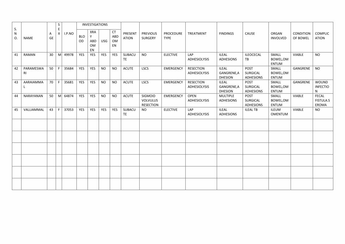
S.NO.
NAME
AGE
S EX
I.P.NO
INVESTIGATIONS PRESENTATION
PREVIOUS SURGERY
PROCEDURE TYPE
TREATMENT
FINDINGS
CAUSE
ORGAN INVOLVED
CONDITION OF BOWEL
COMPLICATION
BLOOD
XRAY ABDOMEN
USG
CT ABDOMEN
41 RAMAN 30 M 49978 YES YES YES YES SUBACUTE
NO ELECTIVE LAP ADHESIOLYSIS
ILEAL ADHESIONS
ILEOCECAL TB
SMALL BOWEL,OMENTUM
VIABLE NO
42 PARAMESWARI
50 F 35684 YES YES NO NO ACUTE LSCS EMERGENCY RESECTION ADHESIOLYSIS
ILEAL GANGRENE,ADHESION
POST SURGICAL ADHESIONS
SMALL BOWEL,OMENTUM
GANGRENE NO
43 AARAIAMMAL
70 F 35681 YES YES NO NO ACUTE LSCS EMERGENCY RESECTION ADHESIOLYSIS
ILEAL GANGRENE,ADHESION
POST SURGICAL ADHESIONS
SMALL BOWEL,OMENTUM
GANGRENE WOUND INFECTION
44 NARAYANAN 50 M 64874 YES YES NO NO ACUTE SIGMOID VOLVULUSRESECTION
EMERGENCY OPEN ADHESIOLYSIS
MULTIPLE ADHESIONS
POST SURGICAL ADHESIONS
SMALL BOWEL,OMENTUM
VIABLE FECAL FISTULA.SEROMA
45 VALLIAMMAL 43 F 37053 YES YES YES YES SUBACUTE
NO ELECTIVE LAP ADHESIOLYSIS
ILEAL ADHESIONS
ILEAL TB ILEUM OMENTUM
VIABLE NO

COIMBATORE MEDICAL COLLEGE HOSPITAL
COIMBATORE-18
DEPARTMENT OF SURGERY
STUDY OF ADHESIVE INTESTINAL OBSTRUCTION AND ITS
MANAGEMENT IN COIMBATORE MEDICAL COLLEGE HOSPITAL
DR. P. MURUGADASAN. M.S., POST GRADUATE
ACADEMIC PERIOD: SEPTEMBER 2011-NOVEMBER 2012
PROFORMA
Name: Age: Sex: Male/Female
S.No: IP No: occupation:
Diagnosis:
Treatment:
COMPLAINTS: 1. Pain Yes/No
1. Duration
2. Nature
3. Site
2. Associated symptoms
1. Vomiting Yes/No
2. Diarrhea Yes/No
Constipation Yes/No
3. Malena Yes/No
4. Abdominal distension Yes/No
5. Fever Yes/No
6. Urinary symptoms Yes/No
PAST HISTORY: 1. HISTORY OF PREVIOUS SURGERY Yes/No
1. Emergency/Elective
2. Incision
3. Time interval between previous surgery
and presentation
2. H/o previous similar episodes Yes/No
3. Known HT/DM/COPD/CAHD/EPILEPTIC Yes/No
CLINICAL FINDINGS:
1. Abdominal distension Yes/No
2. Bowel sounds Yes/No
3. Movement of abdomen with respiration Yes/No
4. Any palpable mass in the abdomen Yes/No
5. Free fluids Yes/No
6. Previous surgical scars Yes/No
7. Per rectal examination Yes/No
8. Other complaints Yes/No
9. General condition of the patient
RELEVANT INVESTIGATIONS:
1. X-ray abdomen erect Yes/No
2. Ultrasound abdomen scan Yes/No
3. CT Scan abdomen Yes/No
TREATMENT GIVEN:
1. Laparotomy and adhesiolysis Yes/No
2. Laparoscopic adhesiolysis Yes/No
3. Laprotomy and resection anastamosis Yes/No
4. Colostomy Yes/No
COMPLICATIONS
PATIENT CONSENT FORM
STUDY: STUDY OF ADHESIVE INTESTINAL OBSTRUCTION AND
ITS MANAGEMENT IN COIMBATORE MEDICAL COLLEGE
HOSPITAL
This study has been explained to me in my own language and I
understood the following
1. What the study involves
2. That the refusal to participate will not affect my treatment in any
way
3. That I may withdraw to take part in this study
Signature of the patient:
Full name of the patient:
Address:
Date:
Witness: (should be a person not connected with the study)
I have been present while the procedure to be performed has been
explained to the patient and I have witnessed his/her consent to take part.
Signature of the witness:
Full name of the witness:
Address:
Date:

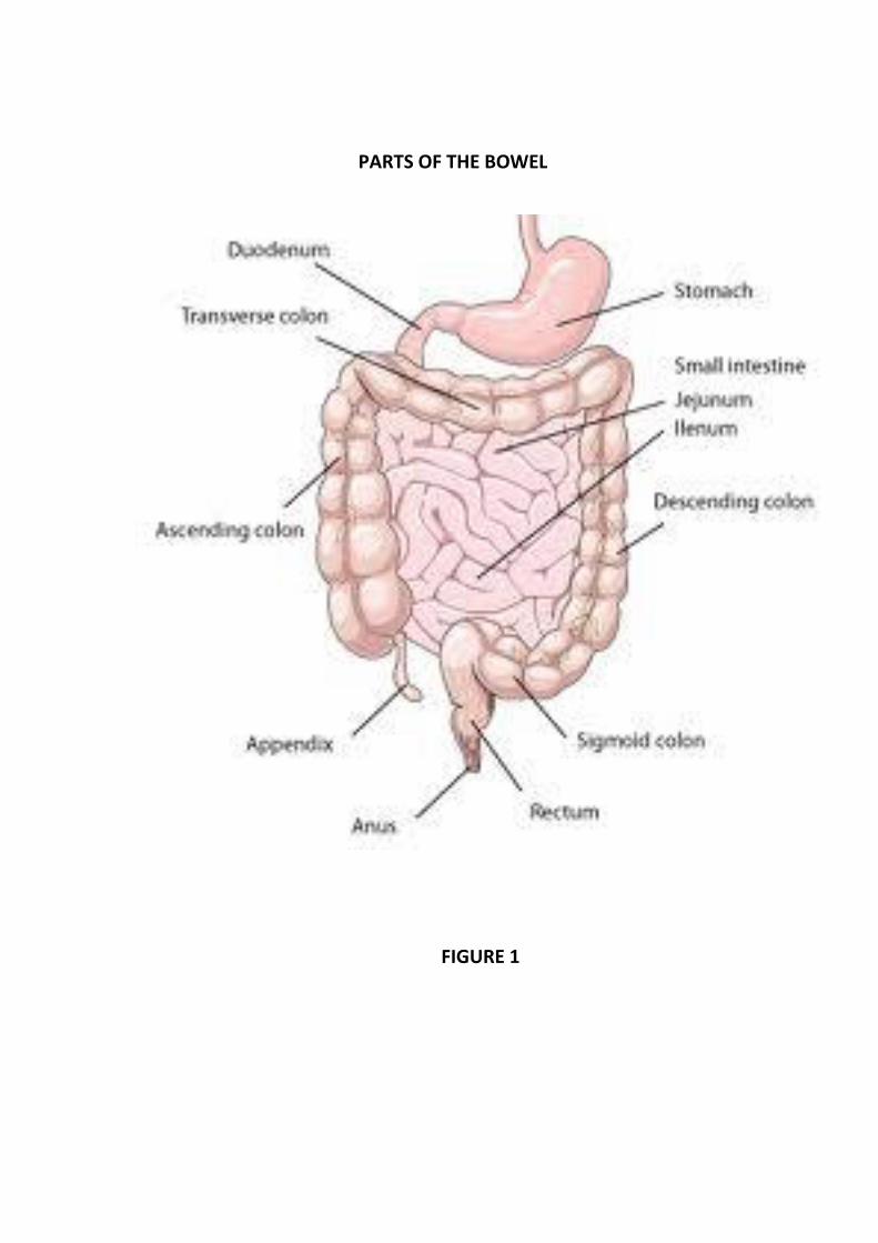
PARTS OF THE BOWEL
FIGURE 1
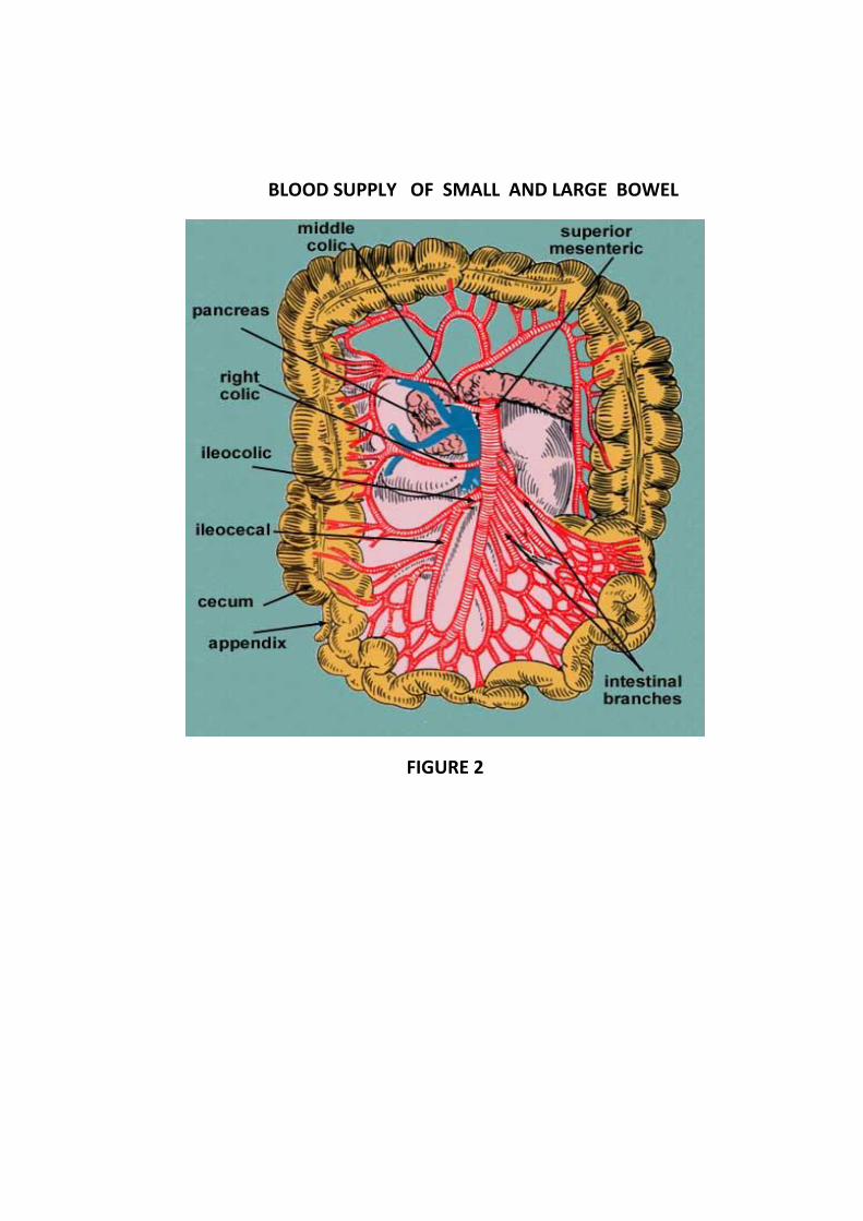
BLOOD SUPPLY OF SMALL AND LARGE BOWEL
FIGURE 2
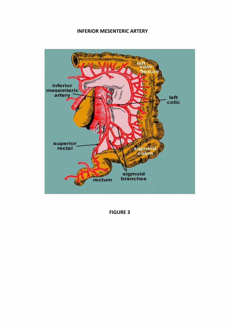
INFERIOR MESENTERIC ARTERY
FIGURE 3
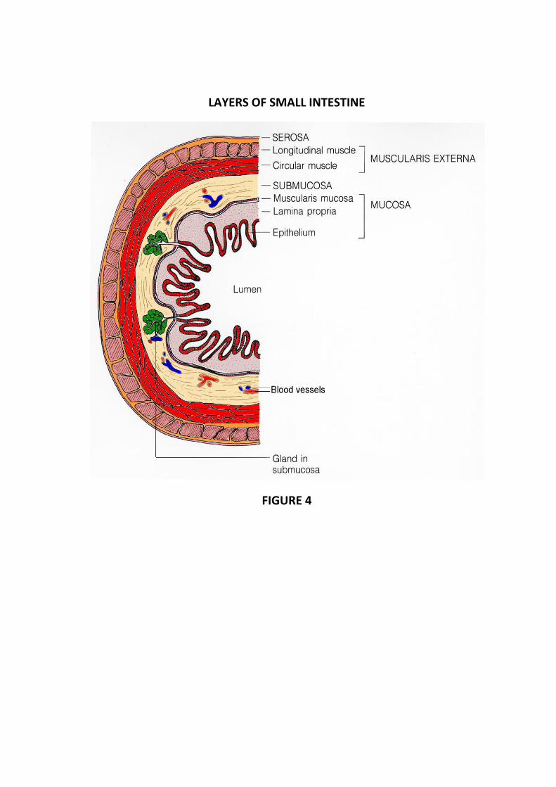
LAYERS OF SMALL INTESTINE
FIGURE 4
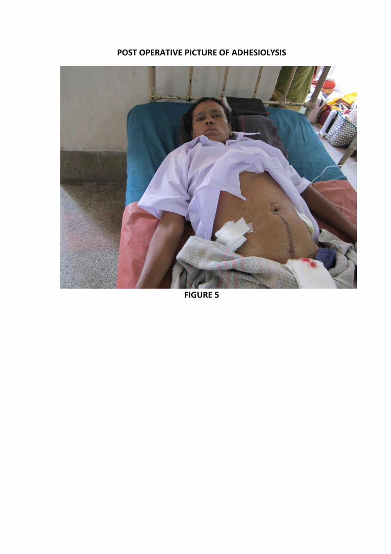
POST OPERATIVE PICTURE OF ADHESIOLYSIS
FIGURE 5
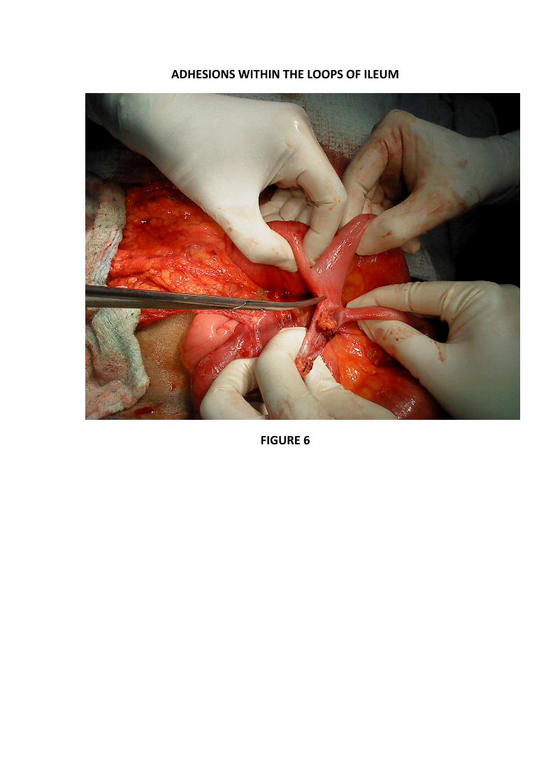
ADHESIONS WITHIN THE LOOPS OF ILEUM
FIGURE 6

BOWEL TO BOWEL ADHESION
FIGURE 7
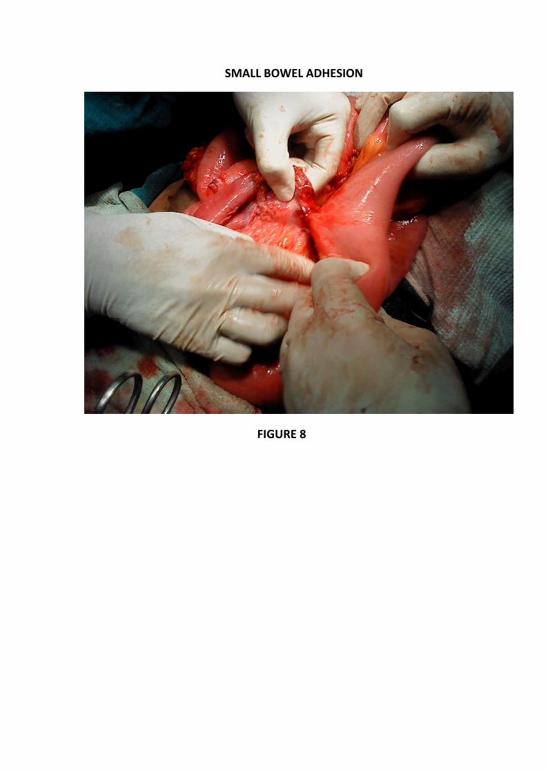
SMALL BOWEL ADHESION
FIGURE 8