Embed Size (px)
Citation preview
Version 09 19
Study Guide for all Emcare courses
© Copyright Emcare Services Limited. No part of this may be used or reproduced in any form or by any
means, or stored in a database or retrieval system, without prior written permission of Emcare.
1
Disclaimer
Emcare Services Ltd. provide this document as part of our service to our
customers. All the information in this document is published in good faith
and for general information purposes only.
Emcare Services Ltd. is not responsible for, and expressly disclaims all
liability for, damages of any kind arising out of use, reference to, or
reliance on any information contained within this document. While the
information contained within the document is periodically updated, no
guarantee is given that the information provided in this document is
correct, complete, and up-to-date.
This document is not intended to replace the information taught on any of
our courses, or to replace information contained in the New Zealand
Resuscitation Council manual: Resuscitation: A Guide for Advanced
Rescuers (1st Ed.).
Although this document may include links providing direct access to other
internet resources, including web sites, Emcare Services Ltd. is not
responsible for the accuracy or content of information contained in these
sites.
Copyright © 2016 Emcare Services Ltd. All rights reserved.
The information in this document may not be reproduced in any material
form or transmitted to any persons without permission from Emcare
Services Ltd. under the Copyright Act 1994.
Please contact Emcare Services Ltd. if you would like to use any of the
information.
Individuals and organisations wishing to make Emcare Services Ltd.
content accessible through their web sites are encouraged to create
hypertext links to the required content.
2
CPR Guidelines – Main Points
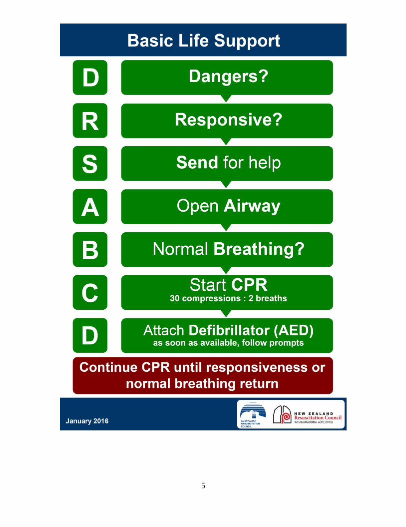
Basic Life Support
• Follow the DRS-ABCD approach
• The need for CPR is determined by unresponsiveness and the absence
of normal breathing
• If the unconscious person is unresponsive and not breathing normally
after the airway has been opened, start chest compressions at once
Chest Compressions
• Compression/ventilation ratio is 30:2 for adults and children for non-
health professionals
• Compression/ventilation ratio is 30:2 for adults and 15:2 in
children/infants for healthcare providers
• Minimise all interruptions to chest compressions
• Hard and Fast and Fresh. Allow the chest to recoil fully
o Hard (i.e. depress by approximately 1/3 of the front-back
dimension of the chest)
o Fast (i.e. chest compressions are at 100 - 120 compressions per
minute)
o Fresh (i.e. Change compression person when tired, or every 2
minutes). Plan the change so that there is minimal interruption to
compressions
Rescue Breaths
• In New Zealand, the NZRC emphasises rescue breathing in all ages
• These breaths are attempted. If unsuccessful, do not delay the
resumption of chest compressions
• Each breath is given over one second, and with enough volume to
produce chest rise
• If the chest does not move, the airway is obstructed and/or there is an
ineffective seal around the nose and mouth, or insufficient air is being
blown into the lungs
• In adults, the focus is on early defibrillation – send for help first, and
compressions as a priority over rescue breathing
• In children and infants 2 attempted initial breaths are given, enough to
produce chest rise. In child/infant collapse – send for help fast (1
minute of CPR before going for help)
Pulse Checks
• The pulse is not checked during the initial assessment and then only if
there are signs of life or it is noted that there is a rhythm present that
could be associated with cardiac output
Precordial Thump
• The precordial thump has largely been omitted
3
• A single precordial thump is administered to the centre of the chest
during a witnessed adult collapse, AND the patient is cardiac
monitored, AND you see they are in Ventricular Tachycardia, AND
there is no defibrillator immediately available
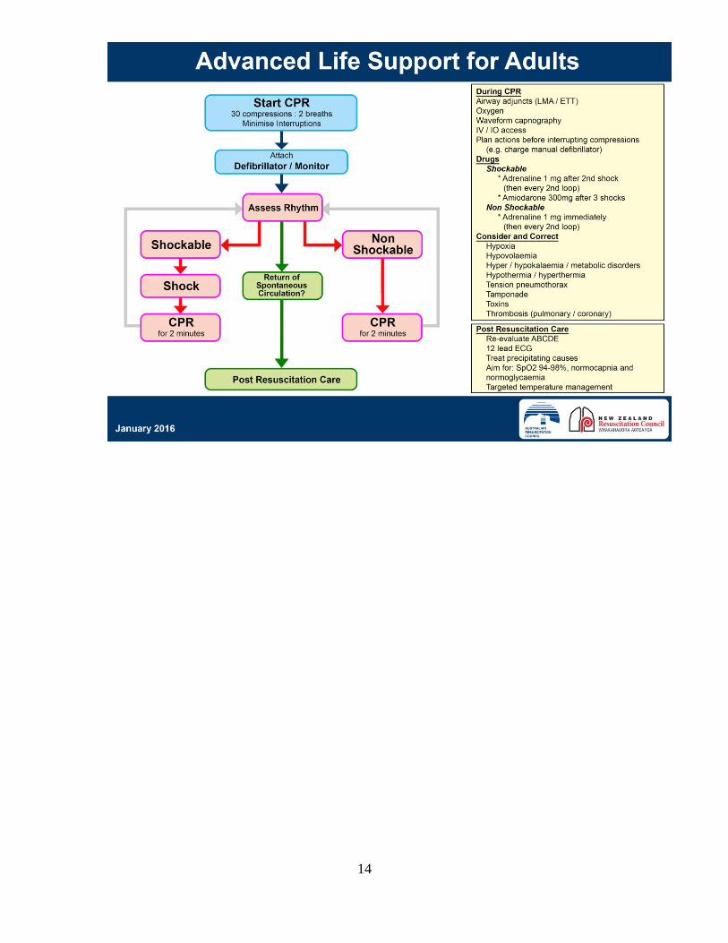
Manual Defibrillation
• All manual defibrillation shocks are given at the default setting for the
machine, or 200J, or maximum energy for the machine if no default
available.
• All manual defibrillation shocks are given at 4 J/kg in children.
• Defibrillations are given as a single shock followed by two (2) minutes
of CPR
• Reanalyse every 2 minutes
AEDs • AEDs are pre-programmed to deliver a certain amount of energy
• AEDs can be used for ALL ages
• Turn the AED on and follow the voice prompts – do not interrupt chest
compressions to apply the AED pads
• Pads should be placed 8 cm away from implanted devices
• Pads may be placed one on the front of the chest and one on the
upper back (between the shoulder blades) for smaller children
Drug Management • Adrenaline
o Shockable
▪ 1 mg is given after the second shock/defibrillation and
then every second loop (2 minutes = 1 loop)
o Non-shockable
▪ 1mg given as soon as possible
o Children/Infants
▪ 10mcg/kg (0.1ml/kg 1:10,000)
• Amiodarone is given in persistent VF and pulseless VT after the third (3rd)
shock/defibrillation. In adults Amiodarone 300mg is given. In children
and infants 5mg/kg Amiodarone is given.
4
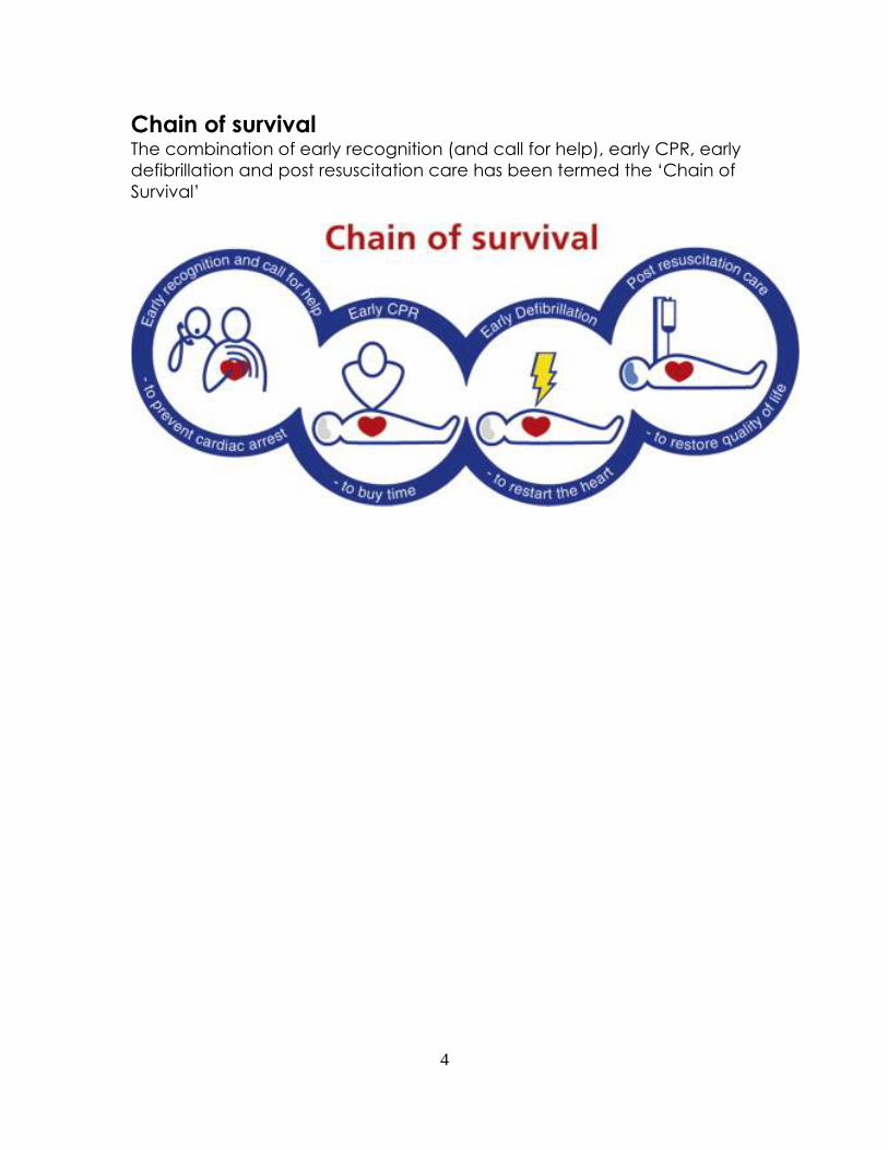
Chain of survival The combination of early recognition (and call for help), early CPR, early
defibrillation and post resuscitation care has been termed the ‘Chain of
Survival’
5
6
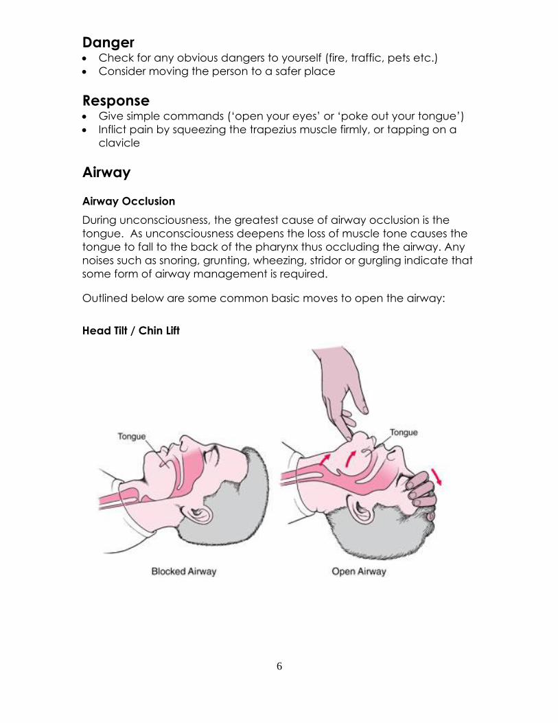
Danger • Check for any obvious dangers to yourself (fire, traffic, pets etc.)
• Consider moving the person to a safer place
Response • Give simple commands (‘open your eyes’ or ‘poke out your tongue’)
• Inflict pain by squeezing the trapezius muscle firmly, or tapping on a
clavicle
Airway
Airway Occlusion
During unconsciousness, the greatest cause of airway occlusion is the
tongue. As unconsciousness deepens the loss of muscle tone causes the
tongue to fall to the back of the pharynx thus occluding the airway. Any
noises such as snoring, grunting, wheezing, stridor or gurgling indicate that
some form of airway management is required.
Outlined below are some common basic moves to open the airway:
Head Tilt / Chin Lift
7
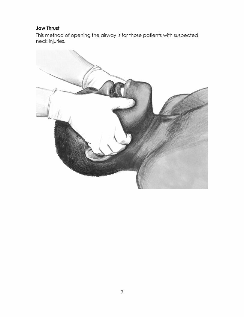
Jaw Thrust
This method of opening the airway is for those patients with suspected
neck injuries.
8
Breathing
After establishing that the airway is clear and open, the next step is to
establish the presence (or absence) of breathing.
The ‘look, listen, feel’ technique quickly confirms whether breathing is
present.
Look, listen feel:
• LOOK for the rising or falling of the lower chest or upper abdomen.
• LISTEN for breathing by placing your ear close to the patient’s nose
and mouth.
• FEEL for air movement at the nose and mouth.
If normal or adequate breathing that moves air is absent after the airway
has been opened, proceed with 30 chest compressions and 2
breaths/ventilations for the adult patient.
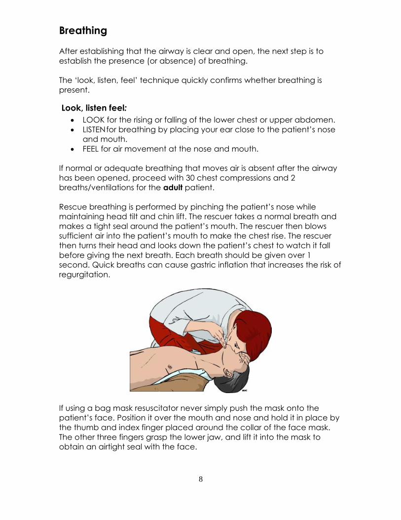
Rescue breathing is performed by pinching the patient’s nose while
maintaining head tilt and chin lift. The rescuer takes a normal breath and
makes a tight seal around the patient’s mouth. The rescuer then blows
sufficient air into the patient’s mouth to make the chest rise. The rescuer
then turns their head and looks down the patient’s chest to watch it fall
before giving the next breath. Each breath should be given over 1
second. Quick breaths can cause gastric inflation that increases the risk of
regurgitation.
If using a bag mask resuscitator never simply push the mask onto the
patient’s face. Position it over the mouth and nose and hold it in place by
the thumb and index finger placed around the collar of the face mask.
The other three fingers grasp the lower jaw, and lift it into the mask to
obtain an airtight seal with the face.
9
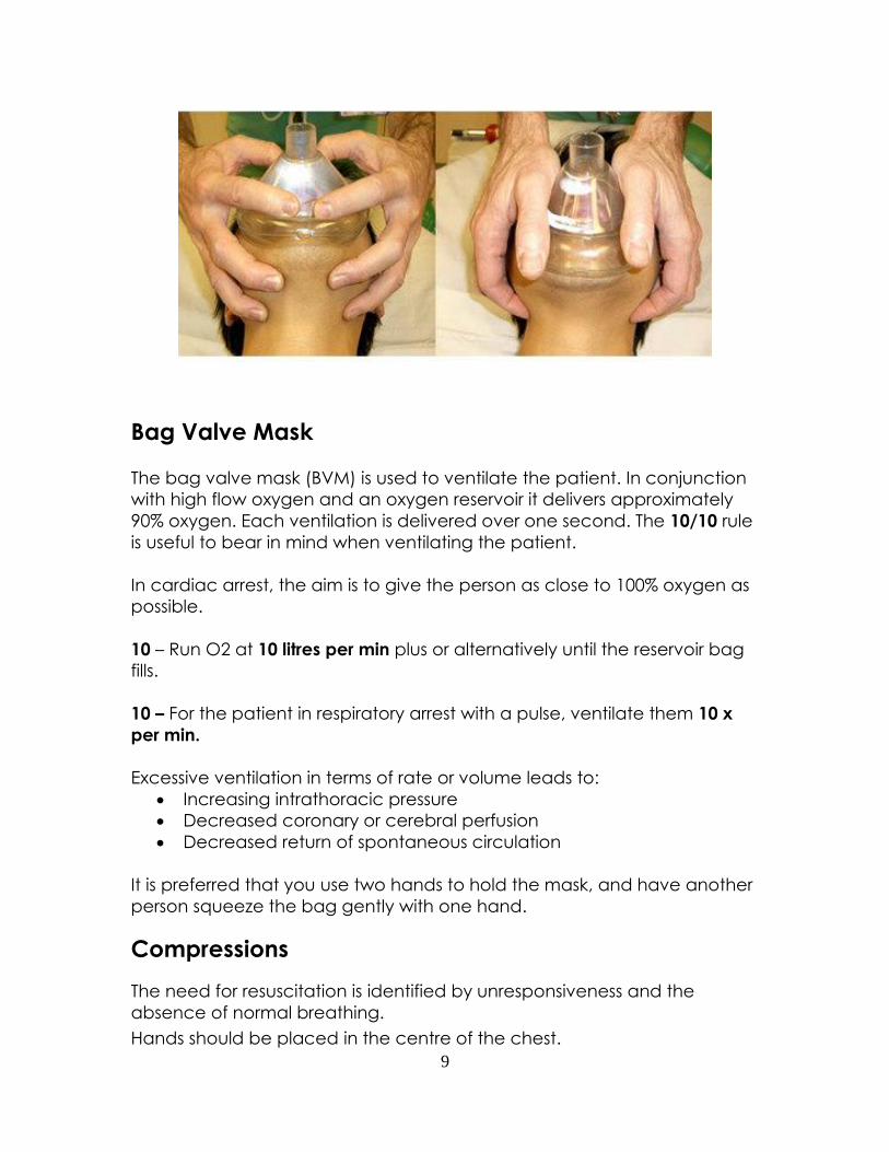
Bag Valve Mask
The bag valve mask (BVM) is used to ventilate the patient. In conjunction
with high flow oxygen and an oxygen reservoir it delivers approximately
90% oxygen. Each ventilation is delivered over one second. The 10/10 rule
is useful to bear in mind when ventilating the patient.
In cardiac arrest, the aim is to give the person as close to 100% oxygen as
possible.
10 – Run O2 at 10 litres per min plus or alternatively until the reservoir bag
fills.
10 – For the patient in respiratory arrest with a pulse, ventilate them 10 x
per min.
Excessive ventilation in terms of rate or volume leads to:
• Increasing intrathoracic pressure
• Decreased coronary or cerebral perfusion
• Decreased return of spontaneous circulation
It is preferred that you use two hands to hold the mask, and have another
person squeeze the bag gently with one hand.
Compressions
The need for resuscitation is identified by unresponsiveness and the
absence of normal breathing.
Hands should be placed in the centre of the chest.
10
Use either a one or two hand technique for children.
In babies, use the ‘two finger’ or the ‘two thumb’ technique.
Compression rate for all ages is 100 - 120/min.
Compression depth should be approximately 1/3 of the depth of the front-
back dimension of the chest. This equates to more than 5cm in adults,
approximately 5cm in children, and 4cm in infants.
Defibrillation
Attach the AED/Defibrillator as soon as it arrives
Rationale for Early Defibrillation
• The most common initial rhythm in sudden cardiac death is Ventricular
Fibrillation (VF)
• The only effective treatment for VF is electrical defibrillation
• The probability of successful defibrillation diminishes rapidly over time
• VF tends to convert to asystole within a few minutes
Safety when Defibrillating
• Always firmly ask people to stand clear when you are about to
defibrillate
• Visually check nobody is touching the patient before you discharge
the shock
• Do not defibrillate over medication patches
• If there is an implanted medical device, the defibrillator pad should
be placed at least 8 cm away from it (Resuscitation – A Guide for
Advanced Rescuers, 1st Ed., p.18)
Paddle and electrode placement
1. If patient’s chest is so hairy electrodes do not contact the skin, then
the use of a razor is indicated.
2. If the person is wet, dry the area that the pads are going to be
applied to.
3. For children less than 8 years old, place one pad on the upper back
between the shoulder blades, and the other pad on the front of the
chest.
Defibrillation energy
An AED delivers a pre-set energy that is unable to be adjusted.
For children less than 8 years of age, it is preferable that a manual
defibrillator is used. If no manual defibrillator is available, use an AED with
paediatric attenuator. If this is not available, use a standard AED.

11
If using a manual defibrillator on an adult, set the machine to its default
setting, or 200J, or maximum energy for the machine if no default
available.
If using a manual defibrillator on a child, set the machine to 4J per kg. If
the energy setting falls between 2 different settings – set the machine to
the higher setting.
Choking
Recognition
Because recognition is the key to successful outcome, it is important to ask
the conscious victim “Are you choking?” This at least gives the victim who
is unable to speak the opportunity to respond by nodding!
Consider the diagnosis of choking particularly if:
• Episode occurs whilst eating, and onset was very sudden
• Adult victim - may clutch his or her neck, or points to throat
• Child victim - there may be clues, e.g. seen eating or playing with
small items just before onset of symptoms
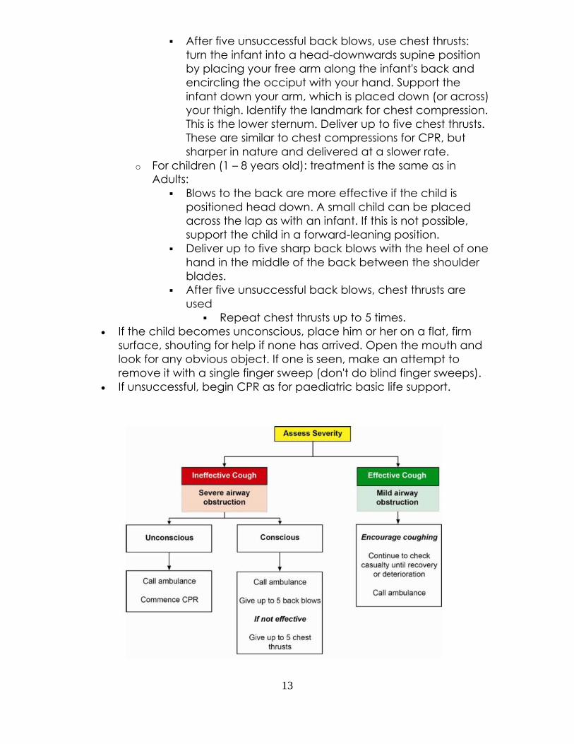
Assess severity
• Effective Cough (Mild Airway Obstruction):
o The patient can breathe, cough effectively and speak
o Children are fully responsive, crying or verbally respond to
questions, may have loud cough (and able to take a breath
before coughing)
• Ineffective Cough (Severe Airway Obstruction):
o Victim unable to breathe or speak/vocalise
o Attempts at coughing are quiet or silent
o Cyanosis and diminishing conscious level (particularly in
children)
o Victim unconscious
Management
Adults
• With an effective cough, encourage the patient to continue
coughing, but do nothing else except monitor for deterioration. If
the obstruction is not relieved – call 111
• Ineffective cough in a conscious patient:
12
o Stand to the side and slightly behind the victim, support the
chest with one hand and lean the victim well forwards (so
that the obstructing object comes out of the mouth rather
than going further down the airway)
o Give up to five sharp back blows between the shoulder
blades with the heel of your other hand (checking after each
if the obstruction has been relieved)
o If unsuccessful, give up to five chest thrusts. Stand behind the
victim put both arms around the chest, identify the same
compression point as for chest compressions, clench one fist,
grasp it with the other hand and pull sharply inwards
o Continue alternating up to five back blows and up to five
chest thrusts until successful or the patient becomes
unconscious
In an unconscious patient:
o Lower the patient to the floor
o Call an ambulance immediately
o Begin CPR
Children
• If coughing effectively, just encourage the child to cough, and
monitor continuously
• If coughing is, or is becoming ineffective, shout for help and assess
the child's conscious level
• If the child is conscious, give up to five back blows, followed by up
to five chest thrusts in infants and children (repeat the sequence
until the obstruction is relieved or the patient becomes
unconscious)
o For infants (<1 year old): back blows and chest thrusts:
▪ In a seated position, support the infant in a head-
downwards, prone position to let gravity aid removal of
the foreign body.
▪ Support the head by placing the thumb of one hand at
the angle of the lower jaw, and one or two fingers from
the same hand at the same point on the other side of
the jaw. Do not compress the soft tissues under the jaw,
as this will aggravate the airway obstruction.
▪ Deliver up to five sharp blows with the heel of your
hand to the middle of the back (between the shoulder
blades).
▪ After each blow, assess to see if the foreign body has
been dislodged and, if not; repeat the manoeuvre for
up to five back blows.
13
▪ After five unsuccessful back blows, use chest thrusts:
turn the infant into a head-downwards supine position
by placing your free arm along the infant's back and
encircling the occiput with your hand. Support the
infant down your arm, which is placed down (or across)
your thigh. Identify the landmark for chest compression.
This is the lower sternum. Deliver up to five chest thrusts.
These are similar to chest compressions for CPR, but
sharper in nature and delivered at a slower rate.
o For children (1 – 8 years old): treatment is the same as in
Adults:
▪ Blows to the back are more effective if the child is
positioned head down. A small child can be placed
across the lap as with an infant. If this is not possible,
support the child in a forward-leaning position.
▪ Deliver up to five sharp back blows with the heel of one
hand in the middle of the back between the shoulder
blades.
▪ After five unsuccessful back blows, chest thrusts are
used
▪ Repeat chest thrusts up to 5 times.
• If the child becomes unconscious, place him or her on a flat, firm
surface, shouting for help if none has arrived. Open the mouth and
look for any obvious object. If one is seen, make an attempt to
remove it with a single finger sweep (don't do blind finger sweeps).
• If unsuccessful, begin CPR as for paediatric basic life support.
14
15
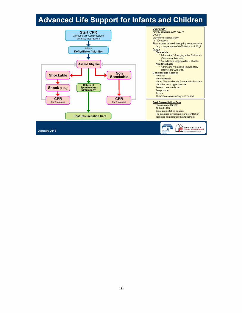
How are children different?
• An infant is less than 1 year of age
• A child is 1 – 8 years of age
• Children 9 years of age and over are treated as adults
• VF may be the cause of approximately 10% of paediatric arrests
• It is rare for a child to have a primary cardiac arrest and is generally
seen in those children with pre-existing congenital or acquired
cardiac disease. Most cardiac arrests are secondary to hypoxia
• Therefore, the paediatric arrest algorithm places rescue breaths not
compressions as first priority - 2 attempted breaths are given first in
Advanced Life Support
• Other causes of cardiac arrest include trauma, dehydration, and
sepsis
• Cardiac compressions are 100 - 120 compressions per minute
• If a manual defibrillator is not available infants ≤ 1year and children
1-8 yrs. may be treated with an AED if it has a dose attenuator. If
neither is available, use the pre-set AED energy levels
• Manual defibrillation is given at 4 Joules/kg
Adrenaline: 10mcg/kg (0.1ml/kg of 1: 10,000 ) intravenous or intra-osseous
dose
• Give 1st dose after second shock for patients with a VT/VF arrest
• Give 1st dose as soon as possible for patients in PEA or asystolic
arrest
• Adrenaline is given immediately post defibrillation and followed with
2 minutes of CPR
• Give every alternate loop or every 4 minutes thereafter
Amiodarone: 5mg/kg in 10mls of 5% Dextrose
• Give Amiodarone for VT/VF arrests after the 3rd defibrillation
16
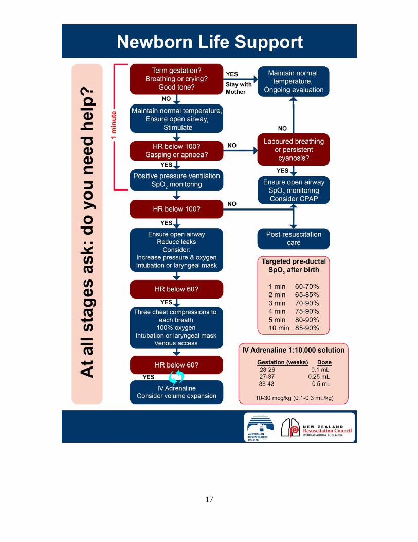
17
18
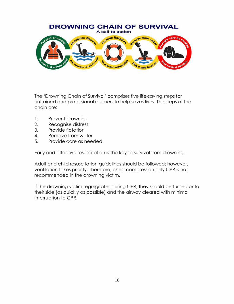
The ‘Drowning Chain of Survival’ comprises five life-saving steps for
untrained and professional rescuers to help saves lives. The steps of the
chain are:
1. Prevent drowning
2. Recognise distress
3. Provide flotation
4. Remove from water
5. Provide care as needed.
Early and effective resuscitation is the key to survival from drowning.
Adult and child resuscitation guidelines should be followed; however,
ventilation takes priority. Therefore, chest compression only CPR is not
recommended in the drowning victim.
If the drowning victim regurgitates during CPR, they should be turned onto
their side (as quickly as possible) and the airway cleared with minimal
interruption to CPR.
19
Waveform Capnography
Useful capnography resources can be found on the Emcare website at
http://www.emcare.co.nz/capnography.html
Waveform capnography is used to:
1. Differentiate oesophageal from endotracheal intubation
2. To guide ventilation
3. To assess the effectiveness of resuscitation, including the efficacy of
CPR
4. To detect Return of Spontaneous Circulation (ROSC)
(Resuscitation – A Guide for Advanced Rescuers (1st Ed.), p.85)
The following link discusses all the basics that you will need to know about
capnography:
http://www.emdocs.net/interpreting-waveform-capnography-pearls-and-
pitfalls/?utm_content=buffer5174e&utm_medium=social&utm_source=twit
ter.com&utm_campaign=buffer
CPR in Special Circumstances
Pregnancy
The incidence of cardiac arrest in pregnancy is approximately 1: 20,000.
The key differences between standard adult resuscitation and obstetric
resuscitation include:
• Early consideration of peri-mortem resuscitation
• Early intubation
• Lateral displacement (to the left) of the uterus
• Consideration of pregnancy-specific causes of collapse
Any defibrillation for pregnant women is delivered at the same frequency
and energy as for non-pregnant women.
Trauma
Cardiac arrest following trauma has a very poor prognosis, and is usually
fatal.
The two most common causes of cardiac arrest from trauma are hypoxia
from airway obstruction and blood loss.
20
If cardiac arrest occurs secondary to trauma, the person will usually be in a
non-shockable rhythm. Standard CPR guidelines should be followed.
Additional considerations for the management of trauma include:
• Jaw thrust to open the airway
• Control of catastrophic external haemorrhage
• Getting the person to hospital for definitive surgical management
In the heavily bleeding person, the best fluid replacement is blood. If blood
is not available, small amounts of 0.9% Saline may be administered to the
unstable person until blood is available.
21
Medical Emergencies
The following information has been adapted or taken from the following
website http://www.resus.org.uk/pages/MEdental.pdf
The ‘ABCD MOVE’ approach to the sick patient
This section provides guidance on the initial approach and management
of common medical emergencies which may arise.
In all circumstances, it is advisable to call for medical assistance as soon as
possible by dialing 111 and summoning an ambulance.
Early recognition of the ‘sick’ patient is to be encouraged. Pre-empting any
medical emergency by recognising an abnormal breathing pattern, an
abnormal patient colour or abnormal pulse rate, allows appropriate help
to be summoned e.g. ambulance, prior to any patient collapse occurring.
A systematic approach to recognising the acutely ill patient based on the
‘ABCD MOVE’ principle.
General principles
1. Follow the Airway, Breathing, Circulation, Disability, Monitoring,
Oxygenation, Venous access, and Exposure/ECG approach (ABCD MOVE)
to assess and treat the patient.
2. Treat life-threatening problems as they are identified before moving to
the next part of the assessment.
3. Continually re-assess starting with Airway if there is further deterioration.
4. Assess the effects of any treatment given.
5. Recognise when you need extra help and call for help early. This may
mean dialing 111 for an ambulance.
6. Use all members of your team. This will allow you to do several things at
once, e.g. collect emergency drugs and equipment, dial 111.
7. Organise your team and communicate effectively.
8. The aims of initial treatment are to keep the patient alive, achieve some
clinical improvement and buy time for further treatment whilst waiting for
help.
9. Remember - it can take a few minutes for treatment to work.
10. The ABCD MOVE approach can be used irrespective of your training
and experience in clinical assessment or treatment. Individual experience
and training will determine which treatments you can give. Often only
simple measures such as laying the patient down or giving oxygen are
needed.
22
First steps
• In an emergency, stay calm. Ensure that you and your staff are safe.
• Look at the patient generally to see if they ‘look unwell’.
• In an awake patient ask, “How are you?” If the patient is
unresponsive, gently shake them and ask, “Are you all right?” If they
respond normally, they have a clear airway, are breathing and have
brain perfusion. If they speak only in short sentences, they may have
breathing problems. Failure of the patient to respond suggests that
they are unwell. If they are not breathing and have no pulse or signs
of life, start CPR according to current resuscitation guidelines.
Airway (A)
Airway obstruction is an emergency.
1. Look for the signs of airway obstruction:
• Airway obstruction causes ‘paradoxical’ chest and abdominal
movements (‘see-saw’ respirations) and the use of the accessory
muscles of respiration e.g. neck muscles. Central cyanosis (blue lips
and tongue) is a late sign of airway obstruction. In complete airway
obstruction, there are no breath sounds at the mouth or nose.
• In partial airway obstruction, air entry is diminished and usually noisy
- Inspiratory ‘stridor’ is caused by obstruction at the laryngeal
level or above.
- Expiratory ‘wheeze’ suggests obstruction of the lower airways,
which tend to collapse and obstruct during expiration. This is
most commonly seen in patients with asthma or chronic
obstructive pulmonary disease.
- Gurgling suggests there is liquid or semi-solid foreign material in
the upper airway.
- Snoring arises when the pharynx is partially occluded by the
tongue or palate.
2. Airway obstruction is an emergency:
• In most cases, only simple methods of airway clearance are needed
- Airway opening manoeuvres – head tilt/chin lift or jaw thrust.
- Remove visible foreign bodies, debris or blood from the airway
(use suction or forceps as necessary).
- Consider simple airway adjuncts e.g. oropharyngeal airway.
3. Give oxygen initially at a high inspired concentration:
• Use a mask with an oxygen reservoir. Ensure that the oxygen flow is
sufficient (15 litres per minute) to prevent collapse of the reservoir
during inspiration.
• If you have a pulse oximeter, titrate the oxygen delivery aiming for
normal oxygen saturation levels (94-98%). In very sick patients this
23
may not be possible and a lower oxygen saturation (more than 90%)
is acceptable for a short period of time.
Breathing (B)
During the immediate assessment of breathing, it is vital to diagnose and
treat immediately life-threatening breathing problems, e.g. acute severe
asthma.
1. Look, listen and feel for the general signs of respiratory distress: sweating,
central cyanosis (blue lips and tongue), use of the accessory muscles of
respiration (muscles of the neck) and abdominal breathing.
2. Count the respiratory rate. The normal adult rate is 12 to 20 breaths per
minute and a child’s rate is between 20 and 30 breaths per minute. A high,
or increasing, respiratory rate is a marker of illness and a warning that the
patient may deteriorate and further medical help is needed.
3. Assess the depth of each breath, the pattern (rhythm) of respiration and
whether chest expansion is equal and normal on both sides.
4. Listen to the patient’s breath sounds a short distance from their face.
Gurgling airway noises indicate airway secretions, usually because the
patient cannot cough or take a deep breath. Stridor or wheeze suggests
partial, but important, airway obstruction.
5. If the patient’s depth or rate of breathing is inadequate, or you cannot
detect any breathing, use a bag and mask (if trained) or pocket mask
ventilation with supplemental oxygen while calling urgently for an
ambulance.
6. Hyperventilation and panic attacks are relatively common in stressful
situations. In most patients these will resolve with simple reassurance.
Circulation (C)
Simple faints or vasovagal episodes are the most likely cause of circulation
problems. These will usually respond to laying the patient flat and if
necessary raising the legs. The systematic ABCD MOVE approach to all
patients will ensure that other causes are not missed.
1. Look at the colour of the hands and fingers: are they blue, pink, pale or
mottled?
2. Assess the limb temperature by feeling the patient’s hands: are they cool
or warm?
3. Measure the capillary refill time. Apply cutaneous pressure for five
seconds on a fingertip held at heart level (or just above), or on the patients
shoulder with enough pressure to cause blanching. Time how long it takes
for the skin to return to the colour of the surrounding skin after releasing the
pressure. The normal refill time is less than three seconds. A prolonged time
suggests poor peripheral perfusion.
Other factors (e.g. cold surroundings, old age) can also prolong the
capillary refill time.
24
4. Count the patient’s pulse rate. It may be easier to feel a central pulse
(i.e. carotid pulse) than the radial pulse.
5. Weak pulses in a patient with a decreased level of consciousness and
slow capillary refill time suggest a low blood pressure. Laying the patient
down and raising the legs may be helpful. In patients who do not respond
to simple measures urgent help is needed and an ambulance should be
summoned.
6. Cardiac chest pain typically presents as a heaviness, tightness, or
indigestion like discomfort in the chest. The pain or discomfort often radiates
into the neck or throat, into one or both arms (more commonly the left) and
into the back or stomach area. Some patients experience the discomfort
in one of these areas more than in the chest. Sometimes pain may be
accompanied by belching, which can be misinterpreted as evidence of
indigestion as the cause. The patient may have known stable angina and
carry their own glyceryl trinitrate (GTN) spray. If they take this, the episode
may resolve. Give a single dose of aspirin and consider the use of oxygen.
Disability (D)
Common causes of unconsciousness include profound hypoxia,
hypercapnia (raised carbon dioxide levels), cerebral hypoperfusion (low
blood pressure), hypoglycaemia (low blood glucose levels) or the recent
administration of sedatives or analgesic drugs.
1. Review and treat the ABCs: exclude hypoxia and low blood pressure.
2. Check the patient’s drug record for reversible drug-induced causes of
depressed consciousness.
3. Examine the pupils (size, equality, and reaction to light).
4. Make a rapid initial assessment of the patient’s conscious level using the
AVPU method: Alert, responds to Vocal stimuli, responds to Painful stimuli or
Unresponsive to all stimuli.
5. Measure the blood glucose to exclude hypoglycaemia, using a glucose
meter. If below 3.0 mmol per litre give the patient a glucose containing
drink to raise the blood sugar or glucose by other means.
6. Nurse unconscious patients in the recovery position if their airway is not
protected.
Monitoring
Attach monitoring to the patient.
You may not have all the monitoring equipment mentioned below. The aim
is to gather as much information about your patient as possible.
1. Pulse – is it present? Is it strong or weak? Is it regular or irregular?
2. Respirations – count the patients respiratory rate for a full minute. Ensure
that the patient is not talking while you are doing this. If the patient
appears short of breath, ask them to count to ten aloud. Whatever
25
number they reach equals the number of words that they can speak per
breath.
3. Blood Pressure – more useful as a trend over time, but this can easily
answer if your patient’s blood pressure is too high or too low.
4. Pulse Oximetry – useful in assessing the patients oxygen levels.
Remember that pulse oximetry is perfusion dependant, so you will not
get accurate readings on the patients finger if their blood pressure is low.
a. Normal oxygen levels are 94% or above
b. If the patient has COPD, normal oxygen levels will be 88% - 92%
5. Temperature – An increased temperature may indicate sepsis, a low
temperature may indicate advanced sepsis.
6. 3-lead ECG – this will very quickly highlight any rhythm abnormalities that
the patient may have.
Oxygenation
Oxygen is a drug, and like all drugs is given when there is a need.
The aim of oxygen is to raise the patients oxygen levels to normal or near
normal.
Give the least amount of oxygen required to bring oxygen levels to 94% or
above if you suspect your patient is having a heart attack.
For seizures, stroke and trauma, giving oxygen at 6 Litres per minute via a
simple face mask (Hudson Mask) is a safe starting point.
Venous Access
Depending on your skill level, consider gaining intravenous (IV) access if you
are significantly concerned about your patient, or if you have called for an
ambulance.
Gaining IV access will enable the administration of IV fluids or medication if
indicated.
Exposure/ECG (E)
To assess and treat the patient properly the loosening or removal of some
of the patient’s clothes may be necessary. Respect the patient’s dignity
and minimise heat loss. This will allow you to see any rashes (e.g.
anaphylaxis) or perform procedures (e.g. defibrillation).
Perform a 12-lead ECG if there is an indication (you suspect a heart attack
or you notice abnormalities on the patients 3-lead ECG).
26
Common medical emergencies
Asthma
Patients with asthma (both adults and children) may have an attack
anywhere at any time. Most attacks will respond to a few ‘activations’ of
the patient’s own short-acting beta2-adrenoceptor stimulant inhaler such
as salbutamol. Repeat doses may be necessary.
If the patient does not respond rapidly, or any features of severe asthma
are present, an ambulance should be summoned. Patients requiring
additional doses of bronchodilator should be referred for medical
assessment after emergency treatment.
If the patient is unable to use the inhaler effectively, additional doses should
be given through a large-volume spacer device.
If the response remains unsatisfactory or if the patient develops
tachycardia, becomes distressed or cyanosed (blueness around the lips or
extremities), arrangements must be made to transfer them urgently to
hospital.
Symptoms and Signs
Clinical features of acute severe asthma in adults include:
• Inability to complete sentences in one breath
• Respiratory rate > 25 per minute
• Tachycardia (heart rate > 110 per minute)
Clinical features of life threatening asthma in adults include:
• Cyanosis or respiratory rate < 8 per minute
• Bradycardia (heart rate < 50 per minute)
• Exhaustion, confusion, decreased level of consciousness
Treatment
Whilst awaiting ambulance transfer, oxygen should be given.
Assuming the patient’s nebuliser is unavailable, up to 10 activations from
the salbutamol inhaler should be given using a large-volume spacer device
and repeated every 10 minutes if necessary until an ambulance arrives.
All emergency ambulances in New Zealand carry nebulisers, oxygen and
appropriate drugs.
If bronchospasm is part of a more generalised anaphylactic reaction and
there are 'life-threatening' signs, an intramuscular injection of adrenaline
should be given.
The perceived risk of giving patients with chronic obstructive pulmonary
disease too much oxygen is often quoted but this should not distract from
the reality that ALL sick, cyanosed patients with respiratory difficulty should
be given high flow oxygen until the arrival of the ambulance. This short term
27
measure is far more likely to be of benefit to the patient than any risks of
causing respiratory depression.
If any patient becomes unresponsive always check for ‘signs of life’
(breathing and circulation) and start CPR in the absence of signs of life or
normal breathing (ignore occasional ‘gasps’).
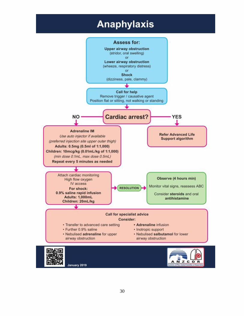
Anaphylaxis
Anaphylaxis is a severe, life-threatening, generalised or systemic
hypersensitivity reaction. It is characterised by rapidly developing life-
threatening airway and/or breathing and/or circulation problems usually
associated with skin and mucosal changes.
Anaphylactic reactions may follow the administration of a drug or contact
with substances such as latex in surgical gloves. In general, the more rapid
the onset of the reaction, the more serious it will be. Symptoms can develop
within minutes and early, effective treatment may be lifesaving.
Anaphylactic reactions may also be associated with additives and
excipients in medicines. It is wise therefore to check the full formulation of
preparations which may contain allergenic fats or oils (including those for
topical application, particularly if they are intended for use in the mouth).
Symptoms and signs
The lack of any consistent clinical manifestation and a wide range of
possible presentations can cause diagnostic difficulty. Clinical assessment
helps make the diagnosis.
Signs and symptoms may include:
• Urticaria, erythema, rhinitis, conjunctivitis
• Abdominal pain, vomiting, diarrhoea and a sense of impending
doom
• Flushing is common, but pallor may also occur
• Marked upper airway (laryngeal) oedema and bronchospasm may
develop, causing stridor, wheezing and/or a hoarse voice
• Vasodilation causes relative hypovolaemia leading to low blood
pressure and collapse. This can cause cardiac arrest (although very
rarely)
• Respiratory arrest leading to cardiac arrest
Clinical Criteria for Diagnosing Anaphylaxis
Anaphylaxis is highly likely when any one of the following three criteria is
fulfilled (taken from the World Allergy Organization Guidelines for the
Assessment and Management of Anaphylaxis
28
https://www.worldallergyorganizationjournal.org/article/S1939-
4551(19)30413-2/fulltext
1. Acute onset of an illness (minutes to several hours) with involvement of
the skin, mucosal tissue, or both (e.g. generalised urticaria, itching or
flushing, swollen lips-tongue-uvula)
AND AT LEAST ONE OF THE FOLLOWING:
A) Respiratory compromise (e.g. dyspnoea, wheeze-bronchospasm,
stridor, reduced peak expiratory flow (PEF), hypoxaemia)
B) Reduced blood pressure or associated symptoms of end-organ
dysfunction (e.g. hypotonia [collapse], syncope, incontinence)
OR
2. Two or more of the following that occur rapidly after exposure to a
Likely allergen for that patient (minutes to several hours)
A) Involvement of the skin-mucosal tissue (e.g. generalized urticaria, itch-
flush, swollen lips-tongue-uvula)
B) Respiratory compromise (e.g. dyspnoea, wheeze-bronchospasm,
stridor, reduced PEF, hypoxemia)
C) Reduced blood pressure or associated symptoms (e.g. hypotonia
[collapse], syncope, incontinence)
D) Persistent gastrointestinal symptoms (e.g. crampy abdominal pain,
vomiting)
OR
3. Reduced blood pressure after exposure to known allergen for that
patient (minutes to several hours)
A) Infants and children: low systolic blood pressure (age-specific) or
greater than 30% decrease in systolic blood pressure
B) Adults: systolic blood pressure of less than 90 mm Hg or greater than
30% decrease from that person's baseline
OR
More simply – if there are 2 or more body systems involved
Treatment
Use an ABCD MOVE approach to recognise and treat any suspected
anaphylactic reaction. First-line treatment includes managing the airway
29
and breathing with restoration of blood pressure (laying the patient flat,
raising the feet) and the administration of oxygen.
If two or more body systems are involved:
– Initial treatment), adrenaline should be given intramuscularly
(anterolateral aspect of the middle third of the thigh) in a dose of
0.5mg/500 micrograms (0.5 mL adrenaline injection of 1:1000); an
autoinjector preparation delivering a dose of 300 micrograms (0.3 mL
adrenaline injection 1:1000) is available for immediate self-administration
by those patients known to have severe reactions. This is an acceptable
alternative if immediately available. The dose is repeated if necessary at 5
minute intervals according to patient response
The paediatric dose for adrenaline is based on the child’s approximate age
or weight:
1 year (10kg) = 0.1 ml (1:1000 adrenaline)
5 years (20kg) = 0.2 ml (1:1000 adrenaline)
10 years (30 kg) = 0.3 ml (1:1000 adrenaline)
12 years (40kg) = 0.4 ml (1:1000 adrenaline)
In any unconscious patient always check for ‘signs of life’ (breathing and
circulation) and start CPR in the absence of signs of life or normal breathing
(ignore occasional ‘gasps’).
In less severe cases any wheeze or difficulty breathing can be treated with
a salbutamol inhaler as detailed above in the section on Asthma.
All patients treated for an anaphylactic reaction should be sent to hospital
by ambulance for further assessment, irrespective of any initial recovery.
Antihistamine drugs and steroids, whilst useful in the treatment of
anaphylaxis, are not first line drugs. Once the emergency phase is over, all
patients should receive corticosteroids and given antihistamines for any
residual itch.
30
31
Cardiac emergencies
The signs and symptoms of cardiac emergencies include chest pain,
shortness of breath, fast and slow heart rates, increased respiratory rate, low
blood pressure, poor peripheral perfusion (indicated by prolonged capillary
refill time) and altered mental state.
If there is a history of angina the patient will probably carry glyceryl trinitrate
spray (GTN) and they should be allowed to use them.
Where symptoms are mild and resolve rapidly with the patient’s own
medication, hospital admission is not normally necessary.
Sudden alterations in the patient’s heart rate (very fast or very slow) may
lead to a sudden reduction in cardiac output with loss of consciousness.
Medical assistance should be summoned by dialing 111.
Myocardial infarction
The pain of myocardial infarction is similar to that of angina but generally
more severe and prolonged.
Symptoms and signs of myocardial infarction
• Progressive onset of severe, crushing pain in the centre and across
the front of chest. The pain may radiate to the shoulders and down
the arms (more commonly the left), into the neck and jaw or through
to the back
• Skin becomes pale and clammy
• Nausea and vomiting are common
• Pulse may be weak and blood pressure may fall
• Shortness of breath
Initial management of myocardial infarction
Call 111 immediately for an ambulance.
Allow the patient to rest in the position that feels most comfortable; in the
presence of breathlessness this is likely to be the sitting position. Patients who
faint or feel faint should be laid flat; often an intermediate position
(dictated by the patient) will be most appropriate.
Reassure the patient as far as possible to relieve further anxiety.
Give aspirin in a single dose of 300 mg orally, chewed. Ambulance staff
should be made aware that aspirin has already been given as should the
hospital. Any treatment (including dental) carried out that might
contraindicate this must be brought to the attention of the ambulance
crew.
32
Oxygen may be administered if the patient has oxygen levels less than 94%,
is cyanosed (blue lips) or level of conciousness deteriorates.
If the patient becomes unresponsive always check for ‘signs of life’
(breathing and circulation) and start CPR in the absence of signs of life or
normal breathing (ignore occasional ‘gasps’).
33
Epileptic seizures
Symptoms and signs
• There may be a brief warning or ‘aura’
• Sudden loss of consciousness, the patient becomes rigid, falls, may
give a cry, and becomes cyanosed (tonic phase)
• After a few seconds, there are jerking movements of the limbs; the
tongue may be bitten (clonic phase)
• There may be frothing from the mouth and urinary incontinence
• The seizure typically lasts a few minutes; the patient may then
become floppy but remain unconscious
• After a variable time the patient regains consciousness but may
remain confused
• Fitting may be a presenting sign of hypoglycaemia and should be
considered in all patients, especially known diabetics and children
An early blood glucose measurement is essential in all actively fitting
patients (including known epileptics)
• Check for the presence of a very slow heart rate (<40 per minute)
which may drop the blood pressure. This is usually caused by a
vasovagal episode. The drop in blood pressure may cause transient
cerebral hypoxia and give rise to a brief seizure
Treatment
During a seizure try to ensure that the patient is not at risk from injury but
make no attempt to put anything in the mouth or between the teeth (in the
mistaken belief that this will protect the tongue). Do not attempt to insert
an oropharyngeal airway or other airway adjunct while the patient is
actively fitting.
Give oxygen.
Do not attempt to restrain convulsive movements.
After convulsive movements have subsided place the patient in the
recovery position and reassess.
If the patient remains unresponsive always check for ‘signs of life’
(breathing and circulation) and start CPR in the absence of signs of life or
normal breathing (ignore occasional ‘gasps’).
Check blood glucose level to exclude hypoglycaemia. If blood glucose
<3.0 mmol per litre or hypoglycaemia is clinically suspected, give
oral/buccal/IV glucose, or glucagon.
After the seizure the patient may be confused (‘post-ictal confusion’) and
may need reassurance and sympathy.
The patient should not be sent home until fully recovered and they should
be accompanied. It may not always be necessary to seek medical
attention or transfer to hospital unless the convulsion was the patients first
seizure, atypical, prolonged (or repeated), or if injury occurred.
34
The National Institute for Clinical Excellence (NICE) guidelines suggest the
indications for sending to hospital are:
• Status epilepticus
• High risk of recurrence
• First episode
• Difficulty monitoring the individual’s condition
Medication should only be given if seizures are prolonged (convulsive
movements lasting 5 minutes or longer) or recur in quick succession. In this
situation an ambulance should be summoned urgently.
With prolonged or recurrent seizures, ambulance personnel will often
administer IM or IV Midazolam which is usually rapidly effective in stopping
any seizure.
Hypoglycaemia
If food is omitted after having insulin, the blood glucose will fall to a low level
(hypoglycaemia). This is usually defined as a blood glucose <3.0 mmol per
litre, but some patients may show symptoms at higher blood sugar levels.
Patients may recognise the symptoms themselves and will usually respond
quickly to glucose. Children may not have such obvious features but may
appear lethargic.
Symptoms and signs
• Shaking and trembling
• Sweating
• Headache
• Difficulty in concentration / vagueness
• Slurring of speech
• Aggression and confusion
• Fitting / seizures
• Unconsciousness
Treatment
The following staged treatment protocol is a suggested depending on the
status of the patient. If any difficulty is experienced or the patient does not
respond, the ambulance service should be summoned immediately;
ambulance personnel will also follow this protocol.
Confirm the diagnosis by measuring the blood glucose.
Early stages - where the patient is co-operative and conscious with an
intact gag reflex, give oral glucose (sugar (sucrose)), milk with added sugar,
glucose tablets or gel). If necessary this may be repeated in 10 -15 minutes.
35
In more severe cases - where the patient has impaired consciousness, is
uncooperative or is unable to swallow safely, give buccal glucose gel, or
administer IM Glucagon, or administer 100ml of 10% Glucose IV.
• Glucagon should be given via the IM route (1mg in adults and
children >5 years old or >20 kg, 0.5mg if <5 years old or <20 kg).
Remember it may take 5-10 minutes for Glucagon to work and it
requires the patient to have adequate glucose stores. Thus, it may be
ineffective in anorexic patients, alcoholics or some non-diabetic
patients.
• Re-check blood glucose every 10 minutes to ensure that it has risen
to a level consistently greater than 3.5 mmol per litre, in conjunction
with an improvement in the patient’s mental status.
• If any patient becomes unconscious, always check for ‘signs of life’
(breathing and circulation) and start CPR in the absence of signs of
life or normal breathing (ignore occasional ‘gasps’).
It is important, especially in patients who have been given Glucagon, that
once they are alert and able to swallow, they are given a drink containing
glucose and if possible some food high in carbohydrate. The patient may
go home if fully recovered and they are accompanied by an adult. Their
General Practitioner should be informed and they should not drive. If you
are unsure, phone 111.
Syncope
Inadequate cerebral perfusion (and oxygenation) results in loss of
consciousness.
This most commonly occurs with low blood pressure caused by vagal over
activity (a vasovagal attack, simple faint, or syncope). This in turn may
follow emotional stress or pain. Some patients are more prone to this and
have a history of repeated faints.
Symptoms and signs
• Patient feels faint / dizzy / light headed
• Slow pulse rate
• Low blood pressure
• Pallor and sweating
• Nausea and vomiting
• Loss of consciousness
Treatment
Lay the patient flat as soon as possible and raise the legs to improve venous
return.
Loosen any tight clothing, especially around the neck and give oxygen.
36
If any patient becomes unresponsive, always check for ‘signs of life’
(breathing, circulation) and start CPR in the absence of signs of life or
normal breathing (ignore occasional ‘gasps’).
Other possible causes
• Postural hypotension can be a consequence of rising abruptly or of
standing upright for too long. Several medical conditions predispose
patients to hypotension with the risk of syncope. The most common
culprits are drugs used in the treatment of high blood pressure,
especially the ACE inhibitors and angiotensin antagonists. When
rising, patients should take their time. Treatment is the same as for a
vasovagal attack.
• Under stressful circumstances, many anxious patients hyperventilate.
This may give rise to feelings of light headedness or faintness but does
not usually result in syncope. It may result in spasm of muscles around
the face and of the hands. In most cases reassurance is all that is
necessary.
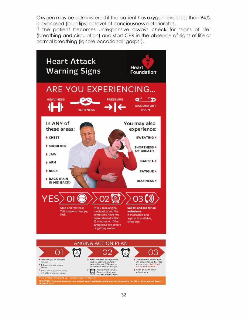
Stroke
Stroke (previously known as cerebrovascular accident) is the second most
common cause of death after heart disease.
A stroke occurs when the supply of blood to part of the brain is suddenly
disrupted or when bleeding from a blood vessel within the skull occurs.
Approximately 80% of strokes are caused by an acute blockage of a blood
vessel supplying part of the brain.
Stroke is a medical emergency.
When stroke is caused by an interruption to the blood supply to a part of
the brain, that area of the brain is damaged and may die. The surrounding
brain tissue is also affected and is at risk of dying. However, if the blockage
can be rapidly cleared and blood supply restored, the amount of damage
to brain tissue can be significantly reduced.
Rapid recognition, protection and support of the airway, breathing and
circulation, and rapid access to definitive stroke care can all contribute
to reducing death, disability and long term effects from stroke.
Symptoms may seem to improve but should still be considered as a stroke.
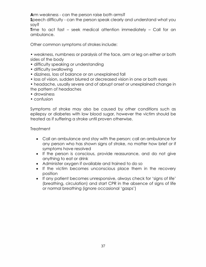
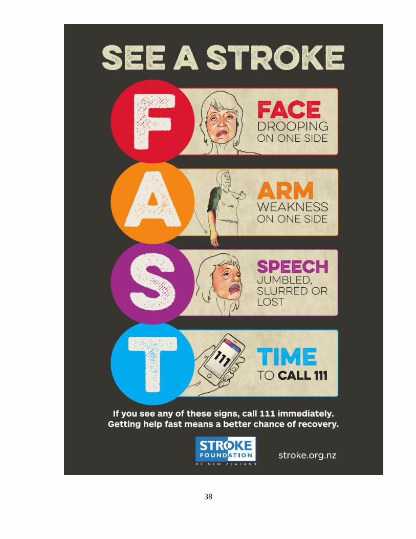
FAST is a simple way for remembering the signs of stroke.
Facial weakness - can the person smile? Has their mouth or eye drooped?
37
Arm weakness - can the person raise both arms?
Speech difficulty - can the person speak clearly and understand what you
say?
Time to act fast – seek medical attention immediately – Call for an
ambulance.
Other common symptoms of strokes include:
• weakness, numbness or paralysis of the face, arm or leg on either or both
sides of the body
• difficulty speaking or understanding
• difficulty swallowing
• dizziness, loss of balance or an unexplained fall
• loss of vision, sudden blurred or decreased vision in one or both eyes
• headache, usually severe and of abrupt onset or unexplained change in
the pattern of headaches
• drowsiness
• confusion
Symptoms of stroke may also be caused by other conditions such as
epilepsy or diabetes with low blood sugar, however the victim should be
treated as if suffering a stroke until proven otherwise.
Treatment
• Call an ambulance and stay with the person: call an ambulance for
any person who has shown signs of stroke, no matter how brief or if
symptoms have resolved
• If the person is conscious, provide reassurance, and do not give
anything to eat or drink
• Administer oxygen if available and trained to do so
• If the victim becomes unconscious place them in the recovery
position
• If any patient becomes unresponsive, always check for ‘signs of life’
(breathing, circulation) and start CPR in the absence of signs of life
or normal breathing (ignore occasional ‘gasps’)
38
39
Paediatrics
Infants and children are not small adults. They are different anatomically and
physiologically, this then means you'll need to have a different approach for
dealing with them
Special Considerations
An ill or injured child will be frightened. This will be caused by the injury/illness and
the feeling and discomfort associated with it. You too, even as a Health
Professional, will frighten that child due to being a stranger.
The sick or injured child will only be able to communicate with you up to their
understanding of vocabulary. This throws in some challenges from the word 'GO'.
You may not be able to get any conversation due to a young child unable to
speak or a child that is howling in pain, this also adds anxiety and confusion to the
child.
Parents and relations might be acting in a manner which may be driven by a
feeling of helplessness. These parents or relations may also become frustrated and
almost 'aggressive' towards you by what may seem a relaxed manner by you
while dealing with their child. A parent wants you to take control straight away
and to be seen doing something constructive with their child. It may seem like a
big task.
Guidelines for Dealing with a Sick Child
Very little time is spent learning and dealing with paediatrics so even experienced
staff can become less confident when dealing with a child.
Following these guidelines will make you job of dealing with a sick child a little
easier.
• Stay calm
• Come down to the child's level
• Who are you? Explain to the child who you are (Name) and that you are
there to help
• Ask mum or dad to hold child
• Has the child got a favourite toy you could give them?
• Be patient and gentle. Take your time if no life threatening injuries are
present
• If possible keep the patient with the parent/carer
• If you have to carry out any procedures explain them to the child and
parents
• Do Not Lie - If something will hurt tell the child it will hurt or their trust in you
will be lost
40
• Remember to constantly reassess the ill or injured child
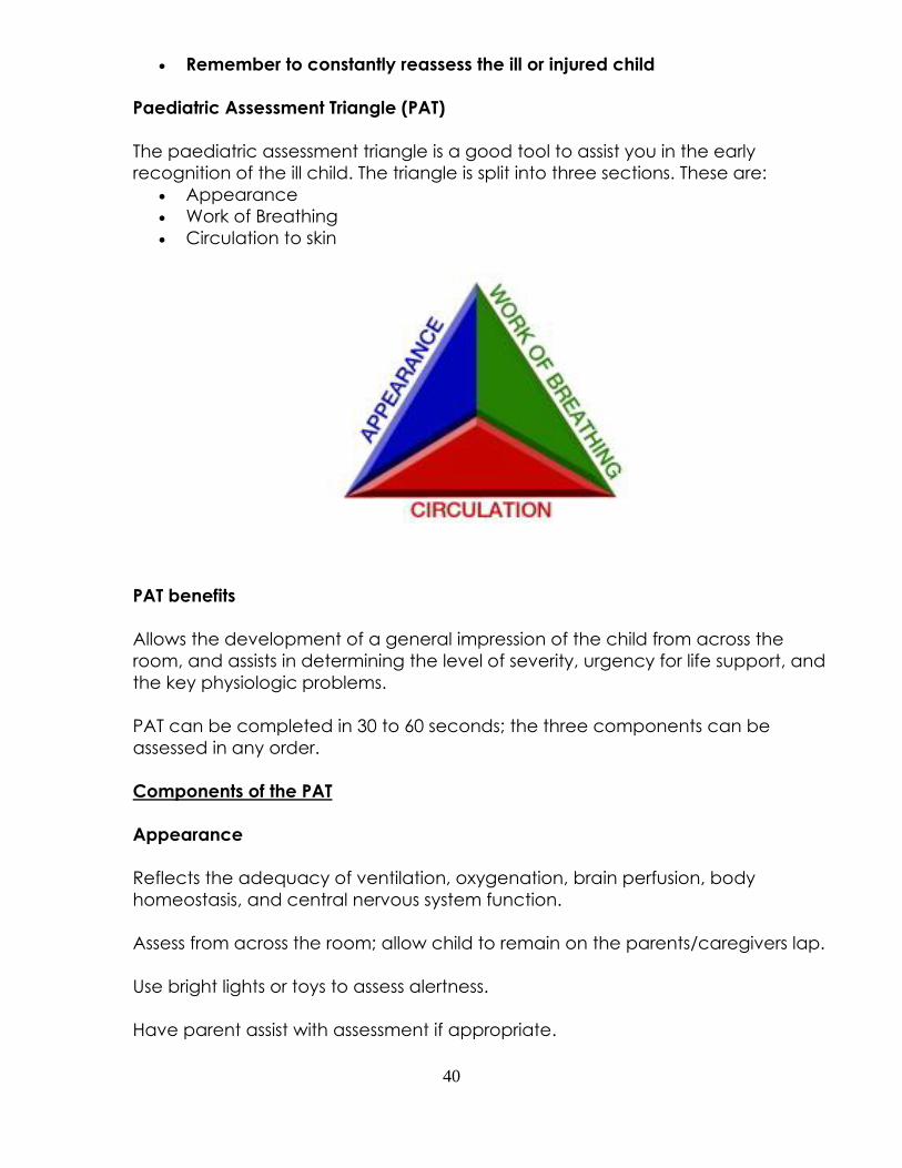
Paediatric Assessment Triangle (PAT)
The paediatric assessment triangle is a good tool to assist you in the early
recognition of the ill child. The triangle is split into three sections. These are:
• Appearance
• Work of Breathing
• Circulation to skin
PAT benefits
Allows the development of a general impression of the child from across the
room, and assists in determining the level of severity, urgency for life support, and
the key physiologic problems.
PAT can be completed in 30 to 60 seconds; the three components can be
assessed in any order.
Components of the PAT
Appearance
Reflects the adequacy of ventilation, oxygenation, brain perfusion, body
homeostasis, and central nervous system function.
Assess from across the room; allow child to remain on the parents/caregivers lap.
Use bright lights or toys to assess alertness.
Have parent assist with assessment if appropriate.
41
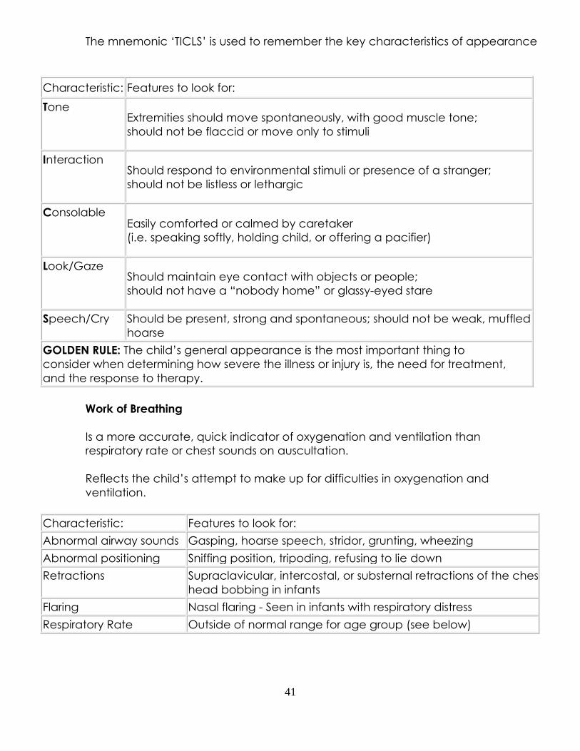
The mnemonic ‘TICLS’ is used to remember the key characteristics of appearance
Characteristic: Features to look for:
Tone Extremities should move spontaneously, with good muscle tone;
should not be flaccid or move only to stimuli
Interaction Should respond to environmental stimuli or presence of a stranger;
should not be listless or lethargic
Consolable Easily comforted or calmed by caretaker
(i.e. speaking softly, holding child, or offering a pacifier)
Look/Gaze Should maintain eye contact with objects or people;
should not have a “nobody home” or glassy-eyed stare
Speech/Cry Should be present, strong and spontaneous; should not be weak, muffled, or
hoarse
GOLDEN RULE: The child’s general appearance is the most important thing to
consider when determining how severe the illness or injury is, the need for treatment,
and the response to therapy.
Work of Breathing
Is a more accurate, quick indicator of oxygenation and ventilation than
respiratory rate or chest sounds on auscultation.
Reflects the child’s attempt to make up for difficulties in oxygenation and
ventilation.
Characteristic: Features to look for:
Abnormal airway sounds Gasping, hoarse speech, stridor, grunting, wheezing
Abnormal positioning Sniffing position, tripoding, refusing to lie down
Retractions Supraclavicular, intercostal, or substernal retractions of the chest wall;
head bobbing in infants
Flaring Nasal flaring - Seen in infants with respiratory distress
Respiratory Rate Outside of normal range for age group (see below)
42
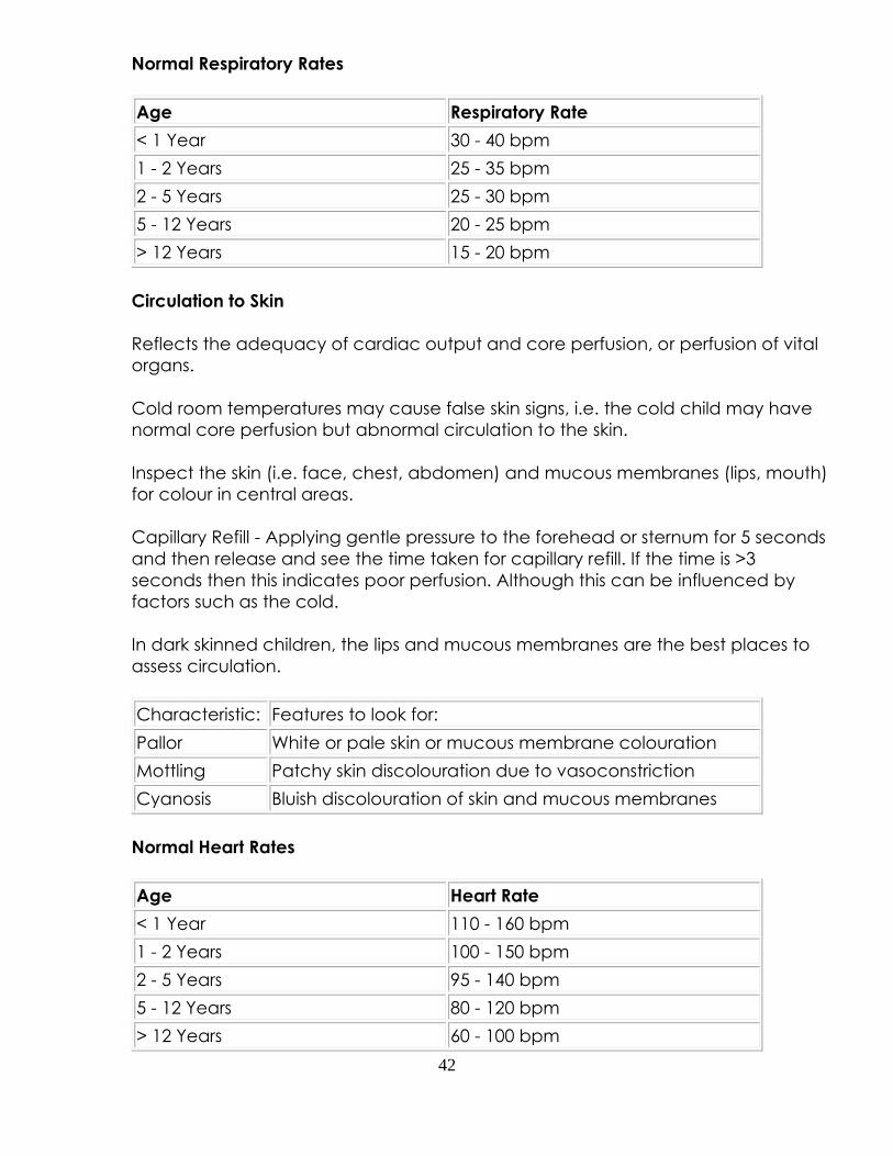
Normal Respiratory Rates
Age Respiratory Rate
< 1 Year 30 - 40 bpm
1 - 2 Years 25 - 35 bpm
2 - 5 Years 25 - 30 bpm
5 - 12 Years 20 - 25 bpm
> 12 Years 15 - 20 bpm
Circulation to Skin
Reflects the adequacy of cardiac output and core perfusion, or perfusion of vital
organs.
Cold room temperatures may cause false skin signs, i.e. the cold child may have
normal core perfusion but abnormal circulation to the skin.
Inspect the skin (i.e. face, chest, abdomen) and mucous membranes (lips, mouth)
for colour in central areas.
Capillary Refill - Applying gentle pressure to the forehead or sternum for 5 seconds
and then release and see the time taken for capillary refill. If the time is >3
seconds then this indicates poor perfusion. Although this can be influenced by
factors such as the cold.
In dark skinned children, the lips and mucous membranes are the best places to
assess circulation.
Characteristic: Features to look for:
Pallor White or pale skin or mucous membrane colouration
Mottling Patchy skin discolouration due to vasoconstriction
Cyanosis Bluish discolouration of skin and mucous membranes
Normal Heart Rates
Age Heart Rate
< 1 Year 110 - 160 bpm
1 - 2 Years 100 - 150 bpm
2 - 5 Years 95 - 140 bpm
5 - 12 Years 80 - 120 bpm
> 12 Years 60 - 100 bpm
43
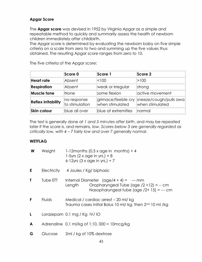
Apgar Score
The Apgar score was devised in 1952 by Virginia Apgar as a simple and
repeatable method to quickly and summarily assess the health of newborn
children immediately after childbirth.
The Apgar score is determined by evaluating the newborn baby on five simple
criteria on a scale from zero to two and summing up the five values thus
obtained. The resulting Apgar score ranges from zero to 10.
The five criteria of the Apgar score:
Score 0 Score 1 Score 2
Heart rate Absent <100 >100
Respiration Absent weak or irregular strong
Muscle tone None some flexion active movement
Reflex irritability no response
to stimulation
grimace/feeble cry
when stimulated
sneeze/cough/pulls away
when stimulated
Skin colour blue all over blue at extremities normal
The test is generally done at 1 and 5 minutes after birth, and may be repeated
later if the score is, and remains, low. Scores below 3 are generally regarded as
critically low, with 4 – 7 fairly low and over 7 generally normal.
WETFLAG
W Weight 1-12months (0.5 x age in months) + 4
1-5yrs (2 x age in yrs.) + 8
6-12yrs (3 x age in yrs.) + 7
E Electricity 4 Joules / Kg/ biphasic
T Tube ETT Internal Diameter (age/4 + 4) = --- mm
Length Oropharyngeal Tube (age /2 +12) = -- cm
Nasopharyngeal tube (age /2+ 15) = --- cm
F Fluids Medical / cardiac arrest – 20 ml/ kg
Trauma cases initial Bolus 10 ml/ kg, then 2nd 10 ml /kg
L Lorazepam 0.1 mg / Kg IV/ IO
A Adrenaline 0.1 ml/kg of 1:10, 000 = 10mcg/kg
G Glucose 2ml / kg of 10% dextrose
44
Medical Emergency Management
This document can be downloaded from our website
http://www.emcare.co.nz/uploads/1/1/4/8/114818101/medical
_emergency_management_2018.pdf
Our recommendation is that you print this off and attach to
your emergency equipment as a quick reference guide
45
Extra Course Resources
Extra course resources can be downloaded from our website.
The ECG Rhythm Study Guide can be found at:
http://www.emcare.co.nz/uploads/1/1/4/8/114818101/emcare
_ecg_rhythm_study_guide_for_core_advanced.pdf
Suggested pre-reading for CORE Advanced can be found at:
http://www.emcare.co.nz/uploads/1/1/4/8/114818101/suggest
ed_pre-reading_for_core_advanced.pdf