Embed Size (px)
Citation preview
y. Ceil sd. 3,231 -244 (1968) 231Printed in Great Britain
STUDIES ON THE MOTILITY OF THE
HELIOZOA
I. THE LOCOMOTION OF ACTINOSPHAERIUMEICHHORNI AND ACTINOPHRYS SP.
C. WATTERS*Department of Biology, Princeton University, Princeton, New Jersey, U.S.A.
SUMMARYAnalysis of cine records indicates that the locomotion of Actinosphaerium eichhorni and
Actinophrys sp. includes a definite rolling motion, in addition to evident horizontal and verticaldisplacements. Such movements could be correlated with significant changes in the lengths ofsupportive axopods, but not with axopodial rowing or sliding movements. The data also do notsupport a model of locomotion based simply on those systematic shifts in the cell's centre ofgravity that would be caused by sequential collapse of supportive axopods. Although activebending of attached axopods cannot be discounted, locomotion would seem to result fromforces generated between the cytosome and substratum by attached axopods undergoingchanges in length. The observations suggest, moreover, that axopodial retraction is moreimportant than elongation in the generation of motive force.
It is proposed that the relative magnitude of each locomotory component is determined bythe dimensional parameters of the particular species. As a consequence, changes in axopodiallength can account for both the 'rolling' and 'gliding' behaviour reported in the literature.
INTRODUCTION
The sun animalcules, or Heliozoa as Haeckel (1866) named the group, are sarco-dines with spherical cytosomes and long, relatively thin and stable pseudopods(Figs. 4, 5). The heliozoan pseudopod, or axopod, has been of particular interest,since in some species it may reach a length of 500 /t (Barrett, 1958). This highly at-tenuated structure consists of an axial core, or axoneme, and surrounding cytoplasmthat is continuous with the cytoplasm of the cytosome (Roskin, 1925; Rumjantzew &Wermel, 1925). The axoneme exhibits a striking form birefringence (Mackinnon,1909), and penetrates deep within the cytosome. More recently, electron-microscopicstudies have shown that this skeletal rod consists of longitudinally oriented micro-tubules ; these are organized into two interlocking sheets coiled around a central axis(Kitching, 1964; Tilney & Porter, 1966). Correlated physiological and ultrastructuralstudies on the lability of these microtubles (Tilney, 1965; Tilney, Hiramoto &Marsland, 1966) have confirmed earlier suggestions that the axoneme is responsiblefor axopodial stability (Schmidt, 1944).
With respect to motility, the axopod has been implicated in locomotion and feeding,
• Present Address: Cell Biology Program, College of Biological Sciences, University ofMinnesota, St Paul, Minnesota 55101, U.S.A.
15 Cell Sci. 3
232 C. Waiters
and contains numerous moving inclusions (see Penard, 1904, among others). Yet lessseems to be known precisely about axopodial motility than is known about thepseudopodial motility of other sarcodines. For instance, the behaviour of supportiveaxopods has not been adequately characterized (Kuhl, 1951), and several differenttypes of locomotory mechanisms have been suggested for these axopods (see Tilney &Porter, 1966). Precise characterization of the various kinds of intracellular motilityhas not been made, and in general, our knowledge of the morphology of preservedmaterial greatly exceeds our understanding of its functional organization. The roleproposed for microtubules in cellular support and motility (Slautterback, 1963;Ledbetter & Porter, 1963) warrants a more thorough study of the motility of theheliozoan axopod.
In the present paper, the locomotion of two common heliozoans, Actinosphaeriumeichhorni and Actinophrys sp., has been re-examined, and a functional role for certainsupportive axopods has been suggested. Other papers will deal with other features ofheliozoan motility, especially with particle movements within the axopods and thecortical surface layer, and with correlated light- and electron-microscopical observa-tions of structures associated with this motility.
MATERIALS AND METHODS
Actinosphaerium eichhorni was collected during the spring and summer of 1965 fromstreams at Bear Mountain, New York, and from Collier's Mill and Batsto, New Jersey.Species identification was based mainly on Leidy's descriptions (1879); generalmorphological features are indicated in Fig. 4 A. This form was grown routinely insmall samples of its native water or in medium supplemented with boiled wheat grainsand an inoculum of mixed ciliates and flagellates (mainly Paramecium, Tetrahymenaand Chilomonas) and small rotifers (Brachionics). In the latter instance, Marshall'smedium was used routinely: 5-0 x IO~5M MgSO4, 5-0 x I O ^ M CaCl2, I ^ X I O ^ M
K2HPO4, r i x I O ^ M KH2PO4; made up in demineralized water (personal com-munication to Dr R. D.Allen). A typical culture maintained at 20 °C in a 14:10light: dark cycle supported a dense population of food organisms for a month. Thedoubling time of A. eichhorni under such conditions was 5-10 days. Subcultures wereinitiated every 2-3 weeks, and only specimens from cultures 1-2 weeks old were usedfor microscopic examination. Except for the medium and the light cycle, the culturetechnique did not differ greatly from the ones employed by Lehrer (1950) andNozawa (1938) in their growth studies on this genus.
A second form, thought to be a species of Actinophrys (Fig. 5) was found routinelyin samples of Sphagnum collected from a cedar swamp near Whiting, New Jersey.To date, attempts to cultivate this organism under more controlled conditions havenot been successful. However, specimens thrive and apparently multiply in 'micro-swamps'—small Sphagnum samples covered with 1 in. of natural swamp water(especially rich in pennulate diatoms) and contained within closed, deep plastic dishes.In all other respects, maintenance was as outlined above.
Specimens were mounted for microscopic examination at high magnification in
Heliozoan locomotion 233
the culture medium between a clean slide and coverslip. The entire preparation,including an air space near the edge, was then sealed with a 1:1:1 mixture of Vaseline,lanolin, and paraffin. Preparation thickness was controlled by supporting slivers ofcoverslips of known thickness or by Turtox plastic rings (no. 320A196; GeneralBiological Supply House, Chicago, Illinois). Microchambers for all horizontal studieswere made after the method of Dellinger (1906), using 2x2 and 1 x 2 in. coverslipsfor sides and the flat edge of a microscope slide fragment as a base.
All cinematographic recordings were made with an Artifiex 16-mm cine cameradriven by a synchronous motor through gear trains for 8 and 16 frames per second(f.p.s.), or through a 'DOM' animation device (Arriflex Corporation, New York, NewYork) for 2 and 1 f.p.s. or for longer periods of time-lapse recording. The film of choiceforrecording locomotion andaxopodial behaviour was High Contrast Pan(Kodak Ltd.,London, England), an extremely fine-grain negative film. Its rated speed (ASA 4-12)was increased about eightfold by development in Diafine (Bauman Chemical Corpora-tion, Chicago, Illinois). Sixteen-mm cin6 prints were analysed with a modified KodakAnalyst (Photo-optical Data Analyzer: L-W Photo Inc., Van Nuys, California).Frame positioning error on projection was less than 0-5 % of the frame length.
RESULTS
Actinosphaerium eichhorni and Actinophrys sp. move with velocities ranging from5 to 100/t/min, sluggish speeds compared to those exhibited by other amoebae(Wolpert, 1965). When unimpeded and viewed at low magnification, their locomotionresembles a very slow and erratic dance, a behaviour documented cinematographicallyfor A. eichhorni by Kuhl (1951). Actinophrys sp. also occasionally displays a backwards-and-forwards rocking motion that does not result in translation (see Penard, 1904).
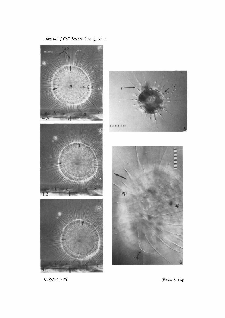
Actinosphaerium slightly compressed between a slide and coverslip continues tomove. If the overlying coverslip restricts the free movements of only some axopods,then locomotion occurs almost as rapidly as normal. An organism actively moving insuch a preparation is shown as viewed from above in Fig. 6. The cytosomes of suchanimals are flattened anteriorly, and their anterior axopods are fewer and shorter thanthe trailing ones (some of which adhere over most of their length to the upper cover-slip). Under such conditions, Actinosphaerium would seem to be rolling forward,because leading axopods slowly detach from the overlying coverslip and pass down-ward and out of focus.
Due to light scattering by the cytosome, the behaviour of axopods passing beneaththe organism can only be inferred from vertical observation. However, these sup-portive axopods could be observed continuously from the side by the technique ofDellinger (1906). Time-lapse cine records (1 f.p.s.) were made of the locomotion thatoccurred within a known focal plane.
Frame-by-frame film analysis indicates that A. eichhorni and Actinophrys sp. roll:for example, in travelling from left to right, their cytosomes revolve in a clockwisedirection. (The term ' clockwise' refers to the direction of cytosome rotation when theorganisms are viewed from the side. If observed from above, the same organisms would
15-2
234 C. Walters
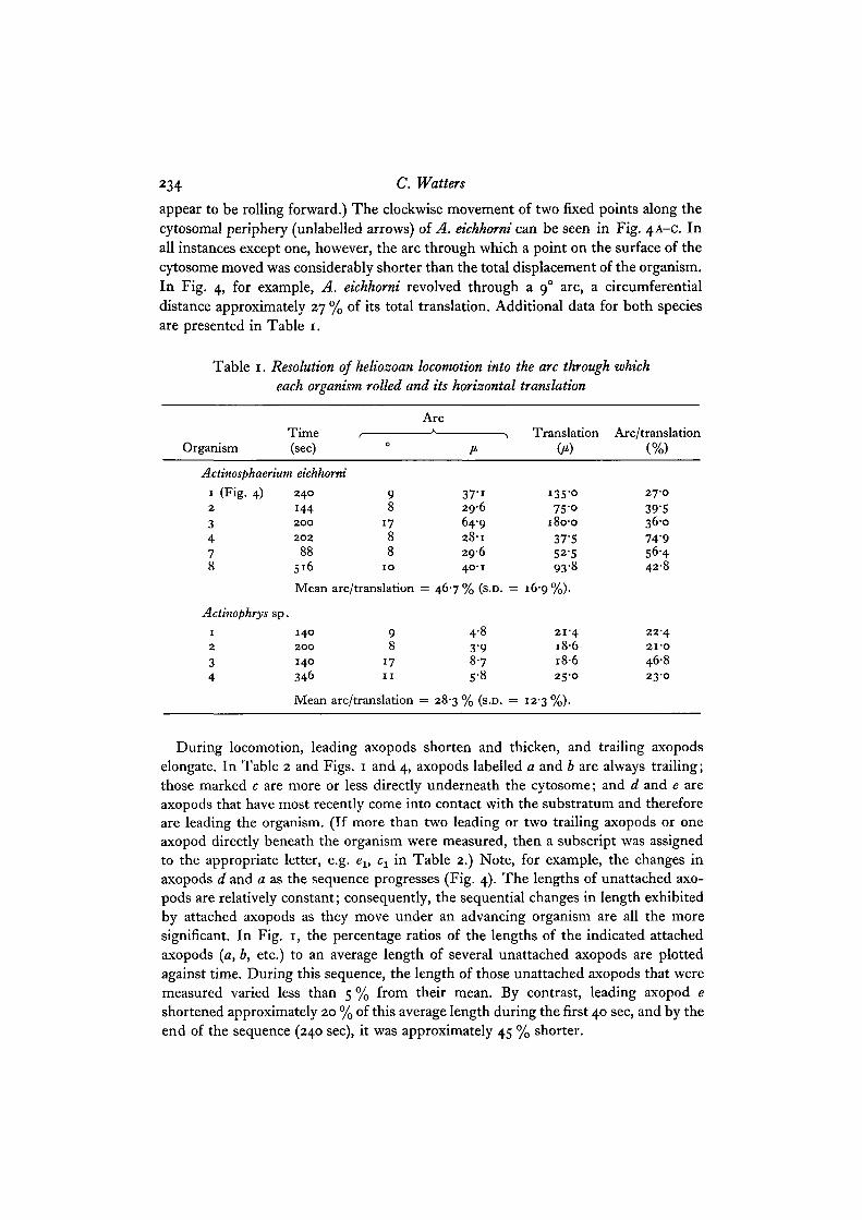
appear to be rolling forward.) The clockwise movement of two fixed points along thecytosomal periphery (unlabelled arrows) of A. eichhorni can be seen in Fig. 4A-C. Inall instances except one, however, the arc through which a point on the surface of thecytosome moved was considerably shorter than the total displacement of the organism.In Fig. 4, for example, A. eichhorni revolved through a 90 arc, a circumferentialdistance approximately 27 % of its total translation. Additional data for both speciesare presented in Table 1.
Table 1. Resolution of heliozoan locomotion into the arc through whicheach organism rolled and its horizontal translation
OrganismTime(sec)
ArcA
Actinosphaerium eichhorni
1 (Fig. 4)2
3478
2 4 0
1442 0 0
2 0 2
88516
Mean
Actinophrys sp.1
2
34
1 4 0
2 0 0
140
346
Mean
9 37-18 296
17 64-98 28-18 296
10 40-1
arc/translation = 46-7 % (s.D. =
9 4-88 3-9
17 8-711 s-8
arc/translation = 28-3 % (s.D. =
Translation(/*)
i3S-o75 -o
180-037-552-593-8
16-9%).
21-41 8 61 8 625-0
12-3%).
Arc/translation(%)
27-039-536-074-956-44 2 8
22-42 I - O
46823-0
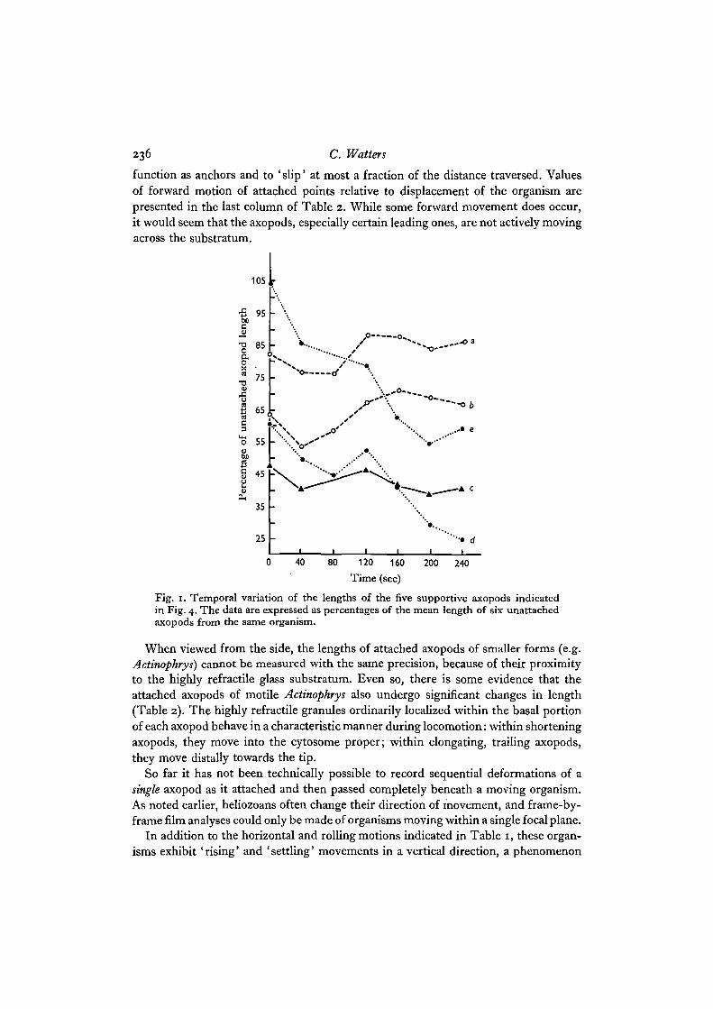
During locomotion, leading axopods shorten and thicken, and trailing axopodselongate. In Table 2 and Figs. 1 and 4, axopods labelled a and b are always trailing;those marked c are more or less directly underneath the cytosome; and d and e areaxopods that have most recently come into contact with the substratum and thereforeare leading the organism. (If more than two leading or two trailing axopods or oneaxopod directly beneath the organism were measured, then a subscript was assignedto the appropriate letter, e.g. ev cx in Table 2.) Note, for example, the changes inaxopods d and a as the sequence progresses (Fig. 4). The lengths of unattached axo-pods are relatively constant; consequently, the sequential changes in length exhibitedby attached axopods as they move under an advancing organism are all the moresignificant. In Fig. 1, the percentage ratios of the lengths of the indicated attachedaxopods {a, b, etc.) to an average length of several unattached axopods are plottedagainst time. During this sequence, the length of those unattached axopods that weremeasured varied less than 5 % from their mean. By contrast, leading axopod eshortened approximately 20 % of this average length during the first 40 sec, and by theend of the sequence (240 sec), it was approximately 45 % shorter.
Heliozoan locomotion 235
Often, trailing axopods continued to lengthen after detachment from the sub-stratum, while leading axopods were not observed to shorten prior to attachment.
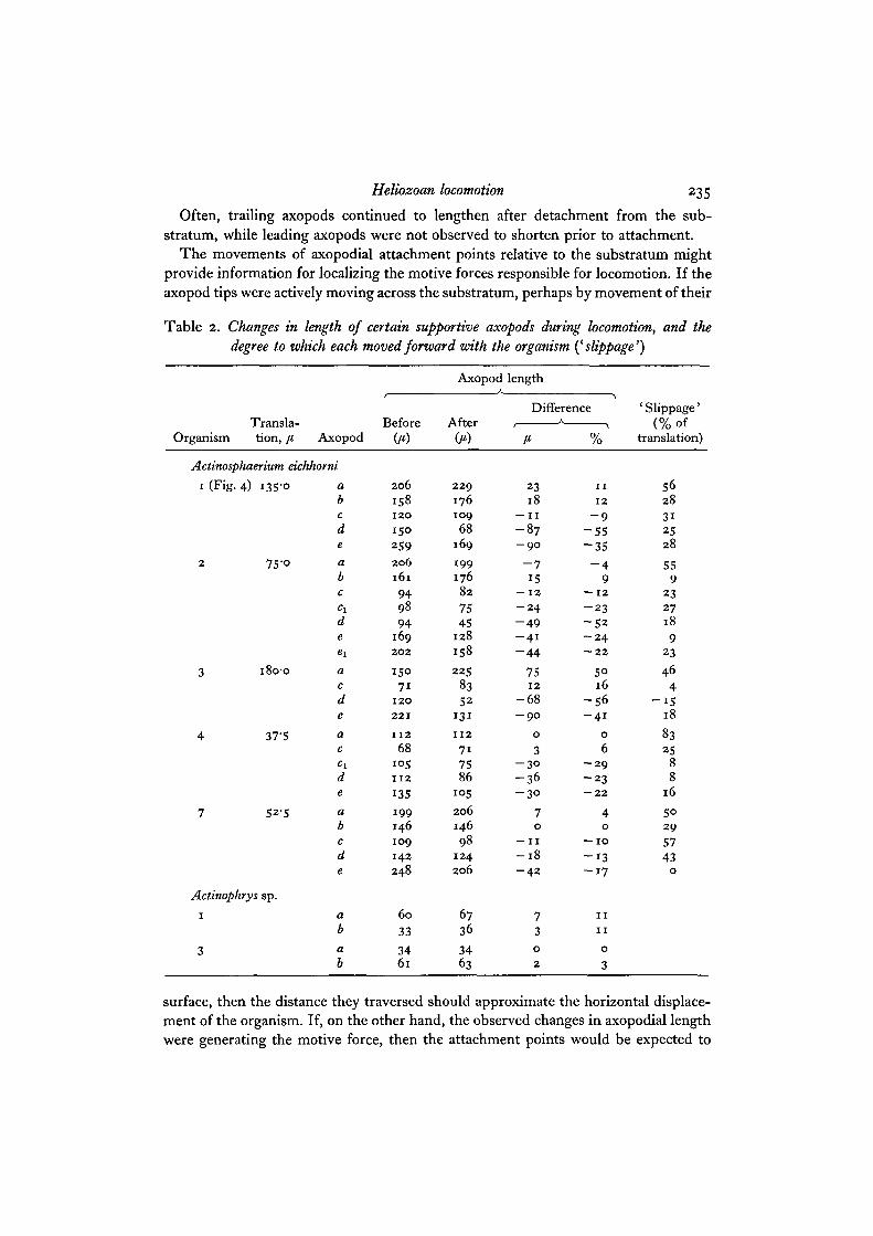
The movements of axopodial attachment points relative to the substratum mightprovide information for localizing the motive forces responsible for locomotion. If theaxopod tips were actively moving across the substratum, perhaps by movement of their
Table 2. Changes in length of certain supportive axopods during locomotion, and thedegree to which each moved forward with the organism {^slippage')
OrganismX i till bid"
tion, [t, Axopod
Actinosphaerium eichhorni
i (Fig.
2
3
4
7
4) 135'°
7S-o
i8o-o
37-5
52-5
Actinophrys sp.
i
3
abcde
abc
deexacdeacc ideabcde
ab
ab
O)
2 0 6
1581 2 0
1502592 0 6161
949894
1692 0 2
15071
1 2 0
2 2 1
1 1 2
68i ° 51 1 2
135
199146109
142248
60
33
3461
Axopod
Aftpr
2 2 91761 0 9
68169
19917682
7545
128
158
225
8352
131
1 1 2
717586
i ° 5
206146
981242 0 6
6736
3463
length
Difference
23l 8
— I I
- 8 7- 9 O
- 715
— 12
- 2 4- 4 9- 4 1
- 4 4
751 2
- 6 8- 9 0
0
3- 3 °- 3 6- 3 0
70
—11
- 1 8- 4 2
73
0
2
1 1
1 2
- 9- 5 5- 3 5
- 49
— 1 2- 2 3
- 5 2- 2 4— 2 2
5°16
- 5 6- 4 1
0
6- 2 9- 2 3— 2 2
40
— 1 0
- 1 3- 1 7
1 1
1 1
0
3
' Slippage'(% of
translation)
562831
2528
559
232718
923
464
- 1 518
8325
88
16
5°2 9
5743
0
surface, then the distance they traversed should approximate the horizontal displace-ment of the organism. If, on the other hand, the observed changes in axopodial lengthwere generating the motive force, then the attachment points would be expected to
236 C. Waiters
function as anchors and to ' slip' at most a fraction of the distance traversed. Valuesof forward motion of attached points relative to displacement of the organism arepresented in the last column of Table 2. While some forward movement does occur,it would seem that the axopods, especially certain leading ones, are not actively movingacross the substratum.
• • • • d
40 80 120 160
Time (sec)
200 240
Fig. 1. Temporal variation of the lengths of the five supportive axopods indicatedin Fig. 4. The data are expressed as percentages of the mean length of six unattachedaxopods from the same organism.
When viewed from the side, the lengths of attached axopods of smaller forms (e.g.Actinophrys) cannot be measured with the same precision, because of their proximityto the highly refractile glass substratum. Even so, there is some evidence that theattached axopods of motile Actinophrys also undergo significant changes in length(Table 2). The highly refractile granules ordinarily localized within the basal portionof each axopod behave in a characteristic manner during locomotion: within shorteningaxopods, they move into the cytosome proper; within elongating, trailing axopods,they move distally towards the tip.
So far it has not been technically possible to record sequential deformations of asingle axopod as it attached and then passed completely beneath a moving organism.As noted earlier, heliozoans often change their direction of movement, and frame-by-frame film analyses could only be made of organisms moving within a single focal plane.
In addition to the horizontal and rolling motions indicated in Table I, these organ-isms exhibit ' rising' and ' settling' movements in a vertical direction, a phenomenon
Heliozoan locomotion 237
first observed for A. eichhorni by Brandt (1878). Such movements are strikingly evidentin side view, but are usually not observed from above, either because of the largerdepth of focus at low magnification or because such vertical motion is restricted byslide and coverslip.
A suitable hypothesis of heliozoan locomotion, therefore, must account for all threedirectional components (horizontal and vertical movements, and rolling), and shouldalso explain exceptional cases where only one or two such movements are evident.
Three incidental observations should be mentioned since they have some bearingon the role of axopods in locomotion, (i) During cytokinesis, two daughter organismsappear to move actively away from each other (see Kuhl, 1953; Kitching, 1964); theyare connected by a cytoplasmic bridge that becomes more and more attenuated untilseparation occurs. Initially, however, neither half possesses trailing axopods, andin this instance the motive force resulting in separation would have to be localized ineither the lateral or the leading axopods. (ii) Specimens of A. eichhorni mounted ina thin layer of 0-8 % purified agar (Difco) appear viable for 24 h and continue tomove, though much more slowly than is normal. After 5-8 h in such a preparation,motile individuals assume a blunt ellipsoid shape with the major axis parallel to thedirection of locomotion. The majority of axopods are trailing the organism; the fewleading ones are relatively shorter and much thicker than normal. They are also thickerthan the remaining agar-embedded axopods of the same organism. Under thesecircumstances, it would seem that the leading axopods are responsible for the elon-gated shape and the movement observed, (iii) Actinosphaerium has frequently beenobserved to climb the vertical glass walls of Dellinger chambers.
DISCUSSION
Heliozoans have been reported to roll, glide or even swim across the substratum(Tregouboff, 1953; Kuhl, 1951), but their motility has not been so critically analysedas has the motility of other sarcodines (see Allen, 1961). It is apparent that theheliozoan axopod is a locomotory appendage, as is the pseudopod of the amoeba, butcan the supposedly different types of locomotion be attributed to a single mode ofaxopodial activity, or must several be invoked?
In the present study, the locomotion of Actinosphaerium eichhorni and a species ofActinophrys was primarily a horizontal displacement accompanied by definite rollingand vertical motions. This locomotion could be correlated with significant changes inthe lengths of certain supportive axopods and seemed to depend on their attachmentto the substratum. If, in fact, these changes do result in locomotion, then the motiveforce so generated would be imparted by the axopods to the cytosome at their pointsof insertion.
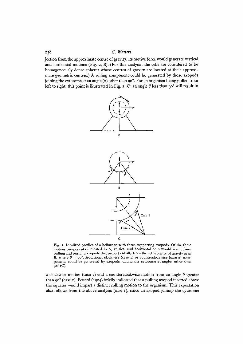
The three components of heliozoan locomotion (Fig. 2, A) could be generated bychanges in the lengths of axopods in a simple vectoral manner, and the angle (6) atwhich an individual axopod joined the cytosome (Fig. 2, C) would determine therelative magnitude of the three motion components it contributed to the resultantlocomotion. In the simplest instance, where an axopod extended along a radial pro-
238 C. Walters
jection from the approximate centre of gravity, its motive force would generate verticaland horizontal motions (Fig. 2, B). (For this analysis, the cells are considered to behomogeneously dense spheres whose centres of gravity are located at their approxi-mate geometric centres.) A rolling component could be generated by those axopodsjoining the cytosome at an angle (6) other than 900. For an organism being pulled fromleft to right, this point is illustrated in Fig. 2, C: an angle 8 less than 900 will result in
Fig. 2. Idealized profiles of a heliozoan with three supporting axopods. Of the threemotion components indicated in A, vertical and horizontal ones would result frompulling and pushing axopods that project radially from the cell's centre of gravity as inB, where 6 = 900. Additional clockwise (case 1) or counterclockwise (case 2) com-ponents could be generated by axopods joining the cytosome at angles other than9O° (C).
a clockwise motion (case 1) and a counterclockwise motion from an angle 6 greaterthan 900 (case 2). Penard (1904) briefly indicated that a pulling axopod inserted abovethe equator would impart a distinct rolling motion to the organism. This expectationalso follows from the above analysis (case i), since an axopod joining the cytosome
Heliozoan locomotion 239
above the equator would form an acute angle 6. Conversely, the forcible elongationof a trailing axopod would produce a clockwise motion for an angle greater than 900,a counterclockwise one for an angle less than 90°.
In all organisms that consistently rolled forward in the direction in which they weremoving (case 1), certain leading axopods often joined the cytosome at angles less than900 (e.g. d in Fig. 4). The angle of trailing axopods, however, rarely varied fromapproximately 900. Active shortening would seem more important than elongation inthe generation of motive force for several other reasons. Shortening axopods generallyexhibited a greater rate of change (Table 3) and less slippage (Table 2) than elongatingones. Under certain circumstances, for example, at cytokinesis, trailing (elongating)axopods are not initially present, yet division is accomplished by the active movementof the two daughter halves away from each other.
Table 3. The rate of change in length of specified axopods during the locomotion ofActinosphaerium eichhorni; the data are collated from Tables 1 and 2
Elongation Retraction (/i/min)
Trailing Directly beneath LeadingOrganism {a, b) (c, ct) (d, e, et)
1 S"8,4'S -2-8 -21-8,-22-52 2-9,6-2 -5-0 , -10-0 -21-7 , -17-1 , -18-33 22-8 -3-6 - 2 0 - 6 , - 1 7 1 , - 1 8 - 34 o-o 0-9, —9-1 —10-9, —9-17 5-4 -8-5 -13-8, -32-3
Average 6-8 — 5-7 —19-8velocity
All of the means differ significantly from each other at the P = 0-05 level (modified Keul'smultiple range test).
Rising and settling movements would occur when an organism moved over axopods(in the 'c' position) that had shortened to varying degrees. Occasionally, upwardmotions in the absence of horizontal displacements were exhibited by organisms withelongating supportive axopods.
The direction of forward motion would be a resultant of all horizontal motion com-ponents, and competition between supportive axopods pulling from different direc-tions would be expected to generate the erratic locomotory behaviour that has beenwell documented for A. eichhorni (Kuhl, 1951). However, locomotion is not alwayspatternless. Specimens of Actinosphaerium have been reported to move parallel to oneanother (Kuhl, 1951), and movement of one organism towards another followed bycontact and immediate reversal of movement has also been observed (unpublishedobservation). Some degree of co-ordination of axopodial behaviour would seem to bepresent, in spite of the absence of ultrastructural evidence (Tilney & Porter, 1966).
An hypothesis relating heliozoan locomotion to the tensile forces generated bychanges in axopodial lengths can account for the phenomena reported in this paper.The hypothesis suggests further that the relative magnitudes of each locomotory com-
240 C. Watters
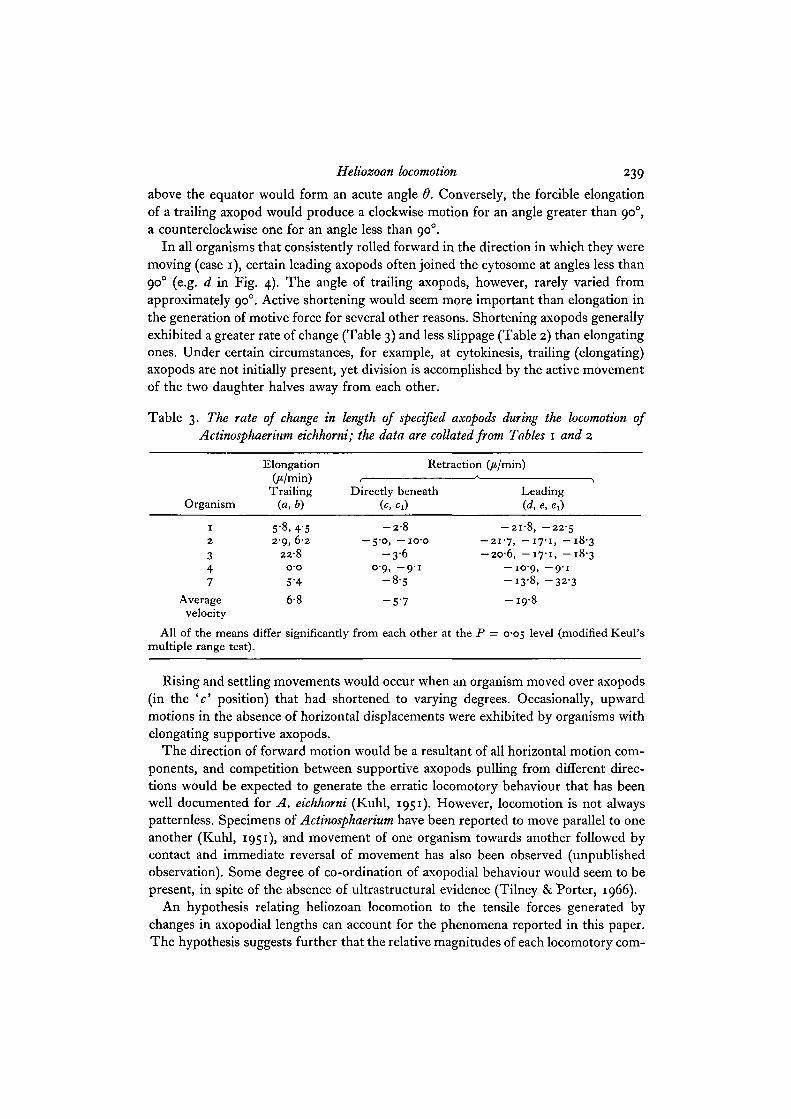
ponent could be related to dimensional parameters such as cytosomal diameter andaxopodial length. For instance, in Fig. 3 the angle 6 discussed above would in turnvary inversely with the angle an axopod made with the substratum (O). The locomotionof heliozoans with small cytosomes and relatively long axopods (such as Actinophrys)should contain a small rolling component, which possibly might not be detected(Fig. 3, B). Larger organisms, such as Actinosphaerium, that possess shorter axopodsrelative to their large cytosomal diameter should exhibit a greater rolling motion(Fig. 3, A). Though the data are by no means conclusive, the larger A. eichhorni seemsto roll more per unit of horizontal displacement than does the smaller Actinophrys withrelatively longer axopods (Table 1).
Fig. 3. Idealized profiles of two different species of heliozoans. Actinosphaerium eich-horni (A) generally possess a larger cytosome and relatively shorter axopods, while themuch smaller Actinophrys sp. displays relatively longer axopods (B). For furtherexplanation, see text.
Since cytosomal size and axopodial length are relatively good taxonomic criteria forthe species studied here (Penard, 1904), the hypothesis provides a single explanationfor the different kinds of locomotion that have been reported for these as well as otherspecies. If not carefully examined, heliozoans such as Actinophrys (Fig. 3, B) mightseem simply to be 'gliding' or 'creeping', while those similar in dimensions toActinosphaerium (Figs. 3, A and 4) would appear to be 'rolling' across the substratum.
Heliozoan locomotion 241
Yet, in both instances, changes in axopodial length would provide the necessarymotive force.
Other possible axopodial mechanisms have been proposed and their variety reflects,in part, the differences in the types of locomotion observed: for example, whether theorganism was 'rolling' or 'gliding'. Although the hypothesis proposed in this papercould account for the apparent observational discrepancies, the explanatory mechan-isms will be examined in more detail.
Conceivably, changes either in the length of axopods or in their position relative tothe substratum could result in locomotion. This study presents the first quantitativedata relating locomotion with changes in axopodial length, although the idea thatattached axopods might pull a heliozoan forward has been discussed before (Penard,1889). Sequential shortening of supportive axopods might also generate a 'rolling'motion if these axopods were asymmetrically located relative to the organism's centreof gravity (Wetzel, 1926). Such a hypothesis, similar to one suggested more recentlyby Tilney & Porter (1966), proposes that locomotion would result more from a shift inthe cell's centre of gravity than from tensile forces generated by axopodial shortening.Since the organism would be rolling forward, the arc through which it moved shouldequal its horizontal displacement, or even slightly exceed it (allowing for slippage), andsubstantial adhesion of the axopods to the substratum should not be necessary. Theobservations and results presented here do not support such an explanation. In noinstance was the arc through which an organism rolled greater than 75 % of its hori-zontal displacement, and in most instances it was less than 50 %. The motive forcegenerated by slight shifts in the centre of gravity should be small, and consequentlyslightly compressed organisms should roll with great difficulty or not at all. In fact,locomotion continued under mild compression, even though the movements of over-lying axopods was obviously impeded. Finally, locomotion of a heliozoan up a verticalsurface cannot result simply from a shift in the organism's centre of gravity.
Changes in axopodial position seem to be of minor importance in locomotion,although their possible significance cannot be completely discounted. Based on time-lapse cinematography, Kuhl (1951) ascribed the locomotion of A. eichhorni to the'rowing movement' (Ortsbewegung) of lateral axopods that transcribed arcs of 8-12°.As Tilney & Porter (1966) have more recently noted, locomotion would result fromsuch rowing motions if the axopods either display a differential speed between effectiveand recovery strokes, or attach to the substratum during their effective stroke and de-tach during recovery. (This latter motion might be more aptly described as 'walking'.)Rowing motions were rarely observed in this study and were never symmetricallyorganized relative to the axis of locomotion. As a consequence, it is difficult to visualizehow directed movements, even of brief duration, could result from such behaviour,especially in those instances where a slightly compressed organism continued to move.More often, axopods displaying rowing motions with differing effective and recoveryspeeds were localized near contractile vacuoles that gradually filled and rapidlycollapsed.
Kitching (1964) also doubts that axopodial rowing movements are responsible forlocomotion. Instead, he suggests that the locomotion of Actinophrys sol might result
242 C. Waiters
from the surface movements of attached axopods. Movement of this cytoplasmicsurface layer, independent of gross changes in the length of the axopods themselves,seems to be implicated in the rejection of material and possibly in feeding (Kitching,i960, 1964). Attached axopods, moving the organism by means of such surface move-ments, should advance at a rate similar to that of the whole organism. Such behaviourwas not observed. In both species studied, the tips of leading axopods, rather thanadvancing, seemed to function as anchors and all attached axopods observed eitherunderwent a change in length or were bent relative to the direction of locomotion.
Finally, attached axopods might propel a heliozoan by active bending (Tilney &Porter, 1966). Bending of attached axopods that are obliquely oriented relative to thedirection of locomotion is a commonly observed phenomenon (Fig. 6), but it is diffi-cult to decide whether such bending is a cause or an effect of locomotion. The datapresented here neither affirm nor negate the importance of bending axopods inlocomotion; however, two further observations are perhaps suggestive of their deriva-tive nature. Only attached axopods of moving organisms bend along their length toany great extent; freely suspended axopods are straight and relatively stiff and one hasthe impression they are passively bent by large motile prey and also by the sub-stratum. However, both attached and unattached axopods exhibit striking changes inlength. Axopodial shortening serves an important feeding role (Looper, 1928; Watters,1966) and, as has been shown here, significant changes in the lengths of attachedaxopods can be correlated with locomotion. Further, the rates of change in lengthexhibited by these attached axopods are similar to those exhibited by unattached ones(Watters, 1966).
Changes in pseudopod lengths are responsible for the locomotion of at least threeother types of cells: Difflugia, a test-bearing sarcodine (Wohlman & Allen, 1968),mesenchyme cells of sea-urchin gastrulae (Gustafson & Wolpert, 1963), and sensoryneurons in culture (Nakai, 1964). In these forms, the ultrastructural basis for thechanges in length has not been adequately characterized, although the work ofWohlman & Allen (1968) strongly implicates 50-A microfilaments in the contractionof the testacean pseudopod. In the case of the Heliozoa, on the other hand, locomotionwould seem to depend on the integrity of the microtubular elements of the axonemeand the structural support they contribute to the axopod. Treatments, such as lowtemperature, colchicine and high hydrostatic pressure, have been shown to affect ad-versely both the stability of axopods and the structure of microtubules (Tilney, 1965;Tilney et al. 1966). Axopods also contain a highly motile cytoplasmic layer that iscontinuous with the equally motile cytosomal surface. In Actinosphaerium eichhornithis latter cytoplasmic layer rapidly forms food cups and, under certain circumstances,extensive cytoplasmic veils (Penard, 1904; Watters, 1966). An understanding of therelationship between locomotion and this associated cytoplasmic motility would co-tribute greatly to our understanding of the dual role proposed for the microtubule instructural support and motility.
Heliozoan locomotion 243
This study formed part of a doctoral dissertation that was presented to Princeton University;it was supported by NIHGMO8691 awarded to Professor R.D.Allen. I wish to thankProfessor Allen for his thoughtful direction and constant encouragement, and also ProfessorLionel Rebhun and Drs Robert Goldman and Alan Wohlman for numerous and helpfuldiscussions.
REFERENCESALLEN, R. D. (1961). Ameboid movement. In The Cell, vol. 2 (ed. J. Brachet and A. Mirsky),
PP- 135-216. New York and London: Academic Press.BARRETT, J. M. (1958). Some observations on Actinosphaerium nucleofilum n.sp., a new fresh
water actinophryid. jf. Protozool. 5, 205-209.BRANDT, K. (1878). Uber die Axenfaden der Heliozoen und die Bewegungen von Actino-
sphaerium. Sber. Ges. naturf. Freunde Berl. pp. 171-178.DELLINGER, O. P. (1906). Locomotion of Amoebae and allied forms, jf. exp. Zool. 3,
337-358.GUSTAFSON, T. & WOLPERT, L. (1963). The cellular basis of morphogenesis and sea urchin
development. Int. Rev. Cytol. 15, 139-214.HAECKEL, E. (1886). Generelle Morphologie der Organismen, vol. 2. Berlin: Reimer.KITCHING, J. A. (i960). Responses of the Heliozoan Actinophrys sol to prey, to mechanical
stimulation, and to solutions of proteins and certain other substances, jf. exp. Biol. 37,407-416.
KITCHING, J. A. (1964). The axopods of the sun animalcule Actinophrys sol (Heliozoa). InPrimitive Motile Systems in Cell Biology (ed. R. D. Allen & N. Kayima), pp. 445-456.New York and London: Academic Press.
KUHL, W. (1951). Mikrodynamische Untersuchungen an der lebenden Zelle von Actino-sphaerium eichhorni Ehrbg., unter Anderung des Zeitmomentes. Protoplasma 40, 555-613.
KUHL, W. (1953). Untersuchungen uber die Cytodynamik der Plasmogamie und temporaren' Plasmabriicken' bei Actinosphaerium eichhorni Ehrbg., unter Anderung des Zeitfaktorsmittels des Zeitrafferfilsm. Protoplasma 42, 133-192.
LEDBETTER, M. D. & PORTER, K. R. (1963). A microtubule in plant cell fine structure. jf. CellBiol. 19, 239-250.
LEHRER, G. M. (1950). Notes on the culture and mechanism of feeding of Actinosphaeriumeichhorni. Biol. Rev. C.C.N.Y. 12, 16-17.
LEIDY, J. (1879). Fresh-water Rhizopods of North America. Washington: U.S. GovernmentPrinting Office.
LOOPER, J. B. (1928). Observations on the food reactions of Actinophrys sol. Biol. Bull. mar.biol. Lab., Woods Hole 54, 485-502.
MACKINNON, D. L. (1909). Optical properties of contractile organs in Heliozoa. jf. Physiol.,Lond. 38, 254-258.
NAKAI, J. (1964). The movement of neurons in tissue culture. In Primitive Motile Systems inCell Biology (ed. R.D.Allen & N. Kamiya), pp. 377-384. New York and London:Academic Press.
NOZAWA, K. (1938). On the voluminal relation between the ectoplasm and endoplasm in aheliozoan, A. eichhorni. Cytologia 9, 185-192.
PENARD, E. (1889). Etudes sur quelques H^liozoaires d'eau douce. Archs. Biol., Paris 9, 123-184.PENARD, E. (1904). Les Heliozoaires d'eau douce. Geneva: Kiindig.ROSKIN, G. (1925). Uber die Axopodien des Heliozoa und die Greifentakeln der Ephelotides.
Arch. Protistenk. 52, 207-216.RUMJANTZEW, A. & WERMEL, E. (1925). Untersuchungen uber den Protoplasmabau von
Actinosphaerium eichhornii. Arch. Protistenk. 52, 217-264.SCHMIDT, W. J. (1944). Polarisationsmikroskopische Beobachtungen an Actinosphaerium
eichhornii. Biol. Zbl. 64, 5-14.SLAUTTERBACK, D. B. (1963). Cytoplasmic microtubules. I. Hydra, jf. Cell Biol. 18, 367-388.TILNEY, L. G. (1965). Microtubules in the heliozoan Actinosphaerium nucleofilum and their
relation to axopod formation and motion, jf. Cell Biol. 27, 107 A.
244 C* Walters
TILNEY, L. G., HIRAMOTO, Y. & MARSLAND, D. (1966). Studies on the microtubules inHeliozoa. III. A pressure analysis of the role of these structures in the formation and main-tenance of the axopodia of Actinospliaerium nucleofilum (Barrett). ,7. Cell Biol. 29, 77-96.
TILNEY, L. G. & PORTER, K. R. (1966). Studies on microtubules in Heliozoa. I. The finestructure of Actinosphaerium nucleofilum (Barrett), with particular reference to the axial rodstructure. Protoplasma 60, 317-344.
TREGOUBOFF, G. (1953). Classe des Heliozoaires. In Traite de Zoologie (ed. P. Grass6), vol. 1 (2),pp. 437-489. Paris: Masson.
WAITERS, C. D. (1966). Studies on the Motility of the Heliozoa. Doctoral Dissertation,Princeton University.
WETZEL, A. (1926). Zur Morphologie und Biologie von Raphidocystis infestans n.sp., einemTemporare aus Ciliaten parasitierenden Heliozoen. Arch. Protistenk. 53, 135-182.
WOHLMAN, A. & ALLEN, R. D. (1968). Structural organization associated with pseudopodextension and cell locomotion in Difflugia. J. Cell. Sci. 3, 105-114.
WOLPERT, L. (1965). Cytoplasmic streaming and amoeboid movement. Symp. Soc. gen.Microbiol. 15, 270-293.
{Received 8 July 1Q67)
In all Figures, each small scale unit represents 10 fi.Fig. 4. Prints from a 16-mm cin6 record of the locomotion of Actinosphaerium eich-horni, as viewed from the side, at the following time intervals: A, O sec; B, 160 sec;c, 240 sec. A. eichhorni possesses numerous axopods (ap) and a cytosome characteristic-ally divided into a vacuolar cortex (C) and a more opaque medullary region (m). As theorganism moves from left to right, supporting axopods (a-e) undergo chracteristicchanges in length. The two unlabelled arrows along the periphery indicate the sametwo axopods throughout the sequence, while the solid bar (r) provides a fixed referencemark on the substratum.Fig. 5. Actinophrys sp. displays a typically spherical cytosome and radiating axopods.Numerous inclusions (j) within the axopods and also a ' contractile vacuole' (cv) areobvious at this magnification.Fig. 6. A slightly compressed A. eichhorni that has continued to move in the directionof the large arrow. A few short, leading axopods (lap) are attached at their tips to theoverlying coverslip, while numerous trailing axopods (tap) are attached along their entirelength. A laterally located bent axopod (bap) can also be seen. The very narrow depth offield achieved both in this figure and in Fig. 5 results from use of the NomarskiDifferential Interference system (Carl Zeiss).
Journal of Cell Science, Vol. 3, No. 2
C. WATTERS (Facing p. 244)