Embed Size (px)
Citation preview

Instructions for use
Title STUDIES ON THE LYMPHONODI OF CATS : I. MACROSCOPICAL OBSERVATIONS ON THE LYMPHONODIOF HEADS AND NECKS
Author(s) SUGIMURA, Makoto; KUDO, Norio; TAKAHATA, Kurahiko
Citation Japanese Journal of Veterinary Research, 3(2), 90-104
Issue Date 1955-08-10
DOI 10.14943/jjvr.3.2.90
Doc URL http://hdl.handle.net/2115/1670
Type bulletin (article)
File Information KJ00002372931.pdf
Hokkaido University Collection of Scholarly and Academic Papers : HUSCAP

STUDIES ON THE LYMPHONODI OF CATS
I. MACROSCOPICAL OBSERVATIONS ON THE LYMPHONODI OF HEADS AND NECKS
Makoto SUGIMURA, Norio KUDO and Kurahiko TAKAHATA
Laboratory of Veterinary Anatomy, Faculty of Veterinary Medicine, Hokkaido Univer8ity, Sapporo, Japan
(Received for Publication, May 2, 1955)
INTRODUCTORY
Up to this time, even though there have been many anatomical studies on the lymphatic system done by numerous investig'dtors, yet about the differences in structures depending on species which should be of interest in the phylogenetic studies the reports have b~en published by only a few investigators, such as GULLAND, SCHUMACHER, RICHTER, BUNTING, BAUM et al., JOLLY, GODBILLE, HEUDORFER and others, using various animals. Many other students have tended to deem the lymphonodi as the same in different animals.
Moreover, the differences in structure depending on location of node, which should be important for functional significations, were only affirmed by a few investigators, such as JOB, JORDAN et LOOPER, ASCHOFF and GYLLENSTEN, while DAWSON et MASUR denied any such fact.
Lately, HORII et al. distinguished "the primary lymphonodus" and "the secondary lymphonodus" - the former only receives directly the lymphatic vessels not passing through any other lymphonodus, while the latter receives the efferent lymphatic vessels of some other lymphonodus, and reported the differences in producing power of lymphocytes and in structures between both types of lymphonodi.
In short, the differences in structure depending on animal species and the location of nodi have not been studied so thoroughly as to be recognized as distinctly proved. So, on these problems the writers hope to throw some light
by means of comparative anatomical studies of the lymphatic system. Cats were selected for materials.
There were only a few macroscopical studies on the lymphatic system of cats, which were made by STROMSTEN, MANABE, MATSUMOTO, TAKESHITA, TOMITA, REIGHARD et JENNINGS and others.
In this paper the appearances of the afferent and efferent lymphatic vessels of each node and the varieties of lymphonodi concerning to their vessels are
JAP. j. VET. RES., VOL. 3, No.2, 1955

Studie8 on the Lymphonodi of Cat8 91
described. For convenience, the study is limited particularly to those of heads and necks.
MATERIALS AND METHODS
The writers used for this research 21 cats, aged 15 days to 7 years, as indicated in table 1.
TABLE 1. Materials Used for This Observation
EXP. NO. SEX AGE WEIGHT (g)
1 0 1 Month 200
2 3 Months 560
3 " 3 700
4 4.5 1050
5 ? 1800
6 2 Years 2800
7 3 3800
8 " 6 " 3500
9 6 " 4750
10 7 4600
11 <f 15 Days 200
12 1 Month 180
13 1 " 210
14 " 1.5 Months 410
15 4 " 1500
16 " 5 1350
17 1 Year 2400
18 " 2 Years 3200
19 3 1800
20 " 4 2250
21 ,. 6 " 3300
The materials were anesthetized and killed by ether. Double solution of India ink bought on the market was injected into various parts of heads and necks before the
temperature of the body had declined, for the purpose of observating the afferent and
efferent lymphatic vessels as accurately as possible. For, injections, the writers used an injection-syringe of 2ml capacity and a needle of
114 mm in diameter on sale in ordinary medical supply shops.
In addition to India ink, the writers also used 4% solution of carmine, 10% solution of cinnabar and about 4~~ solution of Berlin blue which they themselves prepared. Ten

92 SUGIMURA, M. et al.
% solution of cinnabar was useless to make clear the lymphatic vessels, and other
coloring matters leaked out of the lymphatic vessels. But only with Berlin blue were
somewhat good results gained. After all, the India ink was the best injection matter. In the present paper, the descriptions of venous system acc'ompanied with lymphatic
systems are made with reference to HIROTA'S report and the names are given in accord
with the Nomina Anatomica Japonica.
RESULTS
1. Groups of Lymphonodi in Heads and Necks
The lymphonodi of heads and necks were distinguished into 10 groups according to
their positions. The groups were named provisionally as follows: 1) Lymphonodi submand'ibulares mediales; These nodi, of 2.4-0.25 cm in the longest
diameter, touch in front of the union of V. facia.lis and V. transversa hyoidea lying beneath platysma. They were flatly ellipsoid-shape.
2) Lymphonodi submand~'bulare8 laterales; These nodi, of 1.9-0.38 cm in the longest
diameter, touch in front of the union of V. retromandibularis and V. !aciali..." lying beneath platysma. Most of these nodi were depressed with ellipsoid-shape.
3) Lymphonodi submandibulares mediales caudales; These small nodi, of 0.35-0.05 em
in the longest diameter, were scattered about the juncture of V. transversa hyoidea with V. jaciali'1 being situated behind foregoing Lnn. submandibulares med. They were spherical.
4) Lymphonodi subm:"ndibulares latel'ales caudales; These were also small nodi, of 0.36-0.05 em in the longest diameter, which were scattered about the juncture of V. retromandibularis with V. jacialis lying behind foregoing Lnn. submandibulares lat. They were also spherical.
5) Lymphonodi parotidici craniales; These nodi. of 0.86-0.1 em in the longest diameter, were embedded in Gl. parotis lying along V. temporalis communis. Most of these
forms were somewhat depressed with round outlines. 6) Lymphonodi parotidici caudales; These nodi. of 3.1-0.05 cm in the longest di·
ameter, were mostly club-sbaped. but partly gourd-shaped or rounded and embedded in fatty tissues lying behind Gl. parotis, along V. retroauricu,zaris and V. retromandibularis.
7) Lymphonodi retropharyngici; These nodi, of 2.25-0.15 cm in the longest diameter,
were found along V. jugularis 'interna lying behind the pharynx. Lymphonodi of this group were the largest nodi in head and neck regions and mostly kidney-shaped.
8) Lymphonodi cervicales super ficiales dorsales; These nodi, of 3.22-0.1 cm in the longest diameter. were embedded in the fatty tissues lying beneath M. trapezius cel'vicalis and M. omotransversarius, along V. transversa colli. Most of these nodi were flatly
ellipsoid-shaped. but partly gourd-shaped. 9) Lymphonodi cervicales super ficiales ventrales; These nodi, of 1.46-0.08 cm in the
longest diameter, were partly covered by the union of V. jugularis externa and V. transversae coll~ and embedded in the fatty tissues. Their forms were mostly oval.
10) Lymphonodus cervicalis profundus; This small rounded nodus. of 0.45-0.1 em in
the longest diameter, was found along' V. jugularis interna lying beside the trachea of the neck region.

TABLE 2. Number of Lymphonodi Appearing in Each Group of Lymphonodi
EXPERIMENT NO. GROUPS OF
LYMPHONODI TOTAL AVERAGE
Submandibulares med. {~
Submandibulares lat. {~
Submand1·bulares med. f R caud. l L
Submandibulares lat. caud.
ParoUdici cran.
Parotidici caud.
Retropharyngid
Cervicales super ficiales dors.
Cervicales super ficiales vent.
Cervicalis prof.
Total of Each Side
Total of Both Sides
J R
lL fR l L
fR l L
fR lL fR l L
fR l L
fR lL fR l L
1 2 3 4 5 6 7 8 9 10 11 12 13 14 15 16 ·17 18 19 20 21
1 1
1 1
1 1
1 1
1
2
2
1 1
2 1
2 4
3 2
1 1
1 1
2 3
2 2
111
111
111
111
1
112
112
111
21'1
111
5 2
4 4
1 1
1 1
2 3
2 2
3
3
1
1
3
2
1 1
1 1
1 1
1 1
1
1
1
1 1
1 1
4 5
6 2
1 1
1 1
3 2
3 2
1
1
1
1
1
1
3
2
1
1
3
7
1
2
3
1
111
111
1
1 121 1
211 1
1 1
1
1
1
1
1
3
4
1
1
2
1
1
1
1
1
1
2
1
1
1
1
3
3
1
1
2
2
1 1
1 1
1 2
1 1
1 2
1 1
1
1 1
1
1 1
1
3
5
1
1
2
2
6
6
1
1
2
2
1 1
1 1
1 1
2 1
1 1
1 1
1
2
1
1
1 1
1
4 2
7 3
1 1
1 1
2 2
2 2
1
1
1
1
2
1
1
3
1
1
2
2
111
111
111
111
1 2
1 1 1
121
221
1 1
221
5
4
1
1
2
2
2
1
1
1
2
2
5
5
1
1
1
2
1 1 2 1
1 1 2 1
1
1 1
1 1
1 1
1 1
1 1
1 1
2
1
1
1 1
1 2
1 1
2 4
2 4
1 1
1 1
3 2
2 2
1 1
1 1
1
1
1
1
1
1
1
3
3
1
1
2
2
1
1
9 14 14 11 15 15 13 15 9 12 12 16 13 9 11 13 12 13 12 13 11
11 13 13 12 15 14 10 17 9 13 12 13 18 9 11 15 12 14 11 12 10
20 27 27 23 30 29 23 32 18 25 24 29 31 18 22 28 24 27 23 25 21
Remarks: R: Right side. L: Left side.
Average: Average number oflymphonodi on an individual side. *. Average number of lymphonodi on an individual.
44
43
29
36
41
152
43
88
45
5
526
1.05 ± 0.22
1.02 ± 0.15
1.26 ± 0.45
1.33 ± 0.55
1.14 ± 0.35
3.62 ± 1.5
1.02 ± 0.15
2.1 ± 0.46
1.1 ± 0.28
1.0 ±O
* 25.1 ± 4.02
~ :;:: R.. ;;. ~
o ;:z .,.... ~ ~
~ ~
~ ~ o ;:z ~ ..... ,0 ......,
~ ~
~

94 SUGIMURA, M. et al.
2. The Numbers of Lymphonodi
The number of lymphonodi found in heads and necks were distributed into the above-mentioned groups of lymphonodi, as indicated in table 2.
It should be noted with regard to the number of lymphonodi found in heads and necks that the writers could not indicate the differences by individuals, sexes, ages or between right and left sides. But only Lnn. parotidici caud., on an
average 3.62-t-1.5, have shown conspicuous variation of the number.
3. The Existence Ratio of Each Group
In observed 42 sides, each group has been found as indicated in table 3.
TABLE 3. Showing the Ratio 0/ Existence 0/ Each Group
GROUP OF LYMPHONODI
NUMBERS OF SIDES FOUND LYMPHONODI
NUMBERS OF SIDES SEARCHED FOR L YMPHONODI
EXISTENCE RATIOS (%)
GROUPS DIFFERENTIATED BY RATIO OF OCCURRENCE
42 42 42 42 42 41 36 27 23 5
42 42 42 42 42 42 42 42 42 42
100 100 100 100 100 97.6 85.7 64.3 54.8 11.9
Constant ~
Nearly Constant Inconstant
According to the ratio of existence, each group of lymphonodi has been
classified into 3 groups as follows; 1) Group of constant existence (existence ratio of 100,%); This includes Lnn. sub
mandwulares med., Lnn. submandibulares lat., Lnn. parotidici caud., Lnn. retropharyngici and Lnn. cervicales super ficiales dors.
2) Group of nearly constant existence (existence ratio less than 100,% but over 80,%) ;
This includes Lnn. cervicales super jiciales vent. and Lnn. parotidici cran. 3) Group of inconstant existence (existence ratio less than 80%); This includes Lnn.
submandibulares med. caud., Lnn. submandwulares lat. caud. and Ln. cervicalis prof.
4. The Areas of Origin of Afferent Lymphatic Vessels
Each group of the above-named lymphonodi received lymphatic vessels whose
sources were found at respectively corresponding areas in heads and necks, as
indicated in table 4.
J

GROUPS OF
LYMPllONODl
Submandwulares f ~ med. I~~
. Submandwular.. / ~ lat. 1%
Submand"ntlures med. ca"d.
Subm .. ndihulares lat. ca"d.
Parolid'iei "ran.
Parotidic; "a "d.
/F l~ IF
l~ /F l~ /F l~
Rei r<>ph a ryngrci /1 ~ %
Cervical,s IF sUp"·fiC'w l •• dors I ~
Cer .. icnJc~ , / F 8Upt'T ficialePo rent l ~
Crr1'1.CO'Uf prof tE
TABLE 4. Showing the Appearances of Areas of Origin of Afferent Lymphatic Vessels Which Entered into Each of Lymphonodi
.... ° 03 "'I:: u'" .. '" 't:O:i Jl~
~
.~ " -::
" f',
d
.:1 .:1 tl tl
t t I>. ""
l~ ~~
.:1
]·t " <:l
:~ ~~
~.~
~~ :~ ~~
~ i! ~
'" Rj
I 7 42 42 4~ 42 42 42 2.4 16.7 100.0 100.0
31 36 42 42 42 42 73.8 85.7 100.0
35 36 4 35 36 36
100.0 97.2 11.4
342273317 I 1 42 42 39 42 42 7.4 100.0 69.2 78.6 406
5 42 11.9
42 42 2.4 2.4
,;
't; ·8 :.~~ " " .150, " " :Il~
42 42
100.0
r:Fr.IO~IS OF ORIGIN OF AFFERENT LYMPHATIC VESSELS
~ .... " o .~:::; ~8 -§~ cr;"-:
'g
.~ ~ e ~ "''' '~d
37 37
100.0
~ ::s " ~~ t!i:5
40 42 40 42
100.0 100.0
41 35 41 35
100.0 100.0
5 100.0
~ " l: ~
£
27 27
100.0
" ~ ~
.~
!;
" ~ (;
.~
] ~
G
I 42 42 42 2.4 100.0
" ~ ~ rr. .~
G~
42 31 36 42 31 36
100.0 100.0 100.0
.~
] "-
G
32 32
100.0
40 40
100.0
~ '15 ~
-E ~
~ ~
'" <:>.
~ ~
~
" ~ ~ ;:; !:-,; ~~
"'" ~~
,§ 11 " !:
~j ~ t ...;:~
2 22 42 40 4.8 55.0
30 41 73.2
" ~-,; -"'" "<:l " " 1l i~ '" ~ ...::1..:1
" ~-,; :~ ~ ~ ] ~ c ~
~~ ~. ~ ~ ~ E ..3~ ~ c
:~ ] l: ~ ,.;] "" "'''
1 I 33 42 42 42·
2.4 2.4 78.6
23 5 3 1 23 23 23 23
100.0 2\,7 13.0 4.5
Ii 27 27 27 22.2 100.0
6 42 14.3
5 27 18.5
22 27 81.5
5 6 36 36 13.9 16.7
1 34 36 42 42 41 2.4 81.0 85.7
O'~ ~.:l
-"" ~~ "" ".., "'I>.
~ ~~ tl., .~]
~~ ".~ " '" "''''
~ ., " ~~ .~~ ..... ~ ~~ ,,~ " " ...::I",
2 37 36 39 40 24 17 9 2 10 1 36 37 36 39 40 42 40 42 5.6 100.0 100.0 100.0 100.0 57.1 42.5 2\'4
555 5 6 5
100.0 100.0 100.0
I 2 1 I 42 42 42 42 24 4.8 2.4 2.4
41 41 22 26 41 41 41 41
100.0 100.0 53.7 63 . .4
39 40 40 5.1 25.0 2.6
12 10 39 36 42 42 41 42 28.6 23.8 115.l 85.7
41 6 6 4 41 41 41 41
100.0 12.2 12.2 9.8
5 5
100.0
Remarks F Number of side3 where area of ori!!"in was found.
S Number of .,ides where area of origin was sought.
~ e "" ., tl .~ ;:;
a
~ ~
~ ~
~ .,... ~ ~
I ~ ~ ~
~ ~
~
\,

96 SUGIMURA, M. et al.
That is;
1) Lnn. submandibulares med. in all cases receive lymphatic vessels which arise from
Reg. labialis mandihularis, Reg. mentalis and Gl. buccalis, in some cases from Reg.
palpebralis inf., Reg. labialis maxillaris and Cavum oris, in many cases also efferent lymphatic vessels of Lnn. submandibulares lat. and in few cases those from nodi of the
same group.
2) Lnn. Submandwulares lat. in all cases receive lymphatic vessels which begin from Reg. labiali.~ maxillaris, and in a majority of cases they receive those from Reg. palpe
bralis sup. et inf., and efferent lymphatic vessels which arise from Lnn. submandihulares
med. and Lnn. parotidici caud. In only a few cases these lymphonodi were seen to receive
the vessels from Lnn. parotidici cran. and Lnn. submandihulares lat. caud.
3) Lnn. submandibulares med. caud. in all cases receive efferent lymphatic vessels
of Lnn. submandibulares med., in some cases those from Lnn. submandibulares lat., Lnn.
submandihulares lat. caud. and nodi in this group, but never does it receive other lymphatic vessels than efferent lymphatic vessels of other lymphonodi.
4) Lnn: submandihulares lat. caud. in all cases receive efferent lymphatic vessels of Lnn. submandibulares lat., a majority of cases receive those from Lnn. parotidici caud.
and partly Lnn. submandibulares med. and from nodi in this group. In this group it was noted that only efferent vessels of certain lymphonodi enter into lymphonodi of this group.
5) '.Jnn. parotidici cran. in all cases receive lymphatic vessels which arise from Caput
and Gl. paroUs, in a majority of cases, those from Reg. palpebralis sup. and in some cases those from Reg. palpebralis inf. Efferent lymphatic vessels of Lnn. parotidici caud. and
nodi in this group rarely enter into these lymphonodi.
6) Lnn. parotidici caud. in all cases receive lymphatic vessels which arise from Auricula and Gl. parotif:l, in some cases those from surface of Platysma, Caput and Reg.
palpebralis sup. et inf., and Reg. labiaiis maxillari.~ et mandihularis. A·majority of cases in
this group receive efferent lymphatic vessels of Lnn. parotidici cran. and nodi in this group,
in some cases those from Lnn. submandibulares lat. and Lnn. submandibulares lat. caud.
7) Lnn. retropharyngici in all cases receive lymphatic vessels which arise from Apo
neurosis of Collum, Lingua, Cavum oris, Gl. submandibularis, Gl. thyreoidea, Trachea,
Oesophagus and Lnn. submandibulares med., and in some cases those from Gl. parotis and
efferent lymphatic vessels of Lnn. submandwulares lat., Lnn. submandihulares med. caud.
and Lnn. submandibulare.~ lat. caud. They also receive efferent lymphatic vessels of
Lnn. parotidici cran. and Lnn. parotidici caud. Efferent lymphatic vessels of some nodi in this group enter into other nodi of this group.
8) Lnn. cervicales super ficiales dors. in all cases receive lymphatic vessels which arise from the subcutis of Reg. colli dors., Aponeurosis of Collum and Extremitas thoracica,
mostly efferent lymphatic vessels of Lnn. cervicales super ficiales vent. and other nodi of this group, and in some cases those of Lnn. retropharyngici and Lnn. parotidici caud. In
few cases they receive lymphatic vessels which begin from surface of Platysma. In case of lacking Lnn. cervicales super ficiales vent., the efferent lymphatic vessels of Lnn. submandibulares med., Lnn. submandibulares lat., Lnn. submandibulares med. caud.
and Lnn. submandibulares lat. caud. enter into Lnn. ce't·v'lcales super ficiales dors.

Studies on the Lymphonodi of Cats
In this group, 2 nodi in contact or a gourd-shaped nodus were often found, and the
upper nodus in such 2 nodi or the upper part of the gourd-shaped nodus receives
efferent lymphatic vessels of Lnn. parotidici caud, while the lower part receives lymphatic
vessels which begin from Extremitas thoracica and Lnn. cervicales super jiciales vent.
9) Lnn. cervicales super jiciales vent. in all cases receive lymphatic vessels which arise
from the subcutis of Reg. colli vent., Apuneurosis of Collum and Reg. sternalis and
efferent lymphatic vessels of Lnn. submandibulares med., Lnn. submandibulares lat. and
Lnn. parotidici caud., and in some cases they receive those from Lnn. retropharyngei, Lnn. cervicale.'] super jiciales dm·.']. and other lymphonodi of this group. When Lnn.
submandibulares med. caud. and Lnn. submandt'bulares lat. caud. are present, the efferent lymphatic vessels from them enter in all cases into lymphonodi of this group.
10) Ln. cervical'is profundus in all cases receives lymphatic vessels which arise from
Apuneurost's of Collum, Gl. thyreoidea, Trachea, Oesophagus and Lnn.· retropharyngici.
5. Classification of Lymphonodi According to the Sources
of Afferent Lymphatic Vessels
Afferent lymphatic vessels of a lymphonodus have been differentiated into
2 types according to their sources: (a) lymphatic vessels which do not pass
through nodi, (b) the efferent lymphatic vessels of some nodi.
The writers have classified lymphonodi into 3 types according to the state
of above-mentioned lymphatic vessels, as follows; Type Z: Lymphonodus which has solely (a).
Type T: Lymphonodus which has solely (b).
Type ZT: Lymphonodus which has both (a) and (b).
TABLE 5. Number of Writers' Proposed 3 Types Found in Each Group of Lymphonodi
TYPES OF L YMPHONODI TOTAL
GROUPS OF L YMPHONODI Z ZT T
Cases 0/ /0 Cases 0/
/0 Cases % Cases 0/ /0
Submandihulares med. 17 40.5 24 57.1 1 2.4 42 100.0
Submandibulares lat. 2 4.6 41 95.4 43 100.0
Submandibulares med. caud. 22 100.0 22 100.0
Submandibulares lat. caud. 26 100.0 26 100.0
Parotidici cran. 29 70.7 11 26.8 1 2.5 41 100.0
Parotidici caud. 12 8.3 128 88.2 5 3.5 145 100.0
Retropharyngici 42 97.7 1 2.3 43 100.0
Cervicales super ficiales dors. 2 2.3 79 90.8 6 6.9 87 100.0
Cervicales super jiciales vent. 43 95.6 2 4.4 45 100.0
Cervf.calis prof. 5 100.0 5 100.0
98 SUGIMURA, M. et a1.
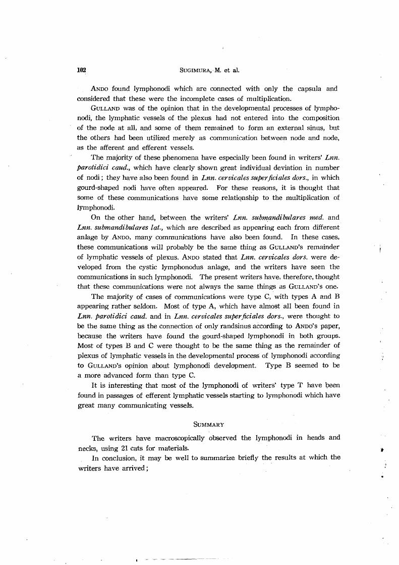
6. The Numbers of the Proposed 3 Types of Lymphonodi
The observed numbers of each type of lymphonodi in each group are set out
in table 5.
In Lnn. 8ubmandihulares med.24 cases among 42 (57.1%) have been taken by type ZT, 17 cases (40.5%) by type Z and only one case (2.4%) by type T.
In Lnn. 8u,bmandibulares lat., the majority of cases (41 cases among 43) (95.4.%) have
been taken by type ZT and only 2 cases (4.6%) by type Z. In . Lnn. 8ubmandihulares med. caud. and Lnn. l'ntbmandibulares lat. caud., all were
found to· be type T.
In Lnn. parotidici cran.,· the majority of cases (29 cases among 41) (70.7%) have been
taken by type Z, 11 cases (26.8%) by type ZT and only one case (2.5%) by type T.
In Lnn. parotidici caud., the majority of cases (128 cases among 145) (88.2%) were type ZT, 12 cases (8.3%) were type Z and 5 cases (3.5%) were type T.
In Lnn. 1'etropharyngici, the majority of cases (42 cases among 43) (97.7%) wet"e found
to be type ZT and only one case (2.3%) was type T. In Lnn. cervicales super ficiales dors., the majority of cases (79 cases among 87) (90.8%)
were type ZT, 6 cases (6.9%) were type T and only 2 cases (2.3%) were type Z. In Lnn. cervicales super ficiales vent., the majority of cases (43 cases among 45) (95.6%)
were type ZT and only 2 cases (4.4%) type T. In Ln. cervicalis. proj., all were type ZT.
TABLE 6. Types of Communication in Each Group of Lymphonodi
TYPES OF COMMUNICATION TOTAL
GROUP OF LYMPHONODI --~--~-.-
Cases % Cases % Cases % Cases ~%
Submandibulares med. 1 100.0 1 100.0
Parotidici cran. 1 100.0 1 100.0 c:
Parotidici caud. 1-4 5 17.2 2 6.9 22 75.9 29 100.0
Cervicales 4 18.2 6 27.3 12 54.5 22 100.0 super ficiales dors.
Submandihulares med. 1 4.8 5 23.8 15 71.4 21 100.0 and lat. .
Submandibulares lat. and 1 50.0 1 50.0 2 100.0 c: lat. caud. (I) (I)
~ 1 20.0 3 60.0 1 20.0 5 100.0 .+0) Parotidici caud. (I) p:)
Parotidici cran. and 1 12.5 1 12.5 6 75.0 8 100.0 caud.
Cervicales 8uper jiciales dorB. and vent. 1 50.0 1 50.0 2 100.0
Total 14 14.6 19 21.3 58 64.1 91 100.0

Studies on the Lymphonodi of Cats
7. About the Communications between Adjacent Lymphonodi by
Lymphatic Vessels Which Allow Reversible Lymph Stream
99
In many cases, it w-as found that communication by lymphatic vessels be
tween adjacent lymphonodi. The forms of communication were 3 types, as
follows;
Type A: the communication consists of only 1 lymphatic vessel through which reversible flow could occur.
Type B: the communication consists of 2 or more lymphatic vessels with
distinct single flowing direction.
Type C: the communication consists of the plexus of lymphatic vessels con
necting among lymphonodi with unconstant flowing directions.
The groups of lymphonodi with each type of the communication are indicated
in table 6.
Out of 91 cases of the communication found in this observation, 29 cases have been
found in Lnn. parotidici caud., 22 cases in Lnn. cervicales super ficiales dors. and only 1 in
each Lnn. submandihu,lares med. and Lnn. parotidici cran., moreover, 21 cases between Lnn. submandwu,lares med. and Lnn. submandwula'res lat., 8 cases between Lnn. parotidici
cran. and Lnn. parotidici caud., 5 cases between Lnn. submandihu,lares lat. and Lnn.
parotidici caud., 2 cases between Lnn. submandihulares lat. and Lnn. submandwulares
lat. cau,d. and also 2 cases between Lnn. cervicales super ficiales dors. and Lnn. cervicales
super ficiales vent.
As indicated in above described results, the communications of lymph among lymphonodi
have been found largely within the writers' group of lymphonodi, while some have been found between lymphonodi of different groups.
The majority of these have been found in Lnn. parotidici caud., Lnn. cervicales super
ficiales dors. and between Lnn. submandibulares med. and Lnn. submandwulares la.t. In
passages of these efferent lymphatic vessels the writers have found many lymphonodi of type T.
In Lnn. parotidici caud., 22 cases among 29 (75.7%) have found to belong to type C, 5 cases (17.2:?;;) to type A and only 2 cases (6.9%) to type B.
In Lnn. cervicales super ficiales dors., 12 cases among 22 (54.5:?;;) have been found to be type C. 6 cases (27.3%) to type Band 4 cases (18.2%) to type A.
In Lnn. submandwulares med., type C has been found only 1 case.
In Lnn. parotidici cran., type B has been found only 1 case.
Out of 21 cases of communication found. between Lnn. sub.mandibulares med. and Lnn. submand~bula1'es lat., 15 cases (71.4%) have been found to belong to type C, 5 cases
(23.8%) to type B and only 1 (4.8%) to type A. In cases of ~mmunication between Lnn. parotidici cran. and Lnn. plt'rotidici caud.,
6 cases among 8 (75%) have been found to be type Cand 1 each case (each 12.5%) to be types A and B.
In communications between Lnn. 8ubmandibulares lat. and Lnn. parotidici caud., 3
cases among 5 (60%) have been found to be type B and each 1 case (each 20~/o),to be types

100 SUGIMURA, M. et al.
A and C.
In c9mmunications between Lnn. submandibulares lat. and Lnn. submandibulares lat. caud., types A and B have been found each 1 case (each 50%).
In communications between Lnn. cervicales super jiciales dors. and Lnn. cervicales 8uper jiciales vent., types B and C have been found each 1 case (each 50%).
DISCUSSION
In this paper, the writers have classified the observed lymphonodi into 10 groups according to their positions.
With regard to Lnn. submandibulares med. eaud. and Lnn. submandibulares lat. eaud., MANABE, REIGHARD et JENNINGS, STROMSTEN, WILLIAM et RICHARD and others have not reported. As each of these unrecorded groups has been found at the rate of 54.8~1,; and 64.3%, the writers have treated them as 1 group. According to previous authors' reports, it was thought these groups of lymphonodi should be included in Lnn. submandibulares med. and Lnn. submandibulares lat. respectively.
Ln. eerviealis profundus, found along V. jugularis interna and beside the trachea, was not also found in above reports relative to cats. From the positions of these lymphonodi, even though found in only 5 sides amongst 42 (11.9%), they are thought to correspond to the anterior, middle or posterior cervical lymphonodus in qogs (SISSON). In cats they were found at all scopes beside neck trachea.
According to the above description, as these lymphonodi were thought to belonged to Le. eervicale profunda in other animals, they were provisionally named Ln. cervicalis profundus.
GULLAND classified lymphonodi into 3 according to the order in which they appear. That is "primary lymphatic gland" which are the first to develop and are all fully developed in foetal life, "secondary lymphatic gland" which are developed in some animals in foetal life, in others not till after birth and "tertiary lymphatic gland" which are formed in adult life on some special occasion, such as during the exceptional activity of some organs or in pathological conditions.
The writers have classified lymphonodi into 3 groups of constant, nearly constan t and inconstant existence according to the difference of ratios at which they exist. Viewing these data it seems clearly that the ratios of the existence of lymphonodi differ according to position.
In the writers' 2 groups of nearly constant and inconstant existence, no definite tendency of their appearance has been found in connection with ages. But that will not allow the conclusion that writers' nearly constant and inconstant groups and GULLAND'S "tertiary lymphatic gland" are the same. At least, writers'
nearly constant group is under suspicion of being abnormal one. The inconstant one which is comparatively small may be the same thing as GULLAND'S "tertiary lymphatic gland".

Studies on the Lymphonodi of Cats 101
In the present paper, the writers have stated that the numbers of lymphonodi
do not particularly vary by individuals, sexes, ages and between right and left
sides, but in their groups of lymphonodi, only Lnn. parotidici caud. has clearly shown a variation of numbers.
For the purpose of determining the source of lymph which enter into each lymphonodus, the starting areas of their afferent lymphatic vessels have been
inspected; it was found that each starting area was correlated respectively to each group of lymphonodi.
It has been found that each lymphonodus acts as a "collecting place of lymph" (writers' type Z), "transporting station of lymph" (type T) or a place with dual
function (type ZT). Each type was shown a different tendency in each of the writers' groups of lymphonodi.
HORII et T AMAKI reported the difference of their so-called primary and secondary lymphonodi.
The present writers have differentiated 3 types of lymphonodi. Their type
Z corresponds to HORn's primary lymphonodi, and types T and ZT to the secondary lymphonodi. Lymphonodi of type T are constantly small and exist
only in contact behind other lymphonodi, of which the efferent lymphatic vessels enter into lymphonodi of type T.
JOB considers that small lymphonodi (so-called "accessory node"), which he had found always in contact with the usually existed lymphonodi in lymphocenter, appear in pathological conditions.
JORDAN et LOOPER stated that such a lymphonodus is found in eontact with
degenerating lymphonodi.
HORII et al. reported that these small nodi are related with the regeneration
of lymphonodi.
SUZUKI and KOIZUMI, using humans and rabbits for material, reported that lymphatic apparatus reaching from small lymphonodus to infiltrations of lymphocytes appears in the fatty tissues of lymphocenter, and further with increase
in age, generally more lymphonodi appear than infiltrations of lymphocytes. If the lymphonodi of present writers' type T were the same thing as the above
described investigator's "accessory node" or small lymphonodi in fatty tissues, it
should be of interest to note the fact that these lymphonodi are very small and
act as only "transporting station of lymph".
In this observation, the writers have noted the communications of lymph
between lymphonodi by lymphatic vessels. According to the styles of the com
munications the writers have classified them into 3 types A, Band C. It was found
that communication exists within writers' one group of lymphonodi, and even between those of other groups.
102 SUGIMURA,~ M. et a1.
ANDO found lymphonodi which are connected with only the capsula and considered that these were the incomplete cases of multiplication.
GULLAND was of the opinion that in the developmental processes of lymphonodi, the lymphatic vessels of the plexus had not entered into the composition of the node at all, and some of them remained to form an external sinus, but the others had been utilized merely as communication between node and node, as the afferent and efferent vessels.
The majority of these phenomena have especially been found in writers' Lnn. paratidici caud., which have clearly shown great individual deviation in number of nodi; they have also been found in Lnn. cervicales .super ficiales dors., in which gourd-shaped nodi have often appeared. For these reasons, it is thought that some of these communications have some relatiqnship to the multiplication of lymphonodi.
On the other hand, between the writers' Lnn. subm,andibulares med. and Lnn. submandibulares lat., which are described as appearing each from different anlage by ANDO, many communications have also been found. In these cases, these communications will probably be the same thing as GULLAND'S remainder of lymphatic vessels of plexus. ANDO stated that Lnn. cervicales dars. were developed from the cystic lymphonodus anlage, and the writers have seen the communications in such lymphonodi. The present writers have, therefore, thought that these communications were not always the same things as GULLAND'S one.
The majority of cases of communications were type C, with types A and B appearing rather seldom. Most of type A, which have almost all been found in Lnn. paratidici caud. and in Lnn. cervicales super ficiales dars., were thought to be the same thing as the connection of only randsinus according to ANDO'S paper, because the writers have found the gourd-shaped lymphonodi in both groups. Most of types Band C were thought to be the same thing as the remainder of plexus of lymphatic vessels in the developmental process of Iymphonodi according to GULLAND'S opinion about lymphonodi development. Type B seemed to be a more advanced form than type C.
It is interesting that most of the lymphonodi of writers' type T have been found in passages of efferent lymphatic vessels starting to lymphonodi which have great many communicating vessels.
SUMMARY
The writers have macroscopically observed the lymphonodi in heads and
necks, using 21 cats for matedals. In conclusion, it may be well to summarize briefly the results at which the
writers have arrived;

Studies on the Lymphonodi of Cats 103
1. The lymphonodi were classified into 10 groups according to position as
follows: Lnn. submandibulares med., Lnn. submandibulares lat., Lnn. submandibulares med. caud., Lnn. submandibulares lat. caud., Lnn. parotidici cran., Lnn. parotidici caud., Lnn. retropharyngici, Lnn. cervicales superficiales dors., Lnn. cervicales superficiales vent. and Ln. cervicalis prof.
2. No numerical difference of lymphonodi has been found by individuals,
sexes, ages or between the right and left sides. About each group of lymphonodi,
however, Lnn. parotidici caud. have shown conspicuous variation in number.
3. Arising from the difference of these existing ratios, the writers' 10 groups of lymphonodi have been divided in 3 groups of constant, nearly constant and
inconstant existence.
4. Each of the starting areas of afferent lymphatic vessels has been corre
lated respectively to each of the writers' grouping of the lymphonodi.
5. The writers have classified lymphonodi into 3 types cf Z, T and ZT
according to the states of their afferent lymphatic vessels and found that these
types appear with different tendency in each group of lymphonodi.
6. In many cases, the writers have found the communication of lymph
among lymphonodi by lymphatic vessels and by these styles of communication
have divided them into 3 types of A, Band C. These types of comm~nication
have been found in writers' groups of lymphonodi, and even between those of
another.
REFERENCES
1) ANDO, S. (1930): Kaibo. Z., 3. 761 (Japanese).
2) ANDREW. (1948): Amer. J. Anat., 82, 104. [HORU1:])].
,3) ASCHOFF, L. (1938-39): Anat. Anz. Erg., 87, 152.
4) BAUM, H. et al. (1908): Anat. Anz., 32, 561-
5) BUNTING, L. (1905): J. Anat. Phys., 39, 56.
6) DAWSON, A. & J. MASUR (1929): Anat. Rec., 42, 46, & Ibw., 44, 143.
7) GODBILLE (1915): Lymphatic Gland in Meat Producing Animals [Jo1)16)].
8) GULLAND, L. (1894): J. Path. & Bact., 2, 447.
9) GYLLENSTEN, L. (1950): Acta Anat., 10, 130 [HORU12)].
10) HEUDORFER, K. (1921): Z. Anat. Entw., 61, 365.
11) HIROTA, K. (1927): Kioto-igalCukai-zasshi, 24, 869 (Japanese).
12) HORII, 1. (1952): Symposium on Hematology, No.6, 15 (Japanese).
13) BaRIl, 1. & S. KOIZUMI (1949): Kaibo. Z., 24, 8 (Japanese).
14) HORII, I. & Y. TAMAKI (1951): Rinpakyu-ni-kansuru-kenkyu, Tokyo I: Japanese).
15) JOB, T. (1915): Anat. Rec., 9, 456.
16) JOB, T. (1922): Amer. J. Anat., 31, 125.
17) JOLLY, J. (1909): Soc. de Biol., 66, 499 & Ibid., 67, 684 [JOB16)].
18) JOLLY, J. (1910): Arch. Anat. Micr., 11 [JOB1b)].

104 . SUGIMURA, M. et al.
19) JORDAN, E. & B. LOOPER (1927): Amer .• J. Anat., 39, 437. 20) KOIZUMI, S. (1948): Report II Division of Anat. Kioto. Univ. [SUZUKI29)].
21) MANABE, S. (1930): Kaiho. Z., 3, 630 (Japanese). 22) MATSUMOTO, T. (1933): Igakukenkyu, 7, 917 (Japanese). 23) NORDMANN, M. (1928): Virch. Arch. Path. Anat. u. Phys., 276, 159. 24) RICHTER, J. (1902): Arch. Mikr. Anat., 60, 469.
25) REIGHARD, J. & H. JENNINGS (1952): Anatomy of the Cat, 3rd Ed., New York. 26) SCHUMACHER, S. (1897): Arch. Mikr. Anat., 48, 145. 27) SISSON, S. (1953): The Anatomy of the Domestic Animals, 4th Ed., Philadelphia
and London. 28) STROMSTEN, A. (1917): Mammalian Anatomy with Special Reference to the Cat
(DAVISON, A), Philadelphia [MANABE2t)].
29) SUZUKI, T. (1952): Acta Scholae Medicinalis Universitatis in Kioto, 30, 174. 30) TAKESHITA, A. (1952): Kumamoto-igakukai-zasshi, 13, 2045 (Japanese). 31) TOMITA, K. (1951): Kaibo. Z., 26, 113 (Japanese). 32) WILLIAM, T. & W. RICHARD (1951): Functional Mammalian Anatomy, 1, New
York.
EXPLANATION OF PLATE
References Common to Figs. 1 and 2
a: Lnn. submandibulares mediales. b: Lnn. submandibulares laterales. c: Lnn. submandibulares mediales caudales. d: Lmi. submandibulares laterales caudales. e: Lnn. parotidici craniales. f : Lnn. parotidici caudales. g: Lnn. retropharyngici. h : Lnn. cervicales super ficiales dorsales. i : Lnn. cervicales super ficiales ventrales. j : Ln. cervicalis profundus. I : V. jugularis communis.
II: V. jugularis externa. III: V. jugularis interna. IV: V. transversa colli. V: V. facialis.
VI : V. transversa hyoidea. VII : V. retromandi bularis.
VIII: V. temporalis communis. IX : V. retroauricularis.
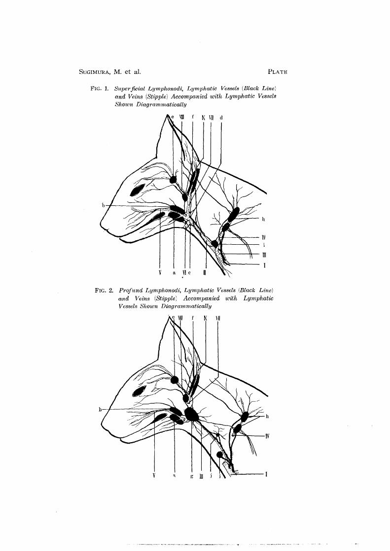
SUGIMURA, M. et al. PLATE
FIG.!. Superficial Lymphonodi, Lymphatic Vessels (Black Line) and Veins (Stipple) Accompanied with Lymphatic Vessels Shown Diagrammatically
t\ VII d
,\;."'I"'---~- ill
v a Vic n
FIG. 2. Profund Lymphonodi, Lymphatic Vessels (Black Line) and Ve'ins (Stipple) Accompanied with Lymphatic Ves.3els Shown Diagrammatically