Embed Size (px)
Citation preview

STUDIES ON THE EXPRESSION OF RESISTANCE OF GOFFEA
SELECTIONS TO HEMILEIA VASTATRIX
by
Teresa Ann Coutinho
Submitted in partial fulfilment of the
requirements for the Doctor of Philosophy degree
in the
Department of Microbiology and Plant Pathology,
University of Natal
Pietermaritzburg
1990

ABSTRACT
Physiological races of Hemileia vastatrix in southern Africa were identified.
Prevalent races were I (2.2%), II (88.9%) and III (2.2%). Six samples could not be
identified. Twelve biotypes of race II were distinguished. In some cases, the
biotypes only occurred in specific regions.
It was established, using fluorescence microscopy, that, in some cases, the
percentages of germinated urediospores that did not form appressoria,
appressorium formation over stomata, and aborted appressoria, were significantly
different between susceptible and resistant selections of the host, and non-hosts.
The sequence of events leading to successful infection was investigated using
scanning electron microscopy (SEM). When a stoma is encountered by a germ
tube tip a uniquely ~haped appressorium forms over one end of the stomatal slit.
A distinct appressorial foot is wedged within the stomatal vestibule. In coffee, a
torpedo-shaped substomatal vesicle initial (SSVI) develops bilaterally from the apex
of the infection wedge, While in bean, the infection wedge protrudes into the
substomatal chamber. The substomatal vesicle (SSV), at 48 hours post inoculation
(hpi) is anchor-shaped. Haustorial mother cells are formed on stubby primary
infection hyphae which curve back onto subsidiary cells. No differences in
appearance of these structures were noted between resistant and susceptible
coffee selections. A much-branched mycelium ramifies through the intercellular
spaces of the mesophyll cells 96hpi. In bean, the SSV began to collapse 48hpi.
Bayfidan ® only slightly suppressed fungal development on the leaf surface.
However, within susceptible tissue, this systemic fungicide had an effect on the

morphology of the fungus. Extracellular material accumulated on the SSVI and the
SSV. The SSV appeared swollen, and disruptions in the vesicle wall was noted.
The discovery of teliospores on locally infected trees led to a SEM study on their
structure, development and germination.
Infection structure formation on the leaf surface, latent period, reaction score and
urediosorus concentration differed between susceptible coffee leaves of different
ages. Generally, mature leaves are more susceptible than very young or old
leaves.
A range of fungicides, mainly systemics, were tested in the field on naturally
infected coffee trees. Various epidemiological and climatic factors influence rust
development in the field. The role of these factors at the fungicide site and in
commercial coffee-growing regions of southern Africa was evaluated.
ii

PREFACE
The experimental work described in this thesis was carried out in the Department
of Microbiology and Plant Pathology, University of Natal, Pietermaritzburg, under
the supervision of Professor F.H.J. Rijkenberg.
All chapters have been prepared as for journal submission, and therefore some
repetition was unavoidable.
There are a few "cited by" references in this thesis. These articles were
unobtainable due to their limited circulation.
I hereby declare that these studies represent original work by the author and have
not been submitted in any form to another University. Where use was made of the
work of others it has been duly acknowledged in the text.
T.A. COUTINHO
iii

ACKNOWLEDGEMENTS
I wish to express my sincere thanks and gratitute to the following people, without
whose co-operation this thesis would not have been possible:
Prof. F.H.J. Rijkenberg for invaluable guidance, advice and his critical appraisal of
the draft manuscripts;
Coffee Growers' Association of South Africa for financial support in the form of a
research grant;
The Council for Scientific and Industrial Research for their generous financial
assistance in the form of a postgraduate bursary;
Drs. C.J. Rodrigues Jr., L. Rijo (CIFC, Portugal), M. St. John Clowes (Tea and
Coffee Research Foundation of Central Africa, Malawi) and G.A. Alvarado
(CENICAFE, Colombia) for seed and cuttings of the various Goftea selections
used in this study;
Mr. S. Terry for advice on fungicide application;
Mr. Vijay Bandu and Mrs. Belinda White of the Electron Microscopy Unit, for their
help, patience and assistance. Mrs. Priscilla Donnelly for her assistance with the
preparation of the plates for this thesis.
Mr. Ken Hosking for the installation of the shade house and watering system;
Fellow postgraduates for useful discussions, and helpful advice;
Cheryl Lennox and Margie Still for their moral support over the past few months;
The coffee farmers, farm managers and representatives of chemical compa"nies for
their invaluable assistance;
iv

My family, for their patience and understanding throughout the course of this
investigation;
and lastly but not least, Michiel van Asch for his continued support and
encouragement throughout this study.
v

FRONTISPIECE
The praises of coffee are sung in the Coffee Cantata "Schweigt stille, plaudet nicht
... " BMV 211, written (1732-35) by J.S. Bach who used the words of Picander
(Christian Friedrich Henrici):
Ei! wie schmeckt der Coffee susse, lieblicher als
tausend Kusse, milder als Muskatenwein
Coffee, Coffee musse ich haben: und wenn jemand
mich will laben, ach so schenkt mir Coffee ein!
which translates roughly into English as:
Oh! how sweet is the taste of coffee, choicer than
a thousand kisses, milder than muscatel wine
Coffee, Coffee must I have: and if anyone
wishes to comfort me, pour me out some coffee.
vi

LIST OF CONTENTS
Page
ABSTRACT
PREFACE iii
ACKNOWLEDGEMENTS iv
FRONTISPIECE vi
LIST OF CONTENTS vi
GENERAL INTRODUCTION 1
LITERATURE CITED 4
CHAPTER 1
IDENTIFICATION OF PHYSIOLOGICAL RACES OF HEMILEIA
VASTATRIX IN SOUTHERN AFRICA 6
INTRODUCTION 6
MATERIALS AND METHODS 7
RESULTS 16
DISCUSSION 21
LITERATURE CITED 25
CHAPTER 2
APPRESSORIUM FORMATION BY HEMILEIA VASTATRIX 28
INTRODUCTION 28
MATERIALS AND METHODS 29
RESULTS 31
DISCUSSION 33
LITERATURE CITED 38
CHAPTER 3
DEVELOPMENT OF INFECTION STRUCTURES OF
HEMILEIA VASTATRIX IN RESISTANT AND
SUSCEPTIBLE SELECTIONS OF COFFEA AND
IN NON-HOSTS
vii
43

INTRODUCTION
MATERIALS AND METHODS
RESULTS
DISCUSSION
LITERATURE CITED
CHAPTER 4
THE EFFECTS OF BAYFIDAN ® GRANULES ON INFECTION
STRUCTURE FORMATION BY HEMILEIA VASTATRIX IN
COFFEA ARABICA CV. CATURRA
INTRODUCTION
MATERIALS AND METHODS
RESULTS
DISCUSSION
LITERATURE CITED
CHAPTER 5
TELIOSPORES OF HEMILEIA VASTATRIX
INTRODUCTION
MATERIALS AND METHODS
RESULTS AND DISCUSSION
LITERATURE CITED
CHAPTER 6
THE EFFECT OF LEAF AGE ON INFECTION OF COFFEA
43
44
47
53
58
61
61
62
65
67
69
73
73
74
77
SELECTIONS BY HEMILEIA VASTATRIX 79
INTRODUCTION 79
MATERIALS AND METHODS 80
RESULTS 83
DISCUSSION 86
LITERATURE CITED 91
viii

CHAPTER 7 . CHEMICAL CONTROL OF THE COFFEE LEAF RUST PATHOGEN,
HEMILEIA VASTATRIX 95
INTRODUCTION 95
MATERIALS AND METHODS 96
RESULTS 100
DISCUSSION 111
LITERATURE CITED 117
APPENDIX 1 121
APPENDIX 2 124
APPENDIX 3 125
APPENDIX 4.1 129
APPENDIX 4.2 131
APPENDIX 4.3 133
APPENDIX 4.4 134
ix
GENERAL INTRODUCTION
Hemileia vastatrix (Berk. et Br.) is the causal organism of leaf rust on the genus
Coftea L. (Javed, 1984). This plant disease was one of the earliest to be studied
sCientifically (Ward, 1882), and, due to its major impact on the world's coffee
industry, it is still the subject of detailed biological investigations.
Penetration is confined to the abaxial leaf surface and results in the formation of
colonies of up to 15mm in diameter. Slight chlorosis of the adaxial surface is
observed. The fungus grows in a radial manner so that mature urediospores are
found in the centre of the affected areas and young spores are produced in
marginal surface tissue. The urediosori are bright yellow to orange in colour. The
tissue in the centre of the affected area becomes necrotic with increasing age. The
economic damage stems from the extensive, premature defoliation of the host
(Waller, 1982). Urediosori are readily attacked by Verticillium lecanii (Zimm.)
Viegas. This hyperparasite was present in all rust samples collected by the present
author, regardless of province, region or site.
The disease is not only of economic importance, but it has a profound social effect
in large areas of the world. Since harvesting of the crop is done manually and the
amount of hand-labour required is determined by the yield, producing a healthy
crop is of paramount importance. In the areas from Panama to Mexico a 20%
infection has been estimated to result in the reduction of labour by as much as 31
million men/day (Muyshondt, 1971, cited by Schieber, 1972).
After oil, coffee is the most important commodity in international trade. It is
produced in approximately 45 countries (Schieber & Zentmyer, 1984). H.
vastatrix can infect all the species of Coftea (Waller, 1982), of which three are
of economic importance, namely C. arabica L., C. canephora Pierre ex
Froehner and C. /iberica Bull ex Hiern (Carvalho et al., 1969). They make up
90%, 9% and 1% of the world's coffee, respectively (Javed, 1984). C. arabica
and C. canephora are grown in the Republic of South Africa, Venda, Gazankulu,
1

Lebowa and KaNgwane. They make up 99.28% and 0.72% respectively of the
total coffee grown in southern Africa (Anon., 1988).
C. arabica is a self-pollinating, allotetraploid species and is the only polyploid
described in this genus. C. canephora (Robusta) and C. liberica are diploid,
cross-pollinating species (Carvalho et a/ ., 1969). Arabica coffee is preferred over
./ all other species because of its superior quality (Van der Vossen, 1985), while
Robusta is known to have a better resistance to rust (Rodrigues et a/. , 1975). C.
liberica has been shown to have a high level of susceptibility (Van der Graaff,
1986). Since Robusta is imported in great quantities for the instant coffee market,
there is a move to increase Robusta plantings in southern Africa (Anon., 1988).
Besides this factor, other reasons for this move include the comparatively low
production costs, the less intensive management required, and its relatively good
resistance to diseases and pests (Anon., 1988).
The earliest report of H. vastatrix in southern Africa was from Natal in 1878
(Rayner, 1960; 1972). By 1904 it was reported to have devastated coffee
growing regions of the Transvaal also (Rayner, 1972). As a result of these
infections, the coffee plantings were completely abandoned in both regions
(Brodrick, 1971). Abandoned trees are still present and have been observed
growing in the wild pathosystems of northern Natal (M.D. Laing, pers. comm.). -In
the 1930s, H. vastatrix was found in many of the warmer areas of southern
Africa, parasitic on indigenous plants of the order Rubiaceae (Gyde, 1932). At
this time, coffee trees still grew in small numbers, but it was found that, if the size
of the plantation was increased, the fungus became established and spread very
rapidly (Gyde, 1932). In the 1960s coffee ,was again planted in Natal and the
disease was observed (Nyenhuis, 1967). In 1969 it was once again recorded in
the Transvaal (Brodrick, 1971). Cultivars of C. arabica resistant to H. vastatrix
are not grown commercially in southern Africa. Thus, coffee rust can still be a
serious problem, especially when chemical control is not included in farming
practices.
- In order to determine the importance of coffee rust to the industry in southern
2
Africa, and to offer advice to giOwers on future plantings of coffee selections
resistant to rust, a survey of the coffee-growing regions was done. This included
collecting rust for race identification, and sampling seed or seedlings from
selections, regarded by the farm managers concerned to be less susceptible, in an
attempt to assess their susceptibility to the rust race/races present in southern
Africa.
Although the infection process of H. vastatrix has been studied since the first
observations by Ward (1882), certain aspects have still not been thoroughly
investigated. Various researchers have reported on factors affecting this process
(Montoya & Chaves, 1981 ; De Jong et al., 1987), however, little work has been
done on the morphology of pre- and post-penetration stages on the susceptible
host. Similarly, there is little information on these processes on and within resistant
coffee selections, non-host plants and fungicide-treated hosts. This study has
attempted to address these deficiencies in our knowledge.
H. vastatrix occasionally produces teliosllO.re-s which will germinate to produce
a basidium and basidiospores (Gopalkrishnan, 1951). The role of basidiospores
is obscure: they. do not infect coffee or any other known host of Hemileia spp.,
and no alternate host of coffee rust has been found. In the literature, reports of
their occurrence are relatively rare. An opportunity to study the morphology of this
stage of the life cycle arose and a scanning electron microscopy study was
undertaken.
Leaf age is known to influence the resistance of Goftea selections to H.
vastatrix. Early reports indicated that younger leaves were more resistant to
infection than older leaves (D'Oliveira, 1957, cited by Kushalappa & Eskes, 1989).
The literature does not provide sufficient evidence to indicate at which stage or
stages in the infection process this resistance mechanism comes into operation.
Infection structure formation as well as subsequent sporulation on leaves of
different ages were therefore investigated.
A field trial investigating the efficacy of chemicals to control coffee rust was
3

included in an attempt to advise growers on which fungicide provides the most
efficient control. The timing of fungicide applications is known to be critical for the
adequate control of coffee rust. In order to provide such information, knowledge
of aspects of the epidemiology of this disease in southern Africa was required. In
depth questionnaires to farm managers, observations and recordings by the . .
present author, as well as a thorough literature search provided an understanding
of the epidemiological situation in this country.
LITERATURE CITED
Anon. (1988) Oorproduksie van Arabica verwag. Landbouweekblad 525,7.
Brodrick H.T.(1971) Coffee and leaf rust. Farming in South Africa 47, 28-
29.
Carvalho A, Ferwerda F.P., Frahm-Leliveld J.A, Medina D.M., Mendes AJ.T. &
Monaco L.C. (1969) Coffee In: Outlines of perennial crop
breeding in the tropics. (Ed. by F.P. Ferwerda & F. Wit) pp. 189-
242. H. Veenman and N.V. Zonen, Wageningen.
De Jong E.J., Eskes AB., Hoogstraten J.G.J. & Zadoks J.C. (1987) Temperature
requirements for germination, germ tube growth and appressorium
formation of urediospores of Hemileia vastatrix. Netherlands
Journal of Plant Pathology 93,61-71.
Gopalkrishnan K.S. (1951) Notes on the morphology of the genus Hemileia.
Mycologia 43,271-283.
Gyde L.M. (1932) Some observations on the genus Hemileia. South African
Journal of SCience 29, 296-300.
Javed Z.U.R. (1984) Leaf rust in Africa and what it means to American programs
In: Coffee rust in the Americas. (Ed. by R.H. Fulton) pp. 15-34.
American Phytopathological Society, St. Paul, Minnesota.
Kushalappa AC. & Eskes AB. (1989) Advances in coffee rust research. Annual
Review of Phytopathology 27, 503-531.
Montoya R. & Chaves G.M. (1981) Effect of temperature and light on germination,
virulence and reproduction period of Hemileia vastatrix Berk. & Br.
4

Tropical Agriculture 7,35998 (Abstract).
Nyenhuis E.M. (1967) Leaf rust attacks coffee in Natal. Farming in South
Africa 42, 11.
Rayner R.W. (1960) Rust disease of Coffee II. Spread of the disease. Wild
Crops 12, 222-224.
Rayner R.W. (1972) Micologia, historia y biologia de la roya del cafeto. Instituto
Interamericano de Ciencias Agricolas de la O.E.A. 94, 18.
Rodrigues C.J. Jr., Bettencourt AJ. & Rijo L. (1975) Races of the pathogen and
resistance to coffee rust. Annual Review of Phytopathology 13,
49-70.
Schieber E. (1972) Economic impact of coffee rust in South America. Annual
Review of Phytopathology 10,491-510.
Schieber E. & Zentmyer G.A (1984) Coffee rust in the western hemisphere.
Plant Disease 68, 89-93.
Van der Graaff N.A (1986) Coffee, Coffea spp. FAO Plant Production and
Protection 70, 49-73.
Van der Vossen H.AM. (1985) Coffee selection and breeding. In: Coffee:
botany, biochemistry and production of beans and beverage.
(Ed. by M.N. Clifford & K.C. Willson) pp.48-96. Croom Helm, London.
Waller J.M. (1982) Coffee rust - epidemiology and control. Crop Protection
1,385-404.
Ward M.H. (1882) Researches on the life history of Hemileia vastatrix, the
fungus of "coffee leaf disease". Botanical Journal 19, 299-336.
5

CHAPTER 1
IDENTIFICATION OF PHYSIOLOGICAL RACES OF
HEMILEIA VASTATRIX IN SOUTHERN AFRICA
INTRODUCTION
In 1932, Mayne presented the first experimental evidence of physiologic
specialization of Hemileia vastatrix Berk. & Br. while working on coffee in India.
His findings necessitated a careful survey of the parasite's specialization
throughout the world, and a search for new sources of resistance in Coftea
arabica L. Dr. A.L.B. d'Oliveira initiated such a programme, and the Centro de
Investigaqao das Ferrugens do Cafeeiro (CIFC) was established in Oeiras, Portugal
in 1955 (Eskes, 1989). Today, 31 rust races have been identified (Bettencourt &
Rodrigues, 1988). According to Rodrigues (1984), 938 rust isolates were obtained
from 37 countries and the number of times each race was identified, expressed as
a percentage of the total number of race cultures established by the CIFC,
indicated that the prevalent races are race II (58.2%), race I (14.4%), race III (8.9%)
and race XV (3.6%). The remaining races constitute about 15% of the total. The
presence of Race II has been reported in 30 out of 33 different world areas where
coffee rust is found, including southern Africa (Rodrigues et al., 1975).
Host genotypes with similar resistance spectra have been classified at the CIFC
into physiological groups or clones. The coffee differentials, routinely used in rust
race identification, are considered as type varieties or clones representing these
different resistance or physiological groups (Bettencourt & Noronha-Wagner,
1971; Rodrigues et al., 1975; Lopes & Godinho, 1976; Eskes, 1989). The
present list of differential hosts consists of clonal lines of five Coftea arabica L.
selections, six tetraploid hybrids of C. arabica x Coftea spp., and six Coftea
spp. selections (Bettencourt & Rodrigues, 1988).
The present survey was undertaken to identify local H. vastatrix races, and use
6

them to screen for the rust resistance of the coffee cultivars commonly grown in
southern Africa. Coffee selections obtained from Malawi, Portugal and Colombia
were also evaluated for resistance, following inoculation with the rust race/races
identified in the present study.
MATERIALS AND METHODS
Rust collection. Rust urediospores were collected from different localities in both
the Transvaal and Natal (Figs. 1, 2 and 3). Urediospores were stored in liquid
nitrogen in gelatin capsules (size 00) placed in Nunc® tubes (1.8 ml). The samples
were obtained from plantations of C. canephora L. and different C. arabica
cultivars. The latter included SL cultivars 14, 28 and 34 (based on appearance
these cultivars appeared to be indistinguishable), Caturra (red- and yellow-cherry
varieties), and Catuai [hybrid between Mundo Nova and yellow Caturra (Anon.,
1987)]. On one estate, Gardenia thunbergia L. was grown in close proximity
to coffee plantings. Rust, similar in appearance to H. vastatrix, was noticed and
collected.
Coffee selection sources. Seeds of a range of Coftea selections were obtained
from the Tea Research Foundation of Central Africa, Malawi, the CIFC, Portugal,
and CENIGAFE, Colombia (Table 1). Cuttings of a range of differentials of C.
arabica (Table 2) were also obtained from the CIFG. According to Dr. L. Rijo
(pers. comm.), these differentials were adequate for the identification of the
majority of coffee rust races. A few of the selections listed in Table 1 belong to
physiological groups different from those used as differentials, and therefore
assisted in the race identification.
Planting conditions for coffee selections. Seeds, seedlings and cuttings were
planted in pre-composted bark medium supplemented at monthly intervals with
N:P:K fertilizer and copper chelate. They were grown under 50% shade at
temperatures between 15 and 30 0G.
7

1 Eastern Transvaal
2 Natal
\
)
Fig. 1 , 2 and 3, ' are detail in Figs, ' ted In m Africa deplc ' ns in southern 'ally cultivated Regia 's commerci where coffee I
8

Fig. 2
o .
CD MHINGA ESTATE
® PHASWANA ESTATE
ZIMBABWE
• Graskop
0) LA ROCHELLE ESTATE
8)WESTFALIA ESTATE
KRUGER
NATIONAL
PARK
,.
M o Z A M B I a U E
Nclspruit
® TOURS ESTATE
® ZOE~NOG ESTATE
(]) BURGERSHALL EXPT. FARM
® SCHOEMANSDAL ESTATE
Eastern Transvaal estates/plantations where coffee is cultivated
9

Fig. 3
ORANGE
FREE
STATE
.LESOTHO
TRANSVAAL
• Newe.slIo
Ladysmilh.
Eslcourl.
Plelerm.rlllburg 0
SWAZILAND
NATAL
Eshowe.
CD ~
0) langer
• S !<a's Kraal
@ • erula'1l
Richard's
Bay
~ 9(8) Umlumbo 10 ~/
11 Port Sh.pslono
TRANSKEI
CD FARM ERICA
o MAYFIELD ESTATE
® ISLAND FARM
o OCEAN LODGE ESTATE
® SHAKA'S KRAAL FIELD STAT.
® OAKFORD PRIORY
0 DRUMMOND FARM
0 ... , __ S,o_--.;..1pO km
® OCEAN VIEW ESTATE
® KIUNDI ESTATE
@ THORNTON FARM
@ ECHO VALLEY FARM
@ BOULDER HILL ESTATE
@ BEAVER CREEK FARM
Natal estates/plantations where coffee is cultivated
10

Table 1 Coffee selections, obtained from various countries, used to evaluate their , resistance/susceptibility to Hemileia vastatrix race/races present in southern Africa
REF. SELECTION PHYSIOLOGICAL INSTITUTE AND COUNTRY OF ORIGIN OF COFFEE CODE GROUP/CLONE* SELECTIONS
A CatillOr 1 A Centro de Investiga~!o das Ferrugens do Cafeeiro, Portugal
B CatillOr 4 A Centro de Investiga~ao das Ferrugens do cafeeiro, Portugal
C S. Agaro J Tea Research Foundation of Central Africa, Malawi
D Geisha C Tea Research Foundation of Central Africa, Malawi
E Caturra E Tea Research Foundation of Central Africa, Malawi
F K7 ? Tea Research Foundation of Central Africa, Malawi
G NC-169 ? Centro Nacional de Investigaciones de cafe, Colombia
H NP-547 ? Centro Nacional de Investigaciones de Cafe, Colombia
I Coffea Hhinga Estate, Transvaal cane~hora ?**
J C. excelsia ?** Burgershall Experimental Station, Transvaal
"I< Groups identified with the aid of a reference table in a research paper by Rodrigues et a/. (1975)
**A specific selection of both C. canephora and C. excelsia are used as differentials (Rodrigues et a/. 1975)
Table 2 Differential selections of Coftea spp. used to identify races of Hemileia vastatrix present in southern Africa
CIFC CODE SELECTION PHYSIOLOGICAL GROUP
87/1 Geisha Group C 128/2 Di11a & A1ghe Group C 110/5 S.4 Agaro Group J
33/1 S.288- 23 Group G 32/1 DK 1/6 Kent's t ype Group D
1343/269 Hybrido de Ti mor Group R 134/4 S.12 Kaffa Group I
11

Leaf disc preparation. Leaf discs have been used with success in determining
resistance to coffee rust (Narasimhaswamy et aI., 1961; Eskes & Toma
Braghini, 1981) and identifying rust races (Ramachandran et al., 1979). The
standardized method described by Eskes (1982a), was used for disc preparation
from third-leaf pairs of the Coftea selections. Leaf discs, 2cm in diameter, were
punched out using a stainless-steel cork borer. The midvein and leaf margins were
avoided. Discs were placed, adaxial surface down, on foam rubber saturated with
tap water in glass trays (29 x 24 x 2.5cm).
Bulking of urediospores. A camel-hair brush (No.1) was used to dust the abaxial
surfaces of leaf discs of cv. Caturra lightly with urediospores collected from the
various localities. Care was taken to prevent possible contamination between
samples. Glass trays, containing these leaf discs, were transferred to a dew
chamber, set at 20°C, and incubated in the dark for 24 h. Following this period,
trays were covered with glass sheets and placed in a constant environment
chamber (Conviron ®) set at 26°C with a 12 h photoperiod. Urediospores from a
single colony of urediosori on the selected leaf disc, from each rust sample, were
removed with a fine brush. The spores were dusted onto three freshly prepared
leaf discs for each rust sample. The discs were incubated as described above.
Again, following the formation of urediosori, urediospores were collected and the
inoculation process repeated until sufficient spores had been accumulated in order
to inoculate the differential leaf discs. During this process, spores from "single
colony" isolations were stored in liquid nitrogen as described previously.
Race identification. Six leaf discs of each differential/selection were inoculated,
as described above, with every rust sample collected and each rust sample was
tested twice. Levels of resistance or susceptibility on the differentials were
determined using the scale described by Eskes & Toma-Braghini (1981) (refer to
Table 3). Once a reaction score was obtained for each differential the results
were compared to Table 4 (from Bettencourt, 1981,cited by Muller, 1984).
Evaluation of the susceptibility of imported Coftea selections to H.
vastatrix. Six leaf discs from each Coftea selection (Table 1) were inoculated,
12

Table 3 A 0 - 9 scale developed by Eskes & Toma-Braghini (1981) to evaluate the reaction types of coffee leaf rust, Hemileia vastatrix
INDEX VALUE
o
1
2
3
4
5
6
7
8
9
CIFC CLASSIFICATION FOR INDIVIDUAL LESIONS
i
fl-,t-
fl,t,o
fl,t,o,o+
fl,t,o,l
fl,t,o,-2
fl,t,o,-3
fl,t,o,-4
t, 2-4
4
DESCRIPTION OF REACTION TYPES FOR LEAVES OR ENTIRE
PLANTS
Immunity, no visible reaction. Minute chlorotic spots, often associated with small tumefactions. Larger chlorotic s~ots, often associated wlth tumefactions. No urediospore production. A mixture of various sizes of chlorotic spots, including very large chlorotic areas. Fewer tumefactions. No urediospore production. A mixture of chlorotic spots of various sizes, with some urediospore formation on large chlorotic lesions*. Sporulation of less than 25% of all lesions. Few tumefactions may occur. Early necrosis of lesions is sometimes observed. As in 4, but with more urediospore formation. Sporulation of less than 50% of all lesions. As in 5, but with increased urediospore production. Sporulation of less than 75% of all lesions. As in 6, but with abundant urediospore production. Sporulation of up to 95% of all lesions. A mixture of lesions with a varying degree of sporulation, sometimes associated with a few tumefactions. Only lesions with abundant sporulation, without marked chlorosis at the lesion border.
* "Lesion" was understood to mean colony of urediosori
Resistar:t [R] = 1, 2 and 3; Moderately Resistant [MR] = 4 and 5; Moderately Susceptible [MS] = 6 and 7; Susceptible = 8 and 9
13

Table 4 Races of Hemileia vastatrix, selected differentials and coffee physiologic groups (after Bettencourt, 1981,cited by Muller, 1984)
PHYSIOLOGIC POSTULATED HOST FACTORS OF RUST RESISTANCE RACE OF RACE HEKILEIA GENOTYPE SH1,5 SH1 SH4,5 SH3,5 SH2,5 SH6 SH1,4 VASTATRIX
Coffea arabica and tetraploid interspecific hybrids (C. arabica x Coffea spp.)
87/1 128/2 110/5 33/1 32/1 1343/269 134/4
COFFEE PHYSIOLOGIC GROUPS
C a J G D R I
I v2,5 R R R R S R R II v5 R R R R R R R III v1,5 S S R R R R R IV v? R R R R R R R VI v? R R R R R R R VII v3,5 R R R KS R R R VIII v2,3,5 R R R S S R R X v1,4,5 S S KS R R R KS XI v? R R R R R R R XII v1,2,3,5 S S R S S R R XIII v5,? R R R R R R R XIV v2,3,4,5 R R S S S R R XV v4,5 R R S R R R R XVI v1,2,3,4,5 S S S S S R S XVII v1,2,5 S S R R R R R XVIII v? R R R R R R R XIX v1,4,? R KS R R R R KS XX v? R KS R R R R MR XXI v? R R R R R R R XXII v5,6 R R R R R S R XXIII v1,2,4,5 S S HS R S R HS XXIV v2,4,5 R R HS R S R R XXV v2,5,6 R R R R S S R XXVI v4,5,6 R R S R R S R XXVII v1,4,6,? R HS R R R HS KS XXVIII v2,4,5,6 R R S R S S R XXIX v5,6,? R R R R R S R XXX v5,? R R R R R R R XXXI v2,5,6,? R R R R S S R XXXII v6,? R R R R R KS R
R = resistant reaction S = susceptible reaction MS = moderately susceptible MR = moderately resistant
14
as described above, with each of the various rust samples, and the reaction types
were assessed using the scale described by Eskes & Toma-Braghini (1981) (refer
to Table 3). The experiment was repeated twice.
Latent period. The latent period was determined on cv. Caturra leaf discs except
where the race/biotype was incapable of infecting this cultivar. In this case the
latent period was determined on the selection which proved to be the most
susceptible to the particular race/biotype. Six leaf discs were inoculated as
described above and used as a single replication. The experiment was repeated
twice. The latent period was taken as the time period from the beginning of
incubation to the time at which 50 per cent of the first urediosori had appeared.
Following their inoculation, discs were incubated as described above.
Evaluation of the susceptibility of Goftea selections, commonly grown in
southern Africa, to H. vastatrix. Four C. arabica selections are commonly
grown in southern Africa, namely, SL 28, Caturra, SL 34 and Catuai (listed in order
of importance). C. canephora is grown on a few estates in relatively small
numbers. Seed/seedling samples were selected on the basis of their reaction to
H. vastatrix in the field and on their rust history. Of interest was the selection of
SL 34 obtained from Drummond. The owner of this estate had previously grown
large quantities of SL 34 down the South Coast of Natal in the 1960s, and due to
a severe rust outbreak had decided to abandon the estate. He selected seed,
which he later planted in Drummond, from one tree that showed no visible
symptoms of rust. At the time of collecting the rust sample, during April, the rust
severity on the Drummond estate was relatively low in comparison to what was
observed on other estates. Twelve leaf discs of such locally grown selections were
inoculated and the disease-reaction type assessed on two different occasions.
The inoculation, incubation and disease assessment methods used are as those
described above.
15

RESULTS
Race identification. All 18 rust samples collected in the Eastern Transvaal from
the seven estates failed to infect any of the differentials (Table 5). From a
comparison between Tables 4 and 5, it is evident that the race/s present in this
area may be one or more of the following : Race II, IV, VI, XI, XIII, XVIII, XXI or XXX.
The differentials obtained were not specific enough to allow further separation of
these races. The sample collected from Gardenia thunbergia failed to infect
any of the differentials indicating that it too may be one of the above races or
another species of Hemileia. Of the 27 rust samples collected in Natal 25 were
incapable of infecting the differentials (Table 6) and the situation as described
above once again applies. Races I and III were identified on samples collected
from the Shaka's Kraal field station and at Oakford Priory, repectively.
Evaluation of the susceptibility of imported Gaftea selections to H.
vastatrix. Following the inoculation of individual leaf discs of the different coffee
selections with the various rust samples, it became evident that if a single race was
present, biotypes existed (Table 7). Rodrigues et a/. (1975), Muller (1984) and
Eskes (1989) noted that only races II, XIII and/or XXX are capable of infecting cv.
Caturra, a member of Physiological Group E. This indicates that all rust samples
collected infecting this cultivar are either one or more of the above races. Races
XIII and XXX are relatively rare, only occurring in the Phillipines and Timor,
repectively; therefore, it is likely that besides races I and III, the other race present
in southern Africa, is race II. The differences in the reaction scores recorded
between isolates of this race, on the Coftea selections (Table 7), may be due to
the presence of biotypes. Dr. C.J. Rodrigues Jr. (pers. comm.) confirmed this
conclusion. The biotypes were given lower case letters and their occurrence in
southern Africa recorded (Fig. 4). The race lie biotype was the most common
followed by lib. Biotypes lIa, c, d, f and 9 were confined to the Eastern Transvaal
while, IIj, k, and m only occurred in Natal. The latent period, even between
biotypes, differed, and their separation was based purely on the reaction score
recorded. Six of the 45 samples were incapable of infecting cv. Caturra (Table 7,
reference code E). This/these race/s could not be distinguished. They may be
16

Table 5 Results of differential testing of races of Hemileia vastatrix collected from various sites and hosts in the Transvaal and independent homelands
COLLECTION HOST DIFFERENTIAL SELECTIONS OF COFFEA SPP.* SITE
87/1 128/2 110/5 33/1 32/1 1343/ 134/4 269
Khinga SL 28 [1]*** 0** 0 0 0 0 0 0 Estate SL 28 [2] 0 0 2 0 0 0 0
Phaslrlana SL 28 0 0 0 0 0 0 0 Estate Gardenia
tbunbergia 0 0 1 0 0 0 0
La Rochelle SL 28 [1] 0 0 0 0 0 0 0 Estate SL 28 [2] 0 0 0 0 0 0 0
Robusta 0 0 0 0 0 0 0
Tours Estate SL 28 [1] 0 0 0 0 0 0 0 SL 28 [2] 0 0 0 0 0 0 0
ZoeJrnog SL 28 0 0 2 0 0 0 0 Estate Caturra 0 0 0 0 0 0 0
Burgershall K7 0 0 0 0 0 0 0 Experimental Catuai 0 0 0 0 0 0 0 Station Caturra 0 0 2 0 0 0 0
Robusta 0 0 2 0 0 0 0 C. excelsia 0 0 0 0 0 0 0
Schoellansdal SL 28 [1] 0 0 0 0 0 0 0 Estate SL 28 [2] 0 0 0 0 0 0 0
* Differentials obtained from the elFe, Oeiras, Portugal ** Mean reaction score (refer to Table 3) on twelve Coftea leaf discs
*** [1] - [2] different sites within a plantation
one or more of the following: Race IV, VI, XI, XVIII or XXI. By evaluating their
reaction to the various C oft ea selection (Table 7) three distinct "races" or possibly
biotypes of one or more of the above, were evident and they were labelled? 1-3 in
the present study. Although the rust sample collected from G. thunbergia failed
to infect C. arabica cultivars or the C. arabica crosses it did infect C.
canephora and to a lesser extent C. exse/sia (Table 7).
17

Table 6 Results of differential testing of races of Hemileia vastatrix collected from various sites and hosts in Natal
* ** ***
COLLECTION HOST DIFFERENTIAL SELECTIONS OF COFFEA SPP.*** SITE
87/1 128/2 110/5 33/1
Ukulinga Caturra 0** 0 0 0 Univeristy Farm Unknown 0 0 0 0
Farm Erica SL 34 0 0 0 0 Caturra 0 0 0 0 Robusta 0 0 2 0 SL 28 0 0 0 0
Island Farm Caturra [lJ* 0 0 0 1 Caturra [2J 0 0 0 0 Caturra [3J 0 0 0 0 Xundo Nova 0 0 0 2
Ocean .Lodge SL 28 [lJ 0 0 0 0 Estate SL 28 [2J 0 0 0 0
Shaka's Kraal SL 28 [1 J 0 0 0 2 Field station SL 28 [2J 0 0 0 0
Caturra 0 0 0 0
Oakford Priory Unknown 9 9 0 0
Drummond Farm SL 34 0 0 0 2
Ocean View SL 28 0 0 0 2 Estate Caturra 0 0 0 0
Kilindi Estate Catuai 0 0 0 0 Caturra 0 0 0 0 SL 28 0 0 0 0
Thornton Farm SL 28 0 0 0 0
Echo Valley SL 28 [lJ 0 0 0 0 Farm SL 28 [2J 0 0 0 0
Boulder Hill SL 28 0 0 0 0 Estate
Beaver Creek SL 28 0 0 0 0 Farm
[1]- [2] = different sites within a plantation
m.ean re~ction score (refer to Table 3) on twe lve Coffea leaf discs Differentials obtained from the CIFC, Oe iras , Portugal
18
32/1 1343/ 269
0 0 0 0
0 0 0 0 0 0 0 0
0 0 0 0 0 0 0 0
0 0 0 0
1 0 0 0 8 0
0 0
1 0
1 0 0 0
0 0 0 0 0 0
0 0
0 0 0 0
0 0
0 0
. -- --
134/4
0 0
0 0 0 0
0 0 0 0
0 0
1 2 2
0
1
0 0
0 0 0
0
0 0
0
2

Table 7 Evaluation of selected Coftea arabica cultivars and C. canephora selections to races of Hemileia vastatrix present in southern Africa
a * **
*** * .... *
ORIGIN OF RACE ORIGIN OF RUST ASSESSMENT SCORE* RACE LATENT SAMPLE
Aa a a a a a a a a a PERIOD
[ESTATE/FARMJ [CV./SELEcrIONJ B C o E F GH I J (days)"
Hhinga SL 28 [1J*** 0 0 6 0 9 4 9 0 4 0 IIa 27 SL 28 [2J 0 0 9 0 9 6 9 0 4 2 IIb 28
Phaswana SL 28 0 0 6 0 9 6 9 3 4 0 lIb 21 Gardenia thunbergia 0 0 0 0 0 0 0 0 9 4 **** 28 (1)
La Rochelle SL 28 [1J 0 0 9 0 9 0 9 0 0 0 IIa 27 SL 28 [2J 0 0 6 0 9 9 9 0 9 1 IIc 20 Robusta 0 0 0 0 9 0 9 0 9 0 lId 27
Tours SL 28 [1] 0 0 3 '3 9 0 9 0 0 0 IIe 27 SL 28 [2] 0 0 3 0 9 4 9 0 0 0 IIe 27
Zoeknog SL 28 0 0 6 3 9 4 9 0 9 0 IIf 25 Caturra 0 0 3 0 9 0 9 0 9 0 lId 28
Burgershall K7 0 0 9 0 9 9 9 3 0 0 lIb 28 Catuai 0 0 0 0 9 0 9 0 0 0 lIe 25 Caturra 0 0 6 4 9 6 9 2 4 1 lIb 20 Robusta 0 0 6 3 9 4 9 0 9 0 IIg 21 C. excelsia 0 0 0 0 9 0 9 0 0 9 IIh 28
Schoemansdal SL 28 [1] 0 0 0 0 9 0 9 0 0 0 IIe 31 SL 28 [2] 0 0 0 4 9 6 9 3 4 0 IIi 28
Ulrulinga Caturra 0 0 0 0 9 0 9 0 0 0 IIe 27 Blue Mountain 0 0 0 0 9 0 9 0 0 0 IIe 27
Farm Erica SL 34 0 0 0 0 9 0 9 0 0 0 IIe 27 Caturra 0 2 0 1 9 7 9 3 0 0 IIi 26 Robusta 2 0 0 0 0 9 0 0 0 0 71 25 (1) SL 28 0 0 0 3 9 0 9 0 0 0 lIe 32 Island Caturra [1] 3 0 0 1 9 5 9 2 0 0 IIi 26 Caturra [2J 0 0 0 0 0 0 0 0 0 0 72 32 (2) Caturra [3J 0 1 0 0 0 0 1 0 0 1 72 32 (2) Mundo Nova 0 0 1 0 0 5 3 3 0 6 73 32 (2) Ocean Lodge SL 28 [1] 0 0 0 0 0 0 0 0 0 0 72 25 (3) SL 28 [2] 0 3 0 3 0 0 0 0 0 0 72 34 (4) Shaka's Kraal SL 28 [1J 2 3 9 2 9 9 9 1 0 7 IIj 27 SL 28 [2J 0 1 0 8 9 9 8 0 0 8 IIj 25 caturra 0 0 0 8 9 9 9 0 0 5 I 25 Oakford Priory Unknown 0 0 9 0 9 3 9 0 9 2 III 20 Drummond SL 34 1 0 0 2 9 0 9 0 0 0 IIt, 32 Ocean View SL 28 2 0 9 0 9 9 9 0 0 3 IIh 23 Caturra 3 0 0 0 8 3· 9 0 0 5 IIk 28 Kilindi Catuai 0 0 7 0 9 0 0 0 0 0 IIe 23 Caturra 0 0 0 0 9 1 9 0 0 0 IIm 32 SL 28 0 0 3 0 9 9 9 0 0 8 IIh 23 Thornton SL 28 0 0 0 0 9 1 9 1 0 7 Ilk 28 Echo Valley SL 28 [1J 0 0 0 0 9 0 9 0 0 0 IIe 34 SL 28 [2J 0 0 0 0 8 0 9 1 0 0 IIe 28 Boulder Hill SL 28 0 0 0 0 9 0 9 0 0 0 lIe 34 Beaver Creek SL 28 0 0 0 0 9 0 9 0 0 0 IIe 32
Refer to Reference code in Table 1
Assessment scale of Eskes & Toma·Braghini (1981) used (refer to Table 3)
Latent pe~iod determined on cv. Caturra [Malawi] or on (1) K7 [Burgershall], (2) Caturra [Kilindi Estate], (3) Catual [Drummond] or (4) Catuai [Kilindi Estate], Sites within a single plantation Species of Hemileia 19

TOTAL NUMBER OF ISOLATES 14
12
10
8
6
4
2
o lIa lib lie lid lie Ilf Ilg Ilh Iii Ilj Ilk 11m III
RUST RACE/BIOTYPE
_ Eastern Transvaal _ Natal
Fig. 4 Races and biotypes of Hemileia vastatrix in southern Africa
Latent period. The latent period, in some cases, differed between isolates of the
same biotype (Table 7). For example, the latent period of biotype lie ranges from
23 to 34 days depending on locality whence it was isolated.
Evaluation of the susceptibility of Gaftea selections, grown in southern
Africa, to H. vastatrix. Refer to Table 8. The highest recorded rating score
on the C. arabica selection SL 28 was obtained following inoculation with race
II biotypes hand m. Biotypes IIj and k were incapable of infecting this selection.
Race III, biotypes IIh and m readily infected SL 34 whereas 1Ij, races ?1 and ?2
failed to infect this cultivar. Cultivar Caturra proved to be the most susceptible to
race II biotypes a and e, and resistant to ? 1, ?2 and ?3. Catuai was equally
susceptible to Race II biotypes a, c, d, e, h, and k, while race ?3, IIj and m failed
to infect this selection. C. canephora selections from Schoemansdal and Island
Estate were resistant to all tested races and biotypes of H. vastatrix. The
remaining Coftea selections were only infected by specific race II biotypes.
20

Table 8 Disease assessment score recorded for selected Gaftea selections commonly. cultivated in the Eastern Transvaal a~d Natal against H. vastatrix races/biotypes present in southern Africa
---------------------------------------------------------------------------------------------------------------------------------------------COFFEE SELECTION oo.IGIN OF COITER
SELECTION RUST RACE/BIOTYPE
ASSESSKEtff SCORE * ------------------ ------------------ --~----;-----;-----;----- ;-----;-----;-----;-----;-----~----~----~-----~-----;-----~-----~-----~--
SL 28 :~~na 4 8 9 8 7 5 9 9 9 8 0 0 9 0 7 0 8
SL 34
caturra
Cattl3i
C. caneiXlora
La Rochelle 0 9 9 9 8 9 9 8 9 9 0 0 9 1 8 0 8 Tours 0 9 9 9 9 9 8 9 9 9 0 0 9 0 7 0 9 Zoeknog 9 9 9 9 9 9 9 7 9 9 0 0 9 0 8 0 9 SchoemanSl:lal 7 7 8 ·9 8 9 8 8 9 9 0 0 9 2 9 0 8 Islarrl Estate 9 9 8 8 9 9 9 9 8 9 0 0 9 1 9 0 9 Kilirrli 8 7 7 8 0 0 8 8 9 3 0 0 9 9 8 8 fnornton 0 4 4 8 8 9 8 8 9 0 0 0 9 8 0 9
K1yfield DrUlWrrl
Zoeknog Islam Estate Kilirrli
Drll!nl!l)nd Kilirrli
La Rochelle furgershall Schoeman SI:la 1 Islarrl Estate Echo Valley
8 9
8 9 o
8 0 7 090
9 9 9 9 9 9 9 0 8
7 9 6 7 9 9 050 0 9 4
999 9 9 9 9 9 7 8 9 9 8 9 0 5 8
o 8 9 0 o 090
o 090 o 990 8 0 8 0
o o
o o o
~
8
o o o
9 9
8 8 9
9 909 9 9 999 0 9 0 5 0 3 979 9 9 8 8 900 900 9
o o o o o
o o o o o
o 0 o 0 o 0 o 0 o 0
o o 0 o 0 o 0 o 0
o o o o o
o 9 o o o
o o o o 3
o 0 o 0 o 0 o 0 o 0
o 0 0 o 0 0 o 0 0 o 0 0 o 0 0
000 004 000 000 000
--------------------------------------------------~-----------------------------------------
* Assessment scale of Eskes & Toma-Braghini (1981) used (refer to Table 3)
DISCUSSION
In order to establish the range of pathogen variation within a defined area, surveys
~ave proved to be a reliable option. Physiological races of many rust fungi have
been successfully identified using this method, for example, P. coronata Cda.
in Canada (Chong & Kolmer, 1990). The ultimate aim of such work is to provide
information of direct relevance to breeding for resistance (Wolfe & Schwarzbach,
1975) and/or in establishing a host gene deployment system (Roelfs, 1984).
21

Results from surveys can also be used to indicate in which region or regions a
cultivar, resistant to a particular race prevalent in that region, can be grown. In
coffee research, the identification of races and new sources of resistance has been
a successful and an ongoing process since 1955 (Eskes, 1989). Researchers,
particularly plant breeders, have exploited gene sources, and resistant material is
now available to the coffee industry (Rodrigues, 1984).
Purification of rust races is usually achieved with single-pustule isolations (Green,
1971). Due to unique nature of urediospore formation of coffee rust, this method
cannot be applied. Monosporic isolations have been attempted, but little success
has been achieved. At the CIFC, infection success for two out of 300 monosporic
cultures has been reported (Carneiro, 1980, cited by Eskes, 1989). Monosoric
isolates have never been tried (Eskes, 1989). Single-colony isolations, used in this
study, to obtain pure cultures was successful. Using a similar technique,
Hoogstraten (1982) reported that from 28 single-colony isolates, obtained from
inoculation with a 50:50 mixture of two races, 27 were pure.
Race identification. Dispite the fact that positive identification of all races could
not be made, it is now clear that physiologic specialization of H. vastatrix does
occur in southern Africa. According to Dr. C.J. Rodrigues, Jr. (pers. comm.) the
assessment score used by the CIFC only characterizes differences in virulence
between races and does not distinguish biotypes. An assessment score
distinguishing between the heterogeneous reaction types frequently found in
coffee, was developed by Eskes & Toma-Braghini (1981), and used successfully
in the present investigation. This scale covers a large range of reaction types and
slight differences recorded may be due to the physiological condition of the host,
or the presence of a race or biotype (Dr. C.J. Rodrigues, Jr., pers. comm.).
Different isolates of the same rust race may have different levels of aggressiveness
(Williams & Owen, 1975; Johnson & Taylor, 1976). Rodrigues (1984) noted
differences in this parameter among isolates of races I, /I and /II of H. vastatrix.
Silva et a/. (1985) reported differences between the aggressiveness of two
isolates of race III and the components of aggressiveness which they recognized
22

included percentage germination, percentage appressoria formed, growth rate,
latent period, etc. They proposed that this variation is unrelated to resistant genes
in the host. Thus there is a possibility that the observed differences between race
II biotypes may be due to differences in aggressiveness.
The appearance of symptoms on G. thunbergia similar to those observed on
Coftea spp. and the failure of urediospores to infect C. arabica (except C.
canephora and to a limited extent C. excelsia) suggested that this fungus is
either a race of H. vastatrix or a species of Hemileia. According to Eskes
(1989), only two known races are incapable of infecting C. arabica and C.
arabica crosses but infect C. canephora readily, namely, VI and XVIII. Dr. C.J.
Rodrigues Jr. (pers. comm.) suggested that the isolate was possibly another
species, H. gardeniae-thunbergia . However, further investigations are
required before this can be verified.
Latent period. The variation in latent period within H. vastatrix race II biotypes
(Table 7) on Coftea selections cannot be satisfactorily explained. The techniques
used in this investigation were standardized; leaves removed from the trees were
of similar age and phYSiological condition, and subsequent to inoculation were kept
under controlled environmental conditions. Differences in the length of the latent
period of rust races have been attributed to leaf age (Eskes & Toma-Braghini,
1982; Parlevliet, 1985; Pretorius et al., 1988), and temperature (Pretorius et al.,
1988; Kushalappa, 1989). In the case of coffee, the latent period differs between
leaves from branches which bear large quantities of fruit and those that bear little
or no fruit (Eskes, 1982b). This factor together with a possible influence of
urediospore density may partially explain the variation in the latent periods.
Variation in the reaction types of coffee commonly cultivated in southern
Africa, and imported selections. Two selections of C. arabica show promise
with regard to their susceptibility to leaf rust, namely, SL 34 and Catuai on the
Drummond and Kilindi Estates, respectively. Both selections were however
susceptible to certain races and biotypes. Generally, the selections of C. arabica
available in southern Africa are highly susceptible to H. vastatrix. Despite the
23

fact that the most popular cultivar, SL 28, is susceptible to most rust
races/biotypes, it has two other major cultural problems (Mr. J . Logan, Coffee
Growers' Association of Zimbabwe, pers. comm.) . Firstly, the tree grows to an
extreme height making harvesting difficult, and, secondly, the berries ripen late in
the year and therefore the plants are not allowed relief from the physiological stress
of bearing prior to flowering. SL 28 has always been grown in southern Africa
despite warnings by Department of Agriculture Officers concerning its susceptibility
to rust (Nyenhuis, 1967). The cultivars Caturra and Catuai are semi-dwarf
varieties; unfortunately they produce berries of a lower quality than the SL varieties
(Anon., 1987). Intensive breeding of C. canephora is presently underway in an
attempt to improve the plantings in this country. Robusta, as it is commonly
known, is resistant to leaf rust. However, certain selections, particularly those
under some form of stress, are susceptible to leaf rust (Coutinho, unpublished
data). The rust samples collected from such material proved to be capable of
infecting specific selections of C. canephora under controlled conditions (refer
to Tables 7 and 8). Thus stress may not be an important factor in the susceptiblility
of C. caneohora to leaf rust. Some C. canephora selections did, however,
prove resistant to all isolates collected in this study (Table 8).
Considerable progress has been made over the past 30 years in the breeding of
material resistant to H. vastatrix. At present, numerous hybrids with Hybrido de
Timor (HOT) as one of the parents, constitute the most advanced resistant material.
available for the control of coffee rust (Varzea et a/. , 1985). Catimor is an
example of such material, and in this investigation proved resistant to all races and
biotypes present in southern Africa. The durability of this resistance is
questionable. A retrospective analysis showed that the resistance of the HOT
common parent has not "broken down" in the preceding 40 years (Varzea et a/. ,
1985). However, there is a move towards breeding for horizontal resistance in
Coftea spp. (Hoogstraten et al. 1982; Van der Graaff, 1986) and some progress
has been made. At present, there are no plantings of resistant selections of C.
arabica in southern Africa and there is an urgent need to introduce resistant
cultivars to the industry.
24

LITERATURE CITED
Anon. (1987) Coffee handbook. (Ed. by W.J.C. Logan & J. Biscoe) pp. 182.
Canon Press, Harare.
Bettencourt A.J. & Noronha-Wagner M. (1971) Genetic factors conditioning
resistance of Coffea arabica L. to Hemileia vastatrix Berk. & Br.
Agronomia Lusitana 31 , 285-292.
Bettencourt A.J. & Rodrigues C.J. Jr. (1988) Principles and practice of coffee
breeding for resistance to rust and other diseases. In: Coffee,
Agronomy (Ed. by R.J. Clarke & R. Macrae) 4, 199-235. Elsevier,
London.
Chong J. & Kolmer J.A. (1990) Virulence and race frequencies of Puccinia
coronata in Canada during 1974-1989 . Phytopathology 80, 1045
(Abstract).
Eskes A.B. (1982a) The use of leaf disc inoculations in assessing resistance to
coffee leaf rust (Hemileia vastatrix). Netherlands Journal of
Plant Pathology 88,127-141.
Eskes A.B . (1982b) Incomplete resistance to coffee leaf rust. In: Durable
resistance in crops (Ed. by F. Lamberti , J.M. Waller & N. van der
Graaff) pp. 291-316. Plenum Press, New York.
Eskes A.B. (1989) Resistance. In: Coffee rust: epidemiology, resistance
and management (Ed. by A.C. Kushalappa & A.B . Eskes) pp. 171-
291. CRC Press, Boca Raton , Florida.
Eskes A.B . & Toma-Braghini M. (1981) Assessment methods for resistance to
coffee leaf rust (Hemileia vastatrix Berk. & Br.). Plant Protection
Bulletin FAO 29, 56-66.
Eskes A.B. & Toma-Braghini M. (1982) The effect of leaf age on incomplete
resistance of coffee to Hemileia vastatrix. Netherlands Journal
of Plant Pathology 88, 215-226.
Green G.J. (1971) Physiologic races of wheat stem rust in Canada from 1919 to
1969. Canadian Journal of Botany 49, 1575-1588.
Hoogstraten J.G.J. (1982) Second technical report on the IAC/ FAO
25

project on durable resistance to coffee leaf rust. lAC,
Campinas, Brazil. pp. 23.
Hoogstraten J.G.J., Eskes AB. & Toma-Braghini M. (1982) Research on
incomplete resistance to coffee leaf rust. ASIC 106 Colloque ,
Salvador, 1982. pp.517-521 .
Johnson R & Taylor AJ. (1976) Spore yield of pathogens in investigations of the
race-specificity of host resistance. Annual Review of
Phytopathology 14, 97-119.
Kushalappa AC. (1989) Biology and epidemiology. In: Coffee rust:
epidemiology, resistance and management (Ed. by A.C.
Kushalappa & AB. Eskes) pp. 13-80. CRC Press, Boca Raton, Florida.
Lopes J. & Godinho I.L. (1976) Physiologic specialization of Hemileia vastatrix
B. & Br. Garcia de Orta , Estudos Agronomicos 3, 13-16.
Mayne W.W. (1932) Physiologic specialization of Hemileia vastatrix . Nature
129, 150.
Muller R.A. (1984) Quelques reflexions a propos de la selection de varietes de
cafeiers resistantes ala rouille orangee (Hemileia vastatrix B. et Br.).
Cafe, Cacao\ The 28, 17-42.
Narasimhaswamy RL. , Namblar N. & Sreenivasan M.S. (1961) Report on work
testing races of leaf disease fungus on coffee selections at Coffee
Research Station, Balehonnur. Indian Coffee 25, 333-336.
Nyenhuis E.M. (1967) Leaf rust attacks coffee in Natal. Farming in South
Africa 42,11 .
Parlevliet J.E. (1985) Resistance of the non-race-specific type. In: The cereal
rusts Vol. II (Ed by AP. Roelfs & W.R Bushnell) pp. 509-510.
Academic Press, Orlando.
Pretorius l.A, Rijkenberg F.H.J. & Wilcoxson RD. (1988) Effects of growth stage,
leaf position and temperature on adult-plant resistance of wheat infected
by Puccinia recondita f.sp. tritici. Plant Pathology 37, 36-44.
Ramachandran M., Sreenivasan M.S. & Vishveshwara S. (1979) Preliminary
survey of physiologic races of coffee leaf rust, Hemileia vastatrix, in
South India. Journal of Coffee Research 9,51.
Rodrigues C.J. Jr. (1984) Coffee rust races and resistance. In: Coffee rust in
26

the Americas (Ed. by R H. Fulton) pp. 41-58. American
Phytopathological Society, St. Paul, Minnesota.
Rodrigues C.J. Jr., Bettencourt AJ. & Rijo L. (1975) Races of the pathogen and
resistance to coffee rust. Annual Review of Phytopathology 13,
49-70.
Roelfs AP. (1984) Race-specificity and methods of study. In: The cereal rusts
Vol. 1 (Ed. by W.R Bushnell & A.P. Roelfs) pp. 132-161 . Academic
Press, Orlando.
Silva M.L., RijoL. & Rodrigues C.J. Jr. (1985) Differences in aggressiveness of two
isolates of race 1\1 of Hemileia vastatrix on the cultivar Caturra of
Coftea arabica. ASIC, 11 8 Col/oque, Lome, 1985. pp.635-645.
Van der Graaff N.A (1986) Coffees, Coftea spp. FAO Plant Production and
Protection 70, 49-73.
Varzea V.M.P., Rodrigues C.J. Jr. & Mexia J.T. (1985) Evaluation of the level of
horizonal resistance to Hemileia vastatrix of some arabica plants of
different physiologic groups when confronted with virulent races. AS I C,
11 8 Col/oque, Lome , 1985. pp.625-633.
Williams RJ. & Owen H. (1975) Susceptibility of barley cultivars to leaf blotch and
aggressiveness of Rhynchosporium secalis races. Transactions
of the British Mycological Society 65, 109-114.
Wolfe M.S. & Schwarzbach E. (1975) The use of virulence analysis in cereal
mildews. Phytopathologische Zeitschrift 82, 297-307.
27

CHAPTER 2
APPRESSORIUM FORMATION BY HEMILEIA VASTATRIX
INTRODUCTION
Many fungal pathogens have developed specialised infection structures for host
plant penetration. One type of such structures is termed the "appressorium", which
can be simple, with very little difference in appearance from the germ tube, or
thick-walled and separated from the germ tube by a septum (Staples & Macko,
1980). The term was introduced by Frank (1883; cited by Emmett & Parbery
1975), who believed the structure to be an adhesive disc. Defining "appressorium"
in terms of morphology only, is regarded by Emmett & Parbery (1975) as
inadequate and inaccurate, and, in their opinion, its definition must be qualified by
stating that it gives rise to infection.
Germinating urediospores of rust fungi cannot usually achieve host penetration
directly, and successful infection depends on the formation of an appressorium
over a stoma. In the genus Coftea, the stomata are confined to the abaxial leaf
surface. Thus, successful infection by Hemileia vastatrix Berk. & Br. can only
occur on this surface (Chinnappa & Sreenivasan, 1968; Harr & Guggenheim,
1978). Studies by Harr & Guggenheim (1978) indicated that the appressorium of
H. vastatrix is unusual in that a "vesicle" is present on its dorsal surface.
Considerable advances have been made recently in the understanding of the
processes involved in infection site recognition (Staples et al., 1985; Hoch &
Staples, 1987; Mendgen et al., 1988). The role of physical and chemical stimuli
in these recognition mechanisms is well documented in the literature (Wynn, 1976;
Grambow & Riedel, 1977; Staples et al. , 1983). In the H. vastatrix-Coftea
interaction the factor, or factors, operating in leaf-surface recognition is/are
unknown. In this study, the reaction of H. vastatrix to leaf surfaces was
examined. A morphological study of appressorium formation on leaves of the host,
28

Coftea arabica L., and a non-host, Phaseolus vulgaris L. was conducted.
MATERIALS AND METHODS
Planting material. Seeds of C. arabica cv. Caturra were obtained from the Tea
Research Foundation of Central Africa, Malawi. Following germination, seedlings
were grown under 50% shade at temperatures between 15 and 30°C in pre
composted pine bark medium supplemented monthly with N:P:K fertilizer and
copper chelate. Intact, detached leaves of P. vulgaris cv. Pinto 650 seedlings,
grown by the Crop Improvement Research Unit, Pietermaritzburg, under
greenhouse conditions (20 to 30°C), were also used in this investigation.
Leaf disc/intact detached leaf preparation. The standardized method,
described by Eskes (1982), was used for leaf disc preparation. Third-leaf pairs of
three-year-old Coftea seedlings were gently rinsed under a stream of tap water
in order to remove soil debris. Leaf discs, 2cm in diameter, were punched out
using a stainless-steel cork borer. The midvein and leaf margins were avoided.
Six Coftea leaf discs and two intact detached leaflets of P. vulgaris were used
for each replication and the experiment was repeated four times. Discs and leaves
were placed, adaxial surface down, onto foam rubber saturated with tap water in
glass trays (29 x 24 x 2.5cm) and inoculated as described below. To obviate
morphogenetic effects induced by gravity, discs were initially attached to the
underside of the lid of a glass tray with double-sided tape arid inoculated. This
practice was discontinued when it was found that the morphology of the
appressorium on such material did not differ from those formed on discs with the
abaxial surface uppermost.
Leaf disc and intact leaf inoculations. Urediospores of H. vastatrix were
freshly collected from infected trees. They were lightly dusted onto the abaxial
surface of C. arabica leaf discs and intact P. vulgaris leaflets with a camel
hair brush (No.1). Glass trays were transferred to a dew chamber, set at 20°C and
incubated in the dark for 24h. Following this period, trays were covered with glass
29

Plate 1
Germination of urediospores of Hemileia vastatrix on the leaf surface of
Coftea arabica, and appressorium formation
Fig. 1 Random extension of urediospore [U] germ tube 24hpi. Appressorium
[A] has formed over a stoma. Note that the germ tube extends over a
closed stoma and by-passes another [arrows]
Fig. 2 Entangled germ tubes [GT] from several urediospores with several
appressorium-like structures [arrows] 24hpi
Fig. 3 Appressorium [A] at the raised lips of the stoma 8hpi. Note the inner
stomatal pore at the base of the vestibule [arrow]

20 pm

sheets and placed in a constant environment chamber (Conviron ®) at 26°C with a
12 h photoperiod.
Light microscopy. At 24 hours-post-inoculation (hpi) three pieces (ca. 5 x 5mm)
of two of the inoculated Goftea leaf discs, and three pieces from two intact P.
vulgaris leaflets, were used for this investigation. This material was fixed in 3%
glutaraldehyde in 0.05M sodium cacodylate buffer (pH = 6.8-7.2) overnight. The
tissue was dehydrated through a graded water:ethanol:tertiary-butanol series and
infiltrated with liquid paraffin and wax from Histosec ® pastilles as described by
Jensen (1962). The embedded material was sectioned at 10 J.Lm , using a rotary
microtome and stained with safranin - fast green following the removal of the wax
with xylene. Sections were viewed with a Zeiss photomicroscope and the stomatal
complexes drawn to scale.
Scanning electron microscopy (SEM). Leaf material was sampled at 2,4, 8 and
24hpi. One Goftea leaf disc and two pieces from each intact P. vulgaris leaflet
were cut at each sample time and from each replication. Each leaf disc was cut
into three pieces (ca. 5 x 5mm). Leaf samples were either viewed directly using an
Emscope ® 8P 2000 cryo-apparatus .orfixed in 3% glutaraldehyde in 0.05M sodium
cacodylate buffer [pH = 6.8-7.2], rinsed in buffer, post-fixed in 2% osmium
tetroxide and dehydrated in an ethanol series. The specimens were critical point
dried (CPO) using a Hitachi ® HCP-2 with carbon dioxide as transition fluid, and
mounted on metal stubs. Prior to coating, some metal stubs with mounted leaf
specimens were gently pressed onto other stubs coated with double-sided tape.
In so doing, the appressoria were stripped from the leaf surface, permitting
examination of both dorsal and ventral surfaces. At 24hpi the infection structures
from three leaf pieces were removed from the leaf surface with double-sided tape
in a similar manner and viewed using cryo-SEM. The tissue was gold/palladium
coated in a Polaron ® sputter coater. Infection structures were viewed with a
Hitachi ® 8-570 operating at either 5, 8 or 10 kV. Measurements of infection
structures were made from SEM micrographs.
30

Plate 2
The appressorium of Hemiieia vastatrix over the stoma of the host, Coffea
arabica
Fig. 1 Appressorium [A] on a germ tube [GT] branch 24 hpi. The appressorial
wedge [AW] is within the stomatal vestibule
Fig. 2 Appressorium [A] terminally delimited from the germ tube [GT} by a
septum [arrow] 24hpi. The appressorial wedge [AW] is again
discernible. Opposite the collapsed germ tube a protruberance [P], cut
off from the appressorium by a septum [arrow], is visible
Fig. 3 Terminal appressorium [A] on the stoma 24hpi. The appressorial
wedge [AW] is visible within the stomatal vestibule. A septum [arrow]
cuts off the germ tube [GT] from the appressorium. No appressorial
protruberance is visible
Fig. 4 Three appressoria [A] over a stoma 24hpi

...

RESULTS
Light microscopy. There are marked differences between the structure of
stomata of C. arabica and P. vulgaris leaves (Figs. 1 A and B). In the former
case, the subsidiary and guard cells are elevated above the epidermal cells; this
is not the case in P. vulgaris leaves. In both Coffea and P. vulgaris,
prominent stomatal lips are raised above the stomatal slit and measure
approximately 4J,Lm and 2J,Lm (n = 10) in height, respectively. The vestibule is a
small chamber which separates the stomatal lips and inner stomatal pore.
Germination. At germination (4hpi), germ tubes (dia. 4.20J,Lm, n = 10) extrude
through germ pores of the urediospore wall. As many as five germ tubes can be
formed from a single urediospore, but only one elongates and extends randomly
over the epidermis, in some cases past or over closed stomata [Plate 1 Fig.1].
Exploratory branches form along its length. On a few occasions, particularly when
the inoculum concentration was high, the germ tubes of several urediospores were
seen to have become entangled causing a mass of appressorium-shaped
structures [Plate 1 Fig. 2]. Fungal morphology at germination was similar on host
and non-host tissue. However, germ tubes were relatively short on P. vulgaris
leaflets in comparison to those on C. arabica leaves.
Appressorium formation on host and non-host tissue. The formation of
appressoria and their shape was similar on both host and non-host tissue.
However, appressoria formed only sporadically on non-host tissue. They were first
observed, in both cases, at 8 hpi.
When the raised subsidiary cell of coffee is encountered by the tip of either a side
branch or the main axis of the germ tube, the tip swells and an appressorium is
formed [Plate 1 Fig. 3]. The near-spherical appressorium usually develops on one
end of the stomatal slit. Following the formation of the appressorium, it is cut off
from the germ tube by a septum which is formed approximately 6J,Lm from the edge
of the appressorial swelling. A wedge-shaped structure develops from the base
of the appressorium into the vestibule of the stoma [Plate 2 Fig. 1, 2 and 3; Plate
31

Plate 3
The upper and lower surfaces of appressoria of Hemileia vastatrix on, and
removed from, the leaf surface of Coftea arabica 24hpi
Fig. 1 Cryo-scanning electron microscopy of the appressorium [A] . The
appressorial wedge [AW] and the protruberance [P] opposite the
collapsed germ tube, are visible [arrow]
Fig. 2 The lower surface of the appressorium [A]. The appressorial wedge
[AW] and the protruberance [P] are noticeable
Fig. 3 Appressorial wedge [AW] produced . on the lower surface of the
appressorium [A]. Note the appressorial wedge has tapered to form the
infection wedge [white/black arrow], and the residue of some plJ1:atively
adhesive material on the appressorium [black arrow]
Fig. 4 A lateral view of an appressorium [A], appressorial wedge [AW] and
infection wedge [arrow]

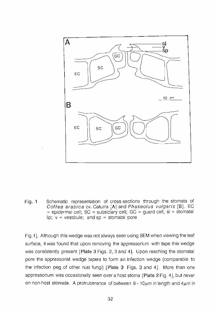
Fig. 1
A
EC
10 pm
8
Schematic representation of cross-sections through the stomata of Goftea arabica cv. Caturra [A] and Phaseolus vulgaris [8]. EC = epidermal cell; SC = subsidiary cell; GC = guard cell; sl = stomatal lip; v = vestibule; and sp = stomatal pore
Fig. 1]. Although this wedge was not always seen using SEM when viewing the leaf
surface, it was found that upon removing the appressorium with tape this wedge
was consistently present [Plate 3 Figs. 2,3 and 4]. Upon reaching the stomatal
pore the appressorial wedge tapers to form an infection wedge (comparable to
the infection peg of other rust fungi) [Plate 3 Figs. 3 and 4]. More than one
appressorium was occasionally seen over a host stoma [Plate 2 Fig. 4], but never
on non-host stomata. A protruberance of between 9 - 10J..Lm in length and 4J..Lm in
32

Plate 4
Lower surfaces of appressoria and germ tubes of Hemileia vastatrix
removed from the leaf surface of Coftea arabica 24hpi
Fig. 1 An enlargement of the insert, Fig. 1b, in Fig . 1a depicts some remnants
of putatively adhesive material [arrows] on the appressorium [A] but not
on the germ tube [GT]
Fig. 2 An enlargement of Fig. 1b illustrates the absence of the putatively
adhesive material from the urediospore [U] and germ tube [arrows]

~.
diameter is often found on the appressorium opposite the collapsed germ tube
[Plate 2 Fig. 2; Plate 3 Fig. 1]. Appressoria without such a protuberance are also
formed [Plate 2 Fig. 3]. Remnants of this protrusion are seen on the surface of
stripped appressoria [Plate 3 Fig. 2].
The lower surfaces of the urediospore, germ tube and appressorium were viewed
upon their removal from the host leaf surface with tape [Plate 4]. Remnants of
putatively adhesive material are absent from the germ tube of both CPD- and cryo
SEM-prepared material [Plate 4 Figs. 1 and 2]. However, remnants were
observed on the appressorium of CPO leaf material [Plate 4 Fig. 1]. Following the
removal of the infection structures, the leaf surface was viewed with cryo-SEM and
no trace of germ tube pathways could be detected.
The number of stomata on leaves of C. arabica cv. Caturra was counted with
the aid of scanning electron microscopy. The mean number of stomata per mm2
was found to be 117 (n = 10).
DISCUSSION
Germ tubes of rust fungi adhere closely to the cuticle of the host in order to
respond to the topographical features essential for growth across leaf epidermal
cells (Wynn, 1981). Directional germ tube extension increases the ability of the
germ tube to locate a stoma more efficiently by reducing wandering (Lewis & Day,
1972). This process is well documented inthe literature. P. graminis f.sp. tritici
Erikss. & E. Henn. germ tubes become orientated perpendicularly to leaf venation
thereby increasing their chance of locating stomata (Lewis & Day, 1972; Lennox
& Rijkenberg, 1989). Gradients in pH at the leaf surface also influence the direction
of germ tube growth of Uromyces viciae-fabae (Pers.) Schroet. (Edwards &
Bowling, 1986).
H. vastatrix germ tubes appear to lack directional growth [Plate 1 Fig. 1].
According to Wynn (1976), random growth may be due to the lack of close
33

adhesion between germ tubes and the leaf surface. This may explain the extensive
exploratory branching of the germ tube of H. vastatrix in attempting to locate a
stoma. In the P. graminis f.sp. tritici/Triticum aestivum L. interaction,
appressorium formation has been found to be negatively correlated with the
degree of branching (Broyles, 1955, cited by Emmett & Parbery, 1975). Niks
(1990) observed a negative correlation between germ tube length of P. hordei
Otth. on Hordeum vulgare L. and the chance of success in the establishment
of a colony, since the formation of a long germ tube and exploratory branches
decreases the amount of energy available to infect the host. The germ tubes of H.
vastatrix are exceptionally long compared to those of other rust fungi, for
example, P. graminis f.sp. triUci (Lennox & Rijkenberg, 1989). Ferreira (1988)
reported that germ tubes of U. transversalis (Thum.) Winter which failed to
locate stomata often reached considerable length. The observation in the present
study that germ tubes of H. vastatrix appear to randomly extend over the leaf
surface is contrary to what would be expected, as there are "a large number of
stomata per square millimetre on C. arabica cv. Caturra leaves. Counts of
stomata made in this study were slightly lower than the 167/mm 2 recorded by
Franco (1939) for an unnamed C. arabica cultivar.
The presence of putative extracellular adhesive material at the interface between
germ tubes and the waxy layer of the host, has been demonstrated on germ tubes
of Puccinia coronata Corda (Onoe et al., 1972, cited by Staples & Macko,
1980), Uromyces appendiculatus Pers. Unger (Epstein et aI., 1987).
Puccinia graminis f.sp. tritici (Harder et al., 1985) and Puccinia
recondita Rob. & Oesm. (C.A. Crookes, pers. comm.). However, the presence
of this material has not been demonstrated consistently. According to Chaubal
(1987), extracellular mucilage was only observed on germ tubes of Puccinia
sorghi Schw. when a cationic detergent or a cationic stain was added to the
fixation solutions used in electron microscopy. The absence of such material on
the germ tubes of H. vastatrix [Plate 4], using cryo-SEM, indicates that the germ
tube must utilize another method of remaining attached to the leaf surface. The
method used by H. vastatrix may involve inter-molecular forces between germ
tube wall components, and host wall. The observation, by the present author, that
34
the abaxial surfaces of C. arabica leaves lack wax crystals, fails to explain the
absence of adhesive material on the H. vastatrix germ tube. Although the leaves
of Phaseolus vulgaris also lack wax extrusions, the germ tubes of U.
appendiculatus are reported to possess extracellular material that may be
involved in binding germ tubes to an inductive surface (Epstein et aI., 1985;
Epstein et aI., 1987). Wynn & Staples (1981) showed that when rust
urediospores of P. graminis f.sp. tritici, P. recondita, P. hordei Otth. and
P. sorghi Schw. were incubated on waxless leaves of their hosts, the germ tubes
neither adhered to the surface nor to the stomata, and consequently, penetration
did not occur.
Appressoria develop in response to stimuli, the nature of which differs in the
different rust species (Wynn & Staples, 1981). In P. recondita, a topographical
stimulus is sufficient to induce appressorium formation (Dickinson, 1970). In C.
arabica the only morphological surface feature that distinguishes the stomata
from the remainder of the leaf surface are the raised subsidiary and guard cells.
These raised areas may well act as thigmotropic stimuli inducing the formation of
H. vastatrix appressoria, although, not infrequently, germ tubes are seen to
traverse stomatal slits. The stomatal guard cells as well as the ridges around the
stomatal opening are thought to provide thigmotropic stimuli inducing the formation
of appressoria of U. appendiculatus (Wynn, 1976). An appressorium
apparently forms when the germ tube tip encounters a stoma. The stomatal lips
of both P. vulgaris and Coftea are prominent (Fig. 1); it is therefore likely that
the stimulus for appressorium formation in H. vastatrix is physical. Dimensions
of the topographical features which determine both germ tube orientation and
appressorium formation have previously been determined (Dickinson, 1949).
Hoch et a/. (1987) defined the exact size of the signal which induced
appressorium formation of U. appendiculatus. This signal was found to be a
ridge or groove in the substrate surface that raises or lowers the elevation by
0.5J..'m. Measurements of the diameter of the germ tube (4.20jJm) of H. vastatrix
correspond to the elevated height of the stomatal lips (4jJm); and when the tip of
a germ tube comes into contact with another germ tube, appressorium-like
structures form [Plate 1 Fig. 2]. This implies that ridges on the surface of
35

approximately 4J..Lm may act as a stimulus inducing appressorium formation in H.
vastatrix.
The method by which the germ tube perceives thigmotropic stimuli is not well
understood. Staples & Hoch (1982) first suggested that the sensing mechanism
involved elements of the cytoskeleton of germ tubes, facilitating reception and
transmission of information about the surface of the host to the nucleus. As is the
case with most actively growing fungi, apical vesicles are observed to be
positioned in the tip of the germ tube and become dispersed during the early
stages of appressorium formation (Staples & Macko, 1980). According to Staples
eta I. (1985), these apical vesicles become positioned in the germ tube at its
interface with the leaf surface thus probably causing the swelling of the germ tube
tip. According to Rijo & Sargeant (1984), vesicles are observed in large numbers
throughout the germ tube of H. vastatrix, particularly near the plasmalemma and
the apex of the tube. The fact that appressoria of this fungus are not consistently
produced at the tip of a germ tube indicates that the process conditioning swelling
of the appressorium in the germ tube of H. vastatrix is not solely associated with
the tip, and that apical vesicles may possibly also arise in a non-apical position.
In P. graminis f.sp. tritici, a chemical, acrolein, produced by the urediospores,
will induce appressorium formation (Macko et aI., 1978). A chemical stimulus
induced by the host rather than by the rust might also be involved. Grambow &
Riedel (1977), and Grambow & Grambow (1978), found that phenols extracted
from the epicuticular wax and cell walls of T. aestivum leaves induced infection
structures of P. graminis f.sp. tritici in conjunction with volatile substances
from the leaf. The role of chemicals in appressorium induction by H. vastatrix
were not investigated in the present study. However, according to Ablanque et a/.
(1988), quantitative and qualitative comparisons of the waxes of different Goftea
selections showed differences between those susceptible and those resistant to
H. vastatrix .
The morphology of the appressorial complex, the appressorium and wedge of H.
vastatrix, is unique. The observations made during the present study confirm
36

· those reported by Harr & Guggenheim (1978). They, however, regarded the
adhering structure, which is termed the appressorial wedge in the present study,
as the appressorium and called what is here referred to as the appressorium, a
dorsal vesicle. To retain the nomenclature used for other rusts, the present author
regards the initial swelling of the germ tube to be the appressprium. One can only
speculate on the function of the appressorial wedge. H. vastatrix is only capable
of infecting the abaxial leaf surface, therefore, the appressorial wedge may have
an attachment role in the absence of adequate adhesive jbinding forces. It is
possible that, to guarantee penetration of the stomatal pore, the appressorium
requires a firm hold and the wedge anchors the appressorium in the stomatal
vestibule. If this is true then the structure here called an appressorium is unique
when compared to appressoria in other rusts.
A protruberance on the appressorium on the side opposite to the germ tube [Plate
2 Fig. 2] has been noticed previously by Kapooria & Mendgen (1985) in the
Uromyces fabae (Pers.) de BaryjVicia faba L. interaction. The appressoria
of this fungus, similarly to those of H. vastatrix, are occasionally delimited by two
septa, and the part of the germ tube confined by the septa enlarges forming the
appressorium. They offered no explanation for its role. Observations imply that the
sensing mechanism of H. vastatrix germ tubes is located at a distance from the
germ tube tip and only once this region reaches the stimulus is an appressoriu~
induced. According to Hoch & Staples (1987), "a germ tube frequently overgrows
the Signal by 5 - 20 J,£m before growth ceases". The patterns of appressorium
development observed in this investigation were consistent on host and non-host
tissue, suggesting that the observed morphology is genetically controlled.
Physical factors, such as temperature, water availability, and light intensity, may
playa role in induction of, and variation in, the morphology of the appressoria of H.
vastatrix. According to De Jong et al. (1987), appressorium formation of H.
vastatrix occurred more rapidly at 13 to 16°C, temperatures lower than those
required for germination. The shape of appressoria was affected by temperature;
at temperatures beween 13 to 19°C they were torpedo-shaped or roundish,
whereas at higher temperatures their shape was predominantly irregular in
37

appearance. In this study, appressoria were induced at a constant temperature of
20°C and no variation in appressorium shape could be found.
More than one appressorium over a stoma have been reported for other host
pathogen combinations (Niks, 1981; Falahati-Rastegar et al., 1983; Lennox &
Rijkenberg, 1989). In a study conducted by Torabi & Manners (1989) the authors
proposed that the proportion of appressoria of P. recondita resulting in
successful penetration was greater when two or more appressoria occurred over
a stoma.
Recognition of stomata by germ tubes of rust fungi occurs with a high degree of
precision (Heath, 1977). Despite the vast amount of information on leaf surface
recognition, much is still unknown. In the present study, the involvement of contact
stimuli in the induction of appressoria by H. vastatrix is presented.
LITERATURE CITED
Ablanque E., Tabacchi R. & Zamudio V. (1988) Contribution to the study of
cuticular waxes of coffee leaves. In: Twelfth international
scientific colloquium on coffee. Horticultural Reviews 59,
6166 (Abstract).
Chinnappa C.C. & Sreenivasan M.S. (1968) Cytology of Hemileia vastatrix.
Caryologia 21, 75-82.
Chaubal R. (1987) Adhesion and g~owth away from surfaces by urediniospore
germ tubes of Puccinia sorghi. Ph.D. thesis, University of Georgia,
Athens.
De Jong E.J., Eskes A.B., Hoogstraten J.G.J. & Zadoks J.C. (1987) Temperature
requirements for germination, germ tube growth and appressorium
formation of urediospores of Hemileia vastatrix. Netherlands
Journal of Plant Pathology 93,61-71.
Dickinson S. (1949) Studies in the physiology of obligate parasitism. I. The stimuli
determining the direction of growth of the germ tubes of rust and mildew
38

spores. Annals of Botany (London) (N.S.) 31,89-104.
Dickinson S. (1970) Studies in the physiology of obligate parasitism. VII. The
effect of a curved thigmotropic stimulus. Phytopathologisc.he
Zeitschrift 69, 115-124.
Edwards M.C. & Bowling D.J.F. (1986) The growth of rust germ tubes towards
stomata in relation to pH gradients. Physiological and Molecular
Plant Pathology 29, 185-196.
Emmett R.W. & Parbery D.G. (1975) Appressoria. Annual Review of
Phytopathology 13, 147-167.
Epstein L., Laccetti L., Staples R.C. & Hoch H.C. (1987) Cell-substratum adhesive
protein involved in surface contact responses of the bean rust fungus.
Physiological and Molecular Plant Pathology 30, 373-388.
Epstein L., Laccetti L., Staples R.C., Hoch H.C. & Hoose W.A. (1985) Extracellular
proteins associated with induction of differentiation in bean rust
uredospore germlings. Phytopathology 75, 1073-1076.
Eskes A.B (1982) The use of leaf disc inoculations in assessing resistance of
coffee to Hemileia vastatrix. Netherlands Journal of Plant
Pathology 88, 127-141.
Falahati-Rastegar M., Manners J.G; & Smartt J. (1983) Factors determining
results of competition between physiologic races of Puccinia hordei.
Transactions of the British Mycological Society 81,233-239.
Ferreira J.F. (1988) A study on the ultrastructure and control of the Gladiolus rust
pathogen, Uromyces transversalis. Ph.D. thesis, University of Natal,
Pietermaritzburg.
Franco C.M. (1939) Relation between chromosome number and stomata in
Goftea. Botanical Gazette 100, 817-827.
Grambow H.J. & Grambow G.E. (1978) The involvement of epicuticular and cell
wall phenols of the host plant in the in vitro development of Puccinia
graminis f.sp. tritici. Zeitschrift fur Pflanzenphysiologie 90,
1-9.
Grambow H.J. & Riedel S. (1977) The effect of morphogenically active factors
from host and nonhost plants on the in vitro differentiation of infection
structures of Puccinia graminis f.sp. tritici. Physiological and
39

Molecular Plant Pathology 11, 213-224.
Harder D.E., Chong J., Rohringer R & Kim W.K. (1985) Structure and
cytochemistry of the walls of urediospores, germ tubes and appressoria
of Puccinia tritici graminis. Canadian Journal of Botany 64,
476-485.
Harr J. & Guggenheim R (1978) Contributions to the biology of Hemileia
vastatrix I. SEM-investigations on germination and infection
of Hemileia vastatrix on leaf surfaces of Coftea arabica.
Phytopathologische Zeitschrift 92,70-75.
Heath M.C. (1977) A comparative study of non-host interactions with rust fungi.
Physiological and Molecular Plant Pathology 10, 73-88.
Hoch H.C. & Staples R.C. (1987) Structural and chemical changes among the
rust fungi during appressorium development. Annual Review of
Phytopathology 25,231-247.
Hoch H.C., Staples R.C., Whitehead B., Comeau J. & Wolf E.D. (1987) Signaling
for growth orientation and cell differentiation by surface topography in .
Uromyces. Science 235, 1659-1662.
Jensen W.A (1962) Botanical histochemistry. Principles and practice.
pp. 88-90. W.H. Freeman and Company, San Francisco.
Kapooria RG. & Mendgen K. (1985) Infection structures and their surface
changes during differentiation in Uromyces fabae.
Phytopathologische Zeitschrift 113,317-323.
Lennox C.L. & Rijkenberg F.H.J. (1989) Scanning electron microscopy of infection
structure formation of Puccinia graminis f.sp . tritici in host and
non-host cereal species. Plant Pathology 38, 547-556.
Lewis B.G. & Day J.R (1972) Behaviour of uredospore germ-tubes of Puccinia
graminis tritici in relation to the fine structure of wheat leaf surfaces.
Transactions of the British Mycological Society 58, 139-145.
Mendgen K., Schneider A, Sterk M. & Fink W. (1988) The differentiation of
infection structures as a result of recognition events between some
biotrophic parasites and their hosts. Phytopathologische
Zeitschrift 123,259-272.
Macko V., Renwick J.AA & Rissler J.F. (1978) Acrolein induces differentiation of
40

infection structures in wheat stem rust fungus. Science 199, 442-
443.
Niks RE. (1981) Appressorium formation of Puccinia hordei on partially
resistant barley and two non-host species. Netherlands Journal of
Plant Pathology 87, 201-207.
Niks RE. (1990) Effect of germ tube length on the fate of sporelings of Puccinia
hordei in susceptible-and resistant barley. Phytopathology 80, 57-
60.
Rijo L. & Sargeant J. (1984) The fine structure of the uredospore and germ tube
of Hemileia vastatrix. Simposio sobre ferrugens do cafeeiro,
Oeiras, 17-20 de Outubro 1983. pp. 297-319.
Staples R.C. & Hoch H.C. (1982) A possible role for microtubules and
microfilaments in the induction of nuclear division in bean rust
uredospore germlings. Experimental 'Mycology 6, 293-302.
Staples R.C. & Macko V. (1980) Formation of infection structures as a recognition
response in fungi. Experimental Mycology 4,2-16.
Staples RC., Grambow H.J., Hoch H.C. & Wynn W.K. (1983) Contact with
membrane grooves induces wheat stem rust uredospore germ lings to
differentiate appressoria but not vesicles. Phytopathology 73, 1436-
1439.
Staples R.C., Hoch H.C., Epstein L. , Laccetti L. & Hassouna S. (1985)
Recognition of host morphology by rust fungi: responses and
mechanisms. Canadian Journal of Plant Pathology 7,314-322.
Torabi M. & Manners J.G. (1989) Appressorium formation of Puccinia
recondita on susceptible and resistant wheat cultivars. Mycological
Research 92, 440-444.
Wynn W.K. (1976) Appressorium formation over stomates by the bean rust
fungus: Response to a surface contact stimulus. Phytopathology
66, 136-146.
Wynn W.K. (1981) Tropic and trudc responses of pathogens to plants. Annual
Review of Phytopathology 19, 237-255.
Wynn W.K. & Staples R.C. (1981) Tropisms of fungi in the host recognition. In:
Plant disease control: resistance and susceptibility (Ed. by
41

R.C. Staples & G.H. Toenniessen), pp. 45-69. Wiley, New York.
42

CHAPTER 3
DEVELOPMENT OF INFECTION STRUCTURES OF HEMILEIA VASTATRIX IN
RESISTANT AND SUSCEPTIBLE SELECTIONS OF THE HOST, AND IN
NON-HOSTS
INTRODUCTION
Many research papers have been published on the spread and control of
Hemileia vastatrix Berk. & Br., but much less is known about the infection
process, which was first described by Ward in 1882. The coffee rust urediospore
germinates, forms an appressorium over a stoma, and gains entry into the
substomatal cavity of the host (Chinnappa & Sreenivasan, 1968; Harr &
Guggenheim, 1978; and Chapter 2 of the present study). Details of early
morphological development within the host are, however, vague and clarification
is needed.
Scanning electron microscopy- has become extremely useful in recent years as a
tool in mycological research. Many previous studies on various host-pathogen
interactions have, until recently, been confined to pre-infection features c:m the
host's surface. However, the advent of an epidermal stripping technique,
described by Hughes & Rijkenberg (1985) for Puccinia sorghi Schw. on Zea
mays L., has made the investigation of early infection structure development within
both host and non-host tissue possible. In recent years this tech{1ique has been
applied to various other rust - host interactions, including those of Uromyces
transversalis (Thum.) Winter - Gladiolus L. (Ferreira & Rijkenberg, 1989) and
Puccinia graminis f.sp. tritici Erikss. & E. Henn. - Triticum aestivum L.
(Lennox & Rijkenberg, 1989).
The objectives of this study were to describe infection structure formation in
resistant and susceptible selections of Coftea spp. and non-hosts, Camellia
sinensis (L.) Kuntze and Phaseolus vulgaris L., with the aid of light,
43

fluorescence and scanning electron microscopy.
MATERIALS AND METHODS
Plant material. Coftea arabica cv. Caturra seed was obtained from the Tea
Research Foundation of Central Africa, Malawi. This cultivar is susceptible to H. ,;
vastatrix (Anon. , 1987). CENICAFE (Centro nacional de investigaciones de
cafe) , Colombia, supplied the lines NP-547 and NC-169, which are F6 progenies
of a cross between C. arabica cv. Caturra and Hybrido de Timor (HOT). NP-
547 gives a resistant, and NC-169 a susceptible, response when inoculated with
H. vastatrix (G.A. Alvarado, pers. comm.). HOT is a population derived from a
single tree, probably a spontaneous hybrid between C. arabica and C.
canephora L., discovered around 1927 in East Timor (Gon~alves et al ., 1977,
cited by Rodrigues, 1984). The majority of HOT selections are resistant to all
known races of H. vastatrix.
Following germination, seedlings were grown under 50% shade at temperatures
between 15 and 30°C in pre-composted pine bark medium supplemented ' monthly
with N:P:K fertilizer and copper chelate. Camellia sinensis and Phaseolus
vulgaris cv. Pinto seedlings were grown, under greenhouse conditions, by the
Department of Horticultural Science, University of Natal, Pietermaritzburg and by
the Crop Improvement Research Unit, Pietermaritzburg, respectively.
Temperatures within the greenhouse ranged from 20 to 30°C.
Leaf disc and intact leaf preparation. The standardized method described by
Eskes (1982), was used for disc preparation from the third-leaf-pairs of Coftea
selections and Camellia sinensis. Intact, detached trifoliate leaves of P.
vulgaris were used. Leaves were gently rinsed under a stream of tap water in
order to remove soil debris. Leaf discs, 2cm in diameter, were punched out using
a stainless-steel cork borer. The midvein and leaf margins were avoided. Discs
and trifoliate leaves of P. vulgaris were placed adaxial surface down, onto foam
rubber saturated with tap water, in glass trays (29 x 24 x 2.5cm).
44

Inoculations. Urediospores of H. vastatrix were freshly collected from infected
trees. They were lightly dusted onto the abaxial surface of leaf discs and intact
leaves with a camel-hair brush (No.1). Second-leaf pairs (non-detached) of year
old seedlings of each Goftea selection were similarly inoculated. The glass trays
with the inoculated discs and trifoliate bean leaves, and inoculated seedlings were
initially placed in a dew chamber at 20°C for 24 h in darkness and later in a
constant environment chamber (Conviron ®) at 26°C with a 12 h photoperiod.
Light microscopy. Six pieces (ca. 5 x 5mm) from two leaf discs of each of the
inoculated Goftea selections, and six pieces (ca. 5 x 5mm) from two intact P.
vulgaris leaves were used for this investigation. These specimens were sampled
at 48 and 72 hours-post-inoculation (hpi) from two replications. The material was
fixed in 3% glutaraldehyde in O.05M sodium cacodylate buffer (pH = 6.8-7.2)
overnight. The tissue was dehydrated through a graded water:ethanol:tertiary
butanol series and infiltrated with liquid paraffin and wax from Histosec ® pastilles
as described by Jensen (1962). The embedded material was sectioned at 10 J.Lm,
using a rotary microtome and stained with safranin - fast green following the
removal of the wax with xylene. Sections were viewed with a Zeiss
photo microscope and the stomatal complexes of bean and coffee drawn to scale.
Fluorescence microscopy. The fluorochrome staining procedure described by
Tiburzy et al. (1983) was used. The surfaces of leaves, sampled 24hpi, were
examined using aZeiss research microscope fitted with epifluorescence equipment
(light source HBO 50; red suppression filter BG 38; exciter filter BP 390-440;
chromatic beam splitter FT 460; barrier filter LP 475). Colour photographs were
taken using Kodak ® Ektachrome 160 Professional 35 mm film.
Experiment 1: Three leaf discs of each resistant and susceptible Goftea selection
were used in a single replication. The experiment was repeated three times. In the
case of the Goftea seedlings, six leaf pieces (ca. 5 x 5mm) were cut from two
attached leaves and used for a Single replication. In this case, the experiment was
repeated twice.
45

Experiment 2: Three leaf discs each of Camellia sinesis and Coftea arabica
cv. Caturra and three pieces of a trifoliate leaf of P. vulgaris (5 x 5mm) were used
in a replication. The experiment was repeated four times.
Scanning electron microscopy (SEM). Leaf specimens sampled at 24, 48, 72
and 96 hpi were fixed in 3% glutaraldehyde in 0.05M sodium cacodylate buffer [pH
= 6.8-7.2], rinsed in buffer and post-fixed in 2% osmium tetroxide in the same
buffer. The leaf pieces were dehydrated in an ethanol series and critical point dried
using a Hitachi ® HCP-2 with carbon dioxide as transition fluid and mounted on
stubs. Immediately following critical point drying, the epidermal stripping technique,
described by Hughes & Rijkenberg (1985), was employed. The technique had to
be modified in the case of coffee, as the epidermis rarely strips from the underlying
cells. The leaf specimen fractures between spongy mesophyll and palisade cells.
Thus, the stub had to be pressed gently onto cellotape numerous times to remove
mesophyll cell remnants. Despite numerous modifications to this technique, the
epidermis of Camellia sinensis leaves did not strip from the underlying tissue,
and as a result, SEM was not accomplished with this plant species. Tissue for SEM
was gold/palladium coated in a Polaron ® E51-00 sputter coater. . Infection
structures were viewed with a Hitachi ® S-570 operating at 5,8 or 10 kV.
Sampling for SEM. Four discs of each selection, and four pieces (5x5mm) from
attached Coftea and detached P. vulgaris leaves were used for a single
replication. One disc/leaf piece was used at each sample time. The experiment
was repeated nine times in the case of coffee leaf discs and three times in the case
of attached coffee and detached bean leaves.
Data recording and processing. For fluorescence microscopy and SEM, the
stage of infection structure formation by the fungus was classified and recorded.
In the case of fluorescence microscopy the following three pre-infection stages
were recorded: (1) germinated urediospores that did not form appressoria; (2)
germinated urediospores that formed appressoria over stomata; and (3)
germinated urediospores that formed appressoria not over stomata. 1(1 the case
of Experiment 2 a fourth category was included, namely, germinated urediospores
46

that formed appressoria on a germ tube side branch. Percentage germination
could not be determined as many ungerminated urediospores are washed off the
leaf pieces during preparation. The whole leaf disc/piece from intact leaf was
scanned and the infection structures, excluding those issuing from large clumps
of urediospores, counted. The mean percentage of the total counts recorded on
each leaf disc/piece of intact leaf, from each replicate, was calculated for all pre
infection stages. Differences between means were tested for significance using the
ANOVA statistical test. Post-penetration categories, using SEM, included: (1)
substomatal vesicle initials (SSVI) formed: this category comprised infection
wedges with unproliferated swollen tips; (2) substomatal vesicles (SSV) formed:
an SSV was considered to have formed once primary infection hypha formation
was initiated; (3) aborted SSVls and SSVs, that is, those observed at later sampling
times (48hpi and later) which had failed to develop further; (4) SSVs with
secondary infection hyphae, and (5) advanced infection stages (ramification of
mycelium through the intercellular spaces).
RESULTS
Light microscopy. Infection structures were, unfortunately, rarely observed using
this technique. However, there are marked differences between the structure of
stomata of Coftea and P. vulgaris leaves (Chapter 2 Fig. 1 A and 8). In the
latter case, the guard celis form an almost even surface with the surrounding
epidermal celis in the substomatal chamber, whereas in coffee the guard celis in
the substomatal chamber are considerably recessed with respect to the subsidiary
cells. In both Coffea and P. vulgaris, prominent stomatal lips are present
around the stomatal slit. The vestibule separates the outer and inner stomatal
pores. Stomatal slit lengths, and guard and subsidiary cell heights of Caturra, NC-
169, NP-547 and P. vulgaris are presented in Table 1. At the 0.05 level there
were no significant differences between the three categories of host selections and
the non-host.
Fluorescence microscopy. Urediospore germination and appressorium
47

Table 1 Measurements of stomatal features in Coftea selections and P. vulgaris (n = 10)
HOST/NON-HOST STOMATAL GUARD CELL SUBSIDIARY SLIT HEIGHT CELL
LENGTH (J.Lm) (J..Lm) HEIGHT (J..Lm)
cv. Caturra 12 8 13
NC-169 15 7 12
NP-547 10 7 11
Bean 14 10 13
formation of H. vastatrix was found to be the same on susceptible and resistant
host, and non-host leaf surfaces (Appendix 1). Internal infection structures of H.
vastatrix were rarely observed in leaves. The number of germinated
urediospores that did not form appressoria, or that successfully formed
appressoria over stomata, or that resulted in aborted appressoria not over stomata,
were in some cases, statistically significantly different between Coftea selections
and non-hosts at LSD 0.05 values.
Experiment 1 : Refer to Table 2. The recorded number of germinated
urediospores that did not form appressoria, and those that successfully formed
appressoria over stomata, was significantly different on all three Coftea
selections. By comparing the values recorded for these two categories, it appears
that the susceptible Caturra, and to a lesser extent NC-169, are more readily
infected than the resistant NP-547. Also, the number of aborted appressoria was
significantly higher on NP-547, than on either Caturra or NC-169. In general, the
percentages of the three categories recorded between leaf discs of Coftea
selections (Table 2) and attached leaves were similar (Table 3). Trends
comparable with those discussed above, were still evident.
Experiment 2: Refer to Table 4. In the comparison between the non-hosts,
Camellia sinensis and P. vulgaris, and the host, cv. Caturra, significantly
more appressoria formed over host stomata. Aborted appressoria occurred more
48
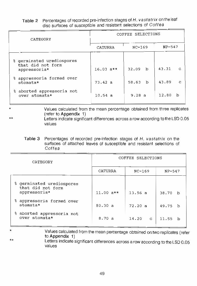
%
~ 0
%
*
**
~ 0
~ 0
%
*
**
Table 2 Percentages of recorded pre-infection stages of H. vas tat r i x on the leaf disc surfaces of susceptible and resistant selections of Goftea
COFFEE SELECTIONS CATEGORY
CATURRA NC-169 NP-547
germinated urediospores that did not form appressoria* 16.03 a** 32.09 b 43.31 c
appressoria formed over stomata* 73.42 a 58.63 b 43.89 c
aborted appressoria not over stomata* 10.54 a 9.28 a 12.80 b
Values calculated from the mean percentage obtained from three replicates (refer to Appendix 1)
Table 3
Letters indicate significant differences across a row according to the LSD 0.05 values
Percentages of recorded pre-infection stages of H. vastatrix on the surfaces of attached leaves of susceptible and resistant selections of Goftea
COFFEE SELECTIONS CATEGORY
CATURRA NC-169 NP-547
germinated urediospores that did not form appressoria* 11.00 a** 13.56 a 38.70 b
appressoria formed over stomata* 80.30 a 72.20 a 49.75 b
aborted appressoria not over stomata* 8.70 a 14.20 c 11.55 b
Values calculated from the mean percentage obtained on two replicates (refer to Appendix 1) Letters indicate significant differences across a row according to the LSD 0.05 values
49

~ 0
~ 0
~ 0
~ 0
*
**
Table 4 Percentage of recorded pre-infection stages of H. vastatrix on leaf disc surfaces from host and non-host plants
COFFEE SELECTIONS CATEGORY
CATURRA TEA BEAN
germinated urediospores that did not form appressoria* 16.06 a** 10.52 b 28.12 c
appressoria formed over stomata* 73.41 a 37.39 b 28.76 b
aborted appressoria not over stomata* 10.54 a 52.09 c 43.12 b
appressoria on a germ tube side branch* 55.70 a 40.96 b 15.98 c
Values calculated from the mean percentage obtained from four replicates (refer to Appendix 1) Letters indicate significant differences across a row according to the LSD 0.05 values
frequently on the non-hosts than on the host. In the case of the non-hosts,
significantly more appressoria formed apically than on a germ tube side branch.
SEM. The time scale involved in the infection process can be established from
Table 5. In the case of the Gaftea selections the following was noted: at 24hpi,
SSVls had formed in Caturra and NP-547, SSVs had already developed in NC-
169 at this time; at 48hpi, SSVs had formed in all three selections; in Caturra,
secondary infection hyph,ae were noticed at 48hpi, and at 72hpi intercellular spread
through the tissue began. In the case of NC-169 and NP-547, secondary infection
hyphae and intercellular spread were noted at 72hpi. In the case of P. vulgaris,
at 24hpi the SSVs had already developed, and secondary infection hyphae were
evident as early as 48hpi. It must be noted that the arrangement of primary and
secondary hyphae occasionally makes their unequivocal identification impossible.
50

Table 5 Total counts of the stages of infection structure development of H. vastatrix within various Coftea selections and a non-host, P. vulgaris, using SEM
*
c
COFFEE SELECTIONS NON-HOST CATEGORY HPI
CATURRA NC-169 NP-547 BEAN
9<-0 SSV initials formed 24 8 8 6 2
9<-0 aborted SSV 48 12 2 2 * initials 72 2 4 2 *
9<-0 SSV formed 24 * 6 * 52
48 18 4 4 53 48 7 c
9<-0 aborted SSVs 72 * 4 6 8
96 * * * 3
9<-0 SSVs with 48 4 * * 21
secondary 72 * 16 6 27 c infection 96 * 4 * 24 c hyphae 96 2
% advanced 72 4 2 2 * infection 96 6 4 2 * Total number of infection structures observed at all 54 54 28 199 samp'le times
stage of fungal development not observed at the particular time under consideration collapsed
In the case of P. vulgaris, the entire epidermis was stripped from the underlying
cells with relative ease. It was for this reason that the total number of infection
propagules examined was much higher for P. vulgaris than for Coftea (Table 5).
Infection structure morphology on the leaf surface of host and non-host
tissue. The appearance of infection structures on resistant and susceptible host
tissue and on the non-hosts was similar. Unlike the abaxial leaf surfaces of
Coftea and P. vulgaris, wax extrusions are present on Camellia sinesis
51

leaves. The leaf surface view of the stomata of Camellia sinensis is unique in
that a prominent ridge in the form of a ring surrounds the complex.
Infection structure morphology within host tissue. Once the stomatal chamber
is entered, a bulbous SSVI develops from the apex of the infection wedge (Plate
1 Fig. 1). Occasionally, two SSVls were observed protruding into the substomatal
chamber (Plate 1 Fig. 2). The initials extend in length and become torpedo
shaped (Plate 1 Fig. 3). Occasionally, amorphous material adhered to the
infection structures in cv. Caturra (Plate 1 Fig. 3). In NP-547, the resistant
selection, the infection wedge appears to extend into the substomatal chamber
(Plate 1 Fig. 1; Plate 2 Fig. 1). This was not observed in either Caturra or NC-
169 tissue. The SSVI soon bifurcates forming two short primary hyphae, the
resultant structure appearing anchor-shaped, because the stubby primary hyphae
curve back onto the subsidiary cells, where, by septum formation each cuts off a
haustorial mother cell (HMC) (Plate 2 Figs. 1,2 and 3). In Plate 1 Fig. 4, the HMC
of the primary hypha appears to form on the mesophyll cells, which was not the
usual situation. The SSVs are orientated at right angles to the stomatal opening.
Secondary infection hyphae emerge directly on the surface of the SSV that, with
reference to the leaf, is adaxial (Plate 2 Fig. 1) and elongate until they reach the
mesophyll cells. Once abutted onto these cells, a septum cuts off a terminal HMC.
Further branching of the secondary hyphae occurs behind the HMC and a much
branched mycelium ramifies through the intercellular spaces of the mesophyll cells
(Plate 2 Fig. 4). No differences in the appearance of these infection structures
between resistant and susceptible hosts were noted.
Infection structure morhology within the non-host, P. vulgaris. The SSVI
was similar to that observed in the host study. However, the infection wedge
protruded through the stoma into the substomatal chamber (Plate 3 Figs. 1-4).
A bulbous structure was evident at the end opposite to the SSV and this structure
did not develop further. After 48hpi, SSVs in various stages of collapse were
observed (Plate 4 Figures 1-4). Again, the SSVs were orientated at right angles
to the stomatal opening. The HMC was the first structure to collapse, and this was
followed by the remaining parts of the infection structure. By 96hpi most infection
52

Plate 1
Infection structure development of H. vastatrix within host's substomatal chamber
24 and 48hpi
Fig. 1 A bulbous substomatal vesicle initial [SSVI] is visible within the substomatal
chamber. Note the presence of the infection wedge (arrow) within the
substomatal chamber
Fig. 2 Two substomatal vesicle initials [SSVI] on the same stoma
Fig. 3 Torpedo-shaped substomatal vesicle initial [SSVI]. The presence of
amorphous material is noticeable [arrows]
Fig. 4 Bifurcate substomatal vesicle [SSV]. Note primary infection hyphae in contact
with the mesophyll cells of host [arrows]


Plate 2
Infection structure development of H. vastatrix within host substomatal chamber
Fig. 1 The substomatal vesicle [SSV] orientated at right angles to the stomatal slit
48hpi. Note the appearance of the infection wedge which has protruded into
the substomatal chamber of NP-547 [arrow]. The primary infection hyphae
[PIH] are appressed to the subsidiary cells [SC] while the initials of the two
secondary infection hyphae [SIH] are clearly evident on the SSV
Fig. 2 The anchor-shaped SSV with primary and secondary infection hyphae 48hpi
Fig. 3 The haustorial mother cells [arrows] are appressed to the subsidiary cells [SC]
and mesophyll cells of the host 72hpi. A septum [s] is seen both between the
infection wedge and the SSV, and cutting off the HMC from the infection hypha
[s]
Fig. 4 The fungus has numerous branched intercellular hyphae [arrows] 96hpi

.. ,

Plate 3
Infection structure development of H . vastatrix within the substomatal chamber of
bean, a non-host, 24hpi
Fig. 1 The infection wedge [IP] protrudes into the substomatal chamber of bean. A
bulb-shaped structure [BSS] is seen at one end of the infection wedge while
at the other end a substomatal vesicle initial [SSVI] is visible
Fig. 2"A septum [arrow] is visible between the bifurcate substomatal vesicle [SSV]
and the infection wedge
Fig. 3 A secondary infection hypha [SIH] has developed from the primary infection
hypha [PIH] on the substomatal vesicle [SSV]
Fig. 4 Primary [PIH] and secondary [SIH] infection hyphae on the SSV. The
arrangement of primary and secondary hyphae (for example, on right of this
micrograph) occasionally makes their unequivocal identification impossible

.. ,
. ...:

Plate 4
Infection structure development of H. vastatrix within the substomatal chamber of
bean, the non-host
Fig. 1 Collapsed haustorial mother cells [arrows] in contact with the non-host's
guard and mesophyll cells 48hpi
Fig. 2 Much-branched infection structure. The haustorium mother cell [arrow] has
collapsed 48hpi. Note the septum between the wedge and the SSV
Fig. 3 Collapsed infection hyphae [arrow] 72hpi
Fig. 4 The entire infection structure collapsed by 72hpi


structures had collapsed.
DISCUSSION
The present investigation on infection structure development of H. vas tat r i x
revealed that the general pattern is similar to that observed for other rust fungi.
During the infection process, the urediospore germ tube sequentially forms an
appressorium, appressorial wedge (refer to Chapter 2 of the present study),
infection wedge, SSV and infection hyphae. The infection structures of Hemileia
vastatrix do, however, have morphological features that are unique.
Fluorescence microscopy: Host study. According to Littlefield & Heath (1979)
and Heath (1981), the development of rusts on the leaf surface is not a determinant
in the resistance of host cultivars. However, in the case of other biotropic fungi,
resistance to pre-infection structure formation can differ between the abaxial and
adaxial leaf surfaces of a host. Amorphous sheets of epicuticular wax on the
abaxial leaf surface of Lolium spp. prevented germinated conidia of Erysiphe
graminis DC. from gaining access to features of the cuticular membrane which
triggers normal infection structure development (Carver et al., 1990). In the
present study, the involvement of epicuticular wax on the Goftea leaves was not
found to be responsible for the apparent pre-infection resistance expressed by the
resistant selection. Significant differences, particularly in the percentages of
germinated urediospores that did not form appressoria and appressoria
successfully formed over stomata, were noted in this study between Coftea
selections. This does indicate that conditions on the leaf surface of susceptible
hosts are more conducive to pre-infection by H. vastatrix than those on the
resistant selection (Table 2 and 3). However, Martins et al. (1985) found that the
primary differences between resistant and susceptible Goftea selections were
only evident after the penetration of the leaf surface by the rust fungus. There are
few reports on reduced levels of appressorium formation by rust fungi in resistant
hosts (Kochman & Brown, 1976; Russell, 1977). Rijo et al. (1982) with the aid
of light microscopy, observed that changes in the morphology of guard and
53

subsidiary cells of immune C. arabica selections following their inoculation with
H. vastatrix. The cytoplasm of the affected cells was granular and they had
"apparently thicker cell walls". These changes were induced without any apparent
penetration by the fungus. It is therefore possible that similar changes occurred in
NP-547 and these alterations in the stomatal complex may account for the failure
of penetration and explain the relatively low numbers of infection structures
observed in the resistant host (Table 5). This factor is unlikely to explain the lack
of recognition by the germ tube and the failure of appressoria to form over stomata.
Measurements of stomatal slit lengths failed to reveal a significant difference
between selections (Table 1).
The time scale involved in the formation of infection structures of H. vastatrix in
host tissue, as reported in the present study, is similar to that observed by Martins
& Moraes (1985). In other rust/host combinations the first sampling was done
6hpi (Lennox & Rijkenberg , 1989). In the present study this was done 24hpi in
accordance with the findings of De Jong et al. (1987) who recorded that, at this
time, appressorium formation would be complete.
Fluorescence microscopy: non-host study. A number of factors have been
implicated in non-host resistance. The surface characteristics of some plant
species could account for non-recognition by the germ tubes of rust fungi. Such
factors may include non-wettability of the leaf surface (Heath, 1977), lack of
directional growth (Wynn, 1976), waxy or hairy leaf surfaces (Heath, 1974; 1977)
and stomatal non-recognition (Wynn, 1976; Heath, 1977). It is evident that, on
host leaves, the germ tubes of H. vastatrix lack directional growth and side
branches are capable of developing appressoria. However, in the non-host, P.
vulgaris , appressoria are generally formed terminally and in some cases on short
germ tubes. Extensive exploration of the leaf surface in this case does not occur,
and many appressoria abort. Thus, a surface feature on the bean leaf may act as
a stimulus inducing appressorium formation by H. vastatrix almost immediately
after germination. Wax extrusions and/or unique surface features may playa role
in non-host resistance. Camellia sinensis leaves are waxy and together with the
prominent ridge surrounding the stomatal complex may account for the low levels
54

of appressoria formed over stomata. This ridge appeared to induce appressoria,
not over stomata, and probably is responsible for the high number of aborted
appressoria on this plant species. Reduced percentages of appressoria over
stomata, due to stomatal non-recognition, have been reported in the case of the
inoculations of T. aestivum and Avena sativa L. with U. appendiculatus
(Pers.) Unger (Wynn, 1976).
SEM study. The sequence of events in infection structure development of H.
vastatrix within host and non-host tissue is summarized diagramatically in Fig.
1. In host tissue, the infection wedge is single, similar to that of Uromyces
transversalis (Ferreira & Rijkenberg, 1989), but unlike the bifurcate infection peg
of Puccinia sorghi (Hughes & Rijkenberg, 1985). The primary infection hyphae
of H. vastatrix are formed in pairs, and their development is synchronous and
at right angles to the stomatal slit (Fig. 1 A and B). According to Ferreira &
Rijkenberg (1989), development of SSVs of U. transversalis is similarly
orientated and they propose that this alignment has co-evolved with, or adapted
to, substomatal chamber orientation. Instances of more than one apparently
functional SSV occupying the same stoma have been reported previously for other
rust fungi (Hughes & Rijkenberg, 1985; Lennox & Rijkenberg, 1990). Amorphous
material on the SSVs occasionally deposited within the susceptible cultivar,
Caturra, was not evident in the other susceptible selection, NC-169 lior in the
resistant selection, NP-547. According to Onoe et al. (1987), similar material is
formed in the association between P. coronata · var. avenae Erikss. and
susceptible tissue, and is thought to contribute to the establishment of
susceptibility. Primary infection hyphae of H. vastatrix curve back onto the
subsidiary cells, and HMCs are formed (Fig. 1 C). Similar to the situations
pertaining in P. sorghi (Hughes & Rijkenberg, 1985), U. transversalis (Ferreira
& Rijkenberg, 1989), and P. graminis f.sp. tritici (Lennox & Rijkenberg, 1989),
secondary infection hyphae arise on the SSV side of the septum separating it from
the HMC (Fig. 1 C and D). The time involved in the formation of the infection
structures of H. vastatrix, within host tissue, is much longer than within any of the
abovementioned rust/host interactions.
55

A
c
E
Fig. 1
B
. 10 ~m
o
. 10 ~m
10 ~m
EC = epidermal cell SC = subsidiary cell GC = guard cell MC = mesophyll cell GT = germ tube A = appressorium P = protuberance A W = appressorial wedge
. 10 ~m
SSVI = substomatal vesicle initial SSV = substomatal vesicle H MC = haustorial mother cell 10 = primary infection hypha 2
0 = secondary infection hypha i
Diagrammatic representation of infection structure development of H. vastatrix within Goftea [A-D] and P. vulgaris [E]
56

It would appear that the intra-tissue defence reaction fails to come into operation
in the resistant Coftea selection, NP-547, within the 96hpi period examined. Light
microscopy studies by Rijo (1972), Rijo & Rodrigues (1978) and fluorescence
microscopy conducted by Martins et a/. (1985), indicated that the differences
between resistant and susceptible hosts of H. vastatrix start to appear 72hpi.
They reported that in the incompatible combinations, fungal growth stopped in the
pre-haustorium stage or sometimes after haustorium formation in the subsidiary
cells. The morphology of the SSV is the same in both reaction types. The
subsequent growth of the fungus is reduced in the resistant host, frequently limited
to a single short hypha which senesces and apparently dies (Rodrigues et al.,
1982). According to Rijo & Rodrigues (1978) and Martins et al. (1985), in the
resistant selections studied, all rust colonies stopped growing after 144hpi. The
differences observed in the present study may be due to the particular resistant
host examined or to the particular rust race.
In non-host tissue, the sequence of events in infection structure development is
similar to that observed in host tissue, but with the following differences. The
infection wedge and a bulb-shaped structure was conSistently visible within the
substomatal chamber (Fig. 1 E) of the non-host. The infection wedge was,
however, also infrequently observed in the substomatal chamber of host tissue
(NP-547). Differences in heights of guard and subsidiary cells (Table 1) could not
adequately explain this feature in either host or non-host tissue. Collapse of the
infection structures occurred in non-host after the development of HMCs from the
secondary infection hyphae. This agrees with the reports by Heath (1974; 1977)
that the defence reaction of rust fungi in non-host tissue usually started after the
first haustorium was formed.
A criticism that can be levelled against the experiments presented in this Chapter
is that differences in the expression of resistance may be different on leaf discs
when compared to attached leaves. Mayama et al. (1975) reported an increase
in susceptibility in detached incompatible wheat leaves floating in different solutions
after inoculation. The normal incompatible reaction of attached wheat leaves to
certain races of Puccinia graminis f.sp. tritici was changed and they became
57

highly susceptible. Eskes (1982) demonstrated that development of H. vastatrix
was the same for leaf discs and attached leaves. However, Martins et aJ. (1986)
noted an increase in susceptibility of Coftea to H. vastatrix in detached leaves
when compared to attached leaves. In the present investigation, there were no
demonstrable differences in the development of infection structures on the leaf
surface and within tissue between leaf discs and attached leaves of resistant and
susceptible selections.
LITERATURE CITED
Anon. (1987) Coftee handbook (Ed. by W.J.C. Logan & J. Biscoe) pp. 27.
Canon Press, Harare.
Carver T.L.W., Thomas B.J., Ingerson-Morris S.M. & Roderick H.W. (1990) The
role of the abaxial leaf surface waxes of Lolium spp. to Erysiphe
graminis. Plant Pathology 39, 573-583.
Chinnappa C.C. & Sreenivasan (1968) Cytology of Hemileia vastatrix.
Caryologia 21, 76-82.
De Jong E.J., Eske~ A.B., Hoogstraten J.G.J. & Zadoks J.C. (1987) Temperature
requirements for germination, germ tube growth and appressorium
formation of urediospores of Hemileia vastatrix. Netherlands
Journal of Plant Pathology 93,61-71.
Eskes A.B. (1982) The use of leaf disc inoculations in assessing resistance of
coffee to Hemileia vastatrix. Netherlands Journal of Plant
Pathology 88,127-141.
Ferreira J. & Rijkenberg F.H.J. (1989) Development of infection structures of
Uromyces transversalis in leaves of the host and a nonhost.
Canadian Journal of Botany 67, 429-433.
Harr J. & Guggenheim R. (1978) Contributions to the biology of Hemileia
vastatrix I. SEM-investigations on germination and infection of
Hemileia vastatrix on leaf surfaces of Coftea arabica.
Phytopathologische Zeitschrift 92, 70-75.
Heath M.C. (1974) Light and electron microscope studies of the interactions of
58

host and non-host plants with cowpea rust - Uromyces phaseoli var.
vignae. Physiological and Molecular Plant Pathology 4,403-
414.
Heath M.C. (1977) A comparative study of non-host interactions with rust fungi.
Physiological and Molecular Plant Pathology 10, 73-88.
Heath M.C. (1981) Resistance of plants to rust infection. Phytopathology 71,
971-974.
Hughes F.L. & Rijkenberg F.H.J. (1985) Scanning electron microscopy of early
infection inthe uredial stage of Puccinia sorghi in Zea mays. Plant
Pathology 34, 61-68.
Jensen W.A. (1962) Botanical histochemistry Principles and Practice.
pp. 90. W.H. Freeman & Company, San Francisco.
Kochman J.K. & Brown J.F. (1976) Effect of temperature, light and host on
prepenetration of Puccinia graminis f.sp. avenae and Puccinia
coronata avenae. Annals of Applied Biology 82, 241-249.
Lennox C.L. & Rijkenberg F.H.J. (1989) Scanning electron microscopy of infection
structure formation of Puccinia graminis f.sp. tritici in host and non
host cereal species. Plant Pathology 38, 547-556.
Littlefield L.J. & Heath M.C. (1979) Ultrastructure of rust fungi. pp. 208-
238. Academic Press, New York.
Martins E.M.F. & Moraes W.B.C. (1985) Studies of the primary infection of coffee
plants by Hemileia vastatrix. Phytopathology 75,742 (Abstract).
Martins E.M.F., Tiburzy R. & Moraes W.B.C. (1985) Histological studies of
compatible and incompatible interactions of coffee leaves and
Hemileia vastatrix. Fitopatologia Brasileira 10, 627-636.
Martins E.M.F., Maria A.C. De, Grundewaldt-Stocker G. & Moraes W.B.C. (1986)
Changes in the resistance of detached coffee leaves by yeast extract
filtrate and heat treatment. F itopatologia Brasileira 11, 899-909.
Mayama S., Rehfeld D.W. & Daly J.M. (1975) The effect of detachment on the
development of rust disease and the hypersensitive response of wheat
leaves infected with Puccinia graminis tritici. Phytopathology
65,1139-1142.
Onoe T., Tani T., Minagawa S. & Sagawa H. (1987) Ultrastructural changes of
59

stomata in relation to specificity to rust fungi In: Molecular
determinants of plant diseases (Ed. by S. Nishimura, C.P. Vance
& N. Ooke) pp. 29-46. Japan Scientific SoCieties Press, Tokyo and
Springer-Verlag, Berlin.
Rijo L. (1972) Histopathology of the hypersensitive reaction t (tumefaction)
induced on Coftea spp. by Hemileia vastatrix. Agronomia
Lusitana 33,427-431.
Rijo L. & Rodrigues C.J. Jr. (1978) Processo de infecqao da Hemileia vastatrix
Berk. & Br. em cultivares susceptfveis e resistentes de Coftea arabica
L. Garcia de Orta, Serie de Estudos Agronomicos, Lisboa 5,1
23-24.
Rijo L., Rodrigues C.J. Jr. & Vasconcelos (1982) Immunity on the coffee-orange
rust association. Histopathological aspects. Garcia de Orta, Serie
de Estudos Agronomicos, Lisboa 9, 101-104.
Rodrigues C.J. Jr. (1984) Coffee rust races and resistance. In: Coftee rust in
the Americas (Ed. by R.H. Fulton) pp. 41-58. American
Phytopathological Society, St. Paul, Minnesota.
Rodrigues C.J. Jr., Rijo L. & Medeiros E.F. (1982) Induction of flecks and
tumefactions on coffee leaves by incompatible and compatible, viable
and non-viable uredospores of H. vastatrix and of other rusts, and by
their leachates. Garcia de Orta, Serie de Estudos
Agronomicos, Lisboa 9, 105-110.
Russell G.E. (1977) Varietal differences in formation of sub-stomatal vesicles by
Puccinia striiformis in winter wheat. Phytopathologische
Z eitschrift 88, 1-10.
Tiburzy R., Martins E.M.F. & Moraes W.B.C. (1983) Visualization of Hemileia
vastatrix structures in coffee leaves by fluorescence microscopy.
Fitopatologia Brasileira 8, 461-466.
Ward H.M. (1882) Researches on the life-history of Hemileia vastatrix, the
fungus of the "coffee-leaf disease". Botanical Journal 19,299-335.
Wynn W.K. (1976) Appressoria formation over stomates . of the bean rust fungus:
response to a surface contact stimulus. Phytopathology 66, 136-
146.
60

CHAPTER 4
THE EFFECTS OF THE FUNGICIDE, BAYFIDAN ®, ON INFECTION
STRUCTURE FORMATION BY HEMILEIA VASTATRIX
IN COFFEA ARABICA CV. CATURRA
INTRODUCTION
Triadimenol (B-( 4-chlorophenoxy-a-( 1, 1-dimethylethyl)-1 H-1 ,2,4-triazole-1-
ethanol) is a systemic triazole fungicide introduced in 1977 (Frohberger, 1978).
It was exclusively developed as a seed treatment for cereals [Baytan ®], and was
later developed as a foliar fungicide [Bayfidan ®] (Kuck & Scheinpflug, 1986).
Triadimenol is readily taken up by the plant roots and apoplastically translocated
to transpiring leaves (Davidse & De Waard, 1984). This fungicide has both high
levels of protectant, and curative action against Hemileia vastatrix Berk. & Br.
(Anon., 1989). According to Dr. M. Deal! (Bayer, pers. comm.), the fungicide has
been further developed into a granular formulation, and combined with an
insecticide, Disyston ®, for use in the coffee industry. It is sold under the trade
name Repulse ® in Malawi, but has not as yet been released in southern Africa.
Triadimenol is a metabolite of triadimefon [Bayleton ®] and is considered to be the
fungitoxic principle (Gasztonyi & Josepovits, 1979). The primary site of action of
triadimenol is considered to be at the level of sterol biosynthesis at C-14
demethylation which, within the fungal metabolism, is inhibited (Buchenauer,
1978). Sterols, are required for growth and reproduction of eucaryotic organisms,
and serve as architectural components of membranes (Siegel, 1981). In wheat,
about 60% of the active ingredient of triadimefon, which penetrated the leaves,
underwent the reduction to triadimenol within 48h at room temperature (Kuck,
1986,cited by Kuck & Scheinpflug, 1986). Similarly, Clark et a/. (1978) found a
56% conversion of triadimefon to triadimenol in 5 days. The spectrum of fungicidal
activity and application rates are, however, similar for both these fungiCides (Kuck
& Scheinpflug, 1986).
61

Previous research papers have dealt with the effect of systemic fungicides on the
ultrastructure of rust fungi (Ferreira, 1988; Guggenheim et aI., 1989). Striking
morphological abnormalities were produced in a number of fungal species by
ergosterol biosynthesis inhibiting (EBI) fungicides; distortion of germ tubes, often
with excessive branching, has been observed in Monilinia fructico/a Wint.
(Sisler et al., 1983) and Botrytis allii Munn. (Richmond, 1984). Germination of
fungal spores is known to be only incompletely suppressed by fungicides having
this action (Kuck et a/., 1982). Consequently, a part of the fungal population
usually completes colonization of the host plant, despite the presence of these
fungicides.
The aim of this study was to determine at what stage, if at all, infection structure
development of Hemi/eia vastatrix would be inhibited in a susceptible Coftea
arabica L. cultivar, following the application of the granular formulation of
triadimenol [Bayfidan ®]. Fluorescence and scanning electron microscopy were
used in this investigation.
MATERIALS AND METHODS
Planting conditions and material. Coftea arabica cv. Caturra seed was
obtained from the Tea Research foundation of Central Africa, Malawi. Following
germination, seedlings were grown in a greenhouse at temperatures between 15
and 30°C in pre-composted pine bark medium supplemented monthly with N:P:K
fertilizer and copper chelate.
Application of fungicide. Six two-year old seedlings were treated monthly with
Bayfidan ® granules. The amount of fungicide used, 5g/seedling, was determined
by taking into consideration the height of the seedlings as well as the container
size. The recommended dose is 38g per cova (Dr. M. Deall, pers. comm.).
Bayfidan ® was applied by randomly scattering the granules over the soil surface.
Six control se~dlings were left untreated. The plants were kept under similar
conditions as described above.
62

Leaf disc preparation. The method described by Eskes (1982) was used for disc
preparation from the third-leaf pairs. Leaves were gently rinsed under a stream of
tap water in order to remove soil debris. Leaf discs, 2cm in diameter, were
punched out using a stainless-steel cork borer. The midvein and leaf margins were
avoided. Discs were placed adaxial surface down, on to foam rubber saturated
with tap water, in glass trays (29 x 24 x 2.5cm). Discs were cut from leaves 28
days after the second application, seven, 14 and 28 days after the third fungicide
application; these times are referred to as A, B, C and D, respectively. The
experiment was repeated three times and eight leaf discs were used in each
replication.
Inoculations. Urediospores of H. vastatrix were freshly collected from infected
trees. They were lightly dusted on to the abaxial surface of leaf discs with a camel
hair brush (No.1). The glass trays were initially placed in a dew chamber at 20°C
for 24 h in darkness, and later moved to a constant environment chamber
(Conviron ®) at 26°C with a 12 h photoperiod.
Sampling. Two leaf discs were sampled at each sample time: 24, 48, 72 and 96
hours-post-inocuiation (hpi). At 24hpi each leaf disc was cut in half and one half
was prepared for fluorescence microscopy and the other for scanning electron
microscopy.
Fluorescence microscopy. The fluorochrome staining procedure described by
Tiburzy et a/. (1983) was used. The leaf samples, 24hpi, were examined using
a Zeiss research microscope fitted with epifluorescence equipment (light source
HBO 50; red suppression filter BG 38; exciter filter BP 390-440; chromatic beam
splitter FT 460; barrier filter LP 475). Colour photographs were taken using KodakR
Ektachrome 160 Professional 35 mm film.
Scanning electron microscopy (SEM). The infection structures formed on the
leaf surface after jor by 24hpi were viewed with either cryo-microscopy, or
prepared for SEM. Cryo-microscopy samples were prepared in an Emscope ® SP
2000 cryo-apparatus. Leaf specimens sampled at 24, 48, 72 and 96hpi were fixed
63

in 3% glutaraldehyde in 0.05M sodium cacodylate buffer [pH = 6.8-7.2], rinsed in
buffer and post-fixed in 2% osmium tetroxide in the same buffer. The leaf pieces
were dehydrated in an ethanol series, critical point dried using a Hitachi ® HCP-2
with carbon dioxide as a transition fluid, and mounted on metal stubs. Immediately
following critical point drying, an epidermal stripping technique, described by
Hughes & Rijkenberg (1985), was employed. For coffee, this technique had to be
modified, as the leaf epidermis most often fractures between the spongy mesophyll
and palisade cell layers, and only rarely does the epidermis strip entirely from the
mesophyll cells. To remove the adherent me sophy II cells, the stub with the
exposed abaxial leaf surface was gently pressed onto cellotape numerous times.
The tissue was gold/palladium coated in a Polaron ® E5100 sputter coater.
Infection structures were viewed with a Hitachi ® 8-570 operating at 5,8 or 10 kV.
Data processing. Data obtained from the leaf exterior were processed separately
from those of the leaf interior, since the stripping method precluded the stripping
of the entire specimen. The stage of infection structure formation of H. vastatrix
was classified and recorded. In the fluorescence microscopy study the following
three pre-penetration stages were considered: (1) percentage of germinated
urediospores that did not form appressoria; (2) percentage of germinated
urediospores that formed appressoria over stomata; and (3) percentage of
urediospores that formed appressoria not over stomata. The entire half of the leaf
disc was scanned and the infection structures, excluding those issuing from large
clumps of urediospores, counted. Percentage germination could not be
determined as many ungerminated urediospores are washed off the leaf pieces
during preparation. A mean percentage of the total counts recorded, from each
replicate, was calculated for pre-infection stages. Differences between means
were tested for significance using the ANOVA statistical test. Post-penetration
counts using SEM considered: (1) number of substomatal vesicle initials (SSVI)
formed: this category comprised infection wedges with unproliferated swollen tips;
(2) number of substomatal vesicles (SSVs) formed: a SSV was considered to
have formed once the primary infection hypha formation was initiated; (3) number
of SSVs with secondary infection hyphae, and (4) number of advanced infections
(ramification of mycelium through the intercellular spaces). Abnormalities in the
64

morphology of these structures were recorded.
RESULTS
Fluorescence microscopy. Refer to Appendix 2 and Table 1. Although
triadimenol had a statistically significant effect (P < 0.05) on the percentages of
germinated urediospores that did not form appressoria and, appressoria formed
over stomata, the effect was not great. In general, at Band D treatment times, the
percentage appressoria formed over stomata was higher on the control than on the
treated leaf discs. Fewer appressoria were formed over stomata at treatment times
Band D. A significantly higher percentage appressoria aborted at time C in
comparison to the other treatment times. No abnormal morphology of the
urediospore, germ tube or appressoria was observed.
Table 1 Mean percentages of counts made of the stages of infection structure development of H. vastatrix onthe leaf surface of cv. Caturra following the application of Bayfidan ® at various time intervals
CATEGORY DAYS AFTER THE SEOOND*/THIRD** APPLICATION OF BAYFIDAN
28 days* [A] 7 days** [B] 14 days** [C] 28 days** [D]
***** % germinated urediospores TREATMENT 6.54 a A 21.98 b A 6.39 abc A 12.84 e A
that did not form OONTROL*** 15.89 ab B 20.82 ab A 17.25 ab AS 9.00 a B appressoria ****
% germinated urediospores TREATMENT 84 .90 a A 73.32 b A 79.95 be A 78.54 e A that formed appressoria CONTROL*** 75 .77 ab B 73.49 ab B 72.79 ab AB 81.34 a B over stomata ****
% germinated urediospores TREATMENT 8.56 ab A 4.70 a A 13.83 e A 8.62 ab AB that formed appressoria OONTROL*** 8.35 ab A 5.69 ab AB 9.96 b A 9.66 b B not over stomata ****
Leaf discs cut from untreated seedlings *** ****
*****
Values calculated from the mean percentage obtained from three replicates (refer to Appendix 2)
•
Lower and upper case letters indicate significant differences across a row and down a column, respectively, according to the LSD 0.05 values
65

SEM. The total counts of infection structures of H. vastatrix observed in cv.
Caturra are presented in Table 2. There is a marked decrease in the number of
structures seen with an extended fungicide treatment. The number of structures
observed at time 0 is less than half the number observed at time A. Extracellular
material accumulated on the infection structures at times A and Bat 72hpi. This
material was only noted on the SSVI observed at time 0 24hpi. At time B advanced
infection structures collapsed and occasionally hyphae with abnormal morphology
were noted. At time 0 it would appear that further growth of the SSVs, after 48hpi
was inhibited.
Table 2 Scanning electron microscopy total counts of early infection structure development of Hemileia vastatrix on Coftea arabica cv. Caturra after treatment with the fungicide, Bayfidan ®
DAYS AFTER SECOND*/ CATEGORY HPI THIRD** APPLICATION CONTROL
SSVI 24 24
SSVs 24 48
SSVs + 48 secondary 72 infection 72 hyphae
Advanced 72 infection 96
96
Total counts
HPI - hours post inoculation a - collapsed
28* [A]
23
3 13
2 7 2b
*** 8 3b
61
OF BAYFIDAN
7** 14** 28** [B] [C] [D]
13 17 9 4b
1 *** 3b 12 3 5
4 2 *** 2 *** *** 4b
4 12 *** 3a 14 *** 3c
46 48 21
b - collection of extracellular material around the infection structure c - abnormal hyphal structure *** = no structures observed
66
26
26 2
14 12
4 10
94

Infection structure formation in triadimenol-treated tissue occurred as follows:- No
abnormalities of pre-penetration structures were observed (Plate 1 Fig. 1),
confirming the results of the fluorescence microscopy study. A collection of
extracellular material was occasionally observed on SSVls 24hpi at time D (Plate
1 Fig. 2). The SSV was sometimes similar in appearance to that observed in the
control, that is, the fungicide appeared to have no effect on their morphology.
However, on a number of occasions the SSV appeared swollen (Plate 1 Fig. 3 and
4). Extracellular material was observed 72hpi (Plate 1 Fig. 5; Plate 2 Fig. 1), but
it was not clear whether this originated from pathogen or host tissue. The
appearance of the intercellular hyphae was abnormal, compared to the control,
and hypha I branches were often swollen (Plate 2 Figs. 2). Disruptions in the wall
of the infection structures were also observed (Plate 2 Fig. 4). Haustorial mother
cell (HMC) collapse (Plate 2 Fig. 3) was observed 96hpi, followed by collapse of
the entire fungal structure.
DISCUSSION
Pre-penetration infection structure formation. One of the earliest opportunities
for fungicides to interfere with the development of a pathogen is during
germination and penetration of the host. Treatment of C. arabica cv. Caturra
seedlings with triadimenol precluded appressorium formation as a target of attack.
A weak effect on this process was observed in the present study. This observation
confirmed reports that ergosterol inhibiting fungicides (EBI) have little effect on the
infection process from germination to host penetration (Buchenauer, 1977; Kuck
etal., 1982; Paul, 1982; Zobrist eta!., 1982).
Post-penetration infection structure formation. Several workers have
investigated the effects of fungicides on the fine structure of fungi as a step to
elucidating their mode of action (Pring, 1984; Pring & Richmond, 1976). Scanning
electron microscopy and fluorescence microscopy have also been successfully
used to study the effects of fungicides on fungal morphology within host tissue
(Ferreira, 1988; Kuck et al., 1982; Paul, 1982).
67
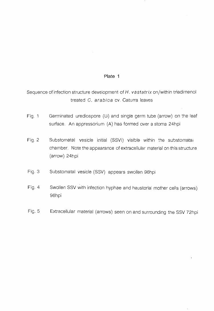
Plate 1
Sequence of infection structure development of H. vastatrix on/within triadimenol
treated C. arabica cv. Caturra leaves
Fig. 1 Germinated urediospore (U) and single germ tube (arrow) on the leaf
surface. An appressorium (A) has formed over a stoma 24hpi
Fig. 2 Substomatal vesicle initial (SSVi) visible within the substomatal
chamber. Note the appearance of extracellular material on this structure
(arrow) 24hpi
Fig. 3
Fig . 4
Fig. 5
Substomatal vesicle (SSV) appears swollen 96hpi
Swollen SSV with infection hyphae and haustorial mother cells (arrows)
96hpi
Extracellular material (arrows) seen on and surrounding the SSV 72hpi


Plate 2
Abnormal infection structure morphology of H. vastatrix following the application
of triadimenol 96hpi
Fig . 1
Fig. 2
Fig. 3
Fig. 4
Extracellular material (arrows) seen surrounding collapsed infection
hyphae and substomatal vesicle
Tips of the intercellular hyphae showing abnormal morphology (arrows)
Collapsed advanced infection structures
Disruptions (arrows) in the walls of this infection structure are evident


In the H. vastatrix/ Goftea cv. Caturra interaction an effect of triadimenol on the
morphology of SSVls, SSVs, and later infection structures was noted with SEM.
The collection of extracellular material on the infection structures may be due to the
disruption of membrane permeability, which would subsequently result in
plasmolysis. Permeability changes of fungal membranes in the presence of EBI
fungicides have been recorded previously. Severe membrane damage is reported
in the yeast, Saccharomyces cerevisiae Hansen., byeconazole (Yamaguchi
et aI., 1981, cited by Kato, 1986). The reasons for disruption of permeability is
thought to be due to small changes in the precise molecular architecture of
membranes (Kato, 1986). Richmond (1984) reported that triadimefon interferes
with the molecular 'mechanism of morphogenesis responsible for the architectural
structure of septa and walls of the fungus Botrytis allii Munn. This factor may
account for the disruptions in the walls of the infection structures observed in the
present study.
Occasionally the infection structures of H. vastatrix within treated tissue
appeared swollen and this may be due to ultrastructural changes occurring within
these structures. Extensive wall thickening of rust fungi was noted in Triticum
aestivum L. and Vicia faba L. treated with triadimefon (Pring, 1984). Hippe &
Grossmann (1982) made similar observations in the treatment of Ustilago
avenae (Pers.) Rostr. sporidia with nuarimol and imazalil nitrate, both EBI
fungicides.
According to Smolka & Wolf (1983), in the Hordeum vulgare L./Erysiphe
graminis DC. complex, the most striking effect after treatment with triadimefon
and triadimenol was the encasement of haustoria at 24 to 48hpi. E. graminis
discontinued its growth after the production of the first haustoria. Similar
observations have been made for other host/pathogen/fungicide combinations,
for example, Phaseolus vulgaris L./Uromyces phaseofi (Pers.) Wint. treated
with oxycarboxin (Pring & Richmond, 1976). However, in the case of the
Puccinia graminis f.sp. tritici Eriks. & E. Henn./T. aestivum interaction
following treatment with triadimenol and triadimefon the development of the first
haustoria was not completely inhibited (Kuck et al., 1982). Paul (1982) noted that
68

triadimefon reduced the number of SSVs and haustoria of Puccinia recondita
Rob. ex Desm. within T. aestivum L. tissue. This fungus was totally inhibited at
the stage of the formation of the first intercellular hyphae. Until transmission
electron microscopy has been carried out with inoculated triadimenol-treated
coffee tissue, no conclusions can be reached on the effects of this fungicide at the
ultrastructural level.
In the present study the onset of fungal disruption by triadimenol occurred between
24 and 48hpi. This relatively late action of the fungicide may be due to the fact that
in the initial stage of development the fungus still draws its reserves of ergosterol
from the urediospore. By 72hpi this supply would be exhausted and fungal growth
would cease. Kuck et a/. (1982) made similar observations in the interaction
between T. aestivum/ P. graminis f.sp. tritici following treatment with
triadimenol and triadimefon.
The excessive branching of intercellular hyphae, particularly at the hypha I tip, may
be due to modifications in the activity of enzymes involved in wall synthesis as has
been reported by Sisler & Ragsdale (1984). Excessive branching of the
intercellular hyphae of P. recondita following treatment of cryproconazole was
reported by Guggenheim et a/. (1989).
It is evident from the present study that there appears to be no similarities between
"natural" resistance conferred by the host genome (Chapter 3) and the "artificial"
resistance conferred by the fungicide, at least in the time frame under
consideration.
LITERATURE CITED
Anon. (1989) Bayfidan ® (BAY KWG 0519) Systemic fungicide: Technical
information. Bayer S.A.
Buchenauer H. (1977) Mode of action of triadimefon in Ustilago avenae.
Pesticide Biochemistry and Physiology 7,309-320.
69

Buchenauer H. (1978) Inhibition of ergosterol biosynthesis by triadimenol in
Ustilago avenae. Pesticide Science 9,507-512.
Clark T., Clifford D.R., Deas AH.B., Gendle P. & Watkins D.AM. (1978)
Photolysis, metabolism and other factors influencing the performance
of triadimefon as a powdery mildew fungicide. Pesticide Science 9,
497-506.
Davidse L.C. & de Waard M.A (1984) Systemic fungicides. Advances in Plant
Pathology 2, 191-257.
Eskes AB. (1982) The use of leaf disc inoculations in assessing resistance to
coffee leaf rust (Hemileia vastatrix). Plant Protection Bulletin
FAO 29, 56-66.
Ferreira J.F. (1988) Effect of bitertanol on the ultrastructure of Uromyces
transversalis infection structures in Gladiolus leaves. pp. 99-114.
Ph.D. thesis, University of Natal.
Frohberger P.E. (1978) ®Baytan, a new systemic broad spectrum fungicide
especially suitable for cereal seed treatment. Pflanzenschutz
Nachrichten Bayer 31,11 -24.
Gasztonyi M. & Josepovits G. (1979) The activation of triadimefon and its role in
the sensitivity of fungicide action. Pesticide Science 10, 57-65.
Guggenheim R, Duggelin M., Mathys D. & Grabski C. (1989) Cryo-SEM
investigations on cyproconazole treated rust fungi in plant cells.
International Symposium of Electron Microscopy applied in
Plant Pathology, Sept. 19-22, 1989, Konstanz, FRG. Abstracts pp.
57.
Hippe S. & Grossmann F. (1982) The ultrastructure of sporidia of Ustilago
avenae after treatment with the fungicides Nuarimol and Imazalil Nitrate.
Pesticide SCience 13, 447-451.
Hughes F.L. & Rikenberg F.H.J. (1985) Scanning electron microscopy of early
infection in the uredial stage of Puccinia sorghi in Zea mays. Plant
Pathology 34, 61-68.
Kato T. (1986) Sterol-biosynthesis in fungi, a target for broad spectrum fungicides.
In: Sterol biosynthesis, inhibitors and anti-feeding
compounds (Managing Ed. by G. Haug & H. Hoffmann). pp. 1-24.
70

Spinger-Verlag, Berlin.
Kuck K.H., Scheinpflug H., Tiburzy R. & Reisener H.J. (1982) Fluorescence
microscopy studies of the effect of Bayleton ® and Baytan® on growth
of stem rust in the wheat plant. Pflanzenschutz Nachrichten
Bayer 35, 209-228.
Kuck K.H. & Scheinpflug H. (1986) Biology of sterol-biosynthesis inhibiting
fungicides. In: Sterol biosynthesis, inhibitors and anti
feeding compounds (Managing Ed. by G. Haug & H. Hoffmann).
pp. 65-96. Spinger-Verlag, Berlin.
Paul V.H. (1982) Studies on the effect of ®Bayleton on pathogenesis of brown rust
of wheat (Puccinia recondita f.sp. tritici). Pflanzenschutz
Nachrichten Bayer 35, 229-246.
Pring RJ. (1984) Effects of triadimefon on the ultrastructure of rust fungi infecting
leaves of wheat and broad bean (Vicia faba). Pesticide
Biochemistry and Physiology 21, 127-137.
Pring RJ. & Richmond D.V. (1976) An ultrastructural study of the effect of
oxycarboxin on Uromyces phaseoli infecting leaves of Phaseolus
vulgaris. Physiological Plant Pathology 8, 155-162.
Richmond D.v. (1984) Effects of triadimefon on the fine structure of germinating
conidia of Botrytis allii.
Physiology 21,74-83.
Pesticide Biochemistry and
Siegel M. R (1981) Sterol-inhibiting fungicides: effects on sterol biosynthesis and
sites of action. Plant Disease 65, 986-989.
Sisler H.D., Walsh R.C. & Ziogas B.N. (1983) Ergosterol biosynthesis: A target of
fungitoxic action. In: Pesticide chemistry: human welfare and
the environment Vol. 3 (Ed. by S. Matsunaka, H.D. Hutson & S.D.
Murphy), pp. 128-134. Pergamon Press, New York.
Sisler H.D. & Ragsdale N.N. (1984) Biochemical and cellular aspects of the
antifungal action of ergosterol biosynthesis inhibitors. In: Mode of
action of antifungal agents (Ed. by A.P.J. Trinci & J.F. Ryley). pp.
257-281. Cambridge University Press, Cambridge.
Smolka S. & Wolf G. (1983) Cytological studies on mode of action of ®Bayleton
(triadimefon) and ®Baytan (triadimenol) on the host-parasite complex
71

barley - Erysiphe graminis f.sp. hordei. Pflanzenschutz
Nachrichten Bayer 56, 97-126.
Tiburzy R., Martins E.M.F. & Moraes W.B.C. (1983) Visualization of Hemileia
vastatrix structures in coffee leaves by fluorescence microscopy.
Fitopatologia Brasileira 8,461-466.
Zobrist P., Colombo V.E. & Bohnen K. (1982) Action offenpropimorph on exterior
structures of Puccinia coronata on oat as revealed by scanning
electron microscopy. Phytopathologische Zeitschrift 105, 11-
19.
72

CHAPTER 5
TELIOSPORES OF HEMILEIA VASTATRIX
INTRODUCTION
Hemileia vastatrix Berk. & Br. has three spore types, namely, urediospores,
teliospores and basidiospores. As yet, the pycnial and aecial stages have not been
found under either natural or controlled conditions (Gopalkrishnan, 1951).
Urediospores are capable of infecting Coftea spp. and other members of the
Rubiaceae (Frean, 1986). Ward (1882), Pole Evans (1909), Gyde (1932), Mayne
(1936), Thirumalachar & Narasimhan (1947) and Gopalkrishnan (1951) were
unable to infect coffee leaves with basidiospores. It is now generally agreed in the
literature, that the life cycle is not completed on the coffee tree. It is for this reason
that this fungus is assumed to be heteroecious. The plant species which is the
alternate host for coffee rust is unknown.
Besides the descriptions of Ward (1882), Thirumalachar & Narasimhan (1947) and
Gopalkrishnan (1951), little is known about the development and structure of the
rarely occurring teliospores. They have been observed on infected coffee grown
in India and Sri Lanka (Nutman & Roberts, 1972), South Africa (Pole Evans, 1909;
Gyde, 1932) and Brazil (Sugimori et al. 1976).
The discovery of teliospores on locally infected trees prompted a scanning electron
microscopy study on their structure, development and germination.
MATERIALS AND METHODS
Infected material. At the end of the winter months, in August, teliospores of H.
vastatrix were noticed on infected leaves collected from trees of C. arabica L.
at Ukulinga experimental station near Pietermaritzburg, South Africa.
73

Induction of basidiospore production. Coffee leaves, containing uredio- and
teliosori, were placed on a glass sheet under a bell-jar which had previously been
lightly sprayed internally with water. This apparatus was placed overnight in a
controlled environment chamber (Conviron ®) at 26°C with a 12h photoperiod.
Scanning electron microscopy. Leaf pieces (ca. 5mm x 5mm) were dissected
from areas of leaves covered in orange urediosori, among which teliosori had been
observed. They were pre-fixed in 3% glutaraldehyde in sodium cacodylate buffer
(pH = 6.8 - 7.2), rinsed in buffer, post-fixed in 2% osmium tetroxide and
dehydrated in an ethanol series. The specimens were critical point dried using a
Hitachi ® HCP-2 with carbon dioxide as a transition fluid and mounted on metal
stubs. The tissue was gold/palladium-coated in a Polaron ® sputter coater. Leaf
pieces were viewed with a Hitachi® S-570 operating at either 8 or 10 kV.
Environmental parameters associated with teliosorus induction. The severity
of H. vastatrix on a few isolated C. arabica trees was monitored monthly at
Ukulinga during 1989, the year in which the teliospores were noticed. The mean
percentage diseased leaf ar~a, used as a rating scale and described by Eskes &
Toma-Braghini (1981) (refer to Table 1), was calculated from the mean of 20
randomly collected leaves. The total rainfall, mean maximum and minimum
temperatures for Ukulinga were obtained from the Agricultural Meteorological
Department at Cedara. The relationship between these parameters and
percentage diseased area is presented graphically in Figs. 1 and 2. The reason
for the inclusion of these data is an attempt to link teliosorus induction to
environmental parameters.
RESULTS AND DISCUSSION
Teliospores of H. vastatrix are described as pedicellate, spherical or napiform
and smooth with a terminal papilla (Ward, 1882). Scanning electron microscopy
confirmed the morphological details decribed above [Plate 1 Fig. 1J. Teliospores
vary in size from 16-25 J.Lm (length) x 19-22J.Lm (width). Immature teliospores lack
74

Fig. 1
Fig. 2
% Diseased Area Rainfall (mm) 40~~~~~-----------------r30
35
30
25
20
Jan Feb Mar Apr May Jun Jul Aug Sep Oct Nov Dec
Months
~ Disease Severity -+- Total Rainfall
25
20
15
The relationship between the total rainfall and percentage diseased area on Goftea arabica leaves infected with Hemileia vastatrix for 1989 at the Ukulinga experimental station, Pietermaritzburg
% Diseased Area Temperature tC) 40~~~~----------~-------r 30
25
20
15
10
5
o Jan Feb Mar Apr May Jun Jul Aug Sep Oct Nov Dec
Months
~ Disease Severity -:.:- max. temperature
~ min. temperature
The relationship between mean minimum and maximum temperatures and percentage of diseased area on Goftea arabica leaves infected with Hemileia vastatrix for 1989 at the Ukulinga experiment station, Pietermaritzburg
75

Fig. 1
Fig. 2
Fig. 3
Fig. 5
Plate 1
Napiform teliospore [T] with a terminal papilla [P] borne on a short
sporophore [8]
Immature teliospores [T] in a teliosorus
Teliosori and a uredio/teliosorus illustrating in situ germ-ination of
teliospores [T]
Teliospore germinating In situ, illustrating the cylindrical promycelium
[arrows]


Table 1 Scale developed by Eskes & Toma-Braghini (1981) to calculate the percentage diseased area of Hemileia vastatrix on Goftea arabica leaves
SCALE NUMBER OF LESIONS* 9:-0 DISEASED AREA
VALUE PER LEAF
0 0 0 1 1 0.6 2 2 1.3 3 4 2.5 4 8 5.2 5 15 9.7 6 29 18.5 7 52 33.1 8 84 54.1 9 118 75.6
* In this study "lesion" was understood to mean a colony of urediosori
the terminal papilla [Plate 1 Fig. 2]. They are borne in clusters on short, ovate
teliosporophores (pedicels) which protrude through the stoma in a manner similar
to those of urediospores. They are commonly found among the urediospores
[Plate 1 Fig. 3], however, they do occasionally occur in teliosori [Plate 1 Figs. 2
and 3]. Gopalkrishnan (1951) has also reported that they occur in the urediosorus
with, or following, the urediospores, or in separate sori. Although the formation of
teliospores could not be investigated, it appears that teliosori and urediosori are
derived from the same, or similar, fertile cells in the substomatal chamber.
According to a study by Gold & Littlefield (1979) on the Melampsora lini
(Ehrenb.) Lev./ Unum usitatissimum L. interaction, few telia developed
independently of prior uredial development. This feature would appear to be
similar in the H. vastatrix/ C. arabica interaction.
Teliospores germinate in situ by producing a promycelium at the apex of the
spore [Plate 1 Figs. 3 and 4]. Ward (1882) reported that the promycelium can
also be formed on the hilum side opposite the papilla. This was not observed in the
present study. According to Vishveshwara & Nag Raj (1960), and Chinnappa &
Sreenivasan (1965), teliospores germinate and produce a cylindrical promycelium
from which four spherical basidiospores are formed. The formation of
76

basidiospores could not be induced in the present study.
Teliospores are reported to be produced on older attached leaves in dry conditions
(Chinnappa, 1965; Waller, 1982). The infected specimens examined in this study
were from fully expanded leaves, that is, the third-leaf pair and older.
It is evident from Fig. 1 that disease severity is highest in August, thereafter there
is a sharp decrease. From these results it would appear that in the seasonal cycle
of H. vastatrix, maximum urediospore production culminates in the formation of
teliospores. The meteorological data indicated that a dry period of less than
2.5mm rainfall per month for 57 days was recorded prior to the period when the
teliospores were first noticed (Fig. 1). The average rainfall per month for the 1989
winter months at Ukulinga was 2.1 mm (much lower than the recorded 11.3mm for
the spring and summer months). Temperature has been implicated inthe induction
of teliospore formation. Groth & Mogen (1978) reported that teliospore formation
of Uromyces appendiculatus (Pers.) Unger on Phaseolus vulgaris L. can
be induced by cold conditions. The mean maximum and minimum temperatures
for the winter months at Ukulinga were 21.5°C and 10.6°C, respectively (Fig. 2).
It would therefore appear that dry, cold conditions are a prerequisite for teliospore
production and peak urediosorus generation by H. vastatrix at Ukulinga.
LITERATURE CITED
Chinnappa C.C. (1965) Studies on the leaf rust (Hemileia vastatrix Berk. &
Br.) of coffee. Indian Coffee 29,9-12.
Chinnappa C.C. & Sreenivasan M.S. (1965) Secondary sporidia in Hemileia
vastatrix. Current Science 34, 668-670.
Eskes A.B. & Toma-Braghini M. (1981) Assessment methods for resistance to
coffee leaf rust (Hemileia vastatrix Berk. & Br.). Plant Protection
Bulletin FAO 29, 56-66.
Frean R.T. (1986) Coffee diseases. Farming in South Africa: Coffee
H.5/1986.
77

Gold R.E. & Littlefield L.J. (1979) Ultrastructure of the telial, pycnial and aecial
stages of Me/ampsora lini . Canadian Journal of Botany 57,
629-638.
Gopalkrishnan K.S. (1951) Notes on the morphology of the genus Hemileia.
Mycologia 43,271-283.
Groth J.v. & Mogen B.D. (1978) Completing the life cycle of Uromyces
phaseoli on bean plants. Phytopathology 68, 1674-1677.
Gyde L.M. (1932) Some observations on the genus Hemileia. South African
Journal of Science 29, 296-300.
Mayne M.W. (1936) Annual report of the coffee Scientific Officer, Mysore Coffee
Experiment Station 14, 1935-1936.
Nutman F.J. & Roberts F.M. (1972) Coffee leaf rust. Kenya Coffee 36, 139-
159.
Pole Evans LB. (1909) Report of the Plant Pathologist, Transvaal Department of
Agriculture, 155-172.
Sugimori M.H., Soave J., Paradela Filho 0., Moraes S.A., Ribeiro I.J.A. & Ito M.F.
(1976) Constatoacao de teliosporos de Hemileia vastatrix Berk. &
Br. no Brazil. Review of Phytopathology 56, 4536 (Abstract).
Thirumalachar M.J. & Narasimhan M.J. (1947) Studies on the morphology and
parasitism of Hemileia species on Rubiaceae in Mysore. Annals of
Botany 11,77.
Vishveshwara S. & Nag Raj T.R. (1960) Nuclear status of basidiospores of
Hemileia vastatrix B. & Br. Indian Coffee 24, 118-119.
Waller J.M. (1982) Coffee rust in Latin America. PANS 18,402-406.
Ward M.H. (1882) Researches on the life history of Hemileia vastatrix, the
fungus of "coffee leaf disease". Botanical Journal 19, 299-336.
78

CHAPTER 6
THE EFFECT OF LEAF AGE ON INFECTION OF COFFEA SELECTIONS
BY HEMILEIA VASTATRIX
INTRODUCTION
Plants change in their susceptibility to disease with age. Generally, as they mature
there is an increase in resistance (Hyde, 1977) which may be followed by a decrease
with further ageing (Parlevliet, 1975). The effect of age on infection does, however,
appear to depend on the particular host-pathogen interaction under investigation. For
example, Padmanabhan & Ganguly (1 954), cited by Eskes & Toma-Braghini (1982)
observed that resistance of rice, Oryza sativa L., to He/minthosporium Link. Fr.
decreased with age, butthat resistance to Pyricularia oryzae Cav. increased with
age. Adult plant resistance to cereal rust diseases has been recognized in many host
pathogen combinations and is characterized by an increasing resistance to infection
with advancing plant age (Hyde & Elahinia, 1989).
In the case of a perennial host, the age of plant organs may be more important than
the age of the whole plant. Sharma et a/ . (1980) studied the Populus
spp.j Me/ampsora larici-populina Kleb. interaction, and found that within ashoot,
the latent period and reaction type were related to leaf age: high susceptibility was
found to be associated with a short latent period. The developmental stage of Goftea
berries also appears to influence resistance to Col/etatrichum cafteanum Noack
(Mulinge, 1970; Van der Graaff, 1981): the young green berries being more
susceptible than ripe fruit.
There are reports on the influence of leaf age on the resistance of Gaftea spp. to
Hemileia vastatrix Berk. & Br. D'Oliveira (1957), cited by Kushalappa & Eskes
(1989), mentioned that mature leaves of G. arabica L. are more resistant than
79
succulent young leaves. Rayner (1961), however, found no difference in leaf age
effects. More recently, Eskes & Toma-Braghini (1982) reported that the effect of leaf
age on resistance varies with particular Coftea spp. and C. arabica selections. A
study on the germination of urediospores on leaves of different ages from the same
cultivar revealed that higher germination occurred on young than on intermediate
and old leaves (Nutman & Roberts, 1963).
In this chapter the frequency of appressorium formation on the leaf surface, and
subsequent sporulation on differently aged leaves of Coftea arabica cv. Caturra,
and two F6 progenies of a cross between C. arabica cv. Caturra and Hybrido de
Timor (HOT), were investigated.
MATERIALS AND METHODS
Planting material and conditiC?ns. Seeds of C. arabica cv. Caturra were obtained ..,
from the Tea Research Foundation of Central Africa, Malawi. CENICAFE (Centro
Nacional de Investigaciones de Cafe), Colombia, supplied the lines NP-547 and NC-
169, the F6 progenies of a cross between C. arabica cv. Caturra and HOT. NP-
547 gives a resistant, and NC-169 a susceptible, response when inoculated with H.
vastatrix (G.A. Alvarado, CENICAFE, pers. comm.). HOT is a population derived
from a single tree, probably a spontaneous hybrid between C. arabica and C.
canephora L., discovered around 1927 in East Timor (Goncalves et a/., 1977 cited, )
by Rodrigues, 1984). The majority of HOT selections are resistant to all known races
of H. vastatrix.
Following germination, seedlings were grown in a greenhouse at temperatures
between 15 and 30°C in pre-composted pine bark medium supplemented monthly
with N:P:K fertilizer and copper chelate.
Leaf ~isc preparation. The third plagiotropic branch, from the top of two-year-old
80

trees of the above lines, was selected. A single branch of each selection was used for
a single replication. The experiment was replicated three times. Leaves from each
node were collected and their position on the branch noted. Seven leaf pairs were
sampled, from the immature shiny first leaf pair to the dark green seventh pair. The
method described by Eskes (1982) was used for leaf disc preparation. Leaves were
gently rinsed under a stream of tap water in order to remove soil debris. Leaf discs,
2cm in diameter, were punched out using a stainless-steel cork borer. The midvein
and leaf margins were avoided. Four leaf discs per each pair of the same age were
used in a replication. Discs were placed, adaxial surface down, on foam rubber
saturated with tap water in glass trays (29 x 24 x 2.5cm) and inoculated as described
below.
Leaf disc inoculation. Urediospores of H. vastatrix were freshly collected from
infected trees. They were lightly dusted onto the abaxial surface of leaf discs with a
camel-hair brush (No.1). The glass trays with the inoculated discs were initially placed
in a dew chamber at 20°C for 24 h in darkness and later transferred to a constant
environment chamber (Conviron ®) at 26°C with a 12 h photoperiod.
Fluorescence microscopy. Discs were examined 24 hours-post-inoculation (hpi).
The fluorochrome staining procedure described by Tiburzy et al. (1983) was used.
The leaf samples were examined using a Zeiss research microscope fitted with
epifluorescence equipment (light source HBO 50; red suppression filter BG 38; exciter
filter BP 390-440; chromatic beam splitter FT 460; barrier filter LP 475). Colour
photographs were taken using Ektachrome 160 Professional 35 mm film. Three
stages of development of the fungus on the leaf surface were recorded and classified,
namely: [ 1] percentage of germinated urediospores that did not form appressoria;
(2) percentage of germinated urediospores that formed appressoria over stomata;
and [3] percentage of germinated urediospores that formed appressoria not over
stomata. The entire leaf disc surface was scanned and infection structures, excluding
those issuing from large clumps of urediospores, counted. Percentage germination
could not be determined as many ungerminated urediospores are washed off the leaf
81
pieces during preparation. The mean percentage of the total counts recorded, from
each replicate, was calculated for all three stages of development of the fungus.
Differences between means were tested for significance using the ANOVA statistical
test.
Latent period, reaction type and urediosorus density. The latent period, the time
period from the beginning of incubation to the time at which 50 per cent of the first
urediosori appeared, was determined. This involved daily inspections of leaf discs
with a hand lens until the urediosori had been .produced. A urediosorus was regarded
as a clump of spores emerging from one stoma. The disease assessment scale (refer
to Chapter 1 Table 3) described by Eskes & Toma-Braghini (1981) 'was used to
determine the reaction type. Urediosorus density was calculated with the aid of a
Zeiss dissecting microscope. The number of uredosori, in a 1 x 1 mm area of a slit
made in cardboard and randomly placed on the infected leaf disc, were counted (n =
10). These latter two parameters were determined for each of the three replications
of the experiment eight weeks after inoculation. The value presented is the mean of
nine readings in the case of latent period and reaction type, and 90 in the case of
urediosorus density. It must be noted that during the eight week period the leaves
have aged. Thus leaf pair 1, will at the time of assessing reaction type, latent period
and uredosorus density, have aged and probably should more correctly be referred
to as Leaf pair 2. This will however depend on the time of the year in which the
experiment was conducted. New leaf formation occurs in September under South
African conditions. This factor was not taken into consideration when assessing the
results.
Ranking. The results obtained for reaction type, urediosorus density and percentage
appressoria over stomata were ranked from lowest to highest value. Latent period,
percentages of germinated urediospores that did not form appressoria and, those that
formed appressoria not over stomata were ranked from highest to lowest value. In
this case, a long latent period, high percentage of germinated urediospores that did
not form appressoria and a high percentage aborted appressoria were regarded as
82

indicators of resistance. Thus, a parameter with a low score would indicate resistance
and one with a high score, susceptibility.
RESULTS
The age of a leaf at the time of inoculation did affect both infection structure
development and sporulation on the leaf surface significantly (Appendix 3 Table A
and B).
Fluorescence microscopy. Refer to Tables 1 and 2, and Figs. 1 and 2. On the leaf
surfaces of Caturra and NC-169 there is a decrease in the number of appressoria
formed with an increase in leaf age. On the resistant host, NP-547, significantly fewer
appressoria are formed on the fourth leaf pair. Appressoria are formed significantly
more readily over stomata of leaf pairs two, three and four of the susceptible
selections. On the youngest leaf pair a significant high percentage of aborted
appressoria was noted compared to percentages obtained for older leaf pairs.
Reaction type, latent period and urediosorus density. The reaction type, in the
case of susceptible lines, cv. Caturra and NC-169, was significantly higher in younger
than older leaves. The reverse was true of the resistant line, NP-547. The latent
period was significantly shorter in the case of NC-169 than on Caturra. In both cases,
the length of the latent period increased with increasing leaf age. The urediosori per
mm2
decreased with increasing leaf age on these susceptible selections.
Ranking. Refer to Table 3. In general, younger leaves of the susceptible selections,
particularly the fully expanded second and third leaf pairs of Caturra, are more
susceptible than the older leaf-pairs. This pattern, clearly indicating an effect of leaf
age on the infection parameters considered for NP-547, was not shown.
83

A HOST
Caturra NC-169 NP-547
B HOST
Caturra NC-169 !,;P-547
c HOST
Caturra NC-169 NP-547
.,.. GERMINATED UREDIOSPORES THAT DID NOT FORM AE.PRESSORIA 70
1
. I 60j . . ... \ , . . - . .. "J ::f ---~: , ~ ~:<~- ~,c ~:;.:;;::;~: l ,+ :. --~-- --.-. -.. ~ .. -.. --. '._._- .. -.--.. --- ... -1 10 --.- - - - .... .. . _-- .-. -~ . . .. - .- - .. - - . ------. - .
O ! ! .
1 3 4 6 e LEAF PAIR NUMBER
LEAF LEAF LEAF LE.<\F PAIR PAIR PAIR PAIR
1 2 3 4
1.12 3 . 66 0.31 5 . 37 3.42 2.21 0 . 52 3.39 2.28 1. 34 1.17 3.27
LEAF PAIR
5
3.40 6 . 98 3 . 89
LEAF LEAF PAIR PAIR
6 7
1. 67 2 . 84 0 . 85 3.44 0.69 2 . 36
,. GERMINATED UREDIOSPORES THAT FORMED APPRESSORIA OVER STOMATA
::. ----- - --- -- - ~ r-.~ .. .. .. .. . .. . .. . . ·~· ·~~.:.~:::._.:._: ·· · · I
40 r ---.- _ .. . _-_._- -. :-:-::~;;--! . ---~~
20 - -_.-- .-._ .. - -- --.- - ---.-- --.- - .--\
o I . ! ! • • I 1
LEAF PAIR
1
3 . 78 7 . 35 4 . 77
3 4 5
LEAF PAIR NUMBER
LE.-\F LL-\F LEAF PAIR PAIR PAIR
2 3 4
4 . 34 0.79 5 .46 2 . 29 1.10 5 . 76 3 . 13 1. 65 4 . 52
6
LEAF PAIR
5
5 . 47 7.91 4 .00
LEAF LE.-\F PAIR PAIR
6 7
3.40 4 . 03 1. 38 5 . 69 1.19 3 . 33
,. GERMINATED UREDIOSPORES THAT FORMED APPRESSORIA NOT OVER STOMATA 40
30 - --------------------._--
1
::r·'C · ----~~==:~;==-l o
2 3 4 6
LEAF PAIR NUMBER
[ - CAr URRA - NC-,Sg . - . NP-547 I LEAF LE.<\F LEAF LEAF LEAF PAIR PAIR PAIR PAIR PAIR
1 2 3 4 5
1. 27 0 . 13 1.15 0 . 93 2.64 3 . 63 0 . 65 0 . 64 1. 80 4.98 2 .02 2.97 0 . 82 2.17 2 . 94
LEAF LEAF PAIR PAIR
6 7
1. 50 0.97 1.00 1. 63 1. 90 1. 51
A-C Standard error of the mean presented in table form. Error bars overlapped when drawn on the graph.
Fig. 1 The effect of leaf age on the developmental stages of H. vastatrix on the leaf disc surface of C. arabicacv. Caturra, and NC-169 and NP-547
84

A HOST
Caturra NC-169 !iP-547
B HOST
Caturra NC-169
c HOST
Caturra NC-169
REACTION SCORE
:~L _ J--- - -J -------oLI __ ~--~--- -~--~-- -._. - -~.- __ ~~~
, 3 4 5
.:-.-.. . . ... ...... -. . .
LEAF PAIR NUMBER
[ ~ CATURRA - HC-16Q - • • NP-547 I LEAF LF.. .. F LE ... F LEAF LEAF PAIR PAIR PAIR PAIR PAIR
1 2 3 4 5
0.59 0 . 62 0 . 60 0 . 31 0 . 27 0 .15 0 . 13 0 . 39 0 . 18 0 0 0 0 . 22 0 .43 0 . 73
LATENT PERIOD (days) 60 I
i <0 L - -- .=..- :--- -________ ---
30 ~
I ~ __ 20;""~ -------~
- -- I
---J
-- I '0:- .... --- - -----.- - - i
0~-~----~4--~5--~--, , F PAIR NUMBER
i LEA
LEAF LEAF LEAF U:'AF LEAF PA IR PAIR PAIR PAIR PAIR
1 2 3 4 5
1. 00 0 . 33 0 0 . 33 0 . 88 0 . 33 0 0 . 33 0 . 88 0 . 67
UREDIOSORllmm '00
1 I 60 ~ ----- -- --- ----- -- - ---1
60 L -
<0 l __ ------ :-J ---- ---J '
LF..-'.F PAIR
1
2 . 65 2. 42
345 LEAf PAIR NUMBER
LEAF LEAF PAIR PAIR
2 3
0 . 79 2.21 2.37 1. 53
LEAF PAIR
4
3.12 1. 43
I
---- I 1 --/1 . i
LEAF PAIR
5
3. 43 4.17
LEAF LF..~F
PAIR PAIR 6 7
0 . 27 0.59 0.09 1. 23 0 . 55 0 . 29
LF..-\F L" '.F PAIR PA!R
6 7
0.33 0 0 0 . 33
LEAF LF..-\F PAIR PAIR
6 7
2.18 4.31 2 . 47 2.84
A-C Standard error of the mean presented in table form. Error bars overlapped when drawn on the graph.
Fig. 2 The effect of leaf age on the reaction type, latent period and urediosorus concentration on the leaf disc surface of C. arabica cv. Caturra and NC-169 and NP-547, following inoculation with H. vastatrix
85

Table 1 Percentages of recorded pre-infection stages of H. vastatrix on the leaf surfaces of C. arabica cv. Caturra, Colombia NC-169 and NP-547
CATEGORY LEAF CATURRA * NC-169 * NP-547 * PAIR
% germinated 1 9.85 a ** A 6.25 a A 15.53 a B urediospores 2 24.87 b A 27.39 b A 40.71 b B that did not 3 27 . 37 b A 32 . 01 be B 41.44 b C form 4 26 . 52 b A 24.49 b A 59.55 e B appressoria 5 38 . 28 e A 38.82 e A 3l. 52 b A
6 31.16 b A 39.29 e B 42.89 b C 7 59.62 d A 51.43 cd AB 39 .43 b C
% germinated 1 56.36 a A 62.25 a A 63.25 a A urediospores 2 59 . 97 a A 59.49 a A 46.05 b B that formed 3 61. 98 a A 58.63 a A 44.42 b B appressoria 4 57 . 58 a A 62.43 a A 31. 64 e B , over stomata 5 43.10 b A 40.98 b A 53.25 ab A
6 58.33 a A 53.66 ab AB 43.17 b B 7 27.34 e A 38.93 b AB 52 .41 b B
% germinated 1 33.79 a A 31. 50 a A 21. 21 a B
*
**
urediospores 2 15 . 15 be A 13 . 12 ab A 13 . 24 be A that formed 3 10.65 d A 9 . 28 e A 14.14 be B appressoria 4 15 . 90 be A 13 . 08 ab AB 8.81 be B not over 5 18 . 62 e A 20.20 b A 15.23 ab A stomata 6 10.50 d AB 7.04 e A 13.94 be B
7 13.04 b d A 9.64 e AB 8.16 e B
Values calculated from the mean percentage obtained on three replicates (refer to Appendix 3) Lower- and upper-case letters indicate significant differences vertically within the category and horizontally between the categories, respectively, according to the LSD 0.05 values
DISCUSSION
The age of leaves at the time of inoculation with H. vastatrix influences the
susceptibility of C. arabica cv. Caturra, NC-169 and NP-547. Susceptibility was
associated with a high recorded reaction type score, that is, according to Chapter 1
Table 3 a seven, eight or nine, shorter latent period and a high number of
urediosori/mm 2 (Table 2). Thus, the susceptible selections generally showed that,
86

Table 2 Macroscopic infection parameters on the leaf surface of C. arabica cv. Caturra, NC-169 and NP-547 inoculated with Hemiieia vastatrix
CATEGORY LEAF CATURRA * NC-169 * NP-547 * PAIR
Reaction type 1 7.87 a A 8 . 30 a A 0 a C 2 5.73 b A 8.43 a B 0 a C 3 8 . 40 a A 8.23 a A 0.42 a B 4 8.40 a A 8.00 a A 0.75 ab B 5 3.73 c A 7.00 a B l. 89 bc A 6 2.27 c A 5.17 b B l. 78 bc A 7 2.60 c A 1.23 c A 2.57 c A
Latent period 1 35.00 a A 19.33 a B (days) 2 37.67 b A 20.00 a B
3 38.00 b A 20.33 a B 4 38.33 b A 20.33 a B 5 40.33 c A 23 . 67 b B 6 42.33 d A 25.00 bc B 7 42.00 d A 25.67 c B
• Uredosori/ 1 68 a A 75 b A 1 x 1 mm 2 52 b A 56 a B
3 50 b A 78 b B 4 46 b A 59 a B 5 46 b A 58 a B 6 20 c A 37 c A 7 45 b A 26 c B
* Lower- and upper-case letters indicate significant differences vertically and horizontally, respectively, according to the LSD 0.05 values
with increasing leaf age, resistance increased, that is, younger leaves were more
susceptible. This observation is confirmed when all six parameters are taken into
consideration (Table 3). This is in agreement with Waller's (1982) observation that
Goftea leaves are at their most susceptible when just fully expanded, that is, leaf
pairs two and three. In the resistant selection, NP-547, an effect of leaf age on pre
infection parameters was not clearly shown. However, the results from assessing
reaction type indicated that resistance is fully expressed in younger leaves and this
resistance decreases with increasing reaf age.
87

Table 3 Ranking of leaf pairs of three Goff ea selections, Caturra, NC-169 and NP-547, on six infection characters *
SELECTION LEAF PAIR CATEGORY TOTAL RANK
1 2 3 4 5 6
CATURRA 1 5 7 7 7 3 1 30 5 2 4 6 6 6 6 4 32 6 3 6 5 5 4 7 6 33 7 4 6 4 4 5 4 3 26 4 5 3 3 3 2 2 2 15 2 6 1 1 1 3 5 7 18 3 7 2 2 2 1 1 5 13 1
NC-169 1 6 7 6 7 6 1 33 7 2 7 6 3 5 5 3 29 4 3 5 4 7 4 4 6 30 5 4 4 4 5 6 7 4 30 5 5 3 3 3 3 2 2 16 2 6 2 2 2 2 3 7 18 3 7 1 1 1 1 1 5 10 1
NP-547 1 1 - - 7 7 1 16 5 2 1 - - 4 4 5 14 4 3 3 - - 3 3 3 12 1 4 4 - - 1 1 6 12 1 5 6 - - 6 6 2 20 6 6 5 - - 2 2 4 13 3 7 7 - - 5 5 7 24 7
* . Characters were ranked from lowest to highest value in the case of reaction type, urediosorus concentration and appressoria over stomata, and from highest to lowest value in the case of latent period, appressoria that did not form and aborted appressoria
1 : reaction type 2: latent period 3: urediosorijmm 2
4: % germinated urediospores that did not form appressoria 5: % germinated urediospores that formed appressoria over stomata 6: % germinated urediospores that formed appressoria not over stomata
In other host-pathogen interactions, other leaf age effects have been noted. According
to Cole (1966), leaves of Nicotiana tabacum L. do not become infected with E.
cichoracearum DC. until fully expanded, and the reason for this may be related to
88

the host cell requirements for nutrients: when the tissue is expanding the pathogen
will probably be less able to compete with the host for essential growth substances at
this time. Jones & Hayes (1971), investigating the Avena sativa L./Erysiphe
graminis DC. f.sp. avenae interaction, found that each leaf exhibits a maximum
level of resistance when just fully expanded, and thereafter susceptibility increases.
An increase in latent periods at adult plant stages was recorded in the T. aestivum
L./ Puccinia striiformis interaction (Hyde & Elahinia, 1989). Pretorius et a/.
(1988) reported that, in the P. recondita f.sp. tritici/T. aestivum interaction, the
latent period increased as the adult-plant-resistant cultivar matured. Little effect on
latent period was noticed in the susceptible cultivar. In the Allium porrum
L./ Puccinia allii interaction, pustule density, pustule length and colony length
decreased with increasing leaf age, and there was a tendency for latent period to
increase with increasing plant age (Jennings et a/. , 1990).
According to Eskes & Toma-Braghini (1982), highly susceptible and highly resistant
Goftea cultivars do not show much variation in resistance to H. vastatrix between
leaves of different ages. Cultivars Caturra and NC-169 would be regarded as highly
susceptible according to the assessment scale developed by Eskes & Toma-Braghini
(1981) [Chapter 1 Table 3], butfindings in the present study indicate that significant
differences do exist. Eskes & Toma-Braghini (1982) found that cultivars with complete
resistance, as is the case with NP-547, showed symptoms on young leaves (flecks
and tumefactions), while adult leaves were symptomless. A fleck is regarded as a
"chlorotic spot" while tumefactions are swollen leaf tissue cells visible as dark green
spots at the infection site or as dark green circles around chlorotic areas (Rijo, 1972).
In the present study, younger leaves were found to be immune while older leaves
showed symptoms, that is, flecks and tumefactions, but no sporulation. It thus
appears that the effect of leaf age differs between Goftea selections. Different rust
races or isolates of the same race can have an effect on leaves of different ages
(Eskes & Toma-Braghini, 1982).
Although the percentage germination was not determined in the present study, there
89

are reports which indicate that H . vastatrix urediospore germination is higher on
young than on intermediate and old coffee leaves (Nutman & Roberts, 1963; Bock,
1962). Russell (1976), investigating urediospore germination of P. striiformis
West. on .adult T. aestivum L. leaves with durable resistance, showed that these
leaves supported a lower germination percentage than younger leaves. It is possible
that the presence of toxic materials from the resident microflora on the leaf surface of
older leaves, or the parasitic members of such microflora, prevents urediospore
germination. According to Rodrigues et a/. (1975), resistance of several Coftea
genotypes was associated with the accumulation of antifungal compounds which
diffuse into the infection droplets from host tissue. They found that urediospore
germination was reduced and germ tube extension suppressed. Experiments by
Martins et a/. (1986) showed that a non-pathogenic fungus on Coftea tissue
induced host metabolites that had antifungal activity and reduced germination of H .
vastatrix urediospores. They noted that this diffusate was more readily produced
on older than younger leaves, and observed that germination was higher on younger
leaves.
In the present study, higher percentages of aborted appressoria on younger leaves
may be due to the presence of immature stomata. Coutinho (unpublished), using
scanning electron microscopy, noted that stomata of the first and second leaf pairs
were not well developed. Thus, on these leaf pairs it is possible that the germ tube tip
fails to recognize the stoma. On older leaf pairs some other factor must operate
preventing the recognition of a stoma by a germ tube and leading to the presence of
aborted appressoria. According to Dickinson (1949), the formation of appressoria
appears to involve responses of the germ tube to the topography of the leaf surface.
This has since been confirmed by Dickinson (1970) and Wynn (1976). The germ
tubes of H . vastatrix appear to lack directional growth, and the contact of the germ
tube tip with the stoma appears to be accidental (Chapter 2). Thus appressoria may
readily abort due to the failure of a germ tube to locate a stoma. Infection structures
are known to abort even on the most susceptible cultivar (Lennox & Rijkenberg,
90

1989). Therefore some mechanism or mechanisms leading to the abortion of
appressoria may operate at, or shortly after, germination and may become more
effective with increasing leaf age. Such mechanisms may be associated with maturity
induced differences in the cell wallar cuticle.
Eskes & Toma-Braghini (1982) found, when assessing resistance of leaves of
different ages, the results differed with the type of inoculation method used. When dry
urediospores were applied (as opposed to a spore suspension) the youngest leaves
had the highest urediosorus density which declined rapidly with increasing leaf age.
Urediospore concentration may also have an effect. Aggregation of too many
urediospores of H. vastatrix in one area may result in autoinhibition (Musumeci et
a/., 1973,cited by Kushalappa, 1989). However, low concentrations of urediospores
of H. vastatrix (10 to 20 spores per droplet) induced less infection that higher
concentrations (150 spores per droplet) (Nutman & Roberts, 1963). A spore
concentration of 15 to 20 spores/cm :1 of leaf tissue is considered optimum (Bock,
1962). According to Aldwinckle (1975), the number of spores of
Gymnosporangium juniperi-virginianae Schw. on Malus sylvestris Mill.,
was positively correlated with inoculum concentration, but the lesion size remained
constant at different concentrations. Thus, these factors should be taken into
consideration when assessing the results obtained in the present investigation.
It must be noted that leaf pair seven was the oldest on the branch of a seedling. In the
case of mature trees in the field , ten to fifteen leaf pairs can be counted on a single
branch. It would be interesting to compare the effect of age on leaves removed from
mature trees under field conditions and, also, from other cultivars or Coftea spp.
LITERATURE CITED
Aldwinckle H.S. (1975) Effect of leaf age and inoculum concentration on the
symptoms produced by Gymnosporangium juniperi-virginianae on
91

apple. Annals of Applied Biology 80, 147-153.
Bock K.R. (1962) Dispersal of uredospores of Hemileia vastatrix under field
conditions. Transactions of the British Mycological Society 45,
63-74.
Cole J.S. (1966) Powdery mildew of tobacco (Erysiphe cichoracearum D.C.).
III. Some effects of irrigation on disease development. Annals of
Applied Biology 57, 435-444.
Dickinson S. (1949) Studies in the physiology of obligate parasitism. I. The stimuli
determining the direction of growth of the germ-tubes of rust and mildew
spores. Annals of Botany 13,89-104.
Dickinson S. (1970) Studies in the physiology of obligate parasitism. VII. The effect
of a curved thigmotropic stimulus. Phytopathologische Zeitschrift
69, 115-124.
Eskes AB. (1982) The use of leaf disc inoculations in assessing resistance to coffee
leaf rust (Hemileia vastatrix). Netherlands Journal of Plant
Pathology 88,127-141.
Eskes A B. & Toma-Braghini M. (1981) Assessment methods for resistance to coffee
leaf rust (Hemileia vastatrix Berk. & Br.). Plant Protection Bulletin
FAD 29, 56-66.
Eskes AB. & Toma-Braghini M. (1982) The effect of leaf age on incomplete
resistance to Hemileia vastatrix. Netherlands Journal of Plant
Pathology 88,215-226.
Hyde P.M. (1977) The effect of leaf age on infection of wheat seedlings by
, Erysiphe graminis and on subsequent colony development.
Phytopathologische Zeitschrift 88,299-305.
Hyde P.M. & Elahinia S.A (1989) The expression of adult-plant resistance in wheat
to Puccinia striiformis . Phytopathologische Zeitschrift 124,31-
38.
Jennings D.M., Ford-Lloyd B.v. & Butler G.M. (1990) Effect of plant age, leaf position
and leaf segment on infection of leek by leek rust, Puccinia allii. Plant
Pathology 39, 591-597.
92
/

Jones I.T. & Hayes J.D. (1971) The effect of sowing date on adult plant resistance of
Erysiphe graminis f.sp. avenae on oats. Annals of Applied
Biology 68,31-39.
Kushalappa A.C. (1989) Biology and epidermiology. In: Coffee rust:
epidemiology, resistance and management (Ed. by A.C.
Kushalappa & AB. Eskes), pp. 13-80. CRC Press, Boca Raton, Florida.
Kushalappa AC. & Eskes AB. (1989) Advances in coffee rust research. Annual
Review of Phytopathology 27,503-531.
Lennox C.L. & Rijkenberg F.H.J. (1989) Scanning electron microscopy study of
infection structure formation of Puccinia graminis f.sp. tritici in host
and non-host cereal species. Plant Pathology 38, 547-556.
Martins E.M.F., Roveratti D.S. & Moraes W.B.C. (1986) Elicitation of stress
metabolites in coffee leaves by a non-pathogen. Fitopato/ogia
Brasileira 11,683-695.
Mulinge S.K. (1970) Development of coffee berry disease in relation to the stage of
the berry growth. Annals of Applied Biology 65, 269-276.
Nutman F.J. & Roberts F.M. (1963) Studies on the biology of Hemileia vastatrix
Berk. & Br. Transactions of the British Mycological Society 46,
27-48.
Parlevliet J.E. (1975) Partial resistance of barley to leaf rust. 1. Effect of cultivar and
development stage on latent period. Euphytica 24,21-27.
Pretorius Z.A, F.H.J. Rijkenberg & R.D. Wilcoxson (1988) Effects of growth stage,
leaf position and temperature on adult-plant resistance of wheat infected
by Puccinia recondita f.sp. tritici. Plant Pathology 37, 36-44.
Rayner R.E. (1961) Spore liberation and dispersal of coffee rust Hemileia
vastatrix B. et Br. Nature 191,725.
Rijo L. (1972) Histopathology of hypersensitive reaction "t" (tumefaction) induced on
Coffea spp. by Hemileia vastatrix Berk. et Br. Agronomia
Lusitana 33, 427-431.
Rodrigues C.J. Jr. (1984) Coffee rust races and resistance. In: Coffee rust in the
Americas (Ed. by R.H. Fulton) pp. 41-58. American Phytopathological
93

Society, St. Paul, Minnesota.
Rodrigues C.J. Jr., Medeiros E.F. & Lewis B.G. (1975) Relationship between a
phytoalexin-like response in coffee leaves (Coffea arabica L.) and
compatibility with Hemileia vastatrix Berk. et Br. Physiological and
Molecular Plant Pathology 6,35-41.
Russell G.E. (1976) Germination of Puccinia striiformis uredospores on leaves
of adult winter wheat plants. Annals of Applied Biology 82,71-78.
Sharma J.K., Heather W.A. & Winer P. (1980) Effect of leaf maturity and shoot age of
clones of Populus species on susceptibility to Me/ampsora larici
populina. Phytopathology 70, 548-554.
Tiburzy R., Martins E.M.F. & Moraes W.B.C. (1983) Visualization of Hemileia
vastatrix structures in coffee leaves by fluorescence microscopy.
Fitopatologie Brasileira 8,461-466.
Van der Graaff N.A. (1981) Selection of Arabica coffee types resistant to
coffee berry disease in Ethiopia. Ph.D. thesis, Agricultural
University, Wageningen.' the Netherlands. pp. 111.
Waller J.M. (1982) Coffee rust epidemiology and control. Crop Protection 1,385-
404.
Wynn W.K. (1976) Appressorium formation over stomata by the bean rust fungus:
Response to a surface contact stimulus. Phytopathology 66, 136-146.
94

CHAPTER 7
CHEMICAL CONTROL OF THE COFFEE RUST
PATHOGEN, HEMILEIA VASTATRIX
INTRODUCTION
Hemileia vastatrix Berk. & Br., the causal organism of leaf rust on Gaftea spp.,
was once such a menace that coffee production in certain regions of the world was
abandoned. The advent of chemical control in the 1930s (Mayne, 1971), heralded
a new era, and in some regions, for example, in southern Africa, coffee was re
established. Numerous fungicide trials have been carried out, usually by the
individual industries of coffee-growing countries (Brodrick, 1971; Javed, 1981;
1982). Today, the application of chemicals is the most widely used method of
combating H. vastatrix. A wide range of fungicides is commercially available to
the grower, and success in preventing and eradicating the disease has been
achieved. This success does, however, depend on technical input which in
developing countries is often not available (Waller, 1982). Advances in the
development of Goftea arabica L. selections resistant to rust are being made
and the exclusive reliance on chemicals will probably decrease.
The importance of the epidemiology of H. vastatrix in developing ' spray
schedules was realized only in the late 1950s (Bock, 1962b). Before chemicals
can be applied, an understanding of the relationship between the fungus and the
host's seasonal physiological behaviour is essential. The development of coffee
rust in the field has been studied and its seasonal periodicity has been reported in
India (Mayne, 1930; 1932), Kenya (Bock, 1962a; Rayner, 1962) and in Brazil
(Kushalappa & Chaves, 1980). Kushalappa and co-workers, over the past
decade, have presented comprehensive models on the development of coffee rust
in time and space (Kushalappa, 1981; Kushalappa, et al. 1983; 1984; 1986).
Numerous factors influence plant disease epidemics. Meteorological factors such
95

as rainfall and temperature play major roles in the build-up of H. vastatrix
(Becker-Raterink, 1984). Other parameters, including, residual inoculum, density
of foliage and crop yield are also very important to disease development (Waller,
1982).
In this component of the study, a range of fungicides, mostly systemic, was tested
on a rust-infected plantation. A rating scale was developed and used to assess
disease incidence. Various epidemiological factors influencing rust development
were also examined not only at the fungicide site, but also in all coffee-growing
regions of southern Africa.
MATERIALS AND METHODS
Fungicide trial
Site. A plantation of Goftea arabica L. on the grounds of the Oakford Priory,
near Verulam, Natal, was selected for this experiment. The cultivar and age of the
trees were unknown. However, according to the Farm Manager, Mr. Fick (pers.
comm.), they were planted in the 1960s. Trees were grown in east/west planted
rows on a north-facing slope. The rows displayed a so-called "hedge-row" effect.
A medium to high incidence of rust was recorded on the approximately 3m tall
trees at the onset of the spraying programme in November 1989. The plantati0l!
had, in previous years, been poorly managed. Prior to the last rating (beginning
of May 1990), the trees were topped.
Experimental design. Four single-tree plots were randomly selected for each
fungicide and each experimental tree was surrounded by four unsprayed trees.
The first spray was applied at the beginning of November 1989, and spraying
continued for six months.
Fungicides. Selected fungicides (Table 1) obtained from various chemical
companies were applied at four-weekly intervals. A CO 2 knapsack sprayer (nozzle
96

03,45 core) with a lance extension, recommended by Mr. S. Terry, BAYER (Pty)
Ltd., Pietermaritzburg, was used. The spray was applied to all sides of the tree with
an up/down motion. Unfortunately, due to the considerable height of the trees it
is questionable whether much fungicide reached the top of the trees. The granular
fungicide was applied by scattering the chemical around the base of the tree under
the canopy.
Table 1 The range of fungicides, their active ingredients and rate of application, selected to control H. vastatrix in the field
FUNGICIDE RATE OF ACTIVE SUPPLIER APPLICATION INGREDIENT
Anvil 160ml/ha hexaconazole ICI BAS 480 OOF 40 g/1001 triazole BASF Bayfidan 250ec 50ml/1001 triadimenol BAYER Bayfidan ec + 50ml/1001 triadimenol + BAYER 1% EN600 armoblen 600 AKZO Bayfidan gr 38 g/tree triadimenol BAYER FBC 25/1 250 g/ha unknown* FBC FBC 25/2 500 g/ha unknown* FBC Kocide 200 g/1001 cuprous PLMSKEM
hydroxide Punch C 50ml/1001 carbendazim/ AGRICURA
flusilazol Systhane 50ml/1001 myclobutanil ROHM & HMS
* Experimental compound
Assessment. Leaf samples were collected at four-weekly intervals from each tree
before the next spray was applied. In the case of Replicate 1, a total of 60 leaves
were collected from the top, middle and bottom on the north, east, south and west
sides of each tree. Twenty leaves, were randomly collected, from all sides and
levels, from each tree sprayed in Replicate 2, 3 and 4.
Rating scale. On the basis of disease severity, representative infected leaf
samples were selected and were drawn to scale. The regions representing
diseased areas as well as control drawings of 0% (no disease) and 100%
(diseased) were assessed with a Micro Measurements System 3/mage Analyser ® ,
97

and the percentage diseased area determined (Table 2). This system is based on
a video camera which counts the pixel numbers of dark (that is, diseased) areas
in the field of view, and can therefore measure the total area of all darkened regions
on a diagram placed under the camera. A visual rating scale was devised on this
basis (Fig. 1). Each leaf collected from the field was rated with the aid of this scale,
and the results recorded. At ths conclusion of the experiment, the mean
percentage diseased area was determined for each treatment and statistically
analysed using the ANOVA test.
Table 2 Rating scale and percentage diseased area, corresponding to diagrams in Fig. 1, used to score coffee leaves infected with H. vastatrix
RATING SCORE PERCENTAGE DISEASED AREA
0 0 1 2.5 3 5.5 5 11. 8 7 22.2 9 34.9+*
* Percentage diseased area greater than 34.9 was given a rating score of 9
Evaluation of epidemiological factors influencing rust development in the
field. Rainfall, temperature and relative humidity records, for the Verulam district,
were obtained from the Weather Bureau in Pretoria. After six fungicide
applications (at the end of May 1990) the overall appearance of the trees in the
entire test area was evaluated, including treated trees. This was achieved by
dividing the area into a grid and evaluating every fifth tree down a row on every
alternate row. The overall appearance of the tree and branch die-back were
visually assessed using a 0 to 10 scale (0: healthy; 10: 75% of the leaves per tree
infected), while overbearing was rated as either present or absent. Overbearing
was associated with leaf chlorosis, and a lack of current leaf formation. Rust
incidence was rated on a simple visual scale of 0 - no rust, 1 - less than 25% of the
98

Fig. 1
RATING SCORE
1 3 5 7 9
Visual rating key for estimating disease severity on Coffea leaves naturally infected with H. vastatrix (bar = 2cm)
leaves on the tree infected, 2 - less than 50% infected, and 3 - 50% and more
leaves infected. The experimental site and examples of the parameters measured
are presented in Plate 1.
Border rows. The influence of the bordering trees within the hedge-row, that is,
down a row, on disease levels of the fungicide-treated trees was evaluated at the
conclusion of the experiment. This was achieved by evaluating the disease levels
of all trees in the hedge-row, border and treated trees, using the 0 - 3 scale
described above.
Latent period. Rayner (1961a) derived a regression equation for the situation in
Kenya which related mean daily maximum temperature (x 1) and mean daily
minimum temperature (x2 ) to the mean latent period (that is, the time to 50%
sporulation) as follows:-
LP = 90.61 - 0.408 (x 1) - 0.44 (x2 ) [temperature in °F] ... .. ..... eq. 1
This basic formula was reassessed in Brazil where certain differences in the
numerical values were found to pertain (Moraes ef a/ . 1976, cited by Kushalappa
99


& Eskes, 1989):-
LP = 103.01 - 0.98 (x 1) - 2.1 (x2 ) [temperature in °C] ...... .... eq. 2
The difference between these equations is due to other factors that are known to
affect the latent period, for example, host physiology. Both equations were used
to obtain an indication of the duration of the latent period in the fungicide plot for
the months during which the trial was conducted.
Climatic factors influencing rust development in southern Africa. The average
rainfall and temperatures recorded for the districts in southern Africa where coffee
is grown, were acquired from the Weather Bureau, Pretoria. The data obtained
were for the years 1920 to 1989. In an attempt to determine the effect of climatic
factors on rust development in southern Africa, a questionnaire was sent to all the
farm managers/owners of coffee farms administered by the Coffee Association of
South Africa. The month/s they recorded with the highest incidence of rust on their
estate was/were noted. For comparative purposes, the relationship between the
month/s with the highest rust incidence and the meteorological data for those
areas, was analysed. The latent periods, as described above, were also calculated
for such estates.
RESULTS
Fungicide trial
Fungicide treatments. The overall treatment effects, the means over each of
seven applications, of the fungicides are presented in Table 3. Appendix 4.1
displays the average percentage diseased area for individual replicates for each
month over the 24-week period. The systemic fungicides, Punch C, Bas 480 OOF
and Bayfidan + EN600, proved to be the most effective. Bayfidan ec, Bayfidan
granules, Anvil, FBC 25/1 and Systhane were less efficient, while the contact
fungicides FBC 25/2 and Kocide were the least effective. All fungicides tested
were able to decrease the percentage diseased area. Statistical analysis (P <
0.05) of differences in percentage diseased area over the 24 weeks indicated the
100

time at which the fungicide became effective (see Appendix 4.2).
Table 3 Overall treatment effects after seven applications of selected fungicides used to control H. vastatrix
FUNGICIDE ~ 0 DISEASED AREA
Punch C 0.93 a* BAS 480 OOF 1. 02 a Bayfidan ec + EN600 1.19 a Bayfidan ec 1. 64 ab Bayfidan granules 1. 74 ab Anvil 1. 74 ab FBC 25/1 1. 80 abc Systhane 2.39 abc FBC 25/2 2.40 be Kocide 2.61 c
Control 5.42 d
* Treatment values with letters in common do not differ significantly at P = 0.05
The effectiveness of each fungicide over the experimental period was compared
to the control (Fig. 2 [A to J]). In the latter case, the percentage diseased area
was relatively low at the start of the experiment and reached a maximum at 12
weeks. The disease level of the control decreased at 16 and 20 weeks and then
once again increased. The time period from the onset of the experiment until a
fungicide became effective, differed for each. Bayfidan ec together with the
penetrant, EN600 [Fig. 2A], and the granular formulation of this fungicide [Fig.
2B] were significantly effective after one application. BAS 480 OOF [Fig. 2C],
Bayfidan ec [Fig. 20] and Anvil [Fig. 2E] only became effective after three
applications, while Systhane [F] gave significant control after four applications.
The percentage diseased area of the Punch C [Fig. 2G] treated trees remained at
low levels throughout the trial. FBC 25/1 [Fig. 2H] and the contact fungicides,
FBC 25/2 [Fig. 21] and Kocide [Fig. 2J], closely followed the disease pattern of
the control but, reduced the percentage diseased area. An analysis of differences
in diseased area between plots revealed, in many cases, that the tree in one plot
was not equally affected when compared with a tree of the same treatment in
another plot. This was especially evident for the trees treated with Bayfidan ec,
FBC 25/2, Punch C and Systhane. In the case of the latter two fungicides, the
101

A % DISEASED AREA B % DISEASED AREA
12 10 ~ CONTROL -t!- BAYFIDAN QR
-t!- BAYflDAN EC • EN&OO 10
-*- CONTROL
8
8 6
6
4
4
2 2
0 ~----±-----~----~~----=-----~----~24 o 4 8 12 4 8 12 16
TIME (weeks) TIME (weeks)
C % DISEASED AREA o % DISEASED AREA
12
10
8
6
4
2
0 0
12 -S- BAS 480 OOF -S- BAYFIDAN EC
~ CONTROL 10 -+- CONTROL
I 8
6
4
4 8 12 16 20 24 4 8 12 16 TIME (weeks) TIME (weeks)
E % DISEASED AREA
12 -S- ANVIL
10 -+- CONTROL
8
6
4
2
oL-----~--__ _L ____ ~ ____ ~~ ____ ~ ____ ~
o 8 12 16 20 24
TIME (weeks)
Fig. 2 The percentage diseased area of Gaftea leaves, naturally infected with H. vastatrix, following the application of fungicides at four-weekly intervals for 24 weeks
102
20 24
20 24

F % DISEASED AREA
10
8
6
4
2
~ SYSTHANE
~ CONTROL
------ r -------(---- ~
O~ ____ -L ______ L_ ____ _L ______ k_ ____ ~ ______ ~
o 4 8 12 16
TIME (weeks)
H % DISEASED AREA
~ FBC 25/1
~ CONTROL
20 24
G % DISEASED AREA
10
8
6
4
2
~ PUNCH C
-*- CONTROL
O~ ____ -L ____ ~~ ____ ~ ____ ~~ ____ _L ____ ~
o 4 8 12 16 20 24
TIME (weeks)
% DISEASED AREA
I~ · .......... ---~
4
O~I--~--+------+------~-----+------~----~ o 4 8 12 16 20
TIME (weeks)
J % DISEASED AREA
10
8
~ KOCIDE
~ CONTROL
4 ..... --- - --..... -
2
8 12 16
TIME (weeks)
OL------L--____ L_ ____ ~ ______ L_ ____ ~ ____ ~
o 4 8 12 16 20 24
TIME (weeks)
Fig. 2 Continued
103
I I
I

FBC 25/2, Punch C and Systhane. In the case of the latter two fungicides, the
percentage diseased area in one replication of each treatment was almost zero and
the trees appeared very healthy and under no stress.
The percentage diseased area, over all treatments, was significantly less at the top
of the tree than at either the middle or bottom (Table 4). No significant differences
(P < 0.05) in diseased area were recorded between leaves rated from different
sides of the trees (Table 4).
Table 4 Mean percentage diseased area, over all treatments, of leaves rated from different positions of selected trees
POSITION/DIRECTION % DISEASED AREA STANDARD ERROR
Top 0 . 93 a* 0.11 Middle l. 68 b 0.23 Bottom l. 63 b 0 . 23
North l.16 a 0.17 East l. 28 a 0.17 South l. 34 a 0 . 21 West l. 27 a 0.17
* Treatment values with letters in common do not differ significantly at P = 0.05
Evaluation of epidemiological factors influencing rust development in the
field. The weather parameters and the percentage diseased area recorded for
untreated trees are presented graphically in Fig. 3 [A to C]. In order to assess
the influence of these parameters, the latent periods for the months during which
the fungicide trial was conducted, were calculated on the basis of formulae
determined by Rayner (1961a) and Moraes et aI., 1976, cited by Kushalappa &
Eskes, (1989) (Table 5). Although the latent periods cannot be accurately
computed, due to the possible influences of other epidemiological factors unique
to the situation in southern Africa, they do give an indication of the relative duration
104

RAINFALL (mm) A % DISEASED AREA 12 350
NOV DEC JAN FEB MAR APR MAY
MONTH (1989/1990)
I ~ "diseased area -+- Rainfall (mm) I
B % DISEASED AREA 12
TEMPERATURE tC)
" . " ----
NOV DEC JAN FEB MAR APR MAY
MONTH (1989/1990)
~" diseased area -+- Min. temperature ..... Max. temperature
C % DISEASED AREA 12
RELATIVE HUMIDITY
----"----,.---.:..--'~l,~~\\\T .. - ;.:~=-----10
- " 8
6
2
o ..L.J,l~&l-...Jm:'1""'''--'' NOV DEC JAN FEB MAR APR
MONTH (1989/1990)
I ~"diseased area --- RH 08hOO .. RH 14hOO I
30
25
20
15
10
5
0
100
80
60
40
20
o
Fig. 3 The percentage diseased area of Coffea leaves naturally infected with Hemileia vastatrix, and weather parameters
105

of this period. The relatively high rainfall Fig. 3 [A] for November may account for
the increase in diseased area in January. Similarly, the high rainfall in March may
account for the increase in diseased area in May. The lower minimum temperature
Fig. 3 [8] in November is optimal for infection structure formation on the leaf
surface (De Jong et al., 1987). Relative humidity at 14hOO Fig. 3 [C] appeared
to have no effect on the course of the epidemic.
Table 5 Latent periods calculated for the seven months of the fungicide trial (from November 1989 to April 1990) using the equations determined by Rayner (1961b) [Eq. 1] and Moraes et at. (1976) cited by Kushalappa & Eskes (1989) [Eq. 2]
MONTH LATENT PERIOD (days)
EQUATION 1 EQUATION 2
November 42 31 December 35 28 January 34 28 February 35 28 March 37 29 April 40 • 31
The results obtained for the overall appearance of the trees in the test area are
presented in Appendix 4.3. The linear correlation coefficient between overall
appearance and rust incidence was -0.58. The other two field characteristics
recorded (die-back and overbearing) were· also not highly correlated with rust
incidence (Table 6). The overall appearance of the trees was highly negatively
correlated with die-back and overbearing. Although the die-back recorded did not
correlate with the present rust incidence, it is possibly as a result of a severe rust
outbreak the previous year. This would not explain the the lack of correlation
between rust incidence and overbearing. In the case of coffee, a high yielding year
follows one or two years of low yield (Kushalappa, 1989a). Thus a tree overbearing
this year indicates that during the previous year the yield was low. No satisfactory
explanation could be found for the lack of correlation between overbearing and low
rust incidence.
106

Table 6 The linear correlation coefficients (r) calculated for the various factors influencing the ratings of individual trees within the plot
INFLUENCING FACTORS r
Rust incidence Overbearing 0.53 Rust incidence Die-back 0.61 Die-back Overbearing 0.61 Die-back overall appearance -0.83 Overbearing overall appearance -0.59
The influence of the neighbouring trees within the hedge-row on disease levels
was evaluated for each replication (refer to Appendix 4.4). A linear correlation
coefficient of 0.37 was obtained for the incidence of rust on untreated
neighbouring, and treated trees within the hedge-row. It is evident from these
results that replicates differed greatly in the above described factors.
Climatic factors influencing rust development in southern Africa. The
relationship between the weather parameters and the months with the highest
incidence of rust, as reported by farm managers/growers is presented graphically
in Figs. 4, 5 and 6. The average rainfall patterns and minimum and maximum
temperatures, in Natal and the Eastern Transvaal over 12 months, were calculated
from the supplied weather data. Eastern Transvaal and Natal have one rainy
season per year with a short dry period over the winter months. The information
supplied by the farmers revealed that there are two peaks in the annual rust cycle
in both regions. The latent periods, as described previously, were calculated for
the different regions (Table 7). The latent periods during the cooler months of the
year are much longer (.±. six weeks) than when the temperature is higher (.±. three
weeks).
107

RAINFALL (mm) HIGHEST DISEASE INCIDENCE 160 ~=~-==-:=~--------------I 3.5
140
120
100
80
60
40
20
~"·······""·".m -···- "X .............. _.......... .......• ......... ........ . 3
2.5
2
1.5
0.5
o ......... l.l,'.='-- =-~=..L 0
JAN FEB MAR APR MAY JUN JUL AUG SEP OCT NOV DEC
MONTH
_ North Eastern TVL
_ South Eastern TVL
EASTERN TRANSVAAL
_ Eastern TVL Central
-- Disease incidence
RAINFALL (mm) HIGHEST DISEASE INCIDENCE 160~~~~~~~--------------------1 6
140.
JAN FEB MAR APR MAY JUN JUL AUG SEP OCT NOV DEC
MONTH
_ South Coast
_ Inland North
NATAL
_ North Coast ~ Inland South
-- Disease incidence
5
4
3
2
o
Fig. 4 Relationship between average rainfall, recorded between 1921 and 1ffi3 in the Eastern Transvaal and Natal, and disease incidence as recorded by farm managers
108

TEMPERATURE (e) 35 ~~~:':"':":''':'':''''::''':''':'::~---':''':------------13.5
HIGHEST DISEASE INCIDENCE
JAN FEB MAR APR MAY JUN JUL AUG SEP OCT NOV DEC
MONTH
~ North (max.) ~ North (min.) --- 01 ..... Incldonco
3
2.5
2
1.5
0 .5
o
TEMPERATURE ( e) 35 ""':::':":":'~':"'--'''':''''::''':''''::~'''''::'':'------------I 3.5
HIGHEST DISEASE INCIDENCE
30
25
20
15
10
5
JAN FEB MAR APR MAY JUN JUL AUG SEP OCT NOV DEC
MONTH
~ Central (max.) ~ Central (min.) """""- DI •• a •• Incidence
3
2.5
2
1.5
0.5
TEMPERATURE (oe) 35 -,--------------------,- 3 .5
HIGHEST DISEASE INCIDENCE
30
25
20
15
10
5
JAN FEB MAR APR MAY JUN JUL AUG SEP OCT NOV DEC
MONTH
~ South (malL) ~ South (min.) ........ 01 ..... Incldonco
3
2.5
2
1.5
0.5
o
Fig. 5 Relationship between minimum and maximum temperature, recorded between 1921 and 1989 in the Eastern Transvaal, and disease incidence as recorded by farm managers
109

TEMPERATURE (el HIGHEST DISEASE INCIDENCE 30~~-=~~~~~~-------------------------, 6
25
20
15
10
5
JAN FEB MAR APR MAY JUN JUL AUG SEP OCT NOV DEC
MONTH
[Si'Sl North Inland (max .) IWZ North Inland (min.)
~ Olleaae Incidence
5
4
2
TEMPERATURE (be l HIGHEST DISEASE INCIDENCE 30":"=-:':":":"'-~~~~'::':"'------------1 6
- 5
L 25
20
15 3
10 2
5 ' 1
o~~~~~~~~~~~~~~~~~~~~~~ o
JAN FEB MAR APR MAY JUN JUL AUG SEP OCT NOV DEC
MONTH
~ Ol.e.a. Incidence ~ South Inland (max.)
TEMPERATURE tel HIGHEST DISEASE INCIDENCE 35 r------~~~~----------------------_, 6
30
25
5
4
3
~~~~~~L£~~~~~~~LD~~~~O
JAN FEB MAR APR MAY JUN JUL AUG SEP OCT NOV DEC
MONTH
~ North Coaat (max.) ~ North Coaat (min.)
~ Diseaee Incidence
TEMPERATURE (el HIGHEST DISEASE INCIDENCE 30r------------------------------------------- 6
25 J5
L 13
2
~~~~~~~~~~~~~~~~LA~~~Jill~O
JAN FEB MAR APR MAY JUN JUL AUG SEP OCT NOV DEC
MONTH
~ South Cout (max.) ~ South CO.lt (min.)
~ South Inland (min.) -- Oll ... e Incidence
Fig. 6 Relationship between minimum and maximum temperature, recorded between 1921 and 1989 in Natal, and disease incidence as recorded by farm managers
110

Table 7 Latent periods calculated for 12 months in the Eastern transvaal and Natal using equations 1 and 2
LOCATION LATENT PERIo.D (days) I
JAN FEB MAR APR HAY JUN JUL AU~ SEP OCT No.V DEC
EASTERN TRANSVAAL
North eq.1* 23 24 35 30 34 38 38 35 31 28 25 24 eq.2** 27 29 32 41 51 61 60 55 45 38 32 29
Central eq.1 30. 31 34 40 . 50 58 59 54 47 41 37 33 eq.2 24 25 26 29 33 37 38 35 32 29 27 26
South eq.1 37 38 41 49 59 68 68 62 54 46 43 40 eq.2 28 28 30 33 38 42 42 39 35 33 31 29
NATAl!
North-inland eq.1 39 39 41 46 53 60. 61 5.7 53 49 45 42 eq.2 29 29 30. 33 36 39 40. 38 . 36 34 32 31
North Coast eq.1 37 36 38 44 51 58 59 56 - 52 48 44 39 eq.2 29 28 29 32 35 38 39 38 3u 34 32 30.
South-inland eq.1 46 46 49 57 67 75 76 70. 62 56 53 48 eq.2 33 33 34 37 42 46 46 43 40. 37 36 34
South Coast eq.1 33 33 36 43 51 59 60. 55 49 44 40. 35 eq.2 26 26 28 31 35 39 39 37 34 32 30. 27
* determined using formula described by Rayner (1961 b) ** determined using formula described by Moraes ef al. (1976) cited by
Kushalappa & Eskes (1989)
DISCUSSION
Fungicides are initially evaluated using laboratory and greenhouse testing, and
based on thei~ success under these conditions the most promising fungicides are
evaluated in the field. Some of the fungicides chosen (Table 1), had already
proved successful commercially in other coffee-growing countries (Okioga, 1978;
Javed, 1984). The inclusion of these fungicides, in the present study, was based
on the knowledge that the performance of a fungicide can vary under different
conditions. Factors such as disease severity and time of fungicide application are
known to affect their efficacy (Kushalappa, 1989b). The site chosen for a trial and
the time C?f the first application is therefore of critical importance. To avoid having
to inoculate the field, the site chosen in the prevent investigation recorded a high
111
..

disease incidence at the onset of the programme. A spray programme against
coffee rust, under southern African conditions, is recommended to begin in
November and continue for six months (farm managers, pers. comm.).
Although the main objective of disease control is to reduce yield loss in coffee, the
evaluation of rust incidence is complicated because of annual variations in berry
yield. It is for this reason that only the disease parameter is employed in evaluating
fungicide efficiency (Kushalappa, 1989b). Various parameters have been used to
quantify disease. According to Kushalappa (1989a), the intensity of coffee rust can
be quantified as incidence, proportion of diseased leaves, or severity, the number
of "lesions" (affected areas or colonies) per leaf or leaf area infected. In field
situations, these parameters have been used successfully (Vine et al . , 1973;
Eskes & Toma-Braghini, 1981). Relating disease area diagrams to disease
progress, as described by Muthappa (1974) and Kushalappa & Chaves (1980),
is particularly useful and more relevant than estimating the proportion of diseased
leaves or counting the number of "lesions" per leaf. In the present investigation,
estimating disease severity by counting the number of "lesions" proved futile as rust
colonies often coalesce.
To establish the amount of disease before and following the application of a
fungicide, the first step is a random sampling of leaves. A rust epidemic builds up
from the lower region of a tree (Rayner, 1961b; Eskes & Toma-Braghini, 1981)
and, generally, the disease intensity is low in the top third of a tree as a result of
direct sun radiation which decreases the amount of free water available for
urediospore germination (Kushalappa, 1989a). Results obtained in the present
study (Table 4) indicated that the percentage diseased area was significantly
higher at the bottom and middle of the tree than on top. In Brazil, the south-eastern
side of a coffee tree normally shows a higher degree of attack than the north
western side (Eskes & Toma-Braghini, 1981). At the fungicide site studied in this
report, there appeared to be no difference in percentage diseased area between
leaves rated from different sides of the same tree (Table 4). This was surprising
as one would expect the leaves of trees within the hedge-row to have a higher
percentage diseased area than the leaves from trees between rows, as disease
112

severity is positively correlated with host density. According to Becker & Kranz
(1977) and Bock (1962a), the lower the host density between trees the slower the
rate of disease development.
In the present investigation, the severity of coffee rust at the onset of the spray
programme was exceptionally high. Under these conditions, a protectant
fungicide, such as Kocide, would be expected to be less successful initially than the
curative, systemic fungicides (Table 1). The former fungicide would, however,
protect the tree from further infection. Generally, systemic fungicides used against
coffee rust are more efficient than protective ones (Kushalappa, 1989b). Bayleton
and Bayfidan, have been commercially applied to coffee with much success
(Javed, 1984; Dr. M. Deall, pers. comm.) and gave good results in the present
study. Other systemics, such as Punch C, the experimental product, BAS 450 OaF,
and Anvil, did show promise as potential control agents. Although, many systemic
fungicides performed well under field conditions, both in the present study and
those by other researchers (Paul & Patel, 1982; Javed, 1982), the major drawback
is their high cost and induction of severe defoliation (Wybou & Stripecke, 1982;
Kushalappa, 1989b). In the present study, the amount of defoliation induced by
the application of systemic fungicides was not estimated.
One would expect the use of protectant fungicides, such as copper-containing
fungicides, to be of limited commercial value since the advent of systemic
fungicides. However, according to Waller (1982) and Kushalappa (1989b),
systemic fungicides, in general, have shown less consistent and efficient control
in the field than copper fungicides; the latter apparently owe their good
performance to their greater persistence in the field. Under field conditions in
Brazil, copper fungicides have presented the best results in rust control when
compared to other fungicides (Muthappa et a/., 1989). Besides being highly
effective and economical, these fungicides correct copper deficiency, increasing
leaf retention and berry yield (Matiello, 1985, cited by Muthappa et a/., 1989).
Efficacy does, however, depend on the amount of metallic copper, and on the
placement of the fungidde at the site of infection, that is, the abaxial surface of the
leaf. However, systemic fungicides do have the following advantages over
113

protectant fungicides: quick reduction of infection level, abortion of urediosori,
elimination of latent infections, and reduction of the inoculum (Muthappa et al .,
1989).
The fungicide trial was not repeated in time due to the high travelling expense
involved. In the event of the field trial described in the present study being repeated
it is advisable that the possible defoliation induced by systemic fungicides be taken
into consideration. The method of disease evaluation used in the present study,
that is, determining percentage diseased area on individual leaves, did not
compare well with the evaluation of the appearance of the whole tree (Fig. 6). It
is therefore advised that single leaf as well as whole tree evaluation be carried out
during the fungicide application period, and that experiments be conducted for
several years to assess the effect of overbearing in subsequent years.
For effective control of coffee rust, a knowledge of climatic conditions is essential.
Among physical environmental factors, rain plays the most important role in the
rate of rust development (Bock, 1962a; Kushalappa et al. , 1983). It provides not
only moisture for germination, but also aids in dispersal of urediospores.
-Consequently, fungicide applications are recommended during the rainy season.
Also, the duration of leaf surface wetness influences the amount of penetration and
disease severity (Kushalappa, 1989a). According to Guzman & Gomez (1987),
five millimetres of rainfall causes canopy saturation and water begins to reach the
abaxial surfaces of coffee leaves by upward splashing. In southern Africa, leaf rust
incidence lags behind the rainfall pattern (Fig. 7) and the latent period would
account for the delay in the appearance of disease symptoms. In the East Rift
Valley districts of Kenya, two peaks of rust progress were found (Bock, 1962a) and
they correspond to the two rainy seasons. The disease progress of coffee rust in
southern Africa also follows a bimodal pattern (Fig. 7). However, Natal and the
Eastern Transvaal Africa have only one rainfall season. Thus factors other than
rainfall are likely to be involved in the disease progress. The relationship between
disease incidence and dew or the duration, per night and per season, of dew
during the year for these regions is not known. It probably does play an essential
role in coffee leaf rust epidemics.
114

The seasonal and daily rhythms, and fluctuations of temperature, influence the rate
of disease development (Kushalappa, 1989a). The temperature during the period
when free water is present on the leaf is a major factor determining urediospore
germination (Kushalappa ef a/., 1983) and influences appressorium formation
(De Jong ef a/., 1987). Temperature also influences the latent period; the shorter
the latent period the greater the number of generations per affected area, and thus
the faster the rate of disease development. According to De Jong ef at. (1987),
germination and appressorium formation by H. vastafrix occurs attemperatures
between 16 and 28°C. The peaks in disease incidence in May/June in Natal and
\ July/August in the Eastern Transvaal, are positively correlated with the optimal
temperatures for spore germination in the month prior to these peaks. Similarly,
the first peak in disease incidence in the Eastern Transvaal during February/March
may be due to optimal temperatures in November/December. This corresponds
to the length of the latent period during the hot summer months. However,
seasonal fluctuations in temperature may either favour or inhibit infection. At
extreme temperatures « 1 0 °c and> 35°C), "lesion" enlargement is limited (Ribeiro
et al., 1978, cited by Kushalappa & Eskes, 1989). According to Becker-Raterink
(1984), high temperatures in Brazil lead to the abortion of rust sporulation and a
sort of heat-induced dormant period. When the temperatures decrease again, the
colonies start sporulating. Similarly, Muthappa (1980) has observed that
infections may remain dormant for several months during a dry period, and only
actively sporulate at the start of the rainy season. According to the data presented
in the present study, the high temperatures experienced in the Eastern Transvaal
during the summer months may delay the rust epidemic until the cooler autumn
temperatures (Fig. 8).
It has long been realised that the physiological condition of the host may
exacerbate rust intensity (McDonald, 1930, cited by Kushalappa & Eskes, 1989).
This can result in a die-back of bearing branches and feeder roots if trees are
carrying a heavy crop, or if defoliation is severe (Waller, 1982). The relationship
between months with the highest recorded rust incidence, as determined by the
farm managers, and the physiological conditioh of the host is presented graphically
in Fig. 10. The increase in disease incidence in the Transvaal and Natal at the
115

HIGHEST INDICENCE OF RUST 10
A E 8
6
4
2
o JAN FEB MAR APR MAY JUN JUL AUG SEP OCT NOV DEC
MONTH
_ Natal _ Tranvaal I A = maximum berry development and leaf formation B = harvesting C = extensive leaf defoliation D = flowering and new leaf formation E = leaf formation
Fig. 10 Relationship between host's physiology and incidence of H. vastatrix on farms/estates in southern Africa
beginning of the year (January until April) is at a period when berry development
and ripening is occurring., High disease levels will place stress on the tree as less
photosyntate is available for berry fill. The trees, within the site chosen in the
present study, did show overbearing (Appendix 4.3). The second increase in
September to November is during the time of flowering and the initiation of leaf
formation. Both factors require an enormous input from the coffee tree. This
physiological stress influences the leaves resistance mechanisms. These factors,
together with the climatic stress due to the less than optimum growing conditions
for coffee in southern Africa (Joubert, 1985), may account for the observed
disease pattern. The increase in leaf abscission, naturally and due to rust
infections, has a great effect on yield. Leaf abscission, as a result of rust, can occur
even when only one rust "pustule" (affected area) is present on the leaf (Nutman
& Roberts, 1970). The effects of the disease on the physiological condition of the
host are cumulative and the amount of disease in any year has an effect on the
116

crop produced the following year.
Once factors influencing coffee rust are known, a predictive system could be
employed. Kushalappa et al. (1983; 1984; 1986) developed such a system and
it has been used effectively to reduce the onset of epidemics and the peak of
disease incidence. Although the present study provides a greater understanding
of factors influencing coffee rust development under South African conditions,
further investigations into the epidemiology of coffee rust in southern Africa may,
in the future, permit such a predictive system to operate successfully.
LITERATURE CITED
Becker S. & Kranz J. (1977) Vergleichende Untersuchungen zur Verbreitung von
Hemileia vastatrix in Kenia. Zeitschrift fur
Pflanzenkrankheiten und Pflanzenschutz 84, 526-539.
Becker-Raterink S.M. (1984) Epidemiology and spread of Hemileia vastatrix.
In: Coffee rust in the Americas (Ed. by R.H. Fulton). pp. 35-40.
American Phytopathological Society, St. Paul, Minnesota.
Bock K.R. (1962a) Seasonal periodicity of coffee leaf rust and factors affecting
the severity of outbreaks in Kenya colony. Transactions of the
British Mycological Society 45, 289-300.
Bock K.R. (1962b) Control of coffee leaf rust in Kenya. Transactions of tfle
British Mycological Society 45,301-313.
Brodrick H.T. (1971) Coffee and leaf rust. Farming in South Africa 47,28-
29.
De Jong E.J., Eskes A.B., Hoogstraten J.G.J. & Zadoks J.C. (1987) Temperature
requirements for germination, germ tube growth and appressorium
formation of urediospores of Hemileia vastatrix. Netherlands
Journal of Plant Pathology 93,61-71.
Eskes A.B. & Toma-Braghini M. (1981) Assessment methods for resistance to
coffee leaf rust (Hemileia vastatrix Berk. & Br.). Plant Protection
Bulletin FAO 29, 56-66.
117

Guzman O. & Gomez L. (1987) Permanence of free water on coffee leaves.
Experimental Agriculture 23,213-220.
Javed z. U. R. (1981) Field trials with new and recommended fungicides for leaf
rust control during 1979. Kenya Coffee 46, 19-24.
Javed Z.U.R. (1982) Trials with new and recommended fungicides for leaf rust
control. Kenya Coffee 47, 199-205.
Javed Z. U. R. (1984) Leaf rust in Africa and what it means to American Programs.
In: Coffee rust in the Americas (Ed. by R.H. Fulton) pp. 15-34.
American Phytopathological Society, St. Paul, Minnesota.
Joubert A.J. (1985) Climatic requirements of coffee. Farming in South Africa
Coffee B.1/1985.
Kushalappa A.C. (1981) Linear models applied to variation in the rate of coffee rust
development. Phytopathologische Zeitschrift 101,22-30.
Kblshalappa A.C. (1989a) Biology and epidemiology In: Coffee rust :
epidemiology, resistance and management (Ed. by A.C.
Kushalappa & A.B. Eskes) pp. 13-80. CRC Press, Boca Raton.
Kushalappa A.C. (1989b) Rust management: an epidemiological approach and
chemical control In: Coffee rust: epidemiology, resistance and
management (Ed. by A.C.Kushalappa & A.B. Eskes) pp. 81-140.
CRC Press, Boca Raton.
Kushalappa A.C., Akutsu M. & Ludwig A. (1983) Application of survival ratio for
monocyclic process of Hemileia vastatrix in predicting coffee rust
infection rates. Phytopathology 73,96-103.
Kushalappa A.C., Akutsu M. , Osequera S.H. , Chaves G.M., Melles C.A., Miranda
J.M. & Bartolo G.F. (1984) Equations for predicting the rate of coffee
rust development based on net survival ratio for monocyclic process of
Hemileia vastatrix. Fitopatologia Brasileira 9,255-271.
Kushalappa A.C. & Chaves G.M. (1980) An analysis of the development of coffee
rust in the field. Fitopatologia Brasileira 5,95-103.
Kushalappa A.C. & Eskes A.B. (1989) Advances in coffee rust research. Annual
Review of Phytopathology 27,503-531.
Kushalappa A.C., Hernandez T.A. & Lemos H.G. (1986) Evaluation of simple and
complex coffee rust forecasts to time fungicide apllication.
118

Fitopatologia Brasileira 11,515-526.
Mayne W.W. (1930) Seasonal periodicity of coffee leaf rust disease Hemileia
vastatrix. Mysore Coffee Experimental Station Bulletin 1-
16.
Mayne W.W. (1932) Annual report of the coffee Scientific Officer. Mysore
Coffee Experimental Station (1931-1932)
Mayne W.W. (1971) The control of coffee leaf disease in South India. World
Crops 23, 206-207.
Muthappa B.N. (1974) A method for estimating the field incidence of coffee leaf
rust. Journal of Coffee Research 4, 40-45.
Muthappa B.N. (1980) Behaviour of Hemileia vastatrix during unfi:wourable
weather. Journal of Coffee Research 10,31-35.
Muthappa B.N., Kannan K., Javed Z.U., Matielio J.B. & Mansk Z. (1989) Chemical
control practices around the world In: Coffee rust: epidemiology,
resistance and management (Ed. by A.C. Kushalappa & A.B.
Eskes) pp. 141-160. CRC Press, Boca Raton.
Nutman F.J. & Roberts F.M. (1970) Coffee leaf rust. PANS 16,606-624.
Okioga D.M. (1978) The role of copper fungicides in the control of coffee
diseases. Kenya Coffee 43,221-230.
Paul V. & Patel N.K. (1982) Studies on the curative and eradicative action of
Bayleton against Hemileia vastatrix, the causal organism of coffee
rust. Pflanzenschutz Nachrichten Bayer 33, 108-122.
Rayner R.E. (1961a) Germination and penetration studies of coffee rust
(Hemileia vastatrix Berk. & Br.). Annals of Applied Biology 49,
497-505.
Rayner R.E. (1961b) Measurements of fungicidal effects in field trials. Nature
190,328-330.
Rayner R.E. (1962) The control of coffee rust in Kenya by fungicides. Annals of
Applied Biology 50, 124-161.
Vine B.H., Vine P.A. & Griffiths E. (1973) Some problems of evaluating fungicides
for use on coffee in Kenya. Annals 0" Applied Biology 75, 317-
385.
Waller J.M. (1982) Coffee rust - epidemiology and control. Crop Protection 1,
119

385-404.
Wybou A. & Stripecke W. (1982) Control of coffee rust (Hemileia vastatrix
Berk. et Br.) with Bayleton in Brazil. Pflanzenschutz Nachrichten
Bayer 33, 123-151 .
120
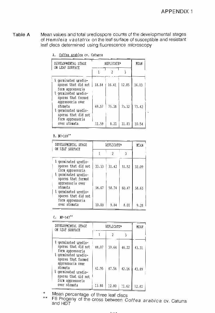
Table A
APPENDIX 1
Mean values and total urediospore counts of the developmental stages of Hemileia vastatrix on the leaf surface of susceptible and resistant leaf discs determined using fluorescence microscopy
A Coffea arabica cv caturra
DEVELOPMENTAL STAGE REPLICATE* MEAN ON LEAF SURFACE
1 2 3
% germinated uredio-spores that did not 18.84 16.41 12.85 16.03 form appressoria
% germinated uredio-spores that formed appressoria over stomata 69.57 75.38 75.32 73.42
% germinated uredio-spores that did not form appressoria over stomata 11.59 8.21 11.83 10.54
B. NC-169··
DEVELOPMENTAL STAGE REPLICATE* MEAN ON LEAF SURFACE
1 2 3
% germinated uredio-spores that did not 33.33 31. 42 31.52 32.09 form appressoria
% germinated uredio-spores that formed appressoria over stomata 56.67 58.74 60.47 58.63
% germinated uredio-spores that did not form appressoria over stomata 10.00 9.84 8.01 9.28
C. NP-547··
DEVELOPMENTAL STAGE REPLICATE* MEAN ON LEAF SURFACE
1 2 3
% germinated uredio-spores that did not 44.07 39.64 46.22 43.31 form appressoria
% germinated uredio-spores that formed appressoria over stomata 41. 95 47.56 42.16 43.89
% germinated uredio-spores that did not form appressoria over stomata 13.98 12.80 11.62 12. 80
* Mean percentage of three leaf discs ** F6 Progeny of the cross between Gaftea arabica cv. Caturra
and HOT

Table B Mean values and total urediospore counts of the developmental stages of Hemileia vastatrix on the leaf surface of susceptible and resistant leaf pieces cut from attached leaves determined using fluorescence microscopy
A Coffea arabica cv caturra . DEVELOPMENTAL STAGE REPLICATE* MEAN ON LEAF SURFACE
1 2 3
% germinated uredio-spores that did not 13.70 8.30 11.00 form appressoria
% germinated uredio-spores that formed appressoria over stomata 82.40 78.20 80.30
% germinated uredio-spores that did not form appressoria over stomata 3.90 13.50 8.70
B. NC-169**
DEVELOPMENTAL STAGE REPLICATE* MEAN ON LEAF SURFACE
1 2 3
% germinated uredio-spores that did not 12.10 15.02 13.56 form appressoria
% germinated uredio-spores that formed appressoria over stomata 71.10 73.30 72.20
% germinated uredio-spores that did not form appressoria over stomata 16.80 11.68 14.20
c. NP-547**
DEVELOPMENTAL STAGE REPLICATE* MEAN ON LEAF SURFACE
1 2 3
% germinated uredio-spores that did not 43.00 34.40 38.70 form appressoria
% germinated uredio-spores that formed appressoria over stomata 44.80
% germinated uredio-54.70 49.75
spores that did not form appressoria over stomata 12.20 10.90 11.55
* Mean percentage of three leaf discs ** F6 Progeny of the cross between Coftea arabica cv. Caturra
and HDT
122

Table C Mean values for the developmental stages of Hemileia vastatrix on the leaf surface of a host and non-host leaf discs determined using fluorescence microscopy
A. Coffea arabica cv. caturra
DEVELOPMENTAL STAGE REPLICATE* MEAN ON LEAF SURFACE
1 2 3 4
% germinated uredio-spores that did not form appressoria
18.84 16.41 12.85 16.14 16.06
% germinated uredio-spores that formed appressoria over stomata 69.57 75.38 75.32 73.35 73.41
% germinated uredio-spores that did not form appressoria over stomata 11.59 8.21 11.83 10.51 10.54
B Camellia sinensis
DEVELOPMENTAL STAGE REPLICATE* MEAN ON LEAF SURFACE
1 2 3 4
% germinated uredio-spores that did not 5.93 15.15 9.69 11.30 10.52 form appressoria
% germinated uredio-spores that formed appressoria over stomata 45.23 27.27 39.21 37.85 37.39
% germinated uredio-spores that did not form appressoria over stomata 48.84 57.58 51.10 50.85 52.09
C. Phaseolus vulgaris
DEVELOPMENTAL STAGE REPLICATE* MEAN ON LEAF SURFACE
1 2 3 4
% germinated uredio-spores that did not 30.90 26.76 21.68 33.14 28.12 form appressoria
% germinated uredio-spores that formed appressoria over stomata 25.53 30.09 31.65 27.78 28 .76
% germinated uredio-spores that did not form appressoria over stomata 43.57 43.15 46.67 39.08 43.12
* Mean percentage of three leaf discs
123

*
A B C D
APPENDIX 2
Mean values for the developmental stages of Hemileia vastatrix on the leaf surface of Gaftea arabica cv. Caturra discs following treatments with the fungicide BayfidanR
DEVELOPMENTAL STATE TREATMENT REPLICATE* MEAN ON LEAF SURFACE
1 2 3
% germinated uredio- A 0 8 . 70 10.92 6.54 spores that did CONTROL 16.67 18.03 12.97 24.23 not form B 27.78 15.56 22.59 2l. 98 appressoria CONTROL 2l. 25 25.48 15.74 20.82
C 8.76 9.17 l. 24 20.22 CONTROL 20.89 11.50 19.37 17.25
D 10.00 16.37 12.16 12.84 CONTROL 19.21 18.34 19.45 19.00
% germinated uredio- A 88.33 85.74 80.63 84.90 spores that formed CONTROL 76 .19 72.73 78.40 75.77 appressoria over B 66.67 79.17 74.12 73.32 stomata CONTROL 72.50 71.29 76.67 73.49
C 74.78 79.75 85.31 79.95 CONTROL 72 .08 78.18 68.10 72.79
D 76.67 77.37 8l. 58 78.54 CONTROL 71 . 49 69.66 72.86 71.34
% germinated uredio- A 11.67 5 . 56 8 . 45 8 . 56 spores that did CONTROL 7.14 9.24 8.63 8.35 not form B 5.55 5.27 3 .2 9 4.70 appressoria over CONTROL 6 .2 5 3.23 7.59 5.69 stomata C 16.46 11.58 13.45 13 .83
CONTROL 7 . 03 10 . 32 12.53 9.96 D 13.33 6.26 6 . 26 8.62
CONTROL 9.30 12.00 7 . 69 9.66
mean percentage on three leaf discs
Inoculated 28 days after second Bayfidan treatment Inoculated 7 days after third Bayfidan treatment Inoculated 14 days after third Bayfidan treatment Inoculated 28 days after third Bayfidan treatment
124

APPENDIX 3
TABLE A
Mean percentages, on nine leaf discs, of the developmental stages of Hemileia vastatrix on the leaf surface of the F6 progeny of a cross between Coftea arabica cv. Caturra and Hybrido de Timor (Colombia). Values were determined on leaf discs using fluorescence microscopy.
DEVELOPMENTAL STAGE CULTIVAR/ REPLICATE MEAN ON LEAF SURFACE LINE
1 2 3
LEAF 1 (youngest leaf)
% germinated uredio- Caturra 7.76 9.31 12.47 9.85 spores that did not NC-169 14.96 0 . 68 3.10 6 . 25 form appressoria NP-547 11.92 13.70 20 . 98 15.53
% germinated uredio- Caturra 57.24 55.56 56.28 56 . 36 spores that formed NC-169 49.61 75 . 08 62 . 07 62 .2 5 appressoria over NP - 547 68 . 87 67.12 53.77 63 . 25 stomata
% germinated uredio- Caturra 35 . 00 35.13 3l. 25 33.79 spores that formed NC-169 35.43 24.24 34.83 3l. 50 appressoria not over NP-547 19 . 21 19.18 25 . 25 2l. 21 stomata
LEAF 2
% germinated uredio- Caturra 33 . 34 2l. 55 19.73 24.87 spores that did not NC-169 23 . 96 26.07 32.13 27.39 form appressoria NP-547 4l. 79 37.47 42.86 40 . 71
% germinated uredio- Caturra 5l. 38 63.16 65 . 38 59 . 97 spores that formed NC-169 61.72 6l. 83 54.92 59.49 appressoria over NP-547 39 . 80 49.34 49.02 46.05 stomata
% germinated uredio- Catur ra 15 .2 8 15. 29 14.89 15.15 spores that formed NC-169 14 . 32 12.10 12.95 13 .12 appressoria not over NP-547 18.41 13 . 19 8.12 13. 24 stomata
LEAF 3
% germinated uredio- Caturra 26 . 60 27.63 27.88 27.37 spores that did not NC-169 33.33 31.42 3l. 52 32.09 form appressoria NP - 547 44 . 07 39.64 40.62 41.44 % germinated uredio- Caturra 60.47 63 . 16 62.32 6l. 98 spores that formed NC-169 56.67 58.74 60.47 58.63 appressoria over NP - 547 4l. 95 47 . 56 43 . 75 44 . 42 stomata % germinated uredio- Caturra 12 . 93 9.21 9.80 10 . 65 spores that formed NC-169 10 . 00 9.84 8 . 01 9.28 appressoria not over NP-547 13.98 12.80 15.63 14.14 stomata
125

TABLE A CONTINUED
DEVELOPMENTAL STAGE CULTIVAR/ REPLICATE MEAN ON LEAF SURFACE LINE
1 2 3
LEAF 4
% germinated uredio- Caturra 14.32 3l.16 34.09 26.52 spores that did not NC-169 20.00 32 . 41 2l.07 24.49 form appressoria NP-547 60.29 65.89 52.47 59.55
% germinated uredio- Caturra 68.18 54.55 50.00 57 . 58 spores that formed NC-169 69.25 50.97 67.06 62.43 appressoria over NP-547 26 . 57 27.70 40 . 66 3l. 64 stomata
% germinated uredio- Caturra 17 . 50 14.29 15.91 15.90 spores that formed NC-169 10.75 16.62 11.87 13 . 08 appressoria not over NP-547 13 .14 6.41 6.87 8.81 stomata
LEAF 5
% germinated uredio- Caturra 44.13 3l.15 39.56 38.28 spores that did not NC-169 50.24 23 . 14 43.08 38.82 form appressoria NP-547 32.17 23.72 38.68 3l. 52
% germinated uredio- Caturra 32.47 50.67 46.15 43.10 spores that formed NC-169 25.36 50.93 46 . 64 40.98 appressoria over NP-547 58.04 56 . 41 45.30 53 . 25 stomata
% germinated uredio- Caturra 23.40 18.18 14.29 18 . 62 spores that formed NC-169 24.40 25 . 93 10 . 28 20.20 appressoria not over NP-547 9 . 79 19.87 16.02 15.23 stomata
LEAF 6
% germinated uredio- Caturra 27.31 33.33 32.85 3l.16 spores that did not NC-169 38.69 40.99 38.20 39.29 form appressoria NP-547 42 . 53 44.31 41.84 42 . 89
% germinated uredio- Caturra 65.00 53.85 56.15 58.33 spores that formed NC-169 56.20 51 . 44 53.35 53.66 appressoria over NP-547 4l. 76 45.54 42.20 43.17 stomata % germinated uredio- Caturra 7.69 12.82 11 . 00 10.50 spores that formed NC-169 5 .11 7 . 57 8.45 7.04 appressoria not over NP-547 15.71 10 . 15 15 . 96 13 . 94 stomata
LEAF 7 (oldest leaf)
% germinated uredio- Caturra 57 . 93 65.94 54 . 98 59.62 spores that did not NC-169 44 . 65 58 . 73 50.91 5l.43 form appressoria NP-547 35 . 75 37.54 45.00 39.43 % germinated uredio- Caturra 28 . 12 20 . 00 33.91 27.34 spores that formed NC-169 48 . 21 28.57 40.00 38.93 appressoria over NP-547 54.00 57 .2 3 46.00 52.41 stomata % germinated uredio- Caturra 13 . 95 14.06 11.11 13.04 spores that formed NC-169 7.14 12 . 70 9.09 9.64 appressoria not over NP-547 10.25 5.23 9.00 8.16 stomata
126

TABLE B
Replicate and mean values on 27 leaf discs of the reaction type, latent period and calculated uredosori concentration on leaf discs of Goftea spp. infected with Hemileia vastatrix
A.
1
**
Gaffea arabica cv. Caturra
LEAF PAIR CATEGORY REPLICATE 1 MEAN
1 2 3
1 Reaction t ype* 9 .0 7.0 7.6 Latent period** 34 37 34 Uredosori/mm2 62 73 70
2 Reaction typ~ 4.5 6.5 6.2 Latent period 37 38 38 Uredosori/mm2 51 53 50
3 Reaction type 9.0 9.0 7 . 2 Latent period 38 38 38 Uredosori/ mm2 45 51 53
4 Reaction type 9 . 0 8 . 0 8.2 Latent period 38 39 38 Uredosori/mm2 48 39 51
5 Reaction type 4.0 4 . 0 3.2 Latent period 40 39 42 Uredosori/mm2 51 37 48
6 Reaction type 2 .0 2 . 8 2.0 Latent period 42 42 43 Uredosori/mm2 17 19 25
7 Reaction type 3 . 5 l.5 2.8 Latent period 42 42 42 Uredosori/mm2 54 37 43
Nine leaf discs were used per replicate Reaction type determined using the scale described in Appendix A in days
127
7.87 35.00 68
5.73 37 . 70 52
8.40 38 . 00 50
8.40 38 . 30 46
3.73 40.30 46
2.27 42.30
20
2.60 42 . 00 45

TABLE B CONTINUED
B. NC-169 (F6 progeny of the cross between C. arabica cv. Caturra and HOT)
LEAF PAIR CATEGORY REPLICATE MEAN
1 2 3
1 Reaction type* 8.5 8.0 8.4 8.30 Latent period** 19 19 20 19.30 Uredosori/mrn2 76 80 70 75
2 Reaction type 8.3 8.3 8.7 8.40 Latent period 20 20 20 20 . 00 Uredosori/mrn2 51 58 60 56
3 Reaction type 9 . 0 7 . 7 8.0 8 . 20 Latent period 20 20 21 20 . 30 Uredosori/mrn2 76 82 78 78
4 Reaction type 8 . 3 7 . 7 7 . 9 8.00 Latent period 20 19 22 20.30 Uredosori/mrn2 57 58 62 59
5 Reaction type 7 . 0 7.0 7.0 7.00 Latent period 23 25 23 23 . 70 Uredosori/mrn2 55 67 51 58
6 Reaction type 5.0 5.3 5.2 5.20 Latent period 25 25 25 25.00 Uredosori/mrn2 31 39 40 37
7 Reaction type 1.0 1.0 1.7 1. 23 Latent period 26 26 25 25.70 Uredosori/mrn2 19 27 30 26
C. NP - 547 (F6 progeny of the cross between C. arabica cv. Caturra and HDT
LEAF PAIR CATEGORY REPLICATE MEAN
1 2 3
1 Reaction type 0 0 0 0
2 Reaction type 0 0 0 0
3 Reaction type 0 . 75 0 0.50 0.42 4 Reaction type 1. 50 0 0.75 0.75 5 Reaction type 3.33 1. 33 1.00 1. 89 6 Reaction type 2.33 2.33 0.67 1. 78 7 Reaction type 3 . 00 2.67 2 . 00 2.56
128

APPENDIX 4.1
Mean percentage of diseased areas of naturally infected leaves with Hemileia vastatrix following the monthly application of selected fungicides over a 24-week period
FUNGICIDE REPLICATION WEEKS AFTER FUNGICIDE J..PPLICATIOII
o 8 12 16 20 24 ------------------- ------------- ------ ------ ------ ------ ------ ------ ------
ANVIL 1 2 3 4
3.78 1.06 1.70 2.07 0.32 2.57 2.26 2.66 2.44 0.14 2.81 3.82 2.48 2.32 0.33 1.28 0.85 2.84 0.20 0.54
1.22 0.63 1.03 1.88 1.06 1. 40 3.42 1.67
------------------- ------------- ------ ------ ------ ------ ------ ------ ------
BAS 480 OOF 1 2 3 4
0.45 0.86 1.76 0.37 0.12 1.08 0.05 0.94 2.44 1.41 0.31 0.14 0.05 0.41 2.04 1.85 2.75 0.60 0.26 0.54 1.14 0.67 2.27 2.48 1.12 0.05 0.59 1.76
------------------- ------------- ------ ------ ------ ------ ------ ------ ------
BAYFIDAN ec 1 2 3 4
1.83 2.31 0.67 0.39 0.09 0.14 0.11 2.73 3.29 3.62 2.15 0.41 0.77 0.45 2.39 2.67 3.09 2.66 1.10 1.59 1.31 3.31 0.83 3.22 1.13 0.63 1.15 1.74
------------------- ------------- ------ ------ ------ ------ ------ ------ ------
BAYFIDAN granules 1 2.80 2.61 2.06 0.41 0.18 0.17 0.~7 2 5.75 4.63 2.17 1.10 0.49 1.57 2.70 3 7.04 2.37 1. 33 0.66 0.55 0.36 0.90 4 0.00 1. 39 1. 34 L41 0.40 1.06 3.11
------------------- ------------- ------ ------ ------ ------ ------ ------ ------
BAYFIDAN + EN600 1 3.03 2.44 1. 31 1.09 0.29 0.23 0.46 2 4.55 1. 09 0.03 0.32 1.17 1. 83 2.23 3 5.04 0.08 0.08 0.23 0.17 0.11 0.00 4 1.57 0.81 1.50 0.75 0.47 1. 88 0.66
-----------------------------------------------------------------------------------

APPENDIX 4.1 CONTINUED
FUNGICIDE REPLICATION WEEKS AFTER FUNGICIDE APPLICATION
o 4 8 12 16 20 24 ------------------- ------------- ------ ------ ------ ------ ------ ------ ------
FBC 25/1 1 2 3 .4
2.48 0.93 1.55 2.08 1.55 1.62 6.38 2.59 0.89 0.12 2.58 2.37
2.09 0.89 1.76 1.15 4.05 0.28 0.80 1.68 1.06 0.59 0.50 0.77 4.57 0.81 1.40 2.87
------------------- ------------- ------ ------ ------ ------ ------ ------ ------
FBC 25/2 1 2 3 4
1.80 1.80 2.01 1.58 0.64 0.66 2.26 6.81 2.35 2.19 4.46 0.45 2.46 0.73 1.71 1.79 1.39 3.64 0.60 1.40 2.12 4.49 6.55 1.95 4.81 0.72 4.16 1.58
------------------- ------------- ------ ------ ------ ------ ------ ------ ------
KOCIDE 1 2 3 4
5.59 1.98 3.01 1.23 0.26 0.41 0.42 3.76 3.38 2.22 5.44 0.66 2.32 5.36 2.57 7.14 1. 49 5.44 0.66 1.94 3.26 0.55 0.17 1.32 1.35 1.60 2.83 6.72
------------------- ------------- ------ ------ ------ ------ ------ ------ ------
PUNCH C 1 2 3 4
0.32 0.25 0.30 0.21 0.10 0.06 0.12 0.65 0.65 0.16 0.93 0.28 1.99 2.10 3.02 1.14 0.62 1.50 0.12 1.74 0.94 0.83 0.91 2.42 0.67 1.42 0.67 2.20
------------------- ------------- ------ ------ ------ ------ ------ ------ ------
SYSTHANE
-------------------
CONTROL
1 2 3 4
-------------
1 2 3 4
5.36 1.52 2.83 1.26 0.30 0.40 0.32 0.17 0.05 0.05 0.00 0.00 0.00 0.00 2.35 2.30 3.23 1.24 0.59 0.51 0.75 4.65 4.41 2.38 6.65 4.70 2.21 2.18
------ ------ ------ ------ ------ ------ ------
2.86 1.67 2.58 11.70 7.62 4.66 4.83 2.92 2.21 6.26 12.06 8.00 2.62 5.20 3.54 2.63 5.80 7.84 4.78 4.11 7.79 3.06 1.77 3.98 8.62 6.14 8.62 8.02
------------------------------------------------------------------------------------
130

APPENDIX 4.2
Statistical differences (P < 0.05) in percentage diseased area in time and with replication following the monthly application of selected fungicides over a 24-week period
FUNGICIDE TIME % DISEASED REPLICATE % DISEASED (weeks) AREA AREA
JJfVIL 0 2.61 a 1 1.54 a 4 2.00 a 2 1.85 a 8 2.42 a 3 2.03 a
12 1. 76 a 4 1. 54 a 16 0.33 b 20 1. 68 ab 24 1. 40 ab
BAS 480 OOF 0 1.03 be 1 0.67 a 4 1. 86 ab 2 0.81 a 8 2.10 a 3 1.31 a
12 0.60 cd 4 1.28 a 16 0.14 cd 20 0.57 cd 24 0.84 cd
BAYFIDAN ec 0 2.57 a 1 0.79 a 4 2.28 a 2 1. 92 ab 8 2.65 a 3 2.12 b
12 1. 58 ab 4 1. 72 ab 16 0.56 b 20 0.91 b 24 0.90 b
BAYFIDAN ec + EN600 0 3.55 a 1 1.26 a 4 1.11 b 2 1.60 a 8 0.73 b 3 0.82 a
12 0.60 b 4 1.09 a 16 0.53 b 20 1.01 b 24 0.84 b
BAYFIDAN granules 0 3.90 a 1 1.20 a 4 2.75 ab 2 2.63 a 8 1. 73 be 3 1.89 a
12 0.90 be 4 1. 24 a 16 0.41 c 20 0.79 be 24 1.72 be
"
131

APPENDIX 4.2 CONTINUED
FUNGICIDE TIME % DISEASED REPLICATE % DISEASED (weeks) }.REA AREA
rBC 25/1 0 2.77 a 1 1.55 a 4 1. 91 ab 2 1. 72 a 8 1. 61 ab 3 1.83 a
12 2.94 a 4 2.10 a 16 0.64 b 20 1.12 ab 24 1. 62 ab
rBC 25/2 0 3.70 a 1 1.54 a 4 3.12 a 2 3.47 b 8 1. 89 ab 3 1. 81 ab
12 3.62 a 4 2.78 ab 16 0.60 b 20 2.17 ab 24 1. 67 ab
KOCIDE 0 3.12 ab 1 1. 84 a 4 3.17 ab 2 3.31 a 8 2.01 ab 3 3.21 a
12 3.37 ab 4 2.08 a 16 0.80 b 20 1. 88 ab 24 3.94 a
PUNCH C 0 1.21 a 1 0.19 a 4 0.74 a 2 0.97 ab 8 0.88 a 3 1. 30 b
12 0.83 a 4 1.30 b 16 0.48 a 20 1.12 a 24 1. 34 a
SYSTHANE 0 3.13 a 1 1.71 b 4 2.07 a 2 0.04 a 8 2.12 a 3 1.57 b
12 2.29 a 4 3.88 e 16 1. 40 a 20 0.78 a 24 0.81 a
CONTROL 0 3.10 ab 1 5.13 a 4 2.07 a 2 5.61 a 8 4.66 be 3 5.21 a
12 10.10 d 4 5.74 a 16 6.64 e 20 5.00 be 24 6.46 e
132

APPENDIX 4.3
Scores on selected trees rated on a five-tree grid scale at the site of the fungicide trial 24-weeks after the first treatment. Overall appearance, die-back, overbearing and rust severity were rated
-- ----- ----- ------------------------ -- ---------- -- -----
ROW 1 LINE 1 OVERALL 1 OVERBEARING 1 DIE-BACK 1 RUST ------ --------- -------------1-- -- ------ ------
1 15 8 yes 1 3 1 1 20 5 yes 1 1 2 1 25 8 no 1 1 1 1 30 7 yes 1 5 2 1 35 4 yes 1 6 2 1 40 6 yes 1 3 3 3 5 2 yes 1 7 1 3 5 3 yes 1 7 1 3 10 3 yes 1 8 3 3 15 9 yes 1 1 0 3 20 4 yes 1 8 2 3 25 10 yes 0 0 3 30 4 yes 6 1 3 35 1 yes 10 2 3 40 10 no 0 0 5 1 5 yes 5 3 5 5 3 yes 8 2 5 5 5 no 4 0 5 10 5 no 7 1 5 15 3 no 7 1 5 20 1 no 9 3 5 25 1 no 8 3 5 30 9 yes 0 1 5 35 10 yes 0 0 5 40 7 no 5 1 7 1 6 yes 5 2 7 5 5 yes 4 2 7 5 10 no 0 0 7 10 5 yes 7 1 7 15 3 yes 9 2 7 20 2 yes 8 3 7 25 5 yes 6 3 7 30 10 no 0 0 7 I 35 6 yes 5 1 7 1 40 10 no 0 1 9 1 1 8 no 2 1 9 I 5 3 no 6 1 9 1 5 10 no 0 0 9 1 10 3 yes 8 3 9 1 15 5 no 1 1 9 I 20 7 no 3 1 9 1 25 1 yes 0 1 9 1 30 3 yes 8 2 9 1 35 5 yes 3 1 9 1 40 7 yes 5 3 11 1 20 1 yes 9 1 11 1 25 5 yes 5 3 11 1 30 8 no 1 2 11 1 35 10 no 0 0 11 I 40 10 no 0 0 ---------- -------- ----------------------- ---------------
1~~

APPENDIX 4.4
Disease severity on a 0 to 3 scale (0 = no infection, 3 = severe infection) on fungicide-treated and border (neighbouring) trees within the four replicates used for the fungicide trial
* **
FUNGICIDE REPLICATE 1
A* B**
Bayfidan ec 1 1 Bayfican gr 1 0 Systhane 1.5 1.5 Kocide 2 1 Anvil 2 1 FBC 25/2 2 3 FBC 25/1 2 0 Punch C 1 2 BAS 480 OOF 1 2 Bayfidan ec +
1% EN 600 0 3 Control 3 3
FUNGICIDE REPLICATE 3
A* B**
Bayfidan gr 2 2 FBC 25/2 3 2 Systhane 2 3 Bayfidan ec 2 3 Bayfidan ec +
1% EN 600 3 2 Punch C 2 2 Kocide 3 3 Anvil 3 3 FBC 25/1 2 3 BAS 480 OOF 2 2 Control 3 3
Fungicide treated trees Neighbouring, untreated tree
FUNGICIDE REPLICATE ' 2
A* B**
Bayfidan ec 1 2 Kocide 3 3 Bayfidan ec +
1% EN 600 1 2 Bayfidan gr 3 3 FBC 25/2 1 2 BAS 480 OOF 2 3 Systhane 0 2 Anvil 2 3 FBC 25/1 3 3 Punch C 3 3 Control 3 3
FUNGICIDE REPLICATE 4
A* B**
Punch C 2 3 FBC 25/2 2 2 Anvil 2 3 Kocide 3 2 Bayfidan gr 3 3 Bayfidan ec +
1% EN 600 3 3 Systhane 3 3 FBC 25/1 3 3 Bayfidan ec 3 3 BAS 480 OOF 2 2 Control 3 3
Note that the design of the experiment was such that the sequence was as follows: untreated, treated, untreated, etc. down a row
134


![Transgenic Expression ofaFungalendo-Polygalacturonase ... · Transgenic Expression ofaFungalendo-Polygalacturonase Increases Plant Resistance to Pathogens and Reduces Auxin Sensitivity1[W]](https://img.pdfslide.us/doc/110x75/5f5599280b86d949f711ae38/transgenic-expression-ofafungalendo-polygalacturonase-transgenic-expression.jpg)