Embed Size (px)
Citation preview

Planta (1993) 190:25-31
Studies on tbe expression of NDH-H, a subunit of tbe N AD(P)H -plastoquinone-oxidoreductase of bigber-plant cbloroplasts Susanne Berger, Ulrike Ellersiek, Peter Westhoff, K1aus Steinmiiller Institut fUr Entwicklungs- und Molekularbiologie der Pflanzen, Heinrich-Heine-Universitiit, Universitiitsstrasse I, W-4000 Diisseldorf I, FRG
Received: 1 October 1992/ Accepted: 9 November 1992
Abstract. The plastid genomes of higher plants contain eleven reading frames (ndhA-K) that are homologous to genes encoding subunits of the mitochondrial NADHubiquinone-oxidoreductase (complex I). The carboxyterminal end of the NDH-H subunit from rice (Oryza sativa L.) was expressed as a fusion protein in Escherichia coli and antibodies against the fusion protein were generated in rabbits. The antibody was used to study the expression of NDH-H, and the following results were obtained: (i) NDH-H is expressed in mono- and dicotyledonous plants, (ii) NDH-H is localized on the stroma lamellae of the thylakoid membrane and (iii) NDH-H is expressed in etioplasts. Together with the finding that two other ndh genes (ndhI and ndhK) are expressed in plastids, these results point to the existence of an NAD(P)H-plastoquinone-oxidoreductase on the thylakoid membrane. The possible function of the enzyme in plastids is discussed and it is suggested that it works in balancing the ATP/ADP and the NADPH/NADP ratios during changing external (i.e. light) or internal (i.e. ATP and NADPH demands of biosynthetic pathways of the plastid) conditions.
Key words: NAD(P)H-plastoquinone-oxidoreductase -ndhH gene - Plastid DNA
Introduction
Besides genes for the well-known multisubunit protein complexes of the thylakoid membrane (PSI and 11, cytochrome b6/f complex and ATPase), the plastid genome contains reading frames that encode components of a putative NAD(P)H-dehydrogenase (Ohyama et al. 1986; Shinozaki et al. 1986; Hiratsuka et al. 1989). Seven of these genes (ndhA-G) have been identified by their
Abbreviations: PVDF=polyvinylidene difluoride; SDS=sodium dodecyl sulfate
Correspondence to: K. Steinmiiller; FAX: 49(211)311-4871
homology to mammalian mitochondrial genes, which encode subunits of the NADH-ubiquinone-oxidoreductase (complex I) of the respiratory chain (Chomyn et al. 1985, 1986). Recently, it was shown that four more reading frames of plastid DNA have homology to subunits of complex I and these were therefore designated as ndh genes: ndhH (Fearnley et al. 1989), ndhI (WU et al. 1989; Dupuis et al. 1991), ndhJ (Videira et al. 1990; Pilkington et al. 1991) and ndhK (Berger et al. 1991; Masui et al. 1991 ; Arizmendi et al. 1992).
The mitochondrial NADH-dehydrogenase is a membrane protein complex that is built up of more than 30 different subunits. It couples the oxidation of NADH and the reduction of ubiquinone with a proton translocation across the inner mitochondrial membrane (Hatefi 1985; Ragan 1987; Weiss et al. 1991). By analogy, the plastid dehydrogenase is thought to act as a NAD(P)Hplastoquinone-oxidoreductase (Ohyama et al. 1988; Marder and Barber 1989). However, at present the function of such an enzyme in plastid metabolism is unknown (see Discussion for details).
Moreover, though it was shown that the ndh genes are transcribed in tobacco (Matsubayashi et al. 1989) and maize (Schantz et al. 1988; Steinmiiller et al. 1989), the existence of an NAD(P)H-dehydrogenase in plastids, either by determination of an NAD(P)H-oxidase activity or by isolation and characterization of the whole protein complex, has not yet been achieved.
Today there are only two reports on the identification ofNDH subunits in chloroplasts. The first one, NDH-K, was formerly thought to encode a PSII subunit and was therefore designated PSII-G (gene psbG) by Steinmetz et al. (1986). This view was questioned by the finding that the protein occurs predominantly in stroma thylakoids and it was suggested that it is an NDH subunit (Nixon et al. 1989). Since a 20-kDa polypeptide with homology to NDH-K (PSII-G) has recently been identified in a complex-I preparation from bovine heart (Masui et al. 1991; Arizmendi et al. 1992), the classification as an NDH-protein is justified. The other subunit, the NDH-I protein (also designated as FRX-B, because of its ho-

26
mology to ferredoxins), was originally found as a protein which binds to the replicative origin of chloroplast DNA (Wu et al. 1989). Again, a homologous polypeptide was detected in a complex-I preparation from bovine heart (Dupuis et al. 1991), demonstrating the identity as an NAD(P)H-dehydrogenase subunit.
We show here that the ndhH gene is also expressed in mono- and dicotyledonous plants, and we demonstrate that NDH-H is located on the stroma thylakoids like NDH-K. Our data provide further evidence for the presence of an NAD(P)H-plastoquinone-oxidoreductase in higher-plant chloroplasts.
Materials and methods
Growth of plants. Spinach (Spinacea oleracea, L. cv. Monatol; Fa. Sperling, Liibeck, FRG), beans (Phaseolus vulgaris, L. cv. Sotexa; Fa. Rhode, Guxhagen, FRG), maize (Zea mays, L. B73; Pioneer Hi-Bred Inc., PJainyiJle, Tex. USA) and Sorghum bic%r, Monch (TX430; Pioneer Hi-Bred Inc.) were grown in a greenhouse. For the illumination experiments, maize and Sorghum were grown in a light-safe growth chamber at 26° C and 90% relative humidity for 8 and lld, respectively. The plants were then illuminated for up to 24 h at 9000 Ix.
Isolation of plant extracts. The isolation ofplastids from the various species and for the illumination experiments was carried out according to procedure I described by Herrmann (1982).
For the isolation of the different plastid fractions from spinach, the initial isolation of plastids was also carried out as described in Herrmann (1982). Broken and intact plastids were then separated by centrifugation through a Percoll step gradient (5 ml plastid suspension and 12 ml 45% and 10 ml 85% Percoll) using the solutions described by Bartlett et al. (1982). The centrifugation was for 10 min at 8000 rpm in a Sorvall HB-4 rotor (Sorvall DuPont de Nemours, Bad Homburg, FRG). The intact chloroplasts were recovered from the 45/85% Percoll interphase and osmotically lysed in 10 mM 4-(2-hydroxyethyl)-l-piperazineethanesulfonic acid (HEPES)-KOH (pH 7.5),4 mM MgCI2 • The subsequent sucrosegradient centrifugation for the separation of the stroma and the envelope and the thylakoid membranes was performed as described in Douce and Joyard (1982).
Grana and stroma thylakoids from spinach were isolated by Triton X-lOO solubilization as described by Berthold et al. (1981) and Ghanotakis et a!. (1984). The solubilized stroma thylakoids were diluted fivefold with supension buffer (50 mM 4-morpholineethanesulfonic acid (Mes)-NaOH (pH 6.0), 15 mM NaCl, 5 mM MgCI2 , I mM sodium ascorbate) and centrifuged for 60 min at 190000 . 9 in a Beckman 45 Ti rotor (Beckman, Miinchen, FRG) at 4° C to pellet any contaminating grana lamellae. The supernatant was precipitated with trichloroacetic acid for sodium dodecyl sulfate (SDS)-gel electrophoresis and immunoblot analysis.
The protein concentration was measured according to Bradford (1976) and chlorophyll was determined with dimethylformamide as described by Moran (1982).
Electrophoresis and Western blotting. Preparative and analytical SDS-polyacrylamide electrophoresis was carried out according to Laemmli (1970) or Schiigger and von Jagow (1987), respectively.
For immunoblot analysis, the proteins were transferred to polyvinylidene difluoride membranes (PVDF, lmmobilon-P; Millipore, Eschborn, FRG). The antigen-antibody complexes were detected either with 12~I-labeled protein A (Westhoff et a!. 1985) or with the ECL Western-blotting-analysis system (Amersham Buchler, Braunschweig, FRG).
Preparation of an antibody against NDH-H. General molecularcloning techniques were performed according to Maniatis et a!.
S. Berger et a!.: Studies on the expression of NOH-H
(1982) and nucleotide and protein sequences were analysed with the PC/GENE software (Genofit, Heidelberg, FRG).
The rice plastid DNA subclone pRP5-2 was constructed by inserting the 3429-bp Sa/l-XhoI fragment from the primary clone pRP5 (Shimada et al. 1989) into the corresponding sites of the vector Bluescript pKS (Stratagene). From pRPS-2, a 19S8-bp Bg/ll fragment (nucleotides 111563-113521, Shimada et al. 1989) was isolated and inserted into the BamHI site of the expression vector pGEMEX2 (Promega, Heidelberg). The resulting plasmid, pGXI958RH contained 68% of ndhH (amino acids 125-393) fused in frame to the reading frame for the first 260 amino acids of the bacteriophage T7 gene-l0 protein. However this construct did not lead to the desired expression in E. coli. Therefore the internal parts of the ndhH reading frame were eliminated by cutting pGX 1958RH with £CoRI and religating. The resulting plasmid, pGXI430RH encompasses the carboxy-terminal 24% (amino acids 300-393) of ndhH.
The expression vectors were transformed into the E. coli strain JM 109(DE3) according to the manufacturer's protocol, and the expression was analysed by electrophoresis of SOS-solubilized bacterial pellets. The fusion protein was isolated by lysis of the bacteria with lysozyme and washings with 3.5 M urea as described in Strebel
et a1. (1986). After a final Qurification by SDS-gel electrophoresis and elution of the desired band, the fusion protein was used to elicit antibodies in rabbits according to standard procedures (Harlow and Lane 1988). The antibodies against DI (PSII-A), CP47 (PSII-B) and PSI-A from spinach are described in Oswald et al. (1990).
Results
Characteristics of NDH-H. The identification of NDH-H as an NAD(P)H-plastoquinone-oxidoreductase subunit encoded in plastid DNA was first described by Fearnley et al. (1989). The mitochondrial counterpart to NDH-H had been known before as the 49-kDa subunit. The protein is localized in the so-called iron-sui fur protein fragment or the peripheral arm of complex I, referring to the old (Hatefi 1985; Ragan 1987) and the new (Weiss et al. 1990) model of the enzyme, repectively. Table 1 shows a sequence analysis of the NDH-H proteins of different organisms. The plastid proteins are clearly more closely related to the cyanobacterial polypeptide than to their mitochondrial counterparts, which are in general some 40 amino acids longer. The existence of transmembrane alpha helices or membrane-associated helices was analysed with three different algorithms of PC/GENE. Though membrane-bound, NDH-H is a relatively hydrophilic protein. The RAOARGOS algorithm indicates one transmembrane-spanning alpha helix for the plastid proteins only when a window of 16 amino acids is used (Table 1, lane 5). With the exception of the cyanobacterial protein, the two other algorithms classify NDH-H in general as a peripheral polypeptide (Table 1, lanes 6, 7).
The NDH-H protein is expressed in mono- and dicotyledons. A rice-plastid DNA fragment, encompassing the carboxy-terminal 94 amino acids of NDH-H was inserted into the expression vector pGEMEX. The resulting fusion protein was purified and injected into rabbits to generate antibodies. The antibody was tested for its specificity against chloroplasts from four different plant species. The total plastid proteins from spinach, beans, maize and Sorghum were separated by SDS-electro-

S. Berger et al.: Studies on the expression of NDH-H
Table 1. Comparison of characteristics of NDH-H polypeptides from various organisms. The sequences were from Hiratsuka et al. 1989 (rice), Maier et al. 1990 (maize), Shinozaki et al. 1986 (tobacco), Ohyama et al. 1986 (liverwort), Steinmilller 1992 (Synechocystis sp. PCC6803), Fearnley et al. 1989 (bovine) and Preis et al. 1990 (Neurospora crassa). The sequences were analysed with subprograms of PC/GENE. 1. Number of amino acids. 2. Molecular mass.
27
3. Amino-acid sequence similarity as percentage of identical amino acids relative to NDH-H of rice. 4. Same as in 3 for the carboxyterminal 94 amino acids. 5,6,7. Determination of the borders of transmembrane alpha helices or membrane-associated helices with subprograms of PC/GENE: 5, with RAOARGOS and windows of 16 or 20 amino acids; 6, with SOAP; 7 with HELIXMEM
Amino Molecular Amino-acid sequence Borders of transmembrane or acids mass similarity I 2 Whole
protein 3
Oryza saliva 393 45685 100 Zea mays 393 45687 96 N.labacum 393 45487 89 M. polymorpha 392 45372 83 Synechocystis 394 45534 70 sp. PCC6803 Bos laurus 430 49192 41 N. crQSsa 436 49243 38
1 2 3 4 kDa
-69 '..-
-46 """"~
.., .--30
- 21.5
- 14.3 Fig. 1. lmmunoblot analysis. The plastid proteins of different plants were separated by SDS-gel electrophoresis, transferred to PVDF membranes and tested with the NDH-H antibody. Lane 1. spinach; lane 2. Sorghum; lane 3. bean; lane 4. maize
phoresis, transferred onto PVDF membranes and tested with the rice NDH-H antibody (Fig. 1). A strong reaction was obtained with chloroplasts from the cereals maize and Sorghum and a distinct reaction was also seen with spinach, while with bean the signal was barely vis-
membrane-associated alpha helices according to:
Last 94 RAOARGOS SOAP HELIXMEM amino acids 5 6 7 4
100 111-127 (16) 98 111-127 (16) 88 111-127 (16)
110-126 (16) 373-389 115-135
372-392 150-171 (16,20) 156-177 (16,20) 416-432 247-268 (16,20) 416-432 (16)
ible. Since the amino-acid sequence similarity for the terminal 94 amino acids between rice and maize is higher (92 identical amino acids) than between rice and tobacco (83 identical amino acids, Table 1), the differences in cross-reactivity may reflect differences between monoand dicotyledonous species.
The calculated molecular mass for the NDH-H polypeptides from rice, maize and tobacco (Table 1) is in good agreement with the approximate molecular mass of the NDH-H polypeptides from the four different plants, estimated after gel electrophoresis (43-45 kDa).
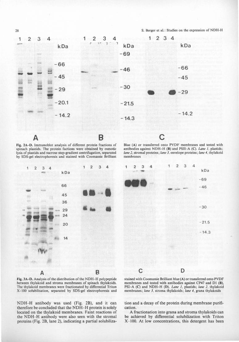
The NDH-H protein is present on the stroma-thylakoid membrane but absent from the chlorop/ast envelope. The distribution of the NDH-H subunit within the chloroplast was investigated in two steps. First, the envelope and thylakoid membranes were analysed to see if they contained the polypeptide and then the distribution of NDH-H between stroma and grana thylakoids was determined. Since NDH-H is a relatively hydrophilic protein (Table 1) and therefore might become solubilized during membrane preparation, the stromal proteins were also included in the analysis.
Lysed spinach plastids were separated by sucrosegradient centrifugation into stroma, envelope and thylakoid-membrane fractions. The analysis of the protein fractions by SDS-gel electrophoresis revealed that the polypeptide compositions of all three fractions were completely different, indicating a clean separation (Fig. 2A). The purity of the chloroplast envelope membranes was further demonstrated by the absence of chlorophyll in the preparation (not shown) and by the Western blot reaction with an antibody against the Dl protein ofPSII. The PSII-A antibody labelled the corresponding protein only in the thylakoid-membrane preparation but not in the stromal proteins or the envelope-membrane fraction (Fig. 2C). The same pattern was obtained when the
28
1 2 3 4 1 2 3 4 - r 1"- 1 - "1 k Da
- 66 .- -.. - - -- 45 - -- f!"- - 29 - -- - 20.1
- - 14.2
A B Fig. 2A-0. Immunoblot analysis of different protein fractions of spinach plastids. The protein factions were obtained by osmotic lysis of plastids and sucrose step-gradient centrifugation, separated by SOS-gel electrophoresis and stained with Coomassie Brilliant
1 2 3 4 2 3 4 ...... k Da
66
45 III ~ I 36
29 • 24
20 ... 14
A B Fig. 3A-0. Analysis of the distribution of the NOH- H polypeptide between thylakoid and stroma membranes of spinach thylakoids. The thylakoid membranes were fractionated by differential Triton X- lOO solubilisation, separated by SOS-gel electrophoresis and
NDH- H antibody was used (Fig. 2B), and it can therefore be concluded that the NDH- H protein is solely located on the thylakoid membranes. Faint reactions of the NDH- H antibody were also seen with the stromal proteins (Fig. 2B, lane 2), indicating a partial solubiliza-
S. Berger et al. : Studies on the expression of NOH-H
1 2 3 4
kD a k Da
- 69
- 46 - 66
- 45
- 30 . - 29
- 21.5
- 14.2 - 14.3
c Blue (A) or transferred onto PVOF membranes and tested with antibodies against NOH- H (8) and PSII- A (C). Lane 1. plastids; lane 2. stromal proteins ; lane 3. envelope proteins; lane 4. thylakoid membranes
1 2 3 4 2 3 4 kDa
- 69
- ~ - - 46
-30
- 21 .5
- 14.3
c D stained with Coomassie Brilliant blue (A) or transferred onto PVOF membranes and tested with antibodies against CP47 and 01 (8), PSJ-A (C) and NOH-H (0). Lane 1. plastids ; lane 2. thylakoid membranes ; lane 3. stroma thylakoids ; lane 4. grana thylakoids
tion and a decay of the protein during membrane purification.
A fractionation into grana and stroma thylakoids can be achieved by differential solubilization with Triton X- JOO. At low concentrations, this detergent has been

S. Serger et al.: Studies on the expression of NDH-H
o 4 24 0 3 6 9 24 kOa - 92.5
.... •• - 69
- 46
A B
- 6 9
- .... - 46 - - --- - -- 30
c D Fig. 4A-D. Immunoblot analysis of the expression of PSI-A (A, H) and NDH- H (C, D) in Sorghum (A, C) and maize (H, D) etiochloroplasts during illumination. The plants were grown for 11 d (Sorghum) or 8 d (maize) in darkness (0) and then illuminated for 4 and 24 (Sorghum) or 3 to 24 h (maize)
shown to solubilize preferentially the stroma lamellae while it leaves the stacked grana thylakoids intact (Berthold et al. 1981). The solubilization of spinach thylakoid membranes according to the improved protocol of Ghanotakis et al. (1984) yielded an almost clean separation between grana and stroma thylakoids. (Fig. 3A). As a control, the different fractions were tested with antibodies against two PSII subunits (CP47 and DI, Fig. 3B) and a PSI subunit (PSI- A; Fig. 3C). The cross-contaminations between the grana- and the stroma-thylakoid fractions were negligible, as can be inferred from the weak signals of CP47 with the stroma lamellae (Fig. 3B, lane 3) and the faint band of the PSI-A protein in the grana preparation (Fig. 3C, lane 4). When the NDH-H antibody was used, a distinct signal was seen with the stroma lamellae but not with the grana-thylakoid membranes (Fig. 3D).
The expression of NDH- H is light-stimulated but lightindependent. Sorghum and maize plants were grown in the dark for several days and then illuminated for up to 24 h. The plastids were then isolated and analysed by an immunoblot. The results presented in Fig. 4 show that appreciable amounts ofNDH- H were already present in the etioplasts of both species and that upon illumination the amount of NDH- H increased slightly (Fig. 4C, D) . As a control, the accumulation of the PSI- A protein was determined. The protein was not detectable in the dark but a 'massive increase occurred during illumination (Fig. 4A, B).
Discussion
The mitochondrial complex I consists of more than 30 different subunits (Ragan 1987; Weiss et al. 1991). It is
29
likey that the plastid NAD(P)H-dehydrogenase is of similar complexity, since so far eleven genes have been characterized (ndhA-K) and several subunits that are involved in electron transport in complex I, e.g. the 51 kDa subunit which carries the FMN- and the NADHbinding sites, or the 78-kDa subunit which contains two iron-sulfur clusters, have not yet been identified in chloroplasts. Thus, the expression of the three subunits (NDH-H, I, K) so far identified is taken as evidence for a functional NAD(p)H-dehydrogenase in plastids.
All ndh genes with homology to plastid ndh-genes have also been isolated from cyanobacteria and the amino-acid sequence similarity between cyanobacterial and plastidial ndh genes (60--80%) is significantly higher than between plastids and mitochondria (20--40%). Therefore, the cyanobacterial and the plastid enzymes are clearly related (Steinmiiller et al. 1989 ; Ellersiek and Steinmiiller 1992). Interestingly, cyanobacteria contain a complete respiratory chain on the cytoplasmic and the thylakoid, or intracytoplasmic, membranes (Molitor and Peschek 1986 ; Peschek 1987). This has been demonstrated by the identification of subunits for NADH-dehydrogenase (Berger et al. 1991), the cytochrome b6/f complex (Kraushaar et at. 1990) and cytochrome c oxidase (Peschek et al. 1988) on both membranes.
From an evolutionary point of view, the inner membrane of the chloroplast envelope is homologous to the cytoplasmic membrane of cyanobacteria (Schnepf and Brown 1971). We therefore investigated whether the NDH- H subunit is also present on both membrane systems in plastids. Our experiments with spinach clearly demonstrate that in chloroplasts the enzyme is restricted to the thylakoid membrane. This agrees with the report of Douce et al. (1973), who found that the plastid envelope does not contain NAD(P)H-cytochrome coxidase activity.
In the generally accepted model of the chloroplast thylakoid membrane, PSII and PSI are confined to the grana and the stroma thylakoids, respectively (Anderson and Andersson 1982; Staehelin and DeWit 1984). We found that NDH- H is also restricted to the stroma lamellae of the thylakoids (Fig. 3). This result is in accordance with the finding of Nix on et al. (1989), who reported that NDH- K (PSII-G) is located preferentially in the stroma-thylakoid fraction.
This raises the question why an NAD(P)H-dehydrogenase has been maintained on the thylakoid membrane during the evolution from endosymbionts to plastids?
Bennoun (1982) showed that both NADH and NADPH were able to reduce the plastoquinone pool in the dark in the PSI-deficient Chlamydomonas mutant F14 and suggested that a complete respiratory pathway from NAD(P)H to H 20 operates in chloroplasts of higher plants. He called this process chlororespiration and suggested that this electron transport serves the reoxidation of reduction equivalents, generated during starch breakdown via the oxidative pentose-phosphate pathway in the chloroplast in the dark, and thus ensures a permanent supply of oxidized carriers.
Besides this function as a valve for reduction equivalents, we propose that the NAD(P)H-dehydrogenase is another device by which the chloroplast coordinates

30
NADPH and ATP synthesis. A calculation of the stochiometry of the light and dark reactions of photosynthesis reveals that additional ATP is required for CO2
fixation (Horton 1985). It is generally assumed that this extra A TP is produced by cyclic photophosphorylation or by photoreduction of O2 (Mehler reaction; Horton 1985; Robinson 1988). Recycling of NADPH via the proton-pumping dehydrogenase would also lead to increased A TP production.
Moreover, it is known that the NADPH/NADP and the ATP /ADP ratios in chloroplasts are regulated very tightly because they are essential for efficient carbon fixation (Horton 1985; Foyer et al. 1990). However, under natural conditions several factors can cause imbalances in these ratios. In general, two categories of factors can be distinguished:, Firstly, there are environmental changes that may lead to variations in A TP and NADPH production, e.g. changes in light quantity and quality. Secondly, differences in the consumption of ATP and NADPH may occur by alterations of the activities of the various metabolic pathways in chloroplasts (e.g. reductive pentose-phosphate pathway, nitrite assimilation or lipid synthesis). If such variations take place, the NAD(P)H-dehydrogenase on the thylakoid membrane may act as a regulator for the coordination between photosynthetic electron flow and ATP and NADPH production.
A recent report which shows that the plastid DNA of the holoparasitic plant Epifagus virgiana has lost ndh genes, together with several genes for components of the photosynthetic electron-transport chain, supports this view (dePamphilis et al. 1991). However, as our illumination experiments demonstrate (Fig. 4), the expression of NDH-H takes place even in the etioplasts in maize and Sorghum. In contrast, the appearance of the chlorophyllbinding proteins of the reaction centers of PSI and PSII is dependent on illumination (Fig. 4, Kreuz et al. 1986; Sutton et al. 1987; Schrubar et al. 1991). Thus, although the concentration of NDH-H increases during illumination, the expression is not coordinated with the synthesis of proteins for the photosynthetic apparatus. Assuming that the NDH-H protein, present in the etioplasts, assembles into a functional enzyme, this result shows that the NAD(P)H-dehydrogenase may have other functions in plastids besides the interaction with photosynthetic electron transport.
We thank Professor M. Sugiura for the rice plastid DNA clone bank, Oliver Buchholz for Sorghum plastid membranes, Pioneer Hi-Bred Inc. for maize and Sorghum seeds and the Deutsche Forschungsgemeinschaft for financial support (SFBI89).
References
Anderson, J.M., Andersson, B. (1982) The architecture of photosynthetic membranes: lateral and transverse organization. Trends Biochem. Sci. 7, 288-292
Arizmendi, J.M., Runswick, MJ., Skehel, J.M., Walker, J.E. (1992) NADH :ubiquinone oxidoreductase from bovine heart mitochondria. A fourth nuclear encoded subunit with a homologue encoded in chloroplast genomes. FEBS Lett 301, 237-242
S. Berger et al.: Studies on the expression of NDH-H
Bartlett, S.G., Grossman, A.R., Chua, N.H. (1982) In vitro synthesis and uptake of cytoplasmically-synthesized chloroplast proteins. In: Methods in chloroplast molecular biology, pp. 1081-1090, Edelman, M., Hallick, R.B., Chua, N.-H., eds., Elsevier, Amsterdam
Bennoun, P. (1982) Evidence for a respiratory chain in the chloroplast. Proc. Natl. Acad. Sci. USA 79, 4342-4356
Berger, S., ElIersiek, U., Steinmiiller, K. (1991) Cyanobacteria contain a mitochondrial complex I-homologous NADH-dehydrogenase. FEBS Lett, 286, 129-132
Berthold, D.A., Babcock, G.T., Yocum, C.F. (1981) A highly resolved, oxygen-evolving photosystem 11 preparation from spinach thylakoid membranes. FEBS Lett. 134,231-234
Bradford, M.M. (1976) A rapid and sensitive method for the quantitation of microgram quantities of proteins utilizing the principle of protein-dye binding. Anal. Biochem. 72, 248-254
Chomyn, A., Mariottini, P., Cleeter, M.WJ., Ragan, C.!', Matsuno-Yagi, A., Hatefi, Y., Doolittle, R.F., Attardi, G. (1985) Six unidentified reading frames of human mitochondrial DNA encode components of the respiratory-chain NADH dehydrogenase. Nature 314, 592-597
Chomyn A., Cleeter, M.WJ., Ragan, C.L, Riley, M., Doolittle, R.F., Attardi, G. (1986) URF6, last unidentified reading frame of human mtDNA, codes for an NADH dehydrogenase subunit. Science 234, 614-618
dePamphilis, C.W., Palm er, J.D. (1990) Loss of photosynthetic and chlororespiratory genes from the plastid genome of a parasitic flowering plant. Nature 348, 337-339
Douce, R., Joyard J (1982) Purification of the chloroplast envelope. In: Methods in chloroplast molecular biology, pp. 239-256, Edelman, M., Hallick, R.B., Chua, N.-H., eds., Elsevier, Amsterdam
Douce, R., Holtz, R.B., Benson A.A. (1973) Isolation and properties of the envelope of spinach chloroplasts. J. BioI. Chem. 248, 7215-722
Dupuis, A., Skehel, J.M., Walker J.E. (1991) A homologue of a nuclear-coded iron-sulfur protein subunit of bovine mitochondrial complex I is encoded in chloroplast genomes. Biochemistry 30, 2954-2960
ElIersiek, U., Steinmiiller, K. (1992) Cloning and transcription analysis of the ndh( A-/-G-£) gene cluster and the ndhD gene in the cyanobacterium Synechocystis sp. PCC6803. Plant Mol. BioI. 20, 1097-1110
Fearnley, I.M., Runswick, M.J., Walker, J.E. (1989) A homologue of the nuclear coded 49 kd subunit of bovine mitochondrial NADH-ubiquinone reductase is coded in chloroplast DNA. EMBO J. 8, 665-672
Foyer, C., Furbank, R., Harbinson, J., Horton, P. (1990) The mechanisms contributing to photosynthetic control of electron transport by carbon assimilation in leaves. Photosynth. Res. 25, 83-100
Ghanotakis, D.F., Babcock, G.T., Yocum, C.F. (1984) Structural and catalytic properties of the oxygen-evolving complex. Biochim. Biophys. Acta 765, 388-398
Harlow, E., Lane, D.P. (1988) Antibodies, a laboratory manual. Cold Spring Harbor Symp. Quant. BioI., Cold Spring Harbor, New York
Hatefi, Y., Ragan, C.L, Galante Y.M. (1985) The enzymes and the enzyme complexes of the mitochondrial oxidative phosphorylation system. In: The enzymes of biological membranes, pp. 1-70, Martonosi, A.N., cd. Plenum Press, New York
Herrmann, R.G. (1982) The preparation of circular DNA from plastids. In: Methods in chloroplast molecular biology, pp. 259-280, Edelman, M., Hallick, R.B., Chua, N.-H .. eds. Elsevier, Amsterdam
Horton, P. (1985) Interactions between electron transfer and carbon assimilation. In: Photosynthetic mechanisms and the environment, pp. 135-187, Barber, J., Baker, N.R., eds. Elsevier, Amsterdam
Hiratsuka, J., Shimada, H., Whittier, R., Ishibashi, T., Sakamoto, M., Mori, M., Kondo, C., Honji, Y., Sun, C.R., Meng, B.Y.,

S. Berger et al.: Studies on the expression of NDH-H
Li, Y.Q., Kanno, A., Nishizawa, Y., Hirai, A., Shinozaki, K., Sugiura, M. (1989) The complete sequence of the rice (Oryza saliva) chloroplast genome: Intermolecular recombination between distinct tRNA genes accounts for a major plastid DNA inversion during the evolution of the cereals. Mol. Gen. Genet. 217, 185-194
Kraushaar, H., Hager, S., Wastyn, M., Peschek, G.A. (1990) Immunologically cross-reactive and redox-competent cytochrome b6/f-complexes in the chlorophyIl-free plasma membrane of cyanobacteria. FEBS Lett. 273, 227-231
Kreuz, K., Dehesh, K., Apel, K. (1986) The light-dependent accumulation of the P700 chlorophyll a protein of the photosystem I reaction center in barley: evidence for translational control. Eur. 1. Biochem. 159, 459-467
Laemmli, U.K. (1970) Cleavage of structural proteins during the assembly of the head of bacteriophage T4. Nature 227, 680--685
Maier, R.M., Dory, L, Igloi, G., Kossel, H. (1990) The ndhH genes of graminean plastomes are linked with the junctions between small single copy and inverted repeat regions. Curr. Genet. 18, 245-250
Maniatis, T., Fritsch, E.F., Sambrook, J. (1982) Molecular Cloning, a laboratory manual. Cold Spring Harbor Laboratory Press, Cold Spring Harbor, New York
Marder, I.B., Barber, J. (1989) The molecular anatomy and function of thylakoid proteins. Plant, Cell Envir 12, 595-614
Masui, R., Wakabayashi, S., Matsubara, H., Hatefi, Y. (1991) The amino acid sequence of the 9 kDa polypeptide and partial amino acid sequence of the 20 kDa polypeptide of mitochondrial NADH:ubiquinone oxidoreductase. J. Biochem. 110,575-582
Matsubayashi, T., Wakasugi, T., Shinozaki, K., YamaguchiShinozaki, K., Zaita, N., Hidaka, T., Meng, B.Y., Ohto, C., Tanaka, M., Kato, A., Maruyama, T., Sugiura M. (1987) Six chloroplast genes (ndhA-F) homologous to human mitochodrial genes encoding components of the respiratory chain NADH dehydrogenase are actively expressed: Determination of the splice sites in ndhA and ndhB pre-mRNAs. Mol. Gen. Genet. 210, 385-393
Molitor, V., Peschek, G.A. (1986) Respiratory electron transport in plasma and thylakoid membrane preparations from the cyanobacterium Anacystis nidulans. FEBS Lett. 195, 145-150
Moran, R. (1982) Formulae for determination of chlorophyllous pigments extracted with N,N-dimethylformamide. Plant Physiol. 69, 1371-1381
Nixon, PJ., Gounaris, K., Coomber, S.A., Hunter, C.N., Dyer T.A, Barber, J. (1989) psbG is not a photosystem two gene but may be an ndh gene. J. BioI. Chem. 264, 14129-14135
Ohyama, K., Fukuzawa, H., Kohchi, T., Shirai, H., Sano, T., Sano, S., Umesono, K., Shiki, Y., Takeuchi, M., Chang, Z., Aota, S.L, Inokuchi, H., Ozeki, H. (1986) Chloroplast gene organization deduced from complete sequence ofliverwort Marchantia polyrnorpha chloroplast DNA. Nature 322, 572-574
Ohyama, K., Kohchi, T., Sano, T., Yamada, Y. (1988) Newly identified groups of genes in chloroplasts. Trends Biochem. Sci. 13, 19-22
Oswald, A., Streubel, M., Ljungberg, U., Hermans, J., Eskins, K., Westhoff (1990) Differential biogenesis of photosystem II in mesophyll- and bundle sheath cells ofNADP malic enzyme-type C4 plants. A comparative protein and RNA analysis. Eur. J. Biochem. 190, 185-194
Peschek, G.A (1987) Respiratory electron transport. In: The Cyanobacteria, pp. 119-161, Fay, P., Van Baalen, eds. Elsevier, Amsterdam
Peschek, G.A., Molitor, V., Trnka, M., Wastyn, M., Erber, W. (1988) Characterization of cytochrome-c oxidase in isolated and purified plasma and thylakoid membranes from cyanobacteria. Methods Enzymol. 167,437-449
Pilkington, S.J., Skehel, J.M., Walker, J.E. (1991) The 30 kilodalton subunit of bovine mitochondrial complex I is homologous to a protein coded in chloroplast DNA. Biochemistry 30,1901-1908
31
Preis, D., van der Pas, I.C., Nehls, U., Rohlen, D.A., Sackmann, U., Jahnke, U., Weiss, H. (1990) The 49 K subunit of NADH: ubiquinone reductase (complex I) from Neurosora crassa mitochondria: primary structure of the gene and the protein. Curr. Genet. 18, 59-64
Ragan, C.1. (1987) Structure of NADH-ubiquinone reductase (Complex I). Curr. Top. Bioenerget. 15, 1-36
Robinson, J.M. (1988) Does O2 photoreduction occur within the chloroplast in vivo? Physiol. Plant. 72, 666-680
Schagger, H., von lagow, G. (1987) Tricine-sodium dodecyl suI fatepolyacrylamide gel electrophoresis for the separation of proteins in the range from 1 to lOO kDa. Anal. Biochem. 166,368-379
Schantz, R., Bogorad, L. (1988) Maize chloroplast genes ndhD, ndhE, and psaC. Sequences, transcripts and transcript pools. Plant Mol. BioI. 11,239-247
Schnepf, E., Brown jr., R.M. (1971) On relationships between endosymbiosis and the origin of plastids and mitochondria. In: Origin and continuity of cell organelles, vol. 2. pp. 299-322, Reinert, J., Ursprung, H., eds. Springer, Berlin
Schrubar, H., Wanner, G., Westhoff, P. (1990) Transcriptional control of plastid gene expression in greening Sorghum seedlings. Planta 183, 101-111
Shimada, H., Whittier, R.F., Hiratsuka, J., Maeda, Y., Hirai, A., Sugiura, M. (1989) A physical map and clone bank of the rice (Oryza sativa) chloroplast genome. Plant Mol. BioI. Rep. 7, 284-291
Shinozaki, K., Ohme, M., Tanaka, M., Wakasugi, T., Hayashida, N., Matsubayashi, T., Zaita, N., Chunwongse, J., Obokata, J., Yamaguchi-Shinozaki, K., Ohto, C., Torazawa, K., Meng, B.Y., Sugita, M., Deno, H., Kamogashira, T., Yamada, K., Kusuda, I., Takaiwa, F., Kato, A, Tohdoh, N., Shimada, H., Sugiura, M. (1986) The complete nucleotide sequence of the tobacco chloroplast genome: its gene organization and expression. EMBO J. 5, 2043-2049
Staehelin, L.A., DeWit, M. (1984) Correlation of structure and function of chloroplast membranes at the supramolecular level. 1. Cell. Biochem. 24,261-269
Strebel, K., Beck, E., Stohmaier, K., Schaller, H. (1986) Characterization of Foot-and-Mouth Disease virus gene products with antisera against bacterially synthesized fusion proteins. J. Virol. 57,983-991
Steinmetz, AA., Castroviejo, M., Sayre, R.T., Bogorad, L. (1986) Protein PSII-G. An additional component of photosystem II identified through its plastid gene in maize. J. BioI. Chem. 261, 2485-2488
Steinmiiller, K., Ley, A.C., Steinmetz, A.A., Sayre, R.T., Bogorad, L. (1989) Characterization of the ndhC-psbG-ORF157/159 operon of maize plastid DNA and of the cyanobacterium Synechocystis sp. PCC6803. Mol. Gen. Genet. 216, 60-69
Steinmiiller, K. (1992) Nucleotide sequence and expression of the ndhH gene of the cyanobacterium Synechocystis sp. PC6803. Plant Mol. BioI. 18, 135-137
Sutton, A., Sieburth, L.E., Bennet, J. (1987) Light-dependent accumulation and localization of photosystem 11 proteins in maize. Eur. J. Biochem. 164, 571-578
Videira, A., Tropschug., M., Wemer, S. (1990) Primary structure and expression of a nuclear-encoded subunit of complex I homologous to proteins specified by the chloroplast genome. Biochem. Biophys. Res. Comm. 171, 1168-1174
Weiss, H., Friedrich, T., Hofhaus, G., Preis, D. (1991) The respiratory-chain NADH dehydrogenase (complex I) of mitochondria. Eur. J. Biochem. 197,563-576
Westhoff, P., Jansson, C., Klein-Hitpa6, L., Berzborn, R., Larsson, C., Bartlett, S.G. (1985) Intracellular coding sites of polypeptides associated with photosynthetic oxygen evolution of photosystem 11. Plant Mol. BioI. 4, 109-120
Wu, M., Nie, Z.Q., Yang, 1. (1989) The 18-kD protein that binds to the chloroplast DNA replicative origin is an iron-sulfur protein related to a subunit of NADH-dehydrogenase. Plant Cell 1, 551-557