Embed Size (px)
Citation preview

Studies on Nutritional and Exercise Physiologyof Zebrafish
著者 Hasumura Takahiroyear 2018その他のタイトル ゼブラフィッシュの栄養生理及び運動生理の研究学位授与大学 筑波大学 (University of Tsukuba)学位授与年度 2017報告番号 12102甲第8574号URL http://doi.org/10.15068/00152278
brought to you by COREView metadata, citation and similar papers at core.ac.uk
provided by Tsukuba Repository

Studies on Nutritional and Exercise Physiology of Zebrafish
A Dissertation Submitted to
the Graduate School of Life and Environmental Sciences,
the University of Tsukuba
in Partial Fulfillment of the Requirements
for the Degree of Doctor of Philosophy in Biological Science
( Doctoral Program in Biological Sciences )
Takahiro HASUMURA

i
Table of contents
Abstract ........................................................................................................................................................ 1
Abbreviations ................................................................................................................................................ 3
General Introduction ..................................................................................................................................... 4
Part I ............................................................................................................................................................. 7
Abstract ..................................................................................................................................................... 8
Introduction .............................................................................................................................................. 9
Materials and methods ............................................................................................................................ 12
Results ..................................................................................................................................................... 17
Discussion ................................................................................................................................................ 20
Conclusions ............................................................................................................................................. 27
Figures .................................................................................................................................................... 28
Part II ......................................................................................................................................................... 37
Abstract ................................................................................................................................................... 38
Introduction ............................................................................................................................................ 39
Materials and methods ............................................................................................................................ 41
Results ..................................................................................................................................................... 46
Discussion ................................................................................................................................................ 48
Conclusions ............................................................................................................................................. 50
Figures .................................................................................................................................................... 51
General Discussion ...................................................................................................................................... 56
Acknowledgements ...................................................................................................................................... 59
References ................................................................................................................................................... 60
1
Abstract
There are few studies on fundamental physiology such as exercise and diet on zebrafish,
although research reports on zebrafish with elaborate and abundant biological information are many
in the fields of Biochemistry, Genetics and Molecular Biology (including Development). Basic
physiology such as exercise and diet is well studied in humans and rodents. However, responses
given by exercise to muscles, relationships between diet and obesity, and technologies for improving
obesity are still unknown, including differences between species. Therefore, I examined the effects of
exercise on skeletal muscle in the field of exercise and muscle physiology, and the effects of the
anti-obesity material tea catechins on the zebrafish obesity model which was reported in part in
obesity / nutrition metabolism research field in order to collect information on zebrafish exercise
physiology and nutritional physiology and deepen insights into this research areas and zebrafish
itself.
In examining the effect of exercise on muscle, I examined zebrafish swimming mode in
detail, identified the drive part of skeletal muscle, observed the hypertrophy of muscle fibers at
muscle fiber level, and clarified exercise quantity‑dependent muscle hypertrophy for the first time.
In addition, in order to investigate the molecular mechanism associated with muscle hypertrophy at
that time, the gene expression analysis of skeletal muscle was performed, and it became clear that
muscle hypertrophic response accompanying exercise is close to that of mammal.
In the study of the relationship between diet and obesity, I analyzed body fat accumulation
in zebrafish diet (overfeeding) induced obesity model and analyzed the anti-obesity effect of tea
catechins known as anti-obesity food material including its mechanism. Specifically, I quantitatively
clarified that body fat which is a problem in obesity, especially visceral fat, accumulates in zebrafish
diet induced obesity using X-ray CT for the first time. And it was confirmed that tea catechins have
body fat, especially visceral fat, reducing effect similarly to mammals. Furthermore, I analyzed the
2
anti-obesity mechanism by gene expression analysis of various organs, confirmed the mechanism of
anti-obesity due to lipid metabolism enhancement in the liver which is also reported in mammals,
and suggested a new anti-obesity mechanism of tea catechins which is the decrease in expression of
SOCS 3 in visceral adipose tissue.
The results of these studies suggested that zebrafish has a close relationship with the
physiology of rodents and humans in relation to the effect of exercises on muscle, nutritional
metabolism related to obesity, and body fat accumulation.
Zebrafish has traditional abundant developmental and genetic biological information and
also has features such as high prolificacy and easy genetic manipulation. In this study, the
physiological state of exercise and meal of zebrafish where information was limited was clarified. By
combining the physiological characteristics of the zebrafish clarified in this study and much
biological zebrafish information originally possessed, it is considered that the zebrafish research
model can be utilized for development preventive improvement technology of sarcopenia,
locomotive syndrome, and metabolic syndrome. I also believe that this will also accommodate social
demands for alternatives to animal testing. On the other hands, the research findings on the
physiology of zebrafish by this study are the information of zebrafish's Agricultural and Biological
Sciences which was insufficient compared to other fish species. The findings concerning exercise,
nutritional metabolism, muscle and fat of zebrafish, which is one of the most detailed research
findings, are thought to be able to lead to study of aquaculture style and edible part of fish. The
multi-faceted research of zebrafish is expected to give further insight into such research and
development areas.

3
Abbreviations
CT Computed tomography
HU Hounsfield unit
EF1α Eukaryotic translation elongation factor 1 alpha 1, like 1
NRF1 Nuclear respiratory factor 1
CS Citrate synthase
MyoD Myogenic differentiation 1
MuRF1 Tripartite motif containing 63
PGC1α Peroxisome proliferator-activated receptor gamma, and coactivator 1 alpha
4
General Introduction
Zebrafish (Danio rerio) are Indian origin which belongs to Cyprinidae (Carp) family and a
freshwater tropical Teleoste of about 4 cm in length. This prolific fish is vigorous and easy to keep
and reproduce. The history of zebrafish are old, it began to be used in development research in the
1930's (Laale 1977), the development of various genetic engineering techniques applied to zebrafish
(for example, ENU (N-Nitroso-N-ethylurea) mutagenesis techniques (Solnica-Krezel 1994, Driever
1996, Haffter 1996), tol2 system of transgenic technology (Kawakami 2004), TALEN/CRISPR-Cas9
of genome editing technology (Huang 2011, Bedell 2012, Friedland 2013, Hwang 2013) and has
advanced development sciences. Many papers currently searched for "zebrafish" are also published
in journals in development fields. A genome project of zebrafish was started relatively early in the
United States (2001), the genome information was already decoded, and many genes have already
been identified and analyzed (Howe 2013). In other words, zebrafish can be said to be a very
biological information-rich fish.
On the other hand, the research field on 'fish' is the research field of Agricultural and
Biological Sciences [Search by Scopus (-2016) TITLE-ABS-KEY(fish) 437746 hits; Field analysis
1st : Agricultural and Biological Sciences 194115 hits (44%), 2nd : Biochemistry, Genetics and
Molecular Biology 109333 hits (25%), including duplication]. For example, as fishes with a lot of
research reports, salmon in which a high aquaculture technique is developed can be mentioned, but
research reports on salmon mainly include research reports on Agricultural and Biological Sciences
[Search by Scopus (-2016):TITLE-ABS-KEY(Salmon) 36859 hit; Field analysis 1st: Agricultural
and Biological Sciences 19002 hit (52%), 2nd
: Biochemistry, Genetics and Molecular Biology 8223
hit (22%), including duplication]. On the other hand, research reports on zebrafish with elaborate and
abundant biological information are many in the fields of Biochemistry, Genetics and Molecular

5
Biology (including Development) [Search by Scopus (-2016) TITLE-ABS-KEY(zebrafish) 30397
hits; Field analysis 1st: Biochemistry, Genetics and Molecular Biology 19730 hits (65%), 2
nd:
Medicine 9090 hits (30%), 3rd
: Agricultural and Biological Sciences 5160 hit (17%), including
duplication], and conversely there are few studies on fundamental physiology such as exercise and
diet.
Basic physiology such as exercise and diet is well studied in humans and rodents. However,
responses given by exercise to muscles, relationships between diet and obesity, and technologies for
improving obesity are still unknown, including differences between species.Regarding the effect of
exercise on muscle, Rodnick et al. (1989) studied the relationship between running distance (total
work) and skeletal muscle mass during voluntary no-load wheel running for 6 weeks by juvenile
Sprague–Dawley rats and reported hypertrophy of the soleus muscle but no dose response. Ishihara
et al. (1998) showed a dose–response relationship between running distance (total work) and skeletal
muscle mass (plantaris) during voluntary load-wheel running for 8 weeks by juvenile Sprague–
Dawley rats. However, these studies did not investigate the effects of changing exercise duration and
did not regulate exercise quantity because they studied voluntary wheel running. In contrast,
treadmill studies in mice have regulated exercise quantity but have not examined the relationship
between exercise quantity (duration/day and overall duration) and skeletal muscle mass (Kemi et al.
2002; Jeneson et al. 2007). Regarding the relationship between diet and obesity, and the
technologies for improving obesity, Oka et al. (2010) demonstrated the usefulness of a zebrafish
model of DIO that shares common pathophysiological pathways with mammalian obesity. DIO
zebrafish have also been used to validate the antiobesity effects of natural products (Tainaka et al.
2011). However, body fat mass, its distribution, and its change, which are important in studying
obesity, were unknown.

6
The cause of these problems was considered to be technically difficult to load exercise with
high accuracy and to detect changes in body fat mass and its distribution in small aquatic organisms
such as zebrafish. Therefore, I examined the effects of exercise on skeletal muscle in the field of
exercise and muscle physiology (Part I), and the effects of the anti-obesity material tea catechins on
the zebrafish obesity model which was reported in part in obesity/nutrition metabolism research field
(Part II) in order to collect information on zebrafish exercise physiology and nutritional physiology
and deepen insight into this research areas and zebrafish itself.

7
Part I
Exercise Quantity‑Dependent Muscle Hypertrophy in Adult Zebrafish (Danio rerio)

8
Abstract
Exercise is very important for maintaining and increasing skeletal muscle mass, and is
particularly important to prevent and care for sarcopenia and muscle disuse atrophy. However, the
dose–response relationship between exercise quantity, duration/day, and overall duration and muscle
mass is poorly understood. Therefore, I investigated the effect of exercise duration on skeletal muscle
to reveal the relationship between exercise quantity and muscle hypertrophy in zebrafish forced to
exercise. Adult male zebrafish were exercised 6 h/day for 4 weeks, 6 h/day for 2 weeks, or 3 h/day
for 2 weeks. Flow velocity was adjusted to maximum velocity during continual swimming (initial 43
cm/s). High-speed consecutive photographs revealed that zebrafish mainly drove the caudal part.
Additionally, X-ray micro computed tomography measurements indicated muscle hypertrophy of the
mid-caudal half compared with the mid-cranial half part. The cross-sectional analysis of the
mid-caudal half muscle revealed that skeletal muscle (red, white, or total) mass increased with
increasing exercise quantity, whereas that of white muscle and total muscle increased only under the
maximum exercise load condition of 6 h/day for 4 weeks. Additionally, the muscle fiber size
distributions of exercised fish were larger than those from non-exercised fish. I revealed that exercise
quantity, duration/day, and overall duration were correlated with skeletal muscle hypertrophy. The
forced exercise model enabled me to investigate the relationship between exercise quantity and
skeletal muscle mass. These results open up the possibility for further investigations on the effects of
exercise on skeletal muscle in adult zebrafish.

9
Introduction
Reduced skeletal muscle mass results in muscle weakness and a limited ability to support
the body. Age-related sarcopenia and disuse muscle atrophy reduce exercise capacity as muscle mass
decreases. Indeed, exercise is very important for maintaining and increasing skeletal muscle mass
and is particularly important for preventing and treating sarcopenia and disuse muscle atrophy (Borst
2004; Melov et al. 2007; Reid and Fielding 2012; Bloomfield 1997). However, the dose–response
relationship between exercise quantity (duration/day and overall duration) and muscle mass is poorly
understood. I assume that the reason for this is a lack of appropriate, high-precision animal models.
Rodnick et al. (1989) studied the relationship between running distance (total work) and
skeletal muscle mass during voluntary no-load wheel running for 6 weeks by juvenile Sprague–
Dawley rats and reported hypertrophy of the soleus muscle but no dose response. Ishihara et al.
(1998) showed a dose–response relationship between running distance (total work) and skeletal
muscle mass (plantaris) during voluntary load-wheel running for 8 weeks by juvenile Sprague–
Dawley rats. However, these studies did not investigate the effects of changing exercise duration and
did not regulate exercise quantity because they studied voluntary wheel running. In contrast,
treadmill studies in mice have regulated exercise quantity but have not examined the relationship
between exercise quantity (duration/day and overall duration) and skeletal muscle mass (Kemi et al.
2002; Jeneson et al. 2007).
Zebrafish (Danio rerio) are vertebrates, and the structure and function of their organs and
tissues are similar to those of humans. Zebrafish have recently been used as animal models in
biomedical research (Lieschke and Currie 2007; Kishi et al. 2009; Best and Alderton 2008; Kishi
2014; Patton et al. 2014; Santoriello and Zon 2012) because of their advantages, such as ease of
genetic manipulation and the low cost of breeding and testing. Studies on exercise and skeletal

10
muscle in zebrafish began to increase in the early 2000s. For example, suppressing myostatin in
zebrafish causes a double-muscle effect. Aging in zebrafish causes skeletal muscle atrophy, decreases
physical ability (Gilbert et al. 2014), and increases the accumulation of oxidized proteins in skeletal
muscle (Kishi et al. 2003). It has also been found that exercise training in zebrafish enhances body
growth (Palstra et al. 2010), leads to white muscle fiber hypertrophy (Palstra et al. 2014), and
enhances expression of genes related to skeletal muscle production, myogenesis, and energy
metabolism (Palstra et al. 2010; Palstra et al. 2014; McClelland et al. 2006). These reports suggest
that skeletal muscle production, changes with aging, and the effects of exercise in zebrafish are
similar to those in mammals. Furthermore, Lin (2012) and Hirata (2009) reported zebrafish as a
useful animal model to study muscle diseases and drug discovery. Moreover, fish have been
proposed to be an exceptional model for investigating vertebrate skeletal muscle function because
their skeletal muscle is composed of red (slow) muscle and white (fast) muscle, as in mammals; but
the muscle is separated into discrete red and white areas (i.e., zebrafish: Fig. 1), unlike in mammals
(Rome 2005). Different muscle types or different muscle fiber types can be studied more easily.
Therefore, zebrafish would be useful for studying the quantitative relationship between exercise and
red/white skeletal muscle mass. However, the previous zebrafish studies did not investigate the
details of the relationship between exercise quantity and skeletal muscle mass. In addition, the
mitochondrial gene expression response to exercise in zebrafish has been suggested to differ from
that of mammals (Lemoine et al. 2010a); however, the expression pattern of genes involved in
muscle catabolism and muscle cell proliferation and differentiation are unknown in zebrafish during
acute exercise.
Here, I identified the area of skeletal muscle in zebrafish that generates the propulsive force
for swimming by analyzing swimming with a high-speed camera and validated the area of skeletal

11
muscle hypertrophy using X-ray micro computed tomography (CT) measurements. I investigated the
effect of exercise duration/day and overall exercise duration on skeletal muscle to reveal the
relationship between exercise quantity and skeletal muscle mass. I also investigated skeletal muscle
gene expression levels during acute exercise training to confirm the muscle response to exercise
intensity and the maximum velocity during continual swimming.
12
Materials and methods
Animals
Adult male zebrafish (Danio rerio) were offspring of fish purchased from a local pet
supplier (Meito-Suien Co., Ltd., Remix, Nagoya, Japan). All fish were kept and raised at
approximately 28°C under a 14-h light and 10-h dark cycle, and water quality conditions were
maintained according to The Zebrafish Book (Westerfield 2007). The fish were concurrently
spawned from the same parent in each experiment described below and were 6–7 months of age.
Exercise setup
The exercise setup for the zebrafish was a 320 L Brettstyle recirculating swim flume, which
was modified from the Personal-Tank PT-70S (West Japan Fluid Engineering Laboratory Co., Sasebo,
Japan). This setup has three swim courses (all 70-cm long, 10-cm wide, and 30-cm high) for exercise
and a non-flow bypass tank (1.7 L) as a non-exercise control. Each swim course or non-flow bypass
tank has a maximum capacity of ten fish. Photoperiod and water were maintained under breeding
conditions.
Experimental design
Experiment 1
Fish were weighed under anesthesia with 0.0075% (w/v) tricaine (Sigma-Aldrich, St. Louis,
MO, USA) and allocated into two groups (n = 12/group) with similar bodyweights. One group
(exercised group) was transferred to the swim courses, and the other group (non-exercised group)
was transferred to the non-flow bypass tanks. The exercised group was exercised 6 h/day, 5
days/week for 4 weeks. Swimming form was studied using a high-speed digital camera

13
EX-ZR1000 (Casio Computer Co., Tokyo Japan) from the dorsal side at 240 fps to observe
propulsive movement. Flow velocity was adjusted for the maximum velocity at which the fish could
swim continuously (initial 43 cm/s) and was readjusted once/week. All fish were fed a full diet twice
daily. The diet was composed of 60% Otohime B2 (Marubeni Nisshin Feed Co., Tokyo, Japan) and
40% gluten (Wako Pure Chemical Industries, Osaka, Japan) (Zang et al. 2011). The day after the
final exercise day, the fish were euthanized with excess anesthetic; their body weight was measured;
and they were gutted by laparotomy. A cross-sectional area of the skeletal muscle on each side was
estimated using X-ray micro-computed tomography (CT). Then, the sample was preserved in 10%
neutral buffered formalin solution (Wako Pure Chemical Industries).
Experiments 2 and 3
Fish were allocated into two groups (n = 8/group) with similar body weights. One group
(exercised group) was transferred to the swim course, and the other group (nonexercised group) was
transferred to the non-flow bypass tank. The exercised group was exercised 6 h/day, 5 days/ week for
2 weeks during experiment 2 and for 3 h/day, 5 days/week for 2 weeks during experiment 3. Flow
velocity was adjusted to the maximum velocity at which the fish could swim continuously (43 cm/s)
and was readjusted once/week. All fish were fed the full diet twice daily, as in experiment 1. The day
after the final exercise day, the fish were euthanized with excess anesthetic, their body weight was
measured, and they were preserved in 10% neutral buffered formalin solution.
Experiment 4
Twenty fish were divided randomly into exercised and non-exercised groups (n = 10/group).
After the fish in the exercised group swam at 21.5 cm/s for 15 min (acclimation), swimming speed
14
was increased to 43 cm/s (maximum velocity at which the fish could swim continuously) for 2 h 45
min. Fish in the non-exercised group were transferred to the non-flow bypass tank. All fish were
euthanized with excess anesthetic and iced immediately after training. Whole bodies were preserved
in RNAlater (Sigma-Aldrich) after laparotomy and stored at 4°C until the gene expression analysis.
Skeletal muscle area measurement by X‑ray micro‑CT
Dissected zebrafish were fixed in a stretched position on a sample holder. The X-ray
micro-CT scan was performed with an in vivo System R_mCT 3D micro-CT scanner (Rigaku Corp.,
Tokyo, Japan). The settings were: voltage 90 kV, current 100 μA, magnification 4×, slice thickness
(scanning width) 50 μm, and shooting time 17 s. The images were viewed and reconstructed with
i-View type R software (J. Morita Mfg., Kyoto, Japan). The CT images were visualized and analyzed
using dedicated CT Atlas Metabolic Analysis ver. 2.03 software (Rigaku). The Hounsfield unit (HU)
value of the muscle, which is a high moisture tissue, was considered to be near the HU of water (HU
= 0) and between the HU of fat tissue (−350.0 to −145.0, according to the manufacturer’s
instructions) and the HU of bone (>224.6 to detect the bone signal). X-ray micro-CT can measure the
volume of total tissue, fat tissue, or bone within the measurement area based on the difference in
X-ray transmittance among tissues. Therefore, I measured of skeletal muscle volume using X-ray
micro-CT because the samples consisted mainly of muscle, fat, and bone. Skeletal muscle
cross-sectional area was measured on the axial section containing the third abdominal vertebra
as the mid-cranial half muscle (Fig. 2, line A) and the axial section containing the posterior end of
the anal fin junction as the mid-cranial half muscle (Fig. 2, line B), which were searched in the 3D
X-ray micro-CT data.
15
Skeletal muscle morphometrics analysis
The morphometrics analysis was done by the same person, who was blinded to the study
design. Red muscle, white muscle, and these muscle fibers were observed in hematoxylin–eosin
(HE)-stained cross-sections of the caudal half muscle (Fig. 2, line B) using a BZ-9000 (Keyence Co.,
Osaka, Japan) microscope. The cross-sectional area of the muscle or muscle fiber was measured
using the manual area definition and BZ-II (Keyence) image analysis software. All skeletal muscle
area and muscle fiber size measurements were made in the whole epaxial quadrant (Fig. 1b). The
total numbers of fibers analyzed in each muscle sample were 106–272 (red muscle) and 441–886
(white muscle).
Gene expression analysis
Skeletal muscle tissues were collected from the caudal half of the muscle and placed in
RNAlater to extract the RNA. Total RNA was extracted using the RNeasy lipid tissue mini kit
(Qiagen K.K., Tokyo, Japan) according to the manufacturer’s instructions. cDNA was synthesized
using the High-Capacity RNA-to-cDNA Kit (Applied Biosystems, Foster City, CA, USA).
Quantitative real-time polymerase chain reaction (PCR) was performed on the cDNA samples using
the TaqMan Fast Universal PCR Master Mix (Applied Biosystems) or the Fast SYBR Green Master
Mix (Applied Biosystems) and the ABI Prism 7500 Fast Real-Time PCR System (Applied
Biosystems) in accordance with the manufacturer’s instructions. TaqMan gene expression assays
included: eukaryotic translation elongation factor 1 alpha 1, like 1 [EF1α: Dr03432748_m1
(GenBank: L47669)], nuclear respiratory factor 1 [NRF1: Dr03074214_m1 (GenBank: AL590150)],
citrate synthase [CS: Dr03434061_m1 (GenBank: CR381531)], myogenin [Dr03138081_m1
(GenBank: AF202639)], myogenic differentiation 1 [MyoD: Dr03138243_g1 (GenBank:

16
BC114261)], F-box protein 32 [atrogin1: Dr03151496_m1 (GenBank: BC052112)], and tripartite
motif containing 63 [MuRF1: Dr03193823_s1 (GenBank: BC071428)]. The SYBR Green primer
sets were: peroxisome proliferator-activated receptor gamma and coactivator 1 alpha [PGC1α
(GenBank: AY998087), forward primer (5′–3′): TGAGGAAAATGAGGCCAACT, reverse primer
(3′–5′): AGCTTCTTCAGCAGGGAAGG]. Baseline and threshold were set manually in accordance
with the manufacturer’s instructions. Relative mRNA expression levels were determined using EF1α
as an endogenous standard.
Statistical analyses
All data are presented as means ± standard errors unless otherwise indicated. The normality
of all data was tested using the Kolmogorov–Smirnov test (with Lilliefors’ correction) for the
histogram analysis of muscle fiber crosssectional area or the Shapiro–Wilk test for all other data.
Comparisons of the muscle cross-sectional area results in experiment 1, the mean cross-sectional
muscle fiber area in experiment 1, and the histogram analysis of the muscle fiber cross-sectional area
in experiments 1–3 between the non-exercised and exercised groups were analyzed using the Mann–
Whitney U test because the data were not normal. All other normal data were analyzed with
Student’s t test. The increase in the skeletal muscle ratio to test the dose-dependency between
exercise quantity and skeletal muscle hypertrophy was analyzed using the Jonckheere–Terpstra test
for trends. Statistical analyses were performed using IBM SPSS Statistics ver. 23 (IBM Corp.,
Armonk, NY, USA). A p value <0.05 was considered significant.

17
Results
Water flow, food consumption, and body weight
Water flow during all exercise training was increased 10% each week, except week 4 in
experiment 1 (Table 1). All fish were able to complete the exercise training. Food consumption of the
exercised training group increased by 45% during experiment 1, by 23% during experiment 2, and by
14% during experiment 3 compared with that in nonexercised fish, although no difference in body
weight was detected between the exercised and non-exercised groups (Table 2).
Identifying the maximum skeletal muscle driving area during swimming
I defined the driving area as the maximum bent position of the body, and considered that this
area generated most of the propulsive force for swimming. The high-speed sequential photography
revealed that zebrafish drove mainly from their caudal half muscle compared with the cranial half
when swimming quickly (Fig. 3). The ratio of the midcaudal half skeletal muscle cross-sectional area
(Fig. 2, line B) to the mid-cranial half skeletal muscle cross-sectional area (Fig. 2, line A) was higher
in exercised fish than that in non-exercised fish (Fig. 4).
Effect of exercise term and duration on skeletal muscle area and fiber size
Red, white, and total muscle cross-sectional areas measured at the ends of experiments 1–3
are shown in Table 2. Red muscle cross-sectional areas of exercise-trained fish in experiments 1–3
were larger than those in non-exercised fish. White and total muscle cross-sectional areas in
experiment 1 were larger than those in non-exercised fish. The skeletal muscle ratios (red, white, and
total) increased in each experiment as calculated from muscle cross-sectional area of individual fish
in the exercised group divided by mean muscle cross-sectional area in the non-exercised group (Fig.
18
5). The red, white, and total muscle ratios increased in response to increased exercise quantity, low:
experiment 3 (2 weeks, 3 h), mid: experiment 2 (2 weeks, 6 h), high: experiment 1 (4 weeks, 6 h).
Mean cross-sectional red and white muscle fiber sizes in experiments 1–3 are shown in Fig. 6. In
experiment 1, mean cross-sectional red muscle fiber sizes were 554.7 ± 24.5 and 705.0 ± 31.6 μm2 in
non-exercised and exercised fish (Fig. 6a), respectively, whereas white muscle fiber sizes were
1543.0 ± 28.6 and 1661.2 ± 40.3 μm2 in non-exercised and exercised fish, respectively (Fig. 6b).
Both red and white muscle fiber sizes from exercised fish were larger than those from non-exercised
fish. In experiment 2, mean cross-sectional red muscle fiber sizes were 478.4 ± 23.5 and 593.7 ± 16.2
μm2 in non-exercised and exercised fish, respectively (Fig. 6c), whereas white muscle fiber sizes
were 1532.8 ± 60.6 and 1544.7 ± 59.2 μm2 in non-exercised and exercised fish, respectively (Fig. 6d).
Red muscle fibers from exercised fish were larger than those from non-exercised fish. In experiment
3, mean cross-sectional red muscle fibers sizes were 496.5 ± 18.9 and 604.7 ± 17.5 μm2 in
non-exercised and exercised fish, respectively (Fig. 6e), whereas white muscle fiber sizes were
1451.4 ± 36.2 and 1502.5 ± 51.9 μm2 in nonexercised and exercised fish, respectively (Fig. 6f). Only
red muscle fibers from exercised fish were larger than those from non-exercised fish. The
cross-sectional red and white muscle fiber size distributions in experiments 1–3 are shown in Fig. 7.
The muscle fiber size distributions of exercised fish, other than white muscle in experiment 2 (Fig.
7d), were larger than those from non-exercised fish.
Effect of exercise training on gene expression in skeletal muscle
The acute mRNA expression response was assessed in genes encoding a regulator of
mitochondrial biogenesis, function, and activity (PGC1α, NRF1, and CS); a muscle differentiation
regulator (MyoD); a muscle development regulator (myogenin); and muscle-specific ubiquitin

19
ligases (atrogin1 and MuRF1) during the single exercise examination in experiment 4 (Fig. 8). All
mRNA levels in exercised fish were significantly higher than those in non-exercised fish.

20
Discussion
Observing the swimming form of zebrafish showed that they drove the caudal-half of the
skeletal muscle to generate high speeds during swimming. The X-ray micro-CT measurements
revealed muscle hypertrophy of the midcaudal half part compared with that of the mid-cranial half
part. These findings show that skeletal muscle in the caudal half hypertrophies as exercise duration
per day and overall duration increases over time in exercised fish (experiments 1–3) compared with
that in non-exercised fish (Fig. 5). Experiments 1–3 revealed skeletal muscle hypertrophy with
increasing duration/day and term, which was considered dose-dependent exercise quantity. White
muscle also hypertrophied with more exercise and increased significantly with duration/day and term
in this study. Additionally, the gene expression patterns observed during a single exercise bout
suggested that exercise intensity in this study was sufficient to affect skeletal muscle. Therefore, I
demonstrated for the first time in zebrafish that exercise quantity dose-dependently increased skeletal
muscle hypertrophy. Skeletal muscle hypertrophy is an adaptation to the quantity of exercise that also
occurs during human exercise training (Konopka and Harber 2014). No forced exercise study has
shown that increasing exercise quantity increases skeletal muscle mass within 4 weeks. Therefore, I
suggest that zebrafish is a promising model to investigate the effect of exercise on skeletal muscle
and to develop technologies, medicine, and foods that will help individuals maintain and increase
skeletal muscle mass.
In this study, I determined that the caudal half skeletal muscle contracted and bent the body
to generate the main propulsive force for swimming, which was analyzed with a high-speed camera
to ensure that exercise quantity was dose-dependently associated with skeletal muscle hypertrophy.
Hypertrophy of skeletal muscle is a response to a new load and the need for increased poster output
to maintain position against increased flow in the case of fish. In mammals, the soleus and plantaris

21
muscles respond to exercise and are often used to investigate the relationship between exercise
quantity and skeletal muscle mass because they drive walking and running. Most mammalian
skeletal muscle studies isolated muscle and weighed each part. However, no study has isolated and
weighed fish skeletal muscle because it has a different structure. Meulen et al. (2006) measured
skeletal muscle cross-sectional area to study skeletal muscle hypertrophy during exercise in juvenile
zebrafish. In their report, exercise quantity-dependent skeletal muscle hypertrophy was not shown
clearly because growth rate in juvenile zebrafish is affected by skeletal muscle hypertrophy. Other
zebrafish studies measured body weight (Palstra et al. 2010) or skeletal muscle fiber size (Palstra et
al. 2014) to investigate the relationship between exercise and skeletal muscle mass. Accordingly, it is
unclear how much exercise causes dose-dependent skeletal muscle hypertrophy. I determined the
skeletal muscle area driving high-speed swimming. Each fish has a respective swimming mode, as
the suffix “-form” (e.g., thunniform) refers to the type of movement and not to the body form. For
example, the mode is classified based on the type of muscle used, such as caudal muscle or the trunk
(e.g., anguilliform, subcarangiform, carangiform, thunniform, or ostraciiform) to the type using the
dorsal, anal, or pectoral fin (e.g., amiiform, balistiform, tetraodontiform, gymnotiform, rajiform,
diodontiform, or labriform) for propulsive movement (Lindsey 1978). Although zebrafish are
assumed to be the caudal type, such as anguilliform, subcarangiform, carangiform, thunniform, or
ostraciiform, there was little detailed knowledge of zebrafish swimming mode until now. Therefore, I
observed high-speed consecutive photographs of zebrafish swimming (Fig. 3) and the appearance
(Fig. 2) of zebrafish with reference to Lindsey (1978), and considered that zebrafish are carangiforms
because they have major characteristics of carangiforms, such as wavelength/body length >1.0;
wavelength visible on body <0.5; amplitude/body length: undulations confined to posterior 1/3; body
shape: mass concentrated anteriorly and peduncle quite narrow; and causal fin: posterior margin

22
notched. The observations of the other two fish were the same. In addition, all fish swimming in this
study displayed the same swimming mode. Rome et al. (1993) reported that most of the power for
swimming originates from the caudal muscle of scup (Stenotomus chrysops), whose swimming form
seems to be subcarangiform or carangiform. Accordingly, I considered that zebrafish drive their
caudal muscle during swimming. However, it is difficult to identify the specific part of the skeletal
muscle in the caudal area that drives swimming the most in zebrafish because the bead of fast
swimming fish was incompletely constant, and the part of the body that bends the most does not
necessarily correspond to the skeletal muscle generating most of the propulsive force. Accordingly, I
assigned the area of the axial section containing the posterior end of the anal fin junction to the
mid-caudal half of the muscle mass and the area of the axial section containing the third abdominal
vertebra to muscle studies isolated muscle and weighed each part. However, no study has isolated
and weighed fish skeletal muscle because it has a different structure. Meulen et al. (2006) measured
skeletal muscle cross-sectional area to study skeletal muscle hypertrophy during exercise in juvenile
zebrafish. In their report, exercise quantity-dependent skeletal muscle hypertrophy was not shown
clearly because growth rate in juvenile zebrafish is affected by skeletal muscle hypertrophy. Other
zebrafish studies measured body weight (Palstra et al. 2010) or skeletal muscle fiber size (Palstra et
al. 2014) to investigate the relationship between exercise and skeletal muscle mass. Accordingly, it is
unclear how much exercise causes dose-dependent skeletal muscle hypertrophy. I determined the
skeletal muscle area driving high-speed swimming. Each fish has a respective swimming mode, as
the suffix “-form” (e.g., thunniform) refers to the type of movement and not to the body form. For
example, the mode is classified based on the type of muscle used, such as caudal muscle or the trunk
(e.g., anguilliform, subcarangiform, carangiform, thunniform, or ostraciiform) to the type using the
dorsal, anal, or pectoral fin (e.g., amiiform, balistiform, tetraodontiform, gymnotiform, rajiform,

23
diodontiform, or labriform) for propulsive movement (Lindsey 1978). Although zebrafish are
assumed to be the caudal type, such as anguilliform, subcarangiform, carangiform, thunniform, or
ostraciiform, there was little detailed knowledge of zebrafish swimming mode until now. Therefore, I
observed high-speed consecutive photographs of zebrafish swimming (Fig. 3) and the appearance
(Fig. 2) of zebrafish with reference to Lindsey (1978), and considered that zebrafish are carangiforms
because they have major characteristics of carangiforms, such as wavelength/body length >1.0;
wavelength visible on body <0.5; amplitude/body length: undulations confined to posterior 1/3; body
shape: mass concentrated anteriorly and peduncle quite narrow; and causal fin: posterior margin
notched. The observations of the other two fish were the same. In addition, all fish swimming in this
study displayed the same swimming mode. Rome et al. (1993) reported that most of the power for
swimming originates from the caudal muscle of scup (Stenotomus chrysops), whose swimming form
seems to be subcarangiform or carangiform. Accordingly, I considered that zebrafish drive their
caudal muscle during swimming. However, it is difficult to identify the specific part of the skeletal
muscle in the caudal area that drives swimming the most in zebrafish because the bead of fast
swimming fish was incompletely constant, and the part of the body that bends the most does not
necessarily correspond to the skeletal muscle generating most of the propulsive force. Accordingly, I
assigned the area of the axial section containing the posterior end of the anal fin junction to the
mid-caudal half of the muscle mass and the area of the axial section containing the third abdominal
vertebra to the mid-cranial half of the muscle mass (Fig. 2) and compared these two parts using
X-ray micro-CT. The results indicated that the mid-caudal half of the muscle hypertrophied in
response to exercise training more than that of the mid-cranial half of the muscle (Fig. 4). In carp,
which are closely related to zebrafish, the red muscle fibers run parallel to the body axis just under
the skin, and the white muscle fibers (approximately 25% as long as red fibers) run in a helical

24
fashion with respect to the spine (Rome 2002). These features suggest that the bending part directly
contact the red muscle and that the part around the bending white muscle are the major drivers. Thus,
hypertrophy of the caudal half of the muscle was considered reasonable. The morphometrics analysis
of the mid-caudal half of muscle cross-sectional area revealed that the mid-caudal half of the muscle
hypertrophied in response to exercise training (Table 2). The specific assignment of the axial section
containing the posterior end of the anal fin junction as mid-caudal half of the muscle was considered
to contribute largely to investigate muscle hypertrophy response to the quantity of exercise in detail.
My results demonstrate that forced exercise in zebrafish increased skeletal muscle mass
dose dependently with the quantity of exercise in short-term. Several rodent studies have also shown
a relationship between exercise quantity and skeletal muscle hypertrophy (Rodnick et al. 1989;
Ishihara et al. 1998; Brown et al. 1992; McMahon et al. 2014). However, these studies were
performed using a voluntary exercise wheel. In contrast, the zebrafish model has advantages of their
innate behavior of swimming against water flow and the ability to undergo forced exercise without
penalty or reward, as in many fish (McClelland 2012). A relatively heavily loaded exercise, which
includes moving through the high density space of water, was forced uniformly on all exercised fish.
In addition, zebrafish are diurnal, like humans. Zebrafish have a simple skeletal structure, and the
caudal half of the zebrafish skeletal muscle generates most of the propulsive force. These advantages
over rodents suggest that the zebrafish exercise model, in which exercise quantity can be accurately
controlled and skeletal muscle can be easily analyzed with minimum influence (McClelland 2012),
can be used, to accurately investigate the relationship between exercise quantity and skeletal muscle
mass.
My results reveal skeletal muscle hypertrophy without an increase in body weight, although
previous reports showed body growth with a suggestion of skeletal muscle hypertrophy by using

25
larval or juvenile zebrafish (Meulen et al. 2006; Palstra et al. 2010). This study used mature adult
zebrafish, which grow very little (Biga and Goetz 2006) or they were fed only enough to exercise. I
am considering this topic for a future study under strict control of nutritional composition and intake.
The muscle fiber data (Fig. 6) show that hypertrophy of the muscle fibers increased muscle
mass, as in mammals. In contrast, the red muscle hypertrophy was suggested after a minimal exercise
(experiment 3) compared with that of white muscle (Table 2; Fig. 5). Red muscle is more active
during sustainable swimming, but not during the escape response, in salmon or carp (Rome 2005),
whose swimming mode is similar to that of zebrafish. The exercise pattern in this study was
sustainable swimming; thus, zebrafish red muscle was considered activated and hypertrophied more
than those of white muscle. Munoz et al. (1994) reported that voluntary wheel exercise by juvenile
rats begins to increase (90% red muscle fibers) soleus muscle mass 1 week after starting the
experiment and began to increase plantaris (90% white muscle fibers) muscle mass 2 weeks after
starting the experiment. In my study, red muscle mass increased when exercise quantity was low, and
white muscle mass increased as exercise quantity increased (Table 2; Fig. 5). Ishihara et al. (1991)
reported that 6.5 weeks of voluntary wheel exercise by juvenile rats causes hypertrophy of the soleus
and plantaris and increases fiber type composition and fiber area of fast-twitch oxidative glycolytic
fibers, which are endurance-related white muscle fibers in the plantaris. The exercise conditions in
my study increased endurance-related white muscle (data not shown), referred to as “pink muscle” in
fish (Syme 2005). Thus, the exercise conditions in my study were equal to those of mammalian
endurance exercise studies performed by Munoz et al. (1994) and Ishihara et al. (1991). The muscle
fiber results indicated that the white muscle fiber size distribution did not change in experiment 2,
even though the fibers became larger in experiments 1 and 3. Although the reason is unclear, it seems
that insufficient caloric intake caused catabolism of white muscle, which was less activated than red

26
muscle. The increase in the feed intake ratio by exercise in experiment 2 (23%) was lower than that
in experiment 1 (45%), despite that the exercise quantity for the first and second week duration was
nearly equal between the two experiments.
PGC1α responds to exercise and growth of mitochondria, in part, through direct interactions
with NRF1 in animals (Konopka and Harber 2014; Uguccioni et al. 2010; Lira et al. 2010; Lemoine
et al. 2010b; Pilegaard et al. 2003), although Lemoine et al. (2010a) reported that the PGC1α gene
expression level does not increase after acute exercise, but its putative target gene (the mitochondrial
enzyme CS) increases 24 h after acute exercise. They discussed differences in PGC1α function
between zebrafish and mammals. In contrast, acute exercise in my study increased PGC1α, NRF1,
and CS gene expression levels. My exercise condition (42 cm/s) was harder than the condition in the
study by Lemoine et al. (2010b) (9 cm/s). Accordingly, zebrafish PGC1α may be less sensitive than
that of mammals and may respond to high-intensity exercise, as shown in my study. Additionally, the
muscle-specific gene expression pattern, which is related to muscle catabolism and muscle cell
proliferation and differentiation, suggested a muscle remodeling response to exercise. Expression of
atrogin1 and MuRF1, which are muscle-specific ubiquitin ligases, increased after acute exercise.
Increased atrogin1 and MuRF1 expression levels have also been reported in humans after acute
exercise (Pasiakos et al. 2010). MyoD and myogenin gene expression, which facilitate muscle
proliferation, differentiation, and regeneration, increased after acute exercise. These genes,
particularly myogenin, increase in human skeletal muscle after acute resistance exercise (Yang et al.
2005). These genes could be responding to the amino acid supply for energy and muscle remodeling
during and after exercise. Further study (e.g., using microarray, such as Palstra et al. 2014, or
RNA-seq on red or white muscle) is needed to analyze the responses of skeletal muscle in detail.
However, the gene expression patterns I observed suggest that exercise training affects muscle
27
remodeling and the properties of mitochondria.
My results demonstrate exercise quantity-dependent skeletal muscle hypertrophy for the
first time in a forced exercise study. This is a significant outcome because the rodent model is not
useful for investigating this relationship. The character of the skeletal muscle response in zebrafish,
as shown in this study, may also contribute to investigations of motor neurons or neuromuscular
junctions using the mutant, transgenic, or knockout zebrafish lines (Santoriello and Zon 2012;
Daikoku et al. 2015; Kishi 2014).
Conclusions
The forced zebrafish exercise model allowed me to investigate the relationship between
exercise quantity and skeletal muscle mass. I revealed that skeletal muscle hypertrophied with
increasing exercise quantity, duration/day, and overall duration. Analyses of caudal half muscle,
which was strongly activated during fast swimming, open the possibility of investigations on the
effects of exercise on skeletal muscle. The results from this study demonstrate that zebrafish is a
promising model to investigate the effect of exercise on skeletal muscle mass.

28
Figures
Fig. 1 Zebrafish cross-sectional muscle diagram.
A Hematoxylin–eosin (HE) stained cross-section of the caudal muscle (Fig. 2, line B). B Diagram of
the cross-section. RM, red muscle fibers; WM white muscle fibers. All muscle area and muscle fiber
size measurements were performed in the “analyzed area” within the dotted line square. Scale bars in
A and B 500 μm

29
Fig. 2 Cross-sectional position for X-ray micro computed tomography (CT) and histological
analysis.
Cross-section at lines A midcranial half muscle and B mid-caudal half muscle in zebrafish were
measured and analyzed as cranial skeletal muscle and caudal skeletal muscle, respectively, by
micro-CT. Histological analysis was performed at line B

30
Table 1 Changes in water flow velocity
Water flow velocity (swimming speed) was equal to swimming speed and was adjusted for the
maximum velocity at which the fish could continue swimming and was readjusted once/week.
Exercise quantity per week is shown (in parentheses)
Table 2 Effect of exercise on body weight and skeletal muscle cross-sectional area
Significant values between non-exercised and exercised groups for each experiment: *** p < 0.001,
** p < 0.01, and * p < 0.05

31
Fig. 3 Consecutive high-speed photographs of zebrafish during fast swimming.
All photographs were taken with a high-speed digital camera (240 frames/s) from the dorsal side. a–f
Consecutive photographs during half of a tail beat cycle (20.8 ms). g A + F overlay. The body part
indicated by the two-headed arrow contracts and bends the body and generates most of the
propulsive force

32
Fig. 4 Muscle hypertrophy in zebrafish after swimming exercise.
The ratio of caudal skeletal muscle cross-sectional area (Fig. 2, line B) to cranial skeletal muscle
cross-sectional area (Fig. 2, line A) was analyzed by X-ray micro- computed tomography. Values are
means ± standard errors. Significant values between non-exercised and exercised groups: *p < 0.05

33
Fig. 5 Effects of changing exercise quantity on increases in the skeletal muscle ratio.
The skeletal muscle mass ratio of each muscle type increased with each increase in exercise quantity;
2 weeks, 3 h/day in experiment 3; 2 weeks, 6 h/day in experiment 2; and 4 weeks 6 h/day in
experiment 1. The ratio values are means ± standard deviations. Increasing trend in the muscle ratio
with the increase in exercise quantity was detected by the Jonckheere–Terpstra test for a trend in red
muscle (p < 0.01), white muscle (p < 0.01), and total muscle (p < 0.01)

34
Fig. 6 Morphometric analysis of mean muscle fiber area in zebrafish after swimming exercise.
A Mean, red muscle fiber cross-sectional area in the non-exercised and exercised groups in
experiment 1. B Mean, white muscle fiber cross-sectional area in experiment 1. C Mean, red muscle
fiber cross-sectional area in experiment 2. D Mean, white muscle fiber cross-sectional area in
experiment 2. E Mean, red muscle fiber cross-sectional area in experiment 3. F Mean, white muscle
fiber cross-sectional area (right side) in experiment 3. All muscle fiber cross-sectional values are
means ± standard errors. Significant values between non-exercised and exercised groups: **p < 0.01,

35
and *p < 0.05
Fig. 7 Histogram of the morphometric analysis results of zebrafish muscle fibers after swimming
exercise.
A Histogram of red muscle fiber cross-sectional area in the non-exercised and exercised groups for
experiment 1. B Histogram of white muscle fiber cross-sectional area in experiment 1. C Histogram
of red muscle fiber cross-sectional area in experiment 2. D Histogram of white muscle fiber
cross-sectional area in experiment 2. E Histogram of red muscle fiber crosssectional area in
experiment 3. F Histogram of white muscle fiber cross-sectional area in experiment 3. All muscle
fiber cross-sectional values are means ± standard errors. Transit distribution of muscle fibers, except
white muscle fibers in experiment 2 (d), to a large size was detected by the Mann–Whitney U test.
Red muscle fiber (p < 0.001) and white muscle fiber (p < 0.001) in experiment 1; red muscle fiber (p
< 0.01) in experiment 2; red muscle fiber (p < 0.001) and white muscle fiber (p < 0.001) in

36
experiment 3
Fig. 8 Effects of a single exercise on skeletal muscle gene expression.
Peroxisome proliferator-activated receptor gamma and coactivator 1 alpha (PGC1α), nuclear
respiratory factor 1 (NRF1), citrate synthase (CS), myogenic differentiation 1 (MyoD), myogenin,
atrogin1, and MuRF1 mRNA expression levels in the caudal skeletal muscle half after acute exercise
in experiment 4. Expression of each mRNA was normalized against corresponding expression in the
non-exercised group. Student’s t test was used for the PGC1α, NRF1, MyoD, myogenin, and
atrogin1 expression levels by because the data were normal. The Mann–Whitney U test was used to
test CS and MuRF1 expression levels because the data were not normal. Values are means ± standard
errors. Significant values between non-exercised and exercised groups: ***p < 0.001, **p < 0.01, *p
< 0.05 by Student’s t test; +++p < 0.001, +p < 0.05 by Mann–Whitney U test

37
Part II
Green Tea Extract Suppresses Adiposity and Affects the Expression of Lipid Metabolism
Genes in Diet-Induced Obese Zebrafish

38
Abstract
Visceral fat accumulation is one of the most important predictors of mortality in obese populations.
Administration of green tea extract (GTE) can reduce body fat and reduce the risk of obesity-related
diseases in mammals. In this study, I investigated the effects and mechanisms of GTE on adiposity in
diet-induced obese (DIO) zebrafish.
Zebrafish at 3.5 to 4.5 months post-fertilization were allocated to four groups: non-DIO,
DIO, DIO + 0.0025%GTE, and DIO + 0.0050%GTE. The non-DIO group was fed freshly hatched
Artemia once daily (5 mg cysts/fish daily) for 40 days. Zebrafish in the three DIO groups were fed
freshly hatched Artemia three times daily (60 mg cysts/fish daily). Zebrafish in the DIO +
0.0025%GTE and DIO + 0.0050%GTE groups were exposed to GTE after the start of feeding three
times daily for 40 days.
Three-dimensional microcomputed tomography analysis showed that GTE exposure
significantly decreased the volume of visceral but not subcutaneous fat tissue in DIO zebrafish. GTE
exposure increased hepatic expression of the lipid catabolism genes ACOX1 (acyl-coenzyme A
oxidase 1, palmitoyl), ACADM (acyl-coenzyme A dehydrogenase, c-4 to c-12 straight chain), and
PPARA (peroxisome proliferator-activated receptor alpha). GTE exposure also significantly
decreased the visceral fat expression of SOCS3 (suppressor of cytokine signaling 3b) which inhibits
leptin signaling.
The present results are consistent with those seen in mammals treated with GTE, supporting
the validity of studying the effects of GTE in DIO zebrafish. My results suggest that GTE exerts
beneficial effects on adiposity, possibly by altering the expression of lipid catabolism genes and
SOCS3.
39
Introduction
According to 2005 World Health Organization estimates, approximately 1.6 billion adults worldwide
were overweight (body mass index [BMI] ≥ 25 kg/m2) and at least 400 million were obese (BMI ≥
30 kg/m2), and these numbers are expected to reach 2.3 billion and 700 million, respectively, by 2015
(Malik et al. 2010). It is widely accepted that obesity results from a positive energy balance
(Spiegelman et al. 2001). In general, obesity can be classified into visceral and subcutaneous types
according to the distribution of fat. Visceral obesity accompanies or precedes components of the
metabolic syndrome, such as hyperinsulinemia and insulin resistance (Ibrahim 2010). Consequently,
visceral fat accumulation is an independent predictor of mortality in men (Kuk et al. 2006).
Green tea, one of the most popular beverages in Asian countries, contains numerous
polyphenols known as catechins, particularly epigallocatechin gallate, epicatechin gallate, and
gallocatechin gallate. Tea and tea components have many beneficial properties, including antioxidant
(Rice-Evans et al. 1996), anticancer (Shankar et al. 2007; Zaveri 2006)), antidiabetic (Matsumoto et
al. 1993), and anti-atherogenic (Miura et al. 2001) activities. Moreover, intake of tea catechins
inhibits DIO in mice (Klaus et al. 2005; Murase et al. 2002) and reduces body weight and body fat in
humans (Kozuma et al. 2005; Nagao et al. 2005, 2007; Takase et al. 2008; Takeshita et l. 2008;
Zhang et al. 2012). These anti-obesity effects of tea catechins seem to involve stimulation of fat
oxidation (Klaus et al. 2005; Dulloo et al. 1999; Shimotoyodome et al. 2005), modulation of
adipogenesis (Furuyashiki et al. 2004), decreased fat synthesis (Shankar et al. 2007; Furuyashiki et al.
2004; Ikeda et al. 2005; Watanabe et al. 1998), and inhibition of digestive enzyme activity and
nutrient absorption (Matsumoto et al. 1993).
Zebrafish (Danio rerio) are vertebrates, and their organs and tissues show similarities to
those of humans in terms of their structure and function. Consequently, zebrafish are increasingly

40
being used as models of human diseases (Kozuma et al. 2005; Best et al. 2008; Kishi et al. 2009;
Lieschke et al. 2007) because of their ease of genetic manipulation and their economic potential for
use in breeding and testing. In addition, lipid metabolism in zebrafish is very similar to that in
humans in terms of intestinal absorption with the aid of bile produced in the liver (James et al. 1987),
transport of fat and cholesterol by lipoproteins (Hölttä-Vuori et al. 2010), β-oxidation (Morais et al.
2007), and storage as triacylglycerols in visceral, subcutaneous, and intramuscular adipocyte depots
(James et al. 1987; Song et al. 2007). Because of these similarities and advantages, zebrafish are used
in lipid metabolism research as a model for lipid-related diseases, including atherosclerosis induced
by high-cholesterol diets (Stoletov et al. 2009) and obesity induced by overexpression of the
endogenous melanocortin antagonist agouti-related protein (AgRP) (Song et al. 2007). Moreover,
Oka et al. demonstrated the usefulness of a zebrafish model of DIO that shares common
pathophysiological pathways with mammalian obesity (Oka et al. 2010). DIO zebrafish have also
been used to validate the antiobesity effects of natural products (Tainaka et al. 2011).
Here, I investigated the effects of GTE on body weight, visceral and subcutaneous fat
accumulation, and the expression of lipid metabolism genes in the liver and visceral fat in DIO
zebrafish.

41
Materials and methods
Ethical approval
This study conformed to the ethical guidelines established by the Institutional Animal Care
and Use Committee of Mie University.
GTE
GTE was prepared and analyzed as previously described (Murase et al. 2006). In brief,
green tea leaves (Camellia sinensis) were soaked in hot water and the resulting extract was reduced
to a powder by spray drying. The extract was then dissolved in hot water and mixed with an equal
volume of chloroform. The aqueous phase was recovered with three volumes of ethanol and the
extract was freeze-dried after removing the solvent. The composition of catechins was measured by
high-performance liquid chromatography. The total catechin content in the GTE was 79.6%–81.8%
(w/w, the sum of all catechins); this comprised epigallocatechin gallate (43.6%–44.4%),
epigallocatechin (20.4%–20.7%), epicatechin gallate (12.0%–12.3%), epicatechin (8.1%–8.3%),
gallocatechin (6.9%–7.0%), gallocatechin gallate (4.4%), and others (3.7%–3.8%). The caffeine
content was 0%–0.1%.
Animals
Male and female zebrafish (AB strain, the Zebrafish International Resource Center, Eugene,
OR, USA) were kept at approximately 28°C under a 14-h light and 10-h dark cycle. Water conditions
of environmental quality were maintained according to The Zebrafish Book (Westerfield 2000).
Experimental design

42
Experiment 1
Male and female zebrafish at 3.5–4.5 months postfertilization were allocated to four groups
(non-DIO, DIO, DIO + 0.0025%GTE, and DIO + 0.0050%GTE) with five fish per 1.7-L tank.
Zebrafish in the non-DIO (control group) were fed freshly hatched Artemia for 120 min once a day
(5 mg cysts/fish daily) for 40 days. Zebrafish in the three DIO groups were fed freshly hatched
Artemia for 120 min three times daily (60 mg cysts/fish daily). Zebrafish in the DIO + 0.0025%GTE
and DIO + 0.0050%GTE groups were exposed to GTE for 105 min, starting 15 min after the start of
feeding (total exposure, 315 min/day), three times daily for 40 days. During feeding, the tank water
flow was stopped; the water in each tank was replaced with fresh water at the end of feeding. Body
weight was measured on days 0, 14, 20, 27, 34, and 40. Body fat volume was measured using
three-dimensional microcomputed tomography (3D micro-CT) following euthanasia on the final day
of the study.
Experiment 2
Female zebrafish were allocated to three groups (non- DIO, DIO, and DIO + 0.0050%GTE)
with five fish per 1.7-L tank. All groups were fed as described in Experiment 1 for 21 days. After
feeding on the final day, the zebrafish were euthanized, immediately transferred into tubes containing
8 mL of RNAlater (Qiagen, Valencia, CA, USA), and stored at 4°C until gene expression analysis.
CT measurement of body fat volume
Zebrafish were fixed in a stretched position on a sample holder. The 3D micro-CT scan was
performed with an in vivo System R_mCT 3D micro-CT scanner (Rigaku Corporation, Tokyo,
Japan). The following settings were used: voltage, 90 kV; current, 100 μA; magnification, ×4; slice

43
thickness (scanning width), 50 μm; and exposure time, 2 min. Images were reconstructed and viewed
using i-View type R software (J. Morita Mfg., Kyoto, Japan). The CT images were visualized and
analyzed using CTAtlas Metabolic Analysis Ver. 2.03 software (Rigaku Corporation). The
Hounsfield unit (HU) value of fat tissue was adjusted to between –350.0 and –145.0 in accordance
with the manufacturer’s instructions. Measurement of body fat volume was limited to the abdominal
cavity, and the initial point of the abdominal cavity was set at the cleithrum (Fig. 9A). Body fat was
then divided into visceral fat and subcutaneous fat along the ribs (Fig. 9B).
RNA extraction and quantitative real-time PCR
The liver and visceral fat were dissected from the stored fish samples prepared in
Experiment 2 and were subjected to RNA extraction. Total RNA was extracted from the livers of fish
in all three groups (non-DIO, DIO, and DIO + 0.0050%GTE) using Isogen (Nippongene, Tokyo,
Japan) with combination with cleanup protocol of RNeasy mini kit (Qiagen K. K., Tokyo, Japan).
cDNA was synthesized using random primers and SuperScript III Reverse Transcriptase (Invitrogen,
Carlsbad, CA, USA). Total RNA was extracted from the visceral fat of fish in the DIO and DIO +
0.0050%GTE groups using an RNeasy Lipid tissue mini kit (Qiagen) and qualified using an Agilent
Bioanalyzer 2100 (Agilent, Santa Clara, CA, USA). Because of poor RNA quality, the samples for
two of five fish in the DIO group were excluded. cDNA was synthesized using a High Capacity
RNA-to cDNA Kit (Applied Biosystems, Foster City, CA, USA). Total RNA could not be extracted
from the visceral fat of fish in the non-DIO group because the amount of visceral fat was too small.
Quantitative real-time PCR was performed on cDNA samples using a TaqMan Fast
Universal PCR Master Mix (Applied Biosystems) or Fast SYBR Green Master Mix (Applied
Biosystems) and an ABI Prism 7500 Fast Real-Time PCR System (Applied Biosystems) in

44
accordance with the manufacturer’s instructions. The TaqMan gene expression assays were as
follows: PPIA (peptidylprolyl isomerase Aa; Dr03152038_m1), ACOX1 (acyl-coenzyme A oxidase 1,
palmitoyl; Dr03147239_m1), ACADM (Dr03120754_m1), PPARA (peroxisome
proliferator-activated receptor alpha b; Dr03149883_m1), and SOCS3 (suppressor of cytokine
signaling 3b; Dr03203997_s1). The primer sets for SYBR Green were as follows: ACC (acetyl-CoA
carboxylase gene, XM_678989, forward primer (5′–3′): ATCATCCCACCCAAACAGAC; reverse
primer (3′–5′): CCCATCACAGAAGGTGGAAC) and FASN (fatty acid synthase gene, XM_682295,
forward primer (5′–3′): ATCTGTTCCTGTTCGATGGC, reverse primer (3′–5′):
AGCATATCTCGGCTGACGTT). The baseline and threshold were set manually in accordance with
the manufacturer’s instructions. The relative mRNA expression levels were determined using PPIA
as an endogenous standard.
Feeding volume assay
The feeding volume assay was conducted as previously described (Tainaka et al. 2011), with
minor modifications, on day 39 of Experiment 1. Briefly, hatched Artemia (5 or 60 mg
cysts/fish/day) were fed to the zebrafish in a 1.7-L tank as described above. For a blank control,
Artemia were placed in a 1.7-L tank without zebrafish (the tank contained water alone). After 90 min,
the number of Artemia not eaten by the zebrafish were counted three times and subtracted from the
number in the blank tank to determine the feeding volume in each tank.
Statistical analysis
All values are presented as means ± standard error of the mean. Statistical analysis was
conducted using analysis of variance followed by Fisher’s partial least-squares difference multiple

45
comparison test. Analyses were conducted using STATVIEW for Windows version 5.0 (SAS Institute
Inc., Cary, NC, USA). Values of P < 0.05 were considered statistically significant.

46
Results
Food intake
Visual observation revealed no marked abnormalities or major differences in feeding
behavior between the three DIO groups (i.e., DIO, DIO + 0.0025%GTE, and DIO+ 0.0050%GTE).
The ratio of Artemia consumed to the amount of Artemia provided (consumption ratio) was
estimated in the feeding volume assay. In male zebrafish, the consumption ratio in the DIO+
0.0025%GTE (40%) and DIO + 0.0050%GTE (37%) did not decrease compared with that in the DIO
group (24%). And also in female, the consumption ratio in the DIO+ 0.0025%GTE (33%) and DIO+
0.0050%GTE (38%) did not decrease compared with that in the DIO group (33%).
Body weight
Significant increases in body weight were observed within 14 days of the start of the
experiment in male and female zebrafish in the DIO group compared with those in the non-DIO
group (Fig. 10). This trend was maintained throughout the 40-day study. From day 14 onward,
exposure to 0.0050% GTE significantly reduced the diet-induced body weight gain in females (P <
0.05 or P < 0.01 compared with the DIO group). By contrast, there were no significant reductions in
diet-induced body weight gain in males exposed to either dose of GTE, or in females exposed to
0.0025% GTE.
Body fat volume
To quantify body fat volume in DIO zebrafish, I performed 3D micro-CT analysis. This
allowed us to quantify the total body fat volume and to separately quantify the visceral and
subcutaneous fat volumes. Fig. 11 compares the body fat volumes between each experimental group.
47
Total body fat, visceral fat, and subcutaneous fat volumes in both sexes in the DIO group were
significantly greater than those in the non-DIO group. Total body fat and visceral fat volumes in male
DIO zebrafish were significantly reduced by exposure to 0.0050% GTE, and those in female DIO
zebrafish were significantly reduced by both doses of GTE. However, in both sexes, there were no
significant differences in subcutaneous fat volume between the DIO group and either GTE-treated
group.
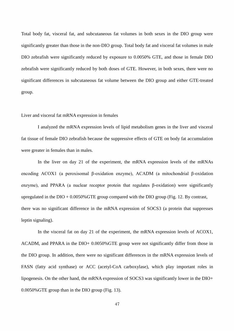
Liver and visceral fat mRNA expression in females
I analyzed the mRNA expression levels of lipid metabolism genes in the liver and visceral
fat tissue of female DIO zebrafish because the suppressive effects of GTE on body fat accumulation
were greater in females than in males.
In the liver on day 21 of the experiment, the mRNA expression levels of the mRNAs
encoding ACOX1 (a peroxisomal β-oxidation enzyme), ACADM (a mitochondrial β-oxidation
enzyme), and PPARA (a nuclear receptor protein that regulates β-oxidation) were significantly
upregulated in the DIO + 0.0050%GTE group compared with the DIO group (Fig. 12. By contrast,
there was no significant difference in the mRNA expression of SOCS3 (a protein that suppresses
leptin signaling).
In the visceral fat on day 21 of the experiment, the mRNA expression levels of ACOX1,
ACADM, and PPARA in the DIO+ 0.0050%GTE group were not significantly differ from those in
the DIO group. In addition, there were no significant differences in the mRNA expression levels of
FASN (fatty acid synthase) or ACC (acetyl-CoA carboxylase), which play important roles in
lipogenesis. On the other hand, the mRNA expression of SOCS3 was significantly lower in the DIO+
0.0050%GTE group than in the DIO group (Fig. 13).

48
Discussion
In this study, I demonstrated that GTE significantly suppressed the accumulation of visceral
fat, but not subcutaneous, in a concentration-dependent manner in zebrafish. This effect was
accompanied by increase hepatic expression of lipid catabolism genes. These results are very
consistent with the findings of previous studies in humans (Kozuma et al. 2005; Nagao et al. 2007)
and rodents (Klaus et al. 2005; Murase et al. 2002; Nagao et al. 2005; Chen et al. 2009; Lee et al.
2009; Sae-Tan et al. 2011). Although it is unclear whether the functions of visceral and subcutaneous
fat in zebrafish are homologous to those in mammals, my findings indicate that body fat distribution
is present in zebrafish as well as in humans and animals and that the site-specific effects of GTE are
similar to those in mammals.
The mechanisms underlying the suppressive effects of GTE on body fat accumulation have
been investigated in rodent models. Some studies have shown that upregulated PPARs and many
lipid-metabolizing enzymes stimulate fat oxidation in the liver (Murase et al. 2002; Chen et al. 2009;
Lee 2009). The hepatic gene expression pattern observed in DIO zebrafish in my study support these
earlier findings. Although it was recently reported that the expression of lipogenic genes, such as
FASN, is downregulated in visceral fat tissue in obese mice (Park et al. 2011), similar changes were
not observed in my study. I assume that the reason for this discrepancy is the difference in obesity
models used. In particular, Park et al. used obese mutant mice with increased FASN gene expression
in adipose tissue (Park et al. 2011), whereas I used a wild-type strain of fish. In support of the
hypothesis that the activities of FASN differs between models of obesity, Letexier et al. reported that
adipose tissue FASN expression is not affected by a high-fat diet in wild-type rodents and is reduced
in obese humans (Letexier et al. 2003). Their results help support the notion that adipose tissue gene
expression profiles differ among species, and the lack of consistency between my data and those of

49
Park et al.(2009). Nevertheless, my present results suggest that the mechanism by which GTE
suppresses body fat accumulation, at least in the liver, in zebrafish is similar to that in rodents.
I also found that GTE significantly decreased the expression of SOCS3 in the visceral fat of
DIO zebrafish. SOCS3 is a negative regulator of leptin signaling and was recently proposed as an
important therapeutic target for obesity (Howard et al. 2004). SOCS3 production in fat is associated
with obesity in humans and rodents as SOCS3 gene expression is increased in subcutaneous fat of
obese patients (Rieusset et al. 2004), and its protein and gene expression levels are increased in
epididymal fat of DIO rodents (Kanatani et al. 2007; Gu et al. 2009). I found that GTE suppressed
adipose tissue SOCS3 expression in zebrafish, which was accompanied by reduced body fat
accumulation. To my knowledge, these results suggest for the first time that SOCS3 expression is
correlated with body fat volume in zebrafish and is involved in the regulation of body fat volume by
GTE.
Another valuable outcome of this study is that 3D micro-CT analysis can be used to analyze
the effect of compounds on adiposity in DIO zebrafish. In humans, visceral fat accumulation, unlike
subcutaneous fat accumulation, is strongly associated with metabolic and vascular risks, particularly
diabetes, hypertension, and stroke, and thus increases the risk of mortality (Ibrahim et al. 2010). CT
is widely used to measure body fat volume in humans (Cnop et al. 2003; Matsuzawa et al. 2002) and
in rodents (Judex et al. 2010; Luu et al. 2009). Although CT is used to evaluate fillet composition in
teleosts, such as salmon and carp (Folkestad et al. 2008; Nanton et al. 2007; Romvári et al. 2002),
to my knowledge, no reports have used CT to measure body fat volume or visceral/subcutaneous fat
volume in small teleosts. My data raise the possibility that micro-CT could be used to measure body
fat volume in such fish. I believe this technique will play an important role in future studies of lipid
metabolism and obesity in zebrafish.
50
Conclusions
I showed that GTE significantly inhibits weight gain and body fat accumulation, and alters
the expression of hepatic lipid catabolism genes in DIO zebrafish. My results suggest that GTE
exerts beneficial effects on adiposity, possibly by altering the expression of lipid catabolism genes
and SOCS3.

51
Figures
Fig. 9 Cross-sectional images taken by three-dimensional micro-computed tomography.
The diagram of the zebrafish shows where the two cross-sectional images A and B were taken. (A)
The red two-headed arrow shows the area where body fat volume was measured (yellow). (B) The
green line shows the boundary between the visceral fat and the subcutaneous fat (orange). The light
green area indicates body fat outside the measuring area. The white arrow indicates the cleithrum.

52
Fig. 10 Effects of green tea extract (GTE) on body weight.
Changes in body weight in male (M) and female (F) zebrafish. Values are means ± SE. **P < 0.01
and *P < 0.05 vs. the diet-induced obese (DIO) group.

53
Fig. 11 Effects of green tea extract (GTE) on body fat volume.
Mean body fat volume in male (M) and female (F) zebrafish after 40 days of feeding. Values are
means ± SE. ***P < 0.001, **P < 0.01, and *P < 0.05 vs. the diet-induced obese (DIO) group.
54
Fig. 12 Effects of green tea extract (GTE) on the expression of lipid metabolism genes in the liver.
ACOX1, ACADM, PPARA, and SOCS3 mRNA expression in the liver of female zebrafish on day
21 of feeding. The expression of each mRNA was normalized against PPIA mRNA expression and
the corresponding expression in the dietinduced obese (DIO) group. Values are means ± SE. **P <
0.01 and *P < 0.05 vs. the DIO group.

55
Fig. 13 Effects of green tea extract (GTE) on the expression of lipid metabolism genes in visceral
fat.
ACOX1, ACADM, PPARA, FASN, ACC, and SOCS3 mRNA expression in the visceral fat of
female zebrafish on day 21 of feeding. The expression of each mRNA was normalized against PPIA
mRNA expression and the corresponding expression in the diet-induced obese (DIO) group. Values
are means ± SE. *P< 0.05 vs. the DIO group.

56
General Discussion
The results of these studies suggested that zebrafish has a close relationship with the
physiology of rodents and humans in relation to the effect of exercises on muscle, nutritional
metabolism related to obesity, and body fat accumulation.
In addition, I found in zebrafish a very interesting feature of muscle hypertrophy precisely dependent
on exercise quantity occurring in a very short period of time, and induction of obesity with
accumulation of body fat in a short period of time due to overfeeding. In rodents, it is difficult to load
running exercise without fear stimulation, but it was found that it can be easily studied by using
zebrafish. I revealed that zebrafish muscle hypertrophy can be detected with high accuracy at the
muscle site (identified by swimming style observation). On the other hands, it was found that
zebrafish has commonalities with human in accumulation of visceral fat due to overfeeding and
distribution of body fat ,including gender difference. Furthermore, I suggested a new mechanism of
this anti-obesity material, tea catechins.
Zebrafish has traditional abundant developmental and genetic biological information and
also has features such as high prolificacy and easy genetic manipulation. And this time, the
physiological state of exercise and meal of zebrafish where information was limited was clarified. By
combining the physiological characteristics of the zebrafish clarified in this study and much
biological information originally possessed by zebrafish, it is considered that the zebrafish research
model can be utilized for development preventive improvement technology of sarcopenia,
locomotive syndrome, and metabolic syndrome. I also believe that this will also accommodate social
demands for alternatives to animal testing.
For example, in Japan, disuse syndrome is listed as the leading cause of Needed Support and
Long-Term Care. Troubles of locomotorium, especially decreasing of muscle mass and muscle

57
strength are serious problems. In humans, age-related muscle mass reduction is known (Nair 2005)
and is referred to as sarcopenia, which has been a global challenge (Evans 1993, 1995). Functional
deterioration of locomotorium such as muscles is regarded as a health problem as a locomotive
syndrome (Nakamura 2008), and research and development to care and improve this is important. In
addition, obesity (overweight) is spreading widely, especially in developed countries (WHO 2017).
And functional deterioration of muscle and other exercisers as a locomotive syndrome is regarded as
a health issue (Nakamura 2008), and development and research of improved techniques for this is
important. Obesity caused by accumulation of visceral fat is regarded as metabolic syndrome,
leading to various fatal diseases (Itoh 2006) proposed a new concept to capture the flow of events
and chain reactions associated with cardiovascular risk: the metabolic domino. In other words,
preventing visceral fat accumulation is also very important for health, and research and development
of such anti-obesity technology has a great need.
Turning to the point of view and paying attention to the fact that zebrafish is a fish, the
research findings on the physiology of zebrafish by this study are the information of zebrafish's
Agricultural and Biological Sciences which was insufficient compared to other fish species. In the
report of the FAO (Food and Agriculture Organization of the United Nations) (2016), consumption of
fishes and shellfishes is growing globally, whereas catch of natural resources by fishery is sluggish
over the last 20 or 30 years, while 30% oceanic fisheries resources are said to be "overused or
depleted", which is a major problem. Meanwhile, in order to solve this problem, sophistication of
aquaculture technology is required, and carp and fun species are the highest in both aquaculture
production and volume growth rate globally. Zebrafish itself is not edible, but it is a carp family fish.
The findings concerning exercise, nutritional metabolism, muscle and fat of zebrafish, which is one
of the most detailed research findings, are thought to be able to lead to study of aquaculture style and

58
edible part of fish. The multi-faceted research of zebrafish is expected to give further insight into
such research and development areas.

59
Acknowledgements
I am most grateful to Prof. Kazuichi Sakamoto (University of Tsukuba) for his continuous
guidance and valuable discussion through my doctoral program.
Also, I would like to express my sincere gratitude to Prof. Yuji Inagaki (University of
Tsukuba), Prof. Chikafumi Chiba (University of Tsukuba), and Prof. Kentaro Nakano (University of
Tsukuba) for their appropriate advice during the preparation of this dissertation.
Further, I wish to express my sincere gratitude to Dr. Shinichi Meguro (Biological Science
Research, Kao Corporation) for giving me the opportunity to carry out this study and for offering the
appropriate advice.
Also, I wish to express my sincere gratitude to Prof. Toshio Tanaka (Mie University
Graduate School of Medicine) and the members of his laboratory for giving me the opportunity to
carry out this study and for offering the appropriate advice, especially zebrafish obesity model.
Also, I am greatly indebted to Dr. Atsushi Oouchi, Mr. Noriyasu Oota, Mr. Yoshihiko
Minegishi, and Ms. Shu Chin for their encouragement and helpful guidance in my research work.
Finally, I would like to appreciate my family who kindly supported my life in University of
Tsukuba.

60
References
Alberti K.G.M.M., Zimmet P., Shaw J., The metabolic syndrome - A new worldwide definition,
Lancet 366(9491), pp. 1059-1062 (2005)
Auer T.O., Duroure K., De Cian A., Concordet J.-P., Del Bene F., Highly efficient
CRISPR/Cas9-mediated knock-in in zebrafish by homology-independent DNA repair, Genome
Research 24(1), pp. 142-153 (2014)
Bedell V.M., Wang Y., Campbell J.M., Poshusta T.L., Starker C.G., Krug Ii R.G., Tan W., Penheiter
S.G., Ma A.C., Leung A.Y.H., Fahrenkrug S.C., Carlson D.F., Voytas D.F., Clark K.J., Essner J.J.,
Ekker S.C., In vivo genome editing using a high-efficiency TALEN system, Nature 491(7422), pp.
114-118 (2012)
Best J.D., Alderton W.K., Zebrafish: An in vivo model for the study of neurological diseases,
Neuropsychiatric Disease and Treatment 4(3), pp. 567-576 (2008)
Biga P.R., Goetz F.W., Zebrafish and giant danio as models for muscle growth: Determinate vs.
indeterminate growth as determined by morphometric analysis, American Journal of Physiology -
Regulatory Integrative and Comparative Physiology 291(5), pp. R1327-R1337 (2006)
Bloomfield S.A., Changes in musculoskeletal structure and function with prolonged bed rest,
Medicine and Science in Sports and Exercise 29(2), pp. 197-206 (1997)
Borst S.E., Interventions for sarcopenia and muscle weakness in older people, Age and Ageing 33(6),
61
pp. 548-555 (2004)
Brown M., Ross T.P., Holloszy J.O., Effects of ageing and exercise on soleus and extensor digitorum
longus muscles of female rats, Mechanisms of Ageing and Development 63(1), pp. 69-77 (1992)
Chang N., Sun C., Gao L., Zhu D., Xu X., Zhu X., Xiong J.-W., Xi J.J., Genome editing with
RNA-guided Cas9 nuclease in Zebrafish embryos, Cell Research 23(4), pp. 465-472 (2013)
Chen N., Bezzina R., Hinch E., Lewandowski P.A., Cameron-Smith D., Mathai M.L., Jois M.,
Sinclair A.J., Begg D.P., Wark J.D., Weisinger H.S., Weisinger R.S., Green tea, black tea, and
epigallocatechin modify body composition, improve glucose tolerance, and differentially alter
metabolic gene expression in rats fed a high-fat diet, Nutrition Research 29(11), pp. 784-793 (2009)
Cnop M., Havel P.J., Utzschneider K.M., Carr D.B., Sinha M.K., Boyko E.J., Retzlaff B.M., Knopp
R.H., Brunzell J.D., Kahn S.E., Relationship of adiponectin to body fat distribution, insulin
sensitivity and plasma lipoproteins: Evidence for independent roles of age and sex, Diabetologia
46(4), pp. 459-469 (2003)
Daikoku E., Saito M., Ono F., Zebrafish mutants of the neuromuscular junction: swimming in the
gene pool, Journal of Physiological Sciences 65(3), pp. 217-221 (2015)
Driever W., Solnica-Krezel L., Schier A.F., Neuhauss S.C.F., Malicki J., Stemple D.L., Stainier
D.Y.R., Zwartkruis F., Abdelilah S., Rangini Z., Belak J., Boggs C., A genetic screen for mutations

62
affecting embryogenesis in zebrafish, Development 123, pp. 37-46 (1996)
Dulloo A.G., Duret C., Rohrer D., Girardier L., Mensi N., Fathi M., Chantre P., Vandermander J.,
Efficacy of a green tea extract rich in catechin polyphenols and caffeine in increasing 24-h energy
expenditure and fat oxidation in humans, American Journal of Clinical Nutrition 70(6), pp.
1040-1045 (1999)
Evans W.J., What is sarcopenia?, Journals of Gerontology - Series A Biological Sciences and
Medical Sciences 50(SPEC. ISSUE), pp. 5-8 (1995)
Evans W.J., Campbell W.W., Sarcopenia and age-related changes in body composition and functional
capacity, Journal of Nutrition 123(2 SUPPL.), pp. 465-468 (1993)
FAO (Food and Agriculture Organization of the United Nations), The State of World Fisheries and
Aquaculture (SOFIA) (2016)
Folkestad A., Wold J.P., Rørvik K.-A., Tschudi J., Haugholt K.H., Kolstad K., Mørkøre T., Rapid and
non-invasive measurements of fat and pigment concentrations in live and slaughtered Atlantic
salmon (Salmo salar L.), Aquaculture 280(1-4), pp. 129-135 (2008)
Friedland A.E., Tzur Y.B., Esvelt K.M., Colaiácovo M.P., Church G.M., Calarco J.A., Heritable
genome editing in C. elegans via a CRISPR-Cas9 system, Nature Methods 10(8), pp. 741-743 (2013)

63
Furuyashiki T., Nagayasu H., Aoki Y., Bessho H., Hashimoto T., Kanazawa K., Ashida H., Tea
catechin suppresses adipocyte differentiation accompanied by down-regulation of PPARγ2 and
C/EBPα in 3T3-L1 cells, Bioscience, Biotechnology and Biochemistry 68(11), pp. 2353-2359 (2004)
Gilbert M.J.H., Zerulla T.C., Tierney K.B., Zebrafish (Danio rerio) as a model for the study of aging
and exercise: Physical ability and trainability decrease with age, Experimental Gerontology 50(1), pp.
106-113 (2013)
Gu H., Liu L., Ma S., Liu Y., Ren Y., Zhai L., Yu F., An L., Yang J., Inhibition of SOCS-3 in
adipocytes of rats with diet-induced obesity increases leptin-mediated fatty acid oxidation, Endocrine
36(3), pp. 546-554 (2009)
Haffter P., Granato M., Brand M., Mullins M.C., Hammerschmidt M., Kane D.A., Odenthal J., Van
Eeden F.J.M., Jiang Y.-J., Heisenberg C.-P., Kelsh R.N., Furutani-Seiki M., Vogelsang E., Beuchle D.,
Schach U., Fabian C., Nüsslein-Volhard C., The identification of genes with unique and essential
functions in the development of the zebrafish, Danio rerio, Development 123, pp. 1-36 (1996)
Hirata H., Zebrafish muscular disease models towards drug discovery, Expert Opinion on Drug
Discovery 4(5), pp. 507-513 (2009)
Hölttä-Vuori M., Salo V.T.V., Nyberg L., Brackmann C., Enejder A., Panula P., Ikonen E., Zebrafish:
Gaining popularity in lipid research, Biochemical Journal 429(2), pp. 235-242 (2010)
64
Hood D.A., Uguccioni G., D'Souza D., Regulation of PPARγ coactivator-1 function and expression
in muscle: Effect of exercise, PPAR Research, 937123 (2010)
Howard J.K., Cave B.J., Oksanen L.J., Tzameli I., Bjoørbæk C., Flier J.S., Enhanced leptin
sensitivity and attenuation of diet-induced obesity in mice with haploinsufficiency of Socs3, Nature
Medicine 10(7), pp. 734-738 (2004)
Howe K., Clark M.D., Torroja C.F., Torrance J., Berthelot C., Muffato M., Collins J.E., Humphray S.,
McLaren K., Matthews L., McLaren S., Sealy I., Caccamo M., Churcher C., Scott C., Barrett J.C.,
Koch R., Rauch G.-J., White S., Chow W., Kilian B., Quintais L.T., Guerra-Assunção J.A., Zhou Y.,
Gu Y., Yen J., Vogel J.-H., Eyre T., Redmond S., Banerjee R., Chi J., Fu B., Langley E., Maguire S.F.,
Laird G.K., Lloyd D., Kenyon E., Donaldson S., Sehra H., Almeida-King J., Loveland J., Trevanion
S., Jones M., Quail M., Willey D., Hunt A., Burton J., Sims S., McLay K., Plumb B., Davis J., Clee
C., Oliver K., Clark R., Riddle C., Eliott D., Threadgold G., Harden G., Ware D., Mortimer B., Kerry
G., Heath P., Phillimore B., Tracey A., Corby N., Dunn M., Johnson C., Wood J., Clark S., Pelan S.,
Griffiths G., Smith M., Glithero R., Howden P., Barker N., Stevens C., Harley J., Holt K.,
Panagiotidis G., Lovell J., Beasley H., Henderson C., Gordon D., Auger K., Wright D., Collins J.,
Raisen C., Dyer L., Leung K., Robertson L., Ambridge K., Leongamornlert D., McGuire S.,
Gilderthorp R., Griffiths C., Manthravadi D., Nichol S., Barker G., Whitehead S., Kay M., Brown J.,
Murnane C., Gray E., Humphries M., Sycamore N., Barker D., Saunders D., Wallis J., Babbage A.,
Hammond S., Mashreghi-Mohammadi M., Barr L., Martin S., Wray P., Ellington A., Matthews N.,
Ellwood M., Woodmansey R., Clark G., Cooper J., Tromans A., Grafham D., Skuce C., Pandian R.,
Andrews R., Harrison E., Kimberley A., Garnett J., Fosker N., Hall R., Garner P., Kelly D., Bird C.,
65
Palmer S., Gehring I., Berger A., Dooley C.M., Ersan-Ürün Z., Eser C., Geiger H., Geisler M.,
Karotki L., Kirn A., Konantz J., Konantz M., Oberländer M., Rudolph-Geiger S., Teucke M.,
Osoegawa K., Zhu B., Rapp A., Widaa S., Langford C., Yang F., Carter N.P., Harrow J., Ning Z.,
Herrero J., Searle S.M.J., Enright A., Geisler R., Plasterk R.H.A., Lee C., Westerfield M., De Jong
P.J., Zon L.I., Postlethwait J.H., Nüsslein-Volhard C., Hubbard T.J.P., Crollius H.R., Rogers J.,
Stemple D.L., The zebrafish reference genome sequence and its relationship to the human genome,
Nature 496(7446), pp. 498-503 (2013)
Huang P., Xiao A., Zhou M., Zhu Z., Lin S., Zhang B., Heritable gene targeting in zebrafish using
customized TALENs, Nature Biotechnology 29(8), pp. 699-700 (2011)
Hwang W.Y., Fu Y., Reyon D., Maeder M.L., Tsai S.Q., Sander J.D., Peterson R.T., Yeh J.-R.J.,
Joung J.K., Efficient genome editing in zebrafish using a CRISPR-Cas system, Nature
Biotechnology 31(3), pp. 227-229 (2013)
Ibrahim M.M., Subcutaneous and visceral adipose tissue: Structural and functional differences,
Obesity Reviews 11(1), pp. 11-18 (2010)
Ikeda I., Hamamoto R., Uzu K., Imaizumi K., Nagao K., Yanagita T., Suzuki Y., Kobayashi M.,
Kakuda T., Dietary gallate esters of tea catechins reduce deposition of visceral fat, hepatic
triacylglycerol, and activities of hepatic enzymes related to fatty acid synthesis in rats, Bioscience,
Biotechnology and Biochemistry 69(5), pp. 1049-1053 (2005)

66
Ishihara A., Inoue N., Katsuta S., The relationship of voluntary running to fibre type composition,
fibre area and capillary supply in rat soleus and plantaris muscles, European Journal of Applied
Physiology and Occupational Physiology 62(3), pp. 211-215 (1991)
Ishihara A., Roy R.R., Ohira Y., Ibata Y., Edgerton V.R., Hypertrophy of rat plantaris muscle fibers
after voluntary running with increasing loads, Journal of Applied Physiology 84(6), pp. 2183-2189
(1998)
Itoh H., Metabolic domino: New concept in lifestyle medicine, Drugs of Today 42(SUPPL. C), pp.
9-16 (2006)
James Henderson R., Tocher D.R., The lipid composition and biochemistry of freshwater fish,
Progress in Lipid Research 26(4), pp. 281-347 (1987)
Jeneson J.A.L., De Snoo M.W., Verlinden N.A.T., Joosten B.J.L.J., Doornenbal A., Schot A., Everts
M.E., Treadmill but not wheel running improves fatigue resistance of isolated extensor digitorum
longus muscle in mice, Acta Physiologica 190(2), pp. 151-161 (2007)
Judex S., Luu Y.K., Ozcivici E., Adler B., Lublinsky S., Rubin C.T., Quantification of adiposity in
small rodents using micro-CT, Methods 50(1), pp. 14-19 (2010)
Kanatani Y., Usui I., Ishizuka K., Bukhari A., Fujisaka S., Urakaze M., Haruta T., Kishimoto T.,
Naka T., Kobayashi M., Effects of pioglitazone on suppressor of cytokine signaling 3 expression:

67
Potential mechanisms for its effects on insulin sensitivity and adiponectin expression, Diabetes 56(3),
pp. 795-803 (2007)
Kawakami K., Transgenesis and Gene Trap Methods in Zebrafish by Using the Tol2 Transposable
Element, Methods in Cell Biology 77, pp. 201-222 (2004)
Kemi O.J., Loennechen J.P., Wisløff U., Ellingsen Y., Intensity-controlled treadmill running in mice:
Cardiac and skeletal muscle hypertrophy, Journal of Applied Physiology 93(4), pp. 1301-1309 (2002)
Kishi S., Using zebrafish models to explore genetic and epigenetic impacts on evolutionary
developmental origins of aging, Translational Research 163(2), pp. 123-135 (2014)
Kishi S., Slack B.E., Uchiyama J., Zhdanova I.V., Zebrafish as a genetic model in biological and
behavioral gerontology: Where development meets aging in vertebrates - A mini-review,
Gerontology 55(4), pp. 430-441 (2009)
Kishi S., Uchiyama J., Baughman A.M., Goto T., Lin M.C., Tsai S.B., The zebrafish as a vertebrate
model of functional aging and very gradual senescence, Experimental Gerontology 38(7), pp.
777-786 (2003)
Klaus S., Pültz S., Thöne-Reineke C., Wolfram S., Epigallocatechin gallate attenuates diet-induced
obesity in mice by decreasing energy absorption and increasing fat oxidation, International Journal of
Obesity 29(6), pp. 615-623 (2005)

68
Konopka A.R., Harber M.P., Skeletal muscle hypertrophy after aerobic exercise training, Exercise
and Sport Sciences Reviews 42(2), pp. 53-61 (2014)
Kozuma K., Chikama A., Hishino E., Kataoka K., Mori K., Hase T., Katsuragi Y., Tokimitsu I.,
Nakamura H., Effect of intake of a beverage containing 540 mg catechins on the body composition
of obese women and men, Progress in Medicine 25(7), pp. 1945-1957 (2005)
Kuk J.L., Katzmarzyk P.T., Nichaman M.Z., Church T.S., Blair S.N., Ross R., Visceral fat is an
independent predictor of all-cause mortality in men, Obesity 14(2), pp. 336-341 (2006)
Laale H.W., The biology and use of zebrafish, Brachydanio rerio in fisheries research. A literature
review, Journal of Fish Biology 10(2), pp. 121-173 (1977)
Lee M.-S., Kim C.-T., Kim Y., Green tea (-)-epigallocatechin-3-gallate reduces body weight with
regulation of multiple genes expression in adipose tissue of diet-induced obese mice, Annals of
Nutrition and Metabolism 54(2), pp. 151-157 (2009)
Lemoine C.M.R., Craig P.M., Dhekney K., Kim J.J., McClelland G.B., Temporal and spatial patterns
of gene expression in skeletal muscles in response to swim training in adult zebrafish (Danio rerio),
Journal of Comparative Physiology B: Biochemical, Systemic, and Environmental Physiology
180(1), pp. 151-160 (2010a)

69
Lemoine C.M.R., Lougheed S.C., Moyes C.D., Modular evolution of PGC-1α in vertebrates, Journal
of Molecular Evolution 70(5), pp. 492-505 (2010b)
Letexier D., Pinteur C., Large V., Fréring V., Beylot M., Comparison of the expression and activity of
the lipogenic pathway in human and rat adipose tissue, Journal of Lipid Research 44(11), pp.
2127-2134 (2003)
Lieschke G.J., Currie P.D., Animal models of human disease: Zebrafish swim into view, Nature
Reviews Genetics 8(5), pp. 353-367 (2007)
Lin Y.-Y., Muscle diseases in the zebrafish, Neuromuscular Disorders 22(8), pp. 673-f684 (2012)
Lindsey C.C., Form, function, and locomotory habits in fish, Fish Physiology 7(C), pp. 1-100 (1978)
Lira V.A., Benton C.R., Yan Z., Bonen A., PGC-1α regulation by exercise training and its influences
on muscle function and insulin sensitivity, American Journal of Physiology - Endocrinology and
Metabolism 299(2), pp. E145-E161 (2010)
Luu Y.K., Lublinsky S., Ozcivici E., Capilla E., Pessin J.E., Rubin C.T., Judex S., In vivo
quantification of subcutaneous and visceral adiposity by micro-computed tomography in a small
animal model, Medical Engineering and Physics 31(1), pp. 34-41 (2009)
Malik V.S., Popkin B.M., Bray G.A., Després J.-P., Hu F.B., Sugar-sweetened beverages, obesity,
70
type 2 diabetes mellitus, and cardiovascular disease risk, Circulation 121(11), pp. 1356-1364 (2010)
Matsumoto N., Ishigaki F., Ishigaki A., Iwashina H., Hara Y., Reduction of Blood Glucose Levels by
Tea Catechin, Bioscience, Biotechnology and Biochemistry 57(4), pp. 525-527 (1993)
Matsuzawa Y., Nakamura T., Takahashi M., Ryo M., Inoue S., Ikeda Y., Ohno M., Sakata T.,
Fukagawa K., Saitoh Y., Sato Y., Shirai K., Miyazaki S., Tokunaga K., Yamanouchi K., Takahashi M.,
Shibasaki T., Nagai M., New criteria for 'obesity disease' in Japan, Circulation Journal 66(11), pp.
987-992 (2002)
McClelland G.B., Muscle remodeling and the exercise physiology of fish, Exercise and Sport
Sciences Reviews 40(3), pp. 165-173 (2012)
McClelland G.B., Craig P.M., Dhekney K., Dipardo S., Temperature- and exercise-induced gene
expression and metabolic enzyme changes in skeletal muscle of adult zebrafish (Danio rerio),
Journal of Physiology 577(2), pp. 739-751 (2006)
Mcmahon C.D., Chai R., Radley-Crabb H.G., Watson T., Matthews K.G., Sheard P.W., Soffe Z.,
Grounds M.D., Shavlakadze T., Lifelong exercise and locally produced insulin-like growth factor-1
(IGF-1) have a modest influence on reducing age-related muscle wasting in mice, Scandinavian
Journal of Medicine and Science in Sports 24(6), pp. e423-e435 (2014)
Melov S., Tamopolsky M.A., Bechman K., Felkey K., Hubbard A., Resistance exercise reverses

71
aging in human skeletal muscle, PLoS ONE 2(5), e465 (2007)
van der Meulen T., Schipper H., Van Den Boogaart J.G.M., Huising M.O., Kranenbarg S., Van
Leeuwen J.L., Endurance exercise differentially stimulates heart and axial muscle development in
zebrafish (Danio rerio), American Journal of Physiology - Regulatory Integrative and Comparative
Physiology 291(4), pp. R1040-R1048 (2006)
Miura Y., Chiba T., Tomita I., Koizumi H., Miura S., Umegaki K., Hara Y., Ikeda M., Tomita T., Tea
catechins prevent the development of atherosclerosis in apoprotein E-deficient mice, Journal of
Nutrition 131(1), pp. 27-32 (2001)
Morais S., Knoll-Gellida A., André M., Barthe C., Babin P.J., Conserved expression of alternative
splicing variants of peroxisomal acyl-CoA oxidase 1 in vertebrates and developmental and nutritional
regulation in fish, Physiological Genomics 28(3), pp. 239-252 (2007)
Munoz K.A., Aannestad A., Tischler M.E., Henriksen E.J., Skeletal muscle protein content and
synthesis after voluntary running and subsequent unweighting, Metabolism 43(8), pp. 994-999
(1994)
Murase T., Haramizu S., Shimotoyodome A., Tokimitsu I., Hase T., Green tea extract improves
running endurance in mice by stimulating lipid utilization during exercise, American Journal of
Physiology - Regulatory Integrative and Comparative Physiology 290(6), pp. R1550-R1556 (2006)

72
Murase T., Nagasawa A., Suzuki J., Hase T., Tokimitsu I., Beneficial effects of tea catechins on
diet-induced obesity: Stimulation of lipid catabolism in the liver, International Journal of Obesity
26(11), pp. 1459-1464 (2002)
Nagao T., Hase T., Tokimitsu I., A green tea extract high in catechins reduces body fat and
cardiovascular risks in humans, Obesity 15(6), pp. 1473-1483 (2007)
Nagao T., Komine Y., Soga S., Meguro S., Hase T., Tanaka Y., Tokimitsu I., Ingestion of a tea rich in
catechins leads to a reduction in body fat and malondialdehyde-modified LDL in men, American
Journal of Clinical Nutrition 81(1), pp. 122-129 (2005)
Nair K.S., Aging muscle, American Journal of Clinical Nutrition 81(5), pp. 953-963 (2005)
Nakamura K., A "super-aged" society and the "locomotive syndrome", Journal of Orthopaedic
Science 13(1), pp. 1-2 (2008)
Nanton D.A., Vegusdal A., Rørå A.M.B., Ruyter B., Baeverfjord G., Torstensen B.E., Muscle lipid
storage pattern, composition, and adipocyte distribution in different parts of Atlantic salmon (Salmo
salar) fed fish oil and vegetable oil, Aquaculture 265(1-4), pp. 230-243 (2007)
National Research Council, Guide for the Care and Use of Laboratory Animals (2011)
Oka T., Nishimura Y., Zang L., Hirano M., Shimada Y., Wang Z., Umemoto N., Kuroyanagi J.,

73
Nishimura N., Tanaka T., Diet-induced obesity in zebrafish shares common pathophysiological
pathways with mammalian obesity, BMC Physiology 10(1), 21 (2010)
Palstra A.P., Rovira M., Rizo-Roca D., Torrella J.R., Spaink H.P., Planas J.V., Swimming-induced
exercise promotes hypertrophy and vascularization of fast skeletal muscle fibres and activation of
myogenic and angiogenic transcriptional programs in adult zebrafish, BMC Genomics 15(1), 1136
(2014)
Palstra A.P., Tudorache C., Rovira M., Brittijn S.A., Burgerhout E., van den Thillart G.E.E.J.M.,
Spaink H.P., Planas J.V., Establishing zebrafish as a novel exercise model: Swimming economy,
swimming-enhanced growth and muscle growth marker gene expression, PLoS ONE 5(12), e14483
(2010)
Park H.J., DiNatale D.A., Chung M.-Y., Park Y.-K., Lee J.-Y., Koo S.I., O'Connor M., Manautou J.E.,
Bruno R.S., Green tea extract attenuates hepatic steatosis by decreasing adipose lipogenesis and
enhancing hepatic antioxidant defenses in ob/ob mice, Journal of Nutritional Biochemistry 22(4), pp.
393-400 (2011)
Pasiakos S.M., McClung H.L., McClung J.P., Urso M.L., Pikosky M.A., Cloutier G.J., Fielding R.A.,
Young A.J., Molecular responses to moderate endurance exercise in skeletal muscle, International
Journal of Sport Nutrition and Exercise Metabolism 20(4), pp. 282-290 (2010)
Patton E.E., Dhillon P., Amatruda J.F., Ramakrishnan L., Spotlight on zebrafish: Translational impact,

74
DMM Disease Models and Mechanisms 7(7), pp. 731-733 (2014)
Pilegaard H., Saltin B., Neufer D.P., Exercise induces transient transcriptional activation of the
PGC-1α gene in human skeletal muscle, Journal of Physiology 546(3), pp. 851-858 (2003)
Reid K.F., Fielding R.A., Skeletal muscle power: A critical determinant of physical functioning in
older adults, Exercise and Sport Sciences Reviews 40(1), pp. 4-12 (2012)
Rice-Evans C.A., Miller N.J., Paganga G., Structure-antioxidant activity relationships of flavonoids
and phenolic acids, Free Radical Biology and Medicine 20(7), pp. 933-956 (1996)
Rieusset J., Bouzakri K., Chevillotte E., Ricard N., Jacquet D., Bastard J.-P., Laville M., Vidal H.,
Suppressor of cytokine signaling 3 expression and insulin resistance in skeletal muscle of obese and
type 2 diabetic patients, Diabetes 53(9), pp. 2232-2241 (2004)
Rodnick K.J., Reaven G.M., Haskell W.L., Sims C.R., Mondon C.E., Variations in running activity
and enzymatic adaptations in voluntary running rats, Journal of Applied Physiology 66(3), pp.
1250-1257 (1989)
Rome L.C., The design of vertebrate muscular systems: Comparative and integrative approaches,
Clinical Orthopaedics and Related Research (403 SUPPL.), pp. S59-S76 (2002)
Rome L.C., Principles of actuation in the mascular system of fish, IEEE Journal of Oceanic

75
Engineering 30(3), pp. 630-646 (2005)
Rome L.C., Swank D., Corda D., How fish power swimming, Science 261(5119), pp. 340-343
(1993)
Romvári R., Hancz Cs., Petrási Zs., Molnár T., Horn P., Non-invasive measurement of fillet
composition of four freshwater fish species by computer tomography, Aquaculture International
10(3), pp. 231-240 (2002)
Sae-Tan S., Grove K.A., Lambert J.D., Weight control and prevention of metabolic syndrome by
green tea, Pharmacological Research 64(2), pp. 146-154 (2011)
Santoriello C., Zon L.I., Hooked! modeling human disease in zebrafish, Journal of Clinical
Investigation 122(7), pp. 2337-2343 (2012)
Shankar S., Ganapathy S., Srivastava R.K., Green tea polyphenols: Biology and therapeutic
implications in cancer, Frontiers in Bioscience 12(13), pp. 4881-4899 (2007)
Shimotoyodome A., Haramizu S., Inaba M., Murase T., Tokimitsu I., Exercise and green tea extract
stimulate fat oxidation and prevent obesity in mice, Medicine and Science in Sports and Exercise
37(11), pp. 1884-1892 (2005)
Solnica-Krezel L., Schier A.F., Driever W., Efficient recovery of ENU-induced mutations from the

76
zebrafish germline, Genetics 136(4), pp. 1401-1420 (1994)
Song Y., Cone R.D., Creation of a genetic model of obesity in a teleost, FASEB Journal 21(9), pp.
2042-2049 (2007)
Spiegelman B.M., Flier J.S., Obesity and the regulation of energy balance, Cell 104(4), pp. 531-543
(2001)
Stoletov K., Fang L., Choi S.-H., Hartvigsen K., Hansen L.F., Hall C., Pattison J., Juliano J., Miller
E.R., Almazan F., Crosier P., Witztum J.L., Klemke R.L., Miller Y.I., Vascular lipid accumulation,
lipoprotein oxidation, and macrophage lipid uptake in hypercholesterolemic zebrafish, Circulation
Research 104(8), pp. 952-960 (2009)
Syme D.A., Functional Properties of Skeletal Muscle, Fish Physiology 23(C), pp. 179-240 (2005)
Tainaka T., Shimada Y., Kuroyanagi J., Zang L., Oka T., Nishimura Y., Nishimura N., Tanaka T.,
Transcriptome analysis of anti-fatty liver action by Campari tomato using a zebrafish diet-induced
obesity model, Nutrition and Metabolism 8, 88 (2011)
Takase H., Nagao T., Otsuka K., Kozuma K., Kataoka K., Meguro S., Komikado M., Tokimitsu I.,
Effects of long-term ingestion of tea catechins on visceral fat accumulation and metabolic syndrome:
Pooling-analysis of 7 randomized controlled trials, Japanese Pharmacology and Therapeutics 36(6),
pp. 509-514 (2008)
77
Takeshita M., Takashima S., Harada U., Shibata E., Hosoya N., Takase H., Otsuka K., Meguro S.,
Komikado M., Tokimitsu I., Effects of long-term consumption of tea catechins-enriched beverage
with no caffeine on body composition in humans, Japanese Pharmacology and Therapeutics 36(8),
pp. 767-776 (2008)
Watanabe J., Kawabata J., Niki R., Isolation and identification of Acetyl-CoA Carboxylase inhibitors
from green tea (camellia sinensis), Bioscience, Biotechnology and Biochemistry 62(3), pp. 532-534
(1998)
Westerfield M., The zebrafish book: a guide for the laboratory use of zebrafish (Danio rerio), 5th edn.
M, Westerfield (2007)
Westerfield M., The Zebrafish Book. A Guide for the Laboratory Use of Zebrafish (Danio Rerio),
(2000)
WHO (World Health Organization),
Obesity and overwight, Fact Sheet (http://www.who.int/mediacentre/factsheets/fs311/en/), (2017)
Xiao A., Wang Z., Hu Y., Wu Y., Luo Z., Yang Z., Zu Y., Li W., Huang P., Tong X., Zhu Z., Lin S.,
Zhang B., Chromosomal deletions and inversions mediated by TALENs and CRISPR/Cas in
zebrafish, Nucleic Acids Research 41(14), (2013)

78
Yang Y., Creer A., Jemiolo B., Trappe S., Time course of myogenic and metabolic gene expression in
response to acute exercise in human skeletal muscle, Journal of Applied Physiology 98(5), pp.
1745-1752 (2005)
Zang L., Morikane D., Shimada Y., Tanaka T., Nishimura N., A novel protocol for the Oral
administration of test chemicals to adult Zebrafish, Zebrafish 8(4), pp. 203-210 (2011)
Zaveri N.T., Green tea and its polyphenolic catechins: Medicinal uses in cancer and noncancer
applications, Life Sciences 78(18), pp. 2073-2080 (2006)
Zhang Y., Yu Y., Li X., Meguro S., Hayashi S., Katashima M., Yasumasu T., Wang J., Li K., Effects
of catechin-enriched green tea beverage on visceral fat loss in adults with a high proportion of
visceral fat: A double-blind, placebo-controlled, randomized trial, Journal of Functional Foods 4(1),
pp. 315-322 (2012)