Embed Size (px)
Citation preview

Journal of Environmental Biology �April, 2007�
Studies on L-asparaginase enzyme of actinomycetes isolated from estuarine fishes
Maloy Kumar Sahu1, K. Sivakumar*1, E. Poorani2, T. Thangaradjou1 and L. Kannan1, 3
1Centre of Advanced Study in Marine Biology, Annamalai University, Parangipettai-608 502, India2Kongunadu Arts and Science College, Coimbatore-614 729, India
3Thiruvalluvar University, Vellore-632 004, India
(Received: March 19, 2005 ; Revised received: July 28, 2005 ; Accepted: August 10, 2005)
Abstract: Actinomycetes were isolated from different organs viz. skin, gills and gut contents of three species of fishes viz. Mugil cephalus (Linnaeus,
1758), Chanos chanos (Forskal, 1775) and Etroplus suratensis (Bloch, 1780) using three different media from the Vellar estuary, situated along the
southeast coast of India. Among the three fishes, M. cephalus harboured highest number of actinomycetes population in all the three body parts
examined followed by C. chanos and E. suratensis. Out of the three body parts of all fishes, gut contents had highest actinomycetes population followed
by gills and skin. Among the three media used for isolation of actinomycetes, Kuster’s agar medium was found to be suitable than the starch casein agar
and glucose asparagine agar media. Out of the 40 strains isolated, only six strains (LA-2, LA-8, LA-15, LA-20, LA-29 and LA-35) showed significant L-
asparagianse activity and were taken up for further studies. Impact of various physical and chemical factors such as pH, temperature, sodium chloride
concentration, carbon sources and amino acids on the growth of actinomycetes and L-asparaginase activity was also studied. Optimum growth and
enzyme activity was noticed under pH 7 to 8, temperature 37 0C, 1-2% sodium chloride concentration, sucrose as carbon source and without any amino
acids. Analysis of the cell components of the isolated strains has revealed the wall type - I (the wall type- I is typical for the genus Streptomyces) and the
strains were micromorphologically similar to the genus Streptomyces. Hence, the morphological, physiological and biochemical along with the
micromorphological results obtained for the L-asparaginase producing strains were compared and the strains were tentatively identified as Streptomyces
aureofasciculus (LA-2), S. chattanoogenesis (LA-8), S. hawaiiensis (LA-15), S. orientalis (LA-20), S. canus (LA-29) and S. olivoviridis (LA-35).
Key words: Estuarine fishes, Actinomycetes, L-asparaginase enzyme, pH, Temperature, Sodium chloride, Amino acids, Carbon compounds
*Corresponding author: E-Mail: [email protected], Tel.: +91-4144-252099 / 243999 (Dir.) / 243223 / 243070 (Ext.) 235, Fax: +91-4144-243999
Introduction
The enzyme L-asparaginase (L-asparagine
amidohydrolase, E. C. 3.5.1.1) has attracted much attention in
the past decades because of its antineoplastic activity (Crowther,
1971; Mc Creadie et al., 1973; Wriston and Yellin, 1973). This
enzyme is used in the treatment of acute lymphatic leukemia
with 60-80% remission (Clausen, 1986; Yoshimoto, 1986). Normal
cells can synthesise their own L-asparaginase with the help of
an enzyme known as L-asparagine synthetase, whereas the
malignant cells deprived of this enzyme requires an exogenous
source of L-asparagine for their growth and multiplication
(Selvakumar et al., 1991). The L-asparaginase splits the L-
asparagine, an important nutrient for cancer cells for the protein
synthesis to make new cancer cells, into L-aspartic acid and
ammonia (Selvakumar et al., 1991). Administration of L-
asparaginase depletes exogenous L-asparagine and causes the
death of malignant cells.
Occurrence of this enzyme has been reported in bacteria,
actinomycetes and fungi (Balakrishnan Nair et al., 1977; Maya
et al., 1992; Mathew et al., 1994). Though actinomycetes are
ubiquitous in nature including water and sediments of the
estuarine environment, the actinomycetes strains present in the
living organisms including fishes shows good enzymatic activity
than the strains isolated from the water and sediment samples
(Dhevendaran and Annie, 1999). Likewise actinomycetes are
providing good L-asparaginase enzyme when compared to
bacterial and fungal sources. Moreover, information on the
occurrence of the L-asparaginase enzyme in actinomycetes
associated with estuarine fishes of the southeast coast of India
is lacking. Hence the present investigation aims to study the L-
asparaginase activity of actinomycetes associated with different
parts of three species of estuarine fishes viz. Mugil cephalus,
Chanos chanos and Etroplus suratensis under different
experimental culture conditions like different pH, temperature,
sodium chloride, carbon sources and amino acids and also to
identify the potential L-asparaginase producing actinomycetes
through chemotaxonomical and conventional methods of
identification.
Materials and Methods
Isolation of actinomycetes: Fish samples Mugil cephalus,
Chanos chanos and Etroplus suratensis were collected using
cast net from the Vellar estuary. Samples were kept in sterile
polyethylene bags and transported to the laboratory under ice
for microbiological analysis. A small portion of the gills and the
skin were removed aseptically with the help of forceps and
scissors from these fishes. Before the alimentary tract of the fish
was removed, the body surface was wiped with 70% ethanol by
using sterile cotton (Sudha et al., 2002). The abdomen was
Journal of Environmental Biology April 2007, 28(2) 465-474 (2007)
©Triveni Enterprises, Lucknow (India) For personal use only
Free paper downloaded from: www. jeb.co.in Commercial distribution of this copy is illegal
Journal of Environmental Biology �April, 2007�
Sahu et al.
opened aseptically and then the alimentary tract was carefully
taken out. The contents of the alimentary tract were squeezed
out with forceps. Then, one gram of the squeezed out gut
contents, gills and skin samples were taken and homogenized
separately in a sterile mortar and pestle. The samples were
serially diluted with filtered and sterilized 50% seawater. One ml
of the serially diluted samples were plated (triplicate) in petriplates
containing Kuster’s agar (KUA), glucose asparagine agar (GAA)
and starch casein agar (SCA) media and incubated at 35oC for
seven days. The leathery colonies of actinomycetes that appeared
on the petriplates were counted from the 5th day onwards upto
28th day. All the colonies that grew on the petriplates were sub
cultured and were maintained in slants.
Assay for L-asparaginase activity: Actinomycetes from different
parts of fishes were screened for L-asparaginase activity. The
packed cells (5mg/ml) were suspended in distilled water and this
was inoculated into 5 ml of glycerol-asparagine broth and
incubated for seven days at 37oC temperature. After the 7th day,
the broth was filtered through whatman number 1 filter paper
and the paper was kept in incubator at 37oC for five days and the
difference in weight was expressed as growth of actinomycetes
in terms of dry weight. Activity of L-asparaginase was measured
by adding 0.5 ml of Nesslers reagent to the filtered cultured broth
(Dhevendaran and Annie, 1999). Within five minutes, a yellow
colour was developed. Then the sample was centrifuged and
absorbance of the supernatant was read using a UV visible
spectrophotometer (Hitachi) at a wave length of 450 nm (Wriston,
1971). The ammonia content was estimated using standard
ammonium chloride solution and protein content of the enzyme
preparation was estimated by following the method of Lowry et al.
(1951). L-asparaginase activity is expressed in International units
(IU)/mg of protein. Ammonium sulphate was used as standard.
Effect of pH, temperature, sodium chloride, carbon
compounds and amino acids on L-asparaginase activity
and growth of the actinomycetes: L-asparaginase activity and
growth of the actinomycetes were measured at different pH,
temperature, sodium chloride concentrations, carbon compounds
and amino acids. The experiments were conducted in 250ml
Erlenmeyer flasks containing the sterilized (at 15 lbs pressure
for 15 minutes) glycerol asparagine broth. The flasks were cooled
and the strain was inoculated and incubated for different
parameters as described below.
Effect of pH: This was studied by varying the pH of the glycerol
asparagine broth by addition of buffer solution ranging from pH 6
to 10. After inoculation of the strain, it was incubated for seven
days at 370 C temperature.
Effect of temperature: After inoculation of the strain in glycerol
asparagine broth, it was incubated at various temperatures such
as 250 C, 290 C, 330 C, 370 C, 410 C and 450 C for seven days.
Effect of sodium chloride concentrations: To study the
tolerance of strains towards sodium chloride concentrations, the
glycerol asparagine broth prepared with distilled water was
incorporated with sodium chloride at varying concentrations such
as 0.05%, 0.1%, 0.5%, 1%, 2%, 3% and 4%. After inoculation of
the strain, it was incubated at 37 oC for seven days.
Effect of various carbon compounds: The glycerol asparagine
broth was used for studying the effect of various compounds such
as glucose, sucrose, lactose, raffinose and mannitol. The broth
was distributed into various flasks and 1% of each carbon source
was then added before inoculation of the strain and it was
incubated for seven days at 37 oC temperature.
Effect of various amino acids: The glycerol asparagine broth
was used for studying the influence of organic matter such as
methionine, tryptophan, L-glutamic acid and threonine. The broth
was distributed into various flasks and 0.8 ml-1 of each amino
acid was then added and incubated for seven days at 37 oC
temperature.
At the end of the incubation period, the cells were
harvested and dried at 35oC temperature in the incubator. Weight
of the cells was taken and expressed as g. dry weight
(Dhevendaran and Annie, 1999). L-asparaginase activity was
determined by the procedure as described earlier.
Taxonomic investigation: The genus level identification was
made for the six strains which showed good enzymatic activity
using cell wall composition analysis and micromorphological
studies (Lechevalier and Lechevalier, 1970). Characterization of
L-asparaginase producing actinomycetes was made by following
the methods described by Shirling and Gottileb (1996) using the
standard culture medium yeast extract-malt extract agar (ISP
medium 2). The species level identification of these strains was
made based on the keys of Nonamura (1974).
Results and Discussion
Actinomycetes population density: Actinomycetes population
density varied in the different parts viz. skin, gills and the gut
contents of all the three species of fishes and also in different
media used.
In the KUA medium: Population density of actinomycetes
recorded from the skin of all the three species of fishes varied
from 0.37 to 0.65 x 10² CFU/g with the minimum (0.37 x 10² CFU/g)
in E. suratensis and the maximum (0.65 x 10² CFU/g) in M.
cephalus. In the case of the gills, it varied from 0.98 to 1.35 x 10²
CFU/g with the minimum (0.98 x 10² CFU/g) in E. suratensis and
the maximum (1.35 x 10² CFU/g) in M. cephalus. In the case of
the gut contents, the maximum was observed (4.15 x 10² CFU/g)
in M. cephalus and the minimum (3.19 x 10² CFU/g), in E.
suratensis (Fig. 1).
466

Journal of Environmental Biology �April, 2007�
L-asparaginase enzyme of actinomycetes
In the GAA medium: Actinomycetes population density recorded
from the skin of all the three species of fishes varied from 0.25 to
0.32 x 10² CFU/g. The highest density (0.32 x 10² CFU/g) was
observed in M. ceplalus, followed by C. chanos (0.31 x 10² CFU/g)
and E. suratensis (0.25 x 10² CFU/g). In the case of the gills,
population density ranged from 0.85 to 1.12 x 10² CFU/g. The
highest density (1.12 x 10² CFU/g) was recorded in M. cephalus.
This was followed by C. chanos (1.01 x 10² CFU/g) and E.
suratensis (0.85 x10² CFU/g). While in the case of gut contents,
population density fluctuated between 2.98 and 3.35 x 10² CFU/
g with the maximum (3.35 x 10² CFU/g) in M. cephalus followed
by C. chanos (3.01 x 10² CFU/g) and E. suratensis (2.98 x 10²
CFU/g) (Fig. 2).
In the SCA medium: The mean population density of
actinomycetes recorded from the skin of all the three species of
fishes varied from 0.19 to 0.29 x 10² CFU/g with the minimum
(0.19 x CFU/g) in E. suratensis and the maximum (0.29 x 10²
CFU/g) in M. cephalus. In the case of the gills, it varied from 0.27
to 0.49 x 10² CFU/g with the minimum (0.27 x 10² CFU/g) in E.
suratensis and the maximum (0.49 x 10² CFU/g) in M. cephalus.
In the case of the gut contents, the maximum was observed (1.45
x 102 CFU/g) in M. cephalus and the minimum (1.01 x 10² CFU/
g), in E. suratensis (Fig. 3).
Actinomycetes populations varied broadly in their density
in the different body parts of all three species of fishes and also
in different media used. Gut contents harboured more population
density compared to other parts of the fishes. The reason for this
could be that the gills and the gut contents are exposed to the
entry of variety of microbial populations including actinomycetes
along with diverse food particles and more numbers of microbes
could have been retained in the guts than the gills as the latter
are continuously washed off with large volume of water. Such
occurrence of actinomycetes could be beneficial to the fishes
either in the production of (microbial) enzymes useful for the
Fig. 1: Actinomycetes population density in different parts of fishes in KUA medium
0
0.5
1
1.5
2
2.5
3
3.5
4
4.5
Mugi cephals Chanos chanos Etroplus suratensis
Fishes
Act
ino
my
cete
s p
opu
latio
n (
10
2 C
FU
/g)
Skin
Gills
Gut contents
Fig. 2: Actinomycetes population density in different parts of fishes in GAA medium
0
0.5
1
1.5
2
2.5
3
3.5
4
Mugi cephals Chanos chanos Etroplus suratensis
Fishes
Act
inom
yce
tes
po
pu
latio
n (
10
2 C
FU
/g)
Skin
Gills
Gut contents
467

Journal of Environmental Biology �April, 2007�
digestion or in the secretion of growth factors and vitamins (by
microbes) which are useful for fishes. Higher percentage of
occurrence of actinomycetes in the gut contents of all the three
fish species upto 3.15 x 10² CFU/g, as observed in the present
study, could be also due to the production of mycoid slime in the
guts which can act as nutrient source for actinomycetes. This would
help in symbiotic or commensal relationship between the host and
actinomycetes. In a recent study from the Veli lake of Kerala state,
this relationship has been emphasized (Dhevendaran and Annie,
1999).
There were marked variations in the number of colonies
of actinomycetes in the three media used (Fig. 4). Maximum number
of colonies was enumerated in KUA medium followed by GAA
medium and SCA medium. Reported that the KUA medium is best
suited for the isolation of actinomycetes from the water, sediment,
seaweed and molluscs samples of the Vellar estuary. The present
study confirms that the same medium can also be used for isolation
of actinomycetes from the fish samples.
L-asparaginase activity: As it is very difficult to obtain sufficient
quantities of L-asparaginase from marine microorganisms, not
many studies on this enzyme have been carried out. Except for
the report of Mathew et al. (1994) and Koshy et al. (1997) on L-
asparaginase from antagonistic Streptomyces sp. isolated from
the Villorita cyprinoids and different samples of the Veli lake,
Kerala, no literature is available from Indian coastal regions. In
the present study, out of 40 actinomycetes strains isolated from
the fishes and tested, only 6 showed good L-asparaginase activity.
Table 1 shows the L-asparaginase activity and growth of
6 strains of actinomycetes, which exhibited good L-asparaginase
activity. All the cultures exhibited activity and growth to different
degrees. The strain LA-29 exhibited maximum enzymatic activity
(35.6 µg ammonia/ml/h) followed by the strain LA-15 (34.5 µg
Fig. 3: Actinomycetes population density in different parts of fishes in SCA medium
0
0.2
0.4
0.6
0.8
1
1.2
1.4
1.6
Mugi cephals Chanos chanos Etroplus suratensis
Fishes
Act
ino
my
cete
s p
op
ula
tio
n (
10
2 C
FU
/g)
Skin
Gills
Gut contents
Fig. 4: Actinomycetes population density in different media
0
0 .5
1
1 .5
2
2 .5
3
3 .5
4
4 .5
K U A G A A S C A
Act
inom
yce
tes
popula
tion
(x103
CFU
/g)
Sahu et al.
Table - 1: Screening for L-asparaginase activity and growth of
actinomycetes
Strain Activity Growth
no. (µµµµg ammonia/ml/h) (g. dry weight)
LA – 2 33.9 0.12
LA – 8 32 0.03
LA – 15 34.5 0.10
LA – 20 31.2 0.04
LA – 29 35.6 0.09
LA – 35 33.1 0.08
468
Journal of Environmental Biology �April, 2007�
ammonia/ml/h) and the strain LA-2 (33.9 µg ammonia/ml/hr).
Mathew et al. (1994) reported the L-asparaginase activity in
Streptomyces sp. isolated from the foregut and hindgut of the
clam, V. cyprinoids. Furthermore, Koshy et al. (1997) have also
noticed more-or-less similar pattern of enzymatic activity in
Streptomyces sp. isolated from the sediments, fishes and
molluscs of Veli lake, Kerala. They have also reported that the
occurrence of Streptomyces in fish and molluscs and synthesis
of L-asparaginase enzyme could be due to the production of
natural substrate by the host organisms that can be ideally used
by Streptomyces. This could be the reason for the production of
L-asparaginase by the isolated strains from the fishes. The strain
LA-29 produced more L-asparaginase (35.6 µg ammonia/ml/h)
with a growth of 0.09 g. in terms of dry weight followed by the
strain LA-15, which produced 34.5 µg ammonia/ml /h of
L-asparaginase in the 0.10 g. of dry weight. However, the strain
LA-8 and strain LA-20 exhibited better enzymatic activity when
compared to their very low growth rate.
Effect of pH, temperature, sodium chloride, carbon
compounds and amino acids on L-asparaginase activity and
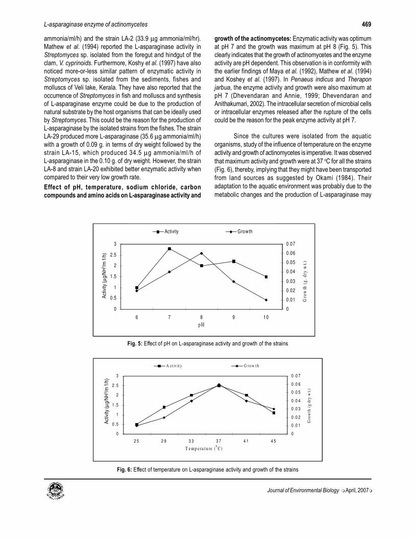
growth of the actinomycetes: Enzymatic activity was optimum
at pH 7 and the growth was maximum at pH 8 (Fig. 5). This
clearly indicates that the growth of actinomycetes and the enzyme
activity are pH dependent. This observation is in conformity with
the earlier findings of Maya et al. (1992), Mathew et al. (1994)
and Koshey et al. (1997). In Penaeus indicus and Therapon
jarbua, the enzyme activity and growth were also maximum at
pH 7 (Dhevendaran and Annie, 1999; Dhevendaran and
Anithakumari, 2002). The intracellular secretion of microbial cells
or intracellular enzymes released after the rupture of the cells
could be the reason for the peak enzyme activity at pH 7.
Since the cultures were isolated from the aquatic
organisms, study of the influence of temperature on the enzyme
activity and growth of actinomycetes is imperative. It was observed
that maximum activity and growth were at 37 oC for all the strains
(Fig. 6), thereby, implying that they might have been transported
from land sources as suggested by Okami (1984). Their
adaptation to the aquatic environment was probably due to the
metabolic changes and the production of L-asparaginase may
0
0 .5
1
1 .5
2
2 .5
3
2 5 2 9 3 3 3 7 4 1 4 5
T e m p e ra t u re (0
C )
Act
ivit
y (
ug
/NH
3/m
l/h
)
0
0 .0 1
0 .0 2
0 .0 3
0 .0 4
0 .0 5
0 .0 6
0 .0 7
Gro
wth
(g
dry
wt.
)
A c t iv it y G ro w t h
0
0.5
1
1 .5
2
2 .5
3
6 7 8 9 10
p H
Act
ivit
y (
ug
/NH
3/m
l/h
)
0
0.01
0.02
0.03
0.04
0.05
0.06
0.07
Gro
wth
(g
. d
ry w
t.)
Activity Grow th
Fig. 5: Effect of pH on L-asparaginase activity and growth of the strains
Fig. 6: Effect of temperature on L-asparaginase activity and growth of the strains
L-asparaginase enzyme of actinomycetes
Act
ivity
(µg/
NH
3 /m
1/h
)A
ctiv
ity (µ
g/N
H3 /m
1/h
)
469
Journal of Environmental Biology �April, 2007�
be unique (Dhevendaran and Anitha Kumari, 2002). It has already
been reported by Maya et al. (1992) that optimum activity and
maximum growth in Bacillus sp. and Moraxella sp. were at 37oC
and Vibrio sp. exhibited maximum L-asparaginase activity at
68oC (Selvakumar et al., 1991) though it was isolated from the
shellfish, Telescopium telescopium from the Vellar estuary.
Recently, Dhevendaran and Annie (1999) and Dhevendaran and
Anitha Kumari (2002) have observed maximum activity of this
enzyme at 37 oC in the mesophilic Streptomyces AQBPI 104 and
AQBTJ 60, isolated from Penaeus indicus and Therapon jarbua of
the Veli lake, Kerala.
Since in the present study, actinomycetes were isolated
from the estuarine fish, it was felt necessary to understand the
influence of various concentrations of sodium chloride on its
enzymatic activity and growth. The activity was recorded even at
0% and the optimum growth was observed at 1-2% sodium
chloride concentration in all the strains (Fig. 7). Selvakumar et
al. (1977), Maya et al. (1992), Mathew et al. (1994) and
Dhevendaran and Anitha Kumari (2002) have also noticed similar
pattern of enzyme activity in the marine sediments, marine
bacteria, streptomycetes and Streptomyces respectively. The
reduced enzyme activity at the higher concentration could be
due to the formation of certain inhibitors in the growth medium or
due to the inhibitory nature of increased sodium chloride level.
Among the different carbon sources used, the enzyme
activity and growth were enhanced in sucrose, whereas lactose
had an inhibitory effect (Fig. 8). Selvakumar et al. (1977) tested
L-asparaginase activity in 24 carbon sources and found that
lactose had inhibitory effect on the marine Vibrio sp. In Moraxella
sp. and streptomycetes, the enzyme activity and growth were
increased by the addition of sucrose and inhibited by lactose as
observed by Maya et al. (1992) and Mathew et al. (1994).
Fig. 9 shows the effect of amino acids on L-asparaginase
activity and growth of the isolated strains. Maximum activity of
enzyme and growth of the strains were noticed without any amino
acids. This observation is in conformity with the earlier findings
of Mathew et al. (1994) and Dhevendaran Anitha Kumari et al.
(2002) in Streptomyces sp. They observed higher L-asparaginase
activity and growth of actinomycetes without any amino acids
and thereby reported that the naturally available amino acids in
the host organisms were sufficient for the maximum enzymatic
activity and growth of actinomycetes.
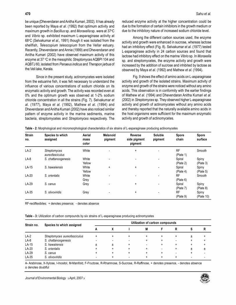
Table - 2: Morphological and micromorphological characteristics of six strains of L-asparaginase producing actinomycetes
Strain Species to which Aerial Melanoid Reverse Soluble Spore Spore
no. assigned mass pigment side pigment pigment chain surface
color pigment
LA-2 Streptomyces White - + - RF Smooth
aureofasciculus (Plate 1)
LA-8 S. chattanoogenesis White - - - Spiral Spiny
Yellow (Plate 2) (Plate 3)
LA-15 S. hawaiiensis White + + - Spiral Spiny
Yellow (Plate 4) (Plate 5)
LA-20 S. orientalis White - - - RF Smooth
Grey (Plate 6)
LA-29 S. canus Grey - + - Spiral Spiny
(Plate 7) (Plate 8)
LA-35 S. olivoviridis Grey - + - RF Spiny
(Plate 9) (Plate 10)
RF-rectiflexibiles; + denotes presence; - denotes absence
Table - 3: Utilization of carbon compounds by six strains of L-asparaginase producing actinomycetes
Strain no. Species to which assignedUtilization of carbon compounds
A X I M F R S R
LA-2 Streptomyces aureofasciculus + + + + + + ± +
LA-8 S. chattanoogenesis - - - + + - + +
LA-15 S. hawaiiensis ± ± + - + + + +
LA-20 S. orientalis + + + + - + ± ±
LA-29 S. canus + + + + + - + -
LA-35 S. olivoviridis - + - + + + - -
A- Arabinose, X-Xylose, I-Inositol, M-Manifold, F-Fructose, R-Rhamnose, S-Sucrose, R-Raffinose, + denotes presence, - denotes absence
± denotes doubtful
Sahu et al.470

Journal of Environmental Biology �April, 2007�
0
0.5
1
1.5
2
2.5
3
3.5
Control M ethionine Tryp tophan L-Glutamic acid Threonine
Amino acids (ml-1
)
Act
ivit
y (
ug
/NH
3/m
l/h
)
0
0.01
0.02
0.03
0.04
0.05
0.06
Gro
wth
(g
. d
ry w
t.)
Activity Growth
Fig. 7: Effect of NaCl concentration on L-asparaginase activity and growth of the strains
Fig. 9: Effects of amino acids on L-asparaginase activity and growth of the strains
Fig. 8: Effects of carbon compounds on L-asparaginase activity and growth of the strains
0
0.5
1
1.5
2
2.5
3
3.5
0 0.05 0.1 0.5 1 2 3 4
NaCl concen tration (%)
Act
ivit
y (
ug
/NH
3/m
l/h
)
0
0.01
0.02
0.03
0.04
0.05
0.06
0.07
0.08
Gro
wth
(g
. d
ry w
t.)
A ct ivity G row th
0
0.5
1
1.5
2
2.5
3
3.5
Control
Gluco
se
Sucros
e
Lacto
se
Raffin
ose
Man
nitol
Carbon s ource (% )
Act
ivit
y (
ug
/NH
/ml/
h)
0
0.01
0.02
0.03
0.04
0.05
0.06
0.07
0.08
Gro
wth
(g
. d
ry w
t.)
A ct ivity G row th
L-asparaginase enzyme of actinomycetes
Act
ivity
(µg/
NH
3 /m
1/h
)A
ctiv
ity (µ
g/N
H3 /m
1/h
)A
ctiv
ity (µ
g/N
H3 /m
1/h
)
471

Journal of Environmental Biology �April, 2007�
Plate - 1: Strain LA 2 - Rectiflexibiles (RF) spores chain (400X)
Plate - 2: Strain LA 8 - Spiral (S) spore chain (400X)
Plate - 3: Strain LA 8 - Spores with spiny surface (15,000X)
Plate - 4: Strain LA 15 - Spiral (S) spore chain (400X)
Plate - 5: Strain LA 15 - Spores with spiny surface (15,000X)
Plate - 6: Strain LA 20-Rectiflexibiles (RF) spore chain (400X)
Plate - 7: Strain LA 29 - Spiral (S) spore chain (400X)
Plate - 8: Strain LA 29 - Spores with spiny surface (7,000X)
Plate - 9: Strain LA 35-Rectiflexibiles (RF) spore chain (400X)
Plate - 10: Strain LA 35 - Spores with spiny surface (4,500X)
Sahu et al.472

Journal of Environmental Biology �April, 2007�
Taxonomic investigation: Of the 40 strains tested for
L-asparaginase activity, only six strains viz. LA-2, LA-8, LA-15,
LA-20, LA-29 and LA-35 showed good activity (Table 1) and hencethese strains were selected for identification.
All the six strains possess LL–Diaminopimelic acid and all
the strains tested contain glycine in their cell wall. Presence of LL –Diaminopimelic acid along with glycine indicates the cell wall
chemotype – I. The strains with chemotype – I do not havecharacteristic pattern of sugars (Lechevalier and Lechevalier, 1970).
The strains belonging to the wall type I are Streptomyces,
Streptoverticillium, Chainia, Actinopycnidium, Actinosporangium,
Elyptrosporangium, Microellobosporia, Sporichthya andIntrasporangium (Lechevalier and Lechevalier, 1970). The
micromorphological observations of the strains, LA-2, LA-8, LA-15, LA-20, LA-29 and LA-35 reveal that all these belong to the
genus Streptomyces. The predominance of Streptomyces in any
actinomycete population is a well known fact (Alexander, 1961).
The morphological, micromorphological, physiologicaland biochemical characteristics obtained for the L-asparaginase
producing strains LA-2, LA-8, LA-15, LA-20, LA-29 and LA-35,tested in the present study are depicted in Table 2, 3. The results
were compared with those of the Streptomyces species given inthe key of Nonomura (1974) and those species described in the
Bergey’s Manual of Determinative Bacteriology (Buchanan and
Gibbons, 1974).
The result shows that except for the absence of
production of melanoid and soluble pigments by the strain LA-2
(Table 2), all the other characters are exactly similar to those ofS. aureofasciculus. Therefore, the strain LA-2 has been tentatively
identified as S. aureofasciculus. In the strain LA-8, except for thedifference in the utilization of inositol and production of soluble
pigment, all the other properties are exactly similar to that of S.
chattanoogenesis and hence this strain has been tentativelyidentified as S. chattanoogenesis (Tables 2, 3). The result further
shows that except for the production of reverse side pigmentsand absence of utilization of mannitol by the strain LA-15 (Tables
2, 3), all the other characters are exactly similar to those of S.
hawaiiensis. Therefore, the strain LA-15 has been tentativelyidentified as S. hawaiiensi. Table 3 shows that the strain LA-20 is
weak in utilization of the carbon compounds viz. fructose. Exceptthis, all the other properties are the same for LA-20 and S.
orientalis. Therefore, the strain LA-20 has been identified as S.orientalis. The strain LA-29 differed from the reference strain S.
canus, by not utilizing the carbon compound viz. rhamnose.
However, LA-29 resembles the reference strain by showing closesimilarity in all the other characters and so the strain LA-29 has
been identified as S. canus (Table 2). Similarly, the strain LA-35is weak in the utilization of arabinose and production of soluble
pigment. Except this, all the other characters of the strain LA-35
are exactly similar to that of S. olivoviridis and hence the strainLA-35 has been identified as S. olivoviridis (Tables 2, 3).
From the present study, it has been inferred that the
finfishes viz. M. cephalus, C. chanos and E. suratensis of the
Vellar estuary are potential candidates for the isolation of L-
asparaginase producing actinomycetes and the growth conditions
have been optimized for culture of these strains and synthesis
of L-asparaginase enzyme under laboratory conditions. Further,
the indigenous constituents of the host organisms and the
environmental factors have influence on the metabolic activity of
microorganisms also. During the present study, six strains were
identified from the different parts of fishes and assigned to different
species of Sterptomyces viz. Streptomyces aureofasciculus (LA-
2), S. chattanoogenesis (LA-8), S. hawaiiensis (LA-15), S.
orientalis (LA-20), S. canus (LA-29) and S. olivoviridis (LA-35),
which possess good L-asparaginase enzyme activity. Further,
studies are needed to develop novel anti cancer drugs of L-
asparaginase enzyme extracted from actinomycetes.
As these fishes are available throughout the year in the
estuary in cheaper rates, hence, it is recommended to utilize the
finfishes viz. M. cephalus, C. chanos and E. suratensis for the
isolation of L-asparaginase producing actinomycetes in the
commercial scale.
Acknowledgments
Authors thank Prof. T. Balasubramaniam, Director,
Centre of Advanced Study in Marine Biology and the authorities
of Annamalai University for providing with necessary facilities.
One of the authors (M.K.S.) is thankful to the Ministry of
Environment and Forest, Government of India for the Fellowship.
References
Alexander, M.: Introduction to soil microbiology. John Wiley and Sons. INC.,
New York (1961).
Balakrish Nair, G., N. Selvakumar, D. Chandramohan and R. Natarajan:
Distribution and activity of L-asparaginase producing fungi in the marine
environment of Porto Novo. Ind. J. Mar. Sci., 6, 172-173 (1977).
Buchanan, R.E. and N.E. Gibbons: Bergey’s manual of determinative
bacteriology. The Williams and Wilkins Co., Baltimore. pp. 747-842
(1974).
Clausen, A.: Determination of L-asparaginase activity in serum by TLC:
Application to the treatment of acute lymphoblstic leukemia. Clinic. Chim.
Acta, 30, 111-116 (1986).
Crowther, D.: L-asparaginase and human malignant disease. Nature, 229,
168-171 (1971).
Dhevendaran, K. and Y. K. Anitha Kumari: L-asparaginase activity in growing
conditions of Streptomyces sp. associated with Therapon jarbua and
Villorita cyprinoids of Veli lake, south India. Ind. J. Mar. Sci., 39(2),
155-159 (2002).
Dhevendaran, K. and K. Annie: Antibiotic and L-asparaginase activity of
Strptomycetes isolated from fish, shellfish and sediments of Veli estuarine
lake along Kerala coast. Ind. J. Mar. Sci., 28, 335-337 (1999).
Koshey, A., K. Dhevendaran, M. I. Georgekutty and P. Natarajan: L-asparaginase
in Streptomyces plicatus isolated from the alimentary canal of the fish,
Gerres filmentosus (Cuvier). J. Mar. Biotechnol., 5, 181-185 (1997).
Lechevalier, M. P. and H. Lechevalier: Chemical composition as a criterion in
the classification of aerobic actinomycetes. Int. J. Syst. Bacteriol., 20,
435-443 (1970).
Lowry, O., N. J. Rosenbrough, A. L. Farr and R. J. Randall: Measurement with
the folin phenol reagent. J. Biol. Chem., 193, 265-275 (1951).
L-asparaginase enzyme of actinomycetes 473

Journal of Environmental Biology �April, 2007�
Mathew, A., K. Dhevendaran, M. I. Geogekutty and P. Natarajan: L-asparaginase
activity in antagonistic Streptomycetes associated with clam Villorita
cyprinoides (Hanley). Ind. J. Mar. Sci., 23, 204-208 (1994).
Maya, K., K. Dhevendaran and P. Natarajan: Studies on L-asparaginase activity
in Bacillus of retting ground. Fish. Technol., 29, 62-66 (1992).
McCreadie, K. B., D. H. W. Ho and E. J. Freireich: L-asparaginase for the
treatment of cancer. Cancer J. Clin., 23, 220-227 (1973).
Nonomura, H.: Key for classification and identification of 458 species of the
Streptomycetes included in ISP. J. Ferment. Technol., 52(2), 78 - 92 (1974).
Okami, Y.: Marine microorganisms as a source of bioactive agents In: Current
perspective in microbial ecology (Eds: M.J. Klug and C. A. Reddy).
American society for microbiology, Washington DC. pp. 615 - 655 (1984).
Selvakumar, N., D. Chandramohan and R. Natarajan: L-asparaginase activity
in marine sediments. Curr. Sci., 46(9), 287-290 (1977).
Selvakumar, N., Vanaja Kumar and R. Natarajan: Part ial purification,
character izat ion and anti-t umor propert ies of L-asparaginase
(antileukemic agent) from a marine Vibrio. In: Bioactive compounds from
marine organisms with emphasis on the Indian ocean (Eds: Mary Frances
Thompson, Rachakonda Sarojini and Rachakonda Nagabhushanam).
Oxford and IBM Publishing Co. Pvt. Ltd., New Delhi, Bombay and
Calcutta. pp. 289-300 (1991).
Shirling, E.B. and D. Gottlieb: Methods for characterization of Streptomycetes
species. Int. J. Syst. Bacteriol., 16, 313 - 340 (1966).
Sudha, K., Nirmala Thampuran and P. K. Surendran: Prevalence of Vibrio species on
fish from pelagic and demersal habitats. Fish. Technol., 39(2), 150-154 (2002).
Wriston, J.C.: L-asparaginase. In: The enzymes. 3rd Edn. Academic Press, London,
New York. 4, 101-121 (1971).
Wriston, J.C. and T.O.Yellin: L-asparaginase: A review. In: Advances in enzymology
(Ed: A. Meister). John Wiley and Sons, Inc., New York. 39, 185-248 (1973).
Yoshimoto, T.: Characterization of polyethylene glycol-modified L-asparaginase
from E. coli and its application of therapy in leukemia. Jap. J. Cancer, 77,
1264-1270 (1986).
Sahu et al.474