Embed Size (px)
Citation preview
STUDIES ON HEMORRHAGIC ANEMIA IN DOGS*
BY J. M. McKIBBIN, A. E. SCHAEFER, C. A. ELVEHJEM, AND E. B. HART
(From the Department of Biochemistry, College of Agriculture, University of Wisconsin, Madison)
(Received for publication, June 15, 1942)
Anemia studies have been carried on in this laboratory for several years in dogs on whole milk rations supplemented in various ways. The anemias have been induced by nutritional and hemorrhagic methods (1, 2). Nu- tritional anemia in puppies receiving such a ration was found curable by the addition of inorganic iron and copper salts, although regeneration was suboptimal in a number of cases (1). When anemia was produced in adult dogs by mild phlebotomy, the response to these mineral supplements was usually good (3), although occasionally it was slow and erratic and under more severe phlebotomy recovery might not occur at all. The most con- sistent failure in blood regeneration occurred when a high level of cobalt was added to the ration (4). The addition of liver extract to these rations resulted in rapid remission from the anemias in all cases. The behavior of dogs is thus comparable to that of rats receiving milk rations. In the lat- ter species iron and copper supplements produce and maintain a normal blood stream but under the strain of hemorrhage evidence indicates that liver supplements furnish additional factors not adequately supplied by the mineralized milk alone (5). It is the purpose of this paper to report inves- tigations into the nature of the substance or substances in liver extract re- sponsible for this stimulation of blood formation.
Recently considerable progress has been made,in the clarification of the problems of dog nutrition and it has been possible for us to obtain normal growth in dogs for a considerable period of time on a highly purified ration supplemented only with synthetic vitamins (6). Phlebotomy and blood regeneration studies on this ration have been most informative and will also be presented in this paper.
EXPERIMENTAL
Hemorrhagic Anemia Xtudies on Mineralized Milk Diet
Adult mongrel dogs were given whole milk twice daily ad l&turn. They were bled weekly from the external jugular vein in amounts usually ranging
* Published with the approval of the Director of the Wisconsin Agricultural Ex- periment Station. Supported in part by a grant from the Wisconsin Alumni Research Foundation. We are indebted to Merck and Company, Inc., Rahway, New Jersey, for generous supplies of thiamine chloride, riboflavin, nicotinic acid, pyridoxine hydrochloride, calcium pantothenate, and choline chloride; to The Wilson Labora- tories, Chicago, Illinois, for the liver extracts and bile salts; and to Abbott Labora- tories, North Chicago, Illinois, for haliver oil.
107
by guest on June 10, 2018http://w
ww
.jbc.org/D
ownloaded from
108 HEMORRHAGIC ANEMIA IN DOGS
from 20 to 25 per cent of their total blood. After 4 to 8 weeks a rather stable state of anemia was reached at a hemoglobin level of 7 to 9 gm. per cent and phlebotomy was discontinued. 20 mg. of cobalt as cobaltous chloride were then added daily to the ration and this addition continued for 1 to 2 weeks followed by 30 mg. of iron as ferric chloride and 3 mg. of each of copper and manganese as cupric and manganous sulfates. After 1 to 2 weeks, during which time the hemoglobin level of the blood increased very little, the liver supplement extract to be tested was given for a period of 2 weeks. Active liver preparations were found to effect a rise in blood hemoglobin to levels of 13 to 14 gm. per cent in this time.
In addition to the hemoglobin determination, red cell counts and hema- tocrit determinations were made routinely and later a plasma iron analysis was also performed routinely. Blood samples for analysis were always taken from the radial vein. An 18 cc. sample was drawn into a 30 cc. syringe, the dead space of the syringe being filled with Wintrobe’s non- shrinking oxalate solution. Hemoglobin determinations were made by the method of Evelyn (7), and hematocrit determinations with Wintrobe tubes. The plasma iron determinations were made on filtrates of blood plasma from a hot trichloroacetic acid precipitation. The filtrates were treated with thioglycolic acid and the ferrous iron then reacted with cx , a’-bipyridine (pH 5.4). The resulting color was determined on the Evelyn photoelectric calorimeter. This method is to be published in detail in the near future.
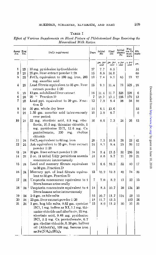
The data from the analysis of blood drawn during the assay of various liver preparations and nutrient materials are given in Table I. For the sake of brevity only the initial and final hemoglobin levels and the initial and maximum plasma iron levels are given. In evaluation of the hemo- globin response to the various supplements it was necessary to minimize errors arising from fluctuations in weight. Assuming 8 per cent of body weight as blood, the total body hemoglobin was calculated from the hemo- globin level of the blood. These values, together with the known amount of hemoglobin removed as the analysis sample, permitted the calculation of the total “hemoglobin made” as given in the last column.
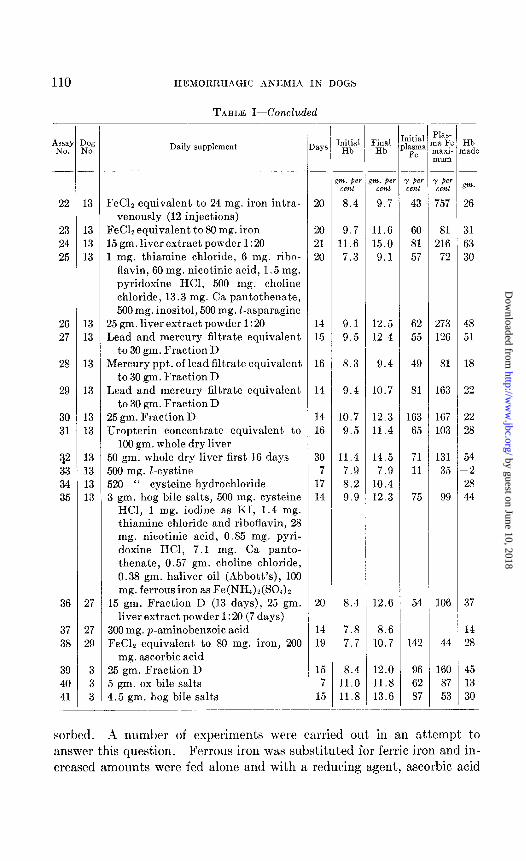
The typical blood picture as seen in the anemia and in the course of the remission is shown in Fig. 1. The anemia is characterized by a fairly high erythrocyte count, extremely small cells, low hematocrit, low hemoglobin, and low plasma iron. The administration of liver extract results in striking increases in hemoglobin, hematocrit, erythrocyte count, and plasma iron but the mean erythrocyte volume and saturation index remain practically unchanged.
The possibility of iron deficiency as suggested by the low mean cell volume and plasma iron values led us to investigate first the question of whether or not the ferric iron as fed was being properly reduced and ab-
by guest on June 10, 2018http://w
ww
.jbc.org/D
ownloaded from
MCKIBBIN, SCHAEFEH, IGLVEHJEM, AND HAR1’ 109
TABLE I
Effect of Various Supplements on Blood Picture of Phlebotomized Dogs Receiving the Mineralized Milk Ration
rc b
‘% To.
1 2 3
4
5 6 7
8 9
10
-
‘ ‘ 13 23 23
23
23 23 23
24 24
24
11 24 12 24
13 24 14 24
15 24
16 24
17 24
18 24
19 24 20 24 21 24
- -
Daily supplement
.O mg. pyridoxine hydrochloride !5 gm. liver extract powder I :20 ?eClz equivalent to 100 mg. iron, 200
mg. ascorbic acid lead filtrate equivalent to 25 gm. liver
extract powder I:20 2 gm. solubilized liver extract !O “ FractionD Lead ppt. equivalent to 30 gm. Frac-
tion D i0 gm. whole dry liver 1.35 gm. ascorbic acid intravenously
over period i2 mg. nicotinic acid, 6.3 mg. ribo-
flavin, 10.5 mg. thiamine chloride, 1 mg. pyridoxine HCl, 12.6 mg. Ca pantothenate, 250 mg. choline chloride
FeC12 equivalent to 60 mg. iron 4sh equivalent to 25 gm. liver extract
powder 1: 20 20 gm. liver extract powder 1:20 1 cc. (4 units) Lilly pernicious anemia
concentrate intravenously Lead and mercury filtrate equivalent
to 30 gm. Fraction D Mercury ppt. of lead filtrate equiva-
lent to 30 gm. Fraction D Uropterin concentrate equivalent to 4
liters human urine orally Uropterin concentrate equivalent to 4
liters human urine intravenously 2-3 gm. ox bile salts 25 gm. liver extract powder I : 20 3 gm. hog bile salts, 0.52 gm. cysteine
HCI, 1 mg. iodine as KI, 1.1 mg. thi- amine chloride and riboflavin, 22 mg. nicotinic acid, 0.66 mg. pyridoxine HCl, 5.5 mg. Ca pantothenate, 0.7 gm. choline chloride, 0.38 gm. halivei oil (Abbott’s), 100 mg. ferrous iron
aa FeN&)&30&
ID.
:
1 1
1
1
-
-
17
15 13
15
16 7
12
14 LO
16
20 14
14 14
15
13
7
14
15 14 13
-
m. per cent
7.7 8.5 7.4
1. pe9 cent 8.5 14.0 9.1
9.1
11.4 11.7 7.3
8.1 5.9
6.5
11.4
11.7 13.1 8.4
13.6 6.7
7.3
7.3 LO.8 8.1 9.4
9.4 13.5 6.6 8.7
8.6 10.2
10.2 12.3
7.0 8.2
8.5
10.7 11.7
8.0
10.7
11.7 13.3 11.2
‘inal Hb
kitial asma Fe
65
73
328 228 36
24
26 25
30 11
53
40
13
30
134
30
‘kS- a Fe laxi- m m
per xnt
73
!28
128 179 38
26
23 30
296 39
Ib ade
m.
14 30 17
23
6 13 10
55 0
13
41 12
34 21
17
23
13
20
15 26 31
by guest on June 10, 2018http://w
ww
.jbc.org/D
ownloaded from
110 HEMORRHAGIC ANEMIA IN DOGS
TABLE I-Concluded
22
23 24 25
26 27
28
29
30 31
?2 33 34 35
36
37 38
39 40 41
Y )W 90.
13
13 13 13
13 13
13
13
13 13
13 13 13 13
27
27 29
3 3 3
Daily supplement D aYS
FeC12 equivalent to 24 mg. iron intra- venously (12 injections)
FeClz equivalent to 80 mg. iron 15 gm. liver extract powder 1: 20 1 mg. thiamine chloride, 6 mg. ribo-
flavin, 60 mg. nicotinic acid, 1.5 mg. pyridoxine HCI, 500 mg. choline chloride, 13.3 mg. Ca pantothenate, 500 mg. inositol, 506 mg. Z-asparagine
15 gm. liver extract powder 1: 20 Lead and mercury filtrate equivalent
to 30 gm. Fraction D Mercury ppt. of lead filtrate equivalent
to 30 gm. Fraction D Lead and mercury filtrate equivalent
to 30 gm. Fraction D 25 gm. Fraction D IJropterin concentrate equivalent to
100 gm. whole dry liver 50 gm. whole dry liver first 16 days 500 mg. Z-cystine 520 “ cysteine hydrochloride 3 gm. hog bile salts, 500 mg. cysteine
HCl, 1 mg. iodine as KI, 1.4 mg. thiamine chloride and riboflavin, 28 mg. nicotinic acid, 0.85 mg. pyri- doxine HCl, 7.1 mg. Ca panto- thenate, 0.57 gm. choline chloride, 0.38 gm. haliver oil (Abbott’s), 100 mg. ferrous iron as Fe(NH4)2(SO&
15 gm. Fraction D (13 days), 25 gm. liver extract powder I :20 (7 days)
300 mg. p-aminobenzoic acid FeClz equivalent to 80 mg. iron, 200
mg. ascorbic acid
20
20 21 20
14 9.1 12.5 15 9.5 12.4
16 8.3 9.4
14 9.4 10.7
14 10.7 12.3 16 9.5 11.4
30 11.4 14.5 7 7.9 7.9
17 8.2 10.4 14 9.9 12.3
25 gm. Fraction D 5 gm. ox bile salts 4.5 gm. hog bile salts
20
14 19
15 7
15
hitial Final Hb Hb
712. @I m. per Y w cent cent cent 8.4 9.7 43
9.7 11.6 60 11.6 15.0 81
7.3 9.1 57
%S- a Fe
mxi- num
I per cent 757
81 216
72
62 273 55 126
49 81
81 163
163 167 65 103
71 131 11 35
75 99
8.4 12.6 54 106
7.8 8.6 7.7 10.7 142 44
8.4 12.0 96 160 11.0 11.8 62 87 11.8 13.6 87 53
litial
‘Y Hb lade
:m.
26
31 63 30
48 51
18
22
22 28
54 -2 28 44
37
14 28
sorbed. A number of experiments were carried out in an attempt to answer this question. Ferrous iron was substituted for ferric iron and in- creased amounts were fed alone and with a reducing agent, ascorbic acid
by guest on June 10, 2018http://w
ww
.jbc.org/D
ownloaded from

MCKIBBIN, SCHAEFER, ELVEHJEM, AND HART 111
(Assays 3, 11, 23, 38, Table I). Ferrous iron was injected intravenously to the level of toxicity symptoms (Assay 22). The response in all cases was far inferior to that given by liver preparations and it was concluded that iron deficiency was not the primary cause of remission failure. That it was a complicat#ing secondary factor seemed altogether probable and so ferrous iron was substituted for ferric iron and thereafter the mineral supplements were supplied continually during the bleeding, anemic, and remission periods.
The organic nature of the fact,or in liver is evident from the failure of the ash fraction of liver extract to produce remission (Assay 12). When the original liver extract was given, a satisfactory response occurred (Assay 13).
t i
I T I I I .I I I 1 I I I
i I 1 1 I 1 1 I I I I 1
0
I
FIG. 1. Effect of equivalent amounts of liver extract ash and liver extract powder on the blood picture of Dog 24 receiving the mineralized milk ration with cobalt. The supplements were initiated at the points indicated by arrows. Hb, blood hemoglobin in gm. per cent; r.b.c., erythrocyte count in millions per c.mm.; m.c.v., mean erythrocyte volume in cu. micra; Fe, plasma iron in mg. per cent; Ht, hemato- crit per cent.
The low mean cell volume also suggested a possible deficiency of pyridox- inc, since this deficiency anemia is also microcytic. Supplements of vita- min l& far in excess of the amount present in the curative dose of liver ex- tract failed to produce improvement (Assay 1). Pyridoxine in supplement mixtures with thiamine, riboflavin, nicotinic acid, calcium pantothenate, choline, asparagine, and inositol produced slight to partial remissions but not at all comparable to those produced by liver extracts (Assays 10 and 25). The slight activity of the water-soluble vitamin mixtures is of inter- est, since several members of this group have been associated with hema- t,opoiesis. Chronic thiamine deficiency in man has been reported by Mason and Mason (8) to be accompanied by a macrocytic hyperchromic anemia, although this was not observed in acute thiamine deficiency. Gobell (9)
by guest on June 10, 2018http://w
ww
.jbc.org/D
ownloaded from
112 HEMORRHAGIC AXEMIA IX DOGS
obtained increases in the red blood cell count of premature anemic infants by the administration of nicotinamide and riboflavin. Dollchen (10) re- ported hemoglobin and erythrocyte responses to pyridoxine, nicotinamide, and riboflavin in a study of hemorrhagic anemia in rabbits. Gyijrgy et al. (11) observed some hemoglobin increase after the administration of ribo- flavin to phlebotomixed dogs. The action of choline is of considerable interest since the observation of Davis (12) that choline is the substance in liver responsible for the depressing effect on cobalt polycythemia. Since a high level of cobalt was used in this ration and may be wholly or in part responsible for remission failure, the action of an antagonizing substance such as choline would deserve investigation. Whatever the role of these water-soluble vitamins in hematopoiesis in dogs may be, it is apparent that they are not of primary importance under the conditions of this study.
A similar neutralizing effect on cobalt toxicity has been attributed to vitamin C by Barron and Barron (13). These investigators observed that intravenous injections of vitamin C would prevent polycythemia in rabbi& if given simultaneously with cobalt. No effective antagonistic action was obtained with vitamin C in our studies if one may use blood regeneration as the criterion for this action (Assays 3, 9, and 38). The low vitamin C content of active liver preparations also argues against the possibility that this vitamin is the active constituent of liver extract.
A cysteine-cobalt complex has been investigated for its biological impli- cations by Griffith et al. (14). They found that acute cobalt toxicity in rats could be prevented by the addition of cysteine or cystine to the diet. In our studies 500 mg. per day of cystine were ineffective over a 7 day period (Assay 33). Cysteine hydrochloride, however, fed at a level of 520 mg. per day showed considerable promise of activity (Assay 34). Since it is cysteine and not cystine which forms the complex with cobalt, the nega- tive result with the latter suggests a failure by the dog to reduce this amino acid to cysteine. At all events, it appears that at least a part of the activ- ity of liver extracts may be attributed to their content of cysteine or small molecular weight peptides containing cysteine, as for example, glutathione. Absorbed cobalt may then be tied into the cobalt-cysteine complex which is incapable of exerting the toxicological actions produced by cobalt alone.
The role of bile salts in the absorption of various substances essential to blood formation has been widely investigated. Seyderhelm et al. (15, 16) found that bile fistula dogs developed an anemia which was autoregulated at about two-thirds the normal hemoglobin level. The anemia was pre- ventable or curable by feeding bile, bile acids, and finally by light-activated ergosterol. Takasu (17) repeated this study of “acholic cachexia” in dogs and found a similar blood picture. He observed, in turn, that bile acids were effective if vitamin D were present. The relation of dietary calcium,
by guest on June 10, 2018http://w
ww
.jbc.org/D
ownloaded from
MCKIBBIN, SCHAEFER, ELVEHJEM, AND HART 113
phosphorus, and vitamin D to iron absorption has been investigated (18). In general an optimum calcium-phosphorus ratio for ossification with ade- quate vitamin D establishes optimum conditions for iron absorption. Con- ceivably these conditions prevent an excess of phosphate in the intestinal tract and a minimum of dietary iron is made unavailable as insoluble ferric phosphate. Mild anemias may then arise from vitamin D deficiency. Josephs (19) found that iron absorption was lowered in infants receiving a diet deficient in vitamin D.
Bile salts may also play a role in iron absorption independent of their relation with the fat-soluble vitamins. Hawkins et al. (20) observed a decreased hemoglobin production in bile fistula dogs which could not be en- tirely accounted for by the decrease in iron absorption. Smith and Cran- da11 (21) found that bile fistula dogs remain persistently anemic in spite of an unimpaired iron absorption as evident from their plasma iron studies.
These relationships suggested the trial of bile salts’ in our experimental anemia. In three trials, two may be considered as indicative of definite activity (Assays 40 and 41); the other showed considerably less stimulation (Assay 19). Although the plasma iron studies in these assays are incom- plete, the available data show no elevation of plasma iron as might be ex- pected if the responses obtained were due to a better absorption of iron, Although none of the responses was comparable to those obtained with liver extracts; nevertheless, it seems apparent that part of the activity of the liver preparations may be attributed to their content of bile salts.
The r61e of xanthopterin or uropterin in blood formation was first sug- gested by the work of Tschesche and Wolf (22). These investigators found that uropterin produced striking increases in the erythrocyte count of young rats made anemic by being fed goat milk. Simmons and Norris (23) working with a nutritional anemia in fingerling Chinook salmon found that intraperitoneal injection of liver extract would produce rapid red blood cell count increases. The active principle of liver was found to be uropterin.
On the basis of the above findings it was decided to try uropterin in this study. No synthesis of the pigment was attempted. Instead, concen- trates of it were prepared from liver and human urine according to the concentration methods of Koschara (24, 25). A preparation made from whole dry liver was fed at a level equivalent to 100 gm. of the original liver per day (Assay 31). One preparation from urine equivalent to 4 liters of urine per day was fed orally (Assay 17), while a more purified preparation was injected intravenously (Assay 18) at the same level. All three prepa- rations showed appreciable activity but were considerably below the respon-
1 Suggested by Dr. David Klein of The Wilson Laboratories, Chicago, Illinois.
by guest on June 10, 2018http://w
ww
.jbc.org/D
ownloaded from
114 HEMORRHAGIC ANEMIA IN DOGS
ses elicited by liver extract. These preparations contained far more urop- terin than would be present in the curative dose of liver extract. The rBle of uropterin, then, seems doubtful, although its deficiency may be at least in part responsible for the failure of blood regeneration.
An attempt at fractionation of the active liver extracts ‘(Fraction D” and “liver concentrate powder 1: 20” (The Wilson Laboratories) was made by use of heavy metal precipitation. In both cases aqueous solutions of the liver extracts were adjusted alkaline to phenolphthalein with NH,OH and sufficient 30 per cent basic lead acetate solution added to insure com- plete precipitation.
The basic lead acetate precipitate was suspended in water and HzS gas bubbled through until all the lead salts were converted into their corre- sponding acids and the lead precipitated as sulfide. This process was re- peated and the combined filtrates neutralized and concentrated under reduced pressure. This preparation was designated as the “lead precipi- tate” and was fed at a level of 30 gm. equivalent of the original Fraction D (Assay 7). The relative inactivity of this preparation can be seen from the character of the hemoglobin and plasma iron changes.
The basic lead acetate filtrate obtained was treated with H2S gas until the excess lead was precipitated. The lead sulfide was then separated by filtration, washed with hot distilled water, and the filtrate and washings neutralized and concentrated under reduced pressure. This preparation designated as the “lead filtrate” was used in Assay 4. Considerable activ- ity was disclosed but the preparation was so difficult to feed at therapeutic levels that it was necessary to work with more purified concentrates.
The lead filtrate was therefore used for the preparation of two mercury fractions. The lead filtrate was adjusted to pH 6.8 and warm saturated HgCl, solution added and the precipitate formed was filtered off. The fil- trate was again adjusted to pH 6.8 and more HgClz solution added until complete precipitation was obtained. Excess mercuric ion was removed with H2S and the mercuric sulfide precipitate was washed with warm water. The combined filtrate and washings were concentrated under reduced prcs- sure and enough ethanol added to precipitate the inorganic salts. The aqueous ethanol-soluble material was filtered off, neutralized, and the alco- hol removed by distillation under reduced pressure. This preparation was designated as the “lead and mercury filtrate” and was used in Assays 15, 27, and 29. Activity was evident in all three trials but in only one (Assay 27) was it equivalent to that of whole liver extract. The other two trials showed considerably less activity.
The mercury precipitate fraction from the lead filtrate was suspended in water and H2S gas passed in until no more HgS was formed. The HgS was washed with boiling water and the filtrate and washings neutralized
by guest on June 10, 2018http://w
ww
.jbc.org/D
ownloaded from
MCKIBBIN, SCHAEFER, ELVEHJEM, AND HART 115
with KOH (Congo red). More H&S was passed in and the precipitate was allowed to settle. This second HgS precipitate was washed with hot water and the combined filtrate and washings neutralized and concentrated under reduced pressure. This preparation was designated as the (‘mercury pre- cipitate of the lead filtrate” and was used in Assays 16 and 28. Slight to moderate activity was observed in these two trials but the preparation seemed somewhat inferior to the mercury filtrate of the lead filtrate.
From the results obtained it seems apparent that the principal regener- ating activity in liver extract is not precipitated by basic lead acetate. More activity seems to pass into the mercury filtrate than into the precipi- tate, but the failure of the complete activity of liver extract to stay in any one fraction indicates that more than one active substance is involved. The possibility that none of these fractions would ever show its true activ- ity must be considered when the actual mechanical difficulties of feeding these preparations are taken into account. The loss of the taste-masking substances as precipitated by basic lead acetate leaves an extremely bitter preparation containing the activity. Nausea and even vomiting some- times accompanied the feeding of this lead filtrate and its two mercury fractions. Optimum blood formation might not be expected to occur under such adverse conditions of assay.
The activity of the lead filtrate fraction suggested the trial of antiper- nicious anemia concentrates, since this substance is not precipitated by basic lead acetate. A preparation obtained from Eli Lilly and Company containing 2 units per cc. was injected intravenously (Assay 14). The activity was only moderate as compared to that of the whole liver extracts and it was concluded that this factor was not involved. The excellent regenerating activity of liver Fraction D, which is very low in the antiper- nicious anemia principle, is better evidence for this conclusion.
The suggestion from the assay of various liver fractions that more than one substance is concerned in blood regeneration under these conditions as well as the activity of the several pure substances recounted above sug- gested the trial of a mixture of all of these active substances. Activity comparable to that of liver extract was obtained by a mixture of the syn- thetic B vitamins, hog bile salts, cysteine, increased iron, iodine, and hal- iver oil (Assays 21 and 35). The poor physical condition of the dogs used for assay after many months of steady experimentation lends considerable weight to the regeneration observed. Especially noteworthy was the ano- rexia and marked loss of weight of Dog 13 just prior to Assay 35. This loss of 3 kilos of body weight was far greater than that usually encountered in our assay work and indicated a severe deficiency or toxicity. The sup- plement mixture afforded the production of a relatively normal mean eryth- rocyte volume in Dog 13 but in neither assay were the plasma iron levels
by guest on June 10, 2018http://w
ww
.jbc.org/D
ownloaded from

116 HEMORRHAGIC ANEMIA IN DOGS
increased appreciably as is the case when cure is effected with liver extracts. It is possible that yet another substance in liver controls the level of iron in the plasma and perhaps its form or state. The general conclusions which may be arrived at from these studies on the milk ration will be considered after the work on the synthetic ration has been discussed.
Anemia Studies on Synthetic Ration
In our studies on the vitamin I3 complex in the nutrition of the dog WC have used a highly purified sucrose-casein ration supplemented wit,h syn- thetic vitamins and liver extracts (26, 27). We have found that if such a ration is supplemented only with synthetic thiamine, riboflavin, nicotinic acid, pyridoxine, pantothenic acid, and choline but no liver extracts a rela- tively normal rate of growth results and the animals may be carried on such a ration for many months in perfectly normal condition (28, 6, 29). However, if any one of these six synthetic vitamins is omitted, a fatal defi- ciency in young puppies will occur in 4 to 6 weeks. The use of such a ration for the study of factors involved in blood regeneration was desirable because (a) it would enable us to produce a ration relatively free of any single dietary factor and (b) it would enable us to rule out the effect of pos- sible unknown dietary factors present in natural food materials such as milk.
Since the dogs receiving the synthetic ration supplemented only with synthetic vitamins appeared to grow and develop normally, it would seem that no other dietary factor is necessary. The normal blood stream which the animals maintain on such a ration argues against the necessity of any other factor essential for blood formation, as was concluded from the studies on the milk ration. If such a factor were existent, it was thought probable that the strain of phlebotomy would clearly establish its deficiency.
Accordingly, four older growing dogs were placed on the basal ration used in all of these studies. It has the following composition: sucrose 66 per cent, acid-washed casein 19, cottonseed oil 8, cod liver oil 3, and salt mixture 4.* In addition the dogs received 100 y each of thiamine chloride and riboflavin, 2 mg. of nicotinic acid, 60 y of pyridoxine hydrochloride, 500 y of calcium pantothenate, and 50 mg. of choline chloride per kilo of body weight per day. These vitamins in aqueous solution were given orally twice weekly by pipette. The dogs were bled at varying intervals, usually every 10 to 13 days, over a period of about 7 months. From 15 to 25 per cent of the total blood in the dog was removed at each bleeding. This volume was measured and its hemoglobin determined. At the end
2 The salt mixture of Phillips and Hart (30) with an additional 1.2 gm. of MnSOa.4H20 per kilo of salt mixture. The mixture contains adequate iron, copper, and manganese but no cobalt salts.
by guest on June 10, 2018http://w
ww
.jbc.org/D
ownloaded from

MCKIBBIN, SCHAEFER, ELVEHJEM, AND HART 117
of the bleeding period the dogs were allowed 2 weeks to recover and at that time a blood sample for analysis was taken in the usual manner. The total blood volume and hemoglobin removed over the period, the weight changes, and the blood chemistry and cytology of the four dogs are given in Table II.
Dogs 190 and 191 after 65 and 79 days respectively on this regimen were then supplemented with 20 mg. per day of cobalt as cobaltous chloride. After 82 days the level of cobalt was increased to 40 mg. per day and con- tinued at this level for the final 58 days of the experiment. From Table II it can be seen that these dogs had very little difficulty in maintaining a nor- mal blood stream even when forced to regenerate four to five such blood streams over the experimental period. The cobalt seemed to produce very little if any deleterious effect on blood building, although at the higher
TABLE II
Blood Regeneration Studies on Four Older Growing Dogs Receiving the Synthetic Ration
Dog No ....................................................... 186
Total bleeding period, days .................... Initial weight, kg .............................. Final weight, kg ............................... Initial blood volume, liters. ................... Final blood volume, liters ...................... Blood removed over period, liters .............. Hemoglobin removed over period, gm .......... Final hemoglobin (2 wks. after final bleeding),
gm. % ....................................... Final hematocrit, 70 ...........................
“ mean erythrocyte volume, cu. micra ..... “ erythrocyte count, millions per c.mm ..... “ plasmairon,-y% ........................
216 216 205 219 11.8 11.05 15.8 14.2 16.95 12.65 16.9 11.8
0.94 0.88 1.26 1.14 1.36 1.01 1.35 0.95 4.82 3.80 5.93 5.09
515 404 685 636
13.3 12.9 12.2 14.5 44.0 44.0 40.5 45.0 63 50 66 54
6.97 8.85 6.16 8.39 102 52 45 52
188 190 191
levels there was a tendency to anorexia and loss of weight in Dog 191. This was quickly corrected by withdrawal of the cobalt at the end of the experiment. This was not observed in Dog 190 and may be explained on the basis of the greater size of this dog and consequent failure to show the chronic toxicity as quickly. It is of interest that the mean erythrocyte volume of the two dogs receiving cobalt was no lower at the end of the ex- periment than that of the two dogs without cobalt. Although these values may be considered somewhat subnormal, they are much higher than was observed on the milk ration.
DISCUSSION
The results obtained on the synthetic ration strongly support the thesis that there are no “secondary anemia” curing factors essential in the nutri-
by guest on June 10, 2018http://w
ww
.jbc.org/D
ownloaded from
118 HEMORRHAGIC ANEMIA IN DOGS
tion of the dog other than those factors supplied. On our particular ration blood regeneration and maintenance were excellent. It is to be emphasized that a completely normal blood stream was produced in 2 weeks after ces- sation of phlebotomy. Perhaps even less time would be required for this regeneration to take place but this was not studied. Of course, it is pos- sible that by variation of the components of the diet a need might be cre- ated for a new factor. But thus far it would seem that of all of the sub- stances necessary for blood building, relatively few are required in the diet and the majority must be synthesized either in the tissues or in the intesti- nal tract. When, however, the tissues do not receive the few fundamental nutrients in proper quantity, there may be an impairment in the synthesis of the body-made substances and a new type of deficiency could arise.
The studies on the milk ration tend to bear out this conception. Min- eralized milk may be regarded as a perfectly complete ration in itself but when used to cure a deficiency already existing or to support an animal under the strain of phlebotomy the border-line concentrations of certain dietary factors in milk may become outright deficient. Synthesis of body- made substances necessary in the regeneration of blood might become im- paired. The responses to bile salts and uropterin support this suggestion, since excellent blood regeneration was observed on the synthetic ration in which neither of these substances was included. The factors which might be expected to be deficient in the milk ration under the strain of our experi- mental conditions are (a) sulfur amino acids, especially cystine and cysteine which are necessary for the detoxication of cobalt, (b) thiamine and nico- tinic acid, (c) vitamin D, and (d) essential fatty acids.
The responses to cysteine indicate that a deficiency of this amino acid is clearly possible. Since the animals maintained their weight fairly well in the anemic state, it seems improbable that a significant thiamine or nico- tinic acid deficiency could be present. The low content of vitamin D and the essential fatty acids in the milk diet may or may not be important. The optimum calcium-phosphorus ratio in milk would lower the require- ment for vitamin D to a minimum but the responses obtained with bile salts might suggest a suboptimal absorption of the vitamin in the anemic animal. The requirement of the dog for linoleic or arachidonic acids for normal growth has not been established. Certainly these fat-soluble sub- stances could not have been furnished by the curative dose of these liver extracts.
The possibility of any amino acid deficiency other than cysteine seems quite remote because of the high quality proteins in milk. Some amino acid deficiencies have been associated with anemias in the various species. Subbarow, Jacobson, and Fiske (31) isolated Z-tyrosine as the active mate- rial in liver extracts which induced reticulocytosis in guinea pigs. They
by guest on June 10, 2018http://w
ww
.jbc.org/D
ownloaded from

MCKIBBIN, SCHAEFER, ELVEHJEM, AND HART 119
also found this acid to be a secondary factor in blood formation in the treatment of pernicious anemia (32). Jacobson and Subbarow (33) have made similar claims for tryptophane and list this acid as another accessory factor in the treatment of pernicious anemia. The need for these two amino acids would probably not become apparent on either ration em- ployed by us, since both contained casein in adequate quantity.
Whatever other factors may be involved in addition to those already mentioned, it seems apparent that milk is deficient in a few substances under our experimental conditions which are directly or indirectly involved in blood regeneration. The ultimate deficiency, be it a dietary essential or a substance normally produced by the tissues, can be cured with liver extract which would be expected to contain both t,ypes of substances. By adding a number of these pure materials in place of liver extract, good regeneration of blood can be obtained.
The small red blood cells observed in our studies on the milk ration even after prolonged liver therapy recall the observations of Leichsenring and Biester (34) on the blood picture of phlebotomized dogs receiving a highly purified casein-sucrose ration supplemented with 3.2 per cent dried yeast. From their data it seems altogether possible that the cell size reduction ob- served in their dogs was of the same nature as that observed in our own.
The exact role of cobalt in the anemia studies on the milk ration is un- certain. Whether it actually exerts an inhibition of hematopoiesis as sug- gested by Frost et al. (4) cannot be established for certain from these studies. A number of points should be considered in the evaluation of this suggestion.
The hematopoietic response obtained with cysteine which removes cobalt as a biologically inactive complex would indicate that a release from an inhibiting effect of cobalt has occurred. That this release is probably not due to the mere addition of a missing amino acid to an inadequate ration is indicated by the results with the synthetic ration in which excel- lent regeneration is given with the same protein at a lower percentage in- take. The failure of cobalt to inhibit hematopoiesis when added to the synthetic raGon indicates that toxic effect on the blood-forming tissues is probably not involved but rather an increased requirement has been cre- ated for an essential factor which is adequately met by the synthetic ration and inadequately met by the milk raGon. It seems probable that the pres- ence of this nutritionally enormous quantity of cobalt in the ration does have some retarding effect on blood regeneration. However, other evi- dence indicates that the cobalt is only one of a number of factors involved in the remission failure.
To investigate the role of cobalt in the anemia two adult dogs were given the mineralized milk ration without the cobalt supplement. When phle-
by guest on June 10, 2018http://w
ww
.jbc.org/D
ownloaded from
120 HEMORRHAGIC ANEMIA IN DOGS
botomized they, too, developed a blood picture identical with that ob- served in the dogs receiving cobalt and showed a similar inability to recover from the anemia. This effect was probably subject to more variation than that exhibited by the dogs receiving cobalt, and a slow remission usually occurred when the bleeding was less severe. Rapid regeneration of blood resulted when these dogs were given liver extracts. It would appear from these studies that an identical deficiency may be produced without added cobalt although the regeneration failure seems much less consistent.
The influence of dietary cobalt on iron transportation and storage in dogs and rabbits has been studied histologically and chemically by Kato (35). Marked increases in total blood iron and liver iron were observed when cobalt was given, but the spleen and bone marrow showed no such deposits unless massive doses of iron were given over a long period of time. The
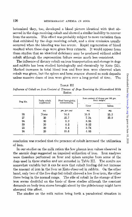
TABLE III
InJEuence of Cobalt on Iron Content of Tissues of Dogs Receiving the Mineralized Milk Ration _
Dog No.
26 27 94 30 23 24
Daily cobalt supplement
Final hemoglobin concentration
mg. gm. per cent mg.
None 14.6 10.8 20 15.7 7.34 20 9.2 8.86 20 8.2 9.52 20 8.4 3.74 20 11.6 4.85
Iron content of tissues per 100 gm. fresh weight
Liver I Spleen
m.
25.3 23.9
conclusion was reached that the presence of cobalt increased the utilization of iron.
In our studies on the milk ration the low plasma iron values observed in the anemic dogs suggested an impaired utilization of iron. Iron analyses were therefore performed on liver and spleen samples from some of the dogs used in these studies and are recorded in Table III. The results are somewhat variable but it can be seen that cobalt feeding did not increase the amount of iron in the liver as Kato observed in rabbits. On the other hand, only two of the five dogs fed cobalt showed a low liver iron, the other three being in the normal range. The role of cobalt in the storage of liver iron seems doubtful on the basis of these studies although the constant demands on body iron stores brought about by the phlebotomy might have obscured this effect.
The studies on the milk ration bring forth a paradoxical situation in
by guest on June 10, 2018http://w
ww
.jbc.org/D
ownloaded from
MCKIBBIN, SCHAEFER, ELVEHJEM, AND HART 121
regard to blood plasma iron content. During the period of. negligible he- moglobin formation the plasma iron was very low. However, when liver was supplied and hemoglobin formation was extremely rapid, the plasma iron invariably rose to very high values. This behavior is quite opposite to that observed by Moore et al. (36) in their studies on human anemia patients. They found that under conditions of rapid blood regeneration, where iron, available for hemoglobin formation, would conceivably be re- moved by the hematopoietic tissues, the plasma iron values were low (pernicious anemia patients in remission, and acute hemorrhage patients). Low values were also found in iron deficiency. High values were found after ingestion of large quantities of iron salts, in clinical states character- ized by diminished hemoglobin formation (aplastic anemia and pernicious anemia in relapse), and in the hemolyt,ic anemias. The failure on the part of our animals to reflect passively a plasma iron level dependent on hemo- globin synthesis suggests instead that the plasma iron level is actively reg- ulated by some substance contained in liver. It is conceivable that an impaired liver function in these deficient animals results in inadequate syn- thesis of such an iron-mobilizing factor.
SUMMARY
1. Adult dogs rendered anemic by phlebotomy failed to respond to iron and copper when high levels of cobalt were added to the whole milk ration. Rapid regeneration of blood occurred when this ration was further supple- mented with whole dry liver or liver extracts.
2. The activity of liver preparations could not be wholly replaced by synthetic B vitamins, bile salts, cysteine, uropterin concentrates, and high levels of iron when fed individually. When these supplements were fed together blood regeneration comparable to that produced by liver prepara- tions was observed. The blood plasma iron, however, was not mobilized as always occurs with liver therapy.
3. Dogs receiving a highly purified ration supplemented only with syn- thctic vitamins retained their ability to regenerate a normal blood stream after subjection to severe phlebotomy for many months. This regenera- tion was complete in 2 weeks.
BIBLIOGRAPHY
1. Potter, V. R., Elvehjem, C. A., and Hart, E. B., J. Biol. Chem., 126, 155 (1938). 2. Frost, D. V., Elvehjem, C. A., and Hart, E. B., J. Nutrition, 21, 93 (1941). 3. Frost, D. V., Potter, V. R., Elvehjem, C. A., and Hart, E. B., J. Nutrition, 19,
207 (1940). 4. Frost, D. V., Spitzer, E. H., Elvehjem, C. A., and Hart, E. B., Am. J. Physiol.,
134, 746 (1941).
by guest on June 10, 2018http://w
ww
.jbc.org/D
ownloaded from
122 HEMORRHAGIC ANEMIA IN DOGS
5. Anderson, H. D., Underwood, E. J., and Elvehjem, C. A., Am. J. Physiol., 130, 373 (1940).
6. Schaefer, A. E., McKibbin, J. M., and Elvehjem, C. A., J. Nutrition, 23, 491 (1942).
7. Evelyn, K. A., J. Biol. Chem., 116, 63 (1936). 8. Mason, R. D., and Mason, H. L., Proc. Stu$ Meetings Mayo Clin., 16, 433 (1941). 9. Gobell, O., Klin. Woch., 18, 1319 (1939).
10. Dollchen, H., Klin. Woch., 19, 220 (1940). 11. Gyorgy, P., Robscheit-Robbins, F. S., and Whipple, G. H., Am. J. Physiol., 122,
154 (1938). 12. Davis, J. E., Am. J. Physiol., 127, 322 (1939). 13. Barron, A. G., and Barron, E. S. G., Proc. Sot. Exp. BioZ. and Med., 35,406 (1937). 14. Griffith, W. H., Pavcek, P. L., and Mulford, D. J., J. Nutrition, 23, 603 (1942). 15. Seyderhelm, R., and Tammann, H., Klin. Woch., 6, 1177 (1927). 16. Seyderhelm, R., Tammann, H., and Baumann, W., Z. ges. exp. Med., 66, 539
(1929). 17. Takasu, M., Deutsch. 2. Chir., 224, 240 (1930). 18. Day, H. G., and Stein, H. J., J. Nutrition, 16, 525 (1938). 19. Josephs, H. W., Bull. Johns Hopkins Hosp., 66, 145 (1939). 20. Hawkins, W. B., Robscheit-Robbins, F. S., and Whipple, G. II., J. Exp. Med.,
67, 89 (1938). 21. Smith, P. W., and Crandall, L. A., Am. J. Physiol., 136, 259 (1942). 22. Tschesche, R., and Wolf, H. J., 2. physiol. Chem., 24.6, 34 (1937). 23. Simmons, R. W., and Norris, E. R., J. Biol. Chem., 140, 679 (1941). 24. Koschara, W., 2. physiol. Chem., 240, 127 (1936). 25. Koschara, W., and Haug, H., Z. physiol. Chem., 269, 97 (1939). 26. McKibbin, J. M., Madden, R. J., Black, S., and Elvehjem, C. A., Am. J. Physiol.,
128, 102 (1939). 27. McKibbin, J. M., Black, S., and Elvehjem, C. -4., Am. J. Physiol., 130, 365
(1940). 28. Schaefer, A. E., McKibbin, J. M., and Elvehjem, C. A., Proc. Sot. Exp. Biol. and
Med., 47, 365 (1941). 29. Schaefer, A. E., McKibbin, J. M., and Elvehjem, C. A., J. BioZ. Chem., 143,
321 (1942). 30. Phillips, P. H., and Hart, E. B., J. BioZ. Chem., 109, 657 (1935). 31. Subbarow, Y., Jacobson, B. M., and Fiske, C. H., New England J. Med., 212,
663 (1935). 32. Jacobson, B. M., and Subbarow, Y., J. Clin. Znv., 16, 573 (1937). 33. Jacobson, B. M., and Subbarow., Y., J. Am. Med. Assn., 116, 367 (1941). 34. Leichsenring, J. M., and Biester, A., Minnesota Agric. Exp. Stat., Tech. Bull. 1.39
(1939). 35. Kato, K., and Iob, V., Am. J. Clin. Path., 10, 751 (1940). 36. Moore, C. V., Doan, C. A., and Arrowsmith, W. R., J. C&n. Inv., 16, 627 (1937).
by guest on June 10, 2018http://w
ww
.jbc.org/D
ownloaded from
Elvehjem and E. B. HartJ. M. McKibbin, A. E. Schaefer, C. A.
IN DOGSSTUDIES ON HEMORRHAGIC ANEMIA
1942, 145:107-122.J. Biol. Chem.
http://www.jbc.org/content/145/1/107.citation
Access the most updated version of this article at
Alerts:
When a correction for this article is posted•
When this article is cited•
alerts to choose from all of JBC's e-mailClick here
tml#ref-list-1
http://www.jbc.org/content/145/1/107.citation.full.haccessed free atThis article cites 0 references, 0 of which can be
by guest on June 10, 2018http://w
ww
.jbc.org/D
ownloaded from