Embed Size (px)
Citation preview

Benha University
Faculty of Veterinary Medicine Animal medicine Department
Studies on Foot and Mouth Disease Virus type O1,A in Sheep
Thesis presented By
Amr Ismail Hassan B.V.Sc Cairo University (2001)
M.V.Sc 2007, Infectious Diseases, Benha University 2007
Under supervision of
Prof. Mohammed Hassanin Ebeid
Professor of Infectious Diseases, Faculty of Veterinary Medicine,Moshtohor Benha University
Prof. Faisal Khalil Hamoda
Professor of Infectious Diseases, Chairman of Animal Medicine Dept.Faculty of Vet. Medicine, Moshtohor, Benha University
Prof. Adel Mohamed Hassan Azab
Chief Researcher, Veterinary serum and Vaccine Research Institute, Abbassia
A Thesis Submitted to
Benha University For the degree of
PhD of Veterinary Medical Science (Infectious Diseases)
2011

Acknowledgment I wish first to thank forever ALLAH for helping me to complete this
work and giving me every thing I need. I would like to take this opportunity to express my cardial
gratitude and deepest thanks for Prof. Dr. Mohamed Hassanin Ebeid, Professor of Infectious Diseases, Faculty of Veterinary Medicine, Benha University, for his valuable help and encouragement giving to me during this work.
I wish to express my deepest appreciation and sincere gratitude to Prof. Dr. Faisal Hamoda, Professor of Infectious Diseases and Head of Animal Medicine Department Faculty of Veterinary Medicine, Benha University and Prof. Dr. Adel Mohamed Hassan Azab and Prof. Dr. Laila Ismail EL-Shahawy, Chief Researcher, Foot and Mouth Disease Department, Veterinary Serum and Vaccine Research Institute, Abbassia, Cairo, for their suggestion and supervision of this work. Also I would like to express my deepest thanks for Pro. Dr. Abd El-Moneim Mohamed Mostafa Professor of Infectious Diseases , Benha Universty.
My great appreciation to the late Prof. Dr. Sinan El-Nakashly, Prof. Dr. Magdy Abdel Atty, Prof. Dr. Hosam Gamal El-Din, Prof. Dr. Abu Baker Aggour, Dr. Samir Mohammed Ali and Dr. Wael Mossad for their assistance to complete the plan of this work.
Also Special thanks for Dr. Safi El-Dean Mahdy, Dr. Ehab El-Sayed, Dr. Mohammed Gamil, Dr. Akram Zakarya , Dr. Assem Abou Bakr and Dr. Ahmed Fathy.
My sincere recognition to all the staff members of Foot and Mouth
Disease Department, Veterinary Serum and Vaccine Research Institute, Abbassia, Cairo,
and all staff members of Animal medicine Department, Benha University.
Amr Ismail

List of contents
1. INTRODUCTION ............................................................................................................................ 1
2. LITERATURE ................................................................................................................................. 3
2.1. FOOT AND MOUTH DISEASE ......................................................................................................... 3 2.2. FMD VIRUS ................................................................................................................................. 3 2.3. TYPING AND SUB TYPING OF FMD VIRUS ...................................................................................... 4 2.4. EPIDEMIOLOGY OF FOOT AND MOUTH DISEASE........................................................................... 5
2.4.1. Foot and mouth disease in Egypt ........................................................................................ 5 2.4.2. Prevalence of Foot and mouth disease in the world ............................................................ 7
2.5. TRANSMISSION OF FMD .............................................................................................................. 9 2.6. PATHOGENESIS OF FMDV ......................................................................................................... 11 2.7. FOOT AND MOUTH DISEASE IN SHEEP ........................................................................................ 13 2.8. PERSISTENCE OF FMD .............................................................................................................. 15 2.9. ANTIGENIC COMPONENTS OF FMD VIRUS .................................................................................. 18
2.9.1. Structural proteins............................................................................................................ 18 2.9.2. Non- structural proteins (NSP)......................................................................................... 19
2.9.2.1. Virus Infection Associated Antigen (VIA) .............................................................................. 19 2.10. INACTIVATION OF FMDV........................................................................................................... 21 2.11. IMMUNITY AGAINST FOOT AND MOUTH DISEASE ........................................................................ 21
2.11.1. Active Immunity ............................................................................................................... 21 2.11.1.1. Immunity after infection ........................................................................................................ 21 2.11.1.2. Immunity after Vaccination ................................................................................................... 24
2.12. ISOLATION AND IDENTIFICATION OF THE VIRUS .......................................................................... 27 2.13. SERUM NEUTRALIZATION TEST (SNT) ........................................................................................ 27 2.14. ENZYME LINKED IMMUNOSORBENT ASSAY (ELISA) .................................................................... 29 2.15. 3ABC-ENZYME LINKED IMMUNOSORBENT ASSAY (3ABC-ELISA) ............................................... 33 2.16. POLYMERASE CHAIN REACTION .................................................................................................. 35
3. MATERIAL AND METHODS ...................................................................................................... 40
3.1. MATERIALS ................................................................................................................................ 40 3.1.1. Animals ............................................................................................................................ 40
3.1.1.1. Sheep .................................................................................................................................... 40 3.1.1.2. Unweaned baby mice ............................................................................................................. 40
3.1.2. Sheep Serum samples ....................................................................................................... 40 3.1.3. Epithelial tissue samples................................................................................................... 43 3.1.4. Oesophageal pharyngeal (op) fluids ................................................................................. 43 3.1.5. Reference virus................................................................................................................. 43 3.1.6. Tissue cultures (established cell lines) .............................................................................. 43

3.1.7. Chemical reagents ............................................................................................................ 44 3.1.7.1. Media .................................................................................................................................... 44
3.1.7.1.1. Growth medium ........................................................................................................ 44 3.1.7.1.2. Maintenance medium ............................................................................................... 44
3.1.7.2. Bovine serum ......................................................................................................................... 44 3.1.7.3. Trypsin .................................................................................................................................. 44 3.1.7.4. Sodium bicarbonate solution ................................................................................................. 44 3.1.7.5. Neomycin .............................................................................................................................. 44 3.1.7.6. Nystatine (antifungal) ............................................................................................................ 45 3.1.7.7. Tween 20 ............................................................................................................................... 45 3.1.7.8. Crystal Violet Stain ................................................................................................................ 45
3.1.8. Reagents used in ELISA ................................................................................................... 45 3.1.8.1. Coating Buffer ....................................................................................................................... 45 3.1.8.2. Phosphate Buffer Saline (PBS)and Bovine Albumin.............................................................. 46 3.1.8.3. Washing Buffer ..................................................................................................................... 46 3.1.8.4. Phosphate Citrate Buffer ....................................................................................................... 46 3.1.8.5. Substrate ............................................................................................................................... 47 3.1.8.6. Stopping Solution .................................................................................................................. 47 3.1.8.7. Conjugate .............................................................................................................................. 47 3.1.8.8. Tween 20 ............................................................................................................................... 47 3.1.8.9. Blocking buffer...................................................................................................................... 47
3.1.9. Prio Check FMDV Non Strctural protein ......................................................................... 47 3.1.10. Nucleic acid recognition reagents and kits ....................................................................... 48
3.1.10.1. Total RNA purification kit ..................................................................................................... 48 3.1.10.2. RNA extraction reagents ........................................................................................................ 49 3.1.10.3. RT-PCR kit ............................................................................................................................ 49 3.1.10.4. PCR kit .................................................................................................................................. 50 3.1.10.5. Primers .................................................................................................................................. 50
3.1.11. Materials used for detection of RT-PCR & PCR products ................................................ 51 3.1.11.1. Tris Acetate EDTA (TAE) gel electrophoresis buffer (40X) .................................................... 51 3.1.11.2. Loading Buffer ...................................................................................................................... 51 3.1.11.3. Agarose ................................................................................................................................. 51 3.1.11.4. Ethidium bromide (Eth Br) .................................................................................................... 51 3.1.11.5. Nucleic acid markers ............................................................................................................. 52
3.1.12. Equipment and supplies ................................................................................................... 52 3.1.12.1. Biological safety cabinet ........................................................................................................ 52 3.1.12.2. Cooling centrifuge ................................................................................................................. 52 3.1.12.3. Vortex mixer .......................................................................................................................... 52 3.1.12.4. Water bath ............................................................................................................................. 52 3.1.12.5. Single and multichannel pipettors (microtitre pipette) ............................................................ 52 3.1.12.6. Disposable syringe filters ....................................................................................................... 53 3.1.12.7. Equipment for cell cultures .................................................................................................... 53 3.1.12.8. Inverted light microscope ....................................................................................................... 53 3.1.12.9. Microtubes with attached cap (Microfuge tubes) .................................................................... 53

3.1.12.10. Thermocycler ......................................................................................................................... 53 3.1.12.11. Electrophoresis unit (set) ....................................................................................................... 54 3.1.12.12. Power supply ......................................................................................................................... 54 3.1.12.13. Transilluminator, UV ............................................................................................................ 54 3.1.12.14. Gel Documentation and Analysis system ................................................................................ 54 3.1.12.15. Other equipment .................................................................................................................... 54
a. Refrigerators and Freezers ................................................................. 54 b. Incubators ........................................................................................ 54
3.2. METHODS .................................................................................................................................. 55 3.2.1. Protocol of sheep vaccination ........................................................................................... 55 3.2.2. Preparation of serum samples .......................................................................................... 55 3.2.3. Oesophageal pharyngeal(op) fluids .................................................................................. 55 3.2.4. Preparation of FMD virus ................................................................................................ 55 3.2.5. Baby mice ......................................................................................................................... 56 3.2.6. Serological tests ................................................................................................................ 56
3.2.6.1. Enzyme linked ImmunoSorbent Assay (ELISA) ..................................................................... 56 3.2.6.1.1. Preparation of ELISA antigen .................................................................................. 56 3.2.6.1.2. Titration of the conjugate ......................................................................................... 56 3.2.6.1.3. Testing of the serum samples using indirect ELISA .................................................. 57
a- Coating ....................................................................................... 57 b- Blocking ..................................................................................... 57 c- Serum dilutions .......................................................................... 57 d- Addition of the conjugate ............................................................ 57 e- Addition of the substrate ............................................................. 57 f- Addition of the stopping solutions ............................................... 57 g- Interpretation of the results ........................................................ 57
3.2.6.2. Serum Neutralization Test (SNT) ........................................................................................... 58 3.2.6.2.1. Staining the SNT microplates used in the test Procedures ......................................... 58
3.2.6.3. PrioCHECK FMDV NS test ................................................................................................... 59 a- Day 1 .......................................................................................... 59 b - Day 2 .......................................................................................... 60
3.2.6.3.1. Interpretation of the Percentage Inhibition ............................................................... 60 3.2.7. Molecular detection of FMDV by RT-PCR and PCR........................................................ 61
3.2.7.1. FMD-RNAextraction-(isolation) ............................................................................................ 61 3.2.7.2. One-step reverse transcriptase-polymerase chain reaction (RT PCR) ..................................... 62 3.2.7.3. Polymerase chain reaction (PCR) .......................................................................................... 62 3.2.7.4. Agarose Gel Electrophoresis of PCR Products ....................................................................... 62
4. RESULTS ....................................................................................................................................... 64
4.1. ISOLATION AND IDENTIFICATION OF FOOT AND MOUTH DISEASE VIRUS FROM FIELD SAMPLES..... 64 4.2. REVERSE TRANSCRIPCATION CHAIN REACTION POLYMERASE (R.T PCR) ................................... 66
5. DISCUSSION ................................................................................................................................. 92
6. SUMMARY .................................................................................................................................... 98

7. REFERENCES ............................................................................................................................. 100

Abbreviations and Symbols
µL Microliter BHK21 Baby Hamster Kidney cells clone 21 CFT Complement Fixation Test CPE Cytopathic Effect D.D.W Double Distilled Water DPI days post infection ELISA Enzyme Linked Immunosorbant Assay FMD Foot and Mouth Disease FMDV Foot and Mouth Disease Virus I/P Intra Peritoneal MEM Minimum Essential Media MLD50 Mice lethal dose 50 nm Nanometer. NSPs Non Structural proteins OD Optical density OIE Office International des Epizootie OP Oesophageol Pharyngeal fluid OPD OrthoPhyneyleneDiamine PBS Phosphate buffer saline PD50 Protective Dose 50 RIP Radio-Immuno Precipitation RNA Ribonucleic acid rpm Revolution per minute RT- PCR Reverse Transcriptase Polymerase Chain Reaction SC Sedimentation coefficient SAT South Africa Territories SNT Serum Neutralization Test TCID50 Fifty tissue culture infective dose µL Microliter UV Ultra Violet VIA Virus Infection Associated Antigen VNT Virus Neutralization test VP Virus protein VSVRI Veterinary Serum and Vaccine Research Institute WPI Week post infection WRL World Reference Laboratory of foot and mouth disease
1
1. Introduction
Foot and Mouth Disease is difficult to control because of it's highly contagious nature, it's ability to infect different domestic and wild life hosts and it's causation by multiple non-cross-protective virus serotypes, (Rodrieguez and Gurbman 2009).
The occurrence of FMD continues to largely reflect economic prospectively
with countries having eradicated the disease and countries struggling or unable to do so. Geographic isolation can favor FMD eradication. Movement of live animal still constitutes the greatest risk for spread of FMD, followed by trade in animal products.FMD virus continues to evolve, giving rise to new strains that cause periodic upsurges in the number of cases and increase the risk of spread into new areas (Knowles et al., 2007).
FMD is considered enzootic in Egypt and many outbreaks have
recurrently occurred involving most governorates (Mousa etal., 1976; Daoud et al., 1988; and EL-Nakashly et al., 1996). The main causative serotypes are O1 & A (Abd El-Rahman et al., 2006)
Routine prophylactic vaccination has been conducted with locally produced
bivalent inactivated serotypes O1 and A Egypt 2006 vaccine. Outbreaks from serotype O1 was in 2000 and 2006, and other serotypes have not been reported since 1972 when serotype A occurred (Aidaros 2002).
On January 2006 clinical cases of FMD were first recognized in cattle farm
and widely spread in Egypt and the virus was isolated and identified by FMD Department and institute for Animal Health United Kingdom (Knowles et al., 2007).
Diagnosis of FMD is based on clinical signs followed by confirmation by
laboratory tests (Giridharan 2005). RT-PCR assays considers an alternative or complementary to classical serological and viral isolation method due to their higher sensitivity, speed and the fact that the handling of infectious virus is not required (Saiz et al 2003)
Recently, There are increase application of tests that detect foot and mouth
disease virus antibodies to non- capsid proteins (NCP) to assess past or present FMD infection/circulation irrespective of vaccination.

2
So, the objective of this study for: Isolation and identification of Foot and mouth disease from field cases.
Reverse Transcripcation Polymerase Chain Reaction (R-T PCR) for
isolated O1 strain. Prevelence of FMDV in different governorate of Egypt using SNT and
ELISA. Detection of NSP antibodies in sera of sheep proved to be positive by
ELISA and SNT in different governorates.

3
2. Literature
2.1. Foot and Mouth Disease
Foot and mouth disease is the most important livestock disease in the world in terms of economic impact. The reason is the ability of disease to cause losses of production, also due to hindering on the trade of animal both locally and internationally, and restrictions on the movement of people which affect the tourism sector, James and Rushton (2002). Foot-and mouth disease virus continues to exist and evolve, thus posing a serious threat to the livestock industry worldwide Mohapatra et al. (2009b), It is also highly infectious and economically devastating disease of livestock, Rodriguez and Gurbman (2009).
2.2. FMD virus
Brown (1973) stated that FMD virus is a member of the family Picornaviridae.
Rueckert and Wimmer (1984) stated that FMDV is a member of genus
Aphthovirus of family Picornaviridae. Picorna viral RNA genomes encode of viral polyprotein precursor, which is processed into the P1 region, containing the capsid proteins VP1, VP2, VP3 and VP4 and the P2 and P3 regions which contain the non-structural proteins.
Franki et al. (1991) classified FMDV as belonged to genus Aphthovirus,
within family Picornaviridae which also includes Enterovirus, Cardiovirus, Rhinovirus and hepatovirus.
Belsham (1993) classified FMDV as a single strand positive sense RNA
virus that belong to the genus aphthovirus in the family Picornaviridae. Kitching (1994) revealed that the FMDV is one of the animal viruses which
are rapidly producing pathogenic mutants. These mutants can spread quickly throughout affected geographical regions leading to severe economic losses.
ICTV (2000) mentioned that FMD was caused by seven types of foot and
mouth disease in the genus Aphtovirus, family Picornaviridae.

4
Domingo et al. (2002) reported that the FMDV is an aphthovirus of the family Picornaviridae. FMDV is non-enveloped particles of icosahedral symmetry.
Musser (2004) mentioned that FMD is caused by RNA virus of the genus
Aphthovirus; 7 immunological distinct serotypes of the virus have been identified. Susceptible species are mainly cattle, sheep, goats, pigs, bison and deer. All body fluids of infected animals can contain the virus and are considered infective.
Buenz and Howe (2006) found that members of the picornavirus family,
including poliovirus and foot-and-mouth disease virus, are widespread pathogens of humans and domestic animals.
Nick et al.(2007) the disease is highly contagious and combined with high
antigenic diversity of the virus makes FMD difficult to control
2.3. Typing and sub typing of FMD virus
Valee and carre (1926) named the classical O, A and C types of FMD virus according of the isolation site (O) being isolated from Oise Valley in France (A) from Allemange (Germeny) and (C) for the third isolated type.
Brooksby (1958) added the so-called exotic types of FMD virus which
involved South Africa Territories (SAT1, SAT2, SAT3) and Asia 1 isolated from Asian countries.
Bachrach (1968) mentioned that seven classified immunological types of
FMDV O, A, C, South African Territory types (SAT1, SAT2, SAT3) and an Asian type designated Asia 1.
Callis et al. (1968) showed that the immunity against one type did not protect
against infection by other types. It was indicated that subtype differentiation could be based on complete or partial lack of cross protection between FMDV strains.
Pereira (1977) identified a total of 65 subtypes of FMD virus classified as A
32, O 11, C 5, SAT1 7, SAT2 4, SAT3 3 and Asia1 3. Anon (1978) mentioned that there was no cross immunity between types but
partial immunity only between subtypes within the same type.

5
Abu El-Zein and Crowther (1980) reported that serological difference
between some subtypes of FMDV was significant enough to recognize each of them apart with safe.
Aggarwal et al. (2002) mentioned that the International Vaccine Bank
(IVB) for FMD at Pirbright holds quantities of seven strains of inactivated FMDV antigen over liquid nitrogen, ready for immediate formulation into vaccine if required. These will protect against the viruses of serotypes that are most likely to threaten the livestock of the UK or other IVB member countries (i.e. serotypes A, O, C and Asia1).
Brown (2002) reported that the extent of the antigenic diversity is such that
animals, whose level of immunity is waning, as for example several months after vaccination, may be susceptible to infection with viruses within the same serotype, although still immune to infection with the virus from which the vaccine had been made.
Domingo et al. (2002) cited that FMD virus is the prototype member of the
Aphthovirus genus of the family Picornaviridae. The virus exists in the form of seven different serotypes A , O, C, SAT1, SAT2, SAT3 and Asia1. But a large number of subtypes have involved within each serotype.
Mason et al. (2002) mentioned that during the last 12 years a strain of
FMDV serotype O, named as PanAsia, has spread from India throughout southern Asia and the Middle East. During 2000, this virus strain caused outbreaks in the Republic of Korea, Japan, Russia (Primorsky Territory), Mangolia and South Africa.
Knowels and Samuel, (2003) reported that Infection or vaccination with
one serotype dose not conferm protection against other serotype
2.4. Epidemiology of Foot and Mouth Disease
2.4.1. Foot and mouth disease in Egypt Zahran (1961) mentioned that different types of FMD virus (SAT2, O and
A) were identified in Egypt, type A and SAT2 were the main causes of outbreaks during 1953, 1958 and 1960. Type ‘O’ virus was the most prevalent in setting up the disease.

6
Awad et al. (1984) reported that field survey on the epizootiology of FMD in Egypt revealed that the percentage of antibodies against the virus infection associated antigen (VIA) in the sera of naturally infected cattle was 88.8% while in contact cattle was 50%. In buffaloes, it was 80% in naturally infected animals and 30.7% in contact ones. The sera of immunized cattle under field conditions were negative to the VIA antibodies. The percentage of VIA antibodies in sera of investigated animals allover Egypt was 13.5%, 18.36% and 15.4% for cattle, buffaloes and sheep respectively.
Moussa et al. (1984) cited that FMD took an enzootic form in Egypt. The
disease appeared each year and attacked susceptible animal causing losses in milk and meat production and sometimes death of young animal.
Omar et al. (1985) recorded in February 1980, an outbreak of FMD in
lactating animals in a village near Alexandria. The isolated FMD Virus was confirmed to be (O).
Daoud et al. (1988) identified a high prevalence of FMD among different
animal species (cattle, buffaloes and sheep) in different provinces in Egypt during 1987 outbreak. The isolated virus was confirmed to be type (O).
Kitching (1990) recorded since 1950, attention was drawn to the importance
of FMD in Egypt after occurrence of several outbreaks and subtype (O1) was the most prevalent isolated strain of FMD virus.
El-Nakashly et al. (1996) isolated FMDV type O1 /93/Egypt in 1993 and
the strain was typed as O1. OIE (2000) Published about outbreak of FMD in Fayoum governorate in
Egypt in September (2000) typed as O1 and cattle and sheep were affected. This indicates that the virus is still being actively transmitted within livestock.
Aidaros (2002) cited that the occurrence of FMD serotype SAT2, A and O
were last reported in Egypt in year 1950, 1972, and 2000, respectively while type O was the only virus isolated.
OIE (2006) published that the Egyptian authorities had reported several
outbreaks of FMD in cattle and buffaloes. The outbreaks were located in eight governorates of Egypt.
Abdel-Rahman et al. (2006) reported that an outbreak of FMD started in
bulls imported and kept in quarantine station at Al-Ismailia Governorate, then spread among local cattle, buffaloes and dairy farms in most governorates in

7
upper and Lower Egypt with 100% morbidity and high mortality rates reach to 80% in newly born calves. The virus isolated from imported and local animals was identified in Egypt as serotype A.
Farag et al. (2006) observed that clinical signs of foot and mouth disease
(FMD) among bulls imported from Ethiopia into a quarantine station at Al-Ismailia Governorate. FMD-WRL reported that the recovered type A/EGY/1/2006 virus was antigenically related to serotype A FMD isolated from Ethiopia, Kenya, Yemen and Saudi Arabia. The recovered type A FMDV re-isolated from local indigenous cattle, buffaloes and dairy animals, fattening bull and backyard allover 10 governorates in upper and Lower Egypt. 100% morbidity, 80% mortality in the newly born calves and 50% losses in milk were recorded in the affected dairy farms. Also, 50% losses were estimated in meat production of fattening bulls.
Ghoneim et al. (2010) cited that Egypt is endemic with two FMDV serotype
(O&A) and the outbreaks still reported since 2006 till now.
2.4.2. Prevalence of Foot and mouth disease in the world
Callis et al. (1968) reported that FMDV has been detected in almost countries of Asia such as Kuwait, Israel, Iraq, Saudi Arabia, Oman, Yemen, United Arab Emirates, Iran, Jordan, Pakistan and India.
Ramarao and Rao (1988) found that the four types of FMDV (O, A, C and
Asia 1) have been isolated from various part of India. Samuel et al. (1990) stated that FMDV serotype 'O' continued to be isolated
from outbreaks in Middle East during the period 1981-1988. Dehoux and Hounsou (1992) gave a brief account on the FMD epidemic of
Borgou Department between November 1990 and April 1991. Morbidity rates of 80-100% were observed in affected cattle herds. Antibodies to types A, O and SAT2 were demonstrated.
Kitching (1998) cited recent outbreaks of FMD in 1996 in the European
countries namely: Bulgaria, Greece, Turkey, Albania and Macedonia. Leforban (1999) recorded outbreaks of FMD type A in 1996 in Macedonia,
Albania and Yugoslavia.

8
Huang et al. (2000) demonstrated that since March 1997 two strains of FMDV have found their way to Taiwan, causing severe outbreaks in pigs and in Chinese yellow cattle.
Benveniti et al. (2001) declared that FMD is one of the most dangerous
diseases of cloven-hoofed animals and is a constant threat in the Middle East and other regions throughout the world despite intensive vaccination programs.
EU FMD Meeting (2001) concluded that outbreak of FMD type O was
confirmed in UK, this outbreak was caused by FMD strain that was responsible for the outbreak in Japan.
Gibbens et al. (2001) confirmed FMD was confirmed in Great Britain. A
major epidemic developed, which peaked around 50 cases a day in late March, declining to under 10 a day by May. By mid-July, 1849 cases had been detected. The main control measures employed were livestock movement restrictions and the rapid slaughter of infected and exposed livestock.
Blanco et al. (2002) reported that during 1999, 11 outbreaks of FMD were
declared in the east and central part of Morocco. All the FMD cases reported were in cattle.
Davis (2002) mentioned that types O, A, and C are the strains that have been
identified in Europe and South America while O, A and ASIA1 are common through Asia. SAT strain 1 and 2 found throughout Africa while SAT 3 is confined to southern Africa. The strain found in the Middle East includes A, O, Asia 1 and SAT 1. FMD is endemic in much of Africa, Asia and parts of South America.
EU FMD Meeting (2002) reported that in Turkey, a total of 29 outbreaks
have been reported, 16 due to type 'O' 11 due to type 'A' and 2 due to type 'Asia1'.
Joe et al. (2002) illustrated that the Republic of Korea has been free for 66
years prior to the introduction of the virus and had recently suspended imports of pork products from neighboring Japan owing to a reported FMD outbreak in that country. On March 2000 a suspected vesicular disease in cattle was reported and confirmed as FMD by the national veterinary research and Quarantine service of the Republic of Korea.
Mason et al. (2002) analyzed the relationship between FMD type O viruses
belonging to the Pan Asia strain. They revealed that all portions of the genomes of these isolates are highly conserved and provided confirmation of close
9
relationship between the viruses responsible for South Africa and UK outbreaks.
Sakamoto et al. (2002) reported that 4 outbreaks of FMD occurred from
March to May 2000 in Miyazaki and Hokkaido prefectures, Japan. FMD virus isolated was achieved by sampling probaing materials from Japanese Black cattle. The FMD was identified as type O by ELISA for antigen detection and nucleotide sequence encoding the VP1 was determined.
Anderson et al. (2003) detected antibody to FMDV serotypes Asia1 and C
during the UK epidemic in 2001. Francois et al. (2004) recorded FMD outbreaks in 62 countries in Africa,
Europe, Middle East, Southern Asia and South-East Asia from the beginning of 2003 and up to Sept. 2004. All other reported outbreaks occurred in countries in which FMD is endemic.
Paiba et al. (2004) isolated FMD type O during the UK 2001 outbreak. Sammin et al. (2004) reported that two different serotypes SAT1 and SAT2
were involved in FMD outbreaks in Zimbabwe in 2003/2004. Bhattacharya et al. (2005) cited that in the state of West Bengal, India,
1,082 FMD outbreaks were reported in the 18 years from 1985 to 2002. Of the prevalent four serotypes, O type FMD virus accounted for the most outbreaks (67%), followed by Asia-1 virus type (15%) and A virus type (14%).
Ryan et al. (2008) recorded that a case of foot-and-mouth disease (FMD) on
a cattle farm in Normandy, Surrey, was confirmed on Friday August 3, 2007, the first case in the UK since 2001.
Hoang (2009) stated that in 2008 FMDV types O and A were reported to be
the prevalent and circulating serotypes causing the endemic outbreaks every month throughout 2008 and the first 2 months of 2009, while FMDV types O and Asia 1 were prevalent in 2007 in Vietnam.
2.5. Transmission of FMD
Burrows (1966) reported that the virus is released from infected animals in blood, milk, pharynx and vagina for variable periods before clinical signs appear on infected animals.

10
Sellers and Parker (1969) mentioned that FMDV was transmitted principally through aerosols and by direct contact with infected animals.
Henderson (1970) recorded that birds contaminated air and wild animals
constituted an uncontrollable source of FMDV infection. McVicar and Sutmoller (1971) reported that milk and milk products could
also play an important role in transmission of FMDV. Gloster et al. (1981) mentioned that FMD could be occurred by airborne.
The virus could be carried for at least 60 kilometers. The two main modes of infection with the disease were inhalation and ingestion under field conditions, an inhalation being the more likely route in cattle.
Moussa et al. (1984) mentioned that rodents may play a role in the
mechanical or biological transmission of FMDV to susceptible cattle. Callis (1996) reported that FMDV could potentially be spread in semen,
food products and by fomites. Bastos et al. (1999) suggested sexual transmission of FMD from carrier
buffalo bulls to domestic cows. Hutber and kitching (2000) said that transmission of FMDV by aerosol
spread can occur over considerable distance, however this is less effective in hot and dry environmental conditions.
Holzhauer et al. (2001) assumed that the disease probably entered the
Netherlands through subclinically infected fattening calves imported from Ireland in late February 2001 that spread the infection to goats housed adjacently.
Alexandersen et al. (2002) mentioned that the contagious nature of FMD is
a reflection of a number of factors, including the wide host-range of the virus, the amount of infectivity excreted by affected animals, the low doses required to initiate infection and many routes of infection.
Dekker et al. (2002) reported that FMD was likely introduced in the
Netherlands by calves imported from Ireland via an FMDV- contaminated resting point in Mayenne, France. The clinical sings of FMD were reported in goats 3 weeks after arrival of the calves.
11
Donaldson and Alexandersen (2002) said that in case of cattle, other important sources of virus that can cause heavy contamination of the environment are saliva, vesicular fluid, epithelium, milk and faeces.
Amass et al. (2003) cited that people could act as mechanical vectors of
FMDV when they move from infected to susceptible animals. Hand washing and changes of clothing were sufficient to reduce the dose of FMDV on people handling.
Uppal (2004) mentioned that small ruminant have been responsible for
epidemic of FMD in cattle in Greece in 1994. Sanson (2005) reported that People, vehicles, livestock and other items can
travel off pastoral livestock farms in New Zealand to other farms either directly or via saleyards over extensive distances. This has implications for the potential spread of infectious diseases such as FMD.
Gloster J. et al. (2008) mentioned that foot and mouth disease virus may
spread by direct contact between animals or via fomites as well as through airborne transmission.
Petrez et al. (2008) documented that the disease was spread by transmission
of virus through direct contact between animals and by indirect contact with fomites containing infectious virus particles, such as contaminated vehicles, feed,or clothing of livestock personnel.
Quan et al., (2009) stated that a strong correlation exists between dose (i.e.
infectiousness of source and intensity of contact) and length of incubation period, severity of clinical disease and efficiency of spread of FMD.
2.6. Pathogenesis of FMDV
Wisniewski (1962) failed to isolate the virus from muscle and heart of
infected cattle, two days after slaughter. The virus was found in the liver 5 days post infection and in the spinal cord and hip lymphnodes 9 days post infection.
Cottral et al. (1963) showed that cattle can be infected with FMDV by
tongue inoculation. The virus rapidily multiplies in the epithelial cells leading to a sever systemic infection.

12
Muntiu et al. (1970) concluded that FMDV could be detected in the blood stream of infected cattle up to seven days post infection. Viraemia in these cattle was observed over 4 days period and on the same day generalized lesions appeared.
Sellers (1971) reported that inoculation of tongue epithelium was the most
sensitive method for initiating infection in cattle. There was a considerable variation in the amount of virus required from each strain to initiate infection. It may be worth while to mention that a minimum infective dose in cattle must contain 10000 virus particles.
Davis (2002) mentioned that FMDV is excreted during viraemia for some
days, thereafter as serum antibody develops viraemia decreases, and the animal ceases to be infectious as the lesion heal.
De Clereq (2002) mentioned that FMD is a very contagious disease because
small dose of virus is infectious, a large amount of virus can be excreted, and there are several routes of infection and excretion. Virus excretion starts 24-48 hours before the onset of clinical signs and declines with the appearance of circulating FMD specific antibody at around 4 to 5 days after infection. Preferred samples for virus detection are the epithelium, vesicular fluid, oesophagopharyngeal fluid probangs, hearinized blood and milk.
Kitching and Hughes (2002) cited the local replication of FMD virus
occurs at the site of entry, in the mucosa of respiratory tract or at a skin or mucous membrane abrasion. The virus then spread throughout the body favoring epithelial tissue in the adult and heart muscle in the juvenile. Lytic changes in the cells of the stratum spinosum and consequent edema give rise to the characteristic vesicles and accumulation of granulocytes, and in the developing myocardium of young animals, to a lympho-histocytic myocadidits.
Sung (2002) recovered FMDV from oesophageal-pharyngeal fluid from both
dairy sheep and dairy cattle, artificially inoculation with 104.6 TCID50 O/Taiwan/99 strain in tongue and feet tissue, four days after inoculation.
Pacheco et al (2008) mentioned that After aerosol exposure of cattle FMDV
first replicates in the pharynx. In 24–48 h the virus invades the blood stream and shortly thereafter lesions appear in the mouth and feet of susceptible animals. Viremia usually disappears after 3–4 days but virus replicates to very high titers (>8 log 10 infectious units per ml) at lesions sites and is shed in the air and body fluids. Between 5 and 10 days after their appearance, lesions resolve and virus is no longer found at the lesion sites and can only be recovered from pharyngeal fluid and tissues.
13
Sellers and Gloster (2008) reveled that in cattle, FMD has been
experimentally reproduced by exposing animals to virus via direct or indirect contact with infected animals, via injection by various routes, by intra-tracheal aerosol infection, by intra-pulmonary implantation, or via respired aerosol.
Juan et al. (2010) suggests that early in FMDV infection of cattle,
replication occurs in the upper respiratory tract within respiratory associated lymphoid tissue.
2.7. Foot and Mouth Disease in Sheep
Shahan (1962) stated that cattle, swine, goats, and sheep are the most commonly affected species, although other ruminants and cloven-footed animals may contact the disease as well.
McVicar and Sutmoller (1968) reported that 3 groups of sheep and goats
were experimentally exposed to FMD virus by different routes including intranasal instillation and intradermolingual injection. These animals were kept and treated with type specific antiserum to prevent carrier state. The results revealed that these animals still acts as a carrier indicating that sheep and goats has a very important role in the spread of the disease.
Kukharov et al. (1973) studied the isolation of FMD virus from cattle and
sheep at various times after recovery from infection. They found that the isolated strains from cattle have a lowered pathogenicity for cattle and Guinea pigs while strains recovered from sheep retained their full pathogenicity for this species. All the isolated viruses were found to be highly pathogenic for pigs.
Forman et al. (1974) reported that FMD carrier state in three species of deer
in UK. Sellers and Gloster (1980) found that the main route of infection was
airborne infection in cattle and sheep up to 20 Km. In some outbreaks movement of the people and vehicles play a role in spreading infection. They added that sheep acts as a source of infection for other species so the ring vaccination of sheep is effective in limiting spread of the disease.
Sharma et al. (1981) studied the patterns of infection and morbidity in
sheep and goats exposed to foot and mouth disease, both naturally and under experimental conditions; they investigated the infection by the presence of

14
viraemia, virus in the pharyngeo-oesophageal region and serum neutralizing antibodies. The authors also recorded that in case of field outbreaks there was greater gap between observed morbidity and actual infection. They concluded that sheep and goats play an important role in the spreading of infection during epizootic of FMD.
Rahman et al. (1988) reported a natural case of FMD in an Indian elephant.
The isolated virus was typed Asia1, it was possibly indirectly transmitted through an outbreak of FMD Asia1 in cattle and buffaloes of the district.
Shawkat et al. (1989) isolated FMD virus type O1 from sheep during 1987
outbreak in Egypt. They studied the pathogenicity of the isolate and concluded that sheep can play a role in the epidemiology of FMD in Egypt.
Fondevila et al. (1995) studied that llamas are resistant to FMD infection,
and they play a minor role in transmitting the virus to domestic livestock. Barnett and Cox (1999) recorded the epidemiological role played by sheep
and goat in transmitting the disease due to the unapparent nature of the disease among those hosts as well as their ability to become carriers representing a reservoir for further infection and spread of the disease.
Ganter et al. (2001) mentioned that sheep and goats might be carriers, so
they play an important role in the epidemiology and transmission of FMD. Shipping and trade with live sheep and goats is much more common world wide than in other FMD susceptible species. Lack of registration and individual identification signs (ear tag) of sheep and goat herd may result in incomplete control measurement under FMD conditions.
Bronsvoot et al. (2002) reported that sheep play important role in
epidemiology and transmition of FMD. Kitching and Hughes (2002) recorded that serotype (O) FMD virus has
been recovered from over 90 % of the positive samples from sheep submitted to the World Reference Laboratory for FMD, Pirbright, U.K. In East Africa where outbreak due to serotype O, A, C, SAT1 and SAT2 are common, predominantly serotype (O) virus was identified in clinically affected sheep and goats.
Hughes et al. (2002a) revealed that the optimal dose of FMDV to infect
sheep and for producing in-contact transmission is about 104 TCID50.

15
Hughes et al. (2002b) mentioned that lesions of FMD may fall to develop in approximately 25% of infected sheep; a further 20% may develop only a single observable lesion.
Amas et al. (2003) found that contact sheep with infected pigs had
developed gross lesions consistent with FMD by 5DPI. Georgiev et al. (2004) cited that Foot and mouth disease have transmitted to
sheep in which infection is frequently sub-clinical Laila et al. (2004) reported that sheep play an important role in the
epidemiology and transmission of FMD. Moreover, FMD is suspected to have transmitted to sheep in which infection is frequently sub-clinical. So, it's of importance to identify animals which have been exposed to the virus and have developed antibodies. Such animals may become carriers and thus be a potential source of new outbreaks.
2.8. Persistence of FMD
Burrows (1966) found that the frequency of recovery of FMDV in esophageal-pharyngeal fluid (OP) samples taken from convalescent cattle after clinical infection was 9-26 weeks and infectivity of the isolated virus was 1-2.4 log10/ml. He also added that the FMDV was recovered from cattle 14-196 days after infection. The chief sites of virus multiplication were the dorsal surface of the soft palate and the pharynx.
Burrows (1968) demonstrated that goats and sheep develop persisted
infection. McVicar and Sutmoller (1968) found that about 50% of a group of goats
and sheep exposed to an infected steer were FMD carriers 4 weeks later. The carrier state may last up to 12 months in some animals.
Sutmoller et al. (1968) observed that virus multiplication was established in
the pharynx of immunized cattle in spite of the presence of high serum antibody titre.
McVicar and Sutmoller (1972) reported that 88 out of 91 (97%) goats
exposed to FMDV became infected and 92% of the infected ones had demonstrated viraemia. They also added that all goats showed viraemia when placed in contact with infected cattle.

16
Kukharov et al. (1973) recorded examination of pharyngeal mucous of
infected cattle and sheep, the virus persisted for at least 271 and 106 days after convalescent, respectively.
Sharma (1979) inoculated 6 sheep with FMDV type "O" (104TCID50/ml)
subcutaneously, intranasaly and intraderolingualy. The animals were viraemic 24 hours after inoculation, and viraemia lasted for 32 to 68 hours.
Arafa (1980) reported that in experimentally infected goats with FMDV, the
duration of FMDV excretion from OP fluid was 4-6 weeks post inoculation. Sharma et al. (1981) stated that 11 out of 19 infected sheep (58%) and 17
out of 20 infected goats (85%) showed the clinical FMD in a work of experimental infection. In field outbreak, (22%) and (17%) of infected sheep and goats, respectively, showed the clinical lesions. The results showed that sub clinical infection of sheep and goats could have an important role in the spread of infection during epidemics.
Ginaru et al. (1986) cited that young buffaloes were infected with FMD by
exposing them to previously infected buffaloes of same age. In acute stages of infection with SAT1 and SAT2 viruses, clear foot lesions developed in most of the buffaloes. During the 1st week following infection, FMDV was found in blood 1-4 days post infection (dpi), in nasal secretion 26 dpi, in saliva 1-6 dpi at a titre of 1.3-4.3 log10 MLD50/ml, and in faeces 1 dpi at a titre of 1.4 log10 MLD50/gm. They added that FMDV could be detected in nasal secretion or saliva of 3 buffaloes up to 4 weeks pi.
Martin et al. (1987) assigned the term “carrier “only to animals that are able
to disseminate infection. Pay (1988) pointed out that the duration of FMD carrier state in sheep and
goats lasted for up to 9 months. Witmann (1990) indicated that FMDV infection could cause a long lasting
virus carrier state in the oesophageal-pharyngeal (OP) region of cattle, sheep, goats, African buffaloes, wildebeest and kudu. Virus could be recovered from OP fluids with low titres for several months up to more than 2 years. During this time, phases of positive virus recovery were interrupted by negative phase. The number of virus carriers decreased as time progresses. The virus carrier state was always accompanied by FMDV antibodies in serum and OP fluid. Vaccinated animals also became virus carriers after FMDV infection, to the same extent as unvaccinated animals. More over experimental contact

17
transmissions of carrier virus to cattle, sheep and goats had failed. Only buffaloes transmit carrier virus to the own species and perhaps to cattle. Nevertheless, virus carriers represent a natural reservoir of FMDV in infected areas and a potential source of antigenically altered virus mutants take place in the animals during the carrier state.
Salt (1993) referred to animals in which FMDV persists in the oesophageal-
pharyngeal region for more than 4 weeks after infection as “carrier ". Yadin and Cloudia (1995) suggested that carrier goats and sheep played
insignificant role in disease transmission. Farag et al. (1998) pointed out that the comparative molecular studies
carried out in FMD WRL (Pirbright, London, UK) revealed closer antigenic relationship of the serotype "O" carrier strain (selected from 21 serotype "O" carrier strain isolated from goats and sheep) to the type "O" viruses that caused outbreaks in the neighboring dairy herds. These results reinforce the evidence that the infected goats and sheep would transmit the virus to the neighboring dairy farms.
Barnett and Cox (1999) concluded that sheep and goats were most likely to
be involved in the transmission of FMDV during the early stages of either clinical or subclinical FMD infection, rather than when they are carriers, and the period of great risk of transmission was up to 7 days after contact with the infection.
OIE annual status (2000) reported that pigs did not become carriers. The
carrier state in cattle was 6 months but it may last up to 3 years, while in African buffalo was 5 years, while in sheep and goats were few months.
Zhang and Kitching (2001) indicated that virus persisted in the basal layer
cells of the pharyngeal epithelium, particularly of the dorsal soft palate. Alexandersen et al. (2002) found that sheep experimentally infected with
the UK 2001 strain showed virus excretion. Firstly, a highly infectious period of around 7 to 8 days, secondly, a period of 1 to 3 days when trace amount of viral RNA were recovered in nasal and rectal swabs; thirdly, a carrier state involving 50% of the sheep.
De Clercq (2002) pointed out that sheep and goats could harbor the FMDV
for up to 9 months after infection, they added that some breeds of cattle could carry the virus for at least 3 years while African buffalo were considered to be life long carriers.

18
Kitching (2002b) cited that the establishment of the carrier state and the
duration of this stage did not only depend on the host species and its breed but also on the strain serotype of FMDV. Also the quantity of virus present in the pharynx of carrier animals could vary considerably over time and the successful recovery of virus would depend on this and other factors, such as the subsequent handling of the sample and the skill of the operator.
Doel (2003) mentioned that the carrier state in FMD appears to last up to 6-9
months in sheep and goats and up to several years in extreme cases in cattle.
2.9. Antigenic components of FMD virus
2.9.1. Structural proteins Bachrach et al. (1963) mentioned that the virus had 140 S sedimentation
rate of FMDV with or without a small amount of 14 S protein degradation products.
Graves et al. (1968) reported that the empty capsid (RNA free) non
infectious with 75 S sedimentation coefficient the antigenicity of it closely related to both 12 S and 140 S components.
Talbot and Brown (1972) mentioned that FMDV consisted of RNA and
protein subunits, which consisted of three relatively large polypeptides (VP1, VP2, VP3) and a smaller polypeptide (VP4). It was found that the molecular weights of them were 34, 30, 26 and 15.5 X10⁶ Dalton, respectively.
Cartwright et al. (1980) recorded that the complete FMD virus infected
particle sedimentation constant lies between 140 S induced the formation of type specific precipitation, complement fixing and neutralizing antibodies in cattle and guinea pigs.
Rueckert and Wimmer (1984) reported that FMDV is a member of the
genus Aphthovirus of the family Picornaviridae. Picornaviral RNA genome encodes a viral polyprotein precursor, which is processed into the P1 region containing the capsid proteins VP1, VP2, VP3, VP4 and P2 and P3 region, which contains the nonstructural proteins.

19
Broekhijsen et al. (1985) found that the capsid polypeptide VP1 of FMDV is capable to elicit neutralizing antibodies. VP1 is known to contain an antigenic determination in the region of amino acids 200-312.
Suroval et al. (1987) recorded that the peptides obtained from sequence
130-160 of VP1 protein were capable of inducing neutralizing antibodies which protect rabbits and guinea pigs from infection mean while sequence 144- 159, 141- 152, 141- 148 and 148- 159 were inactive.
Fox et al. (1989) indicated that amino acid sequence RGD (Arginine-
Glycine- Aspartic acid), at position 145 to 147 and amino acid from C. terminal region of VP1 (positions 203 to 213) contributed to the cell attachment site on FMD virus for BHK cells.
Acharya et al. (1990) mentioned that the FMD genome consists of a single
stranded positive sense RNA molecule that is polyadenylated at 3 ends and terminates at the 5 end with a small covalently attached VP9.They added that the icosahedral capsid WAS composed of 60 copies of each of the four protein of which VP1-3 were partly exposed at the surface the smallest capsid protein VP4 is interland.
Belsham (1993) found that after translation, the polyprotein is cleaved into
four primary cleavage products: namely the amino terminal L-protease, which cleaves at its own carboxyl terminus, P1-2A, the precursor of the capsid proteins, 2BC, and P3, which is cleaved to make the replicative or NSPs 3A, 3B, 3C and 3D (the RNA-dependant-RNA polymerase).
Jackson et al. (2003) concluded that for FMDV, the major structure
proteins, VP1-3 are smaller than their counterparts in other Picornaviruses, especially so VP1, each having a molecular weight of approximately 24000 dalton. The capsid is both thinner with average thickness 33 A0 and smoother, than other Picornaviruses. It also lacks the remarkable surface features such as the pits and canyons described for Picornaviruses.
2.9.2. Non- structural proteins (NSP)
2.9.2.1. Virus Infection Associated Antigen (VIA)
Cowan and Graves (1966) named Virus Infection Associated (VIA) antigen which distinct from the recognized 140 S particle of FMD virus and produce as a result of virus multiplication but not constitute as part of virus moiety.

20
McVicar and Sutmoller (1970) found that Virus Infection Associated Antigen (VIA) antibodies occur only in sera of infected animals and not in sera of immunized animals with inactivated virus vaccine.
Garland et al. (1981) stated that repeated vaccination had been shown to
give rise to VIA antibody.
Villinger et al. (1989) stated that cattle develop antibodies to VIA antigen mainly following the replication of FMDV. They also develop an indirect ELISA for the identification of VIA antibodies in animal sera.
O’Donnell et al. (1997) found that VIA (virus-infection-associated antigen)
or NSP 3D was present in both tissue cultures from which vaccine was prepared and in the viral particle. However, in general no VIAA antibodies were detectable following the initial vaccination, but were not unusual in the sera of animals, which had been given multiple vaccinations.
Brocchi et al. (1998) mentioned that the detection of antibody to non-
structural (NS) proteins of FMD virus has been used to identify past or present infection.
Kitching (2002) reported that the period of time after infection that 2C antibodies may be detected was 12 months while the 3ABC antibodies persist for longer period. The severity of the infection was likely to be the major influence on the levels and the subsequent duration of detection of the NS protein antibodies.
Mason et al. (2003) recorded that Picorna virus proteins derived from the P2
and P3 regions of the genome participate in RNA replication and structural protein folding and assembly. The authors added that the P2 portion of the Picorna virus polyprotein could be processed into three mature polypeptides, 2A, 2B and 2C. They also stated that the non-structural proteins 2C and 3A have membrane-binding properties, 2B enhance membrane permeability and block protein pathways, 3B are required for replication, 3C proteinase (Cpro) performed most of the cleavages of the viral polyprotein and some host cell proteins and 3D polymerase catalyzed the elongation of the nascent RNA chains.
Laila et al. (2004) found that the most reliable single NSP indicator was the
poly-protein 3ABC, antibodies to which appear to provide conclusive evidence of previous infection, whether or not the animal had been vaccinated. Therefore an ELISA detecting antibodies against the non-structural proteins of FMDV detected not only infected animals but also discriminates between infected and
21
vaccinated animals. The FMDV NS detects antibodies directed against the non-structural 3ABC protein of FMDV. The ELISA detects FMDV infected animals independent of the fact that the animal is vaccinated or not. ELISA could be used to test serum samples of cattle, sheep, goats and pigs.
2.10. Inactivation of FMDV
Brown et al. (1963a) concluded that formaline alter the structure of virus and leave residual virus leading to low confidence in formaldehyde treatment of biological products. Laboratories changed to Aziridine which is used now as an inactivator.
Goel and Rai (1985) reported the inactivation of FMDV type O (subtype
O1, O5 and O6) of Indian origin using AEI occurred after 6-12 hours, while inactivation using formaline and heat was incomplete in 48 hours.
Radlett (1989) found that formaline inactivated FMD vaccine may contain
infective virus, the aziridine (AEI) was used successfully for inactivation but its toxicity was high and specialized plan was needed for its manufacture.
Omar et al. (1990) compared between formaline inactivated and BEI
inactivated FMD vaccines. They found that FMD vaccine inactivated with BEI was better in its quality and potency than formaline inactivated one.
Barteling, Cassim (2004) recorded that using Binary Ethyleneimine and
Formaldehyde would be a very fast and safe way for inactivation of foot and mouth disease Virus and enteroviruses.
Nuanualsuwan S et al. (2008) used UV inactivation of foot and mouth virus
in suspension.
2.11. Immunity against Foot and Mouth Disease
2.11.1. Active Immunity
2.11.1.1. Immunity after infection Shahan (1962) revealed that cattle recovered from infection with one type of
FMD virus were immunized to any nature infection to the same type for one to three years but if challenged occur by intradermolingual inoculation of

22
homologous virus during few month only primary vesicle develop in the mouth and not progress to a generalized infection.
Dellers and Hyde (1964) detected 60 hours post inoculation virus
neutralizing antibodies, which persisted for at least 147 days from sheep Infected with FMDV. Initial peak titres occurred by the 10th day.
Gomes et al. (1972) recorded neutralizing antibodies persisted for 18
months in convalescent cattle. McVicar and Sutmoller (1974) stated that carrier animals maintain higher
neutralizing antibody titres to FMDV than convalescent cattle, experimentally infected with FMDV subtype ‘O1’.
Fernandez et al. (1975) detected antibodies against virus infection
associated (VIA) antigen of FMD virus and presence indicated previous exposure to FMD virus.
Lobo et al. (1975) cited that serum contained high titres against VIA
obtained from different types of virus A27 and O1 Vallee. The positive response to VIA antigen of naturally infected cattle was 88% two months after infection. They added that repeated vaccination has no influence on VIA antibodies.
Anderson et al. (1976) reported that goats, which were infected by
inoculation with FMDV, developed serum-neutralizing antibody that reached a peak titre at 14 day and there after declined slowly.
Moussa et al. (1976) mentioned that duration of antibody response in FMD
experimentally infected cattle lasted for a period of 40 weeks. The maximum antibody titre was reached at 10 weeks post infection (p.i.) followed by steady reduction in titre to the 4th month p.i.
Sobko et al. (1976) detected VIA antigen antibodies in the sera of cattle
recovered from FMD infection. Such antibodies were absent from non-infected cattle and cattle immunized with inactivated vaccine.
Graves et al. (1977) described VIA antigen as non-capsid virus particle
produced during virus replication and similar to virus RNA polymerase. They added also that it might be obtained as a by- product following the replication of the virus.
Matsumato et al. (1978) mentioned that the serum neutralization antibodies
rose to high titres within 7 to 10 days after infection of cattle with type ‘O’

23
FMD virus. The level remained high for 4 months; while the virus could be isolated from oesophageal pharyngeal fluid (O.P.) up to 4 weeks post inoculation.
Sharma (1978) detected the antibodies against VIA antigen as early as 8
days and remained detectable for 105 days in infected sheep. Antibody to VIA antigen could not be detected in vaccinated sheep.
Pinto and Hedger (1978) revealed that the VIA antigen test was more
sensitive than the virus isolation for demonstrating infection. Salt (1993) mentioned that infection of susceptible cattle with FMDV results
in rapid rise in serum antibody, which could be detected from around 4 days post-infection. This early antibody was largely IgM, and IgG1 was detectable at 7-10 days post-infection and is highly serotype-specific. Serum antibody levels peaked at 28 days and remain at protective titres for prolonged periods up to 4.5 years in cattle and for life in mice.
Lubroth and Brown (1995) found that the presence of antibodies to protein
2C and to a lesser extent to the polypeptide 3ABC could be used to differentiate the potential carrier convalescent animal from the vaccinated one. Antibodies to 2C could be detected in cattle up to 365 days after infection.
Mackay et al. (1998) concluded that the polyprotein 3ABC was the most
reliable single indicator of infection in both bovine and porcine sera. The immune response to 3ABC appeared early after infection and antibody to 3ABC could be detected for longer than antibody to any other NSP.
Malirat et al. (1998) detected Anti-3ABC antibodies in experimentally
infected animals up to 560 and 742 days post infection. Sorensen et al. (1998) reported detectable antibodies to 3AB and 3ABC in
sheep after 14 days post-infection and to 3D after 22 days post-infection. The same animals were positive to structural proteins by LPB ELISA on day 8 post-infection.
Shen et al. (1999) demonstrated anti-3B antibodies in sera of convalescent
cattle and swine up to 364 and 301 days, respectively. King (2002) argued that the main mechanism of neutralization of
Picornaviruses (such as FMDV) was interference with viral attachment to the host cell. In the case of FMDV, one of the main targets of neutralizing antibodies was the ‘G-H loop’ on viral protein VP1.
24
Ciara Murohy et al. (2002) found that both humoral and cellular responses
were induced as a result of infection with FMDV.
2.11.1.2. Immunity after Vaccination
Mackowiak (1970) studied the suitable technique for assessing the immunity of sheep, these methods are titration of antibodies after vaccination, calculation of an index of protection and the presence or absence of viraemia. He showed that sheep could be successfully vaccinated against FMD using the 1/3 normal cattle dose of a vaccine of good antigenic quality. The resulting immunity lasted for 5-7 months after a primary vaccination and for 12 months after revaccination, and it was recommended that sheep were vaccinated twice in the first year and then annually.
Witmann et al. (1970) concluded that the highest titres of neutralizing
antibodies were achieved after subcutaneous inoculation than that of intramuscular injection of the vaccine even if 108.1 TCID50/dose were applied by intramuscular inoculation.
Muntiu et al. (1971) showed that the duration of immunity of FMD vaccine
varies with breed, age, sex and condition of the vaccinated animal. Wisniewski et al. (1971) mentioned that the adult cattle vaccinated with
conventional FMD vaccine had 1.55, 1.05, 0.94 and 0.84 serum neutralizing indices at 1, 2, 3, 4 months post vaccination respectively.
Muntiu et al. (1974) mentioned that the normal dose of monovalent ‘O’
FMD vaccine contained 10 PD50 protected adult cattle for eight months. Moussa et al. (1974b) reported that the average of serum antibody titre in
Egyptian cattle vaccinated with a locally prepared FMD vaccine was 1.36 at 21 days post vaccination.
Ibrahim et al. (1977) established that Egyptian buffaloes, vaccinated with
locally prepared conventional FMD vaccine, developed serum antibody titres as early as 7 days post vaccination and an average genomic mean of peak neutralization antibody titres of 2.1 log10 at 14 to 21 days.
Abu-EL-Zein and Crowther (1978) concluded that both neutralizing test
and ELISA were the most valuable serological method for measuring the protective antibodies.

25
Sharma (1978) mentioned that the antibody response of vaccinated sheep was similar to that for infected sheep but the antibody to virus infectious associated antigen (VIA) could not be detected in vaccinated sheep.
El-Mikkawi (1980) vaccinated Egyptian cattle and buffaloes with locally
prepared conventional FMD vaccine containing 0.03 ml Guinea pig PD50, and found that the neutralizing antibody titre achieved were so high and reached 2.07 log10 neutralizing titres up to 15 weeks post vaccination.
Sutmoller and Vieira (1980) found that cattle with neutralizing antibody
titre above 1.64 had high chance for protection while titres 1.8 and 1.32 were difficult to interpret in term of protection at challenge.
Student (1980) reported that sheep aged 3 to 4 years old inoculated with
inactivated monovalent FMD vaccine, had neutralizing antibodies were first detectable in the serum at 7 days post vaccination. Antibody titres (average 1: 9.3) were determined by SNT, 1:8.2 by titration in baby mice, 1: 6.8 by color test and 1: 7.02 by CFT based on 50% haemolysisrespectively. Antibodies were detectable in sheep sera for up to 4 months after a single vaccination and for up to 6 months when a second dose of vaccine was given 28 days from the first vaccination.
Abu El-Zein and Crowther (1981) revealed that IgG level was sharply
increased after vaccination with FMD monovalent vaccine and reached constant level at about 35 days post vaccination.
Falchsel et al. (1982) studied the relationship between neutralizing antibody
titre and the relative frequency of occurence of FMD among cattle as examined by probit analysis. Optimum results were achieved by an initial immunization for calves at 5-6 months of age, with a second inoculation 3 months later. Adult cattle required vaccination every other year to maintain 90% of them immune and/or protected.
Pay et al. (1983) found that the vaccination of cattle with inactivated FMDV
elicits a relatively short- lived protection serum response, which lasts only 3-6 months after single vaccination.
Sharma and Murty (1984) reported that in 8 sheep infected with a local
ovine strain of type O aphthovirus, by inoculation into the tongue or foot, neutralizing antibody appeared after 4-6 days with peak titres at 12-18 days, CF antibody and precipitins appeared 16th day and persisted for at least 15 weeks. The indirect FAT was positive between 6 days and at least 15 weeks.

26
Pay and Hingly (1987) stated that higher log SN50 value (2.14) was required
for type ‘O’ vaccine to equate with 50 % protection of cattle than was required for type ‘A’ (1.17) and type ‘C’ (1.41) vaccines. They added also that before 1977 the PA50 value for type ‘O’ vaccine strain was only 1.34 and an antigenic shift was the cause for the large differences between that value and the current PA50 value.
DeClereq et al. (1987) concluded that revaccination of young calves was
effective even with FMD vaccine which was different from the primary vaccine used.
Felfe et al. (1990) studied the seasonal variations of FMD vaccination in
cattle. FMD immunization from February to July proved to be more effective than those performed in the rest of the year. This is confirmed by the formation of higher antibody titres within 14 days post vaccination.
Uluturk et al. (1990) noted that the serum neutralizing index of 57 cattle
given monovalent vaccine against FMD type ‘O’ was 1.76 and 54 animals (94%) resisted the challenge.
Cleland et al. (1994) cited on a vaccination program of 6-8 months old
cattle and buffaloes in a two groups at different times with a trivalent FMD vaccine (type O, A and Asia1). Group 1 at 0 and 180 days, group 2 at 0, 30 and 180 days, the antibody titrers were measured by SNT. Group 2 had significantly higher mean titres and percentage of protected animals (defined as animals with log reciprocal SNT of >1.5) to all 3 serotypes at day 60. At day 180 group 2 had significantly higher mean titres to serotype O and A, but not to Asia1 and there were no difference between the groups in the percentage of animals protected against any of the serotype.
Archetti et al. (1995) showed that FMDV infected cattle regularly mount an antibody response in oesopharyngeal fluids in contrast to vaccinated cattle. Antibodies could be revealed by specific kinetic ELISA. Cattle vaccinated once seldom showed a mucosal antibody response, which could only be detected by a total IgA specific ELISA generally allowed an early detection of FMDV infected cattle. In particular it proved to be more sensitive than the usual indirect, antigen trapping ELISA in experiments on saliva methods.
Fatthia (2003) found that immune response of vaccinated goats with
Alhydragel and DOE Montanide ISA 206 vaccines persisted for 20 and 36 weeks post challenge, respectively.

27
Cox et al., (2008) cited that immunization with a single shot of vaccine containing high antigen payload will protect cattle from clinical disease at 6 months post vaccination and a boost may be unnecessary.
2.12. Isolation and identification of the virus
Roeder and Smith (1987) stated that samples could be checked for the presence of FMD antigen by an indirect sandwich ELISA.
House and House (1989) and Ahl et al. (1996) reported that homogenized
and clarified suspensions of samples must be inoculated into sensitive cell culture, such as primary foetal lamb kidney cells, bovine thyroid cells (BTY), baby hamster kidney (BHK) to isolate and grow virus.
Reid et al. (2001) mentioned that polymerase chain reaction (PCR) could be
used to detect and typing the FMD viral genome.
2.13. Serum Neutralization Test (SNT)
Capstick et al. (1959) employed several types of cell cultures in the neutralization test antibodies developed by infecting cattle or Guinea pigs with FMDV appeared after one week post inoculation .The titre of antibody increased at 3rd week of infection.
Graves (1960) used the so-called color test, which depends on colormetric
reading of the serum neutralization results. Witmann (1965) mentioned the neutralization test for the differentiation
between types and subtypes of FMDV, where unweaned mice were the laboratory host of the test.
Babini (1966) used SNT for investigation of efficiency of vaccines and
duration of immunity. Graves et al. (1972) found that there was good correlation of immunity with
the serum neutralizing antibody titre and thus the degree of protection at the time of challenge. This relation was definitely influenced by subtype of the challenge virus compared with that used in the vaccine.

28
Wisniewski et al. (1974) showed that determination of serum neutralization antibody titres at the time of challenge indicated a correlation between antibody titres and resistance of infection.
Matsumato et al. (1978) reported that serum-neutralizing antibodies rose to
a high titre within 7 to 10 days after infection with FMDV type 'O'. This level continued for 4 months.
Arbelaez et al. (1979) indicated that the micro-neutralization test was very
efficient for the evaluation of antibody levels in bovine sera. El-Mikkawi (1980) vaccinated Egyptian cattle with a locally prepared
conventional vaccine. The neutralizing antibody titres persist up to 15 weeks post vaccination.
Sutmoller and Vieira (1980) found that cattle having neutralizing antibody
titres in excess of 64 would seem to indicate a high level of protection, while titres within the range of 1.8 to 1.32 are particularly different to interpret in terms of protection upon challenge with virulent FMDV.
Abu El-Zein and Crowther (1981) cited that neutralizing test was the most
valuable serological test for measuring protective antibodies. De Simone et al. (1981) reported that the antibody titres of sera from cattle
used in the potency testing of FMD vaccines were assayed both by SNT and by ELISA. The results of the two tests were in good agreement for the sera from vaccinated cattle taken at 21 days post vaccination.
Gaschutz et al. (1986) used SNT to assay the neutralizing antibodies of sera
from cattle of different ages in the Federal Republic of Germany against FMDV. The antibody titres of sera increased with the increase in number of vaccinations of animals.
De Clereq et al. (1987) carried out a serological survey in cattle herds by the
micro-neutralization. In most cases the negative herds were composed of a majority of young un-vaccinated animals or of animals vaccinated once that one year previously.
Bengelsdroff (1989) showed that the SNT proved to be a very suitable aid
for judging the immunological relations between the various strains.

29
Uluturk et al. (1990) noticed that the serum neutralizing index of 57 cattle given a monovalent vaccine against FMD type O was 1.76 and 54 animals (94%) resisted the challenge with virulent virus.
Dhanalakshm and Krishnaswamy (1992) used BHK21 and IBRS2 in
micro-neutralization test for detecting FMD antibodies in 50 healthy cattle aged 2.5-3 years. The animals were vaccinated using a polyvalent vaccine containing FMDV strains: (O, A2, C and Asia1). This method was found sensitive and accurate for monitoring the immune status of vaccinated animals.
Nair and Sen (1992) monitored the antibody responses of sheep vaccinated
with an aluminum hydroxide gel and oil adjuvated FMD vaccine by using SNT and ELISA.
OIE (2000) reported that virus neutralization test is used as FMDV serotype
specific serological test. The quantitative VN microtest of FMD antibody was performed with IB-RS-2, BHK-21, lamb or pig kidney cells in flat-bottomed tissue culture grade microtitre plates. At the World Reference Laboratory (WRL), a titre of 1/45 or more of the final serum dilution in the serum/virus mixture was regarded as positive. Titres 1/16 to 1/32 were considered to be doubtful, and further serum samples are requested for testing. A titre of 1/8 or less was considered to be negative.
Armstrong et al. (2002) mentioned that SNT was considered definitive in
determining the antibody status of live stock. Chung et al. (2002) used SNT for detection of antibody titre with sera
collected from vaccinated pigs after 2.5 years post outbreak in Taiwan 1997. Paiba et al. (2004) cited that using the O1 UK 2001 FMDV in the virus
neutralization test with samples representitive of the uninfected Great Britain sheep population, the test specificity was 100% at a cut-off point of 1/45.
2.14. Enzyme linked immunosorbent assay (ELISA)
Sutmoller and Cowman (1974) employed three immuno-peroxidase techniques (direct, indirect and peroxidase, anti-peroxidase) for detection of FMDV antigen in bovine Kidney cell cultures. They concluded that specificity and technical simplicity of such methods made them useful in the detection of FMDV in infected cell and tissues.
30
Crowther and Abu El-Zein (1979) concluded that ELISA test was successful in the specific detection of FMDV from infected tissue culture or epithelial tissue. The ELISA compared with CFT, being more sensitive and unaffected by anti-complementary factors.
Abu El-Zein and Crowther (1980) by using solid phase ELISA found
immunological activity detected from 4th day post vaccination and levels remained low until 28th day. High antibody titre was detected at 7th day post challenge with increase of (5-10) fold more than the pre-challenged animals. All the cattle (at 21st day) were protected with relatively low levels of protective antibodies detected.
Rai and Lahiri (1981) mentioned that the use of an indirect ELISA was
efficient in detecting FMDV in cell culture fluids, mouse carcasses and cattle tongue epithelium. The technique was also recommended for detecting serum antibody titres.
Hamblin et al. (1986) developed liquid phase blocking ELISA for
quantification of antibodies against FMDV, which might replace the VNT. It is a rapid and relatively simple to perform, reagents, economically and results recorded within 24 hours.
Have (1987) mentioned that ELISA was a rapid and convenient method for
large scale application. The results showed that the technique is more sensitive than CFT and SNT but not entirely type specific.
Holl and Sulk (1988) detected FMD specific antibodies in serum samples
from vaccinated and convalescent cattle by means of ELISA and SNT. A linear correlation between the two tests was obtained with a correlation coefficient of 0.79 for individual values and 0.80% for mean values.
Olver et al. (1988) collected epithelial samples from lesions in the mouth
and feet of calves experimentally infected with FMD type 'O1'. The authors assayed the presence of FMD viral antigens using a double antibody sandwich ELISA and CFT. The titer of infective virus in each sample was also determined. The antigen was detected by ELISA in 70% of mouth samples and 92% of samples from the feet. The CFT was less sensitive and demonstrated antigen in 44% of mouth and 85% of feet samples.
Westbury et al. (1988) found that single dilution blocking ELISA was
sensitive, specific and gave reproducible results. The technique had a potential test quickly and efficiently a large number of sera.

31
Alonso et al. (1990) claimed that ELISA had the same specificity of agar gel immunodiffusion test but was more efficient in detecting small amount of VIA. The method was more satisfactory in the prevention, control and eradication of FMD.
Christensen and Kreider (1992) found that there are correlations between
the neutralizing antibody titre with antibody concentration as determined by ELISA.
Sorensen et al. (1992) developed a blocking ELISA for the detection of
antibodies against FMDV (types SAT1, SAT2 and SAT3) and for the quantification of antibodies on a single dilution of serum. It was a resource saving and proved to be a reliable and precise method for the assessment of antibody levels.
Srinivasan et al. (1992) compared between CFT and indirect sandwich
ELISA in typing O72 FMDV samples including, epithelial tissue and tissue culture supernatants. By ELISA 76.38% of samples were typed whereas by CFT 62.5% were positive. They concluded that ELISA is more sensitive and economical.
Hafez et al. (1993) used indirect sandwich ELISA for local diagnosis of
FMD in the Kingdom of Saudi Arabia. Testing epithelial tissues and/or vesicular fluids, it was possible to carry out serotyping of FMDV before its isolation in cell culture.
Periolo et al. (1993) tested serum samples were collected from vaccinated
with commercial quadrivalent oil vaccines and from un-vaccinated control animals from the FMDV free area of Argentina, using the liquid phase blocking sandwich ELISA (LP-ELISA) test. they was concluded that the use of the LP-ELISA test, a rapid and reliable evaluation of efficacy of FMD commercial vaccines as well as for the assessment of the immunological status of cattle in FMD free and enzootic regions of South America.
Steamer et al. (1993) used ELISA for measuring the combined effect of
concentration and affinity, giving an estimate of the overall biological activity of specific antibodies.
Blacksell et al. (1994) prepared antisera at the central laboratory in Thailand
against the endemic serotypes O, A and Asia1of FMDV. These antisera were used in ELISA for the detection and serotyping of FMDV antigens. ELISA readings of 0.01 optical density (OD) units were considered as a negative result.

32
Niedbalski et al. (1994) studied the response of calves against the trivalent FMD vaccine. They used the liquid phase ELISA for detection of antibodies in sera of the non-vaccinated calves in early stages.
Radostitis et al. (1995) found that a rapid ELISA test was valiable and by
using a special monoclonal antibody, its sensitivity was greater than CFT. Späth et al. (1995) evaluated the antibody response of vaccinated calves
using liquid-phase blocking sandwich ELISA. All calves 20, 30 and 40 days old having high maternal antibody titres respond well to vaccination and 25-57% of vaccinated calves showed protective antibody titres both at 90 and 120 days post vaccination.
Capozzo et al. (1997) compared between the FMD serum antibody activity
of protected and non-protected animals. immunized with inactivated FMD vaccine originated in either bovine tongue tissue (BTT) or BHK-21 cell suspension cultures (BHK), by using liquid phase blocking ELISA. The comparison showed that BTT vaccines induced markedly higher anti-FMD IgG titres than BHK vaccine (mean titres of 1.95 and 1.35 U, respectively).
Smitsaart et al. (1998) applied and developed a liquid phase blocking
sandwich ELISA (LPBE) technique for determination of antibodies produced in cattle with O, A and C FMDV types used in Argentina.
OIE (2000) cited that ELISA was serotype specific, sensitive, quantitative,
quicker to perform, less variable, not dependant on cell cultures and be performed with inactivated antigens.
Mackay et al. (2001) developed a solid phase competition ELISA to
measure antibodies to FMDV. The limit of detection of solid phase ELISA was similar to that of liquid phase assay and both tests had lower limit of detection (i.e. were able to detect lower amounts of antibody) than the virus neutralization test. The specificity of the solid phase ELISA was considerably higher than that of the liquid phase blocking ELISA and almost equivalent to the virus neutralization test.
De Clercq (2002) reported that the virus neutralization test (VNT) is the
reference test to detect antibodies against FMDV. Disadvantages of the test include it takes 2-3 days to complete, requires cell culture facilities and is prefered with live virus. Antibodies can also be detected by ELISA system which is faster and can be performed with inactivated virus.

33
Jacobs et al. (2002) developed a quick and simple Mab based ELISA for the detection of antibodies directed against type O of FMD.
Crowther (2002) Differentiate between foot and mouth disease (FMD)
infected and vaccinated livestock, the target system has been the Enzyme Linked Immunosorbant assay (ELISA) using various antigens from the non-structural protein (NSP) of FMD.
Walid (2004) mentioned that ELISA is sero type specific, sensitive,
quantitative, quicker to perform, less variable, not dependant on cell cultures and be performed with inactivated antigens.
Oem et al. (2005) found that synthetic 2C peptide ELISA can be a
complementary marker to differentiate FMDV-infected from vaccinated on a herd basis.
Ehab (2007) proved that specific antibodies to type O1 and A2006 among
sheep population by ELISA were 89.8 % for O1 and 67.8 % for type A.
2.15. 3ABC-Enzyme linked immunosorbent assay (3ABC-ELISA)
Bergman et al. (1993) developed an enzyme-linked immunosorbent blot assay incorporating a number of non-structural (NS) proteins including the polyprotein 3ABC that capable of differentiating vaccinated from infected cattle with high specificity and sensitivity.
Lubroth et al. (1996) observed that absence of protein 2C from clarified
FMDV vaccine provides the basis for distinguishing convalescent from vaccinated animals.
De Diego et al. (1997) reported that antibodies to 3ABC polyprotein of
FMDV decline within 6 months but still detectable for one year after infection. Silberstein et al. (1997) showed that sera from cattle infected with FMD
reacted with recombinant 3AB1 protein whereas sera from cattle which had been vaccinated against FMDV, not infected or infected with different bovine viruses didn’t recognize the 3AB1 protein. In contrast, anti-virus infection associated antigen (VIAA) antibodies were present in both FMDV- infected and vaccinated animals. They also reported that detection of anti-3AB1 antibodies

34
in sera of experimentally infected cattle obtained between 7 and 560 days post infection.
Sorensen et al. (1998) stated that ELISA using 3AB and 3ABC detected
antibodies from day 8 and day 10 after experimental infection of susceptible cattle and sheep and cattle remain seropositive for more than 395 days.
Shen et al. (1999) developed an ELISA based on a synthetic peptides
corresponding to the highly conserved non structural proteins 2C and 3ABC gave a positive reaction with sera from cattle, pigs, sheep and guinea pigs infected with all seven serotypes of the virus but not with sera from vaccinated animals.
Shawky et al. (2000) produced hyperimmune serum against 3CD antigens
of FMDV, and standardized of the polyclonal antibody against two of non-structural protein of FMDV (protease and polymerase) by using solid phase ELISA. The 3CD ELISA could be used to detect viral activity in animal population using control positive polyclonal antibodies for certification of infected and vaccinated animals.
Hyun et al. (2002) applied a rapid and simple soild-phase
imunochromatographic assay (Biosign FMD kit), to differentiate the infected from vaccinated animal using the 2C and 3ABC protein for capturing specific antibodies.
Bulut et al. (2002) used 3ABC ELISA in measuring antibodies to non-
structural protein (NSP’s) of FMD to differentiate infection from vaccinated. Chung et al. (2002) mentioned that the E.Coli-expressed 3ABC antigen had
been reported to be better than 3AB as the antigen in ELISA for differentiation between infected and vaccinated pigs; this might be due mainly to the conformational differences among the polypeptides produced in different expression systems. They also found that the immune response to non-structural proteins has been reported to develop later than that to structural proteins in the course of infection.
Clavijo et al. (2004) developed a biotinylated 3ABC recombinant protein
and used it in competitive ELISA (cELISA) to detect foot and mouth disease virus.
Laila et al. (2004) Studied the CHEKIT-FMD-3ABC ELISA for
differentiation between infected and vaccinated sheep.

35
Niedbalski (2005) detected foot-and-mouth disease virus infection in vaccinated cattle using three ELISA kits: namely CHEKIT FMD-3ABC, Ceditest FMDV-NS and SVANOIR FMDV 3ABC-Ab ELISA.
Brocchi et al. (2006) used ELISA for the detection of antibodies to the non-
structural protein of foot and mouth disease virus. Iakovleva et al. (2006) used non-structural 3A, 3B and 3AB proteins of
foot-and-mouth disease virus for differentiation of vaccinated and infected cattle.
Lee et al. (2006) measuring the possible presence of antibodies against non-
structural proteins in repeatedly vaccinated calves and beef cattle. This finding demonstrated that a positive reaction to non-structural proteins due to impurities in the FMD vaccine was detectable using commercial tests.
Paton et al. (2006) testing vaccinated animals for the presence of
antibodies to certain non-structural proteins (NSP) of FMDV, which are induced by infection with the virus but not by vaccination with purified FMD vaccines.
2.16. Polymerase chain reaction
Saiki et al., (1985) reported that the polymerase chain reaction (PCR) had already become a widespread research technique. This popularity of the PCR was primarily due to its apparent simplicity and high probability of success.
Becker (1987) showed that use of FMD specific VPl primers enables the rapid diagnosis of types and subtypes of FMD virus prior to sequencing the PCR cDNA product. Direct determination of the nucleotide sequence from VP1 gene provides information on the structure of the antigenic domain of the VP 1 polypeptide.
Saiki et al., (1988) cited that the polymerase chain reaction technique had
provided a powerful means for the rapid diagnosis of many diseases.
Alonso et al., (1990) carried out RT – PCR technique for specific selection and identification of viral sequences that correlate with established serotypes of FMD virus 3D gene region (polymerase gene of foot and mouth disease virus) were carried out.

36
Marquardt and Adam (1990) found that using PCR the time required for
the FMD diagnosis was shortened by analyzing directly clinical samples such as aphtheous or probing material from symptomatic infected animals and by analyzing nasal swabs very early and very late post infection.
Pacciarini et al. (I990) showed that PCR was more advantageous than
serological and tissue culture diagnostic tools which required excess labour, time consuming and subject to the hazard of microbial contamination. So, PCR, as a recent invention in molecular biology, offerred the possibility, rapidity and sensitivity for virus detection.
Meyer et al., (1991) demonstrated that the polymerase chain reaction was
highly specific, rapid and at least sensitive as presently used procedures for FMDV laboratory diagnosis.
Amaral et al. (1993) concluded that the reverse transcription polymerase
chain reaction (RT-PCR) had been used as an additional method for detection. RT-PCR had been dependent on the successful extraction of RNA from samples using substances such as guanidium thiocynate and phenol which can be hazardous and laborious.
Hofner et al (1993) used PCR for amplification of the region of FMD virus
genome that encodes the capsid protein (about 2260 bp). Direct sequencing was carried out on the amplified fragments and showed that the PCR products were> 98% homologous to published FMDV sequences.
House and Meyer (1993) performed PCR technique to detect the presence
of FMDV in OP samples from experimentally infected steers. Stram et al., (1993) mentioned that the PCR was rapidly method of choice
for viral diagnosis. This due to its ease of use, the reduce time required for viral detection and sensitivity. They add that the application of PCR for diagnosis of FMDV become very important as the virus was capable of rapidly producing pathogenic viral protein occured creating large diversity in viral serotypes.
Knowles and Samuel (1994) reported that polymerase chain reaction (PCR)
amplification of reverse transcribed virus RNA promised the possibility of obtaining sequence data from relatively small amounts of RNA and without having to adapt viruses to cell cultures. Determination of the nucleotide sequences of cDNA PCR products has not been straight forward owing to their double-stranded nature. However, recently a new sequencing technique has

37
been developed-cycle sequencing. This is based on the amplification of dideoxy-sequencing products using a thermostable polymerase.
Murphy et al. (1994) examined tissue samples taken from experimentally
infected animals at different times post infection were examined by conventional viral isolation and the PCR technique and they concluded that virus present in the oesopharyngeal fluids during carrier state originate in the pharynx and PCR is more sensitive than standard virus isolation techniques and may be used for rapid detection of FMD virus in specimens obtained during the acute stage of the FMD and for identification of persistently infected cattle.
Locher et al. (1995) found that the restriction enzyme digestion of the DNA generated by PCR with selected endonucleases provided valuable information within hours after the PCR product was available. However, this method presumably does not always permit unambiguous classification of virus isolates because a single point mutation within the amplified sequence can either abolish an existing restriction site orgenerate a new restriction site.
Marquardt et al. (1995) used the PCR to identify a specific genome, in
RNA prepared from nasal swabs of cattle experimentally infected FMD virus. It was more sensitive than enzyme immunoassay, and quicker than virus isolation techniques.
Fawzy et al. (1996) applied the PCR technique to detect the foot-and-mouth
disease virus type O1/EGYPT from epithelium tongue of infected cattle. The amplified segment of viral protein 1(VP1) gene had a molecular size between 298 and 394 bp.
Vangrysperre and Clercq (1996) stated that FMDV types O,A,C,SAT and
Asia 1 could be identified and differentiated using primers selected from the 1D (VP1) genome region. The results were confirmed by direct sequencing of the PCR product.
Callens et al., (1998) developed successfully a multiprimer polymerase
chain reaction (PCR) strategy to detect and to differentiate the seven serotypes of FMDV and vesicular stomatitis virus simultaneously, quickly and accurately. Samples were analyzed by PCR antigen capture ELISA, complement fixation, virus isolation and in certain instances negative stain electron microscopy. Nevertheless the RT-PCR proved extremely sensitive and useful in identifying this spurious reaction (mixed infection).
Reid et al. (1998) cited that RT-PCR could rapidly facilitate the molecular
analysis of field isolates and thus provide important epidemiological

38
information regarding the source of outbreaks. They added that FMDV serotypes O, A and C were detected in cell culture at a dilution of i0’, while RTPCR could only detect serotypes O and A viral RNA up to 105, i.e. 100-fold less sensitive than calf thyroid cells.
Moss and Haas (1999) used RT-PCR (reverse transcription-nested
polymerase chain reaction) for diagnosis of foot and mouth disease. They reported that FMDV could be detected before the onset of clinical symptoms, but in routinely examination in the probang samples the FMDV could be detected after two weeks post infection. Examination of nasal swabs revealed a higher number of infected animals using RT-PCR than the use of plaque test.
Oleksiewicz et al. (2001) used RT-PCR for detection of foot and mouth
disease virus RNA in probang samples. Shawky et al.(2001) isolated FMDV from cattle and buffaloes epithelia of
tongue and foot collected from Ismalia and Monofia governorates in year 2000 this isolates were identified using RT-PCR and CF (complement fixation test).
Zhang et al., (2002) used real-time RT-PCR to measure FMDV RNA in
esophageal-pharyngeal fluid (OP-fluid) samples from cattle experimentally infected with type (O) and contact. Viral replication observed in nasal and mouth swab samples for the 1st week of infection reach to peak at 53 hours, and declined with complete clearance of viral RNA replication occurred in some animals between 7 and 18 days post infection. However, viral RNA persisted in OP-fluid at detectable levels beyond 28 days post infection in persistently infected cattle.
Clavijo et al. (2003) done the polymerase chain reaction as a rapidly tool to
identify FMD infected animals in most outbreaks of foot and mouth disease in South America 2000/2001.
Ferris et al. (2004) performed RT-PCR to detect FMD virus or genome in
vesicular epithelia from eighteen countries. They found no false negative results obtained with RT-PCR since all samples assigned negative by RT-PCR were also negative by ELISA.
Shaw et al. (2004) mentioned that in the recent years, RT-PCR procedures
have become more widely used in FMD diagnosis due to their sensitivity and speed
Zhang et al. (2004) used quantitative real-time RT-PCR to investigate
whether FMDV RNA loads in oesophageal-pharyngeal fluid in the early course

39
of infection was related to the outcome of virus persistence.Analysis of early viral decay/clearance and virus clearance half-life in OP-fluid samples showed that the extent of reduction of viral RNA in OP-fluid samples immediately following peak levels is a critical determinant of the outcome of FMDV persistence.
Giridharan et al. (2005) mentioned that A multiplex PCR(mPCR) clearly
identified the FMD serotypes and in some cases detected dual infections. The test was sensitive and reliable and could be used for serotyping of ELISA negative samples.
Oem et al. (2005) detected a one step TaqMan real-time reverse
transcription polymerase chain reaction (R/T RT-PCR) using a set of primers/probes developed for the detection of foot-and-mouth disease (FMD) virus. The gene-specific probes labeled fluorogen for the internal ribosomal entry site, Leader sequence and 2B regions were used to detect FMD virus (FMDV). This assay specifically detected FMDV both in cell culture preparations and clinical samples, and was capable of distinguishing FMD from other viral diseases similar to clinical signs.
Dukes et al. (2006) revealed that the development of a one-step, reverse
transcription loop-mediated amplification (RT-LAMP) assay enables FMD virus to be detected within an hour in a single tube without thermal cycling. A fragment of the 3D RNA polymerase gene of the virus is amplified at 65 degrees C in the presence of a primer mixture and both reverse transcriptase and Bst DNA polymerase.
King et al. (2006) recognized that reverse-transcription polymerase chain
reaction (RT-PCR) assays could play an important role in the routine detection of FMD virus (FMDV) in clinical samples.
Mohapatra et al. (2007) concluded that the RT-PCR assay targeting FMDV
1D region was found to be more sensitive and authentic in distinguishing serotype A genotypes VI and VII than sandwich ELISA. The ability of RT-PCR to detect viral genomes from even deteriorating tissue materials is evident from the results obtained for clinical samples.
Belak (2007) stated that Molecular techniques, such as real-time PCR, are
replacing conventional techniques such as virus isolation (VI) and antigendetection ELISA (Ag-ELISA) for FMDV diagnosis for several reasons among which are the ease of automation ( Shaw et al, 2007) and rapidity of the results Oem et al., 2005).

40
3. Material and Methods
3.1. Materials
3.1.1. Animals
3.1.1.1. Sheep Seven males Balady sheep of 45-50 kg body weight were kept in animal
house in VSVRI, they were free from external and internal parasites, animals did not show any clinical signs for FMD, no antibodies against FMDV were detected by ELISA and SNT, and the animals were used in the experimental work for studying the effect of repeated vaccination.
3.1.1.2. Unweaned baby mice
92 Unweaned swiss Albino suckling mice of 2-3 days old were used for the titration of FMD virus by intraperitoneal route inoculation (I/P). They were supplied by laboratory animal's house at Veterinary Serum and Vaccine Research Institute (VSVRI) - Abassia- Cairo.
3.1.2. Sheep Serum samples
A total of 852 Serum samples were collected randomly from apparently healthy sheep from slaughter houses or from sheep scattered in farmer’s houses, these animals with no previous report of vaccination and with unknown history of FMD. The samples were collected from Al-Giza, Al-Ismailia, Al-Behira and Al-Menya governates, as shown in table (1) and map.

41
Table (1): Number of sheep sera samples collected from different
governorates and localities.
Governorate Total number
of samples Localities
Number of
serum sample
Al-Menya 174
Al-Menya Samalout
Bany-mazar Magaga Matay
60 25 29 17 43
Al-Ismailia 276
Al-Ismailia Tal El-Kebeer Al-Kassaseen
Faed Kantara
77 49 32 60 58
Al-Giza 133
Abo El-Nomros Hawamdya Badrashein
Kerdasa Saft El-Laban
41 45 16 10 21
Al-Behira 269 Damanhour Etay El-Baroud
Kom Hamda El-Eyoun
Kafr El-Dawr
40 86 68 31 44 Total 852

42
Fig (1): Map of Egypt showed localities of collected samples (Ismailia, Behira, Giza and Menya)
The map showed that this study covered some Egyptian governorates in
lower and Upper Egypt (Behira, Ismailia, Giza, and Menya).
Governorates from where samples were collected

43
3.1.3. Epithelial tissue samples Tongue epithelium samples( five samples from sheep in Al-Ismailia &
Behira).These sample were placed in a transport medium, composed of equal amounts of glycerol and phosphate buffer (glycerol/buffer mixture), and were stored at -70°C until used.
3.1.4. Oesophageal pharyngeal (op) fluids 18 oesophageal scraping were collected (Al-Ismailia and Behira) by means
of a probange-sampling cup with a slightly sharpened edge (Kitching and Donaldson, 1987). Each sample was treated with chloroform and centrifuged at 7000 rpm for Ten minutes to take supernatant and stored at -70°C until used for FMD virus isolation.
The epithelial tissue samples and OP fluids were collected from Al-Ismailia and Behira governorates, where FMD outbreaks were existed.
3.1.5. Reference virus Egyptian FMD virus type (O1) and (A/Egypt/2006), the 7th passage of the
bovine derived virus on BHK21, were used in FMD Department, VSVRI, Abassia, Cairo, Egypt. P.O Box 131
3.1.6. Tissue cultures (established cell lines) A-BHK: Baby Hamster Kidney cells (BHK 21 clone 13). The cells were
supplied by the Animal Virus Institute, Pirbright, UK. They were propagated at FMD) Department, Abbassia, Cairo, using Minimum Essential Medium (MEM) with Earl’s salts and 8-10% sterile newborn calf serum, according to the technique described by (Macpherson and Stocker, 1962).
44
3.1.7. Chemical reagents
3.1.7.1. Media
3.1.7.1.1. Growth medium In general, the growth medium used is cell cultures was minimal essential
media (MEM) with non-essential aminoacids. The medium was supplemented with serum to a percentage of 8-10%. 3.1.7.1.2. Maintenance medium
Hank's balanced salt solution was obtained from Sigma chemical company (USA) in a powdered form and prepared according to the instructions of the manufacturer. The pH of this solution was adjusted between 7.2-7.6 using sterile sodium bicarbonate 7.5%.serum were added to final concentration 2% to media.
3.1.7.2. Bovine serum Newborn calf serum was obtained from Gibco, Invitrogen corporation, New
Zealand; Sigma Aldrich, USA; PAA, Australia; Seiborne, Australia. It was added at a final concentration of 8-10% to the growth media for propagation of the cell cultures.
3.1.7.3. Trypsin
It was supplied by Difco Company in the form of powder and was used as a dispersing agent in cell lines cultures. It was used at a concentration of 0.25% according to (Lennette, 1964).
3.1.7.4. Sodium bicarbonate solution
A 7.5% sodium bicarbonate solution in deionized water, sterilized by Seitz filter, was used for adjustment of the media pH.
3.1.7.5. Neomycin
Antibiotic stock solution of Neomycin, cell culture grade, Sigma lot No.565J22 was prepared in water 50 mg/ml, then filter sterilized and stored at 2-8°C. It was added to both growth and maintenance media to get a final concentration of 50 mg/L. Neomycin is antimicrobial spectrum for Gram negative and Gram positive bacteria.

45
3.1.7.6. Nystatine (antifungal)
Antifungal nystatine suspension (mycostatin), Sigma, Lot No. 64K23111 aseptically processed, endotoxine tested, tissue culture grade. It is added to media at 10.000 unit nystatine/ml and stored at zero °C.
3.1.7.7. Tween 20
It was obtained from Prolabo Company, Paris and used in preparation of the ELISA washing buffer.
3.1.7.8. Crystal Violet Stain:
Crystal violet 1% in methyl alcohol was prepared and filtered through Seitz (EKS). It was used for staining the serum neutralization test tissue culture plates.
3.1.8. Reagents used in ELISA The ELISA reagents were prepared according to Voller et al. (1976) as
following:
3.1.8.1. Coating Buffer
Carbonate bicarbonate buffer (pH 9.6) was prepared from:
Sodium carbonate (Na2CO3) 1.59 gm Sodium bicarbonate (NaHCO3) 2.93 gm
Sodium Azide (NaN3) 0.20 gm Distilled water 1000 ml
It was used for coating the plates. The solution stored at 40C to be used
within 2 weeks.

46
3.1.8.2. Phosphate Buffer Saline (PBS)and Bovine Albumin
It was prepared as described by Voller et al. (1976) as follow:
Sodium chloride (NaCl) 8.0 gm Potassium chloride (KCl) 0.2 gm Potassium dihydrogen phosphate (KH2PO4) 0.2 gm Disodiumdihydrogenphosphate (Na2HPO4.12H2O) 2.9 gm Tween 20 0.5 ml Bovine Albumin 10 gm Distilled water up to 1000ml
The pH was adjusted to 7.4 and the buffer was used as a diluent for
serum samples in ELISA test.
3.1.8.3. Washing Buffer
It was composed of phosphate buffer saline (PBS) with pH 7.2 +0.05% Tween 20. This was used as a liquid phase buffer.
3.1.8.4. Phosphate Citrate Buffer
It was prepared from: Solution A (Citric Acid 19.2 gm/litre).
Solution B (Na2HPO4 Anhydrous 28.4 gm/litre).
And it’s formula as follow:
Solution A 24.6 ml Solution B 25.7 ml
Deionized water 50ml Ortho-phenyl diamine (OPD) 40 mg
H2O2 20% 40ml
pH was adjusted to 5. Phosphate citrate (H2O2 +OPD) was used as substrate buffer. It must be
prepared fresh immediately before used, as it is sensitive to oxidation and must be used once.

47
3.1.8.5. Substrate
Orthophenylene diamine (OPD).It was used as 1 tablet (4mg) for 24 tests.
3.1.8.6. Stopping Solution
1.25 molar sulphuric acid (H2SO4) with pH 3.3 were used for stopping reaction in ELISA plates.
Concentrated H2SO4 120 ml
DDW up to 1 liter
It was used for stopping the reaction in ELISA test.
3.1.8.7. Conjugate
Anti-sheep horse radish peroxidase labeled, anti-species IgG developed in rabbit was used.
3.1.8.8. Tween 20
It was used for preparation of the washing buffer and phosphate buffer saline Tween 20.
3.1.8.9. Blocking buffer
Phosphate buffer saline (PBS) + bovine albumin 1-3% was used for blocking the microtitre plates after process of coating.
3.1.9.Prio CHECK FMDV Non Strctural protein
The kit was obtained from Prionics Lelystad B.V, Platinastraat 33 P.O Box the kit contain FMDV NS antigen captured by the coated mAB.
Test principle: The PrioCHECK FMDV NS is a blocking ELISA. The wells
of the test plate are coated with 3ABC specific monoclonal antibody (mab), followed by incubation with antigen (3ABC protein). Consequently, test plate of ELISA for in-vitro detection of antibodies against FMDV in serum of Cattle, sheep, goats and pigs.
The test is preformed by dispensing the test sample to the wells of a test
plate. After incubation the plate is washed and the conjugate is added. FMDV NS specific antibodies, directed against the non-structural proteins, that may be present in the last sample will bind to the 3ABC protein and will block the binding of the mAb-HPRO. After incubation, the plate is washed and the

48
chromogen (TMB) Substrate is dispensed. After incubation at room temperature (22± 3) the color development measured optically at wavelength of 450nm shows the presence of antibodies directed against the foot and mouth disease virus.
The PrioCHECK FMDV NS is a single dilution test. Serum sample are
tested in a dilution 1:5 dilution. Kit component
Component 1: 5 test plates.
Component 2: 1 vial conjugate (concentrated 30X).
Component 3: 1 vial dilution buffer (concentrated 5X).
Component 4: 2 vials horse serum (lyophilized).
Component 5: 1 vials demineralized water contains 10 ml.
Component 6: 1 vial washing fluid (concentrated 200X).
Component 7: Positive control( ready to use)
Component 8: Weak positive control (ready-to-use).
Component 9: Negative control (ready-to-use).
Component 10: chromogen (TMB) substrate solution (ready-to-use).
Component 11: 1 vial stop solution (ready-to-use).
Additional kit contenet Package insert 1 lid to cover the strips during incubation. Certificate of analysis.
3.1.10.Nucleic acid recognition reagents and kits
3.1.10.1. Total RNA purification kit
It was obtained from V-gene Biotecimology Limited, Industrial Zone
Xiacheng District Hangzhou China, Hangzhou, Zhejiang 310022, China.

49
Kit component
Buffer R-A: (Cell lysis and RNA release) Buffer R-A containing high
concentration of guanidinium that can lyse cells and nucleus rapidly.
Buffer R-B: (Protein-removal buffer).
Buffer R-C: (Phase partition buffer).
By its addition, RNA and free DNA are extracted into the lower-phase.
Buffer R-D: (Phase partition buffer).
By its addition RNA is precipitated, while free DNA remains soluble.
Buffer R-E: (RNA releasing buffer).
RNase-free Water: Used to dissolve RNA.
Buffer TB:
10 mM Tris-HCI, 1 mM Tris-HCI, pH8.5 (RNase-free). Used to dissolve RNA.
3.1.10.2. RNA extraction reagents
Isopropanol: it was obtained from
BDH, England.For precipitation of RNA.
Ethanol:
BDH, England.For washing of RNA.
3.1.10.3. RT-PCR kit
QIAGEN OneStep RT-PCR Kit. QIAGEN, Germany.
Kit Contents
QIAGEN OneStep RT-PCR Enzyme Mix (contains OniniscriptTM
Reverse Transcriptase, SensiscriptTM Reverse Transcriptase, and
HotStarTaq® DNA Polymerase):
Consisted of 20 mM TrisCl, 100 mM KC1, 1 mM dithiothreitol (DII), .1
mlvi EDTA, 0.5% (v/v) Nonidet® P-40, 0.5% (v/v) Tween® 20, 50%
glycerol (v/v), stabilizer; pH 9.0 (20°C).

50
QIAGEN OneStep RT-PCR Buffer, 5x (5x concentrated with 12.5 M
MgCl2):
Contains TrisCl, KC1, (NH4)2S04, 12.5 mM MgC12, DIT; pH 8.7 (20°C).
dNTP Mix: 10 mM each of dATP, dCTP, dGTP, and dflP; ultrapure
quality
RNase-free water: Ultrapure quality, PCR-grade.
3.1.10.4. PCR kit
ExPrime TaqTM DNA Polymerase. GENET BTO, Dankook Univ., Biotech B.I. B-123, Anseo-do3ng, Cheonan-si, Chungnam, Korea, 330-7 1.
Buffer and Reagents ExPrime TaqTM DNA Polymerase with Storage Buffer:
Contain High Fidelity TaqTM DNA Polymerase, 20 mM Tris-HC1 (pH
8.0), 100 mM KCI, 0.5 mM EDTA, 0.1 mM DII, 0.5% Tween 20, 0.5 %
Nonidet P-40, 50% Glycerol.
10X Reaction Buffer:
Contains Tris-HCI (pH 9.0), PCR enhancers, (NH4)2S04, 20mM MgCI2
10 mM dNTPs Mixture: 2.5 mM each of dATP, dCTP, dGTP and dTTP.
3.1.10.5. Primers
All primers were synthesized by Metabion, Germany. Primer 1 (Reverse)
CAA-CTT-CTC-CTG-TAT-GGT-CC Primer 2 (Forward)
CCT-ATG-AGA-ACA-AGC-GCA-TC For coding 3D (422 bp) RT-PCR used to amplify genome fragments of FMD Virus in diagnostic materials, epithelium tongue and OP.

51
3.1.11.Materials used for detection of RT-PCR & PCR products
3.1.11.1. Tris Acetate EDTA (TAE) gel electrophoresis buffer (40X)
Molecular biology grade TAP was obtained from Promega, USA.
TAE consists of Tris base, Glacial acetic acid, 0.5 M EDTA (pH 8)
and DDW. The TAE working buffer (1X) was made by diluting the stock
(40X) solution in DDW. TAP buffer was the used electrophoresis buffer
for agarose gel.
3.1.11.2. Loading Buffer
6X gel loading buffer, according to (Sambrook and Russel, 2001), was consisted of: Glycerol 30%
10% bromophenol blue 0.25%
10% xylene cyanol 0.25%
H2O
It was used for loading of DNA products and DNA ladders onto agarose gel. The samples could be visualized while loading and the dyes served as a marker dye during gel electrophoresis.
3.1.11.3. Agarose Multi ABgarose was purchased from ABgene, UK.
High gel strength agarose, DNase and RNase free, suitable for a wide range of molecular biology techniques was used. It was prepared in 1.7% or 2% concentration for detection of the amplified products in gel electrophoresis.
3.1.11.4. Ethidium bromide (Eth Br)
(2, 7-Diamino-10-ethyl-9-phenylphenanthridium bromide, Homidium bromide). It was obtained from AppliChem, Germany. Eth Br is the most commonly used dye for staining DNA during agarose gel electrophoresis.It fluorescence under UV transilluminator. Excitation with light at a wavelength of 254 nm is absorbed by the DNA and transferred to ethidium
52
bromide. Excitation at 366 nm directly excites the dye. An aqueous stock solution was prepared at concentration of 10 mg/ml and employed at 0.5 µg/ml. The stock solution was stored at 4°C protected from light. It is a powerful mutagen and is moderately toxic.
3.1.11.5. Nucleic acid markers
Set of 100 bp DNA ladder with stain was obtained from SibEnzyme, Russia. Direct loadTM wide range DNA marker 50 bp-10000bp was obtained from Sigma, USA.
3.1.12.Equipment and supplies
3.1.12.1. Biological safety cabinet
Labgard class II laminar flow biological safety cabinet, NuAire,Inc-Plymouth,Minnesota,USA. It was used to minimize risk of contamination of tissue cultures, RNA templates and PCR products.
3.1.12.2. Cooling centrifuge
Sigma2K15,XV.Germany. It was used for centrifugation and/or short spinning of the needed specimen.
3.1.12.3. Vortex mixer
It was used for homogenization and mixing of samples in microtubes.
3.1.12.4. Water bath
Labtech, Korea. It was used to inactivate serum samples inhibitory substances and to thaw frozen stored newly born calf serum, used in tissue cultures.
3.1.12.5. Single and multichannel pipettes (microtitre pipette)
Various pipettors were used: (Volume in µl)
0.5-10, single channels, Eppendorf, Germany.
2-20, single channels, Transferpette, Germany.
10-100, single channels, Transferpette, Germany; Eppendorf,
Germany.

53
100-1000, single channels, Biohit, Finland; Capp, Denmark.
20-200, eight channels, Costar, USA.
20-200, twelve channels, Costar, USA.
Tips of variable volume (µI) (0.5-20, 2-200 and 50-1200) were used.
3.1.12.6. Disposable syringe filters
TPP (0.22 µl), made in Europe; Nalgene (0.22 µl), made in USA. They were used for filtration of OP and epithelial tissue samples before tissue cultures inoculation.
3.1.12.7. Equipment for cell cultures
Roller bottles: Bellco, USA Cell culture flasks:
Greiner bio-one culture flask, Germany, tissue culture treated for maximum cell attachment, sterile, DNase and RNase free Flasks with capacity (ml) 50 and 250.
Micro titer multiwell plates: 96 well plates were obtained from Nunc, Denmark; Greiner, Germany and TPP, Europe.
They were used for cell cultures in virus titration and virus neutralization test.
3.1.12.8. Inverted light microscope
Correct Tokyo, Japan. It was used for cell cultures examination.
3.1.12.9. Microtubes with attached cap (Microfuge tubes)
PCR Tubes: 0.2 ml flat cap tubes, ABgene, UK 0.2 ml domed cap tubes, Costar, USA.
Also, 0.5, 0.6, 1.5 and 2 ml microtubes with attached caps were used. They were used in extraction of RNA as well as in storage of the suspected FMDV samples, RNA templates and PCR products.
3.1.12.10. Thermocycler
Biometra T-gradient thermblock, Gottingen, Germany.

54
3.1.12.11. Electrophoresis unit (set)
Agagel mini horizontal gel electrophoresis apparatus (Horizontal submarine) was purchased from Biometra, Germany.
The electrophoresis system has built-in levels, adjustable feet, double sided flexible combs (8 and 12 well comb), rubber ends block for “tape free" gel casting and fluorescent ruler on the gel tray (UV transparent gel tray). It was designed to electrophorese submerged agarose mini gels for the rapid separation of nucleic acids.
3.1.12.12. Power supply
Bio-Rad, Model No: Power Pac 300, USA.
It offered a power supply for the electrophoretic technique.
3.1.12.13. Transilluminator, UV
It was purchased from Vilber Lourmat, (6X15w, 312 nm tube) France.
For visualization of the ethidium bromide stained nucleic acid.
3.1.12.14. Gel Documentation and Analysis system
It was consisted of: Camera: star light express, Japan, with software for image acquisition. Dark cabinet with UV transilluminator: Harolab, GmbH laborgerate, Germany.
3.1.12.15. Other equipment
a. Refrigerators and Freezers
4°C, -20°C and -70°C refrigerators and freezers were used to store molecular biological reagents, nucleic acid, serum and FMDV samples.
b. Incubators
They were adjusted at 37°C for incubation of tissue cultures.

55
3.2. Methods
3.2.1.Protocol of sheep vaccination The sheep were vaccinated with 1 ml s/c of locally produced bivalent FMD
vaccine. The animals were vaccinated six times with one month interval.
3.2.2.Preparation of serum samples Blood samples were collected in sterile vacutainer for antibody detection.
The collected blood samples were incubated at 37°C for half an hour then put at 4°C for 1 hour and centrifuged at 1500 rpm for 10 minutes. Clear supernatant was aspirated, inactivated at 56°C in water bath for 30 minutes to destroy most of its inhibitory activity and stored at -20°C until used.
3.2.3.Oesophageal pharyngeal(op) fluids The oesophageal scraping was collected by means of a probange-sampling
cup with a slightly sharpened edge (Kitching and Donaldson, 1987). Each sample was treated with chloroform and centrifuged at 7000rpm for 10 minutes to take supernatant and stored at -70°C until used for FMD virus isolation
3.2.4. Preparation of FMD virus Two gram of tongue epithelial tissues (from previously infected cattle with
FMDV, confirmed by ELISA, CFT and Swiss unweaned baby mice inoculation) were weighed and ground using sterile sand with pestle and mortar. Veronal buffer (8 ml) was added to the grind tissues. Chloroform was added to the mixture, which was then centrifuged at 7000 rpm for ten min. at 4°C. The supernatant was withdrawn and used for inoculation of baby Swiss mice two-four days old in serial ten fold dilutions. Paralysis and death of the baby mice after two-seven days post inoculation was indicator for detection and titration of FMDV. The supernatants of other suspected FMDV epithelial tissues were also prepared as previously mentioned.
56
3.2.5.Baby mice
Detection of virus 92 Unweaned swiss Albino suckling mice of two-three days old were used to
detect virus and each sample inoculated in four baby mice I/P in a dose of 0.1ml / mice and deaths were recorded from 48 hours to the 7th day post inoculation.
3.2.6.Serological tests
3.2.6.1. Enzyme linked ImmunoSorbent Assay (ELISA)
3.2.6.1.1. Preparation of ELISA antigen
FMD virus type O1 and type A 2006 were propagated in BHK21 clone 13-cell culture. When 80% cytopathic effect was observed (18: 24 hours after inoculation).The virus suspension was centrifuged at 2000 rpm in cooling centrifuge for 10 minutes; the supernatant was collected and concentrated by poly ethylene glycol - 6000 according to Wagner and McVicar (1969). 3.2.6.1.2. Titration of the conjugate
ELISA microtitre plate was coated with 100µl of 1/100 negative bovine serum in coated buffer for each well. The plate was incubated over night at 4°C and then was washed with washing buffer at least three times. The plate was blocked with (PBS) with 2% bovine albumin and incubated at 37°C for 1 hr.
(Serial dilutions of the conjugate (Rabbit–anti–bovine IgG, horse-radish
peroxidase labeled), from 1/1000 up to 1/22000 were made in the diluting buffer (PBS with bovine albumin).
An amount of 50 microliter (µl) of each dilution was placed in a vertical
column of the wells in a 96 flat bottomed microplate. By means of a micropipette, 50µl of the working solution of orthophenylene diamine (OPD) substrate, freshly prepared, were added to all wells. The plate was covered and placed on a shaker at slow speed (1000 rotate/ minute) at dark place. The reaction was stopped by adding 25µl of sulphoric acid (1.25 Mol) to each well. The plate was read on ELISA reader at a wave length of 492 nanometer (nm). The end point of the dilution should above 1.0 optic density (OD). The titre of the conjugate was 1/20000.

57
3.2.6.1.3. Testing of the serum samples using indirect ELISA
Collected sera were tested against FMD virus antigen using the ELISA technique as described by Voller et al. (1976).
a- Coating ELISA plates were coated with the FMD antigen by adding 100µl of 1/140
diluted antigen (FMDV/O1 or FMDV/A/Egypt 2006) in carbonate bicarbonate buffer (according to titre) the 96 flat bottomed wells. The plate was then kept at 4°C overnight, after that the plate contents were decanted and the plate was washed three times with the washing buffer, and dried as described before.
b- Blocking The coated plate was blocked by adding 100µl of blocking buffer (PBS
buffer with 3% serum bovine albumin) per well and incubated over night at 4°C, then the contents were decanted, the plate washed and dried as before.
c- Serum dilutions To the coated plate, 90µl of diluted buffer (PBS + 1% bovine albumin)
and 10 l of the tested serum were added to every well to make a final dilution 1/10. Each serum sample was run in duplicates, including the control positive (strong and weak positive sera) and negative sera, as well as, the blank control. The plate was covered and incubated at 37°C for 1 hour.
d- Addition of the conjugate The plate was decanted, washed three times using washing buffer, and
then 100µl of the diluted conjugate 1/20000 were added to all the wells. The plate was covered and incubated at 37°C for 1 hour.
e- Addition of the substrate The plate was decanted, and washed 3 times with the washing buffer
solution. Then 100µl of the substrate OPD were added to each well. The plate was covered and incubated for 15 minutes at 37°C. A brownish coloration indicating positive reaction was developed.
f- Addition of the stopping solutions The reaction was then stopped by adding 25µl per well of 1.25 Mol of
sulphoric acid and the plate was read using ELISA reader at 492 nm. g- Interpretation of the results Mean of optical density (OD) of sample or control ODsample – ODnegative ELISA reading = -------------------------- ODpositive – ODnegative

58
The result may be 1.0 or more or less than 1.0 Ratio 1.0 or more than 1.0 means positive result, But the ratio less than 1.0 means negative result.
3.2.6.2. Serum Neutralization Test (SNT)
It was performed in flat bottomed tissue culture microtiter plates Ferreira (1976):
Sera for test were serially diluted into 1:4 in Modified Eagles’s Medium
(MEM). 50 µl from each dilution was distributed into the wells (each dilution in 2
wells). Pretiterated virus, diluted in MEM to contain an estimated 100 TCID50
per 50 µl, was then added to each well. Following incubation at 37 ºC for 1 hours, 150 µl of BHK-21 cell
suspension, containing 3 x 10⁶ cell/ml , was added to each well and the plate sealed with pressure sensitive adhesive cellulose tape.
Control in each plate included a cell control and virus control (1, 10 and 100 TCID50).
Plates were incubated at 37 °C for 48 hours with microscopic examination every 6 hours.
CPE was usually sufficiently advanced at 48 hours and the plates were read using inverted microscope,
Serum neutralization titers were expressed as the reciprocal of the final dilution of serum present in the serum virus mixture at the 50 % end point estimated according to the method of Karber (1931).
3.2.6.2.1. Staining the SNT microplates used in the test Procedures
The media were discarded. The cultured cells were fixed by adding 10% formalin in PBS with pH
7.2- 7.4. The wells were filled with 1 % crystal violet in methyl alcohol for 30
minutes after which excess stain was discarded. The plates were washed with distilled water for at least 5 times. The plates were allowed to dry, Positive wells take a dark violet color while the negative ones have very
faint violet colored

59
3.2.6.3. PrioCHECK FMDV NS test
Solutions to be made in advance Dilution buffer working solution Dilute concentrated dilution buffer (2X) (component 3)1:5 in demineralized
water. E.g for 2 strips prepare 5 ml (2.5ml Dilution buffer (2x) and 2.5 ml dimineralized water.
Additive Equilibrate the vial to 22 + 3oC and reconstitute the lyophilized Additive
(component 4) with 2.5 ml demineralized water (component 5) when not in use store at –20oC.
ELISA buffer Dilute reconstituted additive 1/10 in dilution buffer working solution e.g. for
2 strips prepare 4 ml (add0.4 ml reconstituted additive to3.6ml dilution buffer) unused ELISA buffer can be stored at 5 ±3 C for up to 24 hours.
Conjugate Diluted conjugate (30X) (component 2) 1/30 in freshly prepared ELISA
buffer e.g. for 2 strips prepare 2.1 ml (add 70 µl Conjugate (30x) to 2.03 ml ELISA buffer).
Note: The diluted conjugate must be prepared just before use. Washing fluid
Dilute the washing fluid (200X) (component 6) 1/200 times in demineralized water. The amount of washing fluid is sufficient to prepare a final volume of 12 litres. Stability of the washing solution: 1 week stored at 22 ±3 ºC.
a- Day 1 Incubation with test serum
Dispense 80μl ELISA buffer to all wells of the test plate (component Dispense 20μl of negative control (component 9) to well A1 and B1. Dispense 20μl of weak positive control (component 8) to wells C1and D1. Dispense 20μl of positive control (component 7) to wells E1 and F1. Dispense 20μl of test samples to the remaining wells. Seal the test plate using the enclosed plate sealers. Shake the test plate gently. Incubate overnight (16–18 hrs) at room temperature (20–25°C).

60
b- Day 2 Incubation with conjugate
Empty the test plate after the incubation period and wash the plate 6 times with washing fluid. Tap the plate firmly after the last washing.
Dispense 100 μl diluted conjugate to all wells. Seal the test plate using the enclosed plate sealers. Incubate 1 hour at room temperature (20-25o C).
Incubation with chromogen (TMB) substrate Empty the test plate after the incubation period and wash the plate 6 times
with washing fluid. Tap the plate firmly after the last washing. Dispense 100 μl of chromogen (TMB) substrate solution (component 10) to
all wells. Incubate 20 minutes at room temperature (20-25o C). Add 100 μl of the stop solution (component 11) to all wells. Mix the content of the wells of the test plate prior to measuring.
Reading of the test and calculating the results Measure the optical density (OD) of the wells at 450 nm preferable within
15 minutes after color development has been stopped. Calculate the mean OD450 value of wells A1 and B1 (negative control =
OD450 max). The percentage inhibition (PI) of the controls and the test sera are calculated
according to the formula below. The OD450 values of all samples are expressed as Percentage Inhibition (PI)
relative to the mean OD450 of the negative control (OD450max).
OD450 test sample PI =100 - ------------------------------ x 100 OD450 max Result interpretation Validation criteria 5.1. The OD450 max (mean OD450 of the negative control must be > 1.000. 5.2. The mean PI of the weak positive control must be > 50% 5.3. The mean PI of the positive control must be > 70%
3.2.6.3.1. Interpretation of the Percentage Inhibition
PI = <50% Negative (antibodies against the NS protein of FMDV are absent in the test sample).
PI = ≥50% Positive (antibodies against the NS protein of FMDV are present in test sample.
61
3.2.7.Molecular detection of FMDV by RT-PCR and PCR
3.2.7.1. FMD-RNAextraction-(isolation)
Total RNA purification kit as described by the manufacture’s protocol
Sample collection and homogenization For Animal cells, growing in suspension or single-cell suspension isolated from animal tissues, apply lx l07 cells in a 1.5-ml microfuge tube and centrifuge for 5 mm at 1500 Xg to pellet cells. Discard supernatant.
Flick the bottom of the centrifugation tube to disperse the cell pellet. Suspend the pellet with 0.45 ml of Buffer R-A pre-chilled at 4°C. Cell lysate is homogenized by quickly passing it 10 times through a 1-ml syringe with a 23 - 25-gauge needle to shear genomic DNA. Be careful to minimize foaming. Transfer 0.4 ml of lysate (If the lysate is less than 0.4 ml, make it up to 0.4 ml with Buffer R-A) to a new 1.5-ml microfuge tube.
Add 0.15 ml of Buffer R-E, mix well by vortex or homogenized by quickly passing it 2-3 times through syringe used in step (ii).
Add 0.45 ml of Buffer R-B pre-chilled at 4°C and mix well.
Add 0.45 ml of Buffer R-C pre-chilled at 4°C and mix well. Incubate on ice for 5 mm and centrifuge at ≥12000 Xg for 5 min at 4°C.
Transfer 0.8 ml of the lower-phase into a new 1.5-mI microfuge tube.
In the isolated 0.8 ml of lower-phase, add 0.65 ml of Buffer R-D pre-Chilled at 4°C and mix well. Incubate on ice for 5 min.
Centrifuge at ≥ 12000 Xg for 10 mm at 4°C to precipitate RNA.
Invert the tube quickly to discard liquid phase and precipitate on the Interface. Short spin and remove completely the residual liquid by aspirating with pipette tip.
Washing RNA pellet.
The step (1) and step (2) can be omitted and directly proceed step (3) if the purified RNA is used for RT-PCR, Northern Blot, mRNA preparation and cDNA library construction. (1): Add 100 µl of Buffer R -A and 100 µl o f RNase-free Water, vortex
or pipette with Tip to dissolve RNA pellet completely. (2): Add 160 µl of isopropanol and mix well. Centrifuge at 12000 Xg
for 10 min at 4°C and discard the supernatant.

62
(3): Add 0.5 ml of 50% ethanol pre-chilled at 4°C, and discard the ethanol. Short spin and remove the residual ethanol with Tip. Dry the pellet in the air for 10 min.
Add 50-200 jil of Buffer TE and vortex or pipette with Tip to dissolve RNA pellet completely Centrifuge at 12000 Xg for 1 mm at 4°C.
Transfer the supernatant into RNase-free microfuge tube and keep at -20°C or lower temperature until use.
3.2.7.2. One-step reverse transcriptase-polymerase chain reaction (RT PCR)
It was carried out according to the manufacture’s protocol to perform the reverse transcription and the subsequent PCR in a single reaction tube. The reaction was done in 50 µl reaction volume, containing 10 µl RNA template, 1µM from each gene specific primers, 2 µl dNTP (mix), 2 µl enzyme mix, 10 µl 5x buffer and RNase-free water. Thermocycler program was: (1) 30 min. at 50°C, (2) 15 min. at 95°C, (3) 1 min. at 94°C, (4) 1 min. at 55°C, (5) 1 min. at 72°C; repeating steps (3), (4) and (5) for 30 cycles and finally (6) 10 min. at 72°C. Note: (-) control specimen containing sterile RNase-free water was included at most times.
3.2.7.3. Polymerase chain reaction (PCR)
It was used as described by the manufacture’s protocol to perform 2nd and 3rd (in some times) gene amplicon of the RT-PCR product in order to evaluate the used oligonucleotides. PCR mix (50 µl) consisted of the following: 5 µl PCR product (DNA template), 0.5µ M from each primer, 0.6 mM dNTPs mixture, 2.5 units Taq, 5 µl l0x buffer and sterilized double distilled water. Amplification program was: (1) 94°C for 5 min., (2) 94°C for 30s, (3) 55°C for 30s, (4) 72°C for 1 min.; repeating steps (2), (3) and (4) for 30 cycles and lastly (5) 72°C for 5 min. Also, negative control was included at most times. 10 µl of RT-PCR and/or PCR product were checked on an agose gel.
A positive result was noted for a sample whenever a band of the expected size for the primer was obtained.
3.2.7.4. Agarose Gel Electrophoresis of PCR Products
50 ml of 2% agarose in lx TAE buffer were prepared and then were placed in beaker of boiling water until melted. Allow to cool to about 45°C and add 1 µl ethidium bromide (stock=5 mg/pd) per 10 ml, giving a final concentration of 0.5

63
µg/ml. This can be increased to 1 µg/ml if no ethidium bromide is added to the buffer.
Pour gel and insert well former (comb). Pour buffer lx TAE (containing 0.5 µg/ml ethidium bromide, i.e. 1 µl of 5 mg/ml stock to every 10 ml of buffer) into tank and remove comb from gel.
Samples were prepared on parafilm. 2-4 µl loading buffer + 10 µl PCR product were mixed. Molecular weight marker was also prepared. Load samples into the wells formed in the gel. Electrophorese at 100 volts for 20 min. (minimum) or 10 volts overnight. View and photograph the gel on an UV-transilluminator. Use UV-safety spectacles.

64
4. RESULTS
4.1. Isolation and identification of Foot and mouth disease virus from field samples
Table (2): Isolation of FMDV in T.C and baby mice in samples taken from
Al-Ismailia and Behira governorates Table (2) indected that both FMDV serotype O1&A are exist in the collected
viral samples from Al-Ismailia, Behira.
No. of positive samples for FMD Virus No. of samples and governorates
(E.T) (OP) B.M T.C B.M T.C
Al-
Ismailia 3/3 2/3 3 E.T
7/7 6/7 7 OP Total (10) Behira
2/2 2/2 2 E.T 11/11 10/11 11 OP Total (13)
T.C = BHK 21 clone 13 cell line. B.M= unweaned baby mice. E.T= Epithelial tongue OP= Oesopharyngeal fluid

65
Fig (2): Cell culture (normal and infected cells) used in FMD virus isolation (A) normal BHK-21 cells; (B) infected BHK-21 cells. The
photographs were acquired through 10 objective lens
Table (3): Identification of isolated FMDV from Al-Ismailia and Behira governorates by Indirect ELISA
All the samples either E.T or OP were propagated three successive passages
in BHK cell line (T.C) before use in ELISA.
Governorate
NO.- Type OF Samples
FMDV – seroptype
O1 A Egypt
Al-Ismailia E.T 3 2 1
Behira E.T 2 2 - Al-Ismailia O.P 7 5 2
Behira O.P 11 7 4
A B
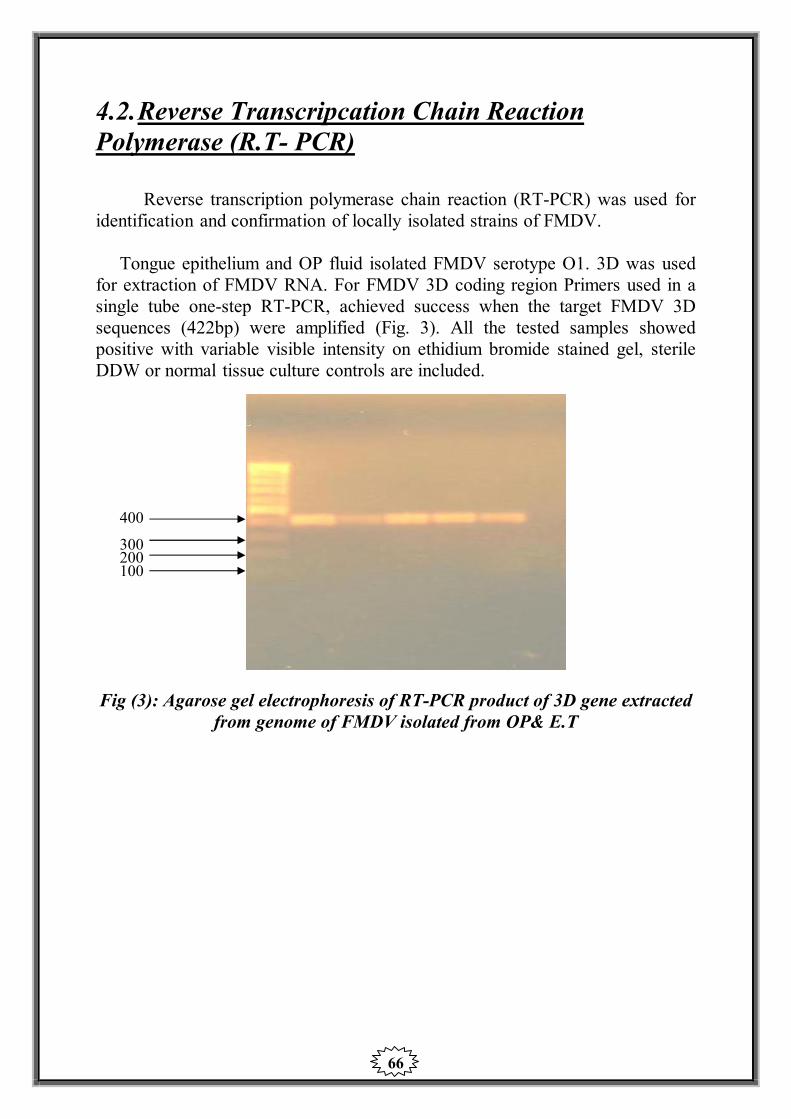
66
4.2. Reverse Transcripcation Chain Reaction Polymerase (R.T- PCR)
Reverse transcription polymerase chain reaction (RT-PCR) was used for identification and confirmation of locally isolated strains of FMDV.
Tongue epithelium and OP fluid isolated FMDV serotype O1. 3D was used
for extraction of FMDV RNA. For FMDV 3D coding region Primers used in a single tube one-step RT-PCR, achieved success when the target FMDV 3D sequences (422bp) were amplified (Fig. 3). All the tested samples showed positive with variable visible intensity on ethidium bromide stained gel, sterile DDW or normal tissue culture controls are included.
400 300 200 100
Fig (3): Agarose gel electrophoresis of RT-PCR product of 3D gene extracted from genome of FMDV isolated from OP& E.T

67
Prevealence of FMD in Upper Egypt (Al-Menya governorate): The results illustarated in following table. Table (4): The prevalence of FMDV specific antibodies against type O1
and A/ Egypt/ 2006 in Al-Menya governorate using SNT. The table showes that the both serotype O1 and A antibodies were scattered
at the locality of Al-Menya governorat except in Matay where O1 antibodies were exist only. The most percentage incidence was at Bany-Mazar the percentage of O1 antibodies was 31% and at Al-Menya the percentage of A antibodies was 20%, while at other localities (Al-Menya, Magaga and Matay). The percentages were ranged between 4.7-20% from FMDV type O1 antibodies and between 5.9-20% for A.
Localities No. of serum
samples
No. of positive
sample
Percentage of
positive samples
O1 A O1 A
Al-Menya 60 10 12 16.6 20
Bany-Mazar 29 9 2 31 6.9
Samalout 25 5 4 20 16
Matay 43 2 - 4.7 -
Magaga 17 2 1 11.8 5.9
Total 174 28 19 16.1 11

68

69
Table (5): Prevalence of FMDV antibodies against type O1 and A/ Egypt /2006 by indirect ELISA in Al-Menya governorate.
When ELISA used (Table 5 & Fig.5) the results were more or less the same
with higher percentages than SNT. The most percentage of O1 antibodies was at Bany-Mazar 31%, while the most percentage of A antibodies was at 25 Al-Menya. The lowest percentage of O1 antibodies was at Matay, while the lowest percentage of A antibodies was at Bany Mazar. The percentage of O1 antibodies ranged between 11.6-31% and between 10.3-25% for A.
Locality
No. of
samples
No. of positive
Sample
Percentage of
Positive samples
O1 A O1 A
Al-Menya 60 11 15 18.3 25
Bany-Mazar 29 9 3 31 10.3
Samlout 25 6 6 23 23
Matay 43 5 - 11.6 -
Magaga 17 3 2 17.6 11.8
Total 174 34 26 19.5 14.9

70

71
Detection of non-strctural protein antibodies in serum of sheep in Al-Menya governorate.
Table (6): PrioCHECK in sera collected from Al-Menya governate.
The table reveladed that Bany Mazar showed the higher percentage (27.6%)
while in Matay showed the lowest percentage (4.6%).
Locality No. of sera
sample
No. of tested
samples
Positive Negative % of
positive
Al-Menya 60 16 10 6 16.6
Bany-Mazar 29 11 8 3 27.6
Samalout 25 9 9 - 36
Matay 43 2 2 - 4.6
Magaga 17 3 3 - 17.6
Total 174 41 32 9 18.4

72

73
Prevealence of FMD in Al-Ismailia: The results illustarated in following table.
Table (7): The prevalence of FMDV specific neutralizing antibodies
against type O1 and A Egypt 2006 in sheep sera in Al-Ismailia governorate. Table 7 & Fig 7 indicated that FMDV antibodies is scattred at Al-Ismailia
governorate when SNT was used, both types of serotypes O1, A antibodies were detected. The higher percentage was recorded at Al-Ismailia (24.6%) for O1 and (32.7%) for strain A at Kantara. The lowest percentage was at Tal El-Keber for O1 (8.2%), while it was (12.2%) for starin A at Tal El-Kebeer.
Locality No. of samples
No. of positive sample Percentage of positive
O1 A O1 A
Al-Ismailia 77 19 11
24.6%
14.3%
Al-Kassassen 32 4 4 12.5 12.5
Tal El-Kebeer 49 4 6
8.2%
12.2%
Fayed 60 9 11
15%
18.3%
Kantara 58 8 19
13.8%
32.7%
Total 276 44 51 15.9% 18.4%

74

75
Table (8): Prevalence of FMDV antibodies against type O1 and A Egypt 2006 by indirect ELISA in Al-Ismailia governorate.
ELISA indicated that Al-Ismailia have the higher percentage of O1
antibodies (19.5%), while Kantra have the higher percentage of A.
Location No. of
samples
No. of positive
sample
Percentage of
positive samples
O1 A O1 A
Al-Ismailia 77 22 18 28.5 23.3
Al-Kassassen 32 5 6 15.6 18.8
Tal El-Keber 49 4 6 8.2 12.2
Fayed 60 10 18 16.7 30
Kantara 58 10 20 17.2 34.4
Total 276 51 68 18.4 24.6

76

77
Detection of non-strctural protein antibodies in serum of sheep. Table (9): PrioCHECK blocking ELISA in sera collected
from Al-Ismailia governorate. PrioCHECK investigation showed a high positive result in Kantara and the
lowest at Tal El-Keber according to the serum samples examined (Table 9 & Fig 9)
Location Total No.
of samples
No. of tested sample
Positive Negative
% of Positive
Al-Ismailia 77 30 12 18 15.5
Al-Kassassen 32 8 5 3 15.6
Tal El-Keber 49 10 6 4 12.2
Fayed 60 20 8 12 13.3
Kantara 58 27 17 10 29.3
Total 276 95 48 47 17.4

78

79
Prevealence of FMD in Al-Giza: The results illustarated in following table.
Table (10): Prevalence of neutralizing antibodies against FMDV type O1
and A Egypt 2006 in sera of sheep at Al-Giza governorate. Kerdasa showed the higher percentage of O antibodies at Al-Giza
governorate (50%), while Badrashine exhibited 31.25% of antibodies against A antibody Saft El-Laban was free from antibodies against A serotype. Abo El-Nomros showed the lowest percentages for both O1 and A it were 14.6% and 7.3% recpectivly.
Locality No. of sample
No. of positive sample
Percentage of positive samples
O1 A O1 A
Abo El-Nomros 41 6 3 14.6% 7.3%
Hawamdya 45 20 8 44.4% 17.7%
Badrashien 16 4 5 25% 31.25%
Kerdasa 10 5 2 50% 20%
Saft El- Labn 21 4 - 19% -
Total 133 39 18 29.3% 13.5%

80

81
Table (11): Prevalence of FMDV antibodies against type O1 by indirect ELISA in sheep sera at Al-Giza governorate.
The same facts appeared when ELISA used Kerdasa & saft El-laban showed
the same criteria indicated when SNT adopted on the same serum samples (Table 11 and Fig 11).
Location No. of sample
No. of positive sample
Percentage of positive sample
O1 A O1 A
Abo El-Nomros 41 7 4 17 9.8
Hawamdya 45 24 10 53.3 22.2
Badrashien 16 5 6 31.25 37.5
Kerdasa 10 6 4 60 40
Saft El- Labn 21 4 - 19 -
Total 133 46 24 34.6 18

82

83
Detection of non-strctural protein antibodies in serum of sheep in Al-Giza governorate.
Table (12): PrioCHECK blocking ELISA in sera
collected from Al-Giza governorate. Table 12 and Fig 12 confirmed that Kerdasa have problem with FMD when
PrioCHECK used 60% percentage was recorded and the range was between 14.6% and 44.4% in the rest of localities at Al-Giza governorate where the serum samples collected.
Location No. of sera
sample
No. of tested
samples Positive Negative Percentage
of positive
Abo El-Nomros 41 9 6 3 14.6
Hawamdya 45 28 20 8 44.4
Badrashien 16 9 3 6 18.75
Kerdasa 10 7 6 1 60
Saft El- Labn 21 4 4 - 19
Total 133 57 39 18 13.5

84

85
Prevealence of FMD in Behira: The results illustarated in following table. Table (13): Prevalence of neutralizing antibodies against FMDV type O1
and A Egypt/ 2006 in sheep sera Behira governorate. The results tabulated in Table 13 and Fig 13 showed that the circulating
antibodies against FMDV serotype O1 was most prevalent at Kafr El-Dawar (Behira) in a percentage of 15.9% and for A was 36.4% at Kafr El-Dawar when SNT used.
Location No. of samples
No. of positive sample
Percentage of positive sample
O1 A O1 A
Damanhour 40 4 12 10 30
Etay El-Baroud 86 6 20 7 23.3
Kom Hamda 68 5 8 7.4 11.8
El-Eyoun 31 4 4 12.9 12.9
Kafr El-Dawar 44 7 16 15.9 36.4
Total 269 26 60 9.7 22.3

86

87
Table (14): Prevalence of FMDV antibodies against type O1 and A Egypt 2006 by indirect ELISA in sheep sera at Bahira governorate. When Elisa used Table 14 and Fig 14 the incidence of the antibodies were
higher in El-Eyoun in a percentage of 16.1% for type (O1) ,Kafr El- Dawar was higher for type A 36.4%.
Location No. of samples
No. of positive sample
Percentage of positive sample
O1 A O1 A
Damanhour 40 5 14 12.5 35
Etay El-Baroud 86 6 24 7 27.9
Kom Hamda 68 6 10 8.8 14.7
El-Eyoun 31 5 5 16.1 16.1
Kafr El-Dawar 44 7 16 15.9 36.4
Total 269 29 69 10.8 25.7

88

89
Detection of non-strctural protein antibodies in serum of sheep in Behira governorate.
Table (15): PrioCHECK blocking ELISA in sheep sera collected from Behira governorate.
Invetigation by PrioCHECK in sheep serum in Behira governorate indicated
that the higher percentage was (36.3%) at Kafr El-Dawar and the lowest percentage was at Kom Hamada.
Location
Total No. of
samples
No. of tasted sample
Positive Negative
Percentage of
Positive
Damanhour 40 16 9 7 22.5
Etay El-Baroud 86 26 19 7 22
Kom Hamda 68 13 5 8 7.3
El-Eyoun 31 8 - 8 -
Kafr El-Dawar 44 23 16 7 36.3
Total 269 86 49 37 18.2

90

91
Table (16): The effect of repeated vaccination of sheep on the formation of NSP of FMDV by the using of PrioCHECK blocking ELISA.
The sheep were vaccinated with 1 ml s/c of locally produced bivalent FMD
vaccine. The animals were vaccinated six times with one month interval. The OD 450 max be > 1.000(-), ± (weak positive) >50% +, > 70 %(+) It is clear from table (15) that the repeated vaccination of sheep with FMD
vaccine for five to six times helps in the appearance of FMDV NS protein. Our result indicated that PrioCHECK blocking ELISA is a good diagnostic tool to detect such protein.
* Non vaccinated. ** Vaccinated for one time.
Months post vaccination Number of sheep 6 5 4 3 2 1
+ + ± - - - 1 + + ± - - - 2 + - - - - - 3 + + - - - - 4 + + ± - - - 5 - - - - - - Control A* - - - - - - Control B**

92
5. Discussion
Foot and Mouth Disease (FMD) virus (genus Aphthovirus, Family
Picornaviridae) principally infects cloven hoofed animals. FMD is caused by 7 immunological distinct serotype (O, A, C, AISA1, SAT1, SAT2 and SAT3). The disease is highly contagious and combined with high antigenic diversity of the virus makes FMD difficult to control (Nick et al., 2007). Infection or vaccination with one serotype dose not conferm protection against other serotypes (FMD serotype are circulate currently or periodically in the Middle East and North Africa (Knowels and Samuel, 2003).
Since the 1950, attention has been drawn to the economic importance of
FMD in Egypt, after several outbreaks of the disease affected cattle, buffaloes, sheep and goats with the predominant isolation of FMD virus serotype O1 (Moussa et al,1974b, Daoud et al, 1988; and El-Nakasly et al., 1996). The last outbreak of serotype (O) was in 2006 (Abd El-Rahman et al, 2006). Other serotypes have not been reported since 1972 (Aidaros 2002). In 2006 clinical cases of FMD was recognized in a cattle farm in Al-Ismailia, Northeastern Egypt. Samples were submitted for laboratory investigation and serotype determination by using virus isolation, antigen ELISA and RT-PCR. FMDV type (A) was confirmed (Knowles et al., 2007). The FMDV type (A) was designated as A/ Egy/2006. So, Egypt is endemic with two FMDV serotype (O&A) and the outbreaks still reported since 2006 till now and the country is usually threatened by FMD outbreaks(Ghoneim et al,2010).
The objective of the study is to screen infection with FMDV and
epidemiology status of both serotypes (O1&A/ Egypt/ 2006) by: Isolation and identification of serotypes (O1&A/ Egypt/ 2006) from
samples collected from different governorates of Egypt (Al-Ismailia, Al-Behira, Al-Giza and Al-Menya by Isolation techniques and antigen ELISA.
Molecular identification of isolates by RT-PCR. Screening of the animal sera to determine the circulating antibodies
against FMDV (O1&A/ Egypt/ 2006) and to detect the non- capsid protein (NCP) by SNT and blocking ELISA.

93
Fig (1) is a map of Egypt showed the governorates in lower and Upper Egypt where the serum and virological samples were collected. These governorates are Al-Ismailia, Al-Behira, Al-Giza, and Al-Menya.
Table (2) showed the types and number of virological samples collected
from Al-Ismailia and Al-Behira governorates where the FMD outbreaks were existed. The samples were epithelial tongue tissue (E.T), oesophageal pharyngeal fluid (OP).3 (E.T) samples from Al-Ismailia and 2 from Al-Behira. While (OP) were 7 samples from Al-Ismailia and 11 samples from Al-Behira were collected.
The sample were treated and inoculated in both unweaned baby mice and
BHK21 cl.13, 2 out of 3 E.T samples collect of Al-Ismailia were positive to FMDV when inoculated in tissue culture(BHK21 cl.13) and 3 out of 3 when inoculated in baby mice. 2 E.T samples were positive when inoculated in both T.C and baby mice. OP samples collected from Al-Behira indicated that, the samples were positive to FMDV when inoculated in T.C and baby mice.
So, the results tabulated in table (2) showed that the collected samples
(E.T&OP) from both Al-Ismailia and Al-Behira governorate contain FMDV. Fig No.2 showed the CPE of FMDV when inoculated in BHK21 cl. 13.
When indirect ELISA to identify the FMDV serotypes table (3), it was
clear that 2 samples were of FMDV type O1 out of 3 E.T and one was of type A/Egypt/2006. 7 OP samples collected from Al-Ismailia showed that 5 samples were of type O1 and 2 of serotype A. Type O1 was detected from 2 E.T samples. 7 OP samples were of serotype O1 and 4 were of serotype A in Al-Behira governorate. The results of tables (2&3) are in agreement with Moussa et al., (1974) who detect and identify FMDV type O1 and Knowles et al., (2007) who confirmA/Egypt/2006 in Egypt also with Ghoniem et al., (2010) who detected FMDV from infected animals ( tissues and secretion).
Nucleic acid recognition tests, such as the RT-PCR are being used
increasingly as a rapid and sensitive diagnostic method (OIE). Fig (3) showed the results of RT-PCR which used to confirm locally strain
of FMDV type O1 (Al-Ismailia and Al-Behira). The E.T and OP isolated FMDV were used for extraction of FMDV RNA and subsequent amplification of FMDV of 3D coding sequences, the primer set was originated from FMDV 3D coding regions. Primers used in single tube one step RT-PCR achieved success when the target was FMDV 3D sequences (422bp) were amplified. All the tested samples (4 E.T and 12 OP) were positive with variable visible

94
intensity on ethidium bromide stained gel. RT-PCR generated a single band corresponding to the size expected.
More or less the same results were showed by (Knowles et al., 2007 and
Ghoniem et al., 2010). Table (4) and Fig (4) showed that the neutralizing antibodies to both
serotypes O1 and A were scattered at the localities of Al-Menya governorates expect in Matay where O1 antibodies was existed only according to the sera collected from sheep. The most percentage incidence was in Bany Mazar the percentage of O1 antibodies was 31 % and at Samalout of A antibodies was 16%, while at other localities(Al-Menya, Magaga and Matay), The percentage were ranged between 4.7- 20% for FMDV type O1 antibodies and between 5.9- 20% for FMDV serotype A antibodies when SNT used.
Table (5) and Fig (5) the results were more or less the same with higher
percentage than SNT. The criteria may be due to that ELISA is more sensitive than SNT. This sensitivity of ELISA could be explained since the ELISA probably detects not only neutralizing antibodies but also other antibodies reacting with antigenic determinates not involved in neutralization test.
When the sheep serum samples were collected from different localities of
Al-Menya governorate were examined by PrioCHECK FMDV non structural protein blocking ELISA to differentiate between antibodies due to vaccination or perivous infection independent of serotype. Bany-Mazar showed the higher percentage (27.6%) while in Matay showed the lowest percentage (4.6%) table (6) and Fig (6).
Table (7) And Fig (7) Indicated that FMDV antibodies is scattered at Al-
Ismailia governorate when SNT was used, both types of serotype O1& A antibodies were detected. The higher percentage was recorded in Al-Isamilia (24.6%) for O1 and (32.7%) for A at Kantara. The lowest percentage was at Tal El- Keber for O1 (8.2%), while was (12.2%) for A at Tal El-keber too.
ELISA indicated that Al-Ismailia have the higher percentage of O1
antibodies (28.5%) while Kantara have the higher perentage of A (34.4%), Table (8) and Fig (8). Any how, the both tests SNT and ELISA indicated that FMDV antibodies one of higher percentage at Al-Ismailia governorate.
Table No. 9 and Fig No.9 showed that the PrioCHECK investigation indicated a high positive percentage (29.3%) at Kantara and the lowest percentage at Tal El-Keber (12.2%) according to the serum samples examined.

95
Table (10) and Fig (10) showed that, at Al-Giza governorate Kerdasa had the higher percentage of O1 antibodies 50%, while Badrashine exhibited 31.25 % of antibodies against A antibodies. Saft El-Laban was free from antibodies against A serotype. Abo El-Nomros showed the lowest percentage for both O1 and A it were 14.6% and 7.3% respectively. The same facts were appeared when ELISA used, Kerdasa and Saft El-Laban showed the same results when SNT adapted on the same serum sample (Table (11) & Fig (11)).
Table (12) and Fig (12) confiremed that Kerdasa have a problem with
FMDV when PrioCHECK used 60% was recorded. At the other localities of Al-Giza the range percentages were between (14.6%-44.4%)
The result of prevalence of neutralizing antibodies aginst FMDV at Al-
Behira governorate tabulated in table (13) and Fig (13), showed that the circulating antibody against FMDV serotype O1 was most prevalent at Kafr El-Dawar in a percentage of 15.9% and for A at Kafr El-Dawar 36.4% when SNT used. When indirect ELISA used to examine the same serum sample collected from Al-Behira table (14) and Fig (14), the incidence of the antibodies were higher at Kafr El-Dawar in a percentage of 15.9% for serotype O1 and 36.4% for serotype A.
PrioCHECK FMDV (NSP) blocking ELISA table (15) and Fig (15)
Indicated that the higher percent of antibodies was at Karfr El-Dawar 36.3% and the lowest percentage, at Kom Hamada 7.3%, El-Eyon was always free.
The serology results exhibited in this study are in agreement of
Bengelsdroff (1989) who stated that SNT proved to be a very suitable aid for judging the immunological relations between the various strains of FMDV, Chirstensen and Kredier (1992) they found that there are correlation between the neutralizing antibody titer and the antibody concentration as determined in ELISA. Again with they monitored the antibody response of sheep vaccinated with aluminum hydroxide gel adjuvanted FMD Vaccine by using SNT and ELISA. OIE (2000) reported that SNT is used as FMDV serotype specific serological test and Armstrong et al., (2002) who mentioned that SNT is considered definitive in determining the antibody status in livestock. De Clereq (2002) concluded that SNT is the reference to detect antibodies against FMDV.
OIE (2000) reported that ELISA is serotype specific, sensitive,
quantitative, quick to perform, less variable, not dependant on cell culture and be performed with inactivated antigen.
In this study the detection of antibodies against non-structural (NS) protein are in Parallel with Bergman et al.,(1993), they developed an Enzyme-linked immunosorbent blot assay incorporating a number of non–structural proteins

96
including the poly protein 3ABC, capable of differentiating vaccinated form infected animals with high specificity and sensitivity and with Sorensen et al.,(1998) they stated that ELISA using 3AB and 3ABC detected antibodies for day 8 and day 10 after experimental infection of susceptible cattle and sheep, they reported also that ELISA detected antibodies against any of the seven serotype of FMDV. Bulut et al., (2002) used 3ABC ELISA in measuring antibodies to non-structural protein of FMDV to differentiate infected from vaccinated animals. Laila et al., (2004) used the Chekit-FMD-3ABC ELISA to differentiate between infected and vaccinated sheep for FMD control in Egypt.
Table (16) indicated that the repeated vaccination (Six times one month
interval) with locally produced bivalent inactivated vaccine adjuvenatd with aluminum hydroxide gel could help in detection of FMDV non-structural (NS) protein. PrioCHECK blocking ELISA is a good diagnostic tool to indicate this protein. This criteria was studied by Lee et al., (2006) they measured the possible presence of antibodies against these non-structural protein in repeatedly vaccinated calves. This is due to impurities in FMD Vaccine (2C) which not present in the clarified FMDV vaccines.
From the already mentioned data, it was clear that, the both serotypes of
FMDV (O1& A/ Egy/2006) still circulating and existing in the governorate under investigation. Moreover about 50% to 80% of sheep were free from antibodies against FMDV,those animals are susceptible to infection with FMD and as we know sheep plays an important in the epidemiology and pathogenicity for cattle and pigs while strains recovered from sheep retained their full pathogenicity for species Sellers and Gloster (1980) who added that sheep act as source of infection for other species so the vaccination of sheep is effective in limiting spread of FMD outbreak, Shawkat et al., (1989) isolated FMDV serotype O1 from Egyptian sheep and they concluded that sheep can play role in the epidemiology of FMD in Egypt; Ganter et al., (2001) mentioned that sheep and goats might be carriers, so they play an important role in the epidemiology and transmission of FMD; Kitching and Hughes (2002) recorded that serotype (O) FMDV has been recovered from over 90% of the positive samples from sheep submitted to the WRL for FMD, Pirbright,U.K, Georgiev et al., (2004) found that FMD have transmitted to sheep in which infection is frequently sub-clinical and Laila et al., (2004) stated that sheep play an important role in the epidemiology and transmission of FMD. More over, FMD is suspected to have transmitted to sheep in which infection is frequently unapparent.
So, it is of importance to identify sheep which have been exposed to the
virus and have developed antibodies. Such animals are carriers and thus be a potential source of new outbreak.

97
In conclusion the vaccination of sheep is very important to reduce the risk
of FMD outbreaks coming from sheep to other susceptible animals in Egypt, also stamping out policy is very useful in FMD eradication.

98
6. Summary
In this study 852 serum sample were collected randlomy from apparently healthy sheep in Al-Giza, Al-Ismailia, Al-Behira and Al-Menya governorates. The serum were submitted for detection of antibodies against FMDV serotypes O1& A using SNT and ELISA. Differentition between vaccinated and previously infected sheep was done by PrioCHECK NSP blocking ELISA.
The results indicatd that, antibodies aginst both serotypes of FMDV O1&A
were detected from the four governorates under investigation. In Al-Menya governorate the percentage of O1 antibodies was 19.54%
(34/174), while antibody percentage against A was 14.94 % (26/174). At the same governorate, the previously infected sheep (according to serum
collected sample) were about 18.4% (32/174), when PrioCheck blocking ELISA used.
In Al-Ismailia governorate the percentage against O1 was 18.4 (51/276)
while for A, it was 24.6 (68/276), mean while sheep previously infected with FMDV were 17.4 % (48/276).
In Al-Giza governorate 34.58% (46/133) of samples were positive to O1
while 18.04 %( 24/133) were positive to A, 29.32% of the collected serum samples showed previous infection with FMDV.
FMDV serotype O1 antibodies percentage was 10.78% (29/269) in Al-
Behira, while it was 25.65% (69/269) for A. 18.2% (49/269) was due to previous infection with the virus.
Moreover, the effect of repeated vaccination on sheep with bivalent FMD
vaccine locally produced was studied. The study showed that the non-strucural protein antibodies of FMDV started to appear since the 4th vaccination. The PrioCHECK FMDV NSP blocking ELISA was used in this study.
On the other hand, virological investigation was done on samples collected
from Al-Ismailia and Al-Behira governorates where FMD outbreaks were existed. The samples were tongue epithelium (E.T) and oesphaigael pharyngeal fluid (OP), Al-Ismailia (3 E.T and 7 OP) and Al-Behira (2 E.T and 11 OP).

99
The samples were inoculated in tissue culture BHK21 clone 13 cell line and in unweaned baby mice. Cytopathic effect (CPE) and classical clinical signs and deaths were observed. ELISA indicated that 2 E.T samples were of serotype O1 and 1 E.T of serotype A while 5 of OP samples were FMDV serotype O1 and 2 of serotype A in Ismilia governorate.
In Al-Behira governorate 2 E.T samples of serotype O1 while 7 OP samples
were of serotype O1 and 4 OP samples were of serotype A. The virological obtained results were confiremd by RT-PCR. E.T & OP isolated FMDV serotype O1 were used for extraction of FMDV RNA and subsequent amplification of 3D coding sequences, primers used in one step.
RT-PCR achived success when the target FMDV 3D sequences (422 bp)
were amplified. All the tested samples showed positive with variable visible intensity on ethidium bromide stained gel.
Our results concluded that:
FMD is still widely spread in Egypt, with higher rate in Upper Egypt than that of Lower Egypt. Egyptain sheep are playing an important role in epidemiology of FMD and issue of sheep vaccination is very important. FMD PrioCHECK test is useful in detection of previously infected animals and realability of the test in differentiation between infected and vaccinated animals. The results of PCR assay documented the occuracy and efficacy of the test rather than that of traditional one ( SNT and ELISA) .
100
7. References
Abdel- Rahman, A. O.; Farag, M. A.; Samira El- Kilany; Eman, M. A.; Manal Abo El- Yazed and Zeidan, S. (2006):
Isolation and identification of FMDV during an outbreak of 2006 in Egypt. Kafr El- Sheikh Vet. Med. J.; 4(1): 2006.
Abu El- Zein, E.N. and Crowther, J.R. (1978): Enzyme-labelled immunosorbent assay techniques in Foot and Mouth
Disease virus research. J. Hyg., 80: 351-390.
Abu El-Zein, E.N. and Crowther, J.R. (1980): Serological comparison of a type SAT2 Foot and Mouth Disease virus
isolated from Sudan with other type SAT2 strain. Bull. Anim. Health Prod. Afr., 27: 245-248.
Abu El- Zein, E.N. and Crowther, J.R. (1981): Detection and quantification of IgM, IgA, IgG1, IgG2 antibodies against
FMD from bovine sera using ELISA. J. Virological Method, 3: 335-365.
Acharya, R.; Fry, E.; Stuart, D.; Fox, G.; Rowlands, D. and Brown, F. (1990):
The structure of FMDV implications for its physical and Biological properties. Vet. Microbiol. 23 (1-4): 21-34 En. 38 ref., 1 st Congress of European Society for Veterinary Virology Laboratory of molecular Biophysics Oxford OX1 3 Gu U.K.
Aggarwal, N.; Cox, S.J.; Statham, R.J. and Barnett, P.V. (2002): International Symposium, 17-20 Sep., Turkey. App., 23.
Ahl, R.; Haas, B.and Marquardt, O. (1996) Improved and new techniques for diagnosis of foot and mouth disease.
Report of the Session of the FAO Research Group of the Standing Technical Committee of the European Commission for the Control of Foot-and-Mouth Disease, 1996,September 2-6, Ma’ale Hachamisha,Israel.p.58-63
Aidaros, H.A. (2002): Regional status and approaches to control and eradication of FMD in The
Middle East and North Africa. Rev.sci.tech.Off.Int. Epiz., 21 (3): 451-458.
101
Alexandersen, S.; Zhang, Z. and Donaldson, A. (2002): Aspects of the persistence of FMD virus in animals. The carrier
problem.Microbes Infect., 4: 1099 -1110.
Alonso, A.; Gomes, M.P.D.; Mortins, M.A. and Sondahl, M.S. (1990): Detection of FMD virus infection associated antigen antibodies comparison of
ELISA and agar gel immunodiffusion test. Preventive Veterinary Medicine, 9 (3): 233-240.
Amaral-Doel, C. M.; Owen, N. E.; Ferris, N. P.; Kitching, R.P.; Doel, T.R. (1993):
Detection of foot-and-mouth disease viral sequences in clinical specimens and ethyleneimine-inactivated preparations by the polymerase chain
Amass, S.F.; Pacheco, J.M.; Mason, P.W.; Schneider, J.L.; Alvarez, R.M.; Clark, L.K. and Ragland, D. (2003):
Procedures of preventing the transmission of FMDV to Pigs and sheep by personnel in contact with infected pigs. Vet. Rec., 153: 137-140.
Anderson, E.C.; Doughty, W.J. and Anderson, J. (1976): The role of sheep and goats in the epizootology of FMD in Kenya. J. of
Hyg., 3: 395-402.
Anderson, J.; Corteyn, M.; Hamblin, P. and Paton, D. (2003): Further Asia 1.Report of Sess. Res. Gr. St. Tech. Committee of the European
Commission for the control of Foot and Mouth Disease. Berne, Swizerland, Sept. 16-19 2003, Appendix 24: 157-165. Validation of the solid phase competitive ELISA for FMDV type C .
Anon (1978): History of Foot and Mouth disease. Vet. Rec., 102: 184 185.
Arafa, M.H. (1980): Studies on the carrier state of goats exposed to FMDV. MVSc Thesis,
University of Cairo.
Arbelaez, G.; Bustos, M.Q.; De-Gerardino, A.G.; De-Lobo, C.A.; Estupinon, A. and Berrera, J. (1979):
Standardization of the micro neutralization technique for FMD virus antibodies. Rev. Inst. Colomb. Agropec., Bogota, 14: 89-92.

102
Archetti, I.L.; Amadori, I.M.; Donn, A.; Salt, J. and Lodetti. (1995): Standardization of the microneutralization technique for FMDV antibodies.
Rev. Inst. Colom. Agrospec. (Bogota), 14: 89-92.
Armstrong, R.M.; Zhang, Z.; Aggarwal, N.; Hamblin, P.A. and Cox, S.J. (2002):
Dedection of Ig against foot and mouth disease virus (FMDV) non structural polyprotein 3ABCin cattle and sheep. European commission for the control of foot and mouth disease.
Awad, F.; Moussa, A.A.M.; Amin, M.; Amer, M. and Daoud, A. (1984): Significance of virus infection associated antigen (VIA) as a tool for
epizootiological survey of FMD in Egypt. Agri. Res. Rev., 62 (5B): 99-106.
Babini, A. (1966): Serum neutralization test for man survey of immunity in cattle vaccinated
against FMD. Atti. Soc. Italy Science Vet., 20: 752-758.
Bachrach, H.L. (1968): Foot and mouth disease. Ann. Rev. Microbiology, 21: 201-244. Progress in
Biophysics and Molecular Biology 60: 241-260.
Bachrach, H.L.; Trautman, R. and Breese, S.S. (1963): Chemical and physical properties of virtually pure FMD virus. Am. J. Vet.
Res., 25: 333-342. Barnett, P.V. and Cox, S.J. (1999): The role of small ruminants in the epidemiology and transmission of foot
and mouth disease. Institute of Farm Animals Health, Pirbright Laboratory, Vet.j., 158 (1): 6-13.
Barteling, S.J.and Cassim, N.I. (2004) Very fast (and safe) inactivation of foot-and-mouth disease virus and
enteroviruses by a combination of binary ethyleneimine and formaldehyde. Dev. Biol. (Basel).; 119:449-55.
Bastos, A.D.S.; Bertschinger, H.Y.J.; Cordel, C.; Van de Vuuren, W.J.; Keet, D.; Bengis, R.G.; Grobler, D.G. and Thomson, G.R.(1999):
Possibility of sexual transmission of FMD from African buffalo to cattle. Vet. Rec., 145 (3): 77-79.
103
Becker, Y. (1987): Computerized secondary structure analysis of FMDV capsid VP1 as basis
for characterization of virus isolates.FMD 17th Conference Office International des Epizootics, Paris, France, 152-168.
Belak, S., (2007): Experiences of an OIE Collaborating Centre in molecular diagnosis of
transboundary animal diseases. a review. Dev. Biol. (Basel) 128, 103–112.
Belsham, G.J. (1993): Distinctive features of FMDV, a member of the Picorna virus family, aspects
of virus protein synthesis, protein processing and structure. Progress in Biophysics and Molecular Biology 60: 241-260
Bengelsdroff, H.J. (1989): Testing the effectiveness of FMD vaccines, the relationship between test
infection results and corresponding neutralization titres of vaccinated cattle. Berl. Munch Tierarztl Wochenschr, Jun, 102: 193-198.
Benveniti, L.; Rogel, Y.; Kunznetzova, L.,; Bujanover, Y. and Stram, Y. (2001):
Gene gun-mediate DNA vaccination against FMDV. Vaccine, Jul, 16-19 (28-29): 3885-3895.
Bergmann, I.E.; Aude de Mello, P. and Neitzert, E. (1993): Diagnosis of persistent aphthovirus infection and its differentiation from
vaccination response in cattle by use of enzyme linked immunoelecctrotramfer blot analysis with bioengineered non-structure virus antigen. Am. J. vet. Res., 5(6): 825-831.
Bhattacharya, S.; Banerjee, R.; Ghosh, R.; Chattopadhayay, A.P. and Chatterjee, A. (2005):
Studies of the outbreaks of foot and mouth disease in West Bengal, India, between 1985 and 2002. Rev. Sci. Tech. Dec; 24(3):945-52.
Blacksell, S.D.; Lunt, R.A.; Chamnanpood, C.; Linchongsubongkok, W.;
Nakarvgkul, N.; Glesson, L.J. and Megkamol, C. (1994): Establishment of a typing enzyme linked immunosorbent assay for Foot and
Mouth Disease antigen using reagents against virus endemic in Thailand. Aust. Animal Health Laboratory, Commonwealth Sci., Indus. Research Organization, Geelong, Victoria.

104
Blanco, E.; Romero, L.J.; El-Harrach, M. and Sanchez-Vizcino, J.M. (2002):
Serological evidence of FMD subclinical infection in sheep population during the 1999 epidemic in Morocco. Vet. Microbial., 85 (1): 13-21.
Brocchi, E.; Bergmann, I.E.; Dekker, A.; Paton, D.J.; Sammin, D.J.; Greiner, M.; Grazioli, S.; De Simone, F.; Yadin, H.; Haas, B.; Bulut, N.; Malirat, V.; Neitzert, E.; Goris, N.; Parida, S.; Sorensen, K. and De Clercq, K. (2006):
Comparative evaluation of six ELISAs for the detection of antibodies to the non-structural proteins of foot-and-mouth disease virus. Vaccine. May 6.
Brocchi, E.; De Diego, M.I.; Berlinzani, A.; Gamba, D. and De Simone, F. (1998):
Diagnostic potential of MAB-based ELISAs for antibodies to non structural protein of Foot and Mouth Disease Virus to differentiate infection from vaccination. Vet. Quart. Vol 20 suppl.2.
Broekhijsen, M.P.; Blom, T.; Powels, P.H.; Barteling, S.; Meloen, R.H.
and Enger-Valk, B.E. (1985): Synthesis of fusion proteins containing antigenic determinant of FMDV. 4th
Meet Europ. Group Mole Biol. Picorna virus, Sept 1-7. Bronsvoot, B.M.D.; Tanya, V.N.; Kitching, R.P.; Nfon, C.; Saidou, H.M.
and Morgan, K.L. (2002): Foot and mouth disease and livestock husbandry practices in Adamawa
Province of Cameroon. Tropical Animal Health and Production. Brooksby, J.B. (1958): The virus of Foot and Mouth disease. Adv. virus Res., 59: 1-37. Brown, F. (1973): Structure-function relationship in Foot and Mouth disease virus. Immunity in
viral and rickettsial disease by A. Kohon and 8 M.A. KLingberg Plenum press, 1973, 19-28.
Brown, F. (2002): A brief of FMD and its causal agent. In FMD control strategies, symposium
proceeding, 2-5 June 2002, Lyon, France: 13-21.
Brown, F.; Hyslop, G.; Crick, J. and Morrow, A.W. (1963a): The use of AEI in the preparation of inactivated FMD vaccine. J. Hyg.
Camb., 61: 337-344.

105
Buenz, E.J. and Howe, C.L. (2006) Trends Microbiol.Jan; 14(1):28-36.Molecular Neuroscience Program,
Department of Neurology, Department of Neuroscience, RO-GU-04-12-NR, Mayo Clinic College of Medicine, 200 First Street SW, Rochester, MN 55905, USA.
Bulut, A.N.; Cokcaltskan, C. and Aplay, B. (2002) A serosurvey to trace non-structural protein to FMD conducted with the sera
from Tharace region of Turkey. EU.FMD meeting 16-20/9/2002, Izmir,Turkey, P 87-89. International Symposium, 17-20 Sep., Turkey. App., 9.
Burrows, R. (1966): Studies on the carrier state of cattle exposed to FMDV. J.Hyg. (Camb.), 64:
81-90. Burrows, R. (1968): Excretion of foot and mouth disease virus prior to the development of
lesions. Vet. Rec., 82: 387-388. Callens, M.; De Clercq K.; Lubroth, J. (1998): Preliminary validation of a RT- PCR for detection and differentiation of
FMDV and VSV by a blind assay on field vesicular samples.European Commission for the Control of Foot- and- Mouth Disease, United Kingdom, September 1998.
Callis, J.J. (1996): Evaluation of the response and risk of FMDV by commodity in International
trade. Res. Sci. Tech., 15 (3): 1075-1085. Callis, J.J.; Mckercher, P.D. and Graves, J.M. (1968): Foot and Mouth Disease. J.A.V.M.A., 135(12): 1798-1802. Capozzo, A.V.E.; Periolo, O.H.; Robiolo, B.; Seki, C.; Torre-JL-La,
Grigera, P.R. and La Torre-JL (1997): Total and isotype humoral responses in cattle vaccinated with foot and
mouth disease virus immunogen produced either in bovine tongue tissue or in BHK-21 cell suspension cultures. Vaccine, 15(6-7): 624-630.
Capstick, P.B.; Sellers, R.F. and Stewart, L. (1959): The neutralization of virus of FMD by immune serum. Arch. Ges. Virus
Forsch, 9: 606-620.

106
Cartwright, B.; Chapman, W.G. and Brown, F. (1980): Serological and Immunological relationships between the 140 S And 12 S
particles of Foot and Mouth Disease Virus. J. Gen. Virol., 50 (2): 369-375.
Christensen, N.D. and Kreider, W.John (1992): Monoclonal antibody neutralization of BVP-1. Department of Pathology,
Microbiology and Immunology, The Mitton S. Hershey Medical Center Heishey,P.A.17033, USA.
Chung, W.B.; Sornesen, K.J.; Liao, P.C.; Yang, P.C. and Jong, M.H.
(2002): Differenation of FMD virus infected from vaccinated by Enzyme Linked
Immuno Sorbent Assay using non- structural protein 3AB as the antigen and application to an eradication program. Clinical Microbiology, Aug.; 40 (8): 2843- 2848.
Ciara Murohy, Zhidong Zhang, Melvyn Quan, Jeannette Knight and
Soren Alexandersen. (2002): International Symposium, 17-20 Sep., Turkey. App., 20. Clavijo, A.; Wright, P. and Kitching, p. (2004): Developments in diagnostic techniques for differentiating infection from
vaccination in FMD. Vet. J., 167: 9-22. Clavijo, A.; Viera-Pereira, P.J.; Bergmann, I. (2003): Use of the Reverse transcription polymerase chain reaction (RT- PCR) for
the rapid diagnosis of foot and mouth disease in South America.Vet. Res. Commun. Jan; 27(1):63-71.
Cleland, P.C.; Chammanpood, P.M.D.; Baldock, F.C. and Gleeson, L.I.
(1994): Serological responses in young cattle and buffalo to two different
vaccination regions in villages in northern Thailand. Thai. J.Vet. Med., 24 (1): 61-73.
Cottral, G.E.; Gailiunas, P. and copion, R.L. (1963): Detection of FMD virus in lymph nodes of cattle through out course of
infection. Porc. U.S. livestock San. A. 67: 463 – 472. Cowan, K.M. and Graves, J.H. (1966): A third antigenic components associated Foot and Mouth Disease infection.
J. Virol., 20: 528-540.

107
Cox, S.; Hamblin, P.; Bankowski, B.; Carr, B.; Paton, D. and Barnett, P. (2008):
Longevity of the protection in cattle following vaccination with emergency FMD from the UK strategic reserve-preliminary results.The global control of FMD-tools, ideas and ideals-Eric, Italy 14-16 October.
Crowther, J.R. and Abu El-Zein, E.M.E. (1979): Application of enzyme linked immunosorbent assay to the detection and
identification of Foot and Mouth Disease viruses. J. Hyg., 83 (3): 513-519. Crowther, J.R. (2002): The use of non structural (NS) antigens of FMDV to assess antibodies in
vaccinated and infected livestock. In FMD control strategies, symposium proceedings, 2-5 June 2002, Lyon, France: 117-118.
Daoud, A.; Omar, A.; El-Bakry, M.; Metwally, N.; El-Mekawi, M. and
El-Kilany, S. (1988): Strain of FMD virus recovered from 1987 outbreak in Egypt. J. Egypt, Vet.
Med. Ass., 48(1): 63 – 71. Davis, G. (2002): Foot and Mouth Disease (FMD). Res. Vet. Sci.; Dec.; 73(3): 195 – 199. De Clereq, K.; Strobbe, R. and Debeco, J. (1987): Foot and Mouth Disease vaccination in Belgium: A serological survey. RPT
Sess. Res. GP Tech. Comm. Control Foot and Mouth Disease (FAO), 34: 81-89. De Clereq, K. (2002): Overview on FMD diagnostic techniques. In FMD control strategies.
Symposium proceedings, 2 – 5 June 2002, Lyon, France: 345 – 351. De Diego, M.; Brocchi, E.; Mackay, D. and De Simone, F. (1997): The non-structural polyprotein 3 ABC of FMDV as a diagnostic antigen in
ELISA to differentiate infected from vaccinated cattle. Archives of virology 142: 2021-2033.
Dehoux, J.P. and Hounsou, V.E.G. (1992): Epidemic of FMD in North Benin during the dry season of 1990 - 1991.
Med. Vet. Pays Tropicaux, 44(3): 261-262

108
Dekker, A.; Bouma, A. and De Jong, M. C. M. (2002): Foot and Mouth Disease Virus transmission between individually housed
calves. FMD control strategies, symposium proceedings, 2-5 June 2002, Lyon, France.
Dellers, R.W. and Hyde, J.I. (1964): Response of sheep to experimental infection with FMDV. Am. J. vet. Res.,
25: 105. De-Simone, F.; Brocchi, E.; Panina, G.F. and Barei, S.(1981): Titration of Foot and Mouth Disease antibodies in cattle sera. Comparative
study of serum neutralization on mice and ELISA. Meeting of Res. GP of the Europ. Comm. for the Control of FMD, Tubingen, September.
Dhanalakshm, K. and Krishnaswamy, S. (1992): Application of the microneutralization test for the detection of FMD viral
antibodies in vaccinated bovines. Cherion, 21 (1-2): 5-8. Andhra Pradesh Agricultural University, Hydersbad, 30, India.
Doel, T.R. (2003): FMD vaccines. Virus Res., 91: 81-99. Domingo, E.; Baranowski E.; Escormis C. and Sorbino F. (2002): Foot and Mouth Disease Virus. Comp. Immunol. Mic. Infect. Disease, Oct.
25 (5:6): 297 – 308. Donaldson, A.I. and Alexandersen, S. (2002): The virological determinants of the epidemiology of FMD. In FMD control
strategies, symposium proceedings, 2-5 June 2002, Lyon, France: 173-180. Donaldson A. I., Alexandersen, Sorensen, J.H.& Mikkelsen, T.(2001): Relative Risks of the uncontrollable (airborne) spread of FMD by different
species. Veterinary Record 148, 602-604. Dukes, J.P.; King, D.P.; Alexandersen, S. (2006): Novel reverse transcription loop-mediated isothermal amplification for rapid
detection of foot-and-mouth disease virus.Arch Virol. 2006 Jun; 151(6):1093-106.
Efficiency and Speed of Transmission of Foot-and-Mouth Disease. J. Comp. Path. 2009, Vol. 140, 225e237.

109
Ehab, I.Mohamed (2007): Studies on FMD in infected and vaccinated sheep M.V.Sc Thesis, Bany Swif
Universty.
El-Mikkawi, M.F. (1980): Some studies on immune response of cattle and buffaloes vaccinated with
foot and mouth disease vaccines inactivated by different chemical inactivators. Ph.D.Thesis (Infectious Diseases), Fac. Vet. Med., Zagazig University.
El- Nakashly, S.A.; Abu Zeid, A.A.; El- Kilany, S. and Abdel Aty, M.M
(1996): Isolation and identification of FMDV during an outbreak of 1993 in Egypt.
7th Sci. Cong., 17-19 Nov., 1996, Fac. Et. Med., Assuit University, Egypt.
EU-FMD meeting (2001): European Commission for the Control of Foot and Mouth Disease/EU
FMD/FAO. Bull. No. 3. EU-FMD (2002): European Commission for the Control of Foot and Mouth Disease/EU
FMD/FAO. EU. FMD Meeting, Azmir, Turkey, 16- 20/9/2002. Falchsel, P.; Hubik, R. and Polak, L. (1982): A study of cattle immunization against FMD types A, O and C.evaluation of
mass immuno-prophylaxis of cattle against FMD in XVI conference of the FMD commission, Paris, 14th-17th September(1982), France, OIE, 275-290.
Farag, M.A.; Alsokayran, A.; Mazloum, K.S. and Al-Bokmy, A.M.
(1998): The role of small ruminants in the epizootology of foot and mouth disease in
Saudi Arabia with reference to the economic impact of the disease on sheep and goats. Veterinary Medical Journal, 40 (79): 23-41.
Farag, M. A.; Samira El- Kilany and Abdel- Rahman, A, O. (2006): The impact of live animal importation on the epizootiology of foot and
Mouth Disease in Egypt. 8th Sci. Vet. Med. Zag. Conference (31 Aug.- 3 Sep. 2006) Hurgada.
Fatthia, A.M. (2003): Vaccination of goats with FMD vaccines. MVSc Thesis, University of
Alexandria

110
Fawzy, H.G.I.; Samira El-Kilany; Wasfy, M. O. and Abdel Rahman,A.O. (1996):
Use of polymerase chain reaction (PCR) technique for the detection of foot mouth disease virus type O1/Egypt.Vet. Med. J. Giza, Vol. 44 No. 3:613-617.
Felfe, P.; Thalmann, G.; Kreinbrink, G. and Schiele, K. (1990): Seasonal variation in the formation of virus neutraliziantibodies in cattle
after vaccination against FMD. VEB Friedreich Leoffler Institute Insel Riems, Betriels des VEB Kombiant Veterinary mpfs toffee.
Fernandez, A.A.; Mello, P.A.D.E.; Gomes, I. and Rosenberg, F. (1975): Use of virus infection associated antigen (VIA) in the detection of cattle
exposed to Foot and Mouth Disease. (En. Es. 6 ref.) Pan American Center. CP. 589 Zc. 00 Rio de Janeiro, R.J. Brazil.
Ferreira M.E.V. (1976): Prubade microneutralization poraestudies de anticueropos de la fibre aftosa.
13th Centropanamericano Fiebre Aftosa, (21/22): 17-24. Ferris, Nigel; Scott, Reid; Donald, King; Ceoff, Hutshings and Andrew,
Shaw. (2004): Prospects for improved laboratory diagnosis of FMD using real time RT-
PCR.Eu Comm. Control of FMD China. Crete. Greece, 11-15 Oct., 2004, Appendix; 42: 261-267.
Fondevila, N.A.; Marcoveccio, F.J.; Biancoviera, J.; O’Donnell, V.k.;
Carllo, B.J.; Schudel, A.A.; David, M.; Torres, A. and Mebus, C.A. (1995): Susceptibility of Lamas to infection with FMDV. J. Vet. Med. Series B.
Moron, Argentina, 42 (10): 595-599. Forman, A.J.; Gibbs, E.P.J.; Baber, D.J.; Herniman, K.A.J. and
Barnett, I.T. (1974): Studies with FMDV in British deer (red, fallow and roe). II: Recovery of
virus and serological response. J.comp. Pathol., 84: 221-228. Fox, G.; Parry, N.R.; Barnett, P.V.; Mc Ginn, B.; Rowland, D.J. and
Brown, F. (1989): The cell attachment site on FMD virus include the amino -acids sequence
RGD (arginine- glycine- aspartic acid). J. Gen. Virol., 70: 625. Francois, Volarcher, J.; Nick, J. Knowles; Rosa Fernandez; Paul, R.
Davis; Rebeca J. Midgley; Bobstatham, Geoff Hutchings, Brenda J. Newman, Nigel P. Ferris and David J. Paton (2004):

111
Global foot and mouth disease situation 2003-2004. Report of Sessi: Res. Gr. St. Tech. Committee of the Eurpean Commission for the control of foot and mouth disease. Chaina, Crete, Greece, 11-15 Oct. 2004. Appendix 21: 137-148.
Franki, R.I.B.; Fauquet, C.M.; Knudson, D.L. and Brown, F. (1991): Classification and nomenclature of viruses.5th Report of Int. Comm. on
taxonomy of viruses suppl. 2. Ganter, M.; Graunke, W.D.; Steng, G. and Worbes, H. (2001): FMD in sheep and goats. PhD Thesis, University of London. Garland, A.J.M.; Baber, D.; Hamblin, C.; Rowe, L.; Barnett, I.T.R.;
Pinto, A.A.; Collen, T. and Donaldson, A.I. (1981): The 1975 FMD epidemic in Malta. II- The detection of carriers and in
apparent infection. British Vet. J., 137 (4): 381-387. Gaschutz, S.; Hess, G. and Witmann, G.J. (1986): Neutralizing antibody response in cattle of different age with respect to the
number of FMD. Rept. Res. Gp. Stand. Tech. Comm. Control of FMD (FAO), Madrid.
Georgi Georgiev; Emiliya Veleva; Liliyana Polihronova and Alessandro
Rossi (2004): Using NSP ELISA (Chekit-FMD-3ABC Bommeli-Intervet) as a Tool for
FMDV Serosurveillance in Bulgeria. European Commission for the Control of Foot and Mouth Disease. Chanina, Crete, Greece 11-15 October 2004 Appendix 72: 447-451.
Ghoneim, N.H.; Abdel Karim, A.K.M.; Laila El-Shahawy and Abd El-Maeim, K.A. (2010):
FMD in animals in Sharkia governorates, Egypt. Trans and Emerg.Dis:1- 4. Gibbens, J.C.; Sharpe, C.E.; Wilesmith, J.W.; Mansley, L.M.;
Michalopoulou, E.; Ryan, J.B. and Hudsun, M. (2001): Descriptive epidemiology of the 2001 FMD epidemic in Great Britain: the
First five months. Vet. Rec., Dec. 15: 149 (24): 729-743. Ginaru, M.D.; Thomson, G.R.; Bengis, R.G. Esterhuysen, J.J.; Bruce,
W. and Pini, A. (1986): Foot and mouth disease in the African buffalo (Syncerous caffer). II.Virus
excretion and transmission during acute infection. Onderstepoort J. of Vet. Res., 53: 75-85.

112
Giridharan P., Hemadri P., Tosh C., Sanyal A., Bandyopadhyay S.K. (2005):
Development and evaluation of a multiplex PCR for differentiation of foot-and- mouth disease strains native to India.J. Virol. Methods 126: 1—11.
Gloster, J.; Blachall, R.M.; Sellers, R.F. and Donaldson, A.I. (1981): Forecasting the airborne spread of FMD. Vet. Rec. (1981). Gloster J, Ryan E, Wright C, Doel C, Parida S, Cox S, Barnett P, Schley
D, Gubbins S, Paton D.(2008): Withdrawn: Foot-and-mouth disease: How much airborne virus do animals
exha Vet J. 2008 Sep 17. Goel, A.C. and Rai, A. (1985): Growth curve, plaque and inactivation studies of FMD virus subtypes O1,
O5 and O6 of Indian origin. Ind.J.Comp.Microbiol.Imm. Infect.Dis., 6(1): 16-28.
Gomes, I.; Fernandez, A.A. and Mello, P.A. (1972): Foot and Mouth Disease circulating antibodies in convalescent cattle. Bull.
Off. Int. des. Epiz., 77 No. 566: 731-741. Graves, J.H. (1960): The differentiation of subtypes (variants) of foot and mouth disease by
serological methods. Amr. J. Vet. Res., 21 (8): 687-690. Graves, J.H.; Cowan, K.M. and Trautman, R. (1968): Immunochemical studies of FMD characterization of RNA free virus like
particles. Virology, 34: 269 – 274. Graves, J.H.; Mackercher, P.D. and Callis, J.J. (1972): Foot and Mouth Disease vaccine: Influence of the vaccine virus subtype on
neutralizing antibody and resistance to disease. Amer. J. Res., 33 (4): 765-768. Graves, J.H.; McVicar, J.W. and Yedloutschnig, R.J. (1977): Review of the VIA reaction in diagnosis of FMD. Proceeding of the annual
meeting of the U.S., Animal Health Association, 81: 256-263. Hafez, S.M.; Farag, M.A.; Mazloum, K.S. and El-Bokmy, A.M. (1993): Application of double sandwich enzyme linked immunosorbent assay for the
diagnosis of foot and mouth disease in Saudi Arabia. National Agriculture and Water Research Center, Ministry of Agriculture and Water, Riyadh, Saudi Arabia, 12(3): 817-830.

113
Hamblin, C.; Barnett, I.T.R. and Crowther, J.R. (1986): A new enzyme linked immunosorbent assay (ELISA) for the detection of
antibodies against FMD virus. II. Application. J. Immunol. Meth., 93: 123- 129. Have, P. (1987): Use of ELISA in diagnosis of viral disease in domestic livestock. Archiv fur
Experimentlle Veterinarmedizin, 41 (5): 645-649. Henderson, W.M. (1970): Foot and mouth disease: a definition of the problem and routs on its
isolation. Br. Vet. J., 126: 115-120. Hoang, V.N., 2009: In 15th Meeting of the OIE Sub-Commission for Foot and Mouth Disease in
South-East Asia, Kota Kinabalu, Sabah, Malaysia,9–13 March. Hofner, M.C.; Carpenter, W.C. and Donaldson, A.L. (1993): Detection of FMD virus RNA in clinical samples and cell culture isolates by
amplification of the capsid coding region. J. Virol. Method. 42(1): 53- 61. Holl, U. and Sulk, I. (1988): Determination of FMD specific antibodies in serum samples of vaccinated and
convalescent cattle by means of enzyme immunoassay. Archiv fur Expermintelle Veterinarmedizin, 42 (3): 470-476.
Holzhauer, M.; Verhoeff, J. and Wuijkuise, L.A. (2001): Deklinishe verschijnselen van monden klauwzeer bijde eerste bevestigde
gevallen op vijf bedrijven in Nederland. Tijdschr. Diergneeskd, 126(8), 282-285.
House, C. and House, J. (1989): Evaluation of techniques to demonstrate foot and mouth disease virus in
bovine tongue epithelium: comparison of sensitivity of cattle, mice, primary cell cultures, cryopreserved cell culture and established cell lines. Vet. Microbiol 1989; 20:99-109.
House, C. and Meyer, R.F. (1993): The detection of foot-and-mouth disease virus in oesophageal-pharyngeal
samples by a polymerase chain reaction technique.J. Virol. Methods. Jun; 43(1):1-6.
114
Huang, C.C.; Jong, Mh. and Lin, S.Y. (2000): Characteristics of FMDV in Taiwan. J.Vet. Med. Sci., 2000 Jul, 82 (7):
6777-6779. Hughes, G.J.; Kitching, R.P. and Woolhouse, M.E.J. (2002a): Dose dependent response of sheep inoculated intranasally with a Type ‘O’
FMDV. J. Comp. Path., 127: 22-29.
Hughes, G.J.l.; Mioulet, V.; Kitching, R.P.; Woolhouse, M.E.J.; Anderson, S. and Donaldson, A.I. (2002b):
FMD infection of sheep: implications for diagnosis and control. Vet. Rec., 150 (23): 724-727.
Hutber, A.M. and Kitching, R.P. (2000): The role of management segregations in controlling intra-herd FMD.
Tropical Animal Health and Production, 32 (5): 285-294. Hyun, B.H.; Um, J.G.; Lee, K.N.; Kim, I.J.; Ku, B.K.; Par, J.Y.; Kim,
C.H.; Shin, N.G.; Kang, J.M.; Cho, I.S.; Joo, Y.S. and An, S.H.( 2002): Rapid serological immunochromatographic assay for differentiating
infection from vaccination in foot and mouth disease using whole blood. In FMD control strategies, symposium proceedings, 2-5 June 2002, Lyon, France.
Iakovleva, A.S.; Shcherbakov, A.V.; Kan'shina, A.V.; Mudrak, N.S. and
Fomin, T.A.(2006): Recombinant non-structural 3A, 3B and 3AB proteins of foot- and-mouth
disease virus: use for differentiation of vaccinate and infectedcattle. Mol Biol (Mosk). 2006 Jan-Feb; 40 (1):165-71.
Ibrahim, M.H.; Moussa, A.A.M.; Hussein, K.; El-Kilany, S.; Stouraitis,
P. and Hussein, M. (1977): Immune response of Egyptian buffaloes to vaccination with FMD vaccine.
Agr. Res. Rev., 55: 181-182. ICTV (2000): The seventh report of the International Committee on Taxonomy of Viruses.
Academic Press. Jackson T.; King A.M.; Stuart D.I. and Fry E. (2003): Structure and receptor binding. Virus Research, 91: 33-46.

115
Jacobs, L.; Chenard, G.; Jelsma, T. and De Clercq, K. (2002): Modification and further validation by different laboratories of a FMDV type
O specific ELISA for detection of antibodies in susceptible species. European Commission for the Control of Foot and Mouth Disease,17-20 September 2002, Izmir,Turkey:242-247.
James, A.D. and Rushton (2002): The economics of FMD. Rev. Sci. tech. Off. Int. Epiz., 21 (3): 637-644. Joe, Y.S.; An, S.H.; Kim, O.K.; Lubroth, L. and Sur, J.H. (2002): FMD eradication efforts in the Republic of Korea. Can. J. Vet. Res., Apr. 66
(2): 122-124. Juan M. Pacheco, Jonathan Arzt, Luis L. Rodriguez (2010):
Early events in the pathogenesis of foot-and-mouth disease in cattle after controlled aerosol exposure .The Veterinary Journal 183, 46–53.
Karber, G. (1931): Beitrag Zurkillekiven Behandlung Pharmakologis Reithenver Suche Naungn
Schmeidebergos. Arch- Epx. Path. Pharmak, 162: 280-283. King, D. P.; Ferris, N.P.; Shaw, A.E.; Reid, S.M.; Hutchings, G.H.;
Giuffre, A.C.; Robida, J.M.; Callahan, J.D.; Nelson, W.M.; Beckham, T.R. (2006):
Detection of foot-and-mouth disease virus: comparative diagnostic sensitivity of two independent real-time reverse transcription- polymerase chain reaction assays. J. Vet. Diagn. Invest. Jan; 18 (1):93-7.
King, A.M.Q. (2002): Epitopes of FMDV and their changeability. In FMD, control strategies,
symposium proceedings, 2-5 June 2002, Lyons, France: 297-304. Kitching, R.P. (1990): FMD in the Middle East and North Africa. Ann. Mtg. Ass. Vet. Teachers,
Res. Workers, Scarborough, April 1990 10 A 38. Kitching, R.P. (1994): Serotypes of FMDV isolated from Egyptian cattle and buffaloes using
polymerase chain reaction. Facsimilr Transmission, European and world reference laboratory for FMD. Pirbright Lab. 4th Annual report, 1994.

116
Kitching, R.P. (1998): A recent history of FMD. J.Comp. Pathol., 118: 89-108.
Kitching, R.P. (2002): The epidemiology of Foot and Mouth Disease in large dairy herds in the
Arabian Peninsula in FMD control strategies, symposium proceedings, 2-5 June 2002, Lyon, France: 109.
Kitching, R.P. (2002b): Identification of FMDV carrier and subclinically infected animals and
differentiated from vaccinated animals. Rev. Sci. Tech. Off. Int. Epiz., 21 (3): 531-538.
Kitching, R.P. and Donaldson, A.I. (1987): Collection and transportation of specimens for vesicular virus investigation.
Rev. Sci. Tech. Off. Int. Epiz., 6: 251-261. Kitching, R.P. and Hughes, G.J. (2002): Clinical variation of oot and mouth disease: sheep and goats. Rev. sci. tech.
Off. Int. Epiz., 21(3): 505-512. Knowles, N. J. and Samuel, A. R. (1994): Paper presented at the session of the research group of the standing technical
committee of the European commission for the control of FMD. Austria, 19-22 Sept.
Knowles, N. J. and Samuel, A. R. (2003): Moluclar epidemiology of Foot and Mouth Disease virus Virus Res 91:65-
80. Knowles, N.; Wadsworth, J.; Reid, S.; Swabey, K.; Kholy, A.; Abd
Elrahman, A.; Soliman, H.; Ebert, K.; Ferris, N.; Hutchings, G.; Statham, R.; King, D. and Paton, D. (2007):
Foot and Mouth Disease serotype A in Egypt. Emerging Infectious Disease. Vol 13, No.10.
Kukharov, V.M.; Provina, N.A.; Sarkisyen, R.A.; Potapapava, A.F. and
Onfriev, V.P. (1973): Carriage of FMDV by animals that have recovered from the disease.
Veterinariya, Moscow, USSR, No. 9: 44-46.

117
Laila, E.El-Shehawy; Samira El-Kilany and Doaud, A.M. (2004): 3AC ELISA fordiagnosis of FMD in Egyptian sheep. App., 68. European
commission for the control of foot and mouth disease. Lee, F.; Jong, M.H. and Yang, D.W. (2006): Presence of antibodies to non- structural proteins of foot- and- mouth disease
virus in repeatedly vaccinated cattle. Vet. Microbiol. Jun 15; 115(1-3):14-20. Leforban, Y. (1999): Prevention measures against FMD in Europe in recent years. Vaccine, 17:
1755-1759. Lennette, E.H. (1964): Diagnostic procedures for viral and rickettsial diseases. A.N. Public Health
Ass. Inc., 3 rd Ed. Brood Way. Lobo, A.C.A.; Gutierrez, C.A. and Marino, J.O.C. (1975): Evaluation of antibodies induced by FMDV infection. 1. Preparation of VIA
(virus infection- associated) antigen and its use in field test. Bulletin de l’Office International des Epizootics, 81 No. 3/4 287-303 (Fr) Lab. de Recherches Med. Vet. Bogota, Colombia.
Locher F., Suryanarayana V. V.S., Tratschin J.-D. (1995): Rapid detection and characterization of foot-and-mouth disease virus by
restriction enzyme and nucleotide sequence analysis of PCR products. J. Clin. Microbiol., 33: 440- 444.
Lubroth, J. and Brown, F. (1995): Identification of foot and mouth disease virus non- structural protein 2C as a
serological indicator to differentiate infected from vaccinated livestock. Research in Veterinary Science, 59: 70-78.
Lubroth, J.; Grubman, M.J.; Burrage, T.G.; Newman, J.F. and Brown,
F. (1996): Absence of protein 2C from clarified foot-and-mouth disease virus vaccines
provides the basis for distinguishing convalescent from vaccinated animals. Vaccine. Apr; 14(5):419-27.
Mackay, D.K.; Bulut, A.N.; Rendle, T.; Davidson, F. and Ferris, N.
(2001): A solid phase competition ELISA for measuring antibody to FMD virus. J.
Virol. Methods, Sep., 97 (1-2): 33-43.

118
Mackay, D.K.J.; Forsyth, M.A.; Davies, P.R.; Berlinzani, A.; Belsham, G.J.; Flint, M. and Rayon, M.D. (1998):
Differentiating infection from vaccination in FMD using a panel of recombinant non-structural proteins in ELISA. Vaccine, 16: 446-459.
Mackowiak, C. (1970): FMD in sheep. Bull. Off. Int. Epiz., 73: 707-714.
Macpherson, I, A. and Stocker,M,G.(1962): Polyma transformation hamster cell colonies.An investigation of generic
factors affecting cell competence. Virol., 16: 147-151. Malirat, V.; Neitzert, E.; Bergmann, I.E.; Marade, E. and Beck, E.
(1998): Detection of cattle exposed to FMDV by means of indirect ELISA test using
bioengineering non-structural polyprotein 3ABC. The veterinary Quarterly 20 (suppt.2): S24-S26.
Marquardt, O. and Adam, K. H. (1990): Foot and mouth disease virus subtyping by sequencing VP1 genes.Vet.
Microbiol., 23: 175-183. Marquardt, O.; Straub, O.C. and Haas, B. (1995): Detection of foot-and-mouth disease virus, independent of type within 24
hours by the polymerase chain reaction applied to nasal swabs Tierarztliche Umschau, 50(5):344-346.
Martin, S.W.; Meek, A.H. and Willeberg, P. (1987): Veterinary epidemiology: principles and methods. Iowa state University
Press, Ames, Iowa, 356pp. Mason, P.W.; Grubman, M.J. and Baxt B. (2003): Molecular basis of pathogenesis of foot and mouth disease virus. V. Res., 91:
9-32. Mason, P.W.; Pacheco, J.M.; Zhao, Q.Z. and Knowles, N.J. (2002): Comparison of the complete genome of Asia, African a European isolates of a
recent FMD virus type "O" Pandemic strain (Pan Asia). Research Group of the Standing Technical Committee of the Eu. FMD Meeting 17-20 Sept. 2002, Izmir, Turkey.
Matsumato, M.; McKercher, P.D. and Nusbaum, K.E. (1978): Secretory antibody response in cattle infected with FMDV. Am. J. Vet. Res.,
39 (7): 1081- 1087.

119
McVicar, J.M. and Sutmoller, P. (1968): Foot and mouth disease in sheep and goats, early virus growth in the pharynx
and udder. Proc. Ann. Mtg. US. Livestock Saint.Ass., 75: 194-199. McVicar, J.W. and Sutmoller, P. (1970): FMD, the agar gel diffusion precipitation test for antibody to virus infection
associated (VIA) antigen as a tool for epizootiologic surveys. Am. J. Epidem., 92: 273-278.
McVicar, J.W. and Sutmoller, P. (1971): FMD in sheep and goats: early virus in the pharynx and udder. Proc. Of 75th
Ann. Meeting animal Health Ass., USA: 104-109. McVicar, J.W. and Sutmoller, P. (1972): Foot and Mouth disease in sheep and goats: early virus growth in the
pharynx and udder. Proc. Ann. Mtg. US Livestock Sanit. Assoc., 75: 194-199. McVicar, J.W. and Sutmoller, P. (1974): Neutralizing activity in the serum and oropharyngeal fluid of cattle after
exposure to FMDV and subsequent re-exposure. Arch. Ges. Virusforsch., 44: 173-176.
Meyer, R.F.; Brown, C.C.; House, C.; House, J.A. and Molitor, T.W.
(1991): Rapid and sensitive detection of foot-and-mouth disease virus in tissues by
enzymatic RNA amplification of the polymerase gene. J. of Virological Methods ,34 (2): 161-172.
Mohapatra, J.K., Sahu, A., Pandey, L., Sanyal, A., Hemadri, D.,
Pattnaik, B., 2009b: Genetic characterization ofA/Egypt/2006 foot-and-mouth disease virus 3A
region in context of the reemergence of VP359-deletion. Mohapatra J.K., Subramaniam S., Tosh C., Hemadri B., Sanyal A.,
Periyasamy T.R. and Rasool T.J. (2007): Genotype differentiating RT-PCR and sandwich ELISA: Handy tools in
epidemiological investigation of foot and mouth disease.Journal of Virological Methods 143(1): 117-21.
Moss, A. and Haas, B. (1999): Comparison of the plaque test and reverse transcriptase nested PCR for the
detection of FMDV in nasal swabs and probing samples. J.of Virological Methods, 80 (1):59-67.

120
Moussa, A.A.M; banoub, S.M.; EL-Said, T.E.; Fahmy, F. Andbohm, H.O. (1974):
Typing and sublyping of strain of strain of FMDV isolated J.Egy.Vrt.Med.assoc., 34 (3-4) 413-419.
Moussa, A.A.M.; Daoud, A.; Hussein, K.; Hassa, N.A.; Fahmy, F.; Azab,
A. and ElShehawy, L. (1984): Prevalence of FMD in Egypt. Agric. Res. Rev., Vol.62 No.5 B, 55-58. Moussa, A.A.M.; Ibrahim, M.H. and Hussein, K. (1976): Preliminary study on antibody response of cattle after Experimental infection
with FMDV. Proc. Of the 13th Arab Vet. Conger., Cairo, 13-18 February. Moussa, A.A.M.; Stouraitis, F.; Ibrahim, M.H.; Daoud, A. and Hussein,
K. (1974b): Foot and Mouth Disease vaccine production in baby hamster kidney (BHK21)
cells in suspension in Egypt. Bull. Off. Int. Epiz., 81 (11-12): 1043-1054. Muntiu, N.; Dohotaru, V.; Bercon, A.; Mirceaseu, G.; Striub, C. and
Tomesau, A. (1970): Studies on the relationship between viraemia and generalized lesions in
challenge, infection with FMD in cattle. Rep. meeting Europ. Comm. Ankara, Turkey: 10 - 15.
Muntiu, N.; Dohataru, V.; Bercan, A.; Micescu, G.; Stribu, C. and
Tomescu, A. (1971): Duration of FMD immunity in one year old cattle, after the first Vaccination
with 10 PD10. European Commission Control, Mtg. Res. Gp. Stand. Tech. Comm. Tubingen, Germany Abstre. FMD Bull., 12 (6): 325-328.
Muntiu, N.; Dohataru, V.; Bercan, A.; Micescu, G.; Stribu, C.
andTomescu, A. (1974): Duration of FMD immunity in one year old cattle, after the first vaccination
with 10 PD50. Arch. Vet., 9 (1-2): 21-28. Murphy,M.L.P.;Meyer,R.F.;Mebus,C.;Schudel,A.andRodrigue Z,M.
(1994): Analysis of sites of foot-and-mouth disease virus persistence in carrier cattle
via the polymerase chain reaction. Arch. Of Virology; 136(3/4):299-307.

121
Musser, J.M. (2004): A practitioner's primer on foot-and-mouth disease. J. Am., Vet. Med. Assoc.,
Apr., 15; 224(8):1261-8. Department of Veterinary Pathobiology, College of Veterinary Medicine, Texas A&M University, College Station, TX 77843.
Nair, S.P. and Sen, A.K. (1992): A comparative study of immune responses of sheep against FMD type Asia
and "O" PEG concentrated aluminium hydroxide gel and oil adjuvanted vaccines. Ind. Vet. Res. Inst. Hebbal, Bangolore 560024 India, 780-786 vaccine Vol. 11 Issue 7.
Niedbalski, W. (2005): Detection of foot-and-mouth disease virus infection in vaccinated cattle. Pol
J. Vet Sci 2005; 8 (4):283-7. Niedbalski, W.; Fitanen, A.; Paprocka, G. and Kesy, A. (1994): Application of liquid phase blocking sandwich ELISA (LPBE) for the
detection of antibodies against foot and mouth disease virus. Bull. Vet. Inst. In Putway, 38 (2): 105-114.
Nick, J.K.; Scott, M.R.; Alaa, A.E; Hatem, M.S; Nigel P.F; Goffrey H.H.
and Parker, D.P. (2007): FMDV serolyre A in Egypt. Emer- injections dis 13:1593-1596. Nuanualsuwan S; Thongtha P; Kamolsiripichaiporn S; Subharat S.
(2008): Int J Food Microbiol. 2008 Sep 30; 127 (1-2):84-90. Epub 2008 Jun 18. Nuenz, S.; Konig, G.; Berinstein, A. and Carrillo, E. (2010): Difference in the virulence of two strains of foot and mouth disease virus
serotype A with the same spatiotemporal distribution. Virus Research 147: 149-152
O’Donnell, V.K.; Smitsarrt, E.; Cetra, B.; Duffy, S.; Finelli, J.; Boyle,
D.; Draghi, G.; Fondevilla, N. and Schudel, A.A. (1997): Detection of virus infection associated antigen and 3D antibodies in cattle
vaccinated against FMD. Revue scientifique et technique (International Office Epizootics), 16: 833-840.
Oem, J.K.; Kye, S.J.; Lee, K.N.; Park, J.H.; Kim, Y.J.; Song, H.J. and
Yeh, M. (2005): J Vet Sci Dec; 6(4):317-25. National Veterinary Research and Quarantine
Service, Ministry of Agriculture, Anyang 430-824, Korea.

122
OIE (2000): Foot and mouth disease, Chapter 2.1.1. In manual of standards for diagnostic
tests and vaccine, 4th Ed. 2000, Paris, 77-92. OIE (2006): Disease information 16 February 2006, 19 (7): 16-19. Oleksiewicz, M. B., Donaldson, A. I. & Alexandersen, S. (2001): Development of a novel real-time RT-PCR assay for quantitation of foot-
and-mouth disease virus in diverse porcine tissues. J. Virol. Methods. 92, 23–35.
Olver, R.E.; Donaldson, A.I.; Gibson, C.E.; Roeder, P.L.; Blanc Smith,
P.M. and Hamblin (1988): Detection of FMD antigen in bovine epithelial samples. Vet. Sci., 44 (3):
315-319. Omar, A.; Arafa, M.H. and Moussa, A.A. (1985): FMD in naturally infected lactating buffaloes. Assiut Vet. Med. J. Vol. 13
No. 25: 81-95. Omar, A.; Bakr, A.; Abdel Samea, M.M. and Bader, S. (1990): Inactivation of FMDV by Aziridine and formalin”. Zagazig Vet.J., 18(3p): 8-
15. Pacciarini, M.; DeSimone,F. and Ferrari,S.(1990): Detection of foot-and-mouth disease virus using PCR. Eur.Comm. for the
Control of Foot-and-Mouth disease, FAO, Lindholm, Denmark, 25-29 June 1990.
Pacheco, J.; Arzt, J. and Rodriguez, L. (2008): Early event in the pathogenesis of Foot and Mouth Disease in cattle after
controlled aerosol exposure.Veterinary Journal. October (16). Paiba, G.A.; Anderson, J.; Paton, D.J.; Soldan, A.W.; Alexandersen, S.;
Corteyn, M.; Wilsden, G.; Hamblin, P.; Mackay, D.K. and Donaldson, A.I. (2004):
Validation of foot and mouth disease antibody screening solid-phase competition of ELISA (SPCE). J. Virol. Methods, 115 (2): 145-158.
Paton, D.J.; De Clercq, K.; Greiner,M.; Dekker, A.; Brocchi,
E.;Bergmann, I.; Sammin, D.J.; Gubbins, S. and Parida, S.(2006):

123
Application of non-structural protein antibody tests in substantiating freedom from foot-and-mouth disease virus infection after emergency vaccination of cattle. Vaccine. 2006 Jul 5.
Pay, T.W.F. (1988): FMD in sheep and goats – a review. FMD Bulletin, 26: 2-13. Pay, T.W.F. and Hingly, P.J. (1987): The use of serum neutralizing antibody assay for the determination of the
potency of FMD vaccine in cattle. Develop. Biol. Standard, 64: 153-161. Pay, T.W.F.; Hingly, P.J.; Radlett, P.J.; Black, L. and O’Reilly, K.J.
(1983): The correlation of 140 S antigen dose with the serum neutralizing antibody
response and with protection from challenge induced by FMD vaccine. Res. Sess. Res. Gp. Stand tech., Comm. Cont. FMD, 52-55 Lelysted, Holland, Rome FAO.
Pereira, H.G. (1977): Subtyping of FMDV. Int. Symp. FMD Lyons 1967. Develop. Biol. Standard,
35: 167-174. (S Korger Basel, 1977). Periolo, O.H.; Seki, C.; Grigera, P.R.; Robiolo, B.; Fernandez, G.;
Moradei, E.; D'Abia, R. and LaTorre, J.L. (1993): Large-scale use of liquid phase blocking sandwich ELISA for the evaluation of
protective immunity against aphthovirus in cattle vaccinated with oil-adjuvant vaccines in Argentina. Vaccine, 11 (7): 754-760.
Petrez Andres M, Guido König, Ernesto Späth and Mark C. Thurmond
(2008): Variation in the VP1 gene of Foot-and-mouth disease virus serotype A
associated with epidemiological characteristics of outbreaks in the 2001 epizootic in Argentina. Journal of Veterinary Diagnostic Investigation Vol. 20 Issue 4, 433-439.
Pinto, A.A. and Hedger, R.S. (1978): The detection of antibody to virus infection associated (VIA) antigen in
various species of African wildlife following natural and experimental infection with FMD. Arch. Of Virol., 57: 307-314.
Quan M, Murphy CM, Zhang Z, Durand S, Esteves I, Doel C,
AlexandersenS (2009):
124
Influence of exposure intensity on the efficiency and speed of transmission of Foot-and-mouth disease. J Comp Pathol. 2009 May; 140 (4):225-37. Epub 2009 Feb 11.
Radlett, P.J. (1989): The inactivation of FMD virus with Aziridine inactivants. Vet.Bull., 27 (1):
1-6. Radostitis, O.M.; Blood, D.C. and Goy, C.C. (1995): Veterinary Medicine, P. 965-973. Educational low priced blooks scheme,
Funded by the British Government, 8th Ed. Rahman, H.; Dutta, P.K. and Dewan, J.N. (1988): Foot and Mouth Disease virus with Aziridine inactivants. Vet. Bull., 27 (1):
1- 6. Rai, A. and Lahiri, D.K. (1981): A micro-enzyme labeled immunosorbent assay (micro- ELISA) for the
detection of Foot and Mouth Disease virus antigen and antibody. Acta. Virol., 25 (1): 49-52.
Ramarao, D. and Rao, B.U. (1988): The occurrence and distribution of Foot and Mouth Disease virus types in
India from 1971 to 1985. Office International des Epizootics, 7(2): 357-364. Ryan E, Gloster J, Reid SM, Li Y, Ferris NP, Waters R, Juleff N,
Charleston B, Bankowski B, Gubbins S, Wilesmith JW, King DP, Paton DJ.(2008):
Clinical and laboratory investigations of the outbreaks of foot-and- mouth disease in southern England in 2007. Vet Rec. 2008 Aug 2; 163(5): 139-47.
Reid, S.M.; Ferris, N.P.; Hutchings, G.H.; De clercq, K.; Newman, B.J.;
Kowele, N.J. and Samuel, A.R. (2001): Diagnosis of foot and mouth disease by RT-PCR within 24 hours: use of
phylogenetic data to evaluate primers for the typing of viral RNA in clinincal samples. Arch Virol 2001; 146:2421-34.
Reid S.M., Forsyth M.A., Hutchings G.H. and Ferris N.P. (1998):
Comparison of reverse transcription polymerase chain reaction, enzyme linked immunosorbent assay and virus isolation for the routine diagnosis of foot-and- mouth disease. J.Virol. Methods 70: 2 13—217.

125
Rodriguez LL, Grubman MJ.(2009): Vaccine. 2009 Nov 5; 27 Suppl 4:D90-4. Roeder, P.L. and Le Blanc Smith, P.M. (1987): Detection and typing of FMDV by ELISA a sensitive rapid and reliable
technique for primary diagnosis. Res. Vet. Sci., 43 (2): 225-232. Rueckert, R.R. and Wimmer, E. (1984): Systematic nomenclature of picorna viral proteins. J. Virol., 50: 957-959. Saiki,R.K.; Gelfand, D.H.; Stoffel, S.; Scharf, S. J.; Higuchi, R.; Horn,
G. T.; Mullis, K. B. and Ehrlic, H. A. (1988): Primer- directed enzymatic amplification of DNA with a thermostable DNA
polymerase. Science, 239: 247. Saiki,R.K.;Scharf,S.;Faloona,F.;Mullis,K.B.;Horn,G.T.; Erlich,H.A. and
Arnheim, N. (1985): Science, 230:1350-1354. Saiz, M.; Dela Morena, D.B. ; Blanco, E.; Nunez, J.I; Fernandez, R.and
sanchez-Vizcaina, T.M. (2003): Detection of FMDV from Culture and Clinical Samples Bye RT- PCR
Coupled to restriction enzyme and sequence analaysis Vet.Res., 34:105-117. Sakamoto, K.; Kanno, T.; Yamaka, M.; Yoshida, K.; Yamazoe, R. and
Murakami, Y. (2002): Isolation of FMDV from Japanese Blake cattle in Miyazaki Prefecture, Japan,
2000. J. Vet. Med. Sci., 2002 Jan, 64 (1): 91-94. Salt, B.C. (1993): The carrier state in FMD, an immunological review. Br. Vet. J., May – Jun.;
149(3): 207 – 23. Sambrook J. and Russel D.(2001): Molecular cloning: A laboratory manual, 3 ed., Cold Spring Harbor
laboratory press, A8.18: 1.73-12.71.
Sammin, D.; Paton, D.; Hutchings, G.; Ferris, N.; Reid scott; Shaw, A.; Knowles, N.; Francois, Valarcher, J.; Parida, S.; Holmes, C.; Gibson, D.; Corteyn, M.; Fernandez, R. and Hamblin Pip (2004):
Serological responses in relation to vaccination and infection in Zimbabwe cattle following outbreaks of FMD. Report of Sessi: Res. Gr. St. Tech. Committee of the Eurpean Commission for the control of foot and mouth disease. Chaina, Crete, Greece, 11-15 Oct. 2004. Appendix 17: 108-117.

126
Samuel, A.R.; Outdridge, E.J.; Areow-Smith, A.E.M.; Kitching, R.P.
and Knowles, N.J. (1990): Antigenic analysis of serotype O FMDV isolates from the Middle East 1981
to 1988. Vaccine, 8(4): 390-396. Sanson, R.L. (2005): A survey to investigate movements off sheep and cattle farms in New
Zealand, with reference to the potential transmission of foot-and Mouth disease. N Z Vet J. Aug; 53(4):223-33.
Sellers, R.F. (1971): Quantitative aspects of the spread of FMD. The veterinary Bulletin, 41(6):
431-439. Sellers, R.F. and Gloster, J. (1980): Quantitative aspects of the spread of foot and mouth disease. J. Hyg. Canb.,
85:129-140. Sellers, R., Gloster, J., 2008: Foot-and-mouth disease: a review of intranasal infection of cattle, sheep and
pigs. The Veterinary Journal 177, 159–168. Sellers, R.F. and Parker, J. (1969): Airborne excretion of FMDV. Journal of Hygiene, 67: 671-677. Shahan, M.S. (1962): The virus of FMD. Ann. N.Y. Acad. Sci., 101: 444-454. Sharma, S.K. (1978): Studies on FMD in sheep with special reference to distribution of the virus
and carrier status. Vet. Res. Bull., 1 (2): 93-95.
Sharma, S.K. (1979): Foot and mouth disease viraemia in sheep. Indian Vet. Medical J., 3(2): 93-
95. Sharma, S.K. and Murty, D.K. (1984): FMD in sheep. I-Antibody response following experimental infection Ind. J.
Anim. Hlth., 23 (2): 177-184. Sharma, S.K.; Singh, P.P. and Murty, D.K. (1981): Biological properties of carrier strains of foot and mouth disease virus type
O isolated from sheep and goats. Ind .J. Anim. Sci., 52: 30-34.

127
Shaw, A.E., Reid, S.M., King, D.P., Hutchings, G.H., Ferris, N.P.,
(2004): Enhanced laboratory diagnosis of foot and mouth disease by real-time
polymerase chain reaction. Rev. Sci. Tech. Off. Int. Epiz. 23 (3), 1003–1009. Shawkat, E.; Yousef, H.; Omar, A.; Metwally, N.; Abdel Samae, M.M.;
Daoud, A. and Ramadan, S. (1989): Pathogenicity of new isolate of foot and mouth virus in susceptible cattle.
Vet.Med.J., 37 (3): 537-547. Shawky, M., Ismail, I., Abd El-Aty, M.M., Hassan, N.M. and Daoud, A.M.
(2001): Isolation and identification of FMD virus in clinical samples from Al-Ismailia
and Menofia governorates in Egypt during year 2000. J. Egypt. Vet. Med. Ass., 61: 251-257.
Shawky, M.; El-Nakashly, S. and Abd El-Rahman, A.O. (2000): Production of reference hyperimmune serum against 3CD antigen of FMD
virus. Suez Canal Vet. Med. J., 111 (1): 2000. Shen, F.; Chen, P.D.; Walfield, A.M.; Ye, J.; House, J.; Brown, F. and
Wang, C.Y. (1999): Differentiation of convalescent animals from those vaccinated against FMD
by a peptide ELISA. Vaccine, 17: 3039-3049.
Silberstein,E.; Kaplan, G.; Taboga, O.; Duffy, S. and Palma, E. (1997): Foot and Mouth disease virus- infected but not vaccinated cattle develop
antibodies against recombination 3AB1 nonstructural protein. Arch Virol., 142(4): 795-805.
Smitsaart, E.N.; Zanelti, M.; Rivera, I.; Fondevila, N.; Compaired,D.; Maradei, E.; Branchi, T.; O'Donnell, V. and Schudel, A.A. (1998):
Assessment using ELISA of the herd immunity levels induced in cattle by FMD oil vaccines. Prev. Vet. Med., 33 (1-4): 283-296.
Sobko, A.I.; Sokolov, L.N.; Koshetsyan, E.G.; Priskoka, V.A.;
Murav’ev, V.K. and Syusyukin, A.A. (1976): Gel diffusion precipitation with ‘VIA’ antigen applied to serum from cattle
immune and susceptible to FMD. Veterinariya, Moscow, USSR, 5: 48. (Vet. Bull., 46: 5584).
Sorensen, K.J.; Madekurozwa, R.L. and Dawe, P. (1992):

128
Foot and Mouth Disease: detection of antibodies in cattle sera by blocking ELISA. Vet. Microbiol., Oct., 32 (3-4): 253-265.
Sorensen, K.J.; Madsen, K.G.; Madsen, E.S.; Salt, J.S.; Nqindi, J. and
Mackay, D.K.J. (1998): Differentiation of infection from vaccination in FMD by the detection of
antibodies to the non-structural proteins 3D, 3AB, 3ABC in ELISA using antigens expressed in baculovirus. Archives of virology, 143: 1461-1476.
Späth, E.J.; Smitsaart, E.; Casaro, A.P.; Fondevila, N.; Leunda, M.R.; Compaired, D.; Buffarini, M. and Pessi, H. (1995):
Immune response of calves to Foot and Mouth Disease virus vaccine emulsified with oil adjuvant. Vaccine, 13 (10): 909-914.
Srinivasan, V.A.; Reddy, G.S. and Nagaiah, K. (1992): Typing of FMD by ELISA. Ind. Vet. J., 69 (4): 249-296. Steamer, W.; Przyklenk, B. and Baverinfeind (1993): Processing raw absorbance data generated by ELISA for measuring specific
antibody activity. Infect. Dis., 1: 53-59. Stram Y., Yadin H., Chai D., Thean M., Laor O. and Becker Y. (1993): Identification of FMDV serotypes using the polymerase chain reaction. Isr. J.
Vet. Med., 48: 57-60. Student, M. (1980): Contribution to the knowledge of antibody titers in the blood serum of sheep
vaccinated and revaccinated against FMD. Vet. Galsn, 34: 627-660 (in Serbo-Croat).
Sung, W.H.T. (2002): Epidemiology of foot and mouth disease in Taiwan. In Foot and Mouth
Disease, control strategies, symposium proceeding, 2-5 June 2002, Lyon, France: pp 97-104.
Suroval, A.Yu.; Volpino, O.M.; Ivanov, V.T.; Chepurkin, A.V.;
Ivanuyashchenkov, V.N.; Burdov, A.N. and Dryagalin, N.Y. (1987): Mimicking protective epitopes of VP1 protein of FMDV type O and A
synthesis peptides. Biorganiches karya kimiya, 13 (8): 1132-1135.
Sutmoller, P. and Cowman, K.M. (1974): The detection of FMDV antigen in infected cell cultures by immunoperoxidase
technique J. Gen. Virol., 22 (2): 287-291.

129
Sutmoller, P.; McVicar, J.W. and Cottral, G.E. (1968): The epizootiological importance of foot and mouth disease carrier.
Experimentally produced foot and mouth disease carriers in susceptible and immune cattle. Arch. Ges. Virus Forsch (23): 227-235.
Sutmoller, P. and Vieira, A. (1980): The relationship of neutralizing antibody titers for FMD and the protection
of cattle. Bull. del Centro an Amer. de Fiebra, 39/40: 57 – 62. Talbot, P. and Brown, F. (1972): A model for FMD virus. J. Gen. Virol., 15: 163 – 170. Uluturk, S.; Erol, N. and Aynagoz, G.D. (1990): Determination of the immunigenicity of FMD vaccines with serum
neutralization tests in parallel with challenge of cattle. So: Dogo-Turk-Veterinerlik-ve-Heyvanci-Li-K-Dergisi 14: 3,421-427 18 ref.
Uppal. (2004): Foot- and- Mouth Disease in small ruminants. European Commission for the
control of FMD, Chania, Greece 2004. Valee, H. and Carre, H. (1926): Sur la pluralite dus virus aphthent. C.R. Acad. Sci. Paris, 174 (11): 1498-
1500. Vangrysperre W. and Clercq K.DE. (1996): Rapid and sensitive polymerase chain reaction based detection and typing of
foot-and-mouth disease virus in clinical samples and cell culture isolates, combined with a simultaneous differentiation with other genomically and/or symptomatically related viruses. Arch. Of Virology, 141(2):331-344.
Villinger, F.; Muller, B.K.; Bruckner, L.; Ackermann, M. and Kihmu,
U. (1989): Antibodies to FMD virus infection associated antigen: use of a
bioengineered VIA protein as antigen in ELISA. Vet. Microbiol., Jul.; 20(3): 235 – 246.
Voller, A; Bidwell, D.E; and Ann Bartlett (1976): Enzyme immuno-assay in diagnostic medicine, theory and practice. Bull.
World Health Organ, Vol. 53: 55-65. Wagner, G, G. and McVicar, J, W.(1969):

130
Foot and mouth disease virus antibodies: Comparison of a tissue culture micro-neutralization test with the assay in suckling mice. Appl.Microbiol., 20(6):995-997
Walid, A. Azab (2004): Serosurvillance of Foot and Mouth Disease Virus in Sharkia Province.
M.V.M.Sc. Thesis Zagazig Universty. Westbury, H.A.; Chamnanpood, P.; Tangchaitrong, S. and Boughty, W.J.
(1988): Single dilution ELISA for detection of serum antibody to FMDV in cattle.
Vet. Micro., 18 (3): 273-283. Wisniewski, J. (1962): Content of FMD virus organs and tissues of cattle after slaughter, determined
by a tissue culture method. Bull. Off. Int. Epiz. 57: 167 – 177. Wisniewski, J.; Kobusiewicz, T.; Szkilink, S.; Baronowski, C.;
Jankowska, J. and Labecka, W. (1974): Evaluation of Foot and Mouth Disease vaccine in terms of PD50 values.
Medycyna Wet., 30 (3): 142-145. Wisniewski, J.; Jankowska, J.; Kobusiewicz, T.; Szkilnik, S. and
Baronowski, C. (1971): Determination of the degree of immunity in cattle on the basis of
neutralizing antibodies after immunization with FMD vaccine type C. Medycyna Weterynaryina, 27 No. 9: 528-531.
Witmann, G. (1965): Antigenic acid immunological relationship between the "A" subtypes of the
foot and mouth disease viruses. Zbl. Bakt. Orig., 194: 1-25. Witmann, G. (1990): Virus carriers in foot and mouth disease. Berliner and Munchener
Tieraztliche Wochenschrift, 103 (5): 145- 150. Witmann, G.; Bauer, K. and Mussgay, M. (1970): Virsuche zur schutzimpfung von schweinen mit vakzinen aus Inactivierten
MKS virus. II- Versuche mit EEI inactivierten virus Und DEAE dextran als adjuvans. Arch. Ges. Virusforsch, 29: 139-158.
Yadin, H. and Cloudia, K. (1995):
131
Small ruminants as FMD virus carrier. Report of the session of the Research group of the standing Technical Committee on Control of Foot and mouth disease; pp 26-32. Vladimir, Russia: Rome: FAO.
Zahran, G.E.D. (1961): FMD in southern region of UAR. Bull. Off. Int. Epiz. , 390-393. Zhang, Z.D. and Kitching, R.P (2001): The localization of persistent foot and mouth disease virus in the epithelial
cells of the soft palate and pharynx. J. Comp. Pathol., 124: 89-94. Zhang, Z.D.; Murohy, C.; Quan, M.; Knight, J. and Alexandersen S.
(2002) Quantification and Duration of FMD virus RNA in bovine esopharyngeal
fluid as determined by real- Time RT- PCR. Research group of the standing technical committee of the EU. FMD meeting 17-20 September 2002, Izmir, Turkey.
Zhang Z., Murphy C., Quan M., Knight J., Alexandersen S. (2004): Extent of reduction of foot-and-mouth disease virus RNA load in
oesophageal-pharyngeal fluid after peak levels may be a criti.

132
الملخص العربي
ع ن المظاھر 852اعتمدت ھذه الدراسة على جم ھ م ام خالی ن اغن ھ مصلیة بشكل عشوائى م عینارات ات الختب عت العین ا . حیث خض ره و المنی ماعلیة و البحی زه و االس ات الجی ن محافظ المرضیھ م
ھ ة بعتریت روس مرض الحمى القالعی د فی ة ض ن وجود االجسام المناعی باستخدام A,O1الكشف ع اختبار المصل المتعادل و اختبار االلیزا .
زا) PrioCHECKكذلك تم التفریق بین االغنام المحصنة و االخرى المصابة سابقا باستخدام (الی
A,O1من خالل النتائج تم الكشف عن االجسام المناعیة ضد مرض الحمى القالعیة لكال من العترتین ة .فى االربع محافظات محل الدراس
ره في د العت ة ض ت نسبة االجسام المناعی %14.94% و 45.19ھى A,O1محافظة المنیا كان
.الیزا PrioCHECK% باستخدام 18.4ت نسبة االغنام المصابة سابقا ھى على التوالى . و كان
رة د العت ة ض ت نسبة االجسام المناعی % 24.6% و 18.4ھى A,O1فى محافظة االسماعلیة كان % .17.4والى و كانت نسبة االغنام المصابة سابقا ھى على الت
رة د العت ة ض % 18.04% و 34.58ھى A, O1فى محافظة الجیزه كانت نسبة االجسام المناعی % .29.32جسام المناعیة المصابة سابقا ھىعلى التوالى . و كانت نسبة اال
% 25.65% و10.78ھى A,O1ضد العترة فى محافظة البحیره كانت نسبة االجسام المناعیة
% .18.2على التوالى . و كانت نسبة االغنام المصابة سابقا ھى
ائى ى ثن اح محل تخدام لق ام باس رر لالغن ین المتك أثیر التحص ك ت ى ذل الوه عل ة ع منت الدراس تضھ العتره ضد مرض الحمى القالعیة و تم الكشف على وجود البروتین الغیر بنائى بدایة من المره الرابع
الیزا. PrioCHECKفى التحصین باستخدام اختبار
ن محافظة زور م ن نسیج اللسان و سوائل ال ات م من ناحیة اخرى فقد تضمنت الدراسة جمع عیناالت و ره حیث ظھرت ح ن العیناالسماعلیة و البحی م حق ث ت ن المرض . حی ة م ى بائی ة ف ات المجمع
ات المزارع النسیجیة ى و CPEو ظھرت عالم ات عل ن نسیج االعراض المرضیة و الوفی ین م عینتره ره O1 اللسان للعت ده للعت ھ واح ره Aو عین زور للعت ن سوائل ال ات م س عین ة خم و O1و ایجابی . Aعینتین للعتره
ـ اما فى محافظة البحیره ائج ال ت نت د دل ره ELISAفق ن نسیج اللسان للعت ین م ة عینت ى ایجابی عل
O1 و ایجابیة تتبع عینات من سوائل الزور لنفس العتره و اربعة عینات للعترهA . . حیث تم استخدام سائل المتسلسل تأكید ھذه النتائج باستخدام اختبار تفاعل البلمره العكسى قد تمو
ان یج اللس زور و نس تخالص ال ین RNAالس خیم ج ة و تض ى القالعی روس الحم ادئ 3D.لفی البین خیم ج ى تض تخدامھ ف د اس ھ عن ت نجاح ى اثب ره العكس ل البلم ار تفاع ى اختب تخدم ف 3Dالمس
(422Pb) ید.كثافتھا على جیل االیثیدیم برومو قد اظھرت جمیع العینات ایجابیتھا مع اختالف

133
جامعة بنها كلية الطب البيطري
قسم طب الحيوان
دراسات عن مرض الحمى القالعیة في االغنام A,O1عترة
رسالة مقدمة من
ط. ب/ عمرو اسماعیل حسن
٢٠٠١جامعة القاھرة -بكالوریوس في العلوم الطبیة البیطریة ٢٠٠٧جامعة بنھا - ماجستیر في العلوم الطبیة البیطریة
ىلللحصول ع
درجة دكتوراه الفلسفة في العلوم الطبیة البیطرية
تحت اشراف األستاذ الدكتور/ محمد حسنین عبید
جامعة بنھا-كلیة الطب البیطري بمشتھر-أستاذ االمراض المعدیة المتفرغ
األستاذ الدكتور/ فیصل خلیل حمودهجامعة -كلیة الطب البیطري بمشتھر- أستاذ االمراض المعدیة ورئیس قسم طب الحیوان
بنھا
األستاذ الدكتور/ عادل محمد حسن عزب رئیس البحوث المتفرغ بمعھد بحوث االمصال واللقاحات البیطریة بالعباسیة
رسالھ مقدمة الي جامعة بنھا
للحصول على درجة دكتوراة الفلسفة في العلوم الطبیة البیطریة
تخصص امراض معدیة
٢٠١١