Embed Size (px)
Citation preview

STUDIES ON FOAM PROCESSING AND
DEGRADATION BEHAVIOUR OF PLA BASED
BLENDS AND COMPOSITES
SANJEEV KUMAR
CENTRE FOR POLYMER SCIENCE AND ENGINEERING
INDIAN INSTITUTE OF TECHNOLOGY DELHI
OCTOBER 2015

© Indian Institute of Technology Delhi (IITD), New Delhi, 2015
STUDIES ON FOAM PROCESSING AND
DEGRADATION BEHAVIOUR OF PLA BASED
BLENDS AND COMPOSITES
by
Sanjeev Kumar
Centre for Polymer Science and Engineering
Submitted
in fulfillment of the requirements of the degree of
Doctor of Philosophy
of the
Indian Institute of Technology Delhi
October 2015

Wxw|vtàxw àÉ Åç ÑtÜxÇàá

Certificate
This is to certify that the thesis entitled “Studies on Foam Processing and Degradation
Behaviour of PLA based Blends and Composites” being submitted by Mr. Sanjeev
Kumar is the report of bonafide research work carried by him under our supervision. This
thesis has been prepared in conformity with the rules and regulations of the Indian Institute of
Technology Delhi, New Delhi. We further certify that the thesis has attained a standard
required for a Ph.D. degree of the institute. The research reported and results presented in the
thesis have not been submitted in part or full to any other institute or university for the award
of any other degree or diploma.
Dr. Anup K. Ghosh
Professor and Head
Centre for Polymer Science and
Engineering (CPSE)
Indian institute of Technology Delhi
New Delhi
India 110016
Dr. Naresh Bhatnagar
Professor
Department of Mechanical Engineering
& Associate Dean (IRD)
Indian Institute of Technology Delhi
New Delhi
India 110016
Date
New Delhi
Acknowledgements
First of all with all gratitude and indebtedness, I express my sincere thanks to the Almighty from
the depth of my heart for blessing me with the two supervisors Prof. Anup K Ghosh and Prof. Naresh
Bhatnagar who showered upon me patronage and erudite guidance throughout my study period, stood by
me in my all good and bad times and honoured me by including in their life domain. They enabled me to this
esteemed benchmark of my life where the completion of my study became a reality. I appreciate the time
and ideas they had given me to conduct my work in a planned and productive manner. The inspirational
and insightful discussion with Prof. Ghosh and Prof. Bhatnagar imbibed the spirit in me which always
kept me going by focusing in my job.
I feel immense honour and pride to acknowledge Indian Institute of Technology Delhi, New Delhi
for providing me excellent environment and facilities throughout my study period. I convey my deep regards
and thanks to the Government of Uttar Pradesh and D.A.V. College, Muzaffarnagar, Uttar Pradesh for
granting me study leave to pursue my PhD degree at IIT Delhi. I also express sincere thanks to KTH
(Royal Institute of Technology), Stockholm, Sweden to provide me opportunity to work there and to give
me exposure towards the latest and useful techniques of material characterizations and analyses.
I express my gratitude and sincere thanks to SRC members Prof. S.N.Maiti, Prof. Puneet Mahajan
and Dr. B. K. Satapathy for their kind support towards completion of my work. My special thanks for
Prof. V. Choudhary and Dr. J. Jacob for their goodwill and support for my study here at CPSE, IIT Delhi
with whom I discussed my experimental analysis, whenever needed as I tried to get benefited with their
depth of knowledge on various topics. I am also thankful to Prof. Shashi Motilal and Mr. Arnav Ghosh
for their inspirational and supporting behaviour toward me. I express special thanks to Prof. I.K.. Verma
and Prof. Ann Christine Albertsson for their affection and kind support, which helped me to explore new
horizons in my work. I am also thankful to Prof. M. Hakkarainen, Dr. Karin Odelious, Dr. Anders
Hoglund for their friendliness and co-operation. I am especially thankful to Dr. M. P. Sawhney, Dr. S.D.
Kaushik and Dr. R.K. Sharma, Dr. Abha Awasthi and Dr. Mayank for their inspiration and
unconditional support in completion of my work.
Peer group learning has been a great lesson to me in CPSE where the students are incredibly
talented and resourceful that helps each other a lot during the course of experiment as well as while
analyzing the raw data. I am indebted to my friends- Dr. Mayank Dwivedi, Dr. Saikat, Dr. Manash, Dr.
Jutika, Mr. Tahir, Mr. Sabapathy, Dr. Sneh, Mr. Satish, Ms. Shikha, Ms. Priyanka, Mr. Rishi, Ms.
Swarna, Ms. Ritima, Mr. Rajendra Singla and Mr. Anindya from the research group of Prof. A.K.Ghosh
for their invaluable help in designing my experiments as well as running the lab instruments. I also thank
Mr. Rajender Malik, Dr. Anil Malik, Dr. Pravin Srivastava, Dr. Deeksha Gupta, Dr. Bhanu Pratap
singh, Mr. Debanga, Mr. H. S. Jaggi, Dr. P. Selvakumar, Mr. Abhishek Gandhi, Col. Anil Yadav, Suchitra
and Narender for their kind cooperation. I also thank all students of CPSE with whomsoever I interacted
and helped me out. I am grateful to the office bearers as well for their kind cooperation in official matters.
In regards to the testing of materials, I thank Mr. Shivkant, Mr. Surinder Sharma and Mr. Ashok
Kapoor of CPSE for their cooperation while sample testing. I am grateful to Mr. Sharma and Mr. Kuldeep
of SEM facility (central) for instructing and helping me to do scan of my samples using the SEM
instrument. The members of the Production Engineering laboratory (PE lab) helped me immensely while
doing a major part of my thesis work. Special thanks to Mr. Vijay Tiwari who supported me so sincerely
that otherwise it would not have been possible to prepare samples. I am grateful to Tulsi ji, Ramchandra ji
and Anil from the same lab for their continuous support while doing my experimental tasks.
I especially thank my parents who have taught me to be hard working and devoted in life,
whatever I do and they both have been great encouragement to me. My sisters and brother have always
supported me with their unconditional love and care and my deep regards are always with them who have
been with me when times are rough.
Date:
Place: New Delhi (Sanjeev Kumar)

Abstract Foaming in biodegradable polymers is of special concern because of their applications
of biomedical importance. The present work focuses on the exploring the foam processing of
poly(lactide) to prepare microcellular foams of with a view to be used as biomedical
scaffolds. The studies were carried out in terms of the material modifications as well as effect
of variation of the processing parameters in order to understand the basic factors which lead
to the development of the microstructure in the polymeric bulk.
As an attempt to understand the detailed effect of variation of the d-content in
poly(lactide) two grades of PLA, one with the highest crystallinity PLLA (PLA 3001D) and
the other with low crystallinity PDLA (PLA 3051D were blended in 0%, 10%, 30%, 50% and
100% ratio to give B00, B10, B30, B50 and B100. As a result d-content differed in different
blends which caused remarkable changes in the % crystallinity as well as crystalline structure
while mechanical and rheological properties were not affected much. These changes in
crystallinity and crystalline structure was found to produce the propound effect of
microstructure when the blend samples were subjected to the foam processing by batch
process using CO2 as PBA (Physical Blowing Agent) under different processing conditions.
In order to explore the role of the filler in the foam processing, the composites were
prepared by adding HA (Hydroxyapatite) in the PDLA with 0%, 1%, 3% and 5% content,
designated as C00, C01, C03 and C05 respectively. While the crystallinity of the various
composites did not change significantly their melt properties varied significantly as
considerable reduction in melting temperature Tm and the shear viscosity was observed. This
in turn produced the remarkable changes in their foam microstructure when foamed by batch
foaming process.
The effects of the material modifications as well as the variation of the microstructure
on their in vitro degradation profile were studied so as to ascertain the synchronization of the
growth rate of the target tissue to be healed inside the body with the rate of degradation of
scaffold. The foamed samples of blends and composites were subjected to the in vitro
degradation at 37 °C in PBS (Phosphate Buffer Saline). The degradation profile showed
different trends initially up to 14 weeks but after that time the degradation rates approached
each other and in the later phase after 48 weeks found to follow the same pattern. The
porosity and pore size were also found to influence the degradation profile as well as the
pattern of degradation products in de-ionized water at 60 °C temperature.

List of Contents List of Figures i
List of Tables ix
List of Equations xi
List of Symbols xii
List of Abbreviations xiii
Chapter 1: Introduction and Literature Survey 1
1.0 Introduction 1
1.1 Biodegradable polymers, bio-plastics and bio-based polymers 2
1.2 Poly(lactic acid), PLA 2
1.3 Advantages of PLA 8
1.4 Applications of PLA 9
1.5 Biomedical applications of PLA: Biomedical scaffolds 10
1.6 Foam processing of polymers 16
1.7 Different foaming techniques 18
A) Blowing processes 19
B) Supercritical CO2 as blowing agent 21
1.7.1 Salt-leaching process 22
1.7.2 Phase separation process 23
1.7.3 Freeze drying process 24
1.7.4 Extrusion foaming process 25
1.7.5 Foaming by injection molding process 26
1.7.6 Solid state batch foaming process 26
1.8 Blowing process: physics behind the process 29
1.8.1 Foaming of polymer/ Supercritical gas solutions 29
1.8.2 Bubble nucleation 32
a) Classical nucleation theory 33
b) Homogeneous nucleation 34
c) Heterogeneous nucleation 36
1.9 Factors affecting foam processing of polymers 39
A) Material properties 39

B) Foaming process variables 42
1.10 Issues in processing of PLA 43
1.11 Modifications of PLA to improve processing 44
1.11.1 Copolymerization 44
1.11.2 Plasticization 45
1.11.3 Surface modification 45
1.11.4 Blending 46
1.11.5 PDLA/PLLA blends 47
1.11.6 Incorporation of bioactive fillers 49
A) PLA-BG (Bioactive glass) composites 50
B) PLA-AWGC (Apatite-Wollastonite Glass Ceramic) composites 51
C) PLA-W (Wollastonite) composites 52
D) PLA-TCP (Tricalcium phosphate) 53
E) PLA-HA (Hydroxyapatite) composites 53
1.12 Degradation of biomedical scaffolds 54
1.13 Recent developments 57
1.14 Lacuna in literature 60
1.15 Motivation and objective 61
1.16 Outlay of the work 62
1.17 Format of Thesis 65
Chapter 2: Materials and Methods 69
2.0 Introduction 69
2.1 Raw materials 80
2.2 Compounding in Twin Screw Extruder (TSE) 82
2.3 Injection Molding 82
2.4 Mechanical properties 83
2.5 Differential Scanning Calorimetry (DSC) 83
2.6 Thermogravimetric Analysis (TGA) 84
2.7 Polarized Light Optical Microscopy (PLOM) 84
2.8 Polarimetry 86
2.9 Capillary rheometry 87
2.10 Dynamic Mechanical Analysis (DMA) 87

2.11 Parallel Plate rheomrtry 88
2.12 Wide Angle X-Ray Diffraction Analysis (WAXD) 89
2.13 Batch Foaming Process set-up 90
2.14 Scanning Electron Microscopy (SEM) 93
2.15 Transmission Electron Microscopy (TEM) 93
2.16 Microstructure analysis of SEM images by ImageJ software 94
2.17 Degradation studies set-up 95
2.18 Mass-loss determination 98
2.19 Size-Exclusion Chromatography (SEC) 98
2.20 Electro-spray Ionization Mass-Spectroscopy (ESI-MS) 99
2.21 Gas Chromatography Mass Spectroscopy (GC-MS) 99
2.22 Media pH variation 100
2.23 Media density variation 100
Chapter 3: Characterization and Foam Processing of PDLA and PLLA
Blends
103
3A: Characterization of blends of PDLA and PLLA 103
3.0 Introduction 103
3.1 Determining d- and l- content 104
3.2 Differential Scanning Calorimetry (DSC) 105
3.3 Thermogravimetric Analysis (TGA) 107
3.4 Wide Angle X-Ray Difraction (WAXD) 109
3.5 Polarized Light Microscopy (PLOM) 111
3.6 Mechanical properties 116
3.7 Dynamical Mechanical Analysis (DMA) 117
3.8 Capillary rheometry 122
3.9 Parallel Plate rheometry 124
3.10 Summary 128
3B: Foam Processing of Blends of PDLA and PLLA 131
3.11 Foaming in PDLA and PLLA 132
3.12 Foaming in blends of PDLA and PLLA 140
3.13 The effect of foaming time 141

3.14 The effect of foaming temperature 143
3.15 The effect of saturation time 146
3.16 The effect of saturation pressure 148
3.17 Conclusions 155
Chapter 4: Characterization and Foam processing of PDLA-HA
composites
159
4A: Characterizations of PDLA-HA composites 159
4.0 Introduction 159
4.1 Differential Scanning Calorimetry (DSC) 160
4.2 Thermogravimetry (TGA) 161
4.3 Wide Angle X-Ray Diffraction Analysis (WAXD) 162
4.4 Scanning Electron Microscopy (SEM) 163
4.5 Transmission Electrom Microscopy (TEM) 164
4.6 Mechanical testing 165
4.7 Dynamical Mechanical Analysis (DMA) 168
4.8 Capillary rheometry 171
4.9 Parallel Plate rheometry 172
4.10 Conclusions 176
4B: Foam Processing of PDLA-HA Composites 179
4.11 Foaming in composites of PDLA-HA 180
4.12 Effect of foaming time 180
4.13 Effect of foaming temperature 183
4.14 Effect of saturation time 186
4.15 Effect of saturation pressure 188
4.16 Conclusions 192
Chapter 5: Degradation Studies on Blends and Composites 195
5.0 Introduction 195
5.1 Degradation of foamed blends in PBS 196
5.1.1 Mass loss with time 196
5.1.2 pH variation of the media 197

5.1.3 Density variation of the media 198
5.2 Degradation studies on composites in PBS 200
5.2.1 Mass-loss with time 200
5.2.2 pH-variation of the media 201
5.2.3 Density variation of the media 202
5.3 Degradation of porous PDLA with different pore-sizes in de-ionized
water
203
5.3.1 Mass-loss with time 206
5.3.2 Scanning Electron Microscopy (SEM) 207
5.3.3 Differential Scanning Calorimetry (DSC) 210
5.3.4 Size Exclusion Chromatography (SEM) 214
5.3.5 Electro-spray Ionization Mass Spectrometry (ESI-MS) 215
5.3.6 Gas Chromatography-Mass Spectroscopy (GC-MS) 217
5.4 Conclusions 219
Chapter 6: Results and Discussion 223
6.0 Introduction 223
6.1 Results and discussion 223
6.2 Conclusions 231
6.3 Future scope 232
References 235
List of Publications and Bio-sketch
i
List of Figures Figure No. Title Page No.
Chapter 1
1.1 Different monomers of PLA 3
1.2 Different types of PLA resulting from polymerisation of different
types of monomers
4
1.3 A typical polymerization process to synthesize PLA 6
1.4 Classification of polymer foams 18
1.5 Foaming methods 19
1.6 Schematic of foam processing using Physical Blowing Agent 21
1.7 Phase diagram of CO2 22
1.8 Schematic showing a typical nucleation process 34
1.9 A bubble nucleated at a smooth planar surface 36
1.10 Schematic representation of a unit cell 38
1.11 Approach adopted to study the fabrication and performance of
PLA foams
62
1.12 Work plan with (a) PDLA and PLLA, (b) PDLA and PLLA,
(c) Degradation in PBS at 37 °C, (d) Degradation in de-ionised
water at 60 °C
64
Chapter 2
2.1 Molecular structure of PLA 69
2.2 (a) FTIR (b) NMR of PDLA 71
2.3 DSC of PDLA (a)First heating scan (b) cooling and second heating scans of PDLA
72
2.4 (a)TGA Thermogram and (b) WAXD of PDLA 73
2.5 (a)FTIR and (b) NMR of PLLA 74
2.6 (a) First heating scan and (b)Cooling curve and second heating
scan of PLLA
75
2.7 (a)TGA Thermogram and (b) WAXD of PLLA 76
2.8 Structure of hydroxyapatite (HA) 77
2.9 (a)FTIR and (b) WAXD of HA 78
ii
2.10 Particle-size distribution of HA 79
2.11 SEM images of (a) un-sintered HA (b) sintered HA 79
2.12 Twin Screw Extruder form Thermo Haake 80
2.13 Screw profile for the twin screw extruder (FS: Forwarding screw
elements, L=D; KB 30/7: Kneading blocks with 30 stagger angle,
7 kneading discs per block, L= 0.25; Ext: Extrusion screw, L =
1.5D)
81
2.14 Injection Molding Machine L&T 82
2.15 Instron 5582 Tensile Testing Machine 83
2.16 Differential Scanning Calorimeter 84
2.17 Thermogravimetric Analyzer 85
2.18 Polarized Light Microscope 85
2.19 Autopol Polarimeter 86
2.20 Capillary Rheometer 88
2.21 Dynamical Mechanical Analyzer 88
2.22 Parallel Plate Rheometer 88
2.23 Wide Angle X-Ray Diffractometer 89
2.24 Batch Foaming Process 91
2.25 Scanning Electron Microscope (SEM) (a) Zeiss EVO (b)TM-1000 94
2.26 Transmission Electron Microscope (TEM) 94
2.27 Schematic showing working of ImageJ software for microstructure
analysis
96
2.28 Shaking Water Bath JULABO SW-22 97
2.29 Size Exclusion Microscopy (SEC) 99
2.30 Electro-Spray Ionization Mass-SpectroscopyESI-MS 100
2.31 Gas chromatography Mass Spectroscopy (GCMS) 100
2.32 Elico pH-Meter 101
2.33 Anton Paar Density-Meter 101
Chapter 3
3.1 DSC Thermo-grams of blends 106
3.2 TGA curves of blends 108
3.3 WAXD curves of different compositions 110

iii
3.4 Graph showing the variation of crystallinity as obtained with d-
content (%) in blends of PDLA and PLLA (a) from DSC (b) from
WAXRD
110
3.5 Spherulites developed in different blends os PDLA and PLLA as
ovbeserved by Polarized Light Microscopy
113
3.6 Spherulites’ growth rates of different blends of PDLA and PLLA
viz. (a) B00, (b)B10, (c)B30, (d)B50 and (e)B100 with respect to
time lapsed keeping cooling rate 1°C/min
114
3.7 Analysis of Polarized Light Optical Microscopy (a) growth rate of spherulites vs. blend composition (b) growth rate vs. d-content (c) Average size vs. composition (d) average size vs. d-content (e) spherulites density vs. composition (f) spherulites density vs. d-content
115
3.8 Stress-Strain curves of blends of PLLA and PDLA 116
3.9 Mechanical properties of blends (a) Young’s Modulus, (b) Tensile
strength, (c) Elongation at yield and (d) Elongation a break
118
3.10 Storage modulus curves of blends by DMA 120
3.11 Loss Modulus curves of blends by DMA 121
3.12 Damping Factor Tanδ of blends by DMA 121
3.13 Viscosity vs. shear rate plots at different temperatures 122
3.14 Viscosity vs. shear rate plots for different blends 123
3.15 Rheometric data from parallel plate viscometer (a) Flow curve at low shear rate, (b) Strain sweep, (c) Storage Modulus, (d) Loss Modulus, (e) Damping factor Tanδ and (f) Complex viscosity
127
3.16 Navigation diagram of the foaming scheme 132
3.17 SEM images of the foamed samples of different thickness of
PDLA and PLLA, where d1=0.8 mm, d2 = 1.6 mm, d3 = 2.4 mm,
d4= 3.2 mm
133
3.18 Analysis of the foamed samples of different thickness of PDLA and PLLA: (a) average cell-size, (b) cell-density. (c) variance and (d) relative foam density
134
3.19 SEM images of the foamed samples of PDLA and PLLA under
different foaming time, t1= 1s, t2 = 3 s, t3 = 5 s, t4 = 7 s
135
3.20 Analyses of the foamed samples of PDLA and PLLA under
different foaming time (a) average cell-size, (b) cell-density, (c)
variance and (d) relative foam density
136

iv
3.21 SEM images of the foamed samples of PDLA and PLLA under
different saturation time, S1 = 1 day, S2 = 3 days, S3 = 7 days, S4
= 14 days
137
3.22 Analyses of the foamed samples of PDLA and PLLA under
different foaming times (a) average cell-size, (b) cell-density, (c)
variance and (d) relative foam-density
137
3.23 SEM images of the foamed samples of PDLA and PLLA under
different saturation pressure, P1 = 10 bar, P2 = 30 bar, P3 = 50
bar, P4 = 80 bar
139
3.24 Analyses of the foamed samples of PDLA and PLLA under
different saturation pressure (a) average cell-size, (b) cell-density,
(c) variance and (d) relative foam density
139
3.25 SEM images of the foamed samples of blends of PDLA and PLLA
foamed under different foaming time, t1 =1 s, t2 = 3 s, t3 = 5 s, t4
= 7 s; for blends (a) = B00, (b)=B10, (c) = B30, (d) = B50, (e) =
B100
142
3.26 Microstructure analysis of the foamed samples of blends under
different foaming time (a) cell-size, (b) cell-density and (c)
variance (d) relative foam density
143
3.27 SEM images of the foamed samples of blends of PDLA and PLLA
foamed under different foaming temperature, T1=160 °C, T2= 180
°C, T3 = 200 °C, T4= 220 °C; where (a) = B00, (b)=B10, (c) =
B30, (d) = B50, (e) = B100
144
3.28 Microstructure analysis of the foamed samples of blends under
different foaming temperature (a) cell-size, (b) cell-density and (c)
variance (d) relative foam density
145
3.29 SEM images of the foamed samples of blends of PDLA and PLLA
foamed under different saturation time, S1= 1 day, S2= 3 days,
S3= 7 days, S4= 14 days; where (a) = B00, (b)=B10, (c) = B30,
(d) = B50, (e) = B100
147
3.30 Microstructure analysis of the foamed samples of blends under
different saturation time (a) cell-size, (b) cell-density and (c)
variance and (d) relative foam density
148

v
3.31 SEM images of the foamed samples of blends of PDLA and PLLA
foamed under different saturation pressure, P1= 10 bar, P2= 30
bar, P3= 50 bar, P4= 80 bar; where (a) = B00, (b)=B10, (c) = B30,
(d) = B50, (e) = B100
149
3.32 Microstructure analysis of the foamed samples of blends under
different saturation pressure (a) cell-size, (b) cell-density and (c)
variance (d) foam density
150
3.33 Correlation between the % crystallinity and the foam morphology
of the blends
154
3.34
Correlation between the foam morphology and the crystalline
morphology of blends
154
Chapter 4
4.1 DSC Thermograms of different composites 160
4.2 TGA curves of composites and HA 162
4.3 Wide Angle Diffraction curves of composites 163
4.4 SEM images of different compositions: (a) C00-neat PLA, (b)
C01-PLA with 1%HA, (c) C03-PLA with 3%HA, (d) C05-PLA
with 5%HA
164
4.5 Transmission Electron Microscopy images of the composites (a)
C01, (b) C03 and (c) C05
165
4.6 Stress-Strain curves of the composites 166
4.7 Mechanical Properties of composites (a) Young’s Modulus, (b)
Tensile Strength, (c) Elongation at yield (d), Elongation at break
(e) Flexural Modulii and (f) Flexural Strengths
167
4.8 Storage Modulus from Dynamical Mechanical Analysis of
composites as a function of temperature
169
4.9 Loss Modulus from Dynamical Mechanical Analysis of
composites as a function of temperature
170
4.10 Damping factor (tan δ) from Dynamical Mechanical Analysis of
composites as a function of temperature
170
4.11 Plot between shear viscosities of the composites as a function of
shear rate
171
vi
4.12 Viscometric data from parallel plate rheometry (a) Flow curve at
low shear rate, (b) Strain sweep, (c) Storage Modulus, (d) Loss
Modulus, (e) Tanδ and (f) Complex viscosity
174
4.13 Navigation diagram for foaming scheme for composites 181
4.14 SEM images of the composites samples foamed under different
foaming time, t1 =1 s, t2 = 3 s, t3 = 5 s, t4 = 7 s; where (a) = C0,
(b) = C01, (c) = C03, (d) = C05
182
4.15 Image J analysis of composites foamed under varying foaming
time (a) avg. cell-size (b) cell-density (c) variance (d) relative
foam-density
183
4.16 SEM images of composite samples foamed under varying foaming
temperature, T1= 160 °C, T2= 180 °C, T3= 200 °C and T4= 220
°C; where (a) C00, (b) C01, (c) C03 and (d) C05
184
4.17 Image J analysis of the composite samples foamed under varying
foaming temperature (a) cell-size (b) cell-density (d) variance (d)
relative foam-density
185
4.18 Samples foamed under different saturation times as S1= 1 day,
S2= 3days, S3=5 days and S4= 7 days; where (a) C00, (b) C01, (c)
C03 and (d) C05
187
4.19 Image J analysis of composites foamed under varying saturation
times (a) cell-size (b) cell-density (c) variance (d) relative foam-
density
188
4.20 SEM images of the foamed composites samples under different
saturation pressures, P1= 10 bar, P2= 30 bar, P3= 50 bar and P4=
80 bar; where (a) C00, (b) C01, (c) C03 and (d) C05
189
4.21 Image J analysis of foamed composite samples under varying
saturation pressure (a) cell-size (b) cell-density (c) variance (d)
relative foam-density
190
4.22 Melting point and average cell-size of composites 191
4.23 Flexural Strength and variance of foamed composites 192
Chapter 5
5.1 Mass loss (%) of foamed samples of blends as plotted against 197
vii
degradation time (a) full time scale (b) initial phase
5.2 pH degradation media of foamed samples of blends as plotted
against degradation time
198
5.3 Density of degradation media of foamed samples of blends as
plotted against degradation time
199
5.4 Molar mass (%) of foamed samples composites as plotted against
degradation time (a) full time scale (b) initial phase
201
5.5 pH degradation media of foamed samples of composites as plotted
against degradation time
202
5.6 Density of degradation media of foamed samples of composites as
plotted against degradation time
203
5.7 Remaining mass as a function of hydrolysis time at 60 ºC for ●
Solid film, □ 0 – 90 µm, ∆ 90 – 300 µm, x 300 – 500 µm, and ○ 90
– 500 µm
206
5.8 Scanning electron micrographs of the cross-section of solid PLA
film hydrolyzed for a) 0 days and b) 14 days in de-ionized water at
60 °C.
208
5.9 Scanning electron micrographs of the surface of porous PLA
scaffolds hydrolyzed in deionized water at 60 °C with initial pore
size range of 90 –300 µm (first row a-c), pore size range of 300 –
500 µm (second row d-f), and pore size range of 90 –500 µm
(third row g-i) hydrolyzed for 0 days (first column a,d,g), 14 days
(second column b,e,h), and 28 days (third column c,f,i).
209
5.10 Melting temperature determined from the first heating scan as a
function of hydrolysis time at 60 °C for ● Solid film, □ 0 – 90 µm,
∆ 90 – 300 µm, x 300 – 500 µm, and ○ 90 – 500 µm
211
5.11 Degree of crystallinity determined from the first heating scan as a
function of hydrolysis time at 60 ºC for ● Solid film, □ 0 – 90 µm,
∆ 90 – 300 µm, x 300 – 500 µm, and ○ 90 – 500 µm
213
5.12 Degree of crystallinity determined from the second heating scan as
a function of hydrolysis time at 60 ºC for ● Solid film, □ 0 – 90
µm, ∆ 90 – 300 µm, x 300 – 500 µm, and ○ 90 – 500 µm
213
5.13 Remaining number-average molar mass of the ● Solid film, □ 0 – 214

viii
90 µm, ∆ 90 – 300 µm, x 300 – 500 µm, and ○ 90 – 500 µm as a
function of hydrolysis time at 60 ºC.
5.14 Size exclusion chromatograms of solid films and porous scaffolds
with a pore size of 0 – 90 µm before and after hydrolysis at 60 ºC
for 7 to 49 days.
215
5.15 Positive ESI-MS spectra of water-soluble degradation products of
(a) solid PLA film after 14 days and of (b) P300/500 after 28 days
of hydrolytic degradation at 60°C in the mass range m/z 150-600.
216
5.16 Oligomeric degradation product profiles for solid and porous PLA
scaffolds recorded by ESI-MS after 28 days of hydrolytic
degradation at 60°C for ● Solid film, □ 0 – 90 µm, ∆ 90 – 300 µm,
x 300 – 500 µm, and ○ 90 – 500 µm
217
5.17 Oligomeric degradation product profiles of solid and porous
structures of PLA recorded by ESI-MS after 49 days of hydrolytic
degradation at 60°C for ● Solid film, □ 0 – 90 µm, ∆ 90 – 300 µm,
x 300 – 500 µm, and ○ 90 – 500 µm
218
5.18 Relative amount of lactic acid monomer extracted from the water
fractions of solid and porous structures of PLA as a function of
degradation time at 60°C. ● Solid film, □ 0 – 90 µm, ∆ 90 – 300
µm, x 300 – 500 µm, and ○ 90 – 500 µm
218

ix
List of Tables Table no. Title Page no.
Chapter 1
1.1 List of some commonly used biopolymers with salient
features
3
1.2 Current producers of PLA 5
1.3 Biomechanical properties of human body tissues 15
1.4 Estimated diffusion co-efficients at 200 °C 31
1.5 Estimated maximum gas solubility at 200 °C and 27.6 MPa 32
1.6 List of important parameters of a few bioactive ceramic
materials
51
Chapter 2
2.1 Main features of PLA 3051D or PDLA 70
2.2 Main features of PLA 3001 D of PLLA 70
2.3 Main features of Hydroxyapatite (HA) 77
2.4 Specifications of the twin screw extruder 80
2.5 Temperature profile of Twin Screw Extruder during
compounding
81
2.6 Formulation of blends 81
2.7 Formulation of Composites 81
2.8 Injection Molding Machine processing parameters 83
2.9 Variation of processing parameters in foaming experiments 92
Chapter 3
3.1 Specific rotation d content (%) of blends 105
3.2 Thermal data as obtained from DSC of blends 106
3.3 TGA data of blends 109
3.4 Crystallinity data from WAXD 109
3.5 Data of Polarized Light Optical microscopy for blends 112
3.6 Mechanical Properties of blends 117
3.7 Dynamical Mechanical Analysis Data 120
3.8 Power law index n-values of blends 123

x
Chapter 4
4.1 Thermal data from DSC for different composites 161
4.2 TGA data for various composites 161
4.3 Mechanical data of the composites 166
4.4 Dynamic Mechanical data of composites 171
4.5 Values of Power Law Index “n” for composites 172
Chapter 5
5.1 Polymer properties before hydrolysis including number-
average molar mass, polydispersity index, glass-transition
temperature, melting temperature, and degree of
crystallinity.
205

xi
List of Equations 1.1 Equation for diffusivity coefficient 31
1.2 Equation for Henry’s law 31
1.3 Equation for free energy due to bubble nucleation 33
1.4 Arrjemious Equation for rate of nucleation 34
1.5 Gibb’s free energy for homogeneous nucleation 35
1.6 Maximum value of free energy for critical size 35
1.7 Equation for critical radius 35
1.8 Free energy of a critical nucleus 35
1.9 Equation for homogeneous nucleation density 35
1.10 Henry’s law for solubility 42
1.11 Fick’s law of diffusion 42
2.1 Equation for percentage crystallinity from DSC 84
2.2 Equation for specific rotation 86
2.3 Equation for calculating d- content 86
2.4 Equation for mass loss 98
2.5 Equation for calculationg water absorption 98
5.1 Equation for determination of porosity 201
xii
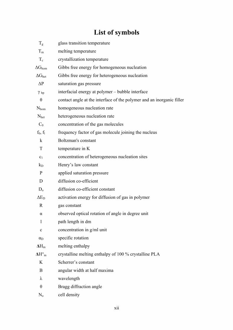
List of symbols Tg glass transition temperature
Tm melting temperature
Tc crystallization temperature
ΔGhom Gibbs free energy for homogeneous nucleation
ΔGhet Gibbs free energy for heterogeneous nucleation
ΔP saturation gas pressure
γ bp interfacial energy at polymer – bubble interface
θ contact angle at the interface of the polymer and an inorganic filler
Nhom homogeneous nucleation rate
Nhet heterogeneous nucleation rate
C0 concentration of the gas molecules
f0, fl frequency factor of gas molecule joining the nucleus
k Boltzman's constant
T temperature in K
c1 concentration of heterogeneous nucleation sites
kD Henry’s law constant
P applied saturation pressure
D diffusion co-efficient
Do diffusion co-efficient constant
ΔED activation energy for diffusion of gas in polymer
R gas constant
α observed optical rotation of angle in degree unit
l path length in dm
c concentration in g/ml unit
αD specific rotation
ΔHm melting enthalpy
ΔH°m crystalline melting enthalpy of 100 % crystalline PLA
K Scherrer’s constant
Β angular width at half maxima
λ wavelength
θ Bragg diffraction angle
No cell density
xiii
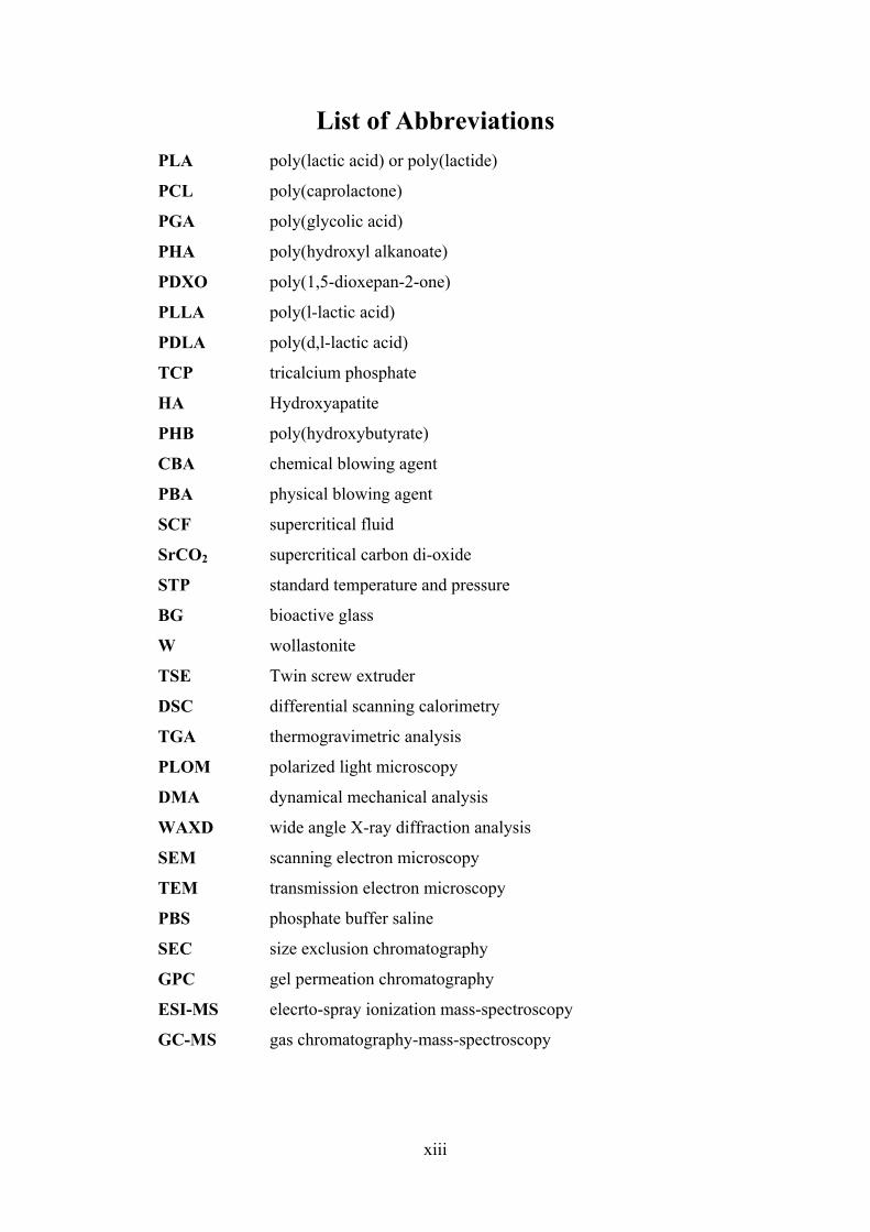
List of Abbreviations PLA poly(lactic acid) or poly(lactide)
PCL poly(caprolactone)
PGA poly(glycolic acid)
PHA poly(hydroxyl alkanoate)
PDXO poly(1,5-dioxepan-2-one)
PLLA poly(l-lactic acid)
PDLA poly(d,l-lactic acid)
TCP tricalcium phosphate
HA Hydroxyapatite
PHB poly(hydroxybutyrate)
CBA chemical blowing agent
PBA physical blowing agent
SCF supercritical fluid
SrCO2 supercritical carbon di-oxide
STP standard temperature and pressure
BG bioactive glass
W wollastonite
TSE Twin screw extruder
DSC differential scanning calorimetry
TGA thermogravimetric analysis
PLOM polarized light microscopy
DMA dynamical mechanical analysis
WAXD wide angle X-ray diffraction analysis
SEM scanning electron microscopy
TEM transmission electron microscopy
PBS phosphate buffer saline
SEC size exclusion chromatography
GPC gel permeation chromatography
ESI-MS elecrto-spray ionization mass-spectroscopy
GC-MS gas chromatography-mass-spectroscopy