Embed Size (px)
Citation preview
Instructions for use
Title STUDIES ON ECHINOCOCCOSIS XXIV : AGE DIFFERENCE IN RESISTANCE TO INFECTION WITHECHINOCOCCUS MULTILOCULARIS IN AKR STRAIN OF MOUSE
Author(s) KAMIYA, Haruo
Citation Japanese Journal of Veterinary Research, 20(3), 69-76
Issue Date 1972-09
DOI 10.14943/jjvr.20.3.69
Doc URL http://hdl.handle.net/2115/1995
Type bulletin (article)
File Information KJ00003418354.pdf
Hokkaido University Collection of Scholarly and Academic Papers : HUSCAP
Jap. J. vet. Res., 20, 69-76 (1972)
STUDIES ON ECHINOCOCCOSIS XXIV
AGE DIFFERENCE IN RESISTANCE TO INFECTION WITH ECHINOCOCCUS MULTILOCULARIS
IN AKR STRAIN OF MOUSE*
Haruo KAMIYA
Department of Parasitology Faculty of Veterinary Medicine
Hokkaido University, Sapporo, Japan
(Received for publication, July 24, 1972)
Age difference in resistance to infection with larval Echinococcus mul#locularis
LEUCKART, 186:i, was investigated in addition to sex difference of AKR strain mice.
1) Mice 29-day-old or younger were highly susceptible to larval Echinococcus
multilocularis.
2) Forty-eight-day-old mice showed the highest resistance to this cestode,
and R3- and 148-day-old mice were less resistant than 48-day-old mice.
:3) Sex difference in resistance was not evident.
4) Susceptibility was 100% in AKR mice.
INTRODUCTION
About larval Echinococcus spp., SCHWABE et al. (1959) reported age resistance
to secondary intraperitoneal infection to unilocular echinococcus in white mice.
The present author could not find any report of age difference in resistance
to infection with larval Echinococcus multilocularis LEUCKART, 1863, in experi
mental animals. He therefore examined age resistance to infection with this
parasite in AKR mice by the oral infection of eggs.
MA TERIALS AND METHODS
AKR mice used for the experiment came from the breeding stocks of
Experimental Animals Laboratory, Hokkaido University. These mice were bred
in a breeding room with a thermostat for keeping the room temperature at
24°C. Suckling mice were weaned at 3 weeks after birth. The mice marked
individually were segregated into their sexes and the mice of same age were
kept in a cage. A syringe with a cannula was used to inoculate each mouse
* This work is a part of Master's thesis by the author at the Graduate School of
Veterinary Medicine, Hokkaido University.
70 KAMIYA, H.
with approximately 330 eggs of Alaskan sHain of- E. multilocularis in physio
logical saline, .. The .egg~ were obta~ned from adul.t ~apeworms of ,experimental
dogs give!} hepatic foci of mice infected experimentally with .larval cestode. I!, ., , ' '. ,~\. ' ~ , I,
The inoculated mice were killed by bleeding 30, 60 and 90 days after the , . ' . ~ , , .
inoculation, echinococcal foci were investigated carefully with naked eyes, the
livers were weighed and the diameter of the large cysts was measured. The
tissue materials were fixed with 10% formalin solution, and paraffine sections for microscopy were stained with hematoxyline-eosin.
RESULTS
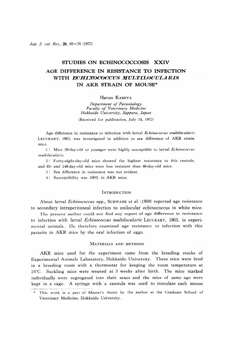
1 Susceptibility
All cases were infected by E. multilocularis and all the echinococcal foci were limited to the liver (tab. 1).
TABLE 1 Susceptibility in AKR mice
AGE AT SUSCEPTIBILITY* INOCULA TION ~~------ --- ---------- ---- ------
"
(Days) Male Female Total
22 4/4 5/5 9/9
29 3/3 3/3 6/6
48 4/4 3/3 7/7
83 4/4 4/4 8/8
148 4/4 :3/3 7/7
Total 19/19 18/18 37/37
*. Number of mice positive/examined
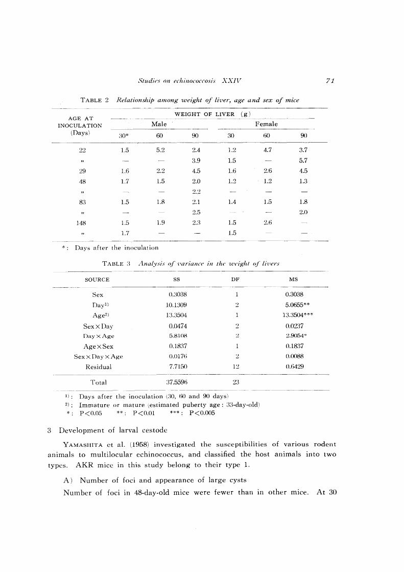
2 Weight of the liver
In the mice 30 days after the inoculation, the weight of livers d~d not increase clearly irrespective of ages, but at 60 and 90 days after the inoculation,
the young mice, less than 30 days old when inoculated, showed apparent
enlargement of the liver (tab. 2).
, The analysis of variance presented in table 3 was made between the imma
ture (22-' and 29-day-old) and the mature (48- and 83-day-old). In this analysis,
'the sexes did not show significant difference in weight of the liver, even at
O~05 level of significance. But it was very significant that the weight of livers
'of' the immature mice was considerably greater than those of the mature (P < 0.005). And, as expected, the increase of weight of livers as the days passed
after the inoculation showed a significant difference (P<O.Ol).
Studies on echinococcosis XXIl Y 71
TABLE 2 Relationship among weight of liver, age and sex of mice
AGE AT WEIGHT OF LIVER (g)
INOCULA TION Male Female (Days) :$0* 60 90 30 60 90
22 1.5 5.2 2.4 1.2 4.7 3.7
3.9 1.5 5.7
29 1.6 2.2 4.5 1.6 2.6 4.5
48 1.7 1.5 2.0 1.2 1.2 1.3
2.2
83 1.5 1.8 2.1 1.4 1.5 1.8
2.5 2.0
148 1.5 1.9 23 1.5 2.6
1.7 1.5
*. Days after the inoculation
TABLE:$ Ana(ysis of '('arianCl' in the '((·cigh! of li'l'ers
SOURCE SS DF MS
Sex O.303S 1 0.3038
Day1) 10.1309 2 5.0655**
Age2 ) 13.3504 1 13.3504***
SexxDay 0.0474 2 0.0237
DayxAge 5.8108 :2 2.9054*
AgexSex 0.18:37 1 0.1837
Sex X Day X Age 0.0176 2 O.OOBR
Residual 7.7150 12 0.6429 -------- --- --------~--------
Total 37.5596 23 ------ ------------- ------- ------ -------
1): Days after the inoculation (:-)0, 60 and 90 days)
2): Immature or mature ~estimated puberty age: :1:i-day-old) *. P<O.05 **: P<O.Ol ***: P<0.OO5
3 Development of larval cestode
YAMASHITA et al. (1958) investigated the susceptibilities of various rodent
animals to multilocular echinococcus, and classified the host animals into two
types. AKR mice in this study belong to their type 1.
A) Number of foci and appearance of large cysts
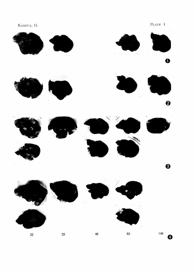
Number of foci in 48-day-old mice were fewer than III other mice. At 30

72 KAMIYA, H.
days after the inoculation, some cysts showed an enlargement only in 22-day-old
male and female mice. At 60 days, almost all cases except both sexes of the
48-day-old and 83-day-old female had large cysts more than 4 mm in diameter.
At 90 days, large cysts appeared in all cases except in a 48-day-old female, and
cysts of young mice increased in size as compared with those in others (figs. 1-4).
B) Brood capsule and scolex formations
1) 22-day-old mice Brood capsules had already been detected in both
sexes 30 days after the inoculation, and brood capsules III a male manifested
scolex formation. At 60 days, matured scolices appeared in both sexes. At 90
days, multilocular cysts were fully developed (figs. 5--10).
2) 29-day-old mice Brood capsules and scolices appeared in both sexes
60 days after the inoculation. At 90 days, brood capsules and immature scolices
increased in number and size in the male, although a few matured scolex could
be found in the female.
3) 48-day-old ITIice DevelopITIent of ITIultilocular cysts was slower than
in the other cases. At 90 days, initial brood capsules without scolex were
recognized in a male (figs. 11-16).
4) 83-day-old mice Brood capsules with immature scolices were detected
60 days after the inoculation in males, although brood capsule formation did
not appear in a feITIale. At 90 days, brood capsule and scolex forITIations were
recognized in males and a female, but other female showed no scolex formation.
Mature scolices did not appear.
5) 148-day-old mice In cases at 60 days after the inoculation, brood
TABLE 4 Relationship among brood capsule formation, scolex formation, age and se.T of the host
MALE FEM},.LE AGE AT
INOCULA TION 30 60 90 30 60 90
(Days) B S B S B S B S B S B S
22 1/1 1/1 1/1 1/1 2/2 2/2 2/2 0/2 1/1 1/1 2/2 2/2*
29 0/1 0/1 1/1 1/1 1/1 1/1 0/1 0/1 1/1 1/1 III 1;1
48 0/1 0/1 0/1 011 1/2 0/2 0/1 0/1 0/1 011 0/1 011
83 011 0/1 1/1 1/1 2/2 2/2 0/1 0/1 0/1 0/1 2/2 1/2
148 0/2 0/2 011 0/1 1/1 1/1 0/2 0/2 1/1 1/1
* . No. of cases showing brood capsule and scolex formations/examined B: Brood capsule formation
s: Scolex formation
Studies on echinococcosis XXIV 73
capsules and scolices were detected in a female. At 90 days, immatured scolices
appeared in a male but matured scolices were not recognized.
Brood capsule formation took place in parallel with scolex formation in all
cases, except the 48-day-old, and mature scolices were found for the first time
in 22-day-old mice 60 days after the inoculation. Up to 90 days, matured
scolices appeared only in young mice.
Relationships among brood capsule formation, scolex formation,! age and
sex of the hosts are summarized in table 4.
DISCUSSION "
The multilocular echinococcus is the larval stage of E. multilocular-is
LEUCKART, 1863, its intermediate host is Rodentia and this species is different
from E. granulosus (BA TSCH, 1786). These facts have been clarified since the
middle of 1950 by RAUSCH & SCHILLER (1951, 1956), VOGEL (1955), YAMASHITA
et a1. (1956, 1958) and others. Histogenetic study of larval E. multilocularis in
rodents has been carried out by several authors. OHBA YASHI (1960) clarified
that the development of this larval cestode became evident with progress of
days after the inoculation. Consequently, it became easy to study multilocular
echinococcosis in experimental animals.
YAMASHITA et a1. (1958) investigated the susceptibilities of various rodent
animals and two types were classified by them according to combination of
morphology of the larva and host tissue reactions. In type 1, the larva develops
rapidly, individual cysts are large in size, scolices are recognizable 1.5-2.5 months and host tissue reactions are slight in degree. Strain AKR mIce
examined in this study belong to this type.
Concerning sex differences, OHBA Y ASHI & SAKAMOTO (1966) reported that
females of two mouse strains exhibited remarkable resistance to this cestode in
varied degrees. AKR mice in this study showed a susceptibility of 100%. In
male and female mice at same age, the appearance of scolices in the males was
slightly earlier than in the females, but sex difference in resistance was not evident.
YAMASHITA et a1. (1958) reported that immature scolices were recognized 2
months after the inoculation in 4-month-old AKR mice and the development at
3 months was still similar to that at 2 months, and that numerous fully de
veloped brood capsules and scolices were found in cases at 5 months after the
inoculation. Comparing the results of YAMASHITA et a1. r)958) with the results
of this study (tabs. 2--4 & figs. 1--16), it is evident that age differences in
resistance do exist in liver enlargement, appearance of large cysts, formation
of brood capsules and subsequent formation of scolices.
74 KAMIYA, H.
About age difference in . resistance to infection with larval Echinococcus
spp., SCHWABE et al. (1959) reported that white mice of unknown sex, which
were inoculated at 48 days of age or younger, were highly susceptible to intra
peritoneal infection with scolices of E. granulosus. In the present study,
however, 48-day-old mice showed the highest resistance to infection with the
multilocular larva. OGLE (1934) discussed the relationship. between sexual maturity of female mice and their environment, especially the influence of tempera
ture. He reported that the mice kept in 22- 25cC reached sexual maturity about
33 days after the birth. On the other hand, the mice kept in 27.S-29c C required about 46 days to reach the sexual maturity. So, the difference between
the results of SCHWABE et al. (1959) and those of the present author might be
caused by the temperature of breeding room.
As for other larval cestodes, GREENFIELD (1942) suggested age resistance in
the albino rat to Cysticercus fasciolaris, and Dow & JARRETT (1960) reported
age,sex and strain differences in susceptibility to this parasite in mice.
Judging from the results obtained in this study, the phenomenon influenced
by the age of host, as same as the strain and sex of the host, must be. also taken into consideration in future experiments with multilocular echinococcosis.
ACKNOWLEDGEMENTS
The author wishes to express his gratitude to Prof. J. YAMASHITA and Dr. M. OHBA YASHI of this Department for their kind direction and review. Further thanks are offered to Prof. T. ISHIKAWA, Department of Veterinary Obstetrics, Faculty of Veterinary Medicine, Hokkaido University, for the statistical analysis to this study.
Studies on echinococcosis XXIV 75
REFERENCES
1) Dow, C. & JARRETT, W. F. H. (1960): Exptl Parasitol., 10, 72
2) GREENFIELD, S. H. (1942): J. Parasit., 28, 207
3) GLLE, C. (1934); ..A.m. J. Physiol., 107, 628
4) OHBA Y ASH!, M. (1960): Jap. J. vet. Res., 8, 134
D) OHBAYASHI, M. & SAKAMOTO, T. (966): Ibid., 14, 65 . "
6) RAUSCH, R. &: SCHILLER, E. L. (1951): Science, 113, 57
7) RAUSCH, R. & SCHILLER, E. L. (1956): Parasitology, 46, 395
8) SCHWABE, C. W., SCHINAZI, L. A. & KILEJIAN; A. (1959): A.m. J. torp. l'vled. Hyg.,
8, 29
9) VOGEL, H. (1955): Dt. med. Wschr., 80, 931 I •
10) YAMASHITA, J., OHBA YASHI, M. & KONNO, S. ~1956): Jap. J. ·I'et. Res., 4, 123
11) Y AMASHIT A, ]., OHBA Y ASHI, M., KITAMURA, Y.,'SUZUKI, K. & OKUGI, M. (1958):
Ihid., 6, 135

EXPLANATION OF PLATES
PLATE I Livers at 60 and 90 days after the inoculation x 7/10
Fig. 1
Fig. 2
Fig. 3
Fig. 4
The number under the photograph shows age at inoculation by
days.
Livers of males at 60 days
Livers of females at 60 days
Livers of males at 90 days
Livers of females at 90 days
KAMIYA, H. PLATE I
o
22 29 48 83 148 o
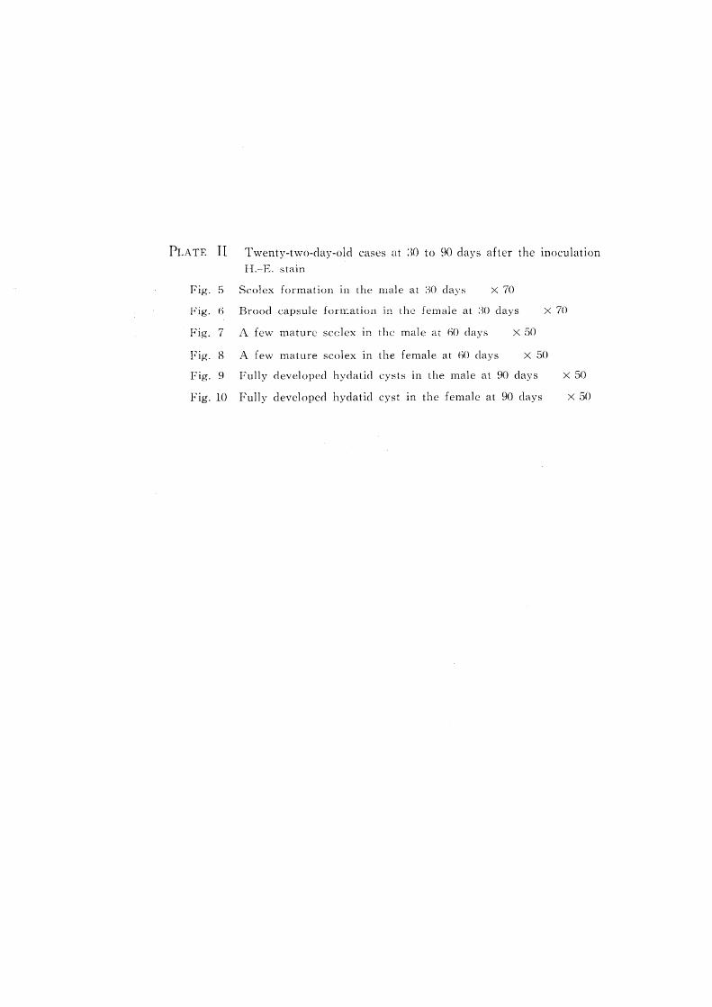
PLATE II Twenty-two-day-old cases at :m to 90 da:ys after the inoculation H.-E. stain
Fig. 5 Scolex formation In the male at :)0 days X 70
Fig. 6 Brood capsule forn:.ation In the female at :30 days X 70
Fig. 7 A few mature scclex In the male at 60 days X 50
Fig. 8 A few mature scolex In the female at m days X 50
Fig. 9 Fully developed hydatid cysts in the male at 90 days X 50
Fig. 10 Fully developed hydatid cyst In the female at 90 days X 50
KAMIYA, H. PLATE II
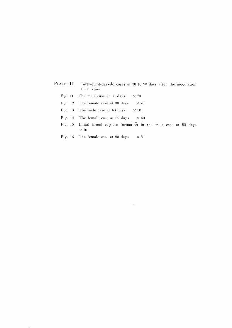
PLATE III
Fig. 11
Fig. 12
Fig. 1:~
Fig. 14
Fig. 15
Forty-eight-day-old cases at :30 to 90 day:,,; after the inoculation H.-E. stain
The male ca:,,;e at ;30 day:,,; X 70
The female case at :m day:,,; X 70
The male ca:,,;c at 60 days X 50
The female case at ()O days X 50
Initial brood capsule formatio~ in the male case at 90 clays
X 70
Fig. 16 The female case at 90 days X 50
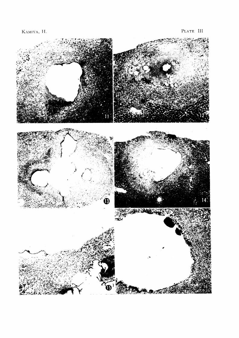
KAMIYA, 11. PLATE III
'"~":t;>-j N
. :"'>'~>~ .,
![Prevalence of Cystic Echinococcosis in Selected Pastoral ... · Echinococcosis is an endemic zoonotic infection found throughout the developing world [1]. It is a neglected emerging](https://img.pdfslide.us/doc/110x75/5f06a2977e708231d418f940/prevalence-of-cystic-echinococcosis-in-selected-pastoral-echinococcosis-is-an.jpg)