Embed Size (px)
Citation preview

STUDIES ON COLCHICINE DERIVATIVES I . Toxicity in Mice and Effects on Mouse Sal-coma 180
BENJAMIN GOLDBERG, PH.D., LOUIS G. ORTEGA, LT., M.c., A.u.s., ABRAHAM GOLDIN, PH.D.,* GLENN E. ULLYOT, PH.D., and EMANUEL B. SCHOENBACH, M.D.
A LTHOUGH colchicine and its deriva- tives have been studied extensively, there have been relatively few reports on the com- parative toxicity of these c0mpounds.5~ 1 O s 1 2 . 13
The present investigation was designed to study the relation of toxicity to chemical structure of a closely related group of col- chicine derivatives. An attempt has been made to obtain relatively precise median lethal dosages (LDbO) in a particular strain of mice. The data thus obtained served as a basis for a comparative study of the antimitotic and other cellular and histological effects of equi- toxic doses.4$ 9 By such procedures, one can differentiate more quantitatively between systemic toxicity and specific biological effects, and relate these to molecular structure.
In studies of the action of colchicine on neoplasms, two outstanding effects have been noted: (1) mitotic arrest (due to inhibition of spindle formation)14 which is often obscured at higher concentration by necrosis, and (2) hemorrhage, which generally has been re- garded as the result of injury to the proliferat- ing capillary endothelium.8. l4 Using these effects as criteria of activity, an attempt was made in this investigation to ascertain the
From the Department of Preventive Medicine, The Johns Hopkins University School of Medicine, Baltimore, and the Biology Section, Medical Division, Army Chemical Center, Maryland; and the Smith, Kline and French Laboratories, Philadelphia, Pennsylvania.
This work was aided in part by a grant from the American Cancer Society to the Department of Preventive Medicine, The Johns Hopkins University School of Medicine, recommended by the Committee on Growth of the National Research Council.
These studies form part of a joint project on the chemotherapy of cancer being conducted at The Johns Hopkins University School of Medicine, De- partment of Preventive Medicine, and the Medical Division, Army Chemical Center, Maryland.
The photomicrographs were prepared by Arthur J. Fisk.
*Present address: Clinical Research Unit, National Cancer Institute, Marine Hospital, Baltimore 11, Maryland.
Received for publication, August 8, 1949.
minimum effective fractions of the LDSo of the various compounds. The compounds which would be considered more desirable from the standpoint of possible therapeutic use are those which are effective far below the LDSo.
METHODS
Throughout these experiments male albino mice, Carworth Farms CFI, weighing 15 to 35 gm., were used. The mice were kept in wire-mesh cages in groups of five, and were given water and Dog Friskies ad libitum.
Trial concentration ranges, for LDSO de- terminations, were obtained from information available in the literature. Dosage levels were run at geometrically spaced intervals, each lower dose being three quarters of the preced- ing dose. In the event that satisfactory data could not be obtained thereby, additional concentration ranges were used. In most instances, the experiments were repeated on three occasions. In each experiment, seven to nine dosage levels were employed with five mice at each level. The diluent of choice was 0.9 per cent NaCl, 10 per cent aqueous gum acacia being used as a suspending agent for the insoluble compounds. Insoluble sub- stances were first triturated in a small amount of the suspending agent and then diluted to correct volume. Care was exercised to keep the material in suspension prior to injection by continuous stirring and agitation. All mice received a single intraperitoneal injec- tion. The total volume of each injection in milliliters was 1 per cent of the weight of the mouse in grams. The mice were observed and mortality recorded for a period of ten to thirteen days after an injection of the com- pounds.
Approximate LDSO figures were obtained by plotting the data on log probability paper

STUDIES ON COLCHICINE DERIVATIVES. I Goldberg, et al.
TABLE 1
TOXICITY DATA ON COLCHICINE DERIVATIVES*
[125
____ Min. eff. anti-
LDbo tumor dose LD.50 Compound Mol. wi. mg.1k-g. pM.IKg. &.lh‘p-. Vehicle
N-benzoyl TMCA
TMCA
N-acetyl TMCA (colchiceine)
N-acetyl
TMCA methyl
colchinol
ether
N-benzoyl TMCA methyl ether
N-acetyl TMCA methyl ether (colchicine)
NHCOCGHs
(NHCOCH, R
R’ NHCOCH, 10.
NHCOCH3
RIOCH,
447.5 > 700 >1600 - 10% G u m acacia
loyo, Gum acacia - 343 200 510
10% Gum 385.4 84 213 50 acacia
10% Gum 357.4 56 157 14 acacia
525.8 ~461 t 120 6 Saline
10% Gum 461 32 69 17 acacia
399 3.5 9 4.5 Saline
NOTE: The structures assigned to colchicine and its derivatives are tentative and as yet indefinite. The original structure by Windaus” may have to be modified in the light of the evidence that the “B” ring may be seven-membered: Barton, Cook, and Loudon,3 and previous papers. Dewar7 has suggested that the “C” ring is also seven-membered, a point of view that has been supported by Arnstein, Tarbell, Huang, and Scott.2
t d-Tartrate derivative used. Value calculated for simple compound.
when feasible. The data was then analyzed by the method of Reed and Muench.
In the tumor studies, the transplantable tumor, sarcoma 180, was employed and im- planted bilaterally into 2- to 3-month-old male Carworth Farms, CF1, mice. The tumor fragments were placed subcutaneously mid- way between the axillary and inguinal regions. The effect of the drugs was observed after a single injection of each compound had been administered intraperitoneally to mice bear- ing seven- or eight-day-old tumors. The mice were sacrified at 6 hours* after injection and the tumors excised and fixed in 10 per cent
*Mice, injected simultaneously, were sacrificed at twenty-four and forty-eight hours for other experi- ments.
_-
formalin or Bouin’s fluid. The tumors were then sectioned at 5 microns and stained with Harris’s hematoxylin and eosin. Gross obser- vations on the amount of hemorrhage and necrosis were recorded at the time of autopsy, and these findings were supplemented by microscopic examination of sectioned tumors. Mitotic counts were performed on ten ad- jacent, non-necrotic, oil immersion fields for each tumor. Appropriate controls, injected with the vehicle, were similarly examined.
RESULTS AND DISCUSSION
The results are summarized in Table 1, where the least toxic compound is listed first and the remainder are listed in the order of

1261 CANCER January 1950
TABLE 2
R ~ S U M ~ OF TOXICITY DATA (MG./KG.) OF COLCHICINE DERIVATIVES
NHCOCsHb
R{o€3
NHz
.{OH
(NHCOCH, R
>700
20 200
84
R’ NHCOCH? 100 (8-10’C.) 56
(NHCOCeH5
Ri\OCH,
< 20
> 20 32
200
30
200
>10
>12.5
10
5
< 25
2-4 (30-32°C.) 3.5 5.0 0.5 to 1 .O KiNHCOCH3 OCHa 60-100 (8-10°C.)
*See Table 1
their increasing toxicity. I t may be observed that there is an approximate two hundredfold increase in order of toxicity on a molar basis from the least to the most toxic compound, all of which apparently can be attributed to fairly simple modifications (a) methylation of the hydroxy methylene substituent of the “C” ring to an enol methyl ether, and (b) acylation of the amino group in the “B” ring.
Excluding N-acetyl colchinol, the tri- methylcolchicinic acid (TMCA) structure may be regarded as basic to the compounds here investigated. Acetylation of the amino group on the “B” ring (colchiceine) results in a compound somewhat more than twice as toxic. Methylation of the hydroxy methylene group of the “C” ring, to form the methyl ether yields a compound (TMCA methyl ether) about four times as toxic. Both these substitutions together (colchicine) increase the toxicity by a factor greater than fifty.
Methylation of the hydroxy methylene group to form the enol methyl ether deriva- tive has, in all cases studied in this report, rendered the analogs more toxic. Acylation (i.e., acetylation or benzoylation) in general has also increased toxicity except in the case of N-benzoyl trimethylcolchicinic acid.
Precise data on solubilities of these com- pounds as related to toxicity are not avail- able but some tentative statements may be made at this time. Acetylation induced greater toxicity than benzoylation. Formation of the enol methyl ethers from the hydroxy meth- ylene groups has given a similar result. In these instances, greater toxicity was associated with greater solubility. This may be an over- simplified interpretation as illustrated by the relatively high toxicity of N-benzoyl tri- methylcolchicinic acid methyl ether, which is not soluble in water.
In Table 2, the toxicity data noted by other investigators are compared with those ob- tained in this study. Colchicine clearly enough is the most toxic compound of the series with the exception of the rather anomal- ous results obtained on frogs (and batsll) a t low temperatures. Furthermore, removal of the enol methyl ether group on Ring C and the acetyl group on Ring B, gives TMCA, which is less toxic than either of the afore- mentioned derivatives in the case of the rat and mouse. However, Fuhner states that TMCA is more toxic than colchiceine to cats and dogs. A benzoyl group in place of the acetyl group appears to result in a decreased
STUDIES ON COLCHICINE DERIVATIVES. I Goldberg, et al. [I27
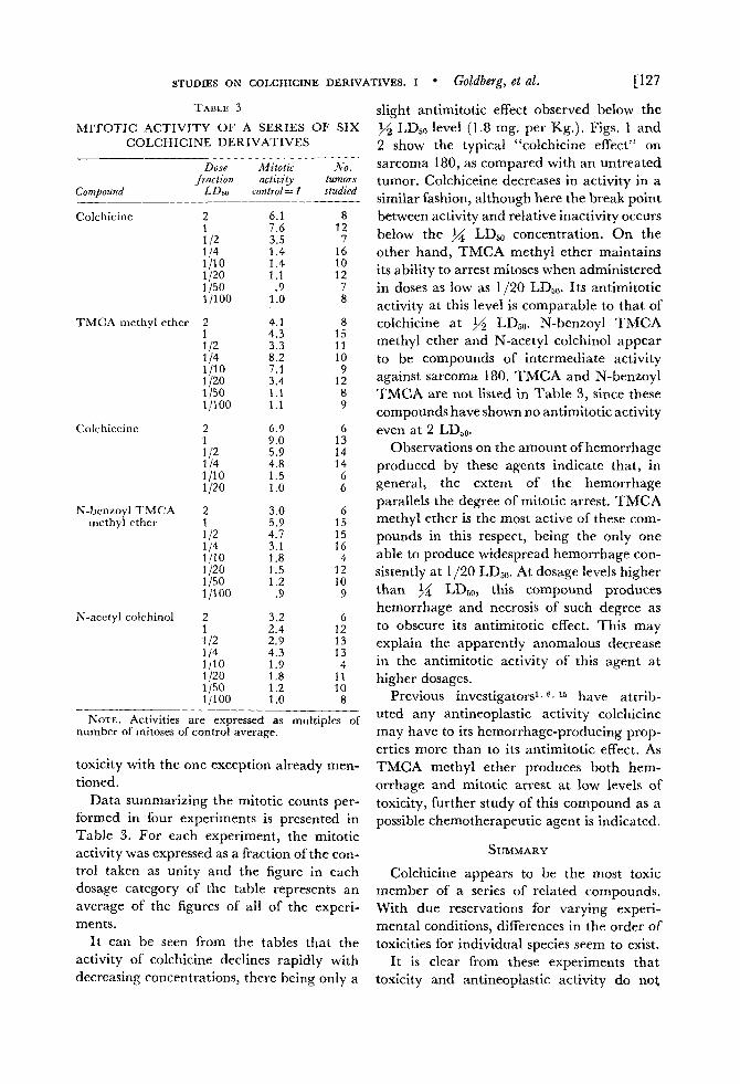
TABLE 3
MITOTIC .ACTIVITY OF A SERIES OF SIX COLCHICINE DERIVATIVES
Dose Jraction
Corn Dound LDso
Mitotic NO. activity tumors
control= 1 studied
Colchicine
TMCA methyl ether
Colchiceine
N-lienzoyl TMCA methyl ether
N-acetyl colchinol
2 1 1 12
1/10 1/20
1 /loo
1 14
1/50
2 1 1 I2
1/20
l j l 00
:;0
1/50
2 1 1 I2
1/10 1 j20
1 14
2 1 1 I2 114 1/10 1/20
1 1100 1 /50
2 1 1 12
1/10 1/20
1 jl00
114
1/50
6.1 8 7.6 12 3.5 7 1.4 16 1.4 10 1.1 12 .9 7
1 .o 8
4.1 8 4.3 15 3.3 11 8.2 10 7.1 9 3.4 12 1 .I 8 1.1 9
6.9 6 9.0 13 5.9 14 4.8 14 1.5 6 1 .o 6
3.0 6 5.9 15 4.7 15 3.1 16 1.8 4 1.5 12 1.2 10
.9 9
3.2 6 2.4 12 2.9 13 4.3 13 1.9 4 1.8 11 1.2 10 1 .o 8
NOTE. Activities are expressed as multiples of number of mitoses of control average.
toxicity with the one exception already men- tioned.
Data summarizing the mitotic counts per- formed in four experiments is presented in Table 3. For each experiment, the mitotic activity was expressed as a fraction of the con- trol taken as unity and the figure in each dosage category of the table represents an average of the figures of all of the experi- ments.
I t can be seen from the tables that the activity of colchicine declines rapidly with decreasing concentrations, there being only a
slight antimitotic effect observed below the LDso level (1.8 mg. per Kg.). Figs. 1 and
2 show the typical “colchicine effect” on sarcoma 180, as compared with an untreated tumor. Colchiceine decreases in activity in a similar fashion, although here the break point between activity and relative inactivity occurs below the M LD,, concentration. O n the other hand, TMCA methyl ether maintains its ability to arrest mitoses when administered in doses as low as 1/20 LD50. Its antimitotic activity at this level is comparable to that of colchicine at LDbO. N-benzoyl TMCA methyl ether and N-acetyl colchinol appear to be compounds of intermediate activity against sarcoma 180. TMCA and N-benzoyl TMCA are not listed in Table 3, since these compounds have shown no antimitotic activity even at 2 LDjo.
Observations on the amount of hemorrhage produced by these agents indicate that, in general, the extent of the hemorrhage parallels the degree of mitotic arrest. TMCA methyl ether is the most active of these com- pounds in this respect, being the only one able to produce widespread hemorrhage con- sistently at 1 /20 LD50. At dosage levels higher than LDLO, this compound produces hemorrhage and necrosis of such degree as to obscure its antimitotic effect. This may explain the apparently anomalous decrease in the antimitotic activity of this agent at higher dosages.
Previous investigators’, 6 , 15 have attrib- uted any antineoplastic activity colchicine may have to its hemorrhage-producing prop- erties more than to its antimitotic effect. As TMCA methyl ether produces both hem- orrhage and mitotic arrest a t low levels of toxicity, further study of this compound as a possible chemotherapeutic agent is indicated.
SUMMARY
Colchicine appears to be the most toxic member of a series of related compounds. With due reservations for varying experi- mental conditions, differences in the order of toxicities for individual species seem to exist.
I t is clear from these experiments that toxicity and antineoplastic activity do not
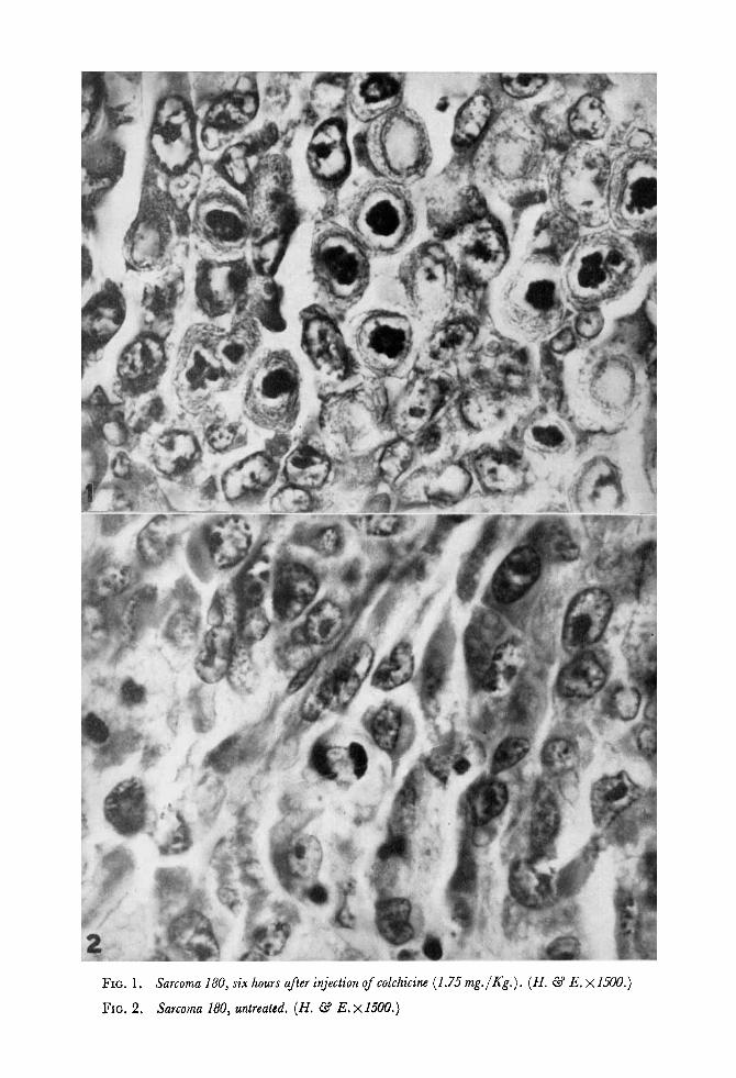
FIG. 1. Sarcoma 180, s i x hours after injection ofcolchicine (1.75 mg./Kg.). (H. @ E. ~1.500.)
FIG. 2 . Sarcoma 180, untreated. ( H . &3 E. ~ 1 5 0 0 . )

STUDIES ON COLCHICINE DERIVATIVES. I. Goldberg, et al. [129
necessarily change in parallel manner with permitted retention of antineoplastic activity. modification in chemical structure. In the The decrease in toxicity was seventeen- series: colchicine, N-benzoyl TMCA methyl fold (9 to 157 micromoles per kilogram) while ether, TMCA methyl ether, N-acetyl col- the minimum effective antineoplastic doses chinol, structural modifications were made varied only fourfold and ranged fi-om 4.5 to that resulted in reduced toxicity, but that 17 micromoles per kilogram.
REFERENCES
1. ANDERVONT, H. B.: Effect of colchicine and bacterial products on transplantable and spontane- ous tumors in mice. 3. Nat. Cancer Inst. 1: 361-366, 1940.
2. ARNSTIIIN, H. R. V.; TARBELL, D. S.; HUANG, H. T., and SCOTT, G. P.: The structure of ring C of colchicine. 3. Am. Chem. SOC. 70: 1669, 1948.
3. BARTON, N.; COOK, J. W., and LOUDON, J. D.: Colchicine and related compounds. Part V. The structure of Windaus’s deaminocolchinol methyl ether. 3. Chem. Sac. [1945]: 176-180, 1945.
4. BERGNER, A. D.: Studies on colchicine deriva- tives. 111. Effect on mitotic activity ofmouse spermato- gonia. Cancrr 3: 134-141, 1950.
5. BRUES, A. M., and COHEN, A,: The effects of colchicine and related substances on cell division. Riochem. J . 30: 1363-1368, 1936.
6. BRUES, A. M.; MARBLE, B. B., and JACKSON, E. B.: Effects of colchicine and radiation on growth of normal tissues and tumors. Am. 3. Cancer 38: 159-168, 1940: addendum 38: 190, 1940.
7. DEWAR, M. J. S.: Structure of colchicine. .Natnre 155: 141-142, 1945.
8. DUSTIN, A,-P., and CHODKOWSKI, K.: 6tude de la cicatrisation par la reaction colchicinique. Compt. rend. de I’Assoc. d. anat. 32: 170-176, 1937.
9. FLEISCHMANN, W., and ULLYOT, G. E.: Studies on colchicine derivatives. 11. Effect on mitotic activity of the corneal epithelium. Cancer 3: 130-133, 1950.
10. FUHNER, H.: Pharmakologische Untersuch- ungen iiber das Colchicin und seine Derivate. Arch. f . exper. Path. u. Pharmakol. 72: 228-238, 1913.
11. HAUSMANN, W.: Ueber den Einfluss der Tem- peratur auf die Inkubationszeit und Antitoxinbildung nach Versuchen an Winterschlafern. ilrch. f . d. ges. Physiol. 113: 317-326, 1906.
12. JACOBJ, C.: Pharmakologische Untersuchung uber das Colchicumgift. Arch. f. exper. Path. u. Phar- makol. 27: 119-157, 1890.
13. LIPPS, H.: Pharmakologische Untersuchungen in der Colchicinreihe. 11. Ueber die Wirkung einiger Colchicinderivate. Der Kapillargiftmechanismus der Colchicinwirkung. Arch. f . exprr . Path. u. Pharmakol. 85: 235-255, 1920.
14. LUDFORD, R . J.: The action of toxic substances upon the division of normal and malignant cells in vitro and in vivo. Arch.f. exper. zeelljarsch. 18: 41 1-441, 1936.
15. LUDFORD, R. J.: Colchicine in the experimental chemotherapy of cancer. 3. Nat. Cancer h i . 6: 89-101, 1945.
16. REED, L. J., and MUENCH, H.: A simple method of estimating fifty per cent endpoints. .4m. 3. Hyg. 27: 493-497, 1938.
17. WINDAUS, A,: Untersuchungen iiber die Kon- stitution des Colchicins. Ann. d. Chem. 59-75, 1924.