Embed Size (px)
Citation preview

STUDIES ON CHANDLERELLA HAWKiNCf, A FILARIAL PARASITE OF CROW
ABSTRACT
Mthesis SUBMITtED' '
IN FULM.LMENT:OF THE:RHQUIREMENTS FOR THE DEGREE OF
DOCTOR .OF PHlLOSdPHY
I N .
/ n o m R Y
SHAHNAZ BANO
SECTION OF PARASITOLOGY DEPARTMENT OF ZOOLOGY
ALIGARH MUSLIM UNIVERSITY ALIGARH
January, 1981

ABSTRACT
The thes i s embodies the r e s u l t s of the s tud i es
on Chand l e r e l l a hauking i , a f i l a r i a l p a r a s i t e of Ind ian
jung l e crou, Corvus macrorhynchos ( U a g l e r ) . Only f our
a s p e c t s , morphology, h i s t o l o g y , h i s t ochemis t r y and
i n ^ i t r o cu l ture have been taken up.
The morpho log i ca l s tud i es inc lude a re d e s c r i p t i o n
of adul t worm u i th an add i t i on of a feu minor d e t a i l s
in the adu l t . The s t ruc tu r e s descr ibed in m i c r o f i l a r i a
are the nuclear s t r u c t u r e s , c epha l i c s t r u c t u r e s ,
pharyngea l thread , Innenkorper and a l so occurrence of
tuo forms of m i c r o f i l a r i a e in blood of crou.
The nuclear s t ruc tu res naue been s tud ied u i th
s p e c i a l r e f e r ence to nuclear landmarks which are of
g r ea t taxonomic vyalu^.
Sometimes the nuclear s t ruc tu r e s do not s u f f i c e
f o r i d e n t i f i c a t i o n of genera and s o e c i e s , the c e p h a l i c
s t r u c t u r e s , pharyngeal thread and Innenkorper have a l so
been s tud i ed .
I t has a lso been proved that the tuo forms of
m i c r o f i l a r i a e , the long and the s h o r t , present in the
blood of crou belono to the same s p e c i e s , C. hauking i .

The s te reoscan s tud ies of the c u t i c u l a r s t ruc tu r e s
of the adult and the m i c r o f i l a r i a have heen done as these
are of g rea t taxonomic importance in c l a s s i f i c a t i o n of
uorms.
The h i s t o l o g i c a l s tud i es inc lude the study of
h i s t o l o g i c a l f e a t u r e s of the body u a l l , musculature,
a l imentary canal and reproduct iue organs as some of these
s t r u c t u r e s prov ide a taxonomic t o o l at var ious l e v e l s .
Among h i s tochemica l •^^tudies, four enzymes, v i z ;
ac id phosphatase, a l k a l i n e phosphatase, adenosine
t r i phospha tase and c a r b o x y l e s t e r a s e have been l o c a l i z e d
as these enzymes are a t t r i b u t e d to d i f f e r e n t func t i ons
and t h e i r d i s t r i b u t i o n i s very s p e c i f i c . Thus the
l o c a l i z a t i o n of these enzymes helos in determining the
va r i ous func t i ons ass igned to d i f f e r e n t organs and par ts
of the body of adult uorm.
In m i c r o f i l a r i a , f i v e en zy r e s , ac id phosphatase,
a l k a l i n e phosphatase, adenosine t r i phospha tase ,
carboxy l e s t e r a s e and a r y l su lphatase , have betin s t u d i e d .
The d i s t r i b u t i o n of these enzymes i s so s p e c i f i c that
t h e i r pa t t e rns help in i d e n t i f i c a t i o n nf d i f f e r e n t
genera and s p e c i e s .

In the present uork only m i c r o f i l a r i a e of
C. haukingi have been CLi l t ivated in v i t r o , in v i eu to
study t h e i r development outsidf-i the body of the hos t .
These s tud ies are important even f o r t e s t i n g the drugs
on p a r a s i t e a lone and a l so f o r c o l l e c t i o n of ES products
uhich may servs as f u n c t i o n a l ant igens aga inst
f i l a r i a s i s .

STUDIES ON CHANDLERELLA HAWKING!, A FILARIAL PARASITE OF CROW
. A THESIS SUBMITTED;^ IN FULFILMENT OF THE REQtjiREMENTS
FpR THE DEGREE OF . .
DOCTOR OF PHILOSbyHY IN
ZOOLOiSY
BY
SHAHNAZ BANO
SECTION OF PARAiil ' l 'OLOUY
DEPARTMENT OF ZOOLOGY ALIGARH MUSLIM UNIVERSITY
ALIGARH
January, 1981

/ J
T5322

l l i p r i ) M u s l i m ALIGARH. M. P. 202001
^Enibersiitp PARAS ITOLOGY RESEARCH L A B O R A T O R Y
DEPARTMENT OF Z O O L O G Y
Director ; Ather H. Siddiqi Ph.D. (Aiig.); Ph.D. (Purdue) 5 3anuary, 1981
This i s to cert i fy that the thesis entitled, "Studies on Chandlerella hamkingi« a f i l a r i a l parasite of crow", which is beirrg submitted by Tslrs. Shahniaz Banoi, embadies original urork done by the candidate herse l f . The entire toark was carried out under my stipervisipn. betuieenB 1976-1980 and that I alloui her ta submit the same in fulfi lment of the requirements for the degree of Doctor of Philcisophy in Zoology of this University.
Q \
ATHER W. SIDDIQI Supervisor

ACKNOULEDGEI ENTS
I uish to extend my s in c e r e thanks to P r o f .
Ather H. S i dd i q i to uhom I am indebted f o r h is
s u p e r v i s i o n , guidance, i n va luab l e sugges t i ons ,
c o n s t r u c t i v e c r i t i c i s m and encouragement throughout
t h i s uork.
I am thank fu l to P r o f . S.i^. Alam f o r p r o v i d ing
me necessary f a c i l i t i e s in the Department of Zoo logy .
I am g r a t e f u l to Dr. Ni tya Nand, D i r e c t o r ,
Cen t r a l Drug Research I n s t i t u t e (CDRl ) , Lucknou, and to
Dr. A.B. Sen, Head, P a r a s i t o l o g y D i v i s i o n , f o r the use
o f research f a c i l i t i e s in the Cent ra l Drug Research
I n s t i t u t e .
The help g iven by Dr. Ran Govind, S c i e n t i s t ,
D i v i s i o n of P a r a s i t o l o g y , CDRI , i s g r a t e f u l l y acknouledged,
f o r he permit ted me to use a l l the f a c i l i t i e s a v a i l a b l e
in his l a b o r a t o r y , e s p e c i a l l y f o r the use of h is cu l ture
room and f o r p r o v i d ing a l l chemica l l y de f ined cu l tu re
media and f o r g i v i n g va luab l e sugges t i ons .
( SHAHIMAZ BAIMO )

N T E N T S
Page No.
I . INTRODUCTINN
I I . HISTORICAL R E U I E U
I I I . STATEI^ENT GF PRGBLEN
L\J. MATERIALS AND HETHOOS
\J, RESULTS AND DISCUSSION
1. Morphology
( a ) L i gh t microscopy
( b ) S t e r eoscan s t u d i e s
2. H i s t o l o gy . . .
3. H i s t o chemis t r y . . .
4. In v i t r o c u l t i v a t i o n
\LL, REFERENCES
\YIL. ABBREVIATIONS
W i n . SUr HARY
1 - 7
8 -49
50 -53
54 -77
78-104
105-110
111-135
136-165
165-173
174-222
223-225
226-228
* * * *

INTRODUCTION
Among the nematodes uhich inc lude a uery l a r g e
number of worms be long ing to d i f f e r e n t groups, the
f i l a r i i d s are of g rea t medica l and v e t e r i n a r y importance.
They cause much human s u f f e r i n g in many par ts o f the u o r l d ,
e s p e c i a l l y in the deve l op ing c oun t r i e s . In e a r l y s tages
o f i n f e c t i o n the pa t i en t s u f f e r s from c h i l l s , f e v e r , aches
and g ene ra l ma la ise . Th i s , i f n e g l e c t e d , r e s u l t s in
in f lammat ion of lymph glands ( l ymphaden i t i s ) and lymph
channels ( l y m p h a n g i t i s ) , thereby s t a r t i n g a l l e r g i c
r e a c t i o n s ending in e l e p h a n t i a s i s . The l a t t e r i s
c h a r a c t e r i s e d by enlargement of c e r t a i n par ts o f the body
due to the depos i t i on of e x t r a c e l l u l a r p r o t e i n , f i b r o s i s
and nec ros i s of t i s s u e s , causing phys i ca l d i scomfor t as
uG l l as d i s a b i l i t i e s r e s u l t i n g in much mental agony.
Not only t h a t , these f i l a r i a l worms cause t r o p i c a l
e o s i n o p h i l i a , i f a c c i d e n t a l l y , man gets i n f e c t e d u i th
f i l a r i a e of animal o r i g i n uhich cannot deve lop
s a t i s f a c t o r i l y in man. These f i l a r i a l worms may cause
d i s turbances in eyes and c e n t r a l nervous system a l s o .
The most common human f i l a r i i d s are Uucherer ia
b a n c r p f t i and Bruq5 a malay i . U. b a n c r o f t i i s p r e va l en t
in warm and humid coun t r i e s l i k e I n d i a , A f r i c a , China,
A rab ia , Nalaya, Formosa and West I n d i e s ; uhereas B, malayi

i s found in I n d i a , na laya , Southeastern Asia and East
I n d i e s e t c . These are the worms commonly r e spons ib l e f o r
l ymphang i t i s , lymphadeni t is and e l e p h a n t i a s i s . The other
f i l a r i i d s i n f e c t i n g man are Onchocerca uolv/ulus,
Dipetalonema pers tans , D. s t r e p t o c e r c a , l^ansonella o z z a r d i ,
Loa loa and D i r o f i l a r i a spp.
Ivulus occurs in Plexico, Guatemala, Sa l vador ,
Northuest Venezue la , A f r i c a and Yemen. I t causes
o n c h o c e r c i a s i s , r e s u l t i n g in deuelopment of f i b r o u s nodules
in the sk in . Dipetalonema perstans i s Found in Congo,
Uganda, South America and northern Argent ina . D. s t r e p t o c e r c a
i s found in Liest and Cent ra l A f r i c a . No e v i d en t symptoms
are produced except a p e r s i s t e n t headache, drousyness,
skin rash and eos inophi l i a. Plansone 11a o z za rd i i s common
in West I n d i e s , Yucatan, Panama, South America and in
nor thern Arqen t i a . The uorms are found in mesentr ies and
v i s c e r a l f a t . No pathogen ic symptoms are produced,
i n f e c t i o n r e s u l t s in an increase of blood e o s i n o p h i l s .
Loa loa i s common in Uest and Cent ra l A f r i c a . Adults
l i v e in sub-cutaneous t i s sue of man and make excurs ions
from place to p lace under skin causing i t c h i n g and c reep ing
s ensa t i on . They shou a s p e c i a l p r e f e r ence f o r c reep ing
in and about the e yes . I t causes pa in l e s s or i t c h y
edematous s u e l l i n g s c a l l e d "Ca labar s u e l l i n g s " which
appear suddenly , l a s t a feu days and then d i sappear , t o

reappear l a t e r someuhere e l s e . These are due to the
a l l e r g i c r e a c t i o n s , to metabo l i c products of the uorms or
t o p r o t e i n s l i b e r a t e d from in ju r ed or exp i r ed uorms.
f l i c r o f - i l a r i a loa may cause f a t a l e n c e p h a l i t i s , uhen they
pene t ra t e i n t o the brain and s p i n a l cord .
Gne spec i e s of D i r o f i l a r i a , 0. conJuct ivae has
been found to p a r a s i t i s e man in Europe, I n d i a , U .S .S .R.
and Tha i l and . I t l i v e s in cyst l i k e tumors of e y e , nose,
arm and mesentry.
The f i l a r i a l p a r a s i t e s of domest ic animals be long
to the spec i e s of Brugia , Dipetalonema, Onchocerca,
S e t a r i a , S t e p h a n o f i l a r i a , P a r a f i l a r j a and D i r o f i l a r i a .
The spec i es of Brugia p a r a s i t i s i n g cats and dogs
a r e , B, malayi in Ind i a ( O r i s s a ) , P'lalaya and Kenya coast
of A f r i c a ; B. pahanqi in Malaya, and B. pa t e i in Pate
i s l a n d and Kenya. They do not produce any pathogenic
symptoms. Only one spec i es of Dipeta lonema, D. reconditum
i s a very common pa ras i t e of dogs in U.S .A . but i t does
not produce any pathogenic symptoms.
The spec i e s of Onchocerca i n f e c t i n g domest ic
animals a re , £ . q ibson i p a r a s i t i c in c a t t l e . ^ I t i n j u r e s
h ides and carcasses by forming hard nodules , 0. i nd i ca
and 0. gut te rpsa i n f e c t c a t t l e and they a l so form skin
nodules . 0. r e t i c u l a t a (O. c e r v i c a l i s ) i nhab i t s the neck
l igament of horses in U.S.A. causing " p o l l i l l " and

f i s t u l o u s w i t h e r s , the m i c r o f i l a r i a e cause papular i t c h i n g
skin s o r e s . 0. a rm i l l a tus causes aneurysms in aor ta of
c a t t l e in A f r i c a ,
The spec i es of S e t a r i a p a r a s i t i c in domestic
animals a r e , 5. equina in horses , 5. l a b i a t o - p a p i l l o s a ,
5. c e r v i and d i g i t a t a in c a t t l e in I n d i a . They do not
cause any damage, but in n o n - s p e c i f i c hos ts , they are
c a r r i e d even to the brain and there i s a danger of invas i on
of c e n t r a l nervous system of sheep, goat and horses e t c .
The ' e p i z o o t i c c e r e b r o - s p i n a l nematod ias i s ' i s found to be
caused by immature S. c e r v i , bes ides t h i s , lumbar p a r a l y s i s
or 'Kumri ' has a l so been caused.
The spec i es of S t e p h a n o f i l a r i a p a r a s i t i s i n g c a t t l e
are S. assamensis causing humpsore in c a t t l e in Assam,
Benga l , Bihar and Or i s sa , z ahee r i causing e a r - s o r e in
Hyderabad and s t e l e s i causing skin and humpsore in
c a t t l e , goat and p i g in I nd i a .
The spec i es of P a r a f i l a r i a i n f e c t i n g domest ic
animals are P. bpv i co l a in c a t t l e and P. m u l t i p a p i l l o s a
in horses in old uo r l d . The uorms p i e r c e the sk in to
d epos i t embryos, causing "summer b l e ed ing " from smal l
"Modules and i n j u r i n g the h ides .
The spec i es of D i r o f i l a r i a i n f e c t i n g domest ic
animals are D. scao i ceps p a r a s i t i c in r a b b i t s , D. immit is
and D. repens p a r a s i t i c in dogs, in a l l uarm c l imates

and southern United S t a t e s . The dogs show r e s p i r a t o r y
d i f f i c u l t i e s .
The f i l a r i i d s i n f e c t i n g w i ld animals are spec i e s
o f Brug ia , Loa, Dipeta lonema, Onchocerca and D i r o f i l a r i a .
Tuo spec i es of Erugia are p a r a s i t i c in u i l d
animals . B. ma lay i , in t i g e r s , s lou l o r i s and an t - ea t e ra
in I n d i a , Halaya, Or issa and Kenya and B. pahangi in
monkeys, slow l o r i s , t i g e r s , other u i l d f e l i n e s and
a n t - e a t e r s in Plalaya.
Loa loa i s found in monkeys in Be lg ian Congo.
One spec i es of Dipetalonema, D. perstans i s a
common pa ras i t e of apes in ra in f o r e s t s of Uest and
Cen t r a l A f r i c a and in chimpanzee in Be lg ian Congo. Spec ies
of Onchocerca are found p a r a s i t i c in ante lopes and one
s p e c i e s of D i r o f i l a r i a , D. tennuis i s found to i n f e c t
r accoons.
The s p e c i a t i o n in p a r a s i t i c nematodes i s usua l l y
based on morphologi c a l charac te rs of adul ts but in f i l a r i a l
p a r a s i t e s , adult i s hard to f i nd and the only a v a i l a b l e
s tage i s m i c r o f i l a r i a e , o f t e n present in blood smears
made at n i gh t . The d iagnos i s o f most human cases of
f i l a r i a s i s i s based on d e t e c t i o n of m i c r o f i l a r i a e in
p e r i p h e r a l b lood . Conseguently attempts are made by
uorkers t o f i n d r e l a t i v e l y simple and r e l i a b l e methods of
d i f f e r e n t i a t i n g the spec i e s of f i l a r i a l uorms by
i d e n t i f y i n g m i c r o f i l a r i a e present in p e r i p h e r a l b lood .

6
r i o r p h o l o g i c a l l y , the c u t i c u l a r s t ruc tu res l i k e
c e p h a l i c p a p i l l a e , genera l body sur face and caudal
p a p i l l a e are of g rea t ualue f o r d i s t i n g u i s h i n g the
d i f f e r e n t f i l a r i a l i n f e c t i o n s . In m i c r o f i l a r i a e usua l ly
the nuclear charac te rs and presence or absence of sheath
i s taken i n t o c ons i d e ra t i on f o r i d e n t i f y i n g the genus
and spec i es o f f i l a r i a l uorms. Uhen these charac te rs are
not s u f f i c i e n t enough, the c epha l i c s t ruc tu res of
m i c r o f i l a r i a e (hook and s p i n e s ) , the pharyngeal thread
and Innenkorper prov ide good taxonomic t o o l s .
The h i s t o l o g i c a l f e a tu r e s l i k e musculature, nuclear
constancy of oesophagus and c e l l s of i n t e s t i n e are of
g r ea t value in taxonomy. I f the change in h i s t o l o g i c a l
s t ruc tu r e s i s s tud ied a f t e r treatment u i th c e r t a i n drugs,
i t may g i ve an important c lue f o r t reatment of f i l a r i a s i s ,
f o r example the drugs may form plugs over o r i f i c e s , they
may deve lop s t e r i l i t y in uorms, e t c .
The h i s tochemica l t e s t s nrov ide a neu taxonomic
t o o l f o r d i s t i n g u i s h i n g the spec i e s of f i l a r i a l worms by
l o c a l i z i n g the phosphatases, sulphatases and e s t e r a s e s
in m i c r o f i l a r i a e . These are very - spec i f i c in t h e i r
d i s t r i b u t i o n in d i f f e r e n t s p e c i e s .
The i j i v i t r o s tud ies g i ve a chance f o r s tudy ing the
l i f e c y c l e s tages outs ide the body of v e c t o r and e ve r y
d e t a i l can be s tud ied without i n t e r f e r e n c e u i th host t i s s u e .

I n v i t r o s tud i es orov/ide 3 chance to t e s t the e f f e c t of
neu drugs on p a r a s i t e on l y . Later the t o x i c i t y t e s t s
can be made in exppr imenta l animals as the nsu drugs can
never be t e s t e d on human sub j ec t s d i r e c t l y .
The techniques f o r d iagnos ing human f i l a r i a s i s and
c l a s s i f y i n g them in t o d i f f e r e n t qrouos are not very
advanced. The morpho l og i ca l , h i s t o l o g i c a l and
h i s t o chemica l t e s t s f o r d i f f e r e n t spec i es are not very
advanced, and the s tud i es on ijn v i t r o c u l t i v a t i o n are
a l s o qu i t e s c a t t e r e d ?nd inadequate . The crou f i l a r i a ,
Chandlere11a haukinqi found in the r i g h t a u r i c l e o f
hear t o f Corvus macrorhynchos ( U a g l e r ) , p rov ides a ve ry
good oppor tun i ty f o r a l l such s t u d i e s . Hence t h i s uiorm
has been chosen as a model snKcies f o r a l l above mentioned
s t u d i e s , as i t i s homologous with human f i l a r i a .

8
HISTORICAL REWIEU
1. (Morphology
( a ) L ight microscopy
( i ) Adult
The genus Chandlere l la was proposed by Yorke
and Maplestone (1926) f o r P i l a r j a bosei Chandler, 1924,
uhich uas descr ibed from racket t a i l e d rjronqo
(Oissemurus parad i scus ) , Cory us crone , P ica p ica and
Certh ia f a m i l i a r j s from the Z o o l o g i c a l Gardens,
Ca l cu t ta . Later Pand i t , f^enon and I y e r (1929) a lso
descr ibed th i s species from r i gh t a u r i c l e o f a emu in
rnadras. G i lbe r t (193D) descr ibed a second spec ies
C. s tan tch insky i from India uhich uas a lso descr ibed
by Sultana (1962) from the heart of a uhite backed
munia (Uroloncha s t r i a t a s t r i a t a ) . Li (1933)
descr ibed C. s inens i s from lungs and trachea of
Corpus s inens i s an'H Urocissa sinens is in China.
Tubangui and f^iasalungan (193?) descr ibed
C. lepidoqrammi from P h i l i p p i n e s from L^pidogramnius
cumingi. Tr lo f f ( 1 ) descr ibed £ . l i e n a l i s from
fxussia from horse. A l i (1957) recorded C. s inens i s
and added one more spec ies C. s ingh i f n m the heart

9
o f Corvus macrorhynchTS in Hyderabad. Yeh (1957)
d e s c r i b e d a neu s n e c i e s C. braz i l i e n s i s from green
b i l l e d touchsn frnrr B r a z i l . Rasheed (1960) r e p o r t e d
C. Co lumb iqa l l i nae fAunus t ine , 19'37) from Crncnpus
phaen icopte rc i s , and d e s c r i b e d ine neu s p e c i e s
C. t hapa r i from 3 c x i c o l a t o rqua ta from Hyderabad.
3ones (1961) d e s c r i b e d f l e x i u a q i n a I j s from
Coruus brachyrhynchos from nontqomery c oun t r y , Qhio.
Sul tana (1962) added t h r e e more spc5cies t o t h i s qenus
C. h ima layans is from Himalayan k e s t r e l (Ce rchne i s
t innuncu lus i n t e r s t i n r t u s ) , C. b u c k l e v i from y e l l o u
f r o n t e d p ied uoorioecker ( L e i o p i r u s mahra t t ens i s
b lan for r i i " ! and C. a ] i i f n m l a r g e p i ed w a g t a i l
( H o t a c i l l a maderaspa tons i s ) from Hyderabad.
C h a t t e r j e e , Son and Bhat tacharya (1965) d e s c r i b ed the
n r esen t s p e c i e s Chand le re lJa haukinqi from hear t o f
Corvus mocrorhynchos from Luckoou.
' i i ) ' M i c r o f i l a r i a
Since the adu l t f i l a r i a l uorms are d i f f i c u l t t i
o b t a i n the s p e c i a t i o n or d i a g n o s i s o f these uorms i s
p o s s i b l e on ly by c h a r a c t e r i z i n g and d i s t i n g u i s h i n g
m i c r o f i l a r i a e of d i f f e r e n t s p e c i e s p r esen t in
o s r i p h u r a l b l ood .

10
Ful lcborn (1Q13) 3tu'^iod the morpho loq ica l
d i f P e r e n c o s in d i f f e r u n t t n i cn f i l a r i a e . Feng (1933)
made a comparatiwe study of the m i c r o f i l a r i a e of
Brugia malayi and UuchBroria h a n c r o f t i and found the
m i c r o f i l a r i a e of 8. malayi to be t w i s t e d in appearance.
I yengar (1939) a l so s tud ied the d i f f e r e n c e s in
morphology of m i c r o f i l a r i a e of U. b a n c r o f t i and
n. malayi and found a c r i n k l e d appearance and uauy
contours in B. ma lay i . Ui lson (1956) gave a Feu
more charac te rs of m i c r o f i l a r i a e f o r d i s t i n q u i s h i n q
these tuo s p e c i e s . The m i c r o f i l a r i a e of B. malayi
are shor t e r than those of U. b a n c r o f t i and are
usua l l y tw i s t ed u i th curv/es and kinks, whereas
m i c r o f i l a r i a e of U. b a n c r o f t i are l onqe r , smooth and
wi th few curuos. The nuc l e i in 'n icrof i l a r i ac of
E. malayi are smudqed and ove r l app ing but they are
d i s c r e t e in U. b a n c r o f t i . T h e r j are two n u c l e i in
t a i l of malayi which arc absent in U. h a n c r o f t i .
Chandler and l\uari (1960 ) , in t h e i r t e x t book
' I n t r o d u c t i o n to P a r a s i t o l o g y ' , i d e n t i f i e d UuchDreria
h a n c r o f t i , Druqia ma lay i , Loa l o a , tJ ineta lonrma
ne r s tans , Plansonella o z z a r d i . Onchocerca uoluulus
and D i r o f i l a r i a i ^ n i t i s by d i s t i n g u i s h i n g thri ir
m i c r o f i l a r i a e on the bas is of orescncc or absence

11
of sheath and d i f f e r r . n cos in nuclear land marks.
Ro th^te in and Broun (1960) separated d i f f e r e n t
m i c r o f i l a r i a e on the bas is of nuclear cha rac t e r s , using
v/ital s t a i n s . Tay l o r (1950) s tud ied .the s t ruc ture of
m i c r o f i l a r i a e of Loa l oa , Uuchereria b a n c r o f t i ,
O i r p f i l a r i a immi t i s , D. rspens and D. ae th i ops .
SchPcher (1962) s tud ied in d e t a i l the s t ruc ture
of m i c r o f i l a r i a of Brugia pahangi and descr ibed a feu
p o s s i b l e f e a t u r e s f o r i t s rap id d i f f e r e n t i a t i o n from
m i c r o f i l a r i a e of B. malayi and B. p a t e i . Schacher,
Geddaui and Church i l l (1967) s tudied the nuclear
number of m i c r o f i l a r i a e and proposed t h e i r number as
a t e s t f o r i n t r a s p e c i f i c groupings and e v o l u t i o n in
Uucherer ia b a n c r o f t i found in d i f f e r e n t geograoh ic
a reas . Schacher and Geddaui (1969) again presented
an ana l y s i s of s p e c i a t i o n and e v o l u t i o n in Uucherer ia
b a n c r o f t i by the study of nuclear constancy ( e u t e l y )
in m i c r o f i l a r i a e .
Pnartinnz Baez (1176) descr ibed the m i c r o f i l a r i a
o f Inchocurca v o l v u l u s . He gave s p c c i a l a t t . jn t i on to
c e p h a l i c and caudal e x t r e m i t i e s and to some of the
c e l l s in major part nf the body. He recommended
s i m i l a r s tud ies in other endemic aroas so as t o search
the poss i b l e d i f ferenc-.s ,

12
Rpqprding c epha l i c and pharyngeal s t ruc tu r e s
and Innonkorper , uery l i t t l e unrk has been done only
in r ecent years .
As fi^^rly as 1966, Sivanandam and Frednr icks
d i s t i n g u i s h e d the m i c r o f i l a r i a e of Brugia pahangi and
B. malayi on the bas is of Innenkorper . Laurence and
Simpson (1968) s tud ied the c epha l i c hook and sp ines
o f m i c r o f i l a r i a e of Brngia , lu'uchereria, Loa,
C a r d i o f i l a r j a Onchocerca, Mansonel la, Uipotalonoma and
L i tomoso ides and found these charac t e rs to be very
s p e c i f i c f o r d i f f e r e n t oenera. Laurence and Simpson
(1969) again s ta ined c epha l i c s t r u c t u r e s , o r a l r i n g ,
pharyngea l thread , Innenkorper and some other important
s t ruc tu r e s l i k e e x c r e t o r y pore , pores of Schuanzgebi Ide
in m i c r o f i l a r i a e of L/uchereria, Brugia , Loa and
C a r d i o f i ] a r j a . Again Laurencc and Simpson ( I 9 7 l )
desc r ibed a l l the s t ruc tu res present in m i c r o f i l a r i a
o f Brug ia , the sheath, the c u t i c l e , the c e p h a l i c space,
the nuclear colur^^n, the sub - cu t i cu l a r c e l l s , the nerv f
r i n g , the e x c r e t o r y pore , the nharyngeal th read , the
Innenkorper , the c e l l , the c e l l s , the anal
v e s i c l e , the SchuanzgobiIde and the t a i l n u c l e i .
Reaarding tuo forms of m i c r o f i l a r i a e , KorkL
( 1 929) descr ibed the frorpholoqy of t y p i r a l and a t y p i c a l

13
forms of m i c r o f i l a r i a e in Uucherer ja b a n c r p f t i . He
(192n) addud that a t y p i c a l forms were much shortpr
and s l i g h t l y t h i c k o r . The length of c epha l i c space
was s h o r t e r , the nuc l e i o v e r l ap , the nerv/e r i ng
s l i q h t l y a n t e r i o r l y p laced and t a i l i s abrupt ly drawn
ou t . Schacher (1962) working on B. pahangi and
B. malayi showed that the body length of m i c r o f i l a r i a e
i s h i gh ly v a r i a b l e and hence cannot be taken as
c r i t e r i a f o r spec i e s d i a g n o s i s .

14
( b ) Stereoscan study
( i ) Adult
Although mic ro topog raph i ca l f e a t u r e s o f the head
r eg i on and the sur face s t ruc ture o f body c u t i c l e hav/e a
g r ea t s i g n i f i c a n c e in nematode taxonomy, yet v e ry
l i t t l e uork has been done to study the f i n e r d e t a i l s of
these s t ruc tu res by s te reoscan s t u d i e s .
A l l i s o n , Ube laker , Uebster and R idd le (1972)
desc r ibed a s i m p l i f i e d technique f o r p repara t i on of
helminths f o r scanning e l e c t r o n microscopy. Tf^isystated
tha t the specimens have t o be t r e a t e d in a manner f o r
making them s e l f suppor t ing in high vacuum and f o r
t h e i r su r f aces to car ry o f f any charge that b i i i lds up
due to e l e c t r o n bombardment. In t h i s uorkthey s tud ied
the a n t e r i o r r eg ion of Su l casca r i s sulcatum.
Dick (1972) s tud ied the u l t r a s t ruc ture of
c u t i c l e of Syphacia obye l a t a . Ueise (1973) s tud ied the
su r f a c e topography of c u t i c l e , a n t e r i o r and caudal ends
and the male and female r ep roduc t i v e system of
^'Temonchus contor tus by scanning e l e c t r o n microscopy.
Dick ^nd Uright (1974a) s tud ied the u l t r a s t r u c t u r o of
the c u t i c l e a s soc i a t ed u i th male r ep roduc t i v e s t ruc tu r e s
in nematode Syphacia o b v e l a t a . They found v a r i a t i o n s
in the c u t i c l e of v e n t r a l copu la to ry s t r u c t u r e s ,
c l o a c d p a p i l l a e , c l o ' i ca , s p i c u l e s , sp i cu l a r sheath,
gubern^culum and other accessory s t r u c t u r e s .

15
Dick and Wright ( i g74b ) i n a separstB papor
s tud i ed the c u t i c u l a r v a r i a t i o n s assoc i a t ed u i t h
e x c r e t o r y pore , vu lva and vagina v e ra . A r i z ono ,
natsuo and Yoshida (1976) did scanning o l e c t r o n
microscopy of S t r o n g y l o i d e s p l an i c eps . They s tud ied
the e x t e r n a l f e a t u r e s of p a r a s i t i c and f r e e l i v i n g
adul t f ema l es , f r e e l i v i n g males, i n f e c t i v e and f i r s t
s tage rhabd i t o i d l a r v a e . Hogger, Es tey , K i s i e l and
Zuckerman (1976) s tud ied the d i f f e r e n c e s in c u t i c u l a r
markings betueen young and o ld females of
Caenorhabd i t i s b r i g q sae . Kikuchi (1976) in h i s three
separa te papers s tud i ed c u t i c u l a r morphology of
A s ca r i s lumbr ico ides and compared i t u i th A. suum,
the c u t i c u l a r morphology of Asca r i d i a columbae and r-ffi •
the c u t i c u l a r s t ruc tu r e s of Terranova dec ip i ens and
Gnothostoma d o l o r e s i . Lopez C aba H e r o and Cast an ys
Cue l l o (1 976) s tud ied the f i n e r d e t a i l s o f c epha l i c
e x t r em i t y of S p l e n d i d o f i l ^ r i a mavis ( L e i p e r )
C^nnert, 1937 u i th s toreoscan e l e c t r o n microscope .
Setasuban (1976) s tud ied the c u t i c u l a r d e t a i l s of
Bathmostomum sangor i Cobbold, 1879 under s t e r eoscon
e l e c t r o n microscope. Uang and F u j i t a (1976) compared
the e x t e r n a l morphology of Ascar i s suum and
A. lumbr ico ides u i th scanning e l e c t r o n microscopy and
found no d i f f e r e n c e in the e x t e r n a l morphology of these
tuo .

16
Barus, K o t r l a and Tenora (1977) d id scanning
e l e c t r o n microscopy of s p i c u l a r sheath of some
t r i c h u r i d s . Shava and Leuis (1977) s tud i ed the
c u t i c l e o f oxyur id nematodes be l ong ing to genus Syphac ia .
Shoho and Uni (1977) s tud i ed the e x t e r n a l
f e a t u r e s of S e t a r i a d i g i t a t a ^ S, m a r s h a l l i , S.m. pande i ,
equina and 3. l a b i a t o - p a p i l l o s a under s t e r eoscan
e l e c t r o n microscope and a m p l i f i e d the d e s c r i p t i o n of
these spe c i e s in the l i g h t of s t e r eoscan s t u d i e s .
They a l s o d i scussed taxonomic importance of
these c h a r a c t e r s . Uertheim and Chabaud (1977) s tud i ed
the c e p h a l i c cha rac t e r s o f the e n t i r e f a m i l y
Pneumospirur idae ( T h e l o z i o i d e a - Nematoda) and in the
l i g h t o f these c h a r a c t e r s , they r e v i s e d the e n t i r e
f a m i l y .
r^archiondo and Sauyer (1978) made a s t e r e o s c a n
study of m i c r o t o p o q r a p h i c a l f e a t u r e s o f head r e g i o n of
Physa l op t e r a f e l i d i s . Tenora, U iger and Barus (1Q78)
d id scanning e l e c t r o n m i c roscop i c s t u d i e s on three
s p e c i e s o f the genus Syphac ia , S. o b v e l a t a , 3. n i qo r i ana
and 3. stroma. They po in ted out s i m i l a r t r a n s v e r s e
s t r i a t i o n s on the body but d i f f e r e n c e in the s t r u c t u r e
of htjad. U i g c r , Barus and Tenora (1 978) s tud i ed the
u l t r a s t r u c t u r e of head and sur f a ce s t r u c t u r e of the
c u t i c l e of Syphacia p e t r u s e u i z i , S. n i g e r i a n a ,
3. f r e d e r i c i and S. stroma. They d i s t i n g u i s h e d these

17
s p e c i e s from each other on head and c u t i c u l a r
s t r u c t u r e s . Uong and Brummer (1978) s tud ied the
c u t i c u l a r morphology of f i v e spec i e s of D i r o f i l a r i a ,
D. immi t i s , 0. corynodes , D. magnilarvatum, D. repens
and D. t ennuis . They a lso observed v a r i a t i o n s in
pa t t e rns on e n - f a c e view of head and v a r i a t i o n s in
the c u t i c u l a r pa t t e rns u i th the po r t i on and aspect
o f the uorm examined. Barus, U i g e r , Tenora and
Stanek (1979) observed the l i p d e n t i c l e s of Toxascar i s - •' ' mi
l e o n i n e , Toxocara canis and T. c a t i under s te reoscan
e l e c t r o n microscope,
( i i ) M i c r o f i l a r i a
Regarding the u l t r a s t r u c t u r e s of m i c r o f i l a r i a e ,
v e r y l i t t l e work has bee;n done. McLaren (1 972) s tud ied
the u l t r a s t r u c t u r e of the m i c r o f i l a r i a e of Dipctalonema
v i t o a L , D. se tar iosum, O i r o f i l a r i a immi t i s , Loa loa and
L i tomoso ides c a r i n i i . U 'Leary (1972) s tud ied the
u l t r a s t r u c t u r a l aspects of m i c r o f i l a r i a e of O i r o f i l a r i a
immit is as r econs t ruc ted from s e r i a l s e c t i o n s .
Tongu (1974) s tud ied the u l t r a s t r u c t u r e of
m i c r o f i l a r i a e of Brugia ma lay i . n i e q e v i l l c , M a r j o l e t
and Ucrmeil (1978) observed the m i c r o f i l a r i a e of
Dipetalonema viteac under SEM. Thoy found 2 types of

m i c r o f i l o r l a e in b lood of hamsters. These d i f f e r in
form of a n t e r i o r end. The l e ss numerous type has a
l o n g , f a i r l y thin (about 2 yum) neck made up of 12-14
r e g u l a r c u t i c u l a r annules. In other t ypo , neck i s
s ho r t e r and made up of 6-8 annules.
18

19
2 . H i s t o l o gy
Not much work on h i s t o l o g y of nematodes has been
done. Whatever has been done i s in Form of s c a t t e r e d
papers and con f ined to only a feu s p e c i e s . Schacher
(1962) s tud ied the h i s t o l o g y of d i f f e r e n t organs of
Brugia pahangi , a f i l a r i i d of c a t , at d i f f e r e n t
important l e v e l s . Later Ansari and Basir (1964) made
a comprehensiue study of h i s t o l o g i c a l c h a r a c t e r i s t i c s
of eve ry organ and system of Set a r i a c e r y j , a f i l a r i a l
p a r a s i t e of bui^'falo. Rest of the uorkers s tud ied the
h i s t o l o g y of i s o l a t e d par ts in some or the other uorm.
V/on S i ebo ld (1838) s tud ied the complex nature
o f the c u t i c l e in Ascar i s lumbr i co ides . A s i m i l a r
pa t t e rn was observed by de nann (1886) in the c u t i c l e
o f Enoplus communis. Goldschmidt (1906) desc r ibed
9 l a ye r s in the c u t i c l e of A s c a r i s . Mart in i (1912)
s tud ied the c u t i c l e o f Oxyuris and found 9 l a y e r s in
i t . Schacher (1962) d id not go i n t o the d e t a i l e d
s t ruc tu r e of the c u t i c l e of Brugia pahangi and he
s imply descr ibed tuo l a y e r s in i t , a c o r t i c a l d e l i c a t e
b a s o p h i l i c l aye r and a f i b r i l l a r r a f r a c t i l e e o s i n o p h i l i c
l a y e r . Ansari and Basir (1964) descr ibed 8 c u t i c u l a r
l a y e r s in S e t a r i a c e r v i . I n g l i s (1964) s tud ied the
s t ruc tu r e of nematode c u t i c l e . Asadov (1973) worked
on the c u t i c l e of T r i c h u r i s o v i s . He observed 6 l a y e r s

20
in the c u t i c l e . Magqnti (1979) s tud ied the c u t i c u l a r
s t r a t a in Enopli<? and proposed a system of nomenclature
based on c u t i c u l a r s t r a t a in nematodes as compared to
Enopl ia model.
Uieus d i f f e r r egard ing the o r i g i n of the c u t i c l o e
a l s o . n i j l l e r (1929) descr ibed i t as a s e c r e t i o n
product . Chitu/ood (1936) be l i eued that var ious l a y e r s
of the c u t i c l e deve lop as pro top lasmic condensat ion
from the l i v i n g c e l l . Ansari and Basir (1964) agree
u i th Chi tuood 's v i e u .
The c u t i c l e i s f o l l o w e d by s u b - c u t i c l e which i s
the middle l aye r o f the body u a l l . i t oua r t (1906)
c a l l e d i t ep ide rm is , uh i l e s tudy ing anatomy of
Oncholaimus. There are d i f f e r e n t v iews r egard ing i t s
o r i g i n . Hamann (1895) regards i t ec todermal whereas
zur j t r a s s e n (1904) b e l i e v e s i t t o bo mesodermal in
o r i g i n . Schacher (1962) did not go i n t o the d e t a i l s
o f the o r i g i n of t h i s l aye r and he s imply desc r ibed
i t s th i cken ings at d o r s a l , v e n t r a l and l a t e r a l chords.
Ansar i and Basir (1964) d i sag ree with the t e rmina logy
and o r i g i n of t h i s l aye r and c a l l e d i t s u b - c u t i c u l a .
The importance of musculature in nematodes was
f i r s t o f a l l emphasised by Schncider (1860 ) . He
sugges ted that thu form, number and arrangement of
muscle c e l l s are of fundamental imoortance in the
c l a s s i f i c a t i o n of nematodes. On the bas i s of these

21
c h a r a c t e r s ho proposed the tnrm p la tymyar ian and
coo lomyar ian . The nematodes having f i b r i l l a r p o r t i o n
o f c e l l s f l a t towards the body c a v i t y are p la t ymyar ian
and those having f i b r i l l a r p o r t i o n o f c e l l s bea r ing
g r o o v e , d i s t a l l y making the sa rcop lasmic par t t o d ip
doun i n t o and between the c o n t r a c t i l e l a y e r s , p resent
on e i t h e r s ide of the c e l l s are coe l omyar ian . Fur ther ,
he took i n t o c o n s i d e r a t i o n the number of muscle c e l l s
i n each s e c t o r , i f f e u , he c a l l e d i t meromyarian, and
i f numerous, po lymyar ian . He s t a t e d tha t meromyarian
forms are p l a t ymyar i an , and polymyar ian forms are
coe l omyar i an . S c h n e i d e r ' s uork l o s t importance when
(Martini (1916) found tha t po lymyar ian and coe lomyar ian
nematodes are meromyarian and p la tymyar ian in f i r s t
l a r v a l s t a g e . Th is proves tha t polymyary and
coe lomyary i s reached in l a t e r s tage o f deve lopment .
Schachcr (1962) d esc r i bed musculature of Bruqi a
pahangi to be coe l omyar i an , not s epara t ed by d o r s a l
and v e n t r a l chords . f^uscle c e l l s in g r a v i d f ema les
are compressed to th in dense l a y e r u i th n u c l e i among
f i b r i l l a r e l ements .
Ansar i and Bnsir (1964) d e s c r i b e d musculature
o f S e t a r i a c a r y i t o be po lymyar ian and coe lomyar ian
t y p e , broken at four p l^ccs by d o r s a l , v e n t r a l and tuo
l a t e r a l chords. They desc r i b ed s p e c i a l i s e d muscles
22
support ing oesophagus, i n t e s t i n e , uulva, anus and
SDicu les , Hirumi, Raski and Gones (1971) s tud ied the
morpho log ica l aspects of platymyarian and sha l l ou
coelomyarian muscles in Trichodorus c h r i s t j e i and
Longidorus e longatus (p lant nematodes). Bogoyav/lens k i i
and Skr jab in (1971) made comparative h i s t o l o g i c a l
i n v e s t i g a t i o n s of the somatic musculature of c e r t a i n
nematodes of the sub-order s p i r u r a t a .
Regarding the body f l u i d , Stewart (1906) s
descr ibed a j e l l y l i k e sut^tance having nuc le i in i t .
Schacher (1962) did not mention anything about body
f l u i d . Ansari and Basir (195A) descr ibed a granular
body f l u i d in Se t a r i a c e r v i . They suggested that t h i s
f l u i d i s necessary as c i r c u l a t o r y system i s absent
and th i s f l u i d may serve as haemocoelomic f l u i d of
o ther i n v e r t e b r a t e s .
The connect ive t i s sue in nematodes has been
descr ibed by Schneider (1902) under the term
" Bindegeuebe" . Looss (1 905) c a l l e d i t " s t rand l i k e
organs'" and Goldschmidt (1906) proposed the term
" I so la t i ongeuebe " .
Schacher (1962) uh i l e studying h i s t o l o g y of
Brugia pahangi did not mention anything about connect ive
t i s s u e . Ansari and Basir (1964) descr ibed nucleated
strands between oesophagus and muscle l aye r in
5. c e r v i .

23
Uork on h i s t o l o g y of oesophagus of nematodes i s
s c a t t e r e d . f^art in i (190S) and Chituood (1 930) s tud ied
the oesophagus of nematodes and descr ibed that the
oesophagus i s covered e x t e r n a l l y as w e l l as i n t e r n a l l y
by a membrane c a l l e d ' t un i ca p r o p r i a ' and regard i t
t o be the s e c r e t i o n of marg inal n u c l e i ,
Schacher (1962) descr ibed tuo oar t s of oesophagus,
the muscular and the g landular in B. pahangi ; the
muscular part i s deeply e o s i n o p h i l i c u i th l a rge nuc l e i
and t r i a n g u l a r lumen and the g landular part i s g ranu la r ,
vacuo la t ed u i th many gland and suppor t ing c e l l n u c l e i .
He descr ibed the presence of a b i l obed oesophago-
i n t e s t i n a l v a l v e , h i s t o l o g i c a l l y s i m i l a r to oesophagus.
Ansari and Basir (1964) a l so descr ibed tuo
pa r t s in oesophagus of 5. c e r v i , e x t e r n a l l y covered
by ' t un i ca pro .pr ia ' , u i th t r i r a d i a t e lumen, supported
by s p e c i a l muscles ca l lnd the somato-oesophageal
muscles. They a lso descr ibed o e s o p h a g o - i n t e s t i n a l
v a l v e , through uhich oesophagus opens i n t o the i n t e s t i n e .
Looss (1905) cons idered the o e s o p h a g o - i n t e s t i n a l va lue
as a product of d i f f e r e n t i a t i o n of i n t e s t i n e . Plagath
(1919) descr ibed i t as a s p e c i a l i s e d part of
oesophagus.
Chituood (1931) proposed the term oesophago-
i n t e s t i n a l v/alv/e. Onmori, Yoshimura and I sh igooka
(1976) did a comparat ive study of oesophageal lumen
24
in s t r o n q y l o i d e a . They presented the cross s e c t i o n s
o f oesophageal r eg i on of 13 spec i e s of Ancy los tomat idae ,
6 o f 3 t r o n g y l i d a e , one of Stephanuridae and § ' o f
T r i chonemat idae , and rccogn ised 5 types of oesophagea l
c u t i c u l a r l i n i n g s . They suggested that s t ruc ture of
oesophagus in cross s e c t i on a l l ows the d i f f e r e n t i a t i o n
of s p e c i e s . Sood and Seha jpa l (1979) s tud ied the
oesophagus of H. contor tus and found the lumen to be
l i n e d u i th c u t i c l e .
The h i s t o l o g y of i n t e s t i n e of nematodes has
a l s o not r e c e i v e d much a t t e n t i o n . Chituood (1930)
gave an i n t e r e s t i n g method f o r c l a s s i f y i n g nematodes
depending on the number, the form, the he ight and the
shape of c e l l s in cross s e c t i on and grouped the
nematodes in t o d i f f e r e n t c a t e g o r i e s . Those having
l a rge number of c a l l s are termed as myriocytous and
those having i r r e g u l a r lumen are an i socy tous .
Schacher (1962) descr ibed i n t e s t i n e to be made
up of a th in hya l ine outer membrane and a b a s o p h i l i c
granular ep i the l ium conta in ing 5-9 nuc l e i per s e c t i o n
at mid r e g i o n . Ansari and Basir (1964) whi le
d e s c r i b i n g h i s t o l o g y of S. c e r v i gave t h e i r op in ion
that t h i s e f f o r t of c l a s s i f i c a t i o n i s uneven and
t roub lesome. They descr ibed the i n t e s t i n e of c e r v i
t o be myriocytous and an i socy tous . They suggested
that myriocytous nematodes are always an i socy tous .

25
Bogolepova (1975) s tud ied the h i s t o l o g i c a l
c h a r a c t e r i s t i c s o f i n t e s t i n a l s t ruc tu r e of Arnidostomum
a n s e r i , Uncinar ia s tenocephala and Oesophagostomum
uenulosum. Bogoya\ylenskii (1976) s tud i ed the
h i s t o l o g y of f i n e s t ruc tu res o f nematodes. Sood and
Seha jpa l (1979) found i n t e s t i n a l ep i the l ium prouided
with deve loped brush borders in H_. c on to r tus .
Uieujs among workers d i f f e r r ega rd ing u a l l of
the i n t e s t i n e which c ons i s t s of a s i n g l e l ayer o f
e p i t h e l i a l c e l l s , prov/ided u i th conspicuous l i n i n g on
the i n t e r n a l sur face knoun as 'Stabchensaum' or
b a c i l l a r y l a y e r .
Daegersk ide Id (1894) found that b a c i l l a r y l aye r
known as 'Stabchensaum' i n t e r n a l l y and the outer
e p i t h e l i a l c e l l l a y e r o f the i n t e s t i n e c ons i s t s o f
long separate rods . Looss (1905) descr ibed
'Stabchensaum' as c u t i c u l a r l a y e r . Quack (1913) made
a d e t a i l e d study of the i n t e s t i n a l c e l l s and
concluded that the rods descr ibed by 3aege r sk i o e Id
(1B94) are l o n g i t u d i n a l s e r i e s o f alv/eoli and i n t e r -
a l v e o l a r substance. Hether ington (1923) s ta t ed that
s u b - b a c i l l a r y l aye r c ons i s t s of f i n e granules which
appear to be connected with each other and with the
rods which form the b a c i l l a r y l a y e r . I^ueller (1929)
suggested that b a c i l l a r y and s u b - b a c i l l a r y l a y e r s

26
c o n s t i t u t e neuromotor system in A s c a r i s . Chituood
(1930) exp la ined the nature of b a c i l l a r y l aye r and
gave three p o s s i b i l i t i e s .
1. B a c i l l a r y l a y e r i s a dewelopmont of minute
tubules uhich aid in absorp t ion or e x c r e t i o n .
2. I t i s a s e c r e t i o n product of p r o t e c t i v e
nature .
3. I t i s a l a y e r o f amalgamated degenerate
c i l i a .
Ansari and Basir (1964) descr ibed b a c i l l a r y
l a y e r c o n s i s t i n g of c i l i a l i k e rods . The rods are
usua l l y fused t o g e the r forming a membrane, but at
p l a ces they are s epa ra t e . They found t h i s l aye r to
be high in card iac part of i n t e s t i n e but lou in r e c t a l
p a r t . There, i s a l so a granular sub-baci l l a r y l a y e r or
' d e c k s c h i c h t ' cor responding to the l a y e r o f basa l
granules in c i l i a t e d ep i the l ium uhich i s f o l l o w e d by
e x t e r n a l pro top lasmic zone of i n t e s t i n e . They a lso
desc r ibed the basal lame l la p i e s en t outer to the
pro top lasmic zone.
Only Ansari and Basir (1964) desc r ibed an
i n t e s t i n o - r e c t a l va l v e surrounded by sph inc te r muscles.
They desc r ibed the rectum to be l i ned by c u t i c l e and
supported by s p e c i a l muscles, depressor ani and d i l a t o r
an i .

27
Uorkors l i k e Eberth (1363 ) , Hamann (1395 ) ,
Ehlers (1899) and V/oltzenloge 1 (1902) descr ibed
r e c t a l g lands. Looss (1905 ) , whi le s tudy ing
Ancylostoma concluded that these are made of connectik/e
t i s s u e . Mart in i (1913) a l so b e l i e v e d that these are
the r e c t a l l i gaments . flagath (1919) took them as
sarcoplasm of the sph inc te r muscles, as none of these
workers could see the duct from these s t ruc tu r e s
l e ad ing in t o rectum. Chituood (1930) uas f i r s t t o
observ/e the o r i f i c e s of r e c t a l glands in R h a b d i t i s ,
f^acrac is , Cephalobe l l u s and Hystr ingnathus. Baker
(1936) conf irmed Chi tuood 's obse r va t i ons in He t e rak i s .
Schacher (1962) did not mention anything about these
s t r u c t u r e s . Ansari and Basir (1964) though observed
these s t ruc tures in S e t a r i a c e r v i but could not
conclude t h e i r nature as they were unable to observe
any duct or any o r i f i c e from these s t r u c t u r e s . Only
Ansar i and Basir (1964) s tud ied the h i s t o l o g y of
the c l oaca j f male in S. c e r v i .
The h i s t o l o g i c a l anatomy of female r ep roduc t i v e
system was f i r s t s tud ied by Chitwood (1929) where he
desc r ibed o va r i e s t o begin with a cap c e l l , which
cove rs the b l ind end of ovary and a part o f o v a r i a l
e p i t h e l i u m . Musso (1930) gave a comple te l y d i f f e r e n t
v iew and regarded the cap c e l l t o be u n d i f f e r e n t i a t e d
germinal stem c o l l which gave r i s e both to e p i t h e l i a l

37
and germinal c u l l s . Schacher (1962) descr ibed tuo
T u a r i e s , op i s thode Iph i c with germinal and growth
zones and a t e rm ina l cap c e l l . In 3 e t a r i a ce ru i
Ansar i and Basir (1964) s t a t ed that ovary i s d i v i s i b l e
i n t o tuo zones , the germinal zone f o l l o w e d by growth
zone. The t i p of the germinal zone i s covered by the
a p i c a l c e l l .
The h i s t o l o g i c a l anatomy of o v iduc ts was f j r s t
s tud i ed by Schacher (1962) in Brugia pahangi. He
observed the ov iduc ts with narrow lumen surrounded
by th i ck cubo ida l ep i the l ium of about 6 c e l l s per
s e c t i o n . Later Ansari and Basir (1964) descr ibed
the h i s t o l o g y of o v iduc ts of S e t a r i a c e r v i , having
narrow lumen surrounded by cubo ida l c e l l s . Onushko
(1974) s tud ied the h i s t o l o g i c a l s t ruc tu r e of female
g e n i t a l organs of Syngamus t rachca at d i f f e r e n t s tages
o f post-embryonic development.
D i f f e r e n t workers put f o r t h d i f f e r e n t v iews
r ega rd ing ^hu o r i g i n of rcceptaculum semin is . "'lagath
(1919) b e l i e v e d i t to be part of o v i duc t . Chitwood
and Chitwood (1933) descr ibed i t as a part o f u terus .
Schacher (1962) stat(>d that the wa l l s of seminal
r e c e p t a c l e are th i ck made of high cubo ida l c e l l s . He
d id not mention i t s o r i g i n . Ansari and Basir (1964)
b e l i e v e that seminal r e c e p t a c l e i s a mod i f i ed par t of
u terus .

29
Schacher (1962) dDscribod the h i s t o l o g y of
uterus in B. oahangi where th ^ u a l l i s tuo l aye red
c o n s i s t i n g o f an outer musclo coat and an inner
ep i the l ium of t a l l c e l l s , Ansari and Basir (1964)
a l s o descr ibed the h i s t o l o g y of uterus of S e t a r i a ce ru i
where the u a l l s c o n s i s t of outer muscular coat o f
c i r c u l a r muscles and inner l aye r of l a r g e , columnar,
nuc l ea t e c e l l s .
Schacher (1962) descr ibed the h i s t o l o g y of
vag ina in B. pahangi. He d i v i d ed i t i n t o tuo r e g i o n s ,
vag ina u te r ina and vag ina vc/ra . Vagina u te r ina has a
t h i c k outer muscle coat and an e q u a l l y th i ck inner
l a y e r o f cubo ida l c o l l s whereas vag ina \je^Ta i s made
o f s e v e r a l l a y e r s of c i r c u l a r and ob l ique muscles.
Ansar i and Basir (1964) a l so descr ibed the vag ina of
S e t a r i a c e r v i having the same c h a r a c t e r i s t i c s . The
d e t a i l s of vu lva have been descr ibed f o r the f i r s t
t ime by Ansari and Basir (19G4) in S e t a r i a c c r v i .
5ood and Knur (1977) s tud ied the h i s t o l o g y of vulva of
Haemonchus conto r tus . They found an outer c o l o u r l e s s
and innur th i ck s c l e r o t i s u d l aye r with smal l
p ro top lasmic co re . The e x t r a s w e l l i n g s are formed
by c u t i c l e .
Mo other worker j j r l i e r than Schacher (1962)
s tud i ed the h i s t o l o g i c a l d e t a i l s of male r ep roduc t i v e

30
system. HG s ta tod TNO tubular s o l i d t e s t i s u i th two
zones , the qGrminal and th^ growth zone in Brugia
pahangi . He observ/f-d thy t e s t i s s t a r t i n g as a s i n g l e
c c l l , f o l l o u c d by a suminal v u s i c l e u i th wa l l s o f
v a r i a b l e th i ckness . I t i s b a s o p h i l i c and leads in t o
the vas de f e rens u i th ten nuc l e i per s e c t i o n . The
vas de f e rens i s f o l l o w e d by a muscular e j a c u l a t o r y
duct which opens outs ide the body through c l oaca .
Ansari and Basir (1964) descr ibed s i m i l a r c h a r a c t e r i s t i c s
o f male r ep roduc t i v e system in Set a r i a c e r y i .
Schacher (1962) dcscr ibcd the capitulum of l e f t
s p i c u l e to bo tubular and r i g h t s p i c u l e beg inning with
t h i c k hol low tube with a med ia l l y d i r e c t e d a l a , the
lumen i s g radua l l y s h i f t e d and reduced to form smal l
channel in B. pahangi . Ansari and Bas ir (1964)
desc r ibed two unequal and d i s s i m i l a r sp i cu l e s in
S e t a r i a c e r v i . The l e f t sp i cu l e i s l a r g e r than r i g h t ,
having long p i t t e d sha f t f o l l o w e d by t a p e r i n g b l ade .
Tht. blade i s f l anged assuming gu t t e r shape. The
r i g h t sp i cu l e i s s t o u t , curved and boat shaped. They
desc r ibed thu s o i c u l e s t o be covered by a c u t i c u l a r
l a y e r h .v inq a pro top lasmic co re .

a i
3. H is tochemist ry
( a ) Acid phosphatase
As e a r l y as 19^6, Chourihury, Dasqunta, Ray and
Bhaduri s tudied the h is tochet i i ca 1 pa t t e rn of m i c r o f i l a r i a
o f Liuchereria b a n c r o f t i and found ac id phosphatase to
be absent in the Innenkbrper,
P i t i t h o r y (1T66) uas f i r s t to r epo r t the enzyme
a c t i v i t y d i f f e r e n c e s in m i c r ^ f i ] a r i a e j f d i f f e r e n t
s p e c i e s . This p rov ides a p o s s i b i l i t y of a neu taxonomic
t o o l f o r i d e n t i f y i n g the f i l a r i a l uorms.
The f e a s i b i l i t y of s epara t ing f i l a r i a l worms has
subsequent ly been deminstrated by Cha l i f oux and Hunt
( 1971 ) . These unrkers shiued s t r i k i n g l y d i f f e r e n t pa t t e rns
f o r ac id phosphatase a n t i u i t y in m i c r o f i l a r i a e of
O i r o f i l a r i a immit is and Dipetalonema reconditum even in
mixed i n f e c t i o n s .
Balbo and Abate ^1972) not only cnnfirmed the
obse r va t i ons of C h a l l f o j x and Hunt hut r epor ted a t h i r d
d i s t i n c t pa t t e rn f o r O i r i f i l a r i r ^ repens. Cha l i f oux ,
Hunt, Garc ia , Sehgal and Comiskey (1^73) went s t i l l
f u r t h e r f o r c l a s s i f y i n g the d i f f e r e n t f i l a r i a l uorms.
They combined ac id phosphatase a c t i v i t y u i th the
i n f o rma t i on on l e m t h and prencs of sheath to compose
a dichotomous key i d e n t i f y i n n 11 snec i es of m i c r o f i l a r i a e
from the neu world mnkeys .

32
Hedinqton, (^ontpDmery, Deruis and Hockmeyer (1975)
d i f f e r e n t i a t e d the m i c r o f i l a r i a e i f Bruqia pahangi and
s u b - p e r i o d i c B. ma l a y i by l o c a l i z i n q the acid phosphatase
ac t i v/ i t y .
Teruedou and Huff (1976) s tud ied the ac id
phosphatase a c t i v i t y in m i c r o f i l a r i a e of Uucherer ia
b a n c r o f t i . Braun-flunz i nqe r and Southgate (1977) s tud ied
the ac id phosphatase a c t i v i t y in m i c r o f i l a r i a e of
Onchocerca uo l vu lus , causing onchocercJas is in Uest A f r i c a ,
Four d i s t i n c t pa t t e rns of enzyme s t a i n i n g uere observed .
They concluded that a number of b i o l o g i c a l s t r a i n s or
v a r i a n t s of Hnchocprca vo l vu lus c o - e x i s t in Uest A f r i c a
and suggested f o r f u r t h e r research that could r e s u l t in
the p r a c t i c a l a p p l i c a t i o n in onchoce r c i a s i s c o n t r o l
programme. G'^ar (1977) s tud ied the d i s t r i b u t i o n of
a c i d phosphatase in l a r v a l s tages of IJuchereria b a n c r o f t i ,
Brugia ma lay i , B. pahangi and D i r o f i l a r i a immit is in
mosquito , and sunqested that an accurate d iagnos i s of
these g e n e r a could be made on the bas is of pa t t e rn of
d i s t r i b u t i o n . ']mar and Kuhlou (1977) d i f f n r e n t i a t e d
the m i c r o f i l a r i a e of Loa loa and iJinetalonema pcrstans
from Cnmeroun on the b f s i s of ncid phosphatase a c t i v i t y .
Omar (1978) performed hi s t T C h e i ca 1 d i f f e r e n t i a t i o n in
m i c r o f i l a r i a e of Inchocerca vo l vu lus by l o c a l i z i n g
ac id phosphatase, in IJn 5t A f r i c a n r a i n - f o r e s t s ( L i b e r i a )

33
Sudan-savana (Upper V o l t a ) , Guatemala and Yemen. He
d i s t i n g u i s h e d 5 d i s t i n c t s t a i n i n g pa t t e rns , Frequent ly
present in a s i n g l e i n f e c t e d person. Aaeinn, s t r a i n
d i f f e r e n c e or other f a c t o r s uere cons idered to be
causing these d i f f e r e n c e s , 'Irnar and Schulz-Key (1978)
s tud ied the ac id phosphatase a c t i v i t y in l a r v a l s tages
of Onchocerca volvyulus deve lop ing in the v/ector,
S imu lium dam no s urn. l/oen and Blotkamp (197 8) s tud ied
ac id phosphatase a c t i v i t y in m i c r o f i l a r i a e j f Oipntalonema,
D i r o f i l a r i a , Dnchocerca and S e t a r i a spp. Th.3y added that
s t a i n i n g pa t t e rn f o r ac id phosnhatase a c t i v i t y may hn Ip
in d i f f e r e n t i a t i o n of m i c r o f i l a r i a e of d i f f e r e n t spec i e s
w i th in the same host . Yen and f'iak (197R) l o c a l i z e d
the acid phosohatase a c t i v i t y in m i c r o f i l a r i a e of
Brug ia , Uucherer ia , 0 i r o f i l a r i a and B r e i n l i a . They
concluded that ac id ohosphatc^so a c t i v i t y in m i c r o f i l a r i a e
g i v e s s u f f i c i e n t l y c h a r a c t e r i 3 t i c and c ons i s t en t r e s u l t s
f o r di f f e re nt i a'".i 3n of oven c l o s e l y r e l a t e d snocius
l i k e Brugia malpyi and B. pahanni, O i r i f i l a r i a rcpens
and D, immit is and B r e i n l i a b o o l i a t i from B, s u r q e n t i .
The enzyme rUs t r i bu t i on of Plalaygian and ru ra l s t r a i n
of b'. b a n c r o f t i uos d i f f e r e n t f r m those of othtT
re g ions .

34
Not much a t t on t i on has beun paid to the enzyme
l o c a l i z a t i o n sf adult unrms. (^aki, I t o and Yanagisaua
(1977) s tudied acid nhospbatase m adult D i r o f i l a r i a
immit i s . f^aki and Yanaqisaua (lQgO) jbserund high acid
phosphatase a c t i v i t y in D i n f i l a r i a immit is at pH 3.Q-5.R.
The body u a l i and reproduct i ve organs showed high acid
phosphatase a c t i v i t y .
Among other nematodes Er]angur and Gorshon (1970)
s tud ied acid phosphatase a c t i v i t y in Turbatr ix a c e t i .
Rui tonbrrg and LoenHorsloot (1Q71) Found acid phosphatase
in i n t e s t i n a l t r a c t and brush borders of larvae of
Anisakis sp. f^eznik ^ 9 7 1 ) l o c a l i z e d acid phosphatase
a c t i v i t y in the c u t i r l n of Ascar id ia q a l l i and Ascar is
lumbr ico ides . B o l l a , L 'e inste in and Lou (1974) worked
on the acid phosphatase a c t i v i t y in deve lop ing and
pqeing Nippostrongylus b r a s i l i e n s i s . They found nreates t
a c t i v i t y in th i rd staqo larvae and T-day p o s t - i n f e c t i o n
adu l t s . Seniuta (1975) l o c a l i z e d acid phosphatase in
deve lopmentt^l s tages of T r i c h i n c l l a s p i r a l i s . He
observed that a c t i v i t y var i ed uith embryonal, m i g ra t ing ,
deve lop ing muscular and invas ivn larvae and i n t e s t i n a l
uorms. Sharma, Singh and Sharna (1976) studied ac id
phosphatase in Stephanurus duntatus. They found maximum
a c t i v i t y at pH 4 .0 , in orsoohc.gu3, i n t e s t i n e , ovary ,

35
u t e r i n e u a l l , cqqs , c u t i c l e and in the t e s t i s . f'laki and
Yanagisaup (1930) obsRrv/ed hioh ac id phosphatase a c t i u i t y
at pH 4 . 5 - 6 . 0 in Ang ios t rongy lus cantonens is . Host of
the a c t i v i t y uas l o c a l i z e d in the body u a l l .
( b ) A l k a l i n e phosphatase
Choudhury, Dasgupta, Ray and Bhaduri (1956)
l o c a l i s e d a I k a l i n e phosphatase in m i c r o f i l a r i a e of
Uuchere r i a b a n c r o f t i . They found Tnnenkorper to be h igh ly
p o s i t i v e . A f e e b l e r e a c t i o n l'3s observed in the c u t i c l e
and n u c l e i . R oy ch ludhury, L a h i r i , Fenedle and Nabu ^ 975)
r epor t ed a lka l inp phosphatase c i c t i v i t y in the v e c t o r
Aedes aegyp t i a f t e r f e ed ing i t on a dog p o s i t i v e f o r
D i r o f i l a r i a repens. Those authors a l so s tud ied enzyme
a c t i v i t y in the adult uorms and found i t to bo g r ea t e r
in male than in f emale .
Ueen and Blotknmp f i g 7 3 ) s tud ied the a l k a l i n e
phosphatase a c t i v i t y in m i c n f i l n r i a e of Oipeta lonoma,
D i r o f i l a r i a , nnchocerca find Se t a r i a spp . , and suggested
tha t the pat terns of enzyme a c t i v i t y may aid to the
d i f f e r e n t i a t i o n of m i c r o f i l a r i a e of d i f f e r e n t spec i e s
u i t h i n the same host .
Among other nematodes, Segidn ( I 9 7 l ) l o c a l i z e d
a l k a l i n e phosphatase in nerve c e l l s of A s ca r i d i a g a l l i .
Seniuta (1975) s tud ied a l k a l i n e phosphatase in
deve lopmenta l s taqes of T r i c h i n o l l a s p i r a l i s and found
i t to be absent.

36
( c ) Adenosine trip'iosphafca'je
Only Couinriiiar, Cauande and Harinath (1 974)
de tec ted adinosine t r iphosphatase a c t i v i t y in homogenates
of n i c r o f i l a r i a e Liuchererid b a n c r o f t i . No other record
in f i l n r i a l uorms i s a v a i l a b l e .
Among other nematodes Pouyakel (116S) studied
adenosine t r iphosph?tase a c t i v i t y in muscle t i s sue
mitochondria of p ig Ascar i s . Ruitenberg and Loenders loot
( l 9 7 1 ) found adenosine t r iphosphatase in c u t i c l e of
Anisakis l a rvae . Reznik (1971) found AtPase in c u t i c l e
of both the spec ies v i z . , Ascar id ia g a l l i and Ascar is
lumbr ico ides . Bok (1973) studied d i s t r i b u t i o n of
adenosine t r iphosphatase in sorne hplminths. Hayashi (1973)
de tec ted adenosine t r iphosphatase f o rma t i i n by
mitochondria from Ascar is lumbricoides su i s . Pavlov
(1973) studied the r o l e of adenn3ine t r iphosphatase in
t ranspor t of amino-acids in -tscaris suwv. Van den Bossche
(1974) detec ted a low ,-mlecular c j n t en t of membrane
bound adenosine t r iphosphatase in mitochondria of
Ascar is suum muscles. Heznik (1975) found magnesium-
dependent adenosine t r iphosohatase in i n t e s t i n a l
ep i the l ium, medial nerve cord, somatic musculature,
oesophageal and reproduct ive system of Ascar id ia g a l l i .
Seniuta (1975) l o c a l i z e d adenosine t r iphosphatase in

37
developmental staqes of Tr ichine11a s p i r a l i s . He obsaryed
that the activ/ity va r i ed with embryonal, m ig ra t ing ,
deve lop ing muscular and inva^iue larvae and i n t e s t i n a l
worms. Ahn found adenosine tr inhosphatasn
a c t i v i t y in sub-cut i cu la r s y n c y t i a l c e l l s , s e r i a l zone
of i n t e s t i n a l ep i the l ium and r e t i c u l a r c e l l s of Ascar is
suum. He a lso s tudied the uptake of '^^C ATP a f t e r
1 4
incubat ing the uorms in Tyrode so lu t i on conta in ing C ATP
and found that r a d i o - a c t i v e l a b e l uas h igh ly concentrated
in i n t e s t i n a l e p i t h e l i a l c e l l s and sub-cut i cu lar t i s s u e s .
Ualker (1977) studied the r e l a t i o n s h i p between temp^^rature
change and mi tochondr ia l ATPase a c t i v i t y . ( d ) Carboxyl e s t e rases
Hart and Lee (1966) l o c a l i z e d the d i s t r i b u t i o n of
cho l i n e s t e r a s e a c t i v i t y and i n h i b i t i o n by anthe lmint ics
in var ious nematode pnrasiL<33. Ueznik (1 969) s tudied
the d i s t r i b u t i o n of o'-in-speci f i c oster-^ses in the body
u a l l of Ascar id ia g a l l i . Sanderson (1969) s tudied
cho l ines t r rasB a c t i v i t y in animal o^ ' r^s i t i c nematodes.
Erhnger d»d Crershon (1 970) studied cho l i nestn rase a c t i v i t y
in r e l a t i o n to the age of Turbntr ix a c e t i . Reznik and
Didenko (1 970) l o c a l i z e d the Cc rbmic estnrd3i?s in
c u t i c u l a r , sub-cut icu 1 ^ r nnd muscle, l ayers of the body
u a l l and al imentary t r a r t of ^-dult T r i chur i s niuris.

38
They found tha t n o n - c e l l u l a r c u t i c l e does not con ta in
c a r b o n i c e s t e r a s e s but the s u b - c u t i c u l a r l a y e r and muscle
l a y e r con ta in n o n - s p e c i f i c e s t e r a s e s and c h o l i n e s t e r a s e .
N o n - s p e c i f i c e s t e r a s e s uere found in c e l l s of a l imen ta r y
t r a c t .
Reznik (1971) found c o n s i d e r a b l e amount o f
c a r b o x y l i c e s t e r a s e in c u t i c l e , hypodermis , muscle and
i n t e s t i n a l e p i t h e l i u m of A s c a r i d i a q a l l i and A s c a r i s
l u m b r i c o i d e s . He fl97l) a l s o worked on ca rbon i c
e s t e r a s e s o f 3 p a r a s i t i c nematodes and found tha t
c u t i c l e of A s c a r i s suum and A s c a r i d i a g a l l i con ta ined
n o n - s p e c i f i c e s t p r a s e s 3nd tha t of T r i c h u r i s conta ined
c h o l i n e s t e r a s e . Cho l ines tn rase occurred in i n t e r c e l l u l a r
spaces o f gut e p i t h e l i u m of A s c a r i s suum and A s c a r i d i a
galli but not in Goithelium o f T r i c h u r i s . Ruitenberg
and Loonders lon t (1971) examinr-d l a r vae o f An i sak i s sp.
and found n o n - s p e c i f i c e s t . rase a c t i v i t y in c x c r o t o r y
organ and sugges ted tha t t h i s h i s t o c h e m i c a l t e s t may
be used f o r testing viability o f l a r va e and o f f e e t o f
a n t h e l m i n t i c s . They (1971) again studied the histochemical
nature o f e x c r e t o r y organ o f An i sak i s l a r vae and found
s t r o n g activity f o r n o n - s p e c i f i c e s t e r a s e s in l a t r r a l
p a r t o f the organ and moderatr activity in central a r ea .
Sanderson (I97l) l o c a l i B e d the distributiin o f
c h o l i n e s t e r a s e a c t i v i t y in some s p e c i e s o f S t r o n g y l o i d e a
and Oxyuro idea .

39
Tan and Zam ( i T T l ) found n o n - s p e c i f i c e s t e rases
in p c r i e n t r i c f l u i d of Ascar js suum. Gupta and Sood
(1974) domonstratod n o n - s p e c i f i c es t e rase act iv/ity in
the body wa l l of Hetorakis n u s i l l a -- nd discussed i t s
f u n c t i o n a l s i g n i f i c a n c e . rialyutina (1974) studied the
aco t ycho l ines t e rase system in nerve and muscle t i s sues
o f Ascar id i a p a l l i . Sanderson (1974) studied a c e t y l -
cho l i n e s t e r a s e in IMippostronqylus b r a s i l i e n s i s .
Goh and Davey (1176) studied nervous system of
Phocanema Hecipens using a c e t y l c h o l i n e method. They
found a c e t y l c h o l i n e s t e r a s e to be present ns smal l
d i s c r e t e granules in nerve r ing and 6 l ong i tud ina l nerve
cords . I j r ight and Aunn (1 976) de tec ted the cho l ines t e rase
a c t i v i t y in th^' nr-rvc r ing in i^anrgrel lus r e d i v i v u s ,
Cnenorhabdit is c l eqans , Prionchulus punctatus,
Aphelcnchus dvenau and TJitylonrhus d i p sac i . They found
that u l t r a son i c oretreatment of i n t a c t or cut nematodes
improved qro;^tly the st<' ining cons is tency at the nerve
r i n g . Yoat-3 and J g i l v i o (1976) s tudied the enzymic
p r o p e r t i e s nnd poss ib le p h y s i o l o g i c a l ro l e of a c e t y l -
cho l ines t e rase in Nippostrongylus b r a s i l i e n s i s and
Necator americanus.
Nuosu (197S) studied age r e l a t e d changes in
es te rase and aco ty1cho l ines te rnse a c t i v i t i e s of the
i n f e c t i v e larvae of Ancylostoma tubaeforma. Dang, Ning

40
and Uanq (1979) detbrmincd cho l inestL^rase a c t i v i t y in
aui3r 2000 i n t e c t Ancylostoma duodenalo and over 1000
Necntor amoricjnus rccovGred Fnm 2 05 pa t i en t s and
t r e a t e d u i th one nf thfj SGV' r a l ant ho In i n t i cs . Terenina
(1979) s tud ied the c h o l i n n s t i r a s e a c t i v i t y in female
g e n i t a l t r a c t s of 7 spec i es of nematodes in d i f f e r e n t
sub-o rde rs . The enzy^ie uas a c t i v e in areas of muscles
and ova.
( e ) A r y l sulphatase
This enzyme has m t so f a r been recorded in
nematodes. Anonq othf r he Im i nt iis Bog i tsch (1 966) s tudied
a ry I su lpho tase in Int^jrmediate l aye r of Hymenolepis
diminuta c y s t i c e r c o i d .

41
4. ^ \/itro cul t iv/at ion
Attempts by workers to c u l t i v a t e ^ ^/itro, the
m i c r o f i l a r i a e of d i f f e r e n t f i l a r i a l uorms, have met
LJith l i t t l e success . I n i t i a l l y the m i c r o f i l a r i a e were
c u l t i v a t e d in uhole b lood . In f a c t the c u l t i v a t i o n
in v i t r o of m i c r o f i l a r i a e of the f i l a r i a l nematodes
p a r a s i t i c in uarm blooded hosts uas f i r s t s u c c e s s f u l l y
c a r r i e d out by Hobson (1948 ) . This uork ujas rev iewed
by Ear l (1959) , uho c a r r i e d out a s e r i e s of iri v i t r o
exper iments on the maintenance o f the m i c r o f i l a r i a e of
D i r o f i l a r i a immi t i s , the dog heart worm, in a v a r i e t y
o f media at 37 C. The m i c r o f i l a r i a e surv i ved f o r
4 days in medium 199 alone but uihen supplemented u i th
10% i n a c t i v a t e d dog serum, the m i c r o f i l a r i a e surv i v ed
f o r 43 days at 37 C; they surv i v ed f o r 61 days when
serum supplement uas increased to 30%. He obta ined
s i m i l a r r e s u l t s uhen supplemented medium 199 with ox or
horse se ra . The r e s u l t s did not improve by using
kidney t i s sue cu l ture o v e r l a i d u i th medium 199 + 10%
i n a c t i v a t e d dog serum ( s u r v i v a l t ime 40 days o n l y ) .
Tay lo r (1 960a) c u l t i v a t e d ijn v i t r o , the
m i c r o f i l a r i a e of D. immit is in a v a r i e t y of media and
cu l tu r e s of mosquito uhole gut. She used medium 199
supplemented u i th horse serum, raw l i v e r or mosquito
e x t r a c t s , rod blood c e l l s -jr d i l u t e T y r o d e ' s s o l u t i o n
at 22 C, in an attempt to induce development of the

42
mosquito s t a g e s . The m i c n f i l a r i a e surv i ved f o r 14
days , no grouth or development occurred . She (1950a)
mainta ined ^ v i t r o , the m i c r o f i l a r i a e of Uuchereria
b a n c r o f t i and Loa loa f o r 10-14 days, in a v a r i e t y of
media conta in ing serum, blond c e l l s and some chemica l l y
d e f i n e d components. The m i c r o f i l a r i a e of U. b a n c r o f t i
exsheathed but no development occurred uhereas there
uaa an i n d i c a t i o n of the f i r s t s tage of development in
m i c r o f i l a r i a e of Loa l o a . In that she observed , the
G c e l l s 1 and 2, to have d i v i d e d .
In the f o l l o w i n g year 1961 Sawyer and Ue ins t e in
attempted to determine the importance o f var i ous
i n o r g a n i c ions f o r v i t r o s u r v i v a l of D . immit j s
m i c r o f i l a r i a e . He t e s t e d the i no r gan i c components of
E a r l e ' s , Gey ' s , Krebs-Ringer-phosphate and Locke 's
s o l u t i o n . In mod i f i ed Locke 's s o l u t i o n , the
m i c r o f i l a r i a e surv i ved f o r 35 h at 37 C and 55 h at
27 C. In Krebs-Ringer-phosphate u i th 0.25^^ g lucose
and 1.5;?^ sodium c a s e i n a t e , b u f f e r e d at nH 7 .2 , the
m i c n f i l a r i a e surv i ved f o r 5 days at 27 C but only
f o r 40 h at 37 C.
This uork was l a t e r extended by Sauyer and
Ue ins t e in (1 961, 1 9 6 3 a , b , c ) . They obta ined the
development of m i c r o f i l a r i a e to the l a t e f i r s t s tage
( e a r l y 'sausage s t a g e ' ) in culturt:s of hepar inated
b lood at 22 or 27 C. This development occurred as

43
e a r l y as 3 days i f cu l ture but no f u r t h e r development
occurred throughout the 21 day cu l ture pe r i od . They
c u l t i v a t e d in v i t r o , the m i c r o f i l a r i a e of Oipetalonema
in the some manner and obtainu^i 'sausage s t a g e ' a f t ^ r
12 days.
Sauycr and Ue ins te in (ig63b) incubated the
m i c r o f i l a r i a e of D. immit is at 22 or 27 C in human
blood or in r abb i t b lood or in Locke 's s o lu t i on
con ta in ing a suspension of dog blood c e l l s . Tho
development to 'sausage s t a g e ' uas obta ined in these
media. They a lso observed the s u r v i v a l o f m i c r o f i l a r i a e
f o r s e v e r a l weeks Liithnut development in dog plasma or
horse serum. In chemica l l y d e f i n ed cu l ture medium
(NCTC 109) they su r v i v ed only f o r 7 days and no
development occurred . Sawyer and Ue ins te in (1953c)
went on to f i nd the optimum concen t ra t i on of horse
serum which would promote the growth to the ' sausage '
form in NCTC 109. They obta ined the maximum y i e l d of
30^ at 27 C with 5-10f i n a c t i v a t e d serum in NCTC 1G9
and the s u r v i v a l t i n e increased to B days. A d e f i n i t e
inc rease in width a l so occurred in l e ss than 30 h of
i ncuba t i on . They found that gass ing the cu l tu r es
e v e r y 2-3 days with 5/'. CO2 in a i r was b e n e f i c i a l but
the medium was not changed during c u l t u r e . The
m i c r o f i l a r i a e did not deve lop at 37 C and the s u r v i v a l
time was reduced t i 4 days.

44
Weinste in (1963) app l i ed his technique f o r
c u l t i v a t i n g m i c r o f i l a r i a e of L i tomo3oides c a r i n i i .
Exshe^thnent uas induced by l y t i c enzymes o m i t t i n g
saponin which uas t i x i c . 'Sausage s t a g e ' uas obta ined
in 6 days in NCTC 109 u i th 20% i n a c t i v a t e d human serum
and a n t i b i o t i c s , supolemented u i th amino-ac ids , suaars
and organ ic ac ids of Grace 's medium. Here a lso he
found that the gass ing a i r u i th CQ2 uas b e n e f i c i a l .
Sauyer and Cheever (1962) r epo r t ed that
m i c r o f i l a r i a e found in Columbian marmoset (Oedipomidas)
su r v i v ed f o r 22 days in a suspension of marmoset
kidney c e l l s in medium 199 + c a l f serum. During t h i s
t ime m i c r o f i l a r i a e shoued an increase of 25% in l ength
but t h i s development uas supposed to be abnormal as
normal development to sausage s tage i s accompanied
by shor ten ing and f a t t e n i n g process .
Ue ins te in (1 963) c u l t i v a t e d ijn v i t r o the
m i c r o f i l a r i a e of Uucherer ia b a n c r p f t i in the same manner
as m i c r o f i l a r i a e of D i r o f i l a r i a immit is and obta ined
'sausaqo s t a g e ' in 4-7% of m i c r o f i l a r i a e . He added
a d d i t i o n a l amino-ac ids , sugars and o rgan i c ac ids to
supplement the medium, 40% of the m i c r o f i l a r i a e shoued
s i gns of development Jj]_ v i t r o . The time of Jevelopment
uas cons ide rab l y pro longed as compared to the time
r e q u i r e d in mrma l h i s t .

45
Plost s u c c e s s f u l c u l t i v a t i o n of m i c r o f i l a r i a e
has been achiev/ed by Uood and Su i t o r (1966) using
m i c r o f i l a r i a e of i'^acacanema formosana from the blood
of Taiwan monkey Hacaca c y c l o p j s . They used Aedes
a e g y p t i c e l l l i n e s o v e r l a i d u i th a medium con ta in ing
0.5^ haemolymph from Phi losamia cynth ia + 10^ f o e t a l
bov ine serum in Grace ' s i n s ec t c e l l cu l ture medium GHA,
P l i c r o f i l a r i a e uere separated from hepar inated blood
by t reatment with saponin b e f o r e being cu l tu r ed . The
development to t h i r d s tage took p lace at 22 C but not
at 28 C. [Medium uas not changed throughout the 33 day
cu l tu r e pe r i od . Liithin 5 days the l a rvae had reached
'sausage s t a g e ' and the movement uas maximum. Between
6-9 days la rvae grew r a o i d l y in l ength and narrowed
s l i g h t l y and in 14-19 days the l a rvae reached the
i n f e c t i v e s t a g e .
'Jc instoin (1970) r epor t ed the deve l ipmont of
Dinetalonema reconditum m i c r o f i l a r i a e to 'sausage' f )rm " • ,
us ing uholc blood and blood f r a c t i o n s from ;iog and
other mammals.
A ik i (1971) r epor t ed a c t i v e exsheathment of
Uucherer ia b a n c r o f t i and Brugia pahangi m i c r o f i l a r i a e
in th i ck blood f i l m s , kopt in moist chambers and on
agar p l a t e s at d i f f e r e n t t emperatures , s a l i n i t i e s and
oH. At 15-20 C, more than 50% m i c r o f i l a r i a e of

46
U. banc r o f t i exshGathed in 4 h, 80% in h and 90% in
12 h. On agar platGs 90% nf U. b a n c r o f t i m i c r o f i l a r i a e
cxsheathed u i th in 8 h and 98.2^ of B. pahangi did so in
1 h. The rate dec l ines at higher temperature. Ib'hen
agar p la te cu l tures uere exposed at 20 C f o l l o u i n q 8 h
s to rage at 5 C the exsheathment ra te uiaa a c c e l e r a t e d .
Lack of sa l ine or concentra t ions ouer 2% k i l l e d the
m i c r o f i l a r i a e . The best concentrat ion was D.9% and
optimum pH uas 5.8 - 6.8.
Cupp (1972) uorking in a Uorkshop on Dev/elopment
of F i l a r i a c in Hosquitoos, U.S.-Japan Co-oporativ/e
Medical Science Program, Un i ve r s i t y of C a l i f o r n i a ,
Los Ange les , has repor ted development to l a t e L
s tage in D, immit is and D. corynodes in mosquito c e l l
l i n e s uhich appeared enhanced by i nsec t haemolymph.
luamoto (1972) made ex t ens i v e j j i v i t r o
Gxperiments with D i r o f i l a r i a immit is and Uuchercria
b a n c r o f t i . He found that 50% of D. immitis micr i f i 1 a r i r u
surv ived f i r 216 h at 5 C, 61 h at 21 C and 9 h at
37 C in s a l i n e . He fiiund that 100% m o r t a l i t y iccurred
at 426, 352 and 48 h of c u l t i v a t i i n , r e s p e c t i v e l y . At
21 C 50% surv ived f i r 74 h in d i s t i l l e d uator , 61 h
in s a l i n e and 216 h in rabb i t plasma and 254 h in
plasma of un in fec ted dogs. He found that s u r v i v a l time
o f U. b a n c r o f t i m i c r o f i l a r i a uas shor ter than D. immitis

47
m i c r o f i l a r i a at a l l temperatures and in a l l c o n d i t i o n s .
In t ravenous t r a n s f u s i o n of D. immit is i n t o dogs shouerl
m i c r o f i l a r i a o t b o present in p e r i p h e r a l c i r c u l a t i o n
f i r at l e a s t 50 days. Houever in r a b b i t s , m i c r o f i l a r i a e
d isappeared a f t e r 21 days. U. b a n c r o f t i m i c r o f i l a r i a e
cou ld not be demonstrated in p e r i p h e r a l c i r c u l a t i o n of
r a b b i t s or dogs, but on autopsy immodiatoly a f t e r
giv/ing i n f e c t i o n moderate number of m i c r o f i l a r i a e were
found in lungs, l i v o r and k idneys .
K le in and Bradley (1974) c u l t i v a t e d m i c r o f i l a r i a e
of D. immit is in NCTC 109 f o r 2-7 days and f o r 6-17 days
in Grace 's medium supplemonted with 20% heat i n a c t i v a t e d
horse serum. The la rvae showed stomal and anal p l a t e
f a r beyond those seen in L s t a g e , yet no e c d y s i s
occur red .
riuaiko and f^kufya ( i g 7 4 ) used s e v e r a l media f o r
c u l t u r i n g in v i t r o , the m i c r o f i l a r i a e o f Onchocnrca
V o l vu lus and G. qut turosa . They .ibserved the
m i c r o f i l a r i a e to su rv i v e f o r lonqnst pe r i od if time
(8-10 and 5-6 days r e s p e c t i v e l y ) in d i s t i l l e d uat r
c o n t a i n i n g 10% c a t t l e serum and 3% g lucose . D i lu t e
T y r o d e ' s s o lu t i on 1:1 v/v u i th d i s t i l l e d uatcr
c on ta in ing 1 0/m c a t t l e serum was found to be e q u a l l y
s u i t a b l e . Devaney and Houel ls (1978) s tud ied the
development in v i t n of a r t i f i c i a l l y exsheathed
m i c r o f i l a r i a e of Brugia pahangi in c o l l l i n e s de r i v ed

48
from Aedes s c u t e l l a r j s , maiayens is , A. acgyp t i and
Amohe l e s s t ephans j . Thoy f jund thot the 'sausage
s t a g e ' icrua dcvc l ipod in these exper iments , extrudes
c e l l s frnm anal pir .- . Thoy ( i g 7 9 ) s tud ied the
devc l ipment i f J r t i f i c i ' l l y exshe-^thed m i c r o f i l a r i a e
of Brugia pahangi and malayi in rrusquiti c e l l l i nes
of 3 spec i es Df Aedos and Anopheles s tephens j at 28 C. ody
Deuelopment :5f m i c r a f i l a r i ae of B, roalayi c o u l ^ b e
s tud ied in Ae des malayensis due t i the lack i f a v a i l a b l e
number nf p a r a s i t e s . Na development occurred in the
absence of mosquito c e l l s . The best r e s u l t s uere
obta ined with A. malaye nsis f o r both B. malayi and
B. pahangi. Up tD 13% ^f larvae reached 'sausage s t a g e '
in 8 days f i r B. pahangi and up to 30^ in 6 days f o r
B. malay i . They (1979) c a r r i ed out amthe r experiment
using cu l ture system fo r maintenance and development
of m i c r o f i l a r i a e if Brugia pahangi and D i r o f i l a r i a
i n r i t i s . In medium NCTC 135 supplemented with 20%
heat i n a c t i v a t e d FB5, 15-20^' i f micr i f i l a r i a e deve loped
t i l a t e f i r s t stage l a r vae . I n c lus i on if insuct c o l l s
d id not impr ive the culturt^s.
Dcvanuy and H^uells ( l g 7 9 ) induced the Gxshe athment
^ ^ ^ Pahangi m i c r o f i l a r i a e in v i t r o . They i s i l a t e d
the m i c r o f i l a r i a e fr-^m C ' t b l -nd and incubat fd f - r 1 h

49
in 20 mH CaCl2 in phosphatG-froe Hank's BSS. A l t e r n a t i v e l y
they UBTG incubstRd f i r 30 minutes in s o l u t i o n s of
endopopt idase (5 .8 uni ts/ml ) or papaya e x t r a c t
protG.^se (3 .0 uni ts/ml ) in c a l c i u m - f r e e BSS. The same
techniques are e f f e c t i v e for m i c r o f i l a r i a e of
Uucherer ia b a n c r o f t i and L i tomosoides c a r i n i i .

50
STATEMENT OF PROBLEM
I t can be seen from the f o r e go inq account of
h i s t o r i c a l r e v i eu that our knowledge on var ious aspects
of f i l a r i a l worms i s qu i t e inadequate and whatever work
has been done i s meagre as w e l l as s c a t t e r e d . The
morphology of d i f f e r e n t spec i es of f i l a r i a l uorns has
been s tudied by a number of workers only by l i g h t
microscopy . The f i n e c u t i c u l a r s t ruc tu r e s by s t e reoscan
e l e c t r o n microscopy, have only been s tud ied in a feu
s p e d e s .
The present work rjyals with a d e t a i l e d
r e r i e s c r in t i on of Chandlere11a haukingi C h a t t e r j i , Sen
and Bhattacharya, 1965; with a d d i t i o n a l d e t a i l s
e s p e c i a l l y those which have been 'hne by s t e reoscan
e l e c t r o n microscopy.
The present knowledqe on the s t ruc tu re of
m i c r o f i l a r i a e i s a l so incomple te . The nuclear land
merks l i k e f i r s t c e l l of nuslear column, nerve r i n g ,
e x c r e t o r y c e l l , H - c e l l , h - c e l l s , anal c e l l , l a s t c e l l
o f nuclear column and orqansof Schwanzqebilde are
poo r l y descr ibed in known poec ies . The c e p h a l i c hook
and sp ines , the o r a l r i n g , the pharynqeal thread and
Innenkoroer are known only in a few s p e c i e s .

51
In the present i n v e s t i g a t i o n , the nuclear
s t ruc tu r e s of m i c r o f i l a r i a of C, hauking i , have been
r e d e s c r i b e d , because in the o r i g i n a l d e s c r i p t i o n , by
C h a t t e r j e e , Sen and Bhattacharya (1965 ) , the f i g u r e
does not shou a c l e a r p i c tu r e o f the nuclear landmarks
which are of g rea t importance in i d e n t i f y i n g or
c l a s s i f y i n g the genera of f i l a r i a l worms.
Sometimes the nuclear s t ruc tu r e s are not
s u f f i c i e n t enough f o r i d e n t i f i c a t i o n of genera, the
c e p h a l i c s t ruc tu r e s which are spec i f i c f o r d i f f e r e nt
genera , have a l so been s tud ied in m i c r o f i l a r i a of
C. hawkingi .
The r e l a t i v e s i z e and p o s i t i o n of Innenkorper
i s a l so of g rea t va lue in the c l a s s i f i c a t i o n of f i l a r i a l
worms and hence the o r a l r i n g , the pharyngeal thread
and the Innenkorper have a lso been s tud ied in long as
w e l l as short forms of m i c r o f i l a r i a e of C, hawkingi .
I t has a lso been proved that the long and the
shor t forms of m i c r o f i l a r i a e belong to C. hawkingi
o n l y , by g i v i n g r a t i o s of body length and s i z e o f
Innenkorper , r e l a t i v e d is tance of Innenkorper , from
a n t e r i o r and p o s t e r i o r ends of the body.
The work on d e t a i l e d h i s t o l o g y of d i f f e r e n t
systems of f i l a r i a l worms i s a l so l i m i t e d to only two
s p e c i e s , Brugia pahangi and S e t a r i a c e r v i . The r e s t

52
of the uork i s s c a t t e r e d and unsystemat ic . In the
f o l l o u i n g pages an attempt has been made to study the
d e t a i l e d h i s t o l o g y of the body w a l l , the musculature,
the d i g e s t i v e and r ep roduc t i v e systems of C. hauking i ,
in v i eu that c e r t a i n h i s t o l o g i c a l cha rac t e r s , l i k e
musculature , the nuclear constancy of oesophagus, the
c e l l s of the i n t e s t i n e prov ide important c r i t e r i a f o r
c l a s s i f y i n g nematodes.
The uork on enzyme l o c a l i z a t i o n i s s c a t t e r e d and
inadequate . As the enzymes are ve ry s p e c i f i c in t h e i r
d i s t r i b u t i o n in d i f f e r e n t genera and s p e c i e s , t h e i r
l o c a l i z a t i o n p rov ides a good taxonomic t o o l f o r
s e p a r a t i n g d i f f e r e n t f i l a r i a e . I n v e s t i g a t o r s showed
s t r i k i n g l y d i f f e r e n t pa t t e rns of phosphatases, comrionly
ac id and a l ka l i n e phosphatases in m i c r o f i l a r i a e of
d i f f e r e n t genera and s p e c i e s . In view of t h i s , the
ac id phosphatase, a l k a l i n e phosphatase, adenosine
t r i phospha tas e , ca rboxy l e s t e rase and a r y l su lpha tase ,
have been s tud ied in m i c r o f i l a r i a e of £. hauking i .
Only four of these , the ac id phosphatase, a l k a l i n e
phosphatase^ adenosine t r iphosphatase and ca rboxy l
e s t e r a s e have been s tud ied in adu l t .
The Uork on in v i t r o c u l t i v a t i o n has been done
on a feu spec i es only and yet not very s u c c e s s f u l .
In the present uork the in v i t r o c u l t i v a t i o n of

53
m i c r o f i l a r i a e of C. haukingi has besn done as these
s tud i e s are of g rea t importance f o r
( a ) s tudying the deve lopmenta l s tages outs ide
the body of the v/ector,
(b ) s tudy ing the e f f e c t of var ious chemicals
and drugs on adult as u e l l as l a r v a l
s tages of the p a r a s i t e , and
( c ) i t may a l so help in the c o l l e c t i o n of ES
products uhich may serve as f u n c t i o n a l
ant igens aga inst f i l a r i a s i s , as somatic
ant igens have f a i l e d to immunise the
i n d i v i d u a l s .

54
(MATERIALS AND PIETHODS
1. Olorphology
( a ) L ight microscopy
( i ) Adult
The adult uorms uere c o l l e c t e d from the hear t
in luke uarm normal s a l i n e (0.8?S NaCl ) e i t h e r by
d issect ing an anaes the t i s ed crou or a d e c a p i t a t e d l i v e
c rou . The heart uas taken out in normal s a l i n e ,
d i s s e c t e d and examined f o r the uorms. The body c a v i t y ,
uhich ge t s f u l l o f b l o od , was a l s o thorough ly uashed
u i t h normal s a l i n e and the con ten ts uere examined f o r
the uorms, as most o f the males escape uhen the heart
i s cut and taken out . The uorms uere uashed thorough ly
in normal s a l i n e and f i x e d in hot 70/S a l c o h o l .
For s tudy ing gross morphology, the uorms uere
c l e a r e d in g l y c e r i n e a l c o h o l , l a c t o p h e n o l or c r e o s o t e .
They uere then p laced on a s l i d e under a c o v e r - g l a s s and
manipulated by r o l l i n g them in c l e a r i n g agent .
D i f f e r e n t s t r u c t u r e s uere s tud i ed under the m ic roscope .
The c e p h a l i c end uas d e c a p i t a t e d and mounted in
e n - f a c e \yieu in g l y c e r i n e - j e l l y f o r s tudy ing c e p h a l i c
p a p i l l a e and amphids.

55
( i i ) n i c r o f i l a r i a
Nuclear s t ruc tu res
For s tudy ing the nuclear s t ruc tu r e s of mic ro -
f i l a r i a e , the smears uere made by tak ing the blood from
uing uein at n i gh t . The smears uere dehaemoglob in ised,
d r i e d in a i r and d i r e c t l y s ta ined u i th Leishman's s t a in
or they uere f i x e d e i t h e r in a c e t i c a c i d - f o r m a l i n -
a l c o h o l (AFA) , methyl a l c oho l or ace tone . The s t a ins
used uere e i t h e r Giemsa's s t a i n , haematoxy l i n - eos in ,
H a l l o r y ' s t r i p l e or H a l l o r y ' s phospho- tungs t i c a c i d -
haematoxy l in .
Cepha l i c s t r u c t u r e s , pharyngeal thread and Innenko'rper
For s tudy ing the c epha l i c s t r u c t u r e s , pharyngea l
thread and Innenko'rper, the smears uere made u i th the
b lood taken from uing ve in Pt n i gh t , from hepar ipated
b lood from heart or from m i c r o f i l a r i a l concen t ra t i ons
suspended in hepar in i sed plasma.
H i c r o f i l a r i a l concen t ra t i ons uere prepared by
l y s i n g RBCs u i th saponin. 5 ml of 0.5% s o l u t i o n of
saponin in normal s a l i n e uas added to 0.5 ml of i n f e c t e d
b lood and kept at 32 C f o r 10 minutes. The contents
uere c e n t r i f u g e d at 15oO rpm f o r 8 minutes, the
supernatant uas taken out and 8 ml of R i n g e r ' s s o l u t i o n
uas added to the sediment , thoroughly mixed and
c e n t r i f u g e d at 1500 rpm f o r 10 minutes. Three such

56
washings were g iven by c e n t r i f u g i n g and d i s ca rd ing the
sunernatant . Then the sediment was suspended in
hepa r in i z ed plasma and smears were made, d r i ed and f i x e d
usua l l y in a c e t i c a c i d - f o r m a l i n a l c o h o l (AFA). Other
f i x a t i v e s used were e t h y l a l c oho l 90% (warm), methanol :
ch lo ro f o rm (2 :1 m/m), Zenker ' s f l u i d or Bouin Dubosq.
The f i x a t i v e was washed thoroughly in running tap water
f o r 4-5 h. The smears were then sub j e c t ed to l i p i d
e x t r a c t i o n by keeping the s l i d e s o ve rn i gh t in methanol :
ch lo ro f o rm (2:1 v / v ) at 50 C and then in absolute
a l c o h o l f o r s e v e r a l days.
A f t e r l i p i d e x t r a c t i o n the smears were sub j e c t ed
e i t h e r to o x i da t i on or su lpha t i on . The o x i d a t i o n was
done with a c i d i f i e d permanganate (0.3% KHnO^ with 0.2 ml
conc. H^SO^ per 100 ml) or p e r i o d i c ac id . The l a t t e r
d id not g i ve very s a t i s f a c t o r y r e s u l t s . The su lphat ion
was done by dehydrat ing the smears in graded e t h a n o l s ,
then immersing them in g l a c i a l a c e t i c ac id f o r 1 minute,
f o l l o w e d by keeping in a mixture of conc. H2S0^ and
g l a c i a l a c e t i c ac id (1:1 v/v ) f o r 20 minutes at room
temperature . The smears were then washed s u c c e s s i v e l y
in g l a c i a l a c e t i c a c i d , tap water and d i s t i l l e d wa te r .
The ox i da t i on was done so as t o in t roduce the
a c i d i c group i n t o the s t ruc tu r e s in m i c r o f i l a r i a e .
A f t e r o x i d a t i o n the smears were s t a ined with bas i c dyes
at low pH. Aldehyde fuchs in 0.5% in 70% e thano l with
57
1.0 ml conc. HCl/10n ml; bas ic fuchs in 0.^% in 0.05 N HCl;
c r y s t a l v i o l e t 0.1% in 0.05 N HCl; neu t ra l red 0.5^ in
70% e thano l u i th 1 ml conc. HC1/10Q ml; a l c i an blue
in 3% a c e t i c ac id and b r i l l i a n t c r e s y l blue Q,1/S in
0.05 N HCl.
The study o f the long and short forms of mic ro -
f i l a r i a e uas done u i th these p r epara t i ons on l y .
Measurements were taken u i th the help of an ocular
micrometer and are in m i l l i m e t e r s unless s t a t ed o t h e r u i s e .
The drauings uere made u i th the help of a Camera luc ida
at proper m a g n i f i c a t i o n . Flany d e t a i l s uere added in
the f i n i s h e d drauings uhich uere inked by Ind ia ink ,
l a b e l l e d and photographed.

58
( b ) Stereoscan study
( i ) Adult
The adult uorms uere taken out from the heart of
anaes the t i s ed crous. They uete kept and thoroughly
washed in normal s a l i n e ( 0 . 8 ^ ) . The uorms uere then
kept in r e f r i g e r a t o r at 4 C so that they become s l u g g i s h .
A f t e r about 1 h the worms uere f i x e d in g lu ta ra ldehyde
in 0.1 n phosphate b u f f e r and l e f t o ve rn i gh t at 4 C in
a r e f r i g e r a t o r . Next morning the f i x a t i v e uas washed
u i th 0.1 n phosphate b u f f e r f o r 10-12 h u i th at l e a s t
5 changes. The worms were then doubly f i x e d with
C a u l f i e l d ' s osmium t e t r a - o x i d e s o l u t i o n f o r 1 h at
4 C. T h e r e a f t e r they uere washed thoroughly with
d i s t i l l e d water and dehydrated in graded a l c o h o l s , i » e . ,
through 30%, 5fJ%, 70%, 80%, qn|% and 95% f o r 10 minutes
each and then tw ice in dry a l c o h o l f o r 30 minutes each.
A f t e r dehydrat ion was complete , the worms were passed
through graded amyl ace ta te s o l u t i o n , 30%, 50%, 70%,
80%, 90%, prepared in abso lute a l c o h o l , f o r 10 minutes
each, f o l l o w e d by two changes in absolute amyl ace ta te
f o r 30 minutes each. Amyl ace ta t e was used as i t i s
m i s c i b l e with both a l c o h o l and l i q u i d CO2, hence i t i s
necessary to in t roduce i t in the t i s sue be f o r e c r i t i c a l
po in t dry ing with l i q u i d C02* The worms uere then
c r i t i c a l l y d r i ed in Balzc^rs Union Type H 9202 C r i t i c a l
po in t D r i e r . The des i r ed par ts were mounted on stubs

59
wi th s i l v e r dag and d r i e d . They uere l a t e r coated
wi th a go ld -pa l l ad ium a l l o y in a Sput ter coa te r
( Po l a ron SEH Coat ing Unit E 5000) . The specimens ucre
observed under Cambridge Stereoscan 180 Scanning
E l e c t r o n r i i c roscope . Photographs u/ere taken an Indu
120 r o l l f i l m of a speed of 125 ASA.
( i i ) M i c r o f i l a r i a
The m i c r o f i l a r i a e were f i r s t separa ted from the
uholG bloot i . For t h i s 50 mg phytohaemagg lut in in ( D i f c o )
uas d i s s o l v e d in 5 ml o f d i s t i l l e d u a t e r . 1 ml pf t h i s
s o l u t i o n uas mixed with 9 ml o f d i s t i l l e d u a t e r . Th is
was used as the s tock s o l u t i o n o f phytohaemagg lut in in .
For s e p a r a t i n g m i c r o f i l a r i a e 0.2 ml of t h i s
s o l u t i o n uas added to 0«5 ml of b lood and the tube was
g e n t l y r o t a t e d f o r 2 minutes f o r mixing p r o p e r l y the
phytohaemagg lut in in wi th the b lood . To t h i s 5 ml of
R i n g e r ' s s o l u t i o n uas addod and c e n t r i f u g e d at IDOO rpm
f o r 3 minutes. The conglomurated RBCs, uhich become
heav i e r u i th phy tohaemagg lu t in in , s e t t l e doun in the
bottom and the c l e a r supernatant , having m i c r o f i l a r i a e ,
was c a r e f u l l y t r a n s f e r r e d to a c e n t r i f u g e tube. This
supernatant was c e n t r i f u g e d at 1500 rpm f o r 10 minutes.
From t h i s the supernatant uas d i scarded anci the sediment
uas washed t h r i c e wi th R i n g e r ' s s o l u t i o n by c e n t r i f u g i n g
and d i s c a r d i n g the supernatant . Thus a l l the t r a c e s o f
phytohaemagg lut in in were removed.

60
The separated m i c r o f i l a r i a e uere f i x e d in 4%
g lu ta ra ldehyde s o l u t i o n in 0.1 n phosphate b u f f e r f o r
24 h at 4 C in r e f r i g e r a t o r . They uere washed t h r i c e
in 0.1 n phosphate b u f f e r by c o n t r i f u g i n g and d i s ca rd ing
the supernatant . They uere then doubly f i x e d in
C a u l f i e l d ' s 1% osmium t e t r a - o x i d e f o r 1 h at 4 C. A f t e r
osmioat ion the m i c r o f i l a r i a e uere uashed thoroughly in
d i s t i l l e d uater and dehydrated in graded e thano l a , i . e . ,
through 50^, 80%, 9^% mi f o r 10 minutes
each and then tu i c e in dry a l c o h o l f o r 30 minutes each,
e v e r y t ime by c e n t r i f u g i n g and d i s ca rd ing the supernatant .
A f t e r dehydrat ion i s complete , the m i c r o f i l a r i a e uere
t r a n s f e r r e d in 1:1 s o l u t i o n o f abso lute a l c oho l and
propy lene ox i d e . They uero resuspended in t h i s l i q u i d
by c e n t r i f u g i n g and d i s ca rd ing the supernatant . A drop
of t h i s l i q u i d having m i c r o f i l a r i a e uas put on a cov/er-
g l a s s , d r i ed in a i r and mounted on specimen stub with
s i l v e r dag. These uere then coated wi th go ld -pa l lad ium
a l l o y in a Sputter coa te r (Po laron SEN coa t ing Unit E 5000) ,
The obse r va t i ons uere made under Cambridge Stercoscan
180 Scanning e l e c t r o n microscope , photographs uere taken
on Indu 120 r o l l f i l m of a speed of 125 ASA.

61
The adult uorms uere c o l l e c t e d in luke uarm
normal s a l i n e (0 ,8^ NaCl) from heart oP an anaes the t i s ed
or decap i t a t ed crouj. They were s t r a i gh t ened in hot
(60-65 C) normal s a l i n e and f i x e d in aqueous Bouin 's or
a l c o h o l i c Bouin 's Dubosq f i x a t i v e f o r a pe r i od of 14-18 h,
A f t e r f i x a t i o n , they were t r a n s f e r r e d to a l c o h o l ,
uhich uas a l so used f o r s t o rage . When des i r ed they were
dehydrated , c l e a r e d and embedded in T i ssu mat (55 C) .
The s e r i a l s e c t i o n s , cut with the help o f a blade
attachment (AO), were f l a t t e n e d on albuminised s l i d e s
and rou t ine techniques uere used f o r s t a i n i n g . DPX uas
used f o r f i n a l l y mounting the s e c t i o n s .
The s ta ins used uere l a y e r ' s haemalum, Harr is
haematoxy l in , Heidenhairts haematoxyl in u i th which i r on
alum 3/? uas used as mordant and 1.5^ f o r d i f f e r e n t i a t i o n .
Eosin uas used f o r c o u n t e r s t a i n i n g . Other s t a in s used
are U o i g e r t ' s I ron haGmatoxylin, counter s t a i n used
be ing Van C i eson ' s p i c r o - f uchsin, l ^ a l l o r y ' s phospho-
t u n g s t i c ac id haematoxy l in . P i c r o f u c h s i n method of
Ka t t ine (1962) uas a lso t r i e d to s t a i n the nuc l e i and
n u c l e o l i . In t h i s method the mordant used c ons i s t s o f
phosphotungst ic and phosphomolybdic ac id (1 ,25^ s o l u t i o n )
i n 1:1 r a t i o (u/v ) .

62
For g e t t i n g ba t t e r d i f f e r e n t i a t i o n , i r on ton ing
uos done. The s e c t i ons were t r e a t e d u i th 1.5%
a f t e r s t a i n i n g u i th hasmatoxyl in and r i n s i n g in d i s t i l l e d
wa t e r . This makes the nuc l e i b lack .
M a l l o r y ' s t r i p l e s t a in was a l so t r i e d . The best
r e s u l t s uere obta ined u i th naye r ' s haemalum which u/as
qu i t e quick a l s o . P i c r o f u c h s i n method a lso gave good
r e s u l t s .
The s e c t i ons were examined under microscope.
Camera luc ida drawings of the s e c t i o n s pass ing through
d i f f e r e n t important r eg i ons were made. The drawings
were inked with Ind ia ink , l a b e l l e d and photographed.

63
3. H is tochemis t ry
( i ) Adult
The adult worms uere taken out in co ld normal
s a l i n e . Thoy uere washed t h r i c e u i th co ld s a l i n e so as
t o remove the deb r i s present on th&ir body. The f i xa t i o r^
uas done in co ld b u f f e r e d formaldehyde f o r 18 to 24 h
at 0-4 C. The worms uere t r a n s f e r r e d to gum sucrose
and kept f o r 12-24 h at 0-4 C and then washed b r i e f l y
in co ld d i s t i l l e d water . T h e r e a f t e r a g e l a t i n block was
made. The embedding was done in g e l a t i n f o r 1 h at
37 C. The block was made in a smal l me ta l i c cup. I t
was then coo led in a r e f r i g e r a t o r , when g e l a t i n had
s o l i d i f i e d comp l e t e l y , the block was hardened in 4%
f o rma l i n f o r 1 h. A f t e r hardening, the block was
trimmed t o the des i r ed s i z e . I t uas kept on a holder
a long with a drop of water under i t and a l lowed to
f roGzc with l i q u i d CQ2 on a carbon d i ox ide bench f r e e z e r .
Uhen the block has p rope r l y f r o z en i t was f i x e d on a
f r e e z e bar in c r y o s t a t . The micrometer was se t at 10 yu
t h i c k n e s s . The s e c t i o n s were cut by a S lec Type HS
Cryos ta t (Cambridge Hocking Microtome) and taken e i t h e r or
d i r e c t l y on c lean s l i d e s ( p r e c h i l i e d ) / i n gum sucrose
s o l u t i o n . For p i ck ing up the s e c t i o n s d i r e c t l y on the
s l i d e , a cover s l i p suc t ion p ick-up was employed.
Clean cover s l i p s or s l i d e s were p icked up by
squeez ing the t e a t and e x p e l l i n g the a i r from c a p i l l a r y

tube . The sucker d i s c was then app l i ed to cov/er s l i p
or s l i d e and tho t e a t uas r e l e a s e d a f t e r p i ck ing up the
s e c t i o n s by p ress ing the s l i d e f i r m l y and s t e a d i l y on
the sur face of the k n i f e .
These s l i d e s unro dr i^d at room temperature f o r
2 h. A f t e r the s e c t i o n s had d r i ed and adhered p r ope r l y
t o the sur face of the s l i d e they were incubated in the
d e s i r e d s u b s t r a t e .
( a ) Acid phosphatase
Tuo methods uera used.
( 1 ) r^odif iud l ead n i t r a t e method
(2 ) Coupling azo-dye method
( 1 ) Mod i f i ed load n i t r a t e method
Sec t ions uure incubated f o r one ha l f to tuo hours
in the f o l l o w i n g s o l u t i o n :
2 v o l s 2% sodium- 5 - g l y c e r o p h o s p h a t e
1 v o l 0.1 .^ -ace t f t e b u f f e r (pH 5 -6 )
1 v o l 2.% l ead ace ta t e
0.3 v o l 1-5% P1gCl2
They were then r insed in d i s t i l l e d uatur and
deve loped in ammoniacal s i l v e r n i t r a t e s o l u t i o n f o r
30 minutes (28?^ ammonia water was added drop by drop to
5% aqueous AgNO^ u n t i l l the p r e c i p i t a t e j u s t d i s s o l v e s )
and r insed in 5% sodium th i osu lpha te f o r 5 minutes and
mounted in g l y c e r i n e j e l l y .

65
) Coupling azo^dye method using hexazo t i z ed p a r a r o s a n i l i n coupler
This method i s qu i t e quick and s e n s i t i v e . The
s e c t i o n s were incubated in a mixture conta in ing 10 mg r\-
naphthyl phosphate in 10 ml of ace ta te b u f f e r . To uhich
750 mg of p o l y v i n y l p y r r o l i d on K60 uas added and s t i r r e d
u e l l t i l l i t became homogeneous. To t h i s 1.0 ml of a
mixture conta in ing 0.5 ml NaN02 + 0.5 ml HPR uas
added and s t i r r e d . The s o l u t i o n was a l lowed to stand
b e f o r e being added to the incubat ion mixture . The pH
uas ad justed t o 5. The incubat ion mixture having t h i s
s o l u t i o n uas f i l t e r e d d i r e c t l y on to the s e c t i o n s and
incubated at 37 C f o r 30 minutes. The s e c t i ons uere
uashed in running water f o r 2 minutes and mounted in
g l y c e r i n e j e l l y .
( b ) A lka l ine phosphatase
Two methods f o r a l k a l i n e phosphatase uere t r i e d .
( 1 ) Gomori 's method
(2 ) Mod i f i ed coup l ing azo-dye method
( 1 ) Gomori 's method
The s e c t i ons uere incubated f o r one ha l f to tuo
hours in the f o l l o u i n g subst ra te c o n t a i n i n g :
10 ml of 3 0 sodium yS-glycerophosphate
10 ml of 2% Sodium d i - e t h y l ba rb i tu ra t e
5 ml d i s t i l l e d uater
20 ml magnesium sulphate

66
They were then r insed in water and t r e a t e d with
2% coba l t s o l u t i o n f o r 5 minutes. They uere washed in
uatsr and t r e a t e d wi th y e l i o u amirionium sulphide ( d i l u t e )
f o r 15 minutes, counte rs ta ined in aqueous eos in f o r
5 minutes and uashed in running water and mounted in
g l y c e r i n e j e l l y .
(2 ) Mod i f i ed coup l ing azo-dye method
The s e c t i ons were incubated in a mixture made by
d i s s o l v i n g 15 mg (10-20 range ) sodium o(-naphthyl phosphate
i n 20 ml of 0.1 PI stock ' t r i s ' b u f f e r (pH 10 ) . To t h i s
20 mg of the s t ab l e d i a z o t a t e o f 5 - c h l o r o - O - t o l u i d i n e
( s a l t 9 t ab l e 55 Pearse (1968) Diazonium S a l t s in
Enzyme H i s t o chem i s t r y ) was added and s t i r r e d w e l l . I t
was f i l t e r e d d i r e c t l y on to the s e c t i o n s in a s u f f i c i e n t
quan t i t y to cover them at 20 C (range 17-22 C) in a
BOD incubator f o r 15-60 minutes. A f t e r that they were
washed in running water f o r 1-3 minutes and counter
s t a in ed in f l aye r ' s haemalum f o r 1-2 minutes. A f t e r t h i s
the s e c t i ons were washed in running tap water f o r
30-60 minutes. The s ta ined s e c t i ons were mounted in
g l y c e r i n e j e l l y .
( c ) Adenosine t r iphosphatase
The s e c t i ons were sub j ec t ed to incubat ion f o r 3 h
in abso lu t e l y f r e sh medium c o n t a i n i n g :
67
0.1 n-sodium ba rb i tu ra t e (2.062 g/100 ml) 20 ml
0.1 Fl-CaCl^ (1.998 q/lOO ml) 10 ml
D i s t i l l e d water 30 ml
Adenosine t r iphosphate (disodium s a l t ) 152 mg
( F i n a l concen t ra t i on 0.005 Pi)
pH UBS ad justed to 9.4 u i th 0.1 PI >JaOH the
mixture uas brought to 100 ml u i th d i s t i l l e d uater
and f i l t e r e d , i f t u r b i d .
A f t e r the incubat ion uas complete the s e c t i ons
uere washed in 3 changes of 1"'' CaCl^, kept in 7.% CoCl^
f o r 3 minutes, uashed b r i e f l y in d i s t i l l e d water and
deye loped in d i l u t e y e l l o u ammonium su lph ide , uashed
tho rough l y , dehydrated, c l ea r ed and mounted in UPX.
( d ) Carboxy lesterasG
Hnly one method i . e . HPR method was used to
demonstrate c a r b o x y I p s t e f a s e in adul t worms. In t h i s
the s e c t i ons were sub j ec t ed to incubat ion in thr
f o l l o w i n q subst ra te at pH 6.5.
7 .5 ml of f].2 f'T phosohate b u f f e r stock s o l u t i o n
2.5 ml d i s t i l l e d wati^r
0.25 ml subst ra te s o l u t i o n (l/J o^-naphthyl
ace ta te in ace tone )
The mixture was gen t l y shaken and 0.8 ml of
HPH was added. This mixture was d i r e c t l y f i l t e r e d on
to the sec t i ons and incubat ion was c a r r i e d out f o r
30 minutes at 30 C. The s e c t i ons were washed b r i e f l y
in wate r , dehydrated in graded e thano ls and mounted in DPX

BB
( i i ) n i c r o f l l a r i a
Smears uicre made u i th the n ight blood from uing
v e in of heav/ily i n f c c t e d crow. They uere d r i ed in a i r
f o r 10-15 minutes, dehaemoglobin ized in co ld R i n g e r ' s
s o l u t i o n f o r 3-5 minutes and then f i x e d in b u f f e r e d
sucrose f o rma l in (pH 7 . 2 ) f o r 10-15 minutes at Q-4 C,
r insed in gum-sucrose s o l u t i o n and s t o r ed in the same
f o r o ve rn i gh t or even f o r longer pe r i ods . Next morning
they uere r insed in co ld s a l i n e and incubated in the
d e s i r ed s u b s t r a t e .
( a ) Acid phosphatase
Tuo methods were used.
l^lodified lead n i t r a t e method
The smears were incubated f o r h in the
f o l l o w i n g s o l u t i o n :
2 v o l s 1% sodium y8-qlycerophosphate
1 v o l 0 .1 a ce ta t e b u f f e r (pH 5 -6 )
1 v o l 2% l ead ace ta te
0.3 v o l 1-5% ngCl2
They uere then r insed in d i s t i l l e d uater and
deve loped in ammoniacal s i l v e r n i t r a t e s o l u t i o n f o r
30 minutes (28% am-nonia uater was added drop by drop
t o 5^ aqueous AgNO^ u n t i l l the p r e c i p i t a t e jus t d i s so l v e s - )
and r insed in 5% sodium th i osu lpha te f o r 5 minutes and
mounted in g l y c e r i n e j e l l y .

m
( 2 ) Coupl ing azo-dye method using hexazo t i z ed p a r a r o s a n i l i n coupler
The f i x e d smears of m i c r o f i l a r i a e were incubated
in a mixture con ta in ing 10 mg c<-naphthyl phosphate in
10 ml of ace ta te b u f f e r . To uhich 750 mg of p o l y v i n y l
p y r r o l i d o n K60 was added and s t i r r e d u e l l t i l l i t became
homogeneous. To t h i s l.n ml o f a mixture con ta in ing
0.5 ml NaN02 (4%) + 0.5 ml HPR uas added and s t i r r e d .
Th i s mixture uas a l lowed to stand be f o r e being added to
the incubat ion mixture . The pH uas ad justed to 5. The
incubat ion mixture having t h i s s o l u t i o n uas f i l t e r e d
d i r e c t l y on to the smears and incubated f o r 30 minutes
at 37 C. The smears were uashed in running uater f o r
2 minutes and mounted in g l y c e r i n e j e l l y .
phosphatase
Only one method i . e . , mod i f i ed coupl ing azo-dye
method uas used to demonstrate the a l k a l i n e phosphatase.
The smears uere incubated in a mixture made by
d i s s o l v i n g 15 mg (10-20 range ) sodium o^-naphthyl
phosphate in 20 ml of 0.1 H stock ' t r i s ^ b u f f e r (pH 10 ) .
To t h i s 20 mg of the s t ab l e d i a z o t a t e o f 5 - ch l o r o -G -
t o l u i d i n e ( s a l t 9 Table 55 Pearse (1968) Diazomium
s a l t s in Enzyme H i s t o chemis t r y ) uas added and s t i r r e d
u e l l . I t uas f i l t e r e d d i r e c t l y on to the smears in a
s u f f i c i e n t quan t i t y to cover them at 20 C in a BOD

70
incubator f o r 15-60 minutes. A f t e r that they uere
washed in running water f o r 1-3 minutes and mounted
in g l y c e r i n e j e l l y .
( c ) Adenosine t r iphosphatase
The smears uure sub j ec t ed to incubat ion f o r 3 h
at 37 C in a b s o l u t e l y f r e sh medium con ta in ing 20 ml of
0,1 n-sodium ba rb i tu ra t e (2.062 g/100 m l ) ; 10 ml of
0.18 n CaCl^ (1 . 998 g/100 m l ) ; 30 ml of d i s t i l l e d water
and 152 mg of adenosine t r iphosphate (disodium s a l t ) .
As soon as ATP was d i s s o l v e d , the pH was ad jus ted to
9.4 with 0.1 n NaOH and the mixture was brought to
100 ml with d i s t i l l e d water . I t was f i l t e r e d , i f
t u r b i d .
A f t e r the incubat ion i s complete the smears wore
washed in 3 changes of CaCl2, t r a n s f e r r e d to 2% C0CI2
f o r 3 minutes, deve loped in d i l u t e y e l l ow ammonium
su lph id e , washed thorough ly , dehydrated , c l e a r ed and
mounted in DPX.
( d ) Carboxy l es t e rase
The smears were incubated at 37 C f o r 3 h in a
mixture prepared by d i s s o l v i n g 1,3 mg o f 5-bromoindoxy1
a c e t a t e in 0.1 ml of absolute a l c o h o l and adding to i t
2 . 0 ml of 0.1 M t r i s - H C l b u f f e r (pH 6.0 or pH 8 . 5 ) ,
71
1.0 ml of 0.05 n potassium ferricyanidG
1.0 ml of 0.05 n potassium f e r rocyan idc
1.0 ml of 0.1 n CaCl2 and the
volume i s made up to 10 ml u i th d i s t i l l e d ua te r .
A f t e r incubat ion the smears uere r insed in ua t e r ,
deihydrated through e thano l grades and c l eared in 1 part
phunol 3 parts xylune and mounted in DPX.
Another method used f o r carboxy l e s t e rase uas
the HPR method. In th i s the smears uere sub jec ted to
incubat ion in the f o l l o w i n g substrate at pH 6 .5 .
7.5 ml of 0.2 PI phosphate b u f f e r stock s o l u t i o n
2.5 ml d i s t i l l e d uater
0.25 ml substrate so lu t i on (1% c/-naphthyl ace ta te
in ace tone ) .
The mixture uas gent ly shaken and 0.8 ml of HPR
uas added.
This mixture was d i r e c t l y f i l t e r e d on to the
smears and the incubat ion uas ca r r i ed out f o r 30 minutes
at 30 C. The smears uere uashed b r i e f l y in ua t e r ,
dehydrateri in graded ethanols and mounted in DPX.
( e ) A r y l sulphatase
Fixed smears uere incubated f o r 1 h in f r e s h l y
prepared mixture of

72
^ -hydroxyqu ino l ine sulphate 50 mg
[\laCl 200 mg
l/cronal ace ta t e b u f f e r pH 6.0 10 ml
Hexazonium p a r a r o s a n i l i n ( f r e s h ) 0,6 ml
pH 5 .8 -6 .0 ( ad jus ted u i th NaOH)
The smears were uashcd in d i s t i l l e d u a t c r ,
dehydrated , c l e a r ed and mounted in DPX.
4. I_n \/itro c u l t i v a t i o n
Only m i c r o f i l a r i a e uere c u l t i v a t e d ^ v i t r o .
N ight blood uas taken from the uing v e in of h e a v i l y
i n f e c t e d jung le crows. Heparin was added t o t h i s blood
(10 uni ts/ml ) to prevent c o - a g u l s t i o n . Whenever des i r ed
the i n f e c t e d crow was s a c r i f i c e d , during day, the lungs
were taken out and kept in warm (37 C) hepar in i z ed
R i n g e r ' s s o l u t i o n f o r sometime and m i c r o f i l a r i a e were
taken out from the lungs by t e a s i n g them g e n t l y . A f t e r
15-20 minutes the p i e c e s of lungs were d iscarded and
m i c r o f i l a r i a e were separated from t h i s R i n g e r ' s s o l u t i o n
c o n t a i n i n g lung exudate . The m i c r o f i l a r i a e could a l so
be obta ined in l a r g e numbers from the blood d i r e c t l y
taken from heart wi th the help of a s y r i n g e .
Be fore c a r r y i ng out v i t r o c u l t i v a t i o n the
concent ra ted y i e l d s of m i c r o f i l a r i a e were obta ined by
s epa ra t i ng them from blood c o rpusc l e s . This could be
done by;
1. Conglomerat ing RBCs
2. Lysing RBCs
D i f f e r e n t reagents were used f o r ob ta in ing the
m i c r o f i l a r i a e from the b lood .
1. Phytohaemagglut in in
Liith phytohaemagglut inin the RBCs get conglomerated
and s e t t l e down qu i ck l y l e a v i n g m i c r o f i l a r i a e f r e e in

74
plasma. 50 mg phytohaemagglut in in ( D i f c o ) was d i s s o l v e d
in 5 ml of d i s t i l l e d u a t e r . 1 ml of t h i s s o l u t i o n
uas mixed with 9 ml of d i s t i l l e d ua t e r . This uas used
as a stock s o l u t i o n . 0.2 ml of t h i s s o l u t i o n uas added
t o 0.5 ml of b lood and the tube uas gen t l y r o t a t e d f o r
2 minutes f o r mixing p rope r l y the phytohaemagglut in in
u i th the b lood. To t h i s tube 5 ml of R i n g e r ' s s o l u t i o n
uas added and c e n t r i f u g e d at 1000 rpm f o r 3 minutes.
The RBCs s e t t l e d down in the bottom and the supernatant
c on ta in ing m i c r o f i l a r i a e was c a r e f u l l y t r a n s f e r r e d t o
a c e n t r i f u g e tube and c e n t r i f u g e d at 1500 rpm f o r
10 minutes. The supernatant uas d iscarded and the
sediment uas uashed t h r i c e u i th R i n g e r ' s s o l u t i o n by
c e n t r i f u g i n g and d i s ca rd ing the supernatant . Thus a l l
the t r a c e s o f phytohaemagglut in in were removed. The
separated m i c r o f i l a r i a e uere examined under the
microscope f o r t h e i r v i a b i l i t y . This uas supposed t o
be the best method as i t gave maximum y i e l d and the
v i s b i l i t y of m i c r o f i l a r i a e uas not a f f e c t e d .
Uith phytohaemagg lut in in , the RBCs get conglomerated
and so they s e t t l e doun qu i ck l y l e a v ing m i c r o f i l a r i a e
f r n e in supernatant . The purpose of adding more R i n g e r ' s
s o l u t i o n immediate ly a f t e r add i t i on of phytohaemagglut in in
uas to prevent s e t t l i n g doun of m i c r o f i l a r i a e which
would have normally happened i f the volume was l e s s .

75
2. Uater
In t h i s method 1P ml of d i s t i l l e d water was added
t o 0.4 ml of i n f e c t e d b lood. I t uias mixed by shaking
g e n t l y and uas a l lowed to stand f o r 2 minutes and
c e n t r i f u g e d at 1500 rpm f o r 8-10 minutes. The
suoernatant was d i s ca rded . The sediment was mixed with
R i n g e r ' s s o lu t i on and washed tw ice by c e n t r i f u g i n g and
d i s c a r d i n g the supernatant . This method did not g i ve
v e r y good y i e l d .
3. Saponin
For l y s i n g RBCs, saponin was found to be very
e f f e c t i v e wi th subsequent t reatment w i th t r y p s i n f o r
g e t t i n g r i d of the remaining c e l l u l a r c o n s t i t u e n t s .
In t h i s method G.5% s o l u t i o n of saponin and 0.1%
s o l u t i o n of t r y p s i n were prepared in normal s a l i n e and
kept in water bath at 36 C. 4.5 ml s o l u t i o n of saponin
was added to 0 .5 ml i n f e c t e d blood and kept at 32 C
f o r 10 mihutes. The contents wore c e n t r i f u g e d at 1500 rpm
f o r a minutes. The supernatant was d iscarded and to
the sediment 8 ml of R i n g e r ' s s o l u t i o n was added,
thorough ly mixed and c e n t r i f u g e d at 1500 rpm f o r
10 minutes. Three such washings were g i ven by c o n t r i f u g i n g
e ve ry t ime at 1500 rpm f o r 10 minutes and d i s ca rd ing
the supernatant . A f t e r the f i n a l wash 4 ml o f 0.1%
t r y p s i n s o l u t i o n in normal s a l i n e was added to the
sed iment , mixed w e l l , incubated f o r 5 minutes at room

76
temperature and c e n t r i f u g e d at 1500 rpm f o r 10 minutes.
The supernatant uas p i p e t t e d out and the sediment uas
uashed t h r i c c with Rinq.'.r 's s o l u t i o n .
Somotimes f i r s t t reatment f a i l e d to lyso a l l
the RBCs in uhich ease a second treatment wi th Q.2%
saponin s o lu t i on uas g iven f o r 2 minutes.
Saponin uas found to be v e r y e f f e c t i v e u i th
subsequent trf.atment u i th t r yps in f o r g e t t i n g r i d of
remaining c e l l u l a r c o n s t i t u e n t s . This method gave
good y i e l d of m i c r o f i l a r i a e but saponin i s found to be
t o x i c t o the m i c r o f i l a r i a e . Since the v i a b i l i t y of
m i c r o f i l a r i a e i s a f f e c t e d , t h i s method did not prove
t o be good f o r ^ v i t r o cu l tu re as the m i c r o f i l a r i a e
did not surv i ve even f o r normal pe r i ods of t ime .
While using t h i s method another problem arose
due to the nuc lea ted RBCs of crou. A f t e r t reatment
with saponin uhen the blood uas c e n t r i f u g e d the
m i c r o f i l a r i a e uere found to be entang led in a mass
having nuc le i o f RBC. Tryps in could not remove t h i s
complete l y .
A number of exper iments uere per formed to
c u l t i v a t e ^ v i t r o the m i c r o f i l a r i a e of Chandlere11a
hsuking i and induce t h e i r development. (*1any s a l t
s o l u t i o n s , chemica l l y d e f i n ed media and b i o l o g i c a l
supplements uere used. The m i c r o f i l a r i a e uere
separa ted from blood and conc rn t ra t ed . This concentratcr i

77
y i e l d of m i c r o f i l a r i a e uas then dispensed i n t o a s e r i e s
of cu l ture v i a l s having d i f f e r e n t media u i th a n t i b i o t i c s
compris ing 100 units p ^ ' n i c i l l i n and SOyug s t r ep tomyc in .
The cu l ture tubes uere then incubated at 22 C in a BOD
incuba t o r .
A l l g lassware were heat s t e r i l i s e d . The bas ic
s a l t s o l u t i o n s uore s t e r i l i z e d at 15 lb pressuru in an
a u t o c l a v e . The b i o l o g i c a l supplements v i z . , serum, CEE
and de f i ned media NCTC 109, E a g l e ' s PIEM, Medium 199,
Cr-IRL 1066 and Grace ' s i n s e c t t i s sue cu l ture medium GP1A
uere s t e r i l i z e d by f i l t e r a t i o n in S e i t z or P l i l l i p o r e
f i l t e r s . The serum u?s i n a c t i v a t e d at 56 C f o r 1 h in
a water b^th be f o r e use. The exper iments uere c a r r i e d
out under axenic c o n d i t i o n s . The media uere not
changed throughout the cu l ture pe r i od as there uas
e v e r y p o s s i b i l i t y o f mort'^.lity and damage to the
m i c r o f i l a r i a e uh i l e c e n t r i f u g i n g and washing due tn
t h e i r d e l i c r t e s t r u c t u r e . P number of media and
supplements uere usud f o r in v i t r o c u l t i v a t i o n .

78
RESULTS AND DISCUSSION
1. (Morphology
( a ) L ight microscopy
( i ) Adult
Chand le re l l a haukingi C h a t t e r j e e , Sen and
Bhst tacharya , 1965.
The body i s e l onga ted u i th at tenuated ends. The
c u t i c l e appears smooth and devo id o f s t r i a t i o n s under
the l i g h t microscope. The mouth i s t e r m i n a l , s l i t - l i k e
w i thout l i p s . There are tuo pa i r s of c epha l i c p a p i l l a e ,
one pa i r sub-uent ra l and the o ther sub -do rsa l . A pa i r
o f amphids are p r esen t , one on each l a t e r a l s i d e .
Plale
The male i s smal l e r than the f ema l e , 11.85-14.21 mm
in l ength and 0,175-0,212 mm in w id th . The c e p h a l i c end
i s b l u n t l y po in ted . The nerue r i n g i s present at
0.137-0.187 mm from the a n t e r i o r end o f the body and
around muscular part of oesophagus. The e x c r e t o r y pore
i s present 0.112-0,125 mm from the a n t e r i o r end of the
body.
The p o s t e r i o r e x t r em i t y i s blunt and curved
making tuo to three c o i l s . There are f i v e pa i r s o f
p o s t - c l o a c a l p a p i l l a e . The s p i c u l e s are sub-equal and
d i s s i m i l a r u i th t runcated prox imal ends and rounded t i p s .
The l e f t sp i cu l e measures 0.059-0, 072 mm and the r i g h t
0.048-0.062 mm in l eng th .

79
The mouth liBads i n t o the oesophagus uhich i s
d i v i s i b l e i n t o tuo p a r t s : a short a n t e r i o r muscular
par t and a long p o s t e r i o r g landular p a r t . The former
measures 0.125-0,212 mm uh i l e the l a t t e r 0 .46-0 .61 mm.
The tuo par ts t o g e the r measure 0. 59-0. 82 mm in l eng th .
The i n t e s t i n e runs in a s t r a i g h t course and cont inues
i n t o rectum. The c l o a c a l aperture i s present at
0 .175-0 .18 mm from the t i p of t a i l .
The t e s t i s i s convo luted occupying approx imate ly
tuo t h i r d s of the body from p o s t e r i o r end, ex tend ing
a n t e r i o r l y up to the middle o f the g landular part of
oesophagus. The uas de f e r ens i s c o i l e d , f o l l o w e d by
v e s i c u l a semina l i s which g radua l l y widens and f i n a l l y
narrows down forming e j a c u l a t o r y duct which opens in t o
the rectum forming common c l oa ca .
Female
The female i s longer than the male, 15.86-21.19 mm
in length and 0 .24-0.31 mm in width . Both the
e x t r e m i t i e s o f body are b lun t l y po in t ed . The nerve
r i n g i s present around muscular po r t i on of oesophagus,
at 0 .12-0 .15 mm from the a n t e r i o r end of body. The
e x c r e t o r y pore i s l o ca t ed at 0 .125-0 .20 mm from
a n t e r i o r end of the body.
The mouth opens i n t o the oesophagus which i s
d i v i s i b l e i n t o two p a r t s , the muscular part 0 .125-0.275 mm
and the g landular part 0 .4 -0 .44 mm, the two par t s

80
t o g e t h e r measure 0 .5 -0 .55 mm. The i n t e s t i n e does not
open to the o u t s i d e . The anal aperture i s a t roph ied
and represented by a depress i on , at 0 .35-0,37 mm from
the p o s t e r i o r end.
The o v a r i e s are p a r a l l e l , o p i s t h o d e I p h i c ; the
tuio u t e r i run s ide by s ide in a convo luted uay and
un i t e t o form a common vag ina . The v/ulva i s present
at 0 .30-0 .50 mm from the a n t e r i o r end of body,
l/ iv iparous , the uterus i s f u l l of deve loped
m i c r o f i l a r i a e .

B1
( i i ) n i c r o f i l a r j a
Nuclear structures
The m i c r o f i l a r i a e measuring 0.142~0.164 by
n,005 mrn are couered by a d e l i c a t e sheath uhich has
been s ta ined by 3 1C i 3 n blus at pH 5 .0 . The body i s
f u l l of nuc l e i but a smal l a n t e r i o r part devo id of
n u c l e i , is known as c epha l i c snoce , r^easurinq
0 .004-0.005 mm in l eng th . The nerue r ing i s present
at 0 .04-0 .05 mm and the e x c r e t o r y pore at 0 .05 -0 .06 mm
from a n t e r i o r end of the body.
The p o s t e r i o r end of m i c r o f i l a r i a conta ins a
s e r i e s of 4 c e l l s and anal v e s i c l e . The f i r s t i s G
c e l l having l a r ge vacuolated nucleus with nuc l e o lu s ,
present at 0 .042-0.044 mm from Dosterior end of the
body and the r e s t are R^ , R. and R^ c e l l s .
The anal v e s i c l e i s seen behind H - c e l l s , present
at 0.016-0.01R mm frnm p o s t e r i o r end o f the body. The
p a i r e d organs seen betuieen thn t i p of t a i l and anal
v e s i c l e are known as Schuanznebi Ide ui'th separa te ducts
opening o u t s i d e , s i t u a t e d at n.n0'3-0.nn4 mm from
poster i i : ) r end of the body. These are pernaps sensory
in f u n c t i o n . The nuclear column cont inues up to the
t i p of t a i l and there i s a s i n a l e nucleus near the
t i p f F i g . 1 ) .

82
Cephal ic s t r u c t u r e s , oharynqeal thread and Innenkorper
The c epha l i c s t ruc tu res in the m i c r o f i l a r i a of
Chand le re l l a haukinqi have been s tud ied with a v i eu
that they are c h a r a c t e r i s t i c of the genera of f i l a r i a l
uorms and t h i s uould help in i d e n t i f i c a t i o n of d i f f e r e n t
genera from m i c r o f i l a r i a e a lone .
Resu l t s
'3f the dyes used a f t e r o x i d a t i o n , aldehyde
fuchs in gave the best r e s u l t s . Liith t h i s s t a i n , at the
t i p o f c epha l i c space, a hook was d i s c e r n i b l e uhich i s
curved at i t s d i s t a l end and b i f u r c a t e d at the base.
iMear the hook there are four smal l t oo th l i k e s t r u c t u r e s ,
the sp ines , arranged in a t r ansve rse rou uhich look
l i k e equi lat'-^'r a l t r a i n g l e s ( F i q . ? ) . There i s a l so
a u e l l d e f ined r i n g l i k e s t ruc tu r e - the o r a l r i n g .
From near the cent re of t h i s r i n g the oharynqeal
thread s t a r t s as a funne l shaped s t r u c t u r e . I t runs
in t e rne t l l y and r e n t r a l l y to end in the Innenkoroor uhich
i s s i t u a t e d in the a n t f r i ir nart of the p o s t e r i o r t h i r d
o f the body of the m i c r o f i l a r i a . The Innenkorper i s
rouqhly r ec tangu lar in shape. The c u t i c l e and the
sheath could a l s o be d e f i n ed very c l e a r l y . The sheath
looks l i k e a smooth cove r ing around the body of the
m i c r o f i l a r i a . The space betueen the c u t i c l e and the
3 he ath remains almost uniform throughout the body
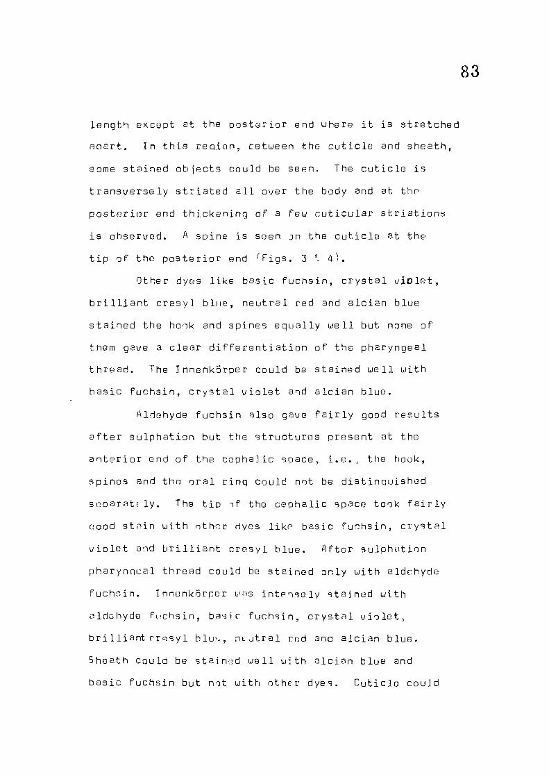
83
l eng th except at the p o s t e r i o r end uhere i t i s s t r e t ched
apa r t . In t h i s r ep i on , te tueen the c u t i c l e and sheath,
some s ta ined o b j e c t s could be seen. The c u t i c l e i s
t r a n s v e r s e l y s t r i a t e d a l l over the body and at the
p o s t e r i o r end th i cken ing of a Feu c u t i c u l a r s t r i a t i o n s
i s observ/ed. A spine i s seen jn the c u t i c l e at the
t i p of the p o s t e r i o r end ' 'F igs . 3 I 4 ) .
Other dyes l i k e bas ic f u chs in , c r y s t a l v i o l e t ,
b r i l l i a n t c r e s y l b lue , neu t ra l red and a l c i an blue
s t a in ed the hook and spines e qua l l y w e l l but none of
them gave a c l e a r d i f f e r e n t i a t i o n o f the pharyngeal
th r ead . The Innenkorper could be s ta ined w e l l wi th
bas i c f u chs in , c r y s t a l v i o l e t and a l c i a n b lue .
Aldehyde fuchs in a lso gave f a i r l y good r e s u l t s
a f t e r su lphat ion but the s t ruc tu r e s present at the
a n t e r i o r end of the c epha l i c space, i . e . , the hook,
sp ines and thn o r a l r ing could not be d i s t i nqu i shed
SGparatf l y . The t i p nf the c epha l i c space took f a i r l y
qood s tn in u i th othor dyes l i k e bas ic fuchs in , c r y s t a l
v i o l e t and b r i l l i a n t c r e s y l b lue . A f t e r su lphat ion
pharynncal thread could be s t a ined only u i th aldehyde
fuchs in . Innenkorper urns i n t e n s e l y s ta ined u i th
aldehyde fuchs in , bas ic f u chs in , c r y s t a l v i o l e t ,
b r i l l i a n t r resy 1 blut^, n t u t r a l rod and a l c i an b lue .
Sheath could be s ta ined u e l l u i th a l c i a n blue and
bas i c fuchs in but not u i th other dyes. C u t i c l e could

84
be d e f i n ed u e l l only when s ta ined u i th aldehyde fuchs in
and bas ic fuchs in . Comparative e f f i c a c y of d i f f e r e n t
s t a i n s a f t e r o x i d a t i o n and sulphaMon has beon
summarised in Table 1.
D iscuss ion
The s t a i n i n g r oac t i ons of the m i c n f i J a r i a e of
Chand l e r e l l a haukinoi are qu i t e s i m i l a r to those of
Brugia (Laurence and Simpson, 1969) u i th some
d i f f e r e n c e s . A f t e r ox id at i on , aldehyde fuchs in q?\yo
almost e qua l l y good r e s u l t s in both ncratodns in
d i f f e r e n t i a t i n g the s t ruc tu r e s in the c epha l i c space
the Innenkorper , the pharyngeal thread , c u t i c l e and
sheath. With bas ic fuchs in f a i r s t a i n i n g of the
c u t i c l e and sheath could be achieved in case of
m i c r o f i l a r i a of Chand le re l l a haukinqi but not u i th
m i c r o f i l a r i a of Brugia, 'Staining r e a c t i on to c r y s t a l
v/iolet ujas s i m i l a r in both the s p e c i e s , though uary inn
in i n t e n s i t y . Uith b r i l l i a n t c r e s y l b lue , neu t ra l red
and a l c i a n blue thpre uas not -^uch d i f f e r e n c e butucjen
the tuo s p e c i e s .
A f t e r su lphat ion a l s o , aldehyde fuchs in gave
the best r e s u l t s . I t s ta ined the c u t i c l e of m i c r o f i l a r i a
o f Chand le re l l a haukinqi mirn i n t e n s e l y than that Tf
Erug ia . But u i th bas i c fuchs in r e s u l t s were not as

ttD
qood as observed by Laurence and Simpson (1969) in
Bruq ia . Uith c r y s t a l v i o l e t , Laurence and Simpson
(1969) r epo r t ed in t ense s t a i n i n g of nharyngea l thread
and poor s t a i n i n g of c e p h a l i c s t r u c t u r e s . But i t has
been j u s t the oppos i t e in ease of C h a n d l r e l l a .
Laurence and Simpson (1Q69) r e p o r t e d in t ense s t a i n i n o
o f c e p h a l i c s t r u c t u r e s and pharyngea l thread wi th
b r i l l i a n t c r e s y l blue but i t uas not so obvious u i t h
C h a n d l e r e l l a . Hinor d i s s i m i l a r i t i e s were a l s o observed
w i th neu t ra l red and a l c i a n b lue . Accord ing t o o r e sen t
e v a l u a t i o n of the s t a i n s s t u d i e d , aldehyde fu chs in
a f t e r o x i d a t i o n seems to be the best f o r r ou t i n e
examinat ion of m i c r o f i l a r i a e in smears.
Usino the d i f f e r e n t s t a i n i n g t e c h n i q u e s , i t has
been p o s s i b l e to study in the m i c r o f i l a r i a o f C h a n d l e r e l l a
haukinqi some important s t r u c t u r e s uhich sbp'v t o
d i f f e r , from s n e c i e s t o s p e c i e s , in t h e i r charc-'ctor
and arrangement. Table 2 g i v e s a compamt i ve summary
o f the d i s t i n q u i s h i n g cha rac t e r s of these s t r u c t u r e s
in m i c r o f i l a r i a o f d i f f e r e n t s p e c i e s of f i l a r i a e so
f a r s tud i ed by Laurence and Simpson (1169 ) . Fasud on
t h i s a key t o a t i n t a t i v e c l a s s i f i c a t i o n of f i l a r i a e
i s appended be lou . Using ^he key i t i s honed that i-he
common spe c i e s of f i l a r i a e could be i d e n t i f i e d , in the
absence of adul t uorns, from m i c r o f i l a r i a e alonn and
uould be u s e f u l in d i a gnos i s e s o e C i a l l y in cases of
z o o n o s i s .

86
Key to the genera of f i l a r i a l uorms based upon c e p h a l i c s t ruc tu r e s
1. Hook b i f u r c a t e at the base u i th sp ines
Hook not b i f u r c a t e , sp ines absent
2. Hook u i th t ransve rse support ing bars
Hook without support ing bars
3* Hook U-shaped
Spines 4, t r i a n g u l a r
Spines 3, t r i a n g u l a r
Spines 3, f a n g - l i k e
2
5
3
A
Uucherer i a
Brugia p a t e i
B, malayi and
B. pahangi
Spines numerous, arranged
i r r e g u l a r l y in 2-3 rous
4. Hook f a n g - l i k e , sp ines 4,
t r i a n g u l a r in shape
5. Hook u i th tuo t r ansve r se suppor t ing bars
Hook without t r ansve r se bars
6. Hook bou-shaped
7. Hook broad p l a t e l i k e u i th c e n t r a l
p o s t e r i o r p r o j e c t i o n
Hook smal l t oo th l i k e s t ruc ture
Hook s t i l l smal l e r
Loa
Chandlerc11a
6
7
C a r d i o f i l a r i a
Onchocerca
f^lansone 11a
Dipetolonema
Hook beak l i k e L i tomoso ides

87
m o
c •H + o + •H D + + 4J tJ ro •D • H X X -1-1 r ro +
CD + + f-i x-Q (/->
4-> (t-tD 1 (-i •H c Q; O' Q) a + + c C M + •M c lO + +
t—. 3 ro JZ
ro CD 0) r-! oi r-t c "D + (D >, ro 4- +
1-1 O 4- 4 O ra U rH JZ JZ TD G jJ C ro x: CJi C_) ro C +
(H • H + 4-Ct- (P H + rl ro CO -r-f cn CJ u ^ c ro n •rH -H O o« CL + 4-•H cc G'l + tl_ + + o f-1 c u • o •H C •H C e Q J o C n
•M U -H O D "D X) •H 4->
o (0 T? -M r: O U CX ro x:
ai a i-i a: r) "D Q c rH -P o rH •iH D C ct- u X c cn n <—i •H TO X) -P c cn ra
;< c • UJ -H
r— -1 CP s; P -C t—1 i HI O -H tC CD =)
x: 1— LJ Ci-ro
! — l-d Q) > -J
JZ Q) 13 r-H u:
>
LJ
+
+ + +
+
4-+
+ + + + + +
Q) o> ro Q -4J X 0) c c o
X) r o o
+ 4- + 4-+ + 4- + 4- 4- 4. +
C C C O c o o o •H •H •H -P -P ro 4J ro ro JZ ro x: "D o -D Q-•H rH •r4 rH X X D r-1 'jn o r.n
C 0) •H (Tl r JZ o 3
• - (
v'O O 4J •H m U1 >. CQ Gj u
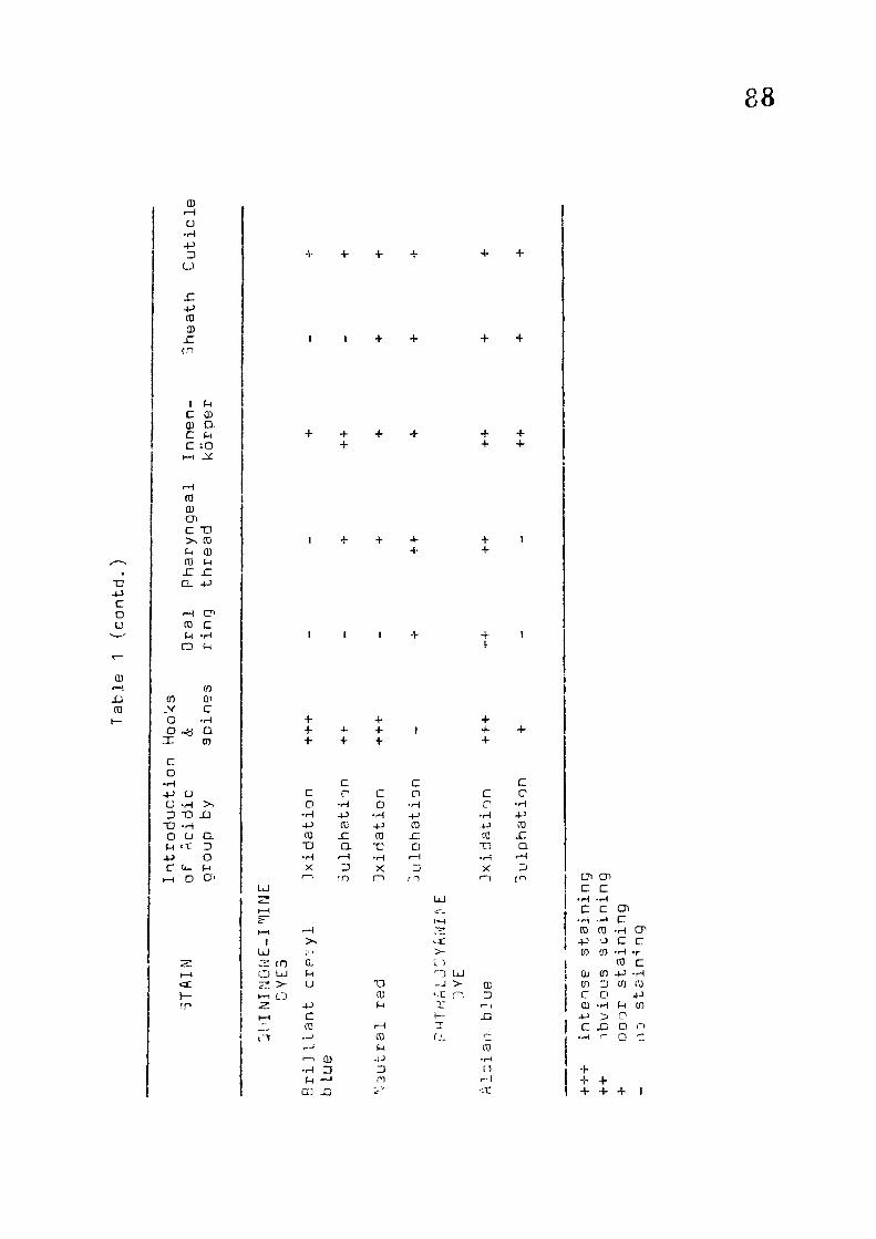
88
CD i-H O -H 3 LJ
n -p ro ID JZ tn
I (-1 C 03 (H CL C f-t C. :0 l-H ^
to QJ cn C T)
(0 U (D 03 (-4 JZ H CL 4-)
rH C CO C f-( -H CD u
cn cn 01 ^ c O -H o oa Q x to c o •H -P O O -H >. D T3 XI -a -H o o M •It -P C (*_ ^ o
(3-3 o u ai
cC I— fO
bj 22* I—(
bJ CO
C) LJ > H-f L.)
+ +
--H >>
0, M U -P c CO • r-' rH CD •H p I—I cn XD
+ +
+ +
+ +
T3 CU M
CO U -U 3 m
LU
I 1 n UJ -J > C n
f,L
cc D
CO
•a:
+ +
+ + + + 4. + 1 4- + + + + +
c c C C c o C c o •H o •H r •H •H -P •H -P •H -P
CO -P CO -p CO CO JO CO JZ CO x: "D Q- -c D TJ Q •H rH •H r-l •H rH X 3 X 3 X 3 1—1 tn rn r-i cn CJi CJI
C C • H .1-4 c c cn •H •rH c CD CO •H CP •P o C c cn cn •H •r
CO c 0) cn -p •H cn 3 cn CO c O -p CD •H u CO -P > n C XJ b r> •H f- Q 'Z
+ + + + + +

89
CO
cr>
c o cn
o D. CD e TD •H
CD o CO OJ CO P "D JC CU. C -P n (0 >-H CD Q CO CO U q; r" c cnjT CD c 4 J P p x: (0 CO -p _J jc D. 3 a-
o n •M c CT' CO C P •M O to 3 CD 3 P (0 CD 3 x: x: -P -M U CO 3 -p c P r-t n -P CD cn P T3
Q) CD o rH in •l-l n CO .—t c X3 CD CO — x: x. CLCJ cn CD CD o (t- •H
o U Ct_ CD o ro •p cn c M
o CO 13 CD t» •H •r-4 XI P •P to c p a p u 6 o cn o •p 03 C-J c
(N m jQ CO
CO Q; TJ x: c
r-t -p Q) (D 4J CO
CO e x: X "-i CD r -p CD p rH cn p Q QJ C XJ t4- cn D P > to o c P CD P CD Oi •p :0 rH ra L, r OJ p Oi c T: JZ r •H p XT C •p c a 4 J (n -p 4-> cu CO
•p c CO c cn x: p (D p n c t CJ L o •p
CP c
cc (-1
o
cn £D c •H o
cn
n o
sa CO D O CD CD r-l 4J
cy (h
o u
CJI c: (0 •H U 4J
0 >—I rH Ci) P -p ^ rp r ; c. : '.a r ru 3 .c i c: ' .c
a CD •rH C r—) r-H
r. CO a 10 ai ^^ -C p cu o Q CD r-i C -P CI ( 3 (It -p O Q C ID U cn O •p c p ffl 0) CD CD (0 •H P -C x: Q cn x: u a -p -p cn L 1
CD TJ CD CD
•P a. (-1
a cn CO Q. -P X ai c c o
T) -P c o o
r o u. S-i c ffl (U
u - -VJ
QJ Q) cr JZ c
•H
L CL' Co cn CJl -r-l
C O C CO J rH
to • (D E
3 -is: r -M
-p TD D C X] ro
•r- l
cr
CD ^ M -H CO r-t
" QJ 0) -H r-H Q) ai CO cn+j> -p c CO (-1 -H CO n o a -r-i JC c M -H p 'H lu loj I cn o -p CO I
CJ •rH
"O cu -p p CD CO
1 CO o o ^ p x: -w o -p 3 CO x: 3 c D. •P (0 -P 1*. o •p 3 -p CO <—t •p ro o o cn CO >nXI 3 CO X) JZ p a XI p
•p - >^ 0 "D c CD - CD cn rH cn c c D Ji: -p T3 p XI P p CO CD •P • H CO D 3 p O IP ai +-> rp Cl 4-> CD •P o rs cn O 1 -D fB o 3 P cn CD -P Q) CT' CU G) -C 3 (D cn c u P c -P u. CO p G XJ ti lU UJ o o CO CO 1 ^^ c p p -p X P
LL XJ cn X) CO CO -u n cn Q
1.0 •r-l
3 p (U
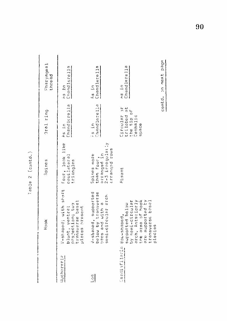
90
X3 -P C o u
CN (D XI (0
cn c
ro u o
OT CD C • H a ro
js: o o
rH CO CO CO ro cu >—1 rH —1 a> •-I <H (
c -a Qj CD (0 (-i f-t
P (D u: D a-m f-H r-H f-i —1 JC x: C X3 C -D C T3
•H C c •H C CD ro CD
cn r CD x: cn x: .i CJ •-C CJ I u
CO >—I rH O P-i ui
C "D •H C
CQ cn JZ
•3. LJ
(B
•H i-i Q) tn O JZ u
ro
CD u ca ,-H
c X) •r-l c CO
CO r
O ft) Ct-O fH "D CJ
ra 03 a rH X} -rH -H 3 o <0 Q -H JC (-( -rJ Qj D CO •H (-t x: CD a CJ -t-j C.J cn
cu CP o ex -p X D c c n
•D -P C O o
CD U
^ t •H CD r-i CO 3 1 ffl c ^ O
ro • H D p o (-1 cn a C-i CP o OJ Q] e; D X) D "O f~t 4-> rH O ffl P c>
Qi CO Ct-- Oi U CJ -p ^ 1—( C 0} c • H cr c
M •H CO c C CO CO CD D 3 •H •H ro p to p cn O O a n (M I p XJ U- CD +J cn -p CO CM ij CL
-P f-i T3 CD C CD (D x: .c -p P u ( f l r—t u CD p p
o (D o CO CO 3 ro O XJ cu x: r—I 3 cn -P a m n 1—) P o cn -p 0) -p <0 c a c r: p -H 3 O £ "O C!
(-1 XJ CJ D (D ID a o •P Cb X} 3 * ^ tn CO p n p P U- -p
c c QJ U) 4-) 3 3 T3 •H CD O P u) a> o tn (-1 - O 0! "C U JJ o cn
TJ M c n •n P Q QJ 1 C cn a p ffl -p cu CD XD c: •P 03 4-> £ a 0! cn a a cn a CO u x: P E P 3 0) (0 -p CD tn CD CO 3 1 CO o O • ro m CD •p -C c c o x : Q CD •p I CL CT, jC C a cn 3 o (0 Q) cn r-l p e J LX u QJ S) CO Q) 1 r-H 'rA 1 (D CO CD o 3 > - p C P p •p
O-•P CI XI .IJ cn CD cn X3 to CO d
(U o
CO i
Ct. r
a 1,5

91
n j-j
c o u
IN OJ XI TO
(T) w c •H C <n
JSf o o X
0} CO x: D 4-; CU ai
rH CXI XJ o TO E O "D w c CO CD Q) C Q) X e: CD GU c X ai rH Jir cn t-i QJ CD o X cn cn C CO c -o Ci_ cnx f-i r-i f-i cnx X •H CD a CD >- (C c -•J D D Ct~ c u o rH ( - ) X U 03 CO •H •r-i -i-i c •H c T) D 03 CD (-1 TJ M "C (-1 0) cn •rH ,—1 ffi T ) X CD X X c c CD X o fC •a c CO CJ X X C J CD •H X tn f-H cn X •H C.) D • H X
-P CU m •H CD r o D CD t-i f-t cn X P OJ a tt- (-1 c CJ O X X X c llJ O . X D o cC o CD o CJ cn 1— cn •H
ro CD t rH
CT r-i 1 X rH C m r. i •D X' CD OJ -H 1 u ca CD c ( - (
a CO 3 X r. n 03 i-H CU X rH X
( c •a 3 ( (-< a r XJ c K •r-1 c U ( (D E CD • rH C a' (-1 ra (i cc D O (-1 CD CO
(0 X •r-t F CD -t-i X CO X X cz u u CO CO Ct <—1 -X
c a in XI et
W y
4J C (D (n X ci
a.
4-> c 03 cn X cC
-p c CD to X u
•rH X C CJ 1—I 1—1 X o ••H
CD n •H rH (-1 •H G) o X 03 X X X o X rH CD r CD X CD CO rH 1—1 a, ••-> c m (U CO CO a u o r o -H fH E CD
•rH u D rH D cn X rv CD o X X u 0) •—1 u X ' u rH Q n X X CD rH n X D rH H n -):> cn CD (-1 o U •H CD 1-4 H n • H G -X O X x-* X CL 3 Q H 'H '0 V cn cn tn
IQ s a3
CD "0 1 1 ; 1 •D u - i C •rJ M n o ai ' I i CO u c r o n 4-' c_ X cn O u c a X r (T' "H •rH o n X

F i g . 1. NucJear s t ruc tu res of n i c r o f 1 l a r i a .
F in . 2. ucphal ir 3tructij"^os jF m ic rD f i 1 ar ia ,
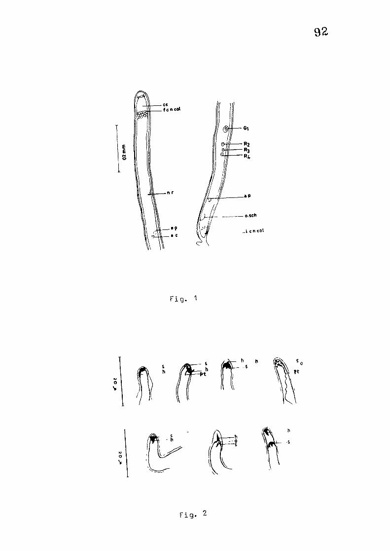
92
E E
F i g . 1
o V
/
fP h h - s
o V
f i g . 2

Fiq . 3. ] r a l r i n g , nharynqeal th read , Innenkorper
and t a i l spine in long Trm of
m i c n f i l a r i a .
F in. "ja-c 3tructurr js in shnrt form of
n i L- n f i 1 r, I i a .

93
( ^ i g . 3
T i g . 4

94
Forms of m ic ro f i l a r i a s
Uhile studying the smears for m i c r o f i l a r i a e , tuo
forma of m ic ro f i l a r i ae uare observ/ed in C, haukingi.
The long and the short forms^ Both tha forma showed
i d e n t i c a l features . The sheath, the cut i c l e , the hook
and spines, o ra l r ing , pharyngeal thread and Innenkorper
uere a l l i d en t i c a l . The extent of pharyngeal thread,
the pos i t ion , shape and size of Innenkorper i s s imi lar
in re lat ion to the body s i z e . The long forms of
m i c r o f i l a r i a e , measuring 136.4-174.9 j j , are found in
comparatively younger crous and the short forms
72.6-94. 6 yu in older crous. Besides these long and tha
short forms of m i c r o f i l a r i a e , a feu intermediate forms
are also ava i l ab l e . The size of Innenkorper, the
storehouse of mucopolysaccharides, also decreases uith
the shortening of the body ( r i g s . 5 & 6 and Tables 3 & 4 ) .
Further, to v e r i f y that these tuo forms belong
to one species , a number of short , intermediate and
long f':irms have been measured, the posit ion of
Innenkorper has been noted and the tuo r a t i o s , between
the body length and distance of Innenkorper from
anter ior end as wel l as between the body length and
distance of Innenkorper from poster ior end, have been
uorked out. I t i s found that these ra t ios remain
almost constant^Table 5 ) .

95
Discussion
In spite Tf the fact that the body length changes
uith the change in the size of Innenkorper, the tuo
forms of mic ro f i l a r i ae show constant oosit ion of
Innenkorper. The nuclear landmarks and the cephalic
structures are s imi lar in both long and short forms.
These observations go to prove that the size of
m i c ro f i l a r i a e decreases uith the decrease in s ize of
Innenkorper - the storehouse of mucopolysaccharides,
due to the immune responses of host. As the in fect ion
gets o lder , the concentration of antigens increases in
the blood and the m ic ro f i l a r i a e do not get proper food
from thei r surroundings. Their size decreases uith the
decrease in size of Innenkorper as m ic ro f i l a r i a e use up
the stored glycogen from i t for their su rv iva l .

Table Length of m i c r o f i l a r i a e , size and p o s i t i o n of" Innonkorper.
96
S. Len th f .S i ze of ,Oistanco of . -Microf i lar ia . T nnenkomer , Innenkorper
• L e n n t h ' B r e a d t h ' a n t e r i o r
1 72. 6 2.2 2.2 52. 8
2 74. 8 2.2 3. 3 56. 1
3 75. 9 4.4 2,2 57.2
4 R2, 5 2.2 2.2 62.7
5 B5. 8 2.2 3.3 71.5
6 85. 8 2.2 3. 3 72. 6
7 86. 9 2.? ''..3 63. 8
3 86. 9 3.3 3.3 58.3
9 88, 0 2.2 3.3 62.7
10 91. 3 7.2 2.2 51 .7
1 1 91 . 3 2.2 3. 3 67. 1
12 12. a 2.2 68,2
13 5 2.2 " . 3 69. 3
14 93. 5 2.2 3.3 57,2
1 5 94. F 1. 1 3 71.5
1 6 94. 6 2.2 3.3 69. 3
17 94. 6 1 . 1 3. 3 DO. 5
1 R 102.3 2.2 3. 3 77.0
1 9 103.4 3, 3 3.3 74. 8
20 n n . o ^.3 3.3 81 .4
21 11 2.2 3. 84.7
contH. on next page

Table 3 ' contr i . )
S .N. Length of ' " ' i i c r o f i l a r i a
' (
t J i z e o f t i lnncnkorpar t
P i s tancs of Innenkorper
'Lenqth'Brparth, I t 1 > 1
from antt^rior end
22 113.3 3.3 3.3 80.3
23 117.7 4.4 4.4 77.0
24 118.8 2.2 2.2 74. S
25 121.0 3.3 4.4 80. 3
26 122. 1 3. 3 3. 3 80.3
27 124. 3 3.3 3.3 78.1
28 124. 3 3.3 3.3 84.7
29 124. 3 4.4 4.4 85. 8
30 125.4 3.3 3 .3 83. 6
31 125.4 3.3 3.3 85. 8
32 126. 5 3.3 3 .3 88. 0
33 127. 6 3.3 4.4 83. 6
34 130. 9 3. 3 4.4 85. 8
35 130. 9 3.3 3. 3 88. 0
36 134.2 3. 3 3.3 88.0
37 135. 3 3.3 4.4 3R.0
38 135. 3 3.3 4.4 94. 6
39 135.3 3.3 3. 3 94. 6
4 0 136.4 4.4 4.4 97. 9
41 136.4 3.3 4.4 92.4
42 137. 5 4.4 3.3 94.6
43 137.5 3.3 4.4 90.2
nontd. on next page

Table 3 ( contd . )
88
S.N.
I
, Length of , 3 i2e of JInnenkorper
t ,Distance of , Innenkorper
f^icrof i l a r i a 1
I Lfjngfeh i Bre adth I t
( from a n t e r i o r , end
44 138.6 2.2 4.4 91 .3
45 13R. 6 3.3 3.3 93.5
46 139,7 3.3 3.3 92*4
47 139.7 4.4 3. 3 93.5
48 un. B 4.4 3. 3 92.4
49 140, 3 3. 3 3.3 9 6. 8
50 140. 8 3.3 3.3 95.7
51 140. 8 3.3 3.3 99. 0
52 140. 8 3. 3 4.4 91 .3
51 141.9 3. 3 3.3 92.4
54 141 . 9 3.3 3.3 95.7
55 141 . 9 3. 3 10B.9
56 143. 0 3. 3 3-3 96. 8
57 143. 0 3.3 4.4 90.2
58 143.0 3.3 3.3 99, 0
59 144. 1 3,3 4.4 1 01 .2
60 145.2 3. 3 3.3 94. 6
61 145.2 4.4 4.4 99. 0
62 145.2 3.3 3. 3 105. 6
63 145.2 3.3 3.3 99, 0
64 147.4 2.2 2.2 101 ,2
65 148.5 3.3 3 .3 1 04, 5
contd. on n^^xt pagB

T ah 1G 3 ( c a n t d . )
99
, 3 i z e of ,Distance o f 3. N. , Length of , Innenkorper ,Inne nkorpe r
, l^icrof i l a r i a ' Length 'Breadth T f
,frnm a n t e r i o r , 8 nd
66 149. 6 3.3 3.3 102.3
67 149. 6 5.5 4.4 102. 3
68 151 . 8 3.3 3 .3 104. 5
69 151 .8 4.4 3.3 104. 5
70 152.9 3.3 4.4 101 .2
71 152. 9 4.4 3.3 105. 6
7 2 152.'^ 3.3 3,3 107. 8
73 152 . 9 3.3 4.4 101.2
74 1 52 . 9 3.3 4.4 102. 3
75 152.9 4.4 4.4 104. 5
76 154. n 4.4 3.3 100.1
77 154. u 3.3 4.4 103.4
78 156.2 3. 3 3, 3 103.4
79 156.2 4.4 3.3 103.4
8 0 157, 3 3.3 4.4 99. 0
81 157. ^ 3.3 3.3 112.2
82 1 58.4 3.3 3.3 103.4
83 158.4 4.4 4.4 106.7
84 15T.5 3.3 3.3 107. 8
85 1 60. 6 3.3 4.4 104. 5
contd. on nfcxt paqc

Tablf-' ' 'contd. )
100
t3izr ' of .Distance of S.N. _ L&ngth of .Innonknrpor . Innenkorper
N i c r o f i l a r i a 'Length 'Breadth , from a n t e r i o r ! 1 ,and
86
37
88
89
90
91
1 62. B
1 63. 9
169.4
1 69.4
170.5
174. 9
4.4
3.3
4.4
3.3
3.3
3.3
4.4
4.4
4.4
3.3
3.3
110. 0
114.4
112.2
122. 1
11 6. 6
114.4
A l l m G n s u r c m e n t s are in p

101
Table 4. Decrease in the s i z e of Innenkorper u i th the shnrtening of the body.
j.!\l. Body lenqth S ize of Innenkorper
1 169.4 4.4 X 4.4
2 158.4 4.4 X 4.4
3 1 56.2 4.4 X 3.3
4 152. 9 4.4 X 3.3
5 151.8 3.3 X 3.3
6 138. 6 3.3 X 3.3
7 135. 3 3.3 X 3.3
8 103.4 3.3 X 3. 3
9 1 n?. 3 2.2 X 3.3
1 0 91 . 3 2.2 X 3. 3
1 1 82. 5 2.2 X 2.2
12 72. 6 2.2 X 2.2
A l l measurements are in P-

102
E o (-1
u 01 a u o ^ c CD c c J—(
o CD o c CD -P tn •H •o •a •
c •D ro C
0 x: •p (H cn O c •H (D -H CD -P
cn n o o Q-X3
cn CD CO JZ -P r-i
r-H c CD CD 3 CD 3 CO -P CO CD XI M
n cn •M n* (-4 •tH CD 4-> -P CO C u:: CO
LO (D JD
CO o -p CO CM n
E P U ci_ C
O- f-i en n CD
Q f-1 Q U O o -.n -w c ^ p CO c Q} -4-) Q) -P cn c CO •H c o C) o
e n M Ct-
•o ' I - p c o a a
a. 0) f-i PI O r c CO C M
CD D cn c 4 J
c c O t-H CD
-C -P c CD
X] o CD
m
CM
rn
to LD cn O • • • • •
CO LO UD CN CM CM CM OJ
r - I D CO o • • « • •
>X3 t n LO Ln t - r - r -
o to
m CM
fi CM
CM CM
a\
cr cn
o CO v£) ro lT)
CJI CM
CM O m cn i n
CO CM r-
c
(D U CO CD -P C cu £ 03 U D cn CO Q E
' I
oi to LD

F i g . S. Photomicronraph of long form of
m i c r o f i l a r i a . X 1100.

»1
103

Fig. 6. Photomicrograph of short form of
m i c n P i l a r i a . X noo.

104
Fig. 6

105
( b ) S tereoscan sturi ies
( i ) Adult
The body c u t i c l c has bpRO doscr ibod to bt. Gmooth
by l i g h t microscopy but i t shous s t rong s t r i a t i o n s at
a n t e r i o r and p o s t o r i o r ends of body. The mid rngion
of body shows f e e b l e s t r i a t i o n s . The mouth i s
e l l i p t i c a l , mounted on a r a i s ed s h i e l d of c u t i c l c ,
Nothing s p e c i a l could be observed r egard ing copha l i e
o a p i l l o e and amphids, only that amphids are su^;n to
have a peduncle. The anal depress ion in female i s
found to be surrounded by f e e b l e uauy s t r i a t i o n s u i th
a feu small ua r t s . Wo d i f f e r e n c e was observed in
c e p h a l i c s t ruc tu res of Tiale and female . The t a i l in
f ema le shous s t r i a t i o n s u i th b l u n t l y rounded t i n . The
t a i l of male dot_3 not show any a d d i t i o n a l f e a t u r e s
( F i g s . 7 - 1 3 ) .
( i i ) H i c r o f i l a r i a o
The s te reoscan s tud i es of m i c r o f i l a r i a e did not
r e u e a l anything s o e c i a l as hhey are cov/erud u i th a
sheath on l y ; the sheath shows wr ink l es at th(i p o s t t r i o r
end in long forms and i t i s wr ink led throughout in
shor t forms ( F i g s . 14 & 15) .

F i g . 7. Scanning e l c c t n n micrograph of a n t e r i o r
pnd of adu l t . X 1500.
F i g . 8, Scanning r l e c t n n micrograph of head
en - face uieu of adu l t . X 1000.

F i g . 7
Fig. 8

F i g . 9. Scanning e l e c t r o n micrograph of genera l
c u t i c u l a r surfacFj in midbody reg inn of
adu l t . X 5000.
F i g . 10. Scanning e l e c t r o n micrograph of vu lva
of f emale . X 700.

107
Fig. 9

/ i g . 11. Scanning e l e c t r o n mirrnqraph of c u t i c u l a r
ornamentat ion around anal depress ion
of f emale . X IIOOG.
F i g . 12. Scanning e l e c t r o n micrograph of t a i l
of f emale . X

108
Fig. 11
Fig . 12

F i g . 13, Scanninq e l e c t r o n minrnqraph of t a i l
o f male. X

109
Fig. 13

F i g . 14. ocannin-. e l e c t r o n micrograph of short
form of m i c r o f i l a r i a . X 3100.
F i g . 15. Scannin-'' e l e c t r o n rriicronranh of a n t e r i o r
find of short f i r r of m i c r o f i l a r i a , X 11000.

134
Fig. 14
F i g . 110

I l l
2. Histology
The present uork deals uith the h i s t o l og i c a l
studies of Chandlere11a haukingi, a f i l a r i a l parasite
of Indian jungle c n u Cprvus macrprhynchos (Uag l e r ) .
BODY IxJALL
The body u a l l consists of cu t i c l e , sub -cut ic le
and muscle layer . These layers are very intimately
associated uith each other.
Cuticle
This is outermost, semitransparent covering of
the body. Besides covering the externa l parts i t passes
through a l l apertures l ike mouth, vulva and c loaca,
forming interna l l in ing of oesophagus, vagina and rectum,
The s u p e r f i c i a l layer of cut ic le forms pap i l l a e ,
cephal ic as u e l l as post -c loaca l and s t r i a t i ons on the
body (The cut ic le shous strong s t r i a t i ons in anterior
and poster ior part of the body and feeb le s t r i a t i ons
in the middle region of the body by stereoscan electron
microscopy). In transverse section the cut ic le shows
f i v e layers (F ig . 15 ) . These layers are as f o l l ows :
1. Outer co r t i c a l layer
2. F i b r i l l a r layer 3. Matrix layer 4. Fibrous layer 5. Basement membrane
112
The external co r t i c a l layer is quits dense as
compared to the other layers . Matrix layer appears l ike
a spongy layer . The f i b r i l l a r layer is perhaps formed
by condensation of matrix layer . In this layer the
f i b r i l s form the netuork. The f ibrous layer is made
up of connective t i s sue . The basement membrane is a
thin layer .
These layers arc not present throughout the
body. Only outer co r t i c a l layer is present in the
l in ing of oesophagus v/agina and cloaca. The cut ic lc is
thicker in male than in female, e spec ia l l y in the
greater part of the middle region of body. This may be
due to the enormous development of uter i in female.
Discussion
The cut ic le of the present form resembles the
cut ic l e of S_. corvi as described by Ansari and Basir
(1964) but in _£, haukinqi only 5 layers could be
observed uhereas Anspri and Basir (196A) described
8 layers in cerv i . Goldschmidt (1906) described
9 layers in the cut ic le of Ascaris lumbricoides.
The nature of cut ic le has also been discussed
by some uorkers. Grubbe (1850) termed i t ch i t in .
Flury (1912) ca l led i t keratin and Plagath (1919) termed
i t cut ic le in case of Ascar is . Mueller (1929)
described the cut ic le as a secretion product, but did
not mention the nature of the product. Chituood (1936)

113
and Ansari and Basir (1964) bsliewed i t to be proto-
plasmic condensati-TH from l iv ing c e l l s . The author
a lso agrees with the vieu of Chj.tunod (1 936) and Anaari
and Basir (1964).
Sub-cut ic le
I t i s present belou the c u t i c u l a r l aye r in the
form of 3 f i n e sheet of protoplasm. I t becomes th in
towards both the e x t r e m i t i e s but becomes t h i c k e r in
the middle reg ion of the body in male and in p o s t e r i o r
par t of the a n t e r i o r one t h i r d of the body in f ema le .
In the middle r eg i on of the body of f ema l e , the
s u b - c u t i c l e gets pressed due to the enormous grouth of
u t e r i . This l a y e r bulges i n t o pseudocoelom at four
p laces in the form of l o n g i t u d i n a l chords; one d o r s a l ,
one v e n t r a l and tuo l a t e r a l s , one on e i t h e r s i d e .
The do r sa l chord i s th inner than the v e n t r a l uh i l e the
l a t e r a l s are most prominent. These chords d i v i d e the
musculature i n t o four groups. The nuc l e i are seen
on ly in the chorda l r e g i o n . A l l the four chords
conta in nerve c e l l s and the l a t e r a l ones lodge l a t e r a l
e x c r e t o r y channels a l s o . The chords begin as smal l
th i ckened s t ruc tu res in the a n t e r i o r r e g i on o f the
body where nuc l e i are not seen in i t . These chords
become mod i f i ed at d i f f e r e n t l e v e l s . In oesophageal
r e g i on they become high g i v i n g support t o the oesophagus
by surrounding i t p a r t l y ( F i g . 1 7 ) , These g i v e support

114
t o vu lva in f ema le . In the r eg i on of gonads the chords
become h igh ly f l a t t e n e d . In t a i l r e g i on they become
inconspicuous.
Discuss ion
This form resembles B. oahangi , as desc r ibed by
Schacher (1962) , in having four sub - cu t i cu l a r chords .
Ansar i and Basir (1964) descr ibed four a d d i t i o n a l
r i d g e s , tuo sub-dorsa l and two s u b - v e n t r a l l y i n g on
e i t h e r s ide of l a t e r a l chords in case of c e r v i .
Hany workers have d iscussed i t s o r i g i n a l s o .
Hamann (1895) regarded i t ec todermal whi l e zur Strassen
(1904) b e l i e v e d i t to be mesodermal in o r i g i n . Stewart
(1906) c a l l e d s u b - c u t i c l e as hypodermis. Schacher
(1962) d id not d iscuss i t s o r i g i n .
flUSCULATiJRE
Ins ide the s u b - c u t i c l e l i e s the somat ic
musculature. The four sub - cu t i cu l a r l o n g i t u d i n a l chords
d i v i d e the musculature i n t o four s e c t o r s . The somatic
musculature ge ts mod i f i ed forming s p e c i a l i s e d muscles
in oesophagea l , i n t e s t i n a l , v u l v a r , anal and sp i cu l a r
r e g i o n .
The somatic musculature i s coe lomyar ian ,
polymyarian t y p e , each muscle c e l l c ons i s t s o f a
prox imal f i b r i l l a r part and d i s t a l p ro top lasmic
nuc lea ted pa r t . The f i b r i l l a r part i s at tached to the

115
s u b - c u t i c l e and pro top lasmic par t l i e s Free in the
pseudocoe l ( F i g . The suppor t ing f i b r i l s are
present in o ro top lasmic pa r t . These f i b r i l s have
connect ions u i t h the nervss running i n s i d e the chords.
The s p e c i a l i s e d muscles suppor t ing oesophagus
are knoun as aomato-oesophageal muscles. They extend
from somatic musculature t o oesophagus to giv/e support
t o i t . These muscles are at tached t o the oesophagus
at the lev/el o f or a l i t t l e behind the nerue r i n g . These
muscles do not form any d e f i n i t e pa t t e rn (F igs . 17 ^ 19 ) .
Those suppor t ing the i n t e s t i n e are termt-a .'s
s o m a t o - i n t e s t i n a l muscles. These are in the form of
bands or a sph inc te r around the i n t e s t i n e ( F i g . 2 0 ) . In
male the sp i cu l e s are supported wi th p r o t r a c t o r and
r e t r a c t o r muscles. In f ema l e , these muscles g i v e
support t o uulua and are knoun as somato-vulvar muscles.
D i scuss i m
Uhi le s tudy ing the musculature of nematodes
Schneider (1B60) found that the form, number and
arrangement of muscles are of g r ea t importance in
c l a s s i f i c a t i o n of nematodes. On t h i s bas is he grouped
the nematodes i n t o tuo groups, p la tymyar ian and
coe lomyar ian . The p latymyar ian are the nematodes in
uhich the f i b r i l l a r po r t i on of muscles i s f l a t towards
the body c a v i t y and coelomyarian are those in uhich
the f i b r i l l a r po r t i on of muscles bear groove and the

116
pro top lasmic part dip down in t o the body c a v i t y
( p s e u d o c o e l ) . Further on the bas is o f the number of
muscle c e l l s in each s c c t o r , he suggested tuo groups of
nematodes, mernmyarian hau/ing feu muscle c o l l s in cach
s e c t o r and po lymyar ian, i f they are numerous,
Har t in i (1916) r e s tud i ed the musculature of
nematodes and suggested that polymyarian and coelomyarian
nematodes are raeromyarian and p latymyar ian by f i n d i n g
the t r a n s i t i o n a l forms between the two in the l a r v a l
s t a g e s .
Schachei' (1 962) did not d iscuss t h i s po in t in the
case of Brugia pahangi .
Ansari and Basir (1964) suggested that polymyary
and coelomyary i s reached in l a t e r s tage of devolnprnent.
They descr ibed somatic musculature in S e t a r i a c e r v i as
polymyarian and coe lomyar ian t ype . They a l so desc r ibed
the s p e c i a l i z e d muscles, the somato-oesophagcal muscles,
s o m a t o - i n t e s t i n a l muscles, somato-uulvar muscles,
somato- r e c t u l muscles, musculus an i , copu la to ry
muscles and sp i cu l a r muscles.
The musculature in Chandlerc11a hawkingi i s
almost s i m i l a r t o the musculature of c e r y j on ly wi th
few minor d i f f e r e n c e s . In the former musculus ani
and copu la tory muscles are not observed .

117
BODY CAUITY
The body c a v i t y i s a pseudocoe l , a n t e r i o r l y in
the r eg ion i f muscular oesnphagus and p o s t e r i o r l y in
t a i l r e g i o n , the body cav/ity i s comple t e l y o b l i t e r a t e d
due to the enormous growth of connec t i ve t i s s u e . In
f ema l e , in the middle r eg i on of the body almost a l l the
space i s f i l l e d wi th c o i l e d r ep roduc t i v e organs. The
body f l u i d i s g ranular . Some nuc leated connec t i ve t i s sue
i s a l s o seen in pseudocoe l in oesophageal r e g i o n .
P i s cus s i on
Goldschmidt (1906) desc r ibed the body c a v i t y in
A s c a r i s t.o be surrounded by ' i s o l a t i o n t i s s u e ' uhich i s
a membranous t i s sue support ing va r i ous organs. S teuar t
(1906) found nuc l e i in j e l l y l i k e mass in Oncholaimus
and b e l i e v e d the body c a v i t y to be f i l l e d u i th
mesenchyme but because hn could not see the c e l l u a l l
around the nuc l e i he did not emphasize his op in i on .
Ansari and Basir (1964) gave d i f f e r e n t v iews r ega rd ing
the body f l u i d . They suoqested that the body f l u i d i s
e i t h e r a f l u i d uhich ge ts co -agu la t ed when the worms
are t r e a t b d wi th f i x a t i v e or i t i s a mesenchymatous
t i s s u e of lou o r g a n i s a t i o n . They a l so presumed i t t o be
a f l u i d in p lace of c i r c u l a t o r y f l u i d .
^^ £• haukingi the body f l u i d i s j e l l y l i k e
granular substance u i th some nuc l e i in i t . I t may be a
f l u i d in p lace of c i r c u l a t o r y f l u i d .

118
CQMNECTI\JE TISSUE
I t i s nuc l ea t ed , granular mass of t i s sue present
around the oesophagus. I t i s not seen in any o ther r eg i on
i n t h i s case .
D iscuss ion
The connec t i ve t i s s u e uas g iven d i f f e r e n t names.
Schneider (19Q2) c a l l e d i t " Bindegeuebe" . L o j s s (1905)
named i t " s t r and l i k e organs" . Goldschmidt (1906)
proposed the term " i s o l a t i o n g e u e b e " f o r i t . Ansari and
Bas i r (1964) suggested i t to be l i k e a mesentry ho ld ing
va r i ous i n t e r n a l organs in t h e i r p l a c e . In Chandlere11a
hawkingi t h i s t i s s u e i s l i k e a mesentry and l i m i t e d
only t o oesophageal r e g i o n .
DIGESTIVE SYSTEM
D i g e s t i v e system cons i s t s of mouth, oesophagus,
i n t e s t i n e , rectum, opening out through c l o a c a l aperture
in male and ending b l i n d l y in anal depress ion in ft jmale.
The mouth i s s imp le , present on ra i s ed c u t i c u l a r
p l a t e . I t leads i n t o oesophagus.
Oesophagus
The oesophagus i s d i v i s i b l e i n t o two p a r t s , the
a n t e r i o r muscular and the p o s t e r i o r g l andu la r . I t i s
covered by a th in membrane, the tun i ca p r o p r i a .

119
The muscular part of oesTphagus i s smal l e r and
narrouer than the g landular part of oesophagus. I t s
u a l l s are th ick^ muscular u i th a t r i r a d i a t e lumen. The
lumen i s supposed to be formed of c u t i c l e uhich covers
the b3dy of nematode e x t e r n a l l y . The g landular part o f
oesophagus i s lonqt^r and broader than the muscular part
i f oesophagus. I t s u a l l s are s y n c y t i a l . This part
o f t e n shows s l i g h t l y muscular charac tor around the lumen,
f r i m uhich muscular rays are seen p r o j e c t i n g i n t o
s y n c y t i a l mass. The lumen i s o f t e n c i r c u l a r . The
muscular part of oesophagus i s supported by somato-
oesaphagea l muscles uhich s t a r t from body u a l l
musculature t o g i ve support to oesophagus (F igs , 17 & 19 ) .
The g landular par t of oesophagus i s supported by
sph inc t e r muscles ( F i g . 21 ) .
A nerve r i ng i s seen around the muscular part
o f oesophagus. I t i s present o b l i q u e l y as in s e c t i o n s
i t i s always Sfjen in semilunar form ( F i g . 2 2 ) . Hugo
quan t i t y of connec t i ve t i s sue i s found to f i l l up the
space between the body w a l l and the oesophagus ( F i g . 2 3 ) .
I n t e s t i n e
The oesophagus i s f o l l o w e d by i n t e s t i n e which
i s th in wa l l ed . The u a l l s are made up of a number of
c e l l s and hence i t i s of myr iocytous typs showing 6-10
nuc l e i per s e c t i o n . The c e l l s of the w a l l are of
e p i t h e l i a l type ( F i g . 20 ) .

Re ctum
The i n t e s t i n e becnmes s l i g h t l y wider forming
rectum uhich ends b l i n d l y in an anal d ep r e s s i i n in
female and opens out through c l oaca in male. Rectum i s
l i n e d i n t e r n a l l y by a c u t i c l e which i s a con t inuat i on
of the e x t e r n a l c u t i c l e ,
Cloaca
Cloaca i s a common c a v i t y formed by recturm,
and e j a c u l a t o r y duct opening i n t o i t . I t i s l i n e d
i n t e r n a l l y by c u t i c l e . Sp icu les are o f t e n seen
p r o j e c t i n g through t h i s o p e n i n g ( F i g . 2 7 ) .
Discuss ion
The nature of oesophagus of C. haukingi i s
s i m i l a r t o the oesophagus ] f B_. pahanqi as desc r ibed by
Schacher (1962) and 3. c e r v i Ansari and Basir (1964 ) .
The only d i f f e r e n c e i s that the g landular part of
oesophagus - ' f ten shows a c i r c u l a r lumen ins t ead of a
t r i a n g u l a r one.
The i n t e s t i n e i s a lso of myr iocytous and
an isocy tous type as c a t e g o r i s e d by Chituood (1930 ) .
The w a l l s hav/e l a r g e number of c e l l s and the lumen i s
i r r e g u l a r . pahangi and S. c e r v i a l so f a l l in the
same c a t e g o r y .

121
m i l REPRODUCTII/E SYSTEM
The male r ep roduc t i v e system i s tubular and
r m m r c h i c . The t e s t i s i s thread l i k e and c o i l e d . I t
beg ins u i th ane c e l l in the muscular par t nf oesophagus
and proceeds backuards and becomos s e v e r a l c e l l t h i c k .
The t e s t i s leads i n t o tubular vas de f e r ens of
approx imate ly same width as that of t e s t i s . The
vas de f e r ens i s qu i t e long and leads i n t o u ider seminal
v e s i c l e which i s f o l l o w e d by muscular e j a c u l a t n r y duct .
The e j a c u l a t o r y duct opens i n t o rectum jus t in f r o n t of
the c l o a c a l aper ture . This common tube i s known as the
c l o a c a .
T e s t i s
The t e s t i s c ons i s t s of two p a r t s , the germinal
zone and the growth zone. The germinal zone s t a r t s
wi th one c e l l but the number inc r eases g radua l l y as i t
proceeds p o s t e r i o r l y . This z :no i s covered over by
f l a t c e l l s . The g. rm c e l l s i n s i d e t h i s cove r ing are
compact ( F i g . ) .
The germinal zone passes i n s e n s i b l y i n t o growth
zone . This zone i s a l so coverea over by f l a t c e l l s .
The c e l l l i n i n g i s th in in the a n t e r i o r r e g i on but
becomes th i ck p o s t e r i o r l y . The c e l l s i n s i d e t h i s
c o v e r i n g are known as spermatogonia ( F i g . 2lf ) .

2
l/as de fe rens
The g n u t h z )ne i s f-^llnuod by vas de ferens uhich
i s a lso nf the same width. The us l l a are glandular and
the lumen i s f u l l i f dcuclopinQ spermatoz:)a uhich are
smal l s phe r i c a l s t ruc tures ( F i g . 2 5 ) .
Seminal v e s i c l e
The uas de ferens uidens to form the seminal
v e s i c l e . This part s t o r e s the sperms. The u a l l s are
made up o f f l a t and narrou e p i t h e l i a l c e l l s . The lumbn
i s f u l l of f u l l y developed spermatozoa ( F i g . 26 ) .
E j a cu l a t o r y duct
The seminal v e s i c lQ i s fo l loujod by muscular
e j a c u l a t o r y duct. The outer layer of u a l l s i s of
c i r c u l a r muscles and the inner l ayer i s of columnar
c e l l s . This duct opens in t D common c loaca ( F i g . 2 7 ) .
Sp icu l es
The sp i cu l es are sub-equal , d i s s i m i l a r and
s c l e r ^ t i z e d s t ruc tu r es . They are covered by a sp i cu la r
sheath made of c i r c u l a r muscles and are supported by
sp i cu l a r muscles ( F i g . 2 8 ) .
Discussion
he male reproduct i ve system of C. haukingi
shous characters s im i l a r to that of Brugia pahangi as
descr ibed by Schachcr (1 962) and Se t a r i a cerui by
ftnsari and Basir (1964) .

•e 23
Schacher (1962) and Ansari and Basir (1964)
d e s c r i b ed t e s t i s f o l l o w e d by seminal v e s i c l e but in t h i s
case t e s t i s i s f o l l o u c d by v/as de feruns and the l a t t e r
l eads i n t o wider seminal v/'isiclo which opens i n t o
c l oaca through a muscular e j a c u l a t o r y duct . Cobb (1905)
and Rauther (1909) descr ibed hair l i k e processes in the
lumen of vas de f e r ens but they are ne i the r desc r ibed by
Schacher (1962) and Ansari and Basir (1964) nor they
are seen in C. haukingi .
FEHALE REPRODUCTIVE SYSTEM
The female r ep roduc t i v e system cons i s t s o f pa i r ed
o v a r i e s , o v i duc t s , receptaculum semin i s , u t e r i and a
common uterus , vag ina and vu l va .
Ovary
The j v a r i o s aru d i d o l p h i c and o p i s t h o d e I p h i c .
They are c o i l e d s t r u c t u r e s . Each ovary c ons i s t s of an
outer e p i t h e l i a l l aye r and an inner germinal chord. A
smal l cap c e l l i s present cove r ing the-: b l ind end of
ovary ( F i g . 29) , Each ovary has two d i s t i n c t zones
the germinal z ine and the growth zone.
The germinal zone s t a r t s jus t behind the a p i c a l
c e l l . This area i s the area of rap id d i v i s i o n . I t
s t a r t s as a s i n g l e c e l l but the number inc reases as i t
proceeds backwards. The c e l l s arc s p h e r i c a l w i th a
124
l a r g e nucleus and a nucleo lus ins ide ( f i g . 30 ) . Thyse
c e l l s are enc losed by e p i t h e l i a l l a y e r . This i s fo l louedr-
by the grouth zone .
The grouth zone i s r e l a t i v e l y longer than
germinal zone. The c e l l s f i l l i n g up t h i s part are kniun
as oogon ia . These oogonia are covered i ve r by a
compara t i v e l y t h i c k e r l aye r of e p i t h e l i a l c e l l s ( F i g . 3 0 ) .
Oviduct
Each ovary i s f a l l o w e d by the ov iduc t . The w a l l
o f the ov iduc t c ons i s t s of an outer l a y e r o f c i r c u l a r mus-
c l e s find an inner l a y e r of columnar c e l l s ( F i g . 3 0 ) '
Uterus and receptaculum seminis
The ov iduct of each s ide i s f o l l n u e d by uterus.
At the junc t i on of ov iduct and utorus the tube becomes
a l i t t l e en la rged f :)rmlng receptaculum seminis f o r
s t o rage of sperms. The wa l l s of receptaculum seminis
ore made up of i r r e g u l a r columnar c e l l s with nuc l e i in
the basa l pa r t . The columnar c e l l s are l i ned e x t e r n a l l y
by a membrane of e p i t h e l i a l c e l l s ( F i g , 3I ) . The
receptaculum seminis narrous diun and proceeds as
uterus proper .
The two u t e r i i run a n t e r i o r l y and uni te forming
common uterus. The u a l l s of uterus arc made up of l ou ,
r e c t angu la r e p i t h e l i a l c e l l s . The lumen i s f u l l of
m i c r o f i l a r i a e , in va r i ous s tages of development ( F i g . 3 2 ) .
125
As i t proceeds a n t e r i n r l y i t s u a l l s become mars and
more th i ck forming muscular uagina.
V/sgina
The vag ina c ons i s t s i f tuio p a r t s , the par t c l o se
t o the uterus i s knoun as vag ina u tc r ina and the par t
opening t o the outs ide i s kniun as vag ina ve^ra or
t rue vag ina . The u a l l s of vag ina u te r ina are made of
columnar c e l l s surrounded by c i r c u l a r muscles ( F i g . 21).
The u a l l s of vag ina ve^ra are th i ck wi th an inner l aye r
o f columnar c e l l s and an out^r l a y e r o f c i r c u l a r and
ob l i que muscles (Figs. 32 33 ) .
D iscuss ion
D i f f e r e n t workers gave d i f f e r e n t op in ion r ega rd ing
the o r i g i n and e x i s t e n c e of cap c e l l present at the
b l i nd end of ova ry . Chituood (1 929) descr ibed t h i s c e l l
as a part o f o v a r i a l ep i the l ium. I^usso (1930) gave a
d i f f e r e n t v i ew . He sa id that the cap c e l l i s an
u n d i f f e r e n t i a t e d germinal stem c e l l which p r o l i f e r a t e s
both qerminal and e p i t h e l i a l c e l l s . Ansari and Basir
(1964) found i t underneath the e p i t h e l i a l c e l l s
c o v e r i ng the germinal zone of o va r y . The author
b e l i e v e s i t t o be a c e l l o f d i s t i n c t o r i g i n .
The receptaculum seminis i s another po in t o f
c o n t r o v e r s y . Hagath (1916) b e l i e v e d i t to be a part
of ov iduct whi le Chituood (1933) descr ibed i t as a

126
m o d i f i e d part of utnrus. The author i s a l so of the
op in ion that t h i s part i s a m o d i f i c a t i o n of uterus as
in young specimens t h i s part i s d i s t i n c t l y se t o f f from
the o v a r i e s and u t e r i but in mature females t h i s
c o n s t r i c t i o n i s l o s t and receptaculum seminis becomes
c on f lu en t wi th uterus . This g i v e s an ev idence that
seminal r e c e p t a c l e i s a mod i f i ed par t of uterus.
Fundamentally the female r ep roduc t i v e system
resembles both the p a r a s i t i c f i l a r i a e B. pahangi and
S. c e r v i so f a r h i s t o l o g i c a l l y s t u d i e d .

F i g . 16. T . T . shouino l a y e r s of c u t i c l e of body.
i q . 17. T . 3 . shouinq hioh chords of s u b - c u t i c l e ,
musr^uI'.' r ' "art of oosociha, us nod sotnato-
o-isonhaqpol nus r l e s .

F i g . 15
127
o m
CU.
mus oes. som.oes.mus.
20/U
F i g . 17

12t
F i g . 18
o IT)
muse. mus.oes. lat.ch.
som.oes.mus.
v.ch.
F i g . 19

F"ig, 20. T . S . shouing i n t e s t i n e and somato-
i n t e s t i n a l muscles.
F i g , 21. T .S , shouing g landular part of
oesophagus and u?gina u t e r i n a .

134
o ui
dorsch. cu. muse.
F i g . 20
gl.oes.
som.oes.mus
F i g . 21

F i g . 22. T .S , shouing nerve r ing around muscular
part of oesophagus.
F i g . 23. T .S . shouing ronnec t i v e t i s sue F i l l i n g
up space body w a l l and
oesophagus.

13.0
O m
F ig . 22
to con.t .
F i g . 23

F i g . 24. T .S shouing germinal and growth zones
of t e b t i s .
F i g . 25. T .S . shouing uas d e f e r e n s .

134
s
V. c h .
F iq . 24
O U)
vas.def.
F i g . 25

F iq . 26. T . 5 . showing seminal v e s i c l e ,
F i g . 27. T . 5 . shouinq n l i a c a .

155
o o in in
F iq . 26
o If)
muse. d e j . sp. sp.s. latch. r e c t .

F ig . 28. T . 3 . shouing rectum, ductus e j a c u l a t o r i u s ,
and sp i cu l e s u i th sheath.
F i g . 29. T.- i . shnuiinr cac c u l l of ovary .

157
o lO
F i g . 28
o u>
cap c
F i g . ?g

F i g . 30. T . 3 . shouing /ones of ovary and ov iduct ,
f^ig. T . 5 . shouing rsceptaculum semim's.

134
F ig . 30
.ov. germ z.
ov. gr.7.
oogn.
F i g . 31

F i g . 32. T .S . shouinq uterus .
F i g . 33. T .S . shouinq ur^gina vera,

135
saoi6
F i g . 32
•saoiB
•jaA'6oA

3. H is tochemis t ry
( i ) Adult
( a ) Acid phosphatase
( b ) A l ka l i n e phosphatase
( c ) Adenosine t r iphosphatase
( d ) Carboxyl e s t e rase
( a ) Acid phosphatase
R e s u l t s :
The c u t i c l e an.j the musculature shou f e e b l y
p o s i t i v e r eac t i on Tor acid ohosphatase a c t i v i t y .
In the a l imentary cana l , the oesophagus, the
i n t e s t i n e and rectum are s t r ong l y p o s i t i v e f o r ac id
phosphatase.
In female reornduct iue system, the germinal and
qrouth zones of oua r i o s shou a moderate ly p o s i t i v e
r e a c t i o n and the e p i t h e l i a l c e l l s c ove r ing the n v f r i e s
shou a s t r o n g l y p o s i t i v e r e a c t i o n . The columnar c e l l s
o f receptaculum seminis shiu a p e cu l i a r type of r eac t i on ,
The e n t i r e c e l l o f the columnar ep i the l ium does not
e x h i b i t the r e a c t i o n but only some l o n g i t u d i n a l strand
l i k e s t ruc tures ins ide the c e l l s shou a p o s i t i v e
r e a c t i o n f o r t h i s enzyme. The wa l l of the u te rus ,
vag ina u te r ina and vanina ve/ra shou s t rong p o s i t i v e
r e a c t i o n .

137
In male, only the a l imentary canal shows a
p o s i t i v e r eac t i on f o r ac id ohosphatase a c t i v i t y ( F i g s . 3 4 - 4 1 )
D iscuss ion
Acid phosphatase has been de t e c t ed h i s t o c h e m i c a l l y
in s i t e s assoc ia t ed with a b s o r p t i v e , s e c r e t o r y and
e x c r e t o r y func t i ons and so a t t e n t i o n has been paid to
the l o c a l i z a t i o n of nhosphatases in r e l a t i o n to these
f u n c t i o n s . For i ns tance , in Hymenolepis diminuta
(Dike and Read, 1971a,b) and Fas c i o l a hepat i ca
(Nansour, 1959; Prober t and Luin, 1974) the tegument
uhich performs abso rp t i v e f u n c t i o n s , shows a s t r o n g l y
p o s i t i v e ac id phosphatase a c t i v i t y . On the o ther hand
in Ascar i s lumbr ico ides nu t r i en t s are absorbed from
the i n t e s t i n e (Sanhueza, P a l r a , Gberhauser, Grrego ,
Parson and S a l i n a s , 1963) , and t h i s organ shous a
s t r ong phos'^hatase a c t i v i t y . In -^any other nematodes,
the lurninal sur face of i n t e s t i n e i s r i ch in ac id
phosphatase i n d i c a t i n g i t s involvRment in absorp t ion of
n u t r i e n t s , as ue11 as s e c r e t o r y f u n c t i o n s .
The hypodprmal ac id phosphatase has been
demonstrated h i s t o chem i ca l l y in doo heart worm,
D i r o f i l a r i a immit is fYanaqisaua and Koyama, 1970) .
S i m i l a r l y in C. haukini^i, a p o s i t i v e r e a c t i o n
in a l imentary canal proyeg that i t i s a c t i v e l y i n vo l v ed
in abso rp t i v e and s e c r e t o r y f unc t i ons .

138
The g landular sur f aces of female r ep roduc t i v e
system shou a p o s i t i v e r e a c t i on and thus i t apnears
tha t ac id phosphntase i s a s soc i a t ed with s e c r e t o r y
f u n c t i o n . In o v a r i e s a moderate ly p o s i t i v e r e a c t i o n
i s present in the c e l l s o f qerminal as w e l l as growth
zones which are a c t i v e l y engaged in m u l t i o l i c a t i o n .
( b ) A l ka l i n e phosnhatase
The a l k a l i n e phosphatase was found to be absent
in C. haukingi adult uorm.

F i g . 34. Photomicr-jqraph of T .S . o f adul t '3houing
ac id phosphatase a c t i v i t y in e p i t h e l i a l
c e l l s , qerminal and qrouth zones of
ovary and i n t e s t i n e . X 400.
F i q . 35. Photomicroqranh af T .' 3 . adul t shauing
ac id phosc.iT' 'j d J a c t i v i t v in mlunnar
Cells of rh;c(?r!taculun semin is , X 400.

£ n
139
•Z-ylB 'AO
'j'S-
•UAdS-J

T i g . 35. Photomicnqraph of T . 3 . of adul t showing
ac id phosphatase a c t i v i t y in uterus. X 400,
T i g . 37. Photomicnqraph pf T . 3 . of adul t shouing
ac id phosphatase a c t i v i t y in vag ina
u t e r ina , X 400.

140
ut.

F iq . 38. Photomicroq]3Dh o f s e c t i o n o f adu l t
shouing ac id phngphatase a c t i v i t y in
vag ina ve ra ( o b l i q u e S R c t i o n ) . X 400.
F i g . 39. PhotomicroL.raDh of T . S . of adu l t shouing
ac id phosph^tese a c t i v i t y on ly in
i n t e s t i n e j f male. X 400.

• -le 141
•••fe.
--J.
r Ti
1 int.^
k

T i g s . 40 41. Phot Tpi rrograohs Df T . 3 . of adul t
shouin^^ a r id phnsonatase a c t i v i t y
only in I n t e s t i n e of male. X 400.

142

( c ) Adenosine t r i p h o s p h a t a s e
R e s u l t s
The c u t i r i B , s u b - c u t i c l e and musculature shou a
modera te l y p o s i t i v e r e a c t i o n f o r t h i s enzyme. The
oesophagus shous a f e e b l e r e a c t i o n u h i l e the i n t e s t i n e
i s s t r o n g l y p o s i t i v e f o r t h i s enzyme.
In the f ema le r e p r o d u c t i v e o rgans , the qprminal
as u e l l as growth zones of the ovary show a p o s i t i v e
r e a c t i o n . The g l andu la r u a l l s of uterus and vag ina
shou a s t r o n g l y p o s i t i v e r p a c t i o n .
The enzyiTG a c t i v i t y i s found to be absent in
male r e p r o d u c t i v e o r g a n 3 ( F i g s . 4 2 - 4 5 ) .
D i s c u s s i o n .
Adenosine t r i phospha tas e occurs in areas uhere
c e l l s are i n vo lued u i t h o r in abso rp t i on or s e c r r t i o n or
t r a n s p o r t of m a t e r i a l o r a c t i v e t u 1 t i p l i c a t i o n .
In C. haukin'^i the muscu l=iturs shows a p o s i t i v e
r e a c t i o n as t h i s enzyme i s s y n t h e s i s e d in m i tochondr ia
o f muscle t i s s u e as demonstrated by Povyake l (1958)
in p i g A s c a r i s .
The i n t e s t i n e shows a p o s i t i v e r e a c t i o n as i t i s
i n v o l v e d in abso rp t i on and s e c r e t i o n as Ru i t onberg
(1972) has found a s t r ong a c t i v i t y in brush borders of

i n t e s t i n e of Anisakis sn. The nvarian c e l l s shou a
pos i t iwe r eac t i on ag the c e l l s are a c t i v e l y inv/olued
in d i v i s i o n in germinal as i i e l l as qrouth zones of
ovary in C. haukingi . Thn LIBIIS of uterus and vagina
are p o s i t i v e fo r th i s enzyme as some s ec r e t o r y processes
are going on in these s t ruc tures .
144

F ie . 46. Photomi cr 3qrapb of T . i . of adult snau 'ing
C 3 r b o x y l e s t e r a s e a c t i t / i t y i n g e r m i n a l
z o n e o r o v a r y a n d i n t e s t i n e . X 4 0 0 .
F i r . 47. Ph^ton icmor ' ' h ,f adult Jinuing
r ^ i b o x y l c - , t ' rc"3<= a c t i v i t y i n n r D U t h
zone Qp ovary. X /i-jT.

T i g . 42. Pho to r " i cn graph of L .S . of adult shouing
adenosine t r iphosohatase a c t i v i t y in
qrouth zone of ovary and g landular part
o f oDSophanus. X 4GO.
i g . 43. Phot ornicro'-^r'p'^ of L .S. of adult shouing
adenosine t r i p m s o h a t a s e a c t i v i t y in
germinal z nne of lUr-ry aod rectum. X 4Q0.

145
i

F i g . 44. Photomi c n a r a p h of L. 3 . nF adult shouing
adenosine t r inhogphatase a c t i i / i t y in
wa l l s o f uaqina. X 4.00.
T i g . 45. Photominronrarjh of L .3 . of adul t shouing
a d G m s i n e t r i o h o s p h a t a g e a c t i v i t y i n
c u t i c l e , m u s c u l a t u r e a n d v/uJva. TOO,
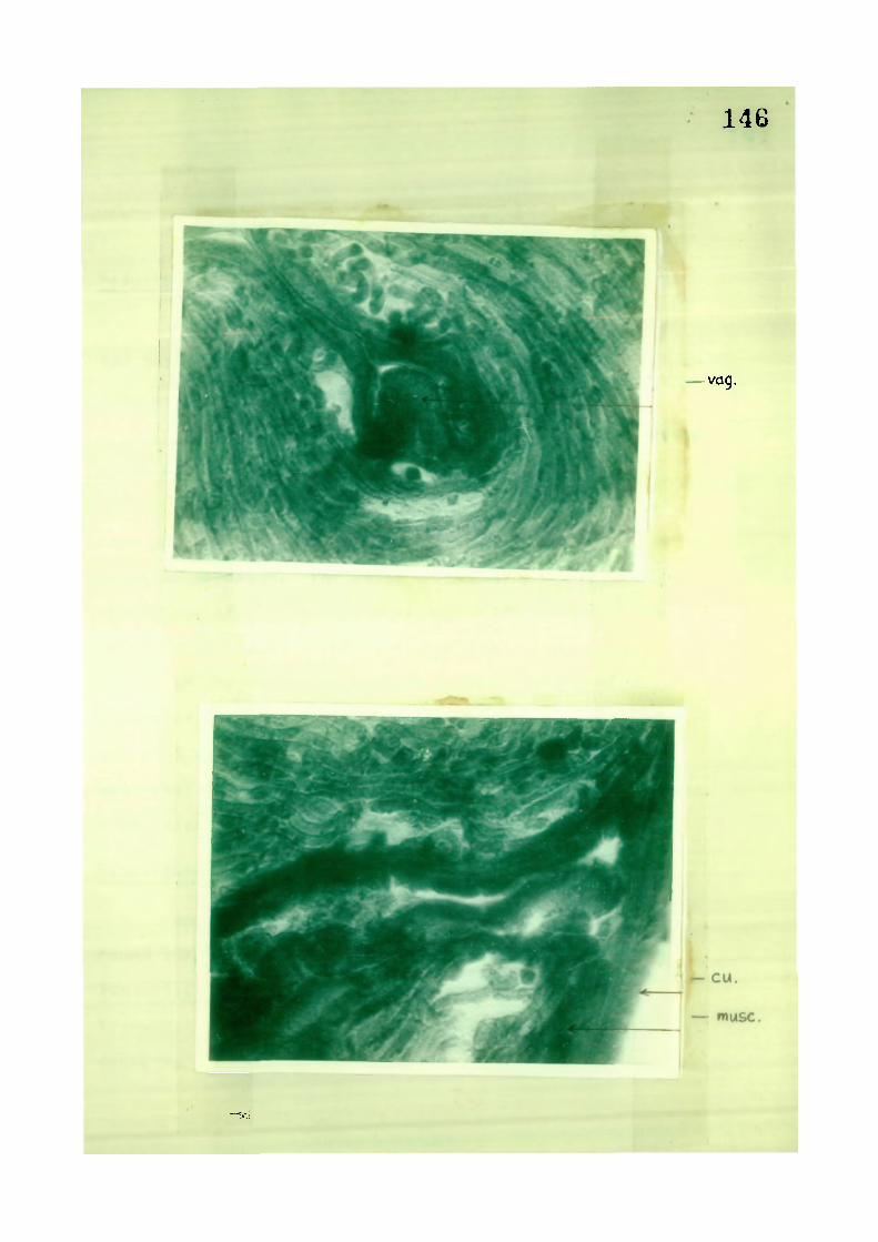
146
-vcjg.
"T'J

(d ) Carboxyl nstorase
R e s u l t s :
The body u a l l shows a f e e b l y p o s i t i v e r e a c t i o n .
The l a t e r a l e x c r e t o r y channels shnu s t r o n g l y p o s i t i v e
re a c t i o n .
Regarding the a l imentary cana l , only the i n t e s t i n e
shows a p o s i t i v e r e a c t i o n .
In the female r eproduc t i v e system, the germinal
zone and e p i t h e l i a l c e l l s c o v e r ing the o v a r i e s , shou a
p o s i t i v e r e a c t i o n . The wa l l s of o v i d u c t , the luminal
ep i the l ium of uterus and inner g landular l i n i n g of the
vag ina show s t r o n g l y o o s i t i v e r e a c t i o n .
In the male r eproduc t i v e system, l i n i n g of
e p i t h e l i a l c e l l s , c ove r ing the t e s t i s , p s o e c i a l l y o f
germinal zone are h igh ly p o s i t i v e f o r na rboxy l e s t e rase
a c t i v i t y . The e p i t h e l i a l c e l l s of seminal v e s i c l e a l so
shou s t r o n g l y p o s i t i v e re a c t i o n / F i g s . 46 -49 ) .
D i s cuss i on :
Workers l i k e Boqitsh (1956) and Halton (1967)
fouhd c a r b o x y l i c e s t e rase in tegumental r eg ions and
suggested that th i s enzyme takes part in nu t r i en t
t r anspo r t through tegumental r e g i o n s . They found the
e s t e rase to be nresent in oesoohagus and i n t e s t i n e and

148
presumed that t h i s enzyme takes part in e x t r a c o r p o r e a l
d i g e s t i o n . The presence oF th i s enzyme in teoumentary
and d i g e s t i v e system has been demonstrated by Bogi tsh
(1 966) •
In C. haukinpi , the i n t e s t i n e shouing p o s i t i v e
r e a c t i o n goes to prov/e that i t takes some part in
d i g e s t i o n . A p o s i t i v e r e a c t i on shoun by e p i t h e l i a l
l i n i n g of male and female r ep roduc t i v e system a f f i r m s
the view that these l i n i n g s take part in nut r i en t
t r a n s p o r t .
A s t r ong l y p o s i t i v e r e a c t i o n e x h i b i t e d by l a t e r a l
e x c r e t o r y channels Further support the v i eu that t h i s
enzyme takes part in the t ranspor t of the e x c r e t o r y
f l u i d .

. - 149
. . M ov.germ.z.
\ •

F iq . 48. PhotnmicrDQraph of T .5 . of adult shouiing
carboxy l es t e rase a c t i v i t y in u t e r i i . X 400,
^ iq . 49. Photornicn "^rcrj'i )f T .3 . nf adult shouinq
carboxy l 'ist-iT-iar-- act lu ity >.n e p i t h e l i a l
c e l l s of t p s t i s . X 400.

150

( i i ) M i c r o f i l a r i a
( a ) Acid phosphatase
( b ) A l ka l i n e phosph.'tase
( c ) Adinosine t r iphosphatase
( d ) Carboxyl e s t e rase
( e ) A r y l sulphatase
( a ) Acid phosphatase
R e s u l t s :
The d i s t r i b u t i o n and i n t e n s i t y o f ac id phosphatase
a c t i v i t y in the m i c r o f i J a r i a of £. haukingi i s presented
in Tab le 6. Smears incubated in subs t ra te f r e e medium
showed no r eac t i on product in m i c r o f i l a r i a .
Strong ac id phosphatase act iv/ i ty uas seen in
''lungebi I de , e x c r e t o r y v e s i c l e , G c e l l and anal v e s i c l e .
Comparat ive ly f e e b l e a c t i v i t y uas observed in the f i r s t
f eu c e l l s of the nuclear coluTin, sub - cu t i cu l a r c e l l s
o f a n t e r i o r ha l f of the body, c e l l s and l a s t
tuo c e l l s of ^hp nuclear colunn, oresent near the t i p
"of the t a i l ( F i q s . & 51 ) ,
D iscuss ion •
The d i s t r i b u t i o n of the acid phosphatase a c t i v i t y
in m i c r o f i l a r i a i s so s n e c i f i c that i t o f f e r s a parameter
f o r d is t inqui s h i n o ths pp^nera, s ' e c i c s and even the

s t r a i n s o f f i l a r i a l unrms. Thus s e v e r a l i n v e s t i g a t o r s
d i s t i n g u i s h e d the s p e c i e s o f f i l a r i a l uorms by l o c a l i z i n g
the d i s t i n c t pa t t e rns of ac id phosohatase a c t i v i t y in
c i r c u l a t i n g rn i c ro f i l a r i a e . C h a l i f o u x and Hunt ( 1 9 7 1 ) ,
Balbo and Abate (1972) d i s t i n g u i s h e d the s p e c i e s o f
D i r o f i l a r i a from Dipeta lonema even in mixed i n f e c t i o n s .
R e d i n g t o n , Plontgomery, Gerv i s and Hockmeyer (1 975 )
d i s t i n g u i s h e d h i s t o c h e m i c a l l y the m i c r o f i l a r i a e o f
Brugia malay i from B. pahangi . Teruedou anri Huff ( 1 9 7 6 )
l o c a l i z e d t h i s enzyme in LJuchereria b a n c r o f t i . Hmar
(1977) con f i rmed the o b s e r v a t i o n s o f p r ev i ous workers
and l o c a l i z e d the enzyme a c t i v i t y in d e v e l o p i n g s t a g e s
in the v e c t o r o f a l l the four f i l a r i a l worms v i z . ,
Uuchere r i a b a n c r o f t i , Brugia m a l a y i , 8. pahangi and
D i r o f i l a r i a i m m i t i s .
Braun-f^unz inqer and Sputhgate (1 977) uent s t i l l
f u r t h e r by di f f e r f? n t i at ing the f our d i f f e r e n t s t r a i n s
o f Onchocerca v o l v u l u s by l o c a l i z i n g aciri phosphatase
in the micro f i l a r i ae (TableT)•
The d i f f e r e n t areas p o s i t i v e f o r ac id phosphatase
i n d i c a t e that t h i s enzyme a c t i v i t y i s p resent on l y at
m e t a b o l i c a l l y a c t i v e r e g i o n s . I t i s supposed t h a t ac id
phosphatase a c t i v i t y i s a s s o c i a t e d u i t h c e r t a i n impor tant
f u n c t i o n a l p rocesses such as a b s o r p t i v e , p h a g o c y t i c ,
e x c r e t o r y or s e c r a t o r y a c t i v i t i e s o f the l i v i n g c e l l s .

153
The m i c r o f i l a r i a of C. haukinqi shous s t rong
a c t i v i t y at the a n t e r i o r end correspondinq to PlungebiJrie
or amphi is , probably due to i t s abso rp t i v e f unc t i on .
S i m i l a r l y the s t rong r e a c t i o n in the e x c r e t o r y and anal
v e s i c l e s i n d i c a t e s the involvement of the enzyme in
e x c r e t o r y or s e c r e t o r y processes . The s i g n i f i c a n c e of
enzyme a c t i v i t y in the C^, R^ and R^ c e l l s , and the
f i r s t feu and l a s t tuo c e l l s of the nuclear column i s
not c l e a r . I t appears , however, that the enzyme i s
i n v o l v e d in c e l l p r o l i f e r a t i o n , s y n t h e s i s , s e c r e t o r y
and absorp t i ve f u n c t i o n s .
The i n t e n s i t y of a c t i v i t y of t h i s enzyme at
Plungebilde i s very s t rong and almost s i m i l a r to that o f
D i r o f i l a r i a immi t i s . S i m i l a r l y the a c t i v i t y in the
e x c r e t o r y v e s i c l e resembles that of B. ma lay i ,
U. b a n c r o f t i and immit is but not B. pahangi in uhich
compara t i ve l y lou a c t i v i t y has been recorded . Here
Innenkorper showed a strong r eac t i on corresponding to
tha t of U. b a n c r o f t i .
Anal v e s i c l e shous s trong a c t i v i t y f o r t h i s enzyme.
This s t ruc tu re shous equa l l y s t rong a c t i v i t y in a l l the
desc r i bed s p e c i e s . Uar iab le i n t e n s i t y of enzyme a c t i v i t y
uas shoun by Schuanzgehi lde and sub - cu t i cu l a r CGlis in
the d i f f e r e n t s p e c i c s .

154
4-> P CO
•H r H - P D O u CO •p
- p OJ n +
j r CJ cn - p 1
X i D CU
- p c n CJ — » —
3 1 N Q)
"O C T3 CD (D r-H U J - P 1 <v JZ JD D a CD e CO o i o ^ ^ ^ —
a aj
10 f) CO u
•—1 -r-t + •H CO (D + CP C CD + c c c >
•H r - - - — — —
- y CTi 3 r— P CD CD JZ a U p « a : 0 +
U E c + CD
O u C CD c
>- - P 1—1 - P t i - - _ - -
•H CQ — ' p
O CD - P cn - P o m CD U ¥ CO •H p - H -t-
o u m CD Q} X - p CO a ixj ro CO _ _ ^ _
- p CD in X I ^ CT. x : Q) P C 1 u . X ) Uj - H (0 •H 2 • P o P ^ ^ _ ,
x : o CD Q cn X J
cu • 1 "D T l •P •H X i + CJ C m +
•P a . c
• D UD C
CD r-H X I cu
1 -CT cu
1 - cn m • P |
•p u 3 Q) Cd j r> c
c n •
1 1 c-'l
+ (-! + (-< + o a I
+ f-i + p + o o I
+ + + fH 4 + O
+ (-. + O I
+ + + + (H + O O 4- O I
I (-1 + o
>> (U rH ro" £ CO •rJ CJ1 D (-J iX)
+ (-( + o I
Q) Oi CD Q
X Q) C c o
T3 -P C O U
+ + + K + M + •ho O
T) 0) C O •rH -P
-P c O Q)
-t- M + M O O
+ + u u o + o
(-1 I o
o c £ •Tj
a.
cJl

155
cu 3 u 4-> LJ CO t •—1 D CD 'n U
1 N CD C T CD '—(
3 •H -C X) o CD cn U
CD .-H O r-| •H
CO CD c CD CC 25
m O-i-i to -iC c OJ c c I—1 >. o (D -p i m O f-l •H u cn X •H l-J (D cr p c CD •rH z IH
Q •a 1—( •H n m C 2 ^ -
CO CD •H U © D-
rn
+ M + o + f-4 o I
O I
+ +
+ + +
oH -o _
X) CD J cn Q) C G r-
f-( + ro ro 03 O + QJ 4-> B Cl-
cl m u ^ a c 0 3 TO D 1- X
+ +
(-1 + o +
(h (-1 O + O I
+ (-4 (-1 + 0 + 0
fH n I
+ u u -f G + O
+ p-1 p + o + o
+ + (H + + o +
+ + t + + o +
n o +
+ (-( u + o + o
+ f-l + f o + +
OD •H •H • ri U f-i 4-1 CD Cl— r-H col U n •H •r-l (D f-l Cl- -P XT o o •r-l O c F D CO •r-l b 3 XJ a
CD •H CD -P •H •H cn P o •H CL cn
O >. CD CL CD rH G> • H •p C -P r-l -p n •rH V CO t-i cn CO a>
o CD CD Q. + + + + H 1

cn X D •P r-t Ol 3 c ra CD rH O
•D CO O o m n •
Q) - -—-
U O t> JZ CT> u -H c rH n ^ CD
CD O Cl-o (0 L3
O' CO X c -p CD •iH D TD CD o rH U cn •H -P XJ CO "D CD
c O -p CO N c C 03 (-1 CO (-1 cu 3 CD cr X <t- c U <u •H cn •H N X) c Q)
n r-i C sr u •H 1 rH .H
c CO CO >> o C CD -P (0 d 5 •H f-l > aa 1 M •rH C CD -P u m Q u CD C (-1 C -p C :C
l-H ^ ffl to (0 -—-CO M -p CD C CJ CO CD -P X •rH 0) u a U (H -H CO CO u to o t-H X --1 x: •H UJ > D C;-
O CD "D U P CJI •r O M C u •H a) -r-l cE B ^ (-1
• CD •a
CD •H r-C X XI 0) CO 1
c 2
tt) o
CI
OJ
( - 1 o
•r-t U Q) -4-' cn O O-
X3 0) C O •H -P C Q] E
O
+ + +
+ + +
+ + U + o +
r) c CM -p cn tJ CD c JSC o X CD C
+ .H + -P rH •r-l . C Ol
CD CJi CD c -P c >-H CD -H CO o O CO C O rH X 3 a CO 3 Ci_ CO
Ci- -P •H CD X) Cu.
+ •P O + f-i + X + o + cn c
•H o r-l -H •p ra N
CD -H > M CO
• O O -P O rH
CJi c -P •H •rH C C to
•rH to •P (-1 -p a ai CO CO 5
CD CD CD nr tn E D + Cw + (-1 + N . C|_ + o + c CO •rH CD -H X] u Cl_ CO CO O M 3 •rH o CD Ci-X CJ o to C P
C' o >. cn -H Xj X E o (0 X CD 0 t CD + f-l p -P O X + o + O 1 (D X -p •-H 3
D. H- e (u Q n X
U 4J CM r ^ U c
p f-l Ct_ CJI o c •H CO -rH (-1 (D C ai •rH -H V p CO I— -P
a 01
[XI LJ o
dj •H CD -P •rH (J1 -P O •H a cn
o >-03 D- CD rH > 5 C 'H -P r -H ^ ra p tn 0, Qi 4-) O CD 0) rn U. 3 ^ + + +
+ + I

F i g . 50, Photomicro qraph of lonn form o^ m i c n f i l a r i a
shouino ac id phosphatase a c t i v i t y . X 900.
F i g . 51. Phot iniur- ipr ' ph of short form of
micri '-MlcMiP nhnuing acid phosnhatase
a c t i v i t y . X 400.

157
t

158
( b ) A lka l ine phosphatase
A l ka l i n e phosphatase uas found to be absent in
m i c r o f i l a r i a of C. haukingi .

159
( c ) Adenosine t r iphosphatase
Resu l ts "
adenosine t r i ph isphatase i s in t ense l y present
in the cepha l i c end, cxc r e t o r y v e s i c l e and G c e l l .
The sheath also shows a f e e b l y p o s i t i v e r e a c t i o n ( F i g . 52)
D i s cuss i on :
Adenosine tr iphosphatase is assoc ia ted u i th
energy metabolism, pembrane t r anspo r t , muscle t i s sue
format ion and absorpt i ve func t i ons . The strong ATPase
a c t i v i t y , e xh ib i t ed by the cepha l i c end, exc re to ry
v e s i c l e and G c e l l indicates the functional a c t i v i t y
and energy rretabolisr" in thesc^ s t ruc tu res .
The cophai i c end takes part in absorpt ion or
membrane t ranspor t of food m a t e r i a l ; the e x c r e t o r y
v c s i c l o shous a strnnq r eac t i on f o r t h i s enzymo, as i t
i s assoc iatpd u i f - tbt metabo l ic processes . G c e l l
shous a c t i v i t y , as i t i s r i ch in chnmat in mater ia ] and
i s f i r s t to d i v i de during development.

'006 X 'A^T^^T^oe aseq-eqcl
-soq-dtjq. b u t s g u e p e c>UTnoi,s b i a t j i x j o j q t u j
JO ujjoJ 5uo-[ JO L|deJboaoTUJo::^oq(j • 'OT j

160
'.v. 1

161
( d ) Carboxyl o s t c rase ( n o n - s p e c i f i c )
Re su I t s
An intense n n n - s o e c i f i c c i r b o x y l e s t e r a se
a c t i v i t y uas 'observed in the c enha l i c r eg i on of
m i c r o f i l a r i a e from a n t e r i o r t i p to the nerve r i n g , in
the e x c r e t o r y v e s i c l e , anal v e s i c l e and one or tuo
c e l l s near the t i p of t a i l . A f eu sub - cu t i cu l a r ce U s
a l s o shoued the carboxy l e s t e r a s e a c t i v i t y ( F i q s . 53 & 54 ) .
D i s cuss i on :
Carboxy l l c estrar hydro lases ( c a rboxy l e s t e r a s e s )
are a l a r g e group of n o n - s p e c i f i c hydro lases a c t ing at
pH optima betuecn 5.0-9,N on simple shor t - cha ined f a t t y
ac id e s t e r s . They r,rn both h y d r o l y t i c and s y n t h e t i c
in a c t i o n . Houiever, ib i s t r u e , as layers (1960) has
po inted out , that the "metabolic func t i ons of the
e s t e r a s e s are not comple te ly knoun.
Tho strong e s t e rase a c t i v i t y in the a n t e r i o r
par t o f the m i c r o f i l a r i a i nd i c a t e s a pos s i b l e f unc t i on
o f tho enzyme in absorpt ion and r e syn thes i s of f a t t y
ac id e s t e r s . The enzyme a c t i v i t y in the nerve r ing
r e g i on might represent a cho l i nos t e r a s e a c t i v i t y s ince
oC-naphthy l a c e t a t e i s a lso hydro lysed by c h o l i n e s t e r a s e s .
Houever, the p o s s i b i l i t y that the e s t e r a s e s are i n vo l v ed
in the synthes is of l i p i d s , c h a r a c t e r i s t i c of nervous
system cannot bo ove r l ooked .

S i m i l a r l y the i^cl l knoun a s s o c i a t i o n of e s t e r a s e
a c t i v i t y ui th c x c r o t o r y func t i on in many animals , o f f e r s
an exp lana t i on f o r tho intense enzyme a c t i v i t y seen in
the e x c r e t o r y and anal v e s i c l e s of m i c r o f i l a r i a .
In the c e l l s o f the p o s t e r i o r end of the
m i c r o f i l a r i a , e s t e r a s e s might be i n vo l v ed in c e l l
p r o l i f e r a t i o n or s y n t h e t i c f u n c t i o n s .
162

F i g . 53. Photomicroaraoh of long form o f
m i c r o f i l a r i a ahouinq narboxy l e s t e rase
a c t i v i t y . X 900.
F i g . 54. Photomicroqrar'h of short form of
m i c r o f i l a r i a shouinq carhoxy l e s t e rase
act iv/ i ty . X QOO.

163

164
( s ) A r y l 'Sulphatase
R e s u l t s :
A d i f f u s n d r ea c t i on a l l over the body has been
obseruedJ^Figs. 55 & 56) .
D i scuss ion ;
The e n t i r e body of the m i c r o f i l a r i a , ra ther the
nuclear column, showed r e a c t i o n f o r n ry l sulphatase
uhereas tho Innenkorpor reg ion gave a nega t i ve r e a c t i o n
in long forms and in short forris a d i f f u s e d r e a c t i o n
i s o resent along the e n t i r e l ength of the body inc lud ing
Innenkorper .

F i g . 55. Photomicrograph of long form of
m i c r o f i l a r i a showing a r y l sulohataao
a c t i v i t y . X 900.
F i g . 56. Photomicroqrrnh of short form of
m i c r o f i l a r i a shouiino n ry l sulphatase
a c t i v i t y . X TOO.

165
-A-

166
4. In v i t r o c u l t i v a t i o n
A l l the ijn v i t r o exper iments uere c a r r i e d out
at 22 C and at the pH 7.5 ad justed with the he lp of
0 .4^ NaHCOj or T r i s b u f f e r and t e s t e d with u n i v e r s a l
i n d i c a t o r .
Experiment No." 1
Ty rode ' s s o l u t i o n (Tyrode , 1910)
R inger-Locke (Locke , 1901)
Hank's BSS (Hanks and Wal lace , 1949)
and E a r l e ' s s a l i n e ( E a r l e , 1943) uere taken
and supplemented with 20 or 50^ of i n a c t i v a t e d bovine
SB r urn.
The m i c r o f i l a r i a e surv i v ed f o r 6-9 days, both in
Tyrode and Rinqpr-Locke uhen supplemented u i th 20/S bovine
serum. The s u r v i v a l t ime became 11-13 days u i th 50/S
serum supplement.
The s u r v i v a l time did not improve much in Hank's
BSS and E a r l e ' s s a l i n e . Uith 20w serum supplement the
s u r v i v a l time uas 5-10 days and u i th 50^' the s u r v i v a l time
became 10-12 days.
No grouth or development could be observed .
Usual ly the m i c r o f i l a r i a e remained ve ry a c t i v e f o r
4-5 days and then they s t a r t e d becoming s lugg i sh and
dy ing g radua l l y .

167
Experiment No. 2
Chemical ly d e f i n ed medio, NCTC 109 (Evans, Bryant ,
F io ramont i , f^cQuilkin, S a n f o r - , U e s t F a l l and Ea r l e , 1956;
McQuilkin, Evans and Ea r l e , 1957).
Eaq l e ' s MEM (Eag l e , 1959)
CnRL - 1066 ( Pa rke r , Cas tor , NcCul lock, 1957)
Grace 's Gi A (Grace , I95f0 uere used supplsmentinf j
^ u i th 20% or 50% bovine serum,
( M i c r o f i l a r i a e surv i v ed f o r 10-12 days in NCTC 109
and in medium 199 u i th 20% serum supplement s l i g h t
inc rease in length (6 j j ) occurred .
The s u r v i v a l time uas reduced to 5-5 days uhen
serum supplement u ' s increased to 50%, I t uas noted
that there uas a gradual decror'se in the pH of the
medium uhich became a c i d i c ( t ) -4 ) a f t e r 5 nays. I t i s ,
t h e r e f o r e , conc luder that e i t h e r the m i c r o f i l a r i a e could
not stand the a c i d i c pH or the concent ra t i on of the
media, uhich may be too r i c h .
The m i c r o f i l a r i a e surv i ved f o r 14 days in ClRL-IOeS,
They remained very a c t i v e in Grace ' s medium uhen
supplemented u i th 20% bovine serum and surv i ved f o r
20-24 days. No growth or development uas observed ,
50% serum supplement uas not used.

168
Experiment No. 3
E a r l e ' s s a l i n e , R inger-Locke and Hank's B53
supplemented with lacta lbumin 5%, yeast e x t r a c t 2%.
In a d d i t i o n , 20/ serum of uh i te ra t or guinea p ig or
bov ine or f o u l were used.
In Ear l a ' s medium, having the above mentioner;
percentage of lac ta lbumin and y e a s t , uhen supplemented
u i th 20^ uhi te ra t serum, the m i c r o f i l a r i a e surv i v ed
f o r 12-16 days; u i th 20'^ guinea p i g serum the s u r v i v a l
t ime became 15-17 days; u i th 20^ bovine scrum, the
m i c r o f i l a r i a e surv i v ed f o r 8-10 days and u i th 2 0>o f o u l
s'erum f o r lR-20 days.
In Rinnor-Locke and Hank's BSS the s u r v i v a l time
did not improve. The range of s u r v i v a l time i s almost
the s 'me.
Experiment No. 4
This cxneriment u- s done by using E a g l e ' s nE* ,
nedium 199, NCTC 109, C' ^IRL-IOee -" nd Grace 's Gi'lA. The
m i c r o f i l a r i a e kept comparat i ve ly more a c t i v e , the
s u r v i v a l time uas increased and sometimes there uas
l i t t l e inc rease in s i z e .
liihen E a g l e ' s NEn, having lacta lbumin (5%) and
yenst e x t r a c t (25^), supplemented u i th 2 0^ uh i te ra t
serum, uas used f o r c u l t i v a t i n g m i c r o f i l a r i a e , they
su rv i v ed f o r 7-10 days but no grouth or development uas
169
obse r v ed ; when guinea p i g sarum was supplemented,
the m i c r o f i l a r i a e surv i v ed f o r 10-11 days, they kept
q u i t e a c t i v e u i th 20^ bovine serum and surv i ved only
f o r 6-8 dcys . Uith f o u l serum supplement the
s u r v i v a l time inc reased to a maximum pe r i od of 20 days.
T h e r e a f t e r the m i c r o f i l a r i a e s t a r t e d becoming s lugg i sh
and dying g radua l l y .
The r e s u l t s in medium 199 and NCTC 109 were
almost s i m i l a r . IMCTC 109 u i th 2% yeast e x t r a c t and 5%
l a c ta lbumin , uhen supplemented with 20% uh i te ra t
serum, the m i c r o f i l a r i a e surv i v ed f o r 12-15 days and
in Medium 199 thoy surv i ved f o r 12-16 days. In both,
s l i g h t increase in length (4 yu) uas observed ; uhen
NCTC 109 and medium 199 were supplemented with 20';
guinea p ig serum, the s u r v i v a l t ime became 17-19 days
in both the media. n i c r o f i l a r i a e kept very a c t i v e and
perhaps due to the u r i g q l i n g of m i c r o f i l a r i a e , the
sheath at the a n t e r i o r end got broken. An increase of
6 yu in length was observed ; uhen supplemented u i th 20%
bov ine serum the m i c r o f i l a r i a e surv i v ed f o r 10-12 days
in NCTC 109 and 8-14 days in medium 199; uhen
supplemented with 20% f o u l serum, the m i c r o f i l a r i a e
su r v i v ed f o r 15-20 days in NCTC 109 and f o r 17-20 days
i n medium 199.
Yeast e x t r a c t (2%) and lacta lbumin (5%) uere
added to CnRL-1066 and Grace 's medium a l so and then 20/
o f d i f f e r e n t sera supplements uere g iven f o r c u l t i v a t i o n
o f m i c r o f i l a r i a e .

In CflRL-1066 u i th 20% uh i t e ra t seru^r), the
m i c r o f i l a r i a e suruiued Tor 14 days; u i th 20% guinea p i g
serum the m i c r o f i l a r i a e suruiued f o r 18-22 days,
breaking of sheath and inc rease in l ength (6 /u) uas J
observed ; with 20% bovine serum they surv i v ed f o r
12-13 days; with 2 0% f o u l serum the m i c r o f i l a r i a e
su r v i v ed f o r 20-24 days.
The Grgce ' s GMA, gave the best r e s u l t s . When
supplemented u i th 20% white ra t serum, the m i c r o f i l a r i a e
kept ve ry a c t i v e , the shenth usua l l y got broken. They
su r v i v ed f o r 14-18 days; with 20% guinea p i g serum
supplement, the m i c r o f i l a r i a e s u r v i v e d f o r 20-22 days;
w i th 20% bovii.e serum supplement, the- m i c r o f i l a r i a e
surv i v ed f o r 12-15 days and u i th 20% f o u l serum
suoplement the m i c r o f i l a r i a e surv i v ed f o r 20-28 days.
Experiment No. 5
In th i s exper iment the m i c r o f i l a r i o e uere
c u l t i v p t e d in Grace ' s G( A and CnRL-1066 supplemented
u i t h 20% crou serum, crou b lood ce l i s ( separa ted by
adding c i t r a t e , d i l u t i n g and c e n t r i f u g i n g ) , minute
f ragments of Aedes l a r v a l gut , 0.5% Aedes (pupa l )
haemolymph, 1.0% g lucose and 0.5% CEE.
The s u r v i v a l t ime inc reased to 25-30 days in
CnRL-1066 and 43-45 days in Grace 's GI A. In Grace ' s
GNA, the m i c r o f i l a r i a e surv i ved f o r the l onges t t ime .

171
most of them showed broken sheath, vet there uas no
t rue ecr iys is , a feu became shortened and th ickened with
broken sheath s t i l l i n t a c t . i o t rue deunlopmental
s tages could be obseruod.
Experiment No. 6
nonolayGrs i f white ra t embryo, chick embryo,
h ea r t , kidney and l i v e r (uh i te rat p r e n a t a l ) uere prepared
and o v e r l a i d u i th Hank's P3S, lacta lbumin 5%, yeast
e x t r a c t 2/ and uhi te rat serum IT"^.. The m i c r o f i l a r i a e
su r v i v ed f o r 5-7 days and then '^ost of them nenetra ted
under the monolayer and d i ed . c3ome of them uero found
l y i n g dead under the monolayer even a f t e r 24.0 hours.
d i s c u s s i o n :
The m i c r o f i l a r i a e of Chand le re l l a haukingi
su r v i v ed f o r about 10-12 days in NCTC 109 and medium 199
u i t h 20% scrum supplf^ment, uh i l e the m i c r o f i l a r i a e of
M r o f i l a r i a immit is ( E a r l , 1959) , surv i ved f o r 4 days
in medium 199 a l one , f o r 43 days u i th 10'/. scrum
supplement and f o r 61 days u i th serum supnlomont.
In the present case , s u r v i v a l time of C. haukingi
decreased to S-6 Hays only uhen the serum supplement
uas increased to 50%.
The s u r v i v a l time of C. haukinqi m i c r o f i l a r i a e
i s compara t i ve l y longer than that of D. immit is

m i c r o f i l a r i a e ' ' jauiytr and U'^ instp in, 1961) uhen cul t iv/ated
in V/itro in R inqer and Locke ' s s o l u t i o n u i t h d i f f e r e n t
supplemonts , Sauy j r and U e i n s t e i n ( 1 9 5 1 ) had used
Krebs -K inge r phosphate i/ith g lucoso and ^ .Sf- sodium
c a s e i n a t e and C. haukingi m i c r o f i l a r i a e uere c u l t u r e d
i n Tyrodo and Hinq^r L i c k e ' s s o l u t i o n wi th 20% bov ine
se rum.
The m i c r o f i l a r i a e of D, immit i s surv/iued f o r
7 days (Sauyer and U e i n s t e i n , 1161) in NCTC 109 and the
m i c r o f i l a r i a e o f C. haukingi s u r v i v e d f o r 10-12 days
i n t h i s medium.
The s u r v i v a l t ime o f m i c r o f i l a r i a e of C. hauking i
in GPIA wi th 20) sprum supplement remained only f o r
20-24 days but the m i c r o f i l a r i a e o f naracanema formosana
(Uood and S u i t o r , 1966) dev(^ loped to i n f e c t i v e s t age
in GnA supplementpd u i th D.S^ i n s e c t haemolymph and 1
f o e t a l bov ine serum.
Bcjsides a l l t h e s e , a number of o ther rombinai-ions ,
c h c m i c a l l y de f in t d m fd i a , ^upnlemented u i th v a r i o u s
n u t r i e n t s and TC mono layers , o v e r l a i d u i t h growth
media , having Hank's 653, l a c t a l b u n i n , yr.ast e x t r a c t
and serum, have been used f o r c u l t i v a t i n n v i t r n ,
the m i c r o f i l a r i a e nf C. haukinni . Th j r e s u l t s
uerp ob ta ined uhen c u l t i v a t ' - d in G!'1A supolemented

173
ui th 20f> ' r r o u S f ^ r u r r , n r o u b l o T d C f j l l s , minute fragments
of Aode3 l a r v a l qut and 0.5/^ Aedes ^pupal) haumolymph,
1.0% g lucose and 0.5'/^ CEE. In such a medium the suruiual
time increased to days.

174
REFEREMCES
Abe, Y . , Hor i , E . , Yana i i , T . , Hno, I . , Ushuku, F.
and Kaqei , .J. ( i 9 6 0 ) . Studies on F i l a r i a and
F i l a r i a s i s . Morpho log i ca l and His tochemica l
s tud i es on r p i r r o f i l a r i a of 'Juchsreria h a n c r o f t i
i^ed. 3. Kaqoshima Uniw. 1058-1114.
Adams, C.U.M. and S l ope r , O.C. ^ 955) . Technique f o r
demonstrat inq neurosecre to ry m a t e r i a l on human
hypothalmus.
Lancet 1: 651-65?.
Ahn, 3 . 0 . MQ^B). H is tochemica l s tud i es on the
a c t i v i t y of /-T°ase and auto rad iog raph ic study
on the uotake of ''^C ATP in As car i s
lumbrico ides t u i 3 .
Korean Centra l 3. P_ed. 30. 5'^-59.
A l i , S.'' . ' 1T56 ) . I t u d i e s in thn nematode o a r a s i t e s
of f i s h e s and b i rds found in Hyderabad S t a t e .
I ndian 3_' He Imintho 1. J •
A l l e n , U.R. and Harkema, R. (1972) , H is tochemica l
enzyme s tud i e s on Pharynnostnmoides
adenoceoha la "Hfickerdite , ' ^ i l l e r and Harkema,
1971, CTrematoMa, OiplnstoTiat i dae ' .
Proc . Helminthnl . 3ac. Uash. 39. 1'^1-153.

175
A l l i s o n , l i . F . , Ube laker , 3.U. , Uebs t e r , R .U .3 r . and
R i d d l e , 3.PI. (1972 ) . P r e p a r a t i o n of Helminths
f o r scanning e l e c t r o n m ic roscopy .
3. P a r a s i t o l . S8: 415-416.
A n s a r i , 3 . A . and B a s i r , . A. ( 1964 ) . H i s t o l o g i c a l
anatomy o f S e t a r l a c e r y i .
I ndian 3. He lmin tho l . Sonographic supplement .
Dec. 1964.
A o k i , Y. ( 1971 ) . Exsheathing phenomenon of
m i c r o f i l a r i a e ijn v i t r o .
Trop . f^ed. Nagasaki 13: 170-179.
A r i z o n o , N. , r a t s u o , K. and Yosh ida , Y. ( 1976 ) .
Scanning e l e c t r o n microscopy of 3 t r o n g y l o i d e s
p l an i c eps Roge rs , 1943.
3apanQqe 3. P a r a s i t o l . 46R-474.
Asadov/, N . i , ( 1973 ) . The f i n e s t r u c t u r e o f Tr i c h u r i s
ovi9 Hbilgaurd, 1795.
Hate r , nauch. Kon fpr . a s o i r . posuy. poluuekov/.
Yubi l eyu / 'zerbaidzhanskooo -e daooq icheskogo
I n s t i t u t A im.W.1. Lenina Baku, U . 3 . S . R . (1973)
307-210.
Paker , A .n . ( 1936 ) . S tud i es on He t e rak i s g a l l i n a r y m
(Gmel in, 1710) Freeborn , 1923, a nematode
p a r a s i t e of f o u l s .
Trans. R. Can. I n s t . 20 179-21 5; 2 V 51-36.

Pa lbo , T. and Mhate, 1. (1972) . H is tochemica l
d i f f e r e n t i a t i o n of m i c r o f i l a r i a e of D i r o f i l a r j a
immi t i s , D i m f i l a r i a renens and Oipeta lonema 9 p.
P a r a s s i t o l n g i a 14: 239-244.
Bano, 3. and Sen, A.B. (1973) . Cephal ic s t ruc tu r e s
in the m i c r o f i l a r i a of Chand le re l l a hauking i .
3. He lminthol . 47: 1 -8 .
Ba rc l ay , R. '1965 ) . F i l a r i a s i s in South-Uest 3abah.
Ann. Trop. f^ed. P a r a s i t o l . 59: 340-345.
Barka, T. and Anderson, P.D. (1962 ) . H is tochemica l
methods f o r ac id-phosphatase using hexazonium
p a r a r o s a n i l i n as coup l e r .
2* Histochem. Cytochem. 10-. 741-7 53.
Bark;,, T. and Anderson, P .3 . (1 963) .
H i s tochemis t r y . Theory P r a c t i c e and
B ib l i og raphy pp. 2 42-2 44.
York, Evanston and London: Hoeber
Medical D i v i s i o n , Harper and Rou.
Ba rne t t , R.3. ''1 962 ) . Pine s t r u c t u r a l l o c a l i z a t i o n
of a c e t y l c h o l i n e s t e r a s e at myoneural j unc t i on .
3. C e l l B i o l . 12: 247.

177
B a r t e l , P^-H. and Hansen, Pl.T. (1962) . D e s c r i p t i o n of
micr 3f i la^ri ae 'of f ^ i c i p s e l l a brev icauda Lyons A
Hansen, 1961. ( F i l a r i o i d e a ) from the black
t a i l e d Jack Rabb i t , u i th notes on m i c r o f i l a r i a e
of hares.
3. P a r a s i t o l . 4R; 43-46.
Barus, U. , K o t r l a , B. and Tenora, F. (1977) . A
scanning e l e c t r o n mic roscop ic study of
sp i cu l a r sheath of some T r i chur ids (Nematoda).
F o l i a P a r a s i t o l . 107-110.
Barus, U. , U i g e r , R . , Tenors, F. and Stanek, n.
(1979) . Scanning e l e c t r o n microscopy of the
l i p d e n t i c l e s o f Toxascar is l e on ina , Toxocara
canis and T. catj^ (iMematoda) .
U a s t n. Ces k . Spo l . Zoo l . 43: 3 -6 .
B a y l i s , PI.A. (1 936) . The fauna of B r i t i s h I n d i a ,
i nc lud ing Ceylon and Burma. Mematoda \Jol. I
Tay l o r and Francis L t d . , London.
P a y l i s , I^.A. (1 939) . The fauna of B r i t i s h I n d i a ,
i nc lud ino Ceylon and Burma. Nematoda l/ol. I I
Tay l o r and Francis L td . , London.
Becke t t , E.B. and '^acdonalri, U.U. (1972) . The
morphology of the r e c t a l p ro t rus ion of
B. nahangi and sub -pe r i od i c B. malayi l a rvae as
a method of d i f f e r e n t i a t i n g between the tuo
s p e c i e s ,
Ann. Trop. ( ''ed. P a r a s i t o l . 66: 1 35-141.

178
Bennet, C.L. (1975) . 3ranning e l e c t r o n microscopy
of Fasc i o l a hepat ica L. during growth and
maturat ion in the mouse.
3. P a r a s i t o l . 6 1 * g 9 2 - B 9 R .
Bi rd , A.F. (1971 ) . The s t ruc ture of nematodes.
Academic P r e s s , Neu York, 318 p.
Bjorkman, N. and T h o r s e l l , U. (1964) . On the Fine
s t ruc ture and r e so rp t iue func t i on of the
c u t i c l e of liv/er f l u k e , F. hepat ica L.
Exp. Ce l l Re^. 31 9-329.
Bog i t sh , B.3. (1 966) . H is tochemica l observ/ations on
Posthodiplostomum minimum TT. Esterases in
sub - cu t i cu l a r c e l l s , h o l d - f a s t organ c e l l s
and the nervous system.
Exp. P a r a s i t o l . 19: 64-70.
Boq i t sh , B.3. (196'^). Fine s t r u c t u r a l l o c a l i z a t i o n
of aci" ' phosphatase and a ry I su lpha tase
a c t i v i t i e s jn the in t e rmed ia te layer of
Hymenolepis diminuta c y s t i c e r c o i d s .
Trans, Am. f l i c r o s c , Soc. TB: 411-419.
179
Boqolepo\/a, I . I . (1975) . H i s t o l o g i c a l cha rac t e r -
i s t i c s o f the i n t e s t i n a l s t ruc ture of the
p a r a s i t i c nGmatodes Amidostomum anse r i ,
Uncinaria s tenoccphala and 'lesophagostomum
v/enulosum (sub o r d e r : S t r o n g y l a t a ) .
Sb. F-<ab. Lenin gr . \jet. Tnst . ( F i z i o l .
Biokhim. Morphol. Selskozyaistv/ennykh
Zhiuotnykh) 203-210.
Bogolepowa, I . I . and Chernozerskaya, N.G. (1971 ) .
H i s t o l o g i c a l and c e r t a i n h i s t ochemica l
c h a r a c t e r i s t i c s of i n t e s t i n e of A s ca r i d j a in
d i f f e r e n t f u n c t i o n a l s t a t e s .
Tr . Wses. I n s t . GeIminto1. im K . I . Skr j ab in
57-62.
Bogoyav/lenskii , Yu.K. (1176) . H i s t o l o g i c a l study
of the f i n e s t ruc ture of nematodes.
Tr . Ge l^mintol. I ab. ( P n b l . Hbshch. Q e l m i n t o l . )
26: 94-98.
Boqoyau l e n s k i i , Yu .< . , Onushko, l\l . 1/. and Andreeua,
A.i^. (1978) . r i i c romorpho log ica l o r g a n i s a t i o n
uf thti male g e n i t a l system of nematodes.
T r . Ge lmintnl . Lab. (Nematody i akan tose f a l y
I'lorpho loQi ya S is temat ika e k o l o g i y a i
f i z z i o l o g i y a ) ^ ; 193-199.

180
Bogoyaw l e n s k i i , Yu. and S k r j a b i n , A.A. (1971 ) .
Comparatiue h i s t o l o g i c a l invyesti ga t ions of
the somatic musculature of CBrtain nematodes
o f the sub-orde^r s p i r u r a t a .
Flatcr. Mauch. Konfer . Unses. Hbshch.
Ge rn in t o l . 49-57.
Bok, S .S . (1973) . H is tochemica l s tud i e s on
Helminths. E s p e c i a l l y o.i the d i s t r i b u t i o n of
adenosine t r i phospha tase .
Korean J. P a r a s i t o l . 11: 126.
B o l l a , R . I . , Ue ins t e in , P . P . and Lou, C. (1974 ) .
Acid phosphatase in deve l op ing and age ing
Nippostrongy lus b r a s i l i e n s i s .
Comp. Biochem. P h y s i o l . 43: 131-145.
Borge rs , M. , Bossche, ri.l/an Hen and Schaper, 3,
(1970) . The u l t r a s t r u c t u r a l l o c a l i z a t i o n of
n n n - s n e c i f i c phosphatases in the i n t e s t i n a l
e p i t h o l i u r of A j c a r i s suum.
3. Histonhom. Cytochem. 1R: 519-521.
braun-'^'unzinqer, H.k. and i ou thge t e , B.A. (1 977).
P r e l im inary s tud ies on the h i s t ochemica l
rlif f e r o n t i at ion of s t r a i n s of Onchocerca
v/olvulus m i c r o f i l a r i a e in Tono.
Gu l l . U. H. ] . S5: 569-575.

181
Cremner, K .C. , ' T g i l u i c , B . . , Ke i th , R.K. and
B e r r i e , D.A. (1Q73). A c e t y l c h o l i n e s t e r a s e
s e c r e t i o n by p a r a s i t i c nematodes I I I .
Oesophagostomum sp.
I n t . 3. P a r a s i t o l . 3: 609-619.
Brug, S .L . (192 9 ) . F i l a r i a malayi n .sp . p a r a s i t i c
in man in the Plalay a r c h i p e l a g o . Trans .7th
Coh^r. Far-East Ass.
Trop. r^ed. Ind ia 1927 3'- 27 9.
Buckley , 3 . 3 .C . (1955 ) . Symposium on L o i a s i s \1_.
The morphology of the l a r v a l s tages in the
v e c t o r : some of the problems inv/olved.
Trans. R. Soc. Trop. ^"ed. Hyg. 122-126.
Buckley , 3 .3 .C . (1960) . On Brugia gen. nou. f o r
Uucherer ia sp. of malayi group.
Ann. Trop. j ^d . Paras i t o l . 7 5-77.
C h a l i f o u x , L. and Hunt, H.D. M971 ) . H is tochemica l
d i f f e r e n t i a t i o n of O i r o f i l a r i a immit is and
Dipetalonema reconditum.
1' '"ted. Assoc. 158: 601-60^^.
Cha l i f oux , L .U . , Hunt, R .D. , Garc ia , F .G . , Schga l ,
P .K . and Comiskey, 3 .R. (1973) . F i l a r i a s i s in
noLJ uor ld monkeys: h i s tochemica l d i f f e r e n t i a t i o n
of c i r c u l a t i n g m i c r o f i l a r i a e .
Lab. Anim. Sc i . 23: 211 -220.

182
Chandler , A.C. ' '1Q24). Neu f i l a r i a e from Indian
b i r d s .
P a r a s i t o l o g y 16. 398-404.
C h a t t e r j e e , R .K . , 3en, A.B. and Bhattacharya, O.K.
(1965 ) . Chandlere11a haukingi sp. nov. a
f i l a r i a l pa ras i t e of the Indian Jungle Crou
Corv/us macrorhynchos Uag l c r .
Ann. Trop. P^ed. P a r a s i t o l . 434-440.
Chen, S.IM. and Houe l l s , F^.E. (1979 ) . The in v i t r o
c u l t i v a t i o n of the i n f e c t i v e laruae and the
e a r l y mammalian s tages of the f i l a r i a l uorm
Brugia pahangi.
Ann. Trop. ^ed. P a r a s i t o l . T^: 473-486.
Cheonq, U.H. , Umar, Hassan B. and Sivanandam, 5.
(1965) . An attnmpt to separa te nixed Brugia
i n f e c t i o n s by b i o l o g i c a l means.
) ingapore i^gd. J. 6: 43.
Chi tuood, B.C. (19?9 ) . Mnt-es on tho copu la to ry sac
of Bhabdi t i s , j t rongy loides_.
Schn. Helm. Soc. Uash . j . Pa ras i t o l . 2R1-292,
Chituoori, B.C. (1930 ) . The s t ruc tu r e of the
esophagus in tho T r i churo iden .
3. P T r a s i t o l . 17: 3S-42.

183
Chituood, B.C. A comoarat i ve h i s t o l o g i c a l
study of c c r t a i n nematodes.
Z. Horphol . 2'37-?R4.
Ch i tuood , O.G. ( 1936 ) . The va lue of e sophagea l
s t r u c t u r e s in nemic c l a s s i f i c a t i o n .
3. P a r a s i t o l . 22: 528.
Chi tuood, B.G. and Chi tuood, n.B. ( 1933 ) . The
h i s t o l o g i c a l anatomy of Cepha lobe l lus p a p i l l i q e r
Cobb, 1920.
Z. Z e l l f o r s h . 19: 309-355.
Chi tuood, B.C. and Chi tuood, '^.B. ( 1940 ) . An
i n t r o d u c t i o n to Nemato logy, Sec t inn I I , ISO n.
Chi tuood, B.G. and Uchr, E.E. (1 934) . The v/alue of
c e p h a l i c c h a r c c t c r s in nematode c l a s s i f i c a t i o n
u i th s p o c i a l r e f e r e n c e to the super f a m i l y
I p i r u r o i den.
i - L ' P ^ras i t cnk 7- ?73-3qh.
Choudhury, A . P . , Djsr .u i ta B. Hay, H.N. and
Bhc-iduri, I\1. U. M 9 5 5 ) . Hi-5tochemical pa t t e rn of
the m i c r o f i l a r i a nf L 'ucherer ia bc 'nc ro f t i in the
p e r i p h e r a l b l ood .
Du l l . Ca l cu t ta Sch. Trop. "led. 4. 23-24.

184
Comiskey, 3 .R. (1973 ) . F i l a r i a s i s in neu world
monkeys. H is tochemica l d i f f e r e n t i a t i o n of
c i r c u l a t i n g m i c r o f i l a r i a e .
Lab. Anim. S c i . 23: 211-270.
Dav i s , O .A . , Bog i t sh , B.3. and Nunnal ly , D,A. (1969)
Cytochomical and b iochemica l obse rva t i ons on
the d i g e s t i v e t r a c t s o f d i g e n e t i c t rematodes .
I I I . N o n - s o e c i f i c e s t e r a s e s in riaematoloechus
medioplex u s.
Exp. P c r a s i t o l . 24: 121-129.
Daus, B. (1962) . H i s t o l o g i c a l study of the caeca l
ep i the l ium of F. hepa t i ca .
P a r a s i t o l o g y 52: 493-493.
Dennis, E .A . , 3harp, and Douglas, R. (1974 ) .
Carbohydrate reservEiS and phosphatase a c t i v i t y
in tf ie mol iLisc-trematode r e l a t i o n s h i p of
l^yt i lus nduLis 1. and '-"roctoeces maeculatus
(Looss , 1901) 'Idhnor, 1911.
3. He Imin tho l . 4R : 1 -14 .
Devaney, E. and Houe l l s , R.E. (1978 ) . The
exshep'thment of m i c r o f i l a r i a e of Brugia pahangi
in v i t r o ' 'Troc. Spring OlbBt., B r i t i s h 3oc.
P a r a s i t . , Canterbury, England, 5-7 A p r i ' 1978.
A b s t r a c t ) .
P a r a s i t o l o o y 77: 34.

1B5
Oev/aney, E. and Houe l l s , R.E. (1 978) . The
development ijn v i t r o of a r t i f i c i a l l y
exsheathed m i c r o f i l a r i a e of Brugia pahangi
(Dcmonstrr t i on ) . In (Medical entomology
Centenary. Symposium proceed ings 23-25 Nov. 1 977,
London, U.K.
R. Soc. Trop. f^ed. Hyg. 1978.' 131.
Devaney, E. and Houe l l s , R.E. (1979 ) . Culture
system f o r maintenance and development of
m i c r o f i l a r i a e .
Annl. Trop. Pled. P a r a s i t o l . 139-144.
Devaney, E. and Houe l l s , R.E. (1979 ) . The
development of exsheathed m i c r o f i l a r i a e of
Brugia pahangi and B. malayi in mosquito c e l l
l i n e s .
Annl. Trop. f c d. P a r a s i t o l . 7_3: 387-392.
Devaney, E. and Houe l l s , R.E. (1 979) . The
exsheathment of Frugi 'j oahanqi m i c r o f i l a r i a e
under c o n t r o l l e d cond i t i ons ijn v i t r o .
Annl, Trap. I^cd. P a r a s i t o l . 73" 227-233.
D ick , T .A. (1972 ) . The u I t r a s t r u c t u r p and func t i on
of the c u t i c l e of Syphacia obve la ta (Hur 'olphi ,
1 802) , Sourat , 1916 and r a l i t e d s p e c i e s .
D i ss . - b s t r . I n t . 33. 1323.

u i ck , T .A. and U r i g h t , K.A. (1974 ) . The u l t r a -
s t ruc tu r s of the c u t i c l e of the nematode
3yphacia obv/elata (Hudolphi , 1802) I I I .
Cu t i c l e a s soc i a t ed u i th the male r ep roduc t i v e
s t ruc tu r e s .
Can. 3. Zoo l . 179-1B2.
D ick , T .A . and Ur i qh t , K.A. (1974 ) . The u l t r a -
s t ruc tu re of c u t i c l e of the nematode
Syphacia jbvyelata (Rudo lph i , 1 302) II/. The
c u t i c l e a s soc i a t ed with the f emale e x c r e t o r y
pore , uulva and uaqina v/pra.
Can. 3. Zoo l . 245-250.
Dike , S.C. and Read,, C.P. ( i g 7 1 a ) . Tequmentary
phosphohydrolases of Hymenolepis diminuta.
3. P a r a s i t o l . 57: 81 -87 .
D ike , 3 . r . and Rr=ad, C.P. ( i q 7 1 b ) . Re l a t i on of
tequmentary phosphohydrolases and sugar
t ransDort in Hyreno lep is diminuta.
3. P a r a s i t o l . 1251 -1255.
Oouvyres, F. U. (1979) . ^ y/jtro development of
T r i chos t r onqy lus axe i from i n f e c t i v e l a rvae
of young adu l t .
3. P a r a s i t o l . 65. 79-14.

187
Dro id z , 3. (197-3). Genetic i s o l a t i o n as a c r i t e r i o n
' f o r riefininq a spec i e s w i th in the p a r a s i t i c
nematodes.
LJiari. P a r a z y t o l . 25; 171-18:^.
E a r l , P .R. (1 959) . F i l a r i a e from dog i_n v i t r o .
Ann. N. Y. Acad. 5 c i . 7^: 163-175.
Eberth, C.3. (186:5). Untersuchunqen uber Nematoden.
77 pp . , L e i p z i g .
Eh l e r s , H. (1899) . Zur Kenntnis der Anatomie und
B i o l o g i e von Oxyuris curvu la .
Arch. Naturg. 1-26.
E r l ange r , N. and Gershon, D. (1970 ) . Studies on
ageing in Nematodes J I . Studies oP the
a c t i v i t i e s o f s e v e r a l enzymes as a func t i on
of age.
Exp. Geronto l . 5: n - l 9 .
Feng, L.C. (193'5), A comparative study of the
anatomy of M i c n f i l a r i a malayi Bruq, 1927 and
T i i c r o f i l a r i a b a n c r o f t i Cobhold, 1877.
Chin. Med. 3. 47: 1214.
F l u r y , r . (1912) . Zur Chemie und T o x i k o l o g i e der
Ascor iden.
Arch. Exp. Pa tho l . 67: 275-392.
188
Franks, (^.B. and S t o l l , N.R. (1 945) . The i s o l a t i o n
o f m i c r o f i l a r i a e f n m blond f o r use as an t i g en .
3. P a r a s i t o l . 31: 158-162.
F r i pp , P . 3 . (1967 ) . H is tochemica l l o c a l i z a t i o n of
e s t e rase a c t i v i t y in Schistosomes.
Exp. Paras j t o l . 330-390.
Fu l l ebo rn , F. (1913 ) . Zur morphologie der
D i r o f i l a r i a immit is Leyd i , 1856.
Zentb l . Bakt. Paras i tkr i . 1; Or iq . 65, 341.
Fu l l ebo rn , F. (1913 ) . Be i t r age zur Plorphologie und
Di f f e r e n t i a l d i a g n o s e der l^dkrof i l a r i e n .
Arch. S c h i f f s - u . Tropenhyg. 17: Beih 1, 7.
G a l l o q l y , R.L. (1972 ) . ATPase and membrane ,
t r anspor t in the ra t tape uorm Hymenolepis
diminutQ.
Diss . Abstr . I n t . 33: 1398.
Gear, N.R. and Fr ipp , ^ . 3. (1974 ) . Comparison of
the c h a r a c t e r i s t i c s of acety I cho l i n e s tp ra s e
present in four s o e c i e s of Schistosoma.
Comp. Biochem. P h y s i o l . 47: 743-7 52.
Goh, 5 . L . and Davey, K.G. (1976 ) . A c e t y l c h o l i n e s t e r a s e
and synapses in the neruous system of
Phocanema dec ip i ens (Nematoda) : a h i s t o -
chemical and u l t r a s t r u c t u r a l s tudy.
Can. 3. Zoo l . 54: 752-771.

189
G o i l , [^.Pl., Suada, T. and Sato , K. (1 973) . Some
b iochemica l s tud i e s on the n o n - s p e c i f i c ac id
phosphomonoeGterases of D i r o f i l a r i a immi t i s .
Jap. 2- £3: 215-218.
GoLdschmidt, R. (1906 ) . H i t t e i l u n g e n zur H i s t o l o q i e
von Asca r i s .
Zoo l . Anz. 29: 719-737.
Govinduar, S . L . , Gauande, T .S . and Har inath, B.C.
(1974) . Enzymes in LJuchereria b a n c r o f t i .
Indian 3. Biochem. Bioohys. 11: 338-339.
Grube, A,E. (1 850) . Uie Femi l i en der Annel iden.
Arch. Naturg. 16: 249-364,
Gupta, A.m. and 3oad, P . P . (1974) . N o n - s p e c i f i c
Bsterase and i t s inuoluement in the dynamism
of outer body cowering of t rematodes , ces todes
and nematodes.
Acta Helminthol . SO: 124-130.
rialton, D.U. (1T67 ) . Studies on c a r b o x y l i c e s t e r a s e
a c t i v i t y in Fasc i o l a hepa t i ca .
3. P a r a s i t o l . 53: 1210-1216.
Hal ton, D.U. (1968 ) . Light and e l e c t r o n microscope
s tud ies of cu rboxy l e s t e rase act iv/ i ty in the
trematoda Haplometra cy 1 indrace? .
3. Po r r - s i t o l . 54: 1 124-1 130.

228
r
Hal ton , D.IJ. r.nd Plorr is , G,P. (1969) . Occurjfence of
chol incstGrnscj and c i l i a t e d sunsory s t ruc tu r e s
in a f i s h g i l l f luko D i c l i dophora mer langi
(TrQmatoda-Plonoqenea).
Z. Paras i t enkd . 33: 21-30.
Hamann, 0. (1895 ) . Die Memat he Iminthen 2 : 1-120.
Har t , R.3. and Lee, R.n. (1966) . Cho l ines t e rase
a c t i v i t i e s o f va r i ous nematode p a r a s i t e s and
t h e i r i n h i b i t i o n by the organophosphate
an the lm in t i c s Haloxon.
Exp. P a r a s i t o l . 18: 332-337.
Hawking, F. (1952 ) . A h i s t o l o g i c a l study of
Onchocerc ias i s t r e a t e d u i th Heterazan.
Br. Med. 3, 1: 992-994.
Hayashi , H. (1973 ) . ATP format ion by mitochondr ia
from Ascar i s lumbr ico ides ua r . su i s
Jap. 3. P a r a s i t o l , 22: 97-107.
Hother ing ton , D.C. (1923) . Comparative s tud ies on
c e r t a i n f e a t u r e s of nematodes and t h e i r
s i g n i f i c a n c e 117.
B i o l . Olonogr. 8: 1-62.

191
riirumi, H. , Rask i , T . 3 . arid Dnnes, (M.a. (1971 ) .
t^rimitiuG musclo c e l l s of Nematedes; Plorpho lo ni r>a 1
aspects nf n 1 atyniyarian and shal low
roe lomyarian muscles in tuo o lan t u a r a s i t i c
nematodes, Tr icho^orus c h r i s t i e i and
Longidorus e l onga tus .
3. U l t r a s t r u c t . Hcs. 34: 517-543.
Hobson, A.O. (1948) . The phys i o l ogy and c u l t i v a t i o n
in a r t i f i c i a l media of nematodes p a r a s i t i c
in the a l imentary t r a c t of animals.
P c r a s i t o l o g y 183-227.
Hoqger, C.H. , Estey , H.H, , K i s i e l , M.J. and
Zuckorm^n, E'.ri. (1976) . ' icanning e l e c t r o n
microscooy of yiung and o ld Caenorhabdi t i s
bri qqsae fo-r- lo3 .
3. Nomito l . 'T: 2'^7-2>P,.
I n q l i s , liJ. C. (1064) . fht' s t ruc ture of nematode
cut i r l e .
Proc . Zoo l . S j c . London 143: 465-5D2.
I n q l i s , U.G. (196S) . npttorp of e v o l u t i o n in
p a r a s i t i c nom-^torics. In Evo lu t i on of P a r a s i t e s ,
ed. A.E.R. Tay l o r (,5rd jymp. Br. Soc. P a r a s i t . )
79, Ox ford : P l a c k u e l l .

192
luamoto, I . (1972) . Studios on s u r v i v a l time of
micro f i l o r i a e .
Trop. Hed. Nagasaki 124-137.
I y enga r , T l . i .T . ( I T^Q ) . D i f f e r e n t i a t i o n of
m i c r o f i l a r i a e of Uuchereria b a n c r o f t i and
F i l a r i a m 13 y i •
Indian 3. Res. 27_ 563.
3 a e g e r s k i o e l d , L .A. (1894) . Be i t rage zur Kenntnis
der Nematoden.
Zoo l . 3ahrb. Abt. Anat. 7: 449-532.
3?S2C Z , F .F .3 r . (1 976) . L ight and e l e c t r o n
microscop ic (both t ransmiss ion and s t e r eoscan )
s tud ies of the oesophageal r eg i on o f female
Rhigonema thysanophora C r i t e s , 1965 (iMematodao
Rh igonemat idae ) (Uo l . I & I I ) .
D iss . Abstr . I n t . 37: 646-647.
3one3, B.R. , Smith, B.F. and L e f l o r e , U.B. (1979) .
The u l t r a s t r u c t u r a l l o c a l i z a t i o n of a c e t y l -
cho l i n e s t r a s e a c t i v i t y in the sco l ex of
c y s t i c e r cus of Hydatigera t a e n i a e f o r m i s .
Trans. Am. ^ i c r o s c . Soc. 98: 154.

1B3
3ones, 3. (1961 ) . Des c r i p t i on of S p l e n d i d o f i l a r i a
f l e x i u a g i n a l i s (iMematoda: Inchoce rc idon ) Fro
eastern crou and key to the spec i e s of genus
S p l e n d i d o f i l a r i a S k r j a b i n .
3. P a r a s i t o l . 47: 437.
m
Jordon, P. (1955) . Notes on e l e p h a n t i a s i s and
hydrocoe l due to Uuchereria b a n c r o f t i .
K ikuch i , S. (1976) . Scanning e l e c t r o n microscopy of
nematodes of mammals and b i r d s , Asca r i d i a
COlumbae.
3. \iet. Pled. 660: 41 9-422.
K ikuch i , S. (1976) . Scanning e l e c t r o n microscopy of
nematodes of mammals and b i r d s , Ascar is
lumbr ico ides .
3. Uet. i^ed. 661 : 487-493.
K ikuch i , S^ (1976 ) . Scanning e l e c t r o n microscopy of
nematodes nf mammals and b i r d s .
3. Uet. Med. 652: 555-557 ; 559-553.
K ikuch i , 3. (1976) . Scanning e l o c t n n microscopy of
nomatortes of mammals and b i r d s .
3. [let. Med. 663: 629-635.

194
K l e i n , 3.B. and [ j radJ fy , . L. (Or. ) (1 974) . Induct ion
of norpho l o n i c a l changes in r n i c r o f i l a r i a e from
D i r o f i l a r i a immit is by in v/itro culturu
tachniquns.
J. P a r r s i t o l . 60- 649.
Klokke, A.H. (1961) . F i l a r i r s i s due to Brugja malayi
in South Borneo ( I n d o n e s i a ) .
Trans. R. Soc. Trop. "lyd, Hyg. 55: 433.
Ko i e , f l 9 7 7 ) . Ttornoscan s tud ies of c e r c a r i a u ,
metacDrcar iae and adult C ryp t oco t y l e l ingua
(C r ep l i n , 1125) r i s rhne i i o r , 1903 ^Trematoda:
Hetcrophyidao) .
3. P a r a s i t o l . 63: 835-B39.
Korke, U.T. (1929) . Ibserva t inns on the atv i l e a l
v a r i e t y of b a n r r j f t i and i t s s i nn i f i c ance , 1\1.
Indian J. fu^ . 16' in23.
K r a l j , M. and Lui , A. (1967) . His tochemica l
s tud i es of cholint-stc raso act iv/i ty in
Paramphistomun^ eeru i and D io lod i sous
subc lavatus .
Uet. Arh. 37: 257-261
Laurence, B.R. and Simpson, ^^.G. (1968) . Cephal ic
and pharynq.^nl s t ru r tu ros in m i c r o f i Inr ine
as reuealnd by s t a i n i n g .
3. He lminthol . 42" 309-3311.

195
Laurence, B.R. and Simpson, f i. G. (1969) . S ta in ing
m i c r o f i l a r i a e f o r s t ruc tu r e s other than n u c l e i .
Trans. R. 3o£. Trpp. r- e d. Hyg. B01-810.
Laurence, B.R. and Simpson, I' i.G. (1971) . J' i cr of i l a r i a
of Brug ja : A f i r s t s tage nematode l a r v a .
3. He lminthol . 23-40.
Lee, C.C. and ( f i l l e r , 3.H. (1957 ) . Fine s t ruc ture
of D i r o f i l a r i a immit is body u a l l musculature.
Exp. P a r a s i t o l . 20: 334-344.
Lee, D.L. (1966 ) . The s t ruc ture and composit ion of
the helminth c u t i c l e .
Adv, P a r a s i t o l . 4: 1R7-254.
Lee, D.L. (1972) . The s t ruc ture of the helminth
c u t i c l e .
Adv. P a r a s i t o l . 10: 347-379.
L i , H.C. (1933) . Report ^n a c o l l e c t i o n of
p a r a s i t i c nematodes, mainly from north China.
Par t I : F i l a r i o i d e a .
P a r a s i t o l o g y 25: 192.
L i l l i e , R.D. (196? ) . H i s t o p a t h o l o g i c t e chn i c and
P r a c t i c a l H i s tochemis t ry , IMeu York.

196
La, 3 . 3 . , Ha l ] , 3 . E . , A H e n d e r , P .A . and K l a i n e r ,
M.S. (1975) . Scanning e l e c t r o n microscopy of
an o p e c o e l i d c e r c a r i a and i t s encystment and
encapsu la t i on in an i n s e c t host .
J, P a r a s i t o l . 61: 4 ^ - 4 1 7 .
Looss, A. (19Q5). The anatomy and l i f e h i s t o r y of
Anchylostoma duodenalG. Rec.
Egypt Gout. Schl . [^cd. 3: 1-158.
Lopez C o b a l l e r o , E.3. and Castanys Cue l l o , F. (1976)
The u l t r a s t r u c t u r e of c epha l i c ex t r emi t y of
3 p l e n d i d o f i l a r i a mauis ( L e i p e r ) Gonnert, 1937
(Nematoda: Aproc to idea ) Abst .
Congr. Nacion. P a r a s i t . 29-30 Sept . -
U2 Oct. 1976.
(Granada, Granada Spain, Assoc iac ion de
P a r a s i t o l o g o s Spanoles 1976 (83) Dep. Z o o l . ,
Fac. G lenc ins . Uni\y. de Granada, Granada,
Spain)
nac f i c ' , O.U.3. and Corson, 3.F. (1922 ) . A neu
spcc i c s of f i l a r i a l larv/a found in the skin
of na t i v e s in the Gold Coast .
Ann. Trop. P a r a s i t o l . 16: 465.
Hagath, T .B. f l 9 n ) . Camallanus americanus nov.Sp,
Trans. Am. H ic rosc . 3oc. 38: 49-170,

197
f^aggnti , A.R. (1979 ) . Role of c u t i c u l a r s t r a t a
nomenclature in systpmnt ics of Nemata.
3. Nematol. 11• 94-98.
^ak i , 3 . , I t o , Y. and Yanagisyua, T. (1977) .
Histochemica l s tud i es on phosphatases of
Nomatodes. 1. On ac id phpsphatases of
Ang ios t rongy lus cantonens i s , D i r o f i l a r i a
immi t i s , Ascar i s lumbr ico ides and T r i c h u r i s
muf is .
3ap. 3. P a r ? s i t o l . 43-44.
nak i , 3. and Yanaqisauja, T. (1980) . Acid
phosphatase a c t i v i t y demonstratDd in the
ncmatories D j r o f j l " r i a immit is and
Png ios t rongy lus cantonensis wi th s p e c i a l
r e f e r ence to the charac t e rs and d i s t r i b u t i o n .
Par - s i t o l o q y 23-38.
f^alyutin-^, T.A. (1974). Study of a c e t y l c h o l i n e -
cho l i nos t s r a so system in the nerve and muscle
t i s s u e s of /^scaridia g a l l i .
Byu l l . Vi s c s . I n s t . Gs Im in t o l . im. K . I .
Skryabina 1_4: 33-84,
Plan, 3 .G.de . (1886 ) . Anatomische Untcrsuchungen
ub^r f r e i l e b e n d e Norsee-Nematoden. 82 p.

Plansour, T. (1959 ) . Studies on the carbohydrate
metabolism oP l i v e r f luke Fasc i o l a hepa t i ca .
Ciochern. Diophys. f^cta. 4-56-464.
Harchiondo, A.A. and Gauyer, T . y . (1978) . Scanning
e l e c t r o n microscopy of th:i head reg ion of
Physa lop te ra f e l i d i s Acker t , 1936.
Proc . He lmintho l . 3oc. Washington 45: 258-260.
Mart inez Baez, m. (1976 ) . Nou d e s c r i p t i o n of the
m i c r o f i l a r i a of Onchocerca vo l vu lus ( Leuckar t ,
1894) R a i l l i e t and Henry, 1910 from Chiapas,
Mexico.
Rev. I n v e s t . Salud. Pub l i ca 136t 139-150.
M a r t i n i , E. (1 908) . Die Konstanz his t o l o q i s c h e r
Elemente bi.i Nematoden nach Mbschluss der
Entu icke lunqsper iode .
\/i rh. Mnnt. Tot . 27 • 132-136.
M a r t i n i , E. '1912 ) . f-omf rkunqnn uber den Bau dc r
J X y u r e n .
Zoo l . Hnz. 39: 49-53.
M a r t i n i , E. (1913) , Ueber die j t e l i u n g der Nematoden
in System.
Vy'orh. Dt^ch. Zoo l . Ges. 233-248.
Ma r t i n i , E. ' '1916). Die f^natomie der Qx!yuris curvule ,
Z. U iss . Zoo l . 116: 137-534.

199
•^atsudo, Koboyashi , J. and Kakai, T. (1969 ) .
I n v/itro m-Tintcnance and f i l a r i c i d a l t e s t o f
L i tomosoides c a r i n i i , the co t ton rat f i l a r i a .
Jap. 3. P a r o s i t o l . 221-228.
McLaren, D.D. (1969 ) . C i l i a r y s t ruc tu r e s in the
m i c r o f i l a r i a of Lpa l oa .
Trans. R. Soc. Trop. Med. Hyg. 63: 290-291.
PicLaren, D.3. (1972 ) . U l t r a s t r u c t u r a l s tud ios on
m i c r o f i l a r i n e (Nematoda: F i l n r i o d e a ) ,
P a r a s i t o l o g y 65: 317-332.
I ^ i e g e u i l l e , n. , H a r j o l c t , M. and Uermei l , C. (1978)
iDbscrvations on the m i c r o f i l a r i a of
t l ipetn ] onem i t e a e with the scanning
e l e c t r o n microscope.
C. R. .\cad. 3 c i . Pa r i s D. 286: 979-999.
(^oore, Pl.N. and Halton, D.U. (1976 ) . F a s r i o j a
h i 'pa t i ca : h i s tochemica l obseruat ions on
juv/enilfcs r nd adul ts and the c y t o p a t h o l o g i c a l
changes induced in i n f e c t e d mouse liuc-r,
Exp. P a r a s i t o l . 40' 212-224.
Pluel lDi , D.F. (1 929) . Studies on the m i c r o s cop i c a l
anatomy and phys i o l ogy of s c a r i s lumbr ico ides
and riscaris me ga locephala ,
Z. Z e l l f o r s c h . 8: 395-396.

200
l^usso, H. (193D). n io Gen i ta l rohren von Asca r j s
lumbr ico ides und mcgalocephala,
Z. Uiss. Zoo l . 137: 274-305.
r^uaiko, G. and P'lkufya, A. (1974 ) . I_n v/itro cu l tu r e
and b iochemica l s tud i es on m i c r o f i l a r i a e of
Onchocerca v/olvulus and 0. gut turosa .
In Annual r epo r t of the Enst A f r i c a n I n s t i t u t e
of na l a r i a and Uector-Borne D i s e r s e s .
January 1972 to Uecembcr 1973 East A f r i c a
(1974 ) , 111-115.
Hyers , D.K. (1960) . Carboxyl Ester Cleavage.
In "The Enzymus"
Uol . 475.
Eds. boyer , r-.D., Lardy, H. and l^yrback, K.
Acnd. P ress . (V. Y.
Nakai , n. and Okamurr, I . (1976 ) . i lbsorvat ions on
i n t e s t i n e of nscar i s suum i^ith scanning
e l e c t r o n micros cone.
Dtp. 3. P a r - s i to 1. 25 : 32 .
Nuosu, (A.G.C. (1978 ) . Agu r e l a t e d changes in e s t ' rr.so
and acety Icho l ines t ( - rase a c t i v i t i e s of the
i n f e c t i v e larvae of Ancylostoma tubaeforma.
I n t . 0, P a r a s i t o l . R: 355-358.

2€i
Ggil\/ie, B .n . , R o t h u e l l , T . L . U . , BremnGr, K .C . ,
3 c h n i t z o r l i n g , H .J . , Nolan, 3. and Ke i th , R.K.
(1973) . A c e t y l c H o l i n e s t e r a s e s e c r e t i o n of
p a r a s i t i c numatodos I . Evyidence of s e c r e t i o n
of the enzyme by a number of s o c c i e s .
I ^ . 3. P a r a s i t o l . 3: 589-597.
Ghmori, Y . , Yoshimura, H. and I sh i qooka , 3. (1976 ) .
Comparative s tud i es on the c u t i c u l a r l i n i n g of
Oesophageal lumen of S t r ongy l o i d ea
( S t r o n g y l i n a : Nematoda) in cross s e c t i o n s .
Jap. 3. P a r a s i t o l . 25: 24-35.
O ' L ea r y , T . P . (1972) . I J l t r a s t ruc tu ra l aspects of
D i r o f i 1 a r i p immit is m i c r o f i l a r i a e as
recons t ruc ted from s e r i a l s e c t i o n a n a l y s i s .
D iss , '^bstr. I n t . 33r 964-965.
• mar, M.S. (1977) . D i s t r i b u t i o n :)f ac id phosphatase
a c t i v i t y in l a r v a l staqus of Uucherer ia
banc roP t i , Prugia ma lay i , pahangi and
D i r o f i l a r i n irrmit is in moSquito.
Tropenmed. P a r a s i t o l . 21: 1 00-108.
Omar, n.S. and Kuhlou, F. (1977) . L o c a l i z a t i o n of
acid phosphatase in m i c r o f i l a r i a of Loa loa and
Dipetalonema perstans from Cameroun.
Tropenmed. i ^a ras i t o l . 28; 552-553.

Omar, (^.5. and 5chulz -Key . H. (1976 ) . Acid
phosph?tcse a c t i v i t y o f Onchocerca volwulus
m ic ro f i l ' - ^ r i ae - f r om Liest A f r i c a and Guatemala,
(unpubl ished donumcnt) UH']/0MCHi/7 6.1 30, 7 p.
Qmpr, ri.5. and 5chul2-Key, H. (1973 ) . Acid
phosphatase act iv/ i ty in l a r v a l s tages of
Tnchoce rcB vyp luulus deve l op ing in v e c t o r
Simuliun damnosum.
Tropenmed. P a r a s i t o l . ?9° 359-363.
Imar , ^T.S. ( 1978 ) . H is tochemica l enzyme s t a i n i n g
pa t t e rns of Qnrhocerca V T I vu lus m i c r o f i l a r i a e
and t h e i r occurrence in d i f f e r e n t
Dnchocerc ias i s araas .
Tropenmed. P n r a s i t o l . 29. 462-472.
Qnushko, N.W. (1974) . H i s t o l o g i c a l s t ruc tu r e of the
q c n i t T l ornans of thf: female Syngamus t rachea
at d i f f e r e n t s tTges of post -embryonic
deuc lopment.
T r . t>e I'minto 1. Lab, ( t k o l o g i y a i r,eografiya
qel 'mintov) 24. 114-119.
O r l o f f , l.\l. (1 947) . U e t e r i n a r i y n , S o v i e t
he Imintho louy . 24 : l ? - ! "^ .

Pc^ndit, C.G., nenon, K.P . Pnd I y e r , P .U.S. (1 929).
A note on Chandlnre l la bose i Yorke and
f^aplostono, 192 6.
Indian 3. Pled. HGS. 16: 959.
P p t i l , H.3. and Hodqi , 3 .5 . (1976 ) . H is tochomica l
l o c a l i z a t i o n of n o n - s p e c i f i c e s t e rase a c t i v i t y
in Paremphistomum c e r v i (Trematoda" —
Paramphistomidae) .
Curr. 3 c i . 45: 625-626.
Poulou, A.\y. (1 973) . L f f u c t of ATP, Uitamin B6 and
DNP in the t r anspor t processes of a s e r i r s of
T f i ino-ac ids in ^'scaris suum.
Tr . Co lmin to l . L^b. (Eko log i ya i taksonomiya
qel'mintov/) 115-11B.
P t a r s o , A.G.E. (1968 ) . H is tochemis t ry t h e o r e t i c a l
rnd apnl iod 1 & 2.
3. & f . Churchi l l Ltd. London.
P i t i t h o r y , 3. (1 966) . L ;3 pho3phomonoesterascs de
H i f f c r o n t f j s r r i i c r n f i l a i r e s .
f^nn. P n r a s i t o l . 41 . 79-81.
Pndykula, H. nnd HRrman, E. (1955 ) . The s p e c i f i t y
of h i s t ochemica l ppth^d f o r adonosino
t r i ohospha tTse .
3. His tochen. Cytochem. 3: 170-195.

204
Pouyake l , L . I . (1 963) . Adsnosinetr iphosphat f ise
a c t i v i t y in musclo t i s sue mitochondr ia of
p i g /iscaris.
i^od. P n r a z i t . 37. 530-533.
Prashad, U.R. and Curaya, S .S . (1978 ) . Phosphatases
in Helminths: E f f e c t of pH and var ious
chemicals and ant ihe Im in t i c s on the enzyme
a c t i v i t i e s .
Uet. P a r a s i t o l . 4: 111-120.
P r o b e r t , A.3. and Luin, T. (1974) . K i n e t i c p r o p e r t i e s
and l o c a t i o n of n o n ~ s a e c i f i c phosphomonoesterases
in sub — c e l l u l a r f r a c t i o n s of Fasc i o l a hepa t i ca .
Exp. P a r a s i t o l . 35: 253-261.
P r o b e r t , M.3. and Luin, T. (1976 ) . Fasc i o l a hepa t i c a :
the presence of p a r t i c l e a s soc i a t ed and so lub l e
n o n - s p e c i f i c ac id -ohosohatases .
Exp. f ^a ras i t o l . 40: 206-211.
Quack, h. (1913) . Uobor den f e i n e r o n Bay der (>?iti:el-
darmzel len o i n i q e r Nematoden.
i * Z e l l f o r s c h . ri_: 1-50.
Ramisz, A. and Szan'kouska, Z. (1969) . H is tochemica l
s tud ios on a c t i v e c h o l i n e s t c r a s o in the l i ,ver
f l u k e ( Fnsc i o l a hepat ica L . ) .
F o l i a Histochem. Cytochem. 7 : 407-416.

205
Ramisz, A. and Sz-nkouska, Z. (1171 ) . Histochemica1
s tud i es on cho l i no s t e r a s e a c t i v i t y in the
musculnr nhTsu oF t r i c h i n e ; l l i a s i s .
Histochemirzne: badania nad nktyuinymi
CholinestGraz-^rni u n i e s n i o u p j f a z i e u l o s n i c y .
Po l . Arch. Uotur. 14: 307-314.
Rasheed, S. ( i 9 6 0 ) . The nemotode p a r a s i t e s of the
b i rds of Hyderabad, I nd i a .
B i o l o g i a 5: 1-116.
Red ington , B .C. , (^ontogomery, C .A . , J e r v i s , H.R.
and Hockmeyer, U.T. (1975) . Histochemica1
d i f f e r e n t i a t i o n nf the m i c r n f i l a r i a e of
Brugia pahangi and sub -pe r i od i c Brugia malay i .
Ann. Trop. Ned. P a r a s i t o l . £9: 489-492.
Re id ; 3 r . , U.A. and Harkema, R. (1970 ) . Carboxy l i c
e s t e r a se a c t i v i t y of Procyotrema marsupi fo rmis
Harkema and r^ i l lur , 1959 (D ip l o s t oma t i dae ) .
3. P a r a s i t o l . 56: 1074-1083.
Rezn ik , C.K. (1969 ) . L o c a l i z a t i o n of n o n - s p e c i f i c
ca rboes t c rase .-nd cho l inRstsrasa in s exua l l y
mature Fasc i o l a heoa t i ca .
P r ob l . P a r a z i t . 1: 199-201.

Rezn ik , G.K. (1971 ) . O i s t r i b u t i o n of n o n - s p e c i f i c
c a r b o x y l i c e s t e r a s e s and f a t in the musculo-
cutanc-ous sac ^nd the mid i n t e s t i n e of
Asca r id i a q a l l i ,
3b. Rab. GeIminto1. posyyaschen. 9 0 - l e t i y u .
so. dnya rozhdeniyn. Akademika K. I ,
3kryabina. f loscou;
Izdatel^. stwo. " KOLOS" 317-321.
Rezn ik , G.K. ( I 9 7 l ) . Hlstochemica1 i n v e s t i g a t i o n
of carbonic e s t e r a s e s in 3 spoc ios of
p a r a s i t i c nematodes.
T r . Uses. I n s t . Ge lminto l . im. K. I .
Skrynbina 17: 115-117.
Rezn ik , G.K. (1975 ) . H is tochemica l study of
Adenosine tr iphosphatascj in A s c a r i d i a g a l l i .
T r . Uses. I n s t . Cel 'mintol . im K. I . Skr jab ina
TRorut i chesk ie Pr able my Uuter inf-rnoi
G e l m i n t o l o a i i 22; 127-136.
Rezn ik , G.K. and Didonko, P . P . (1970) . Histochemica1
s tud ios of carbonic e s t e rases in T r i c h u r i s
muris.
Tr . l/ses. I n s t . Cc l^mintol. im. K . I . Skryabina.
16: 189-193.

207
Robinson, E.D. (1954) . Notos on occurrence and
b i o l o g y oP F i l a r i a l nematodes in 3outh Western
G e 0 r g i n .
3. P a r a s i t o l . 13^.
R o t h s t e i n , M. and Broun, I^.L. ( i 960) . V i t a l s t a i n i n g
and d i f f e r e n t i a t i o n of m i c r o f i l G r* 1 3G «
1' 10'50-1094.
R o t h s t e i n , Pi. and N i cho las , U. L. (1969 ) . Culture
methods and n u t r i t i o n of Nematodes and
Acanthocophala,
I n : F l o r k i n , and Schecr , B.T. ( E d i t o r s ) ,
Chemical Zoology Wol. H I , Echinodermata,
Nematoda and Acanthocophala. IMeu York and
London. Meademic Press Inc . pp. 2B9-32 8.
R o t h u e l l , T . L . U . , G g i l u i e , B.P^. and Love, R.3 .
(1973) . A c e t y l c h o l i n e s t e r a s e s e c r e t i o n by
p a r - j s i t i r nematodes I I . T r i chos t r ongy lus sp.
I n t . 3. P a r a s i t o l . 3: 599-601.
Roy, T .K. (1979) . Histochemicnl s tud i e s on
Ra j l l i e i - in (R_. ) j o h r i (Cestoda; Hfyyaineidae ) .
1. ^ o n - s p e c i f i c and s p e c i f i c phosphatases.
3. Helminthol . 53: 45-49.

208
Roy Choudhury, 3 . P . , L a h i r i , S . K . , Bendle, M.S.
and Nabu, C . J . (1 975) . Phosphatase a c t i v i t y of
v e c t o r and p a r a s i t e .
3. Commun. P i s . 7: 244-250.
Ru i t enberg , E.O. (1972) . Acid phosphatase in the
i n t e s t i n a l c e l l s of tuo nematode larvae
Anisakis sp. and T r i c h i n e l l a s p i r a l l i s .
I n : Van den Bossche H. ( E d i t o r )
Comparative Biochemistry of P a r a s i t e s .
Ncu York, U.S.A. Academic Press Inc . (1972)
pp. 283-295.
Rui tenburg , E.3. and Loenders l oo t , H.3. (1971 ) .
Enzyme h i s t ochemis t r y of An isak is sp. l a r va e .
T i i d s c h r , D ieraeneesk . 16: 247-260.
Ru i tenbprg , E . 3 . and Loenders l o o t , H . 3 . (1971) .
H is tochemlca l p r oou r t i e s of the nx r r e t o r y
organ of Anisakis sp. l o r v a f .
3. Par s i t o J . 57 1149-1150.
Sanderson, B.E. (1969) . Chol inestorasG a c t i v i t y in
animal p a r a s i t i c nematodes ( A b s t r a c t ) .
P a r a s i t o l o g y 59: 22-23.
Sanderson, B .E . ( l 9 7 l ) . Cho l ines t e rase a c t i v i t y in
some sprciGs of S t rongy In idea and Jxyuroidea
(iMematoda) .
Comp. Gen. Pharmacol. 2: 469-472.

t 0 9
Sanderson, B.L. (1974'*. A c e t y l cho l i n e s t e r a s e l e v e l s
in Nippostronqy lus b r a s i l i e n s i s from neonata l
r a t s .
Pn ras i t o l oqy 69 ( T i t l e on ly )
SnnhuBzs, P . , Pa]ma, R . , Iberhauser , E. , Orrego, H.,
Parsons, 0 .3 . and Sa l inas , A. (1968) .
Absorption of carbohydrr'tr s by i n t e s t i n e of
Mscnris lumbnco ides in v i t r o ,
^aturo London ?19: 1062-1 063.
Sauyer, T.K. and Cheevcr, A.U. (1962) . Some
i n t o s t i n n i pa ras i t e s of the co t ton- topoed
ninch (Colombian marmoset) ledipomidas u i th a
notcj in the T u r v i v i l j_n v i t r o of
m i c r o f i l iir<(. nf one of the p a n s i t c s .
Proc. Holminthol . Soc. H ' ' '59-162.
Sauyer, T.K. n d U- inst in , '^.P. M961 ) . Su r v i va l
of O i r o f i l r r i a immitis n i c r o f i l a r i ae in
modi f i ed phyo iaJoq i ra l sa l ine s o lu t i ons .
3. Par-^s i to l . 47" 24.
Sauyer, T.K. and Ueinst .- in. P .P . ( l 963a ) .
Morphologic changrs occurr ing in canine
m i c r o f i l a r i a e maintained in whole blood
cu l tu res .
Am. 3. \Jet. Res. 24: 402-407.

210
Scuyer , T .K . and i Jc ins to in , P . P . ( i g 6 3 b ) . S tud i es
on m i c r o f i l a r i a e o f the dog hear t uorm,
D i r o f i l a r i a immi t i s : s e p a r a t i o n of paras i tRS
from uhnle b l ood .
3. P a r a s i t o l . 49: 39-45.
Sauyor , T .K . and U e i n s t e i n , P . P . ( 1963c ) . The
in v i t r o development of m i c r o f i l a r i a e o f the
dog heart uorm, D i r o f i l a r i a immi t i s to the
sausage form.
J . P a r a s i t o l . 49: 218-224.
Schacher , 3 . F . ( 1962 ) . Deve lopmenta l s t ages o f
Brugia pahangi in the f i n a l hos t .
3. P a r a s i t o l . 48: 693-7 06.
Schacher , 3 .F . (1 962 ) . [Morphology o f the m i c r o -
f i l a r i a of Brugia pahangi and of the l a r v a l
s t a g e s in '^osqui to .
3. Par ^ s i t o l . 48: 679-692.
Schacher , 3 . F . ( 1969 ) . I n t r a s p e c i f i c v a r i a t i o n in
m i c r o f i l a r i a e u i t h d e s c r i p t i o n of Uuchere r ja
l eu i i s i sp. nov. (Mornatoda F i l a r i o i d e a ) from
man in E r a z i l .
Ann. T roo . f^rd. P a r a . s i t o l . 63: 341 -351.

211
Schacher, J . F . r.nd Ccddaui, (1969) . An ana lys i s
of spoc in t ion and f. 'volution in Wuchereria
b a n c r o f t i by study of nuclaar constancy
(Ru tc l y ) in m i c n f i l a r i ao .
Ann. Trop. f^od. P a r a s i t o l . 63: 67.
Schacher, 3 . F . , Geddaui, I^.K. and Church i l l , C. U.
(1967). Nuclear numbc'r of micro f i l a r i ^o as
t e s t f o r i n t r a s p e c i f i c groupings and e vo lu t i on
Uuchererja b a n c r o f t i .
3. P a r a s i t o l . 53: B92.
Schneider , A. ( lR6n ) . Ueber d ie IVuskein und Norven
dur Nematodon.
Arch. An at . Phys i o l , u. U lss . f^ed. 224-242.
Schneider , A. f l 866). (Sonographic der Nematodcn.
357 p . , B - r l i n .
Schne ider , K.C. (1902) . Lehrbuch der uerg le ichendcn
H i s t o l o g i e der T i e r e , 91B pp. 3ena,
Seg idn, G.l/. (1971). Histochemical inv/estigatinn
of the a lka l ine phosphatase- and glycogen
content in the norue t i s sue of Ascar id ia ga 11i .
Tr . G^lmintol . Lab. (Voprosy f l i o l o g i i P i z i o l o g i i
i t i okh imi i Gijl'mintovy Zhivotnykh i R a s t c n i i )
21; P0-R8.

212
Son, A .B . , ChT't tf-T j!}p , R.K. and Ghc'tt acharya, B.K.
(1965). Studies on f i l a r i a s i s in the Indian
Junqle Cnu Corwus macrorhynchos Uaglor .
Ann. Trop. Dud. '-^i^rHsitol. 59: 34 6-3 54.
Seniuto , R. '1975) . ^ytochemical study on some
phosphatases in d i f f e r u n t devo lopmontal s tages
of T r i c h i n e l l a s p r i r a l i s (Nematoda).
Acta P n r a s i t o l . " o l . 23: 593-601.
Sotasuban, P. (197 6 ) . Light microscopy and scanning
e l e c t r o n microscopy of B?"thrnostomum sanguri
Cobbold, 1879, of elenh.'-nts.
Southcjost hbinn 3. I ron . Pled. Publ ic Hl th.
7: ''.90-394.
Shrrma, H.K. , S i n oh, K. and Gharma, P.K. (1 976).
Biochemical Jnd histoch<.ni ca l s tudios on
n o n - s n e c i f j c ph isnhomonoLstrasos of suino
kidney uor^^ ti.:jhrnur is dbntatus (OiM-sinq, 1339)
£• ^''^^-^sit^-nkd. 1 87-1 92.
3hav/a, r.r'I.H, nd Louis , J.hj. (1P77) . j canning
e l e c t r on microscopic s tudios on the c u t i c l e of
oxyurid nomatodes belonging to genus Syphacia.
Pa ra s i t o l o g y 75; 25-26.

Shishou, B.A. (1971) . Cho l ines t e rase a c t i v i t y in the
nematode Lus t roncy l i d es exc i sus 3aqe r sk i o e I d ,
1909 (D ioc tophymidea ) .
P a r a z i l o l o g i y a ^t 341-343.
Shoho, C. and Uni, S. (1977 ) . Scanning e l e c t r o n
microscopy (SE(^) of aqme J jc tar ia spec i e s
( P i l a r i o i d e a : Nematoda).
Z. Paras i t enkd . 93-1QA.
S i e b o l d , C.Th.von (1838) . He In in tho l og i s che Be i t r age
l/ ier ter Be i t r age uber gesch l e ch t s lose
Nematoideon.
Arch. Maturg. 301-314.
Simpson, f . G. and Laurence, E.R. (1 972) . H is tochemica l
s tud ios on n i c r o f i l a r i a e .
P a r a s i t o l o g y 64: 61-BB.
Sivanandam, S. and Oondero, T . J . 3 r . (1972) .
D i f f o r e n t i a r i o n betueen p e r i o d i c and sub-
p e r i o d i c Brugja r a l a y i and Grugia pahangi on
the bas is of m i c r o t i l a r i a l sheath cas t i ng
in v/itro.
Annl. Trop. Med. P a r a s i t o l . 487-496.
Siv/anandam, S. and F r ede r i cks , H.3. (1 966). The
innenkdrner in d i f f e r e n t i a t i o n betueen the
m i c r o f i l a r i a e i f Grugia rahangi and B. rrialayi
( s u b - o e r i o d i c f o rm ) .
Pled. J . " a l aya 20: 337.

Smyth, 3 .0 . (1174 ) . Problems of j j i y/itro ru l t i vyat ion ,
In Third T nt , ^onqr. P a r a s i t o l . t^'iunich,
Auqust .
P roceed ings . U3I. I , Vienna, Aust r ia FACTA.
Sood, f^.L. and '-<aur, '1977) . r i o roho log i ca l and
n i s t o l o g i L a l -.tudies on vu l var c o n f i g u r a t i o n s
in riaemonchus con to r tus .
Fo l i a P a r a s i t o l . 24: 111-115.
5ood, n . L . and 3eha jpd l , K. (1'17T), i ^o rpbo log i ca l ,
histocherr,ical and b iochemica l s tud i e s on the
gut Maemonchus cnntortus Riid. 1 " 03
Z. Paras i t enkd . ^6: 267-273.
Sood, P . P . and Gupta, A.N, (1974) , I m p l i c a t i o n of
n o n - s p e c i f i c e s t e rase in the nervous
s t imu la t i on of suckers in d i g e n e t i c t rematodes .
Z. '^arasitenkd. 4^; 77-83.
S t e u a r t , F.H. ( ignb"! . The anatomy of OnchoJaimus
v u l g o r i s Eas t . , with notes on tuo p a r a s i t i c
nematode s.
S t rassen , O.zur (1904) . Hnthrconema, e ine neue
Gattung F re i l ebender Namatoden.
Zoo 1. 3ahrb. Suppl. 7, Fos tschr . z . T^
Geburstst . H. Uelsmann., 301-346.

215
Sul tana , A. ( 1 9 ^ 2 N G U spec ie3 of Chandlere l la ynrke
and f ^ap j es tme , 1Q25 and jp l end i^ io f i l a r i a
Skrj 'abin 19?'^, From Indian b i rds u i th a
d iscuss ion of r e l a t ed qenera.
1' P ^ r a s i t o l . 48: 707-714.
Sundar Rao, S. and i^aplestone, P.A. (1940) . The
adult of H i c r o f i l a r i a malayi Brug, 1927.
Indian Tied. Gaz, 75: 159-1 60,
Suzuki , T. and Sereqeg , I . C . (1979) . f^asa d i s s s r t i o n
technioue f o r determining i n f e c t i u i t y ra te o f
f i l a r i a s i s uec to rs .
Jap. 3. E ^ . ^ d . 117-121.
Tan, P. and Zam, 3 .G. (1973) . " i so la t ion and
c h a r a c t e r i z a t i o n of a n o n - s p e c i f i c e s t e rase
enzyme of ' ' s c i r i s suuin o e r i e n t r i c f l u i d .
Comp. Giochem. P h y s f o l . 46: 673-632.
T a y l o r , A.E.R. ( I 960a). l^aintenance of f i l a r i a l
worms jj2 u i t r
Exo. P a r a s i t o l . '1: 113-12D.
T a y l o r , A.E.R. (1960b). Studies on the m i c r o f i l a r i a e
of Loa l oa , Uycherer ia b a n c r o f t i , D i r o f i l a r i a
immi t i s , D. re pens and D. ae th iops .
3. Helminthol . 34: 13-26.

216
T a y l o r , P.E.R. and Paker, D.R. (1968 ) . C u l t i v a t i o n
o f p a r a s i t e s v i t r - i .
B lackuo l l S c i e n t i f i c P u b l i c a t i o n s 377 pp.
Tenora , P . , U iqe r , R. and Barus, \l.
jcanning e l e c t r o n microscop ic s tud i es on
three spec i e s of the genus Syphacia (IMematoda) .
Uestn. Cesk. Spo l . Zoo l . 77-79.
Tc r en ina , N.B. (1979) . Cho l incs t e rase a c t i v i t y in
g e n i t a l system of female nematode,
Tr . Ge lminto l . Lab. (Cel fninty Zhivotnykh i
r a s t e n i i ) 140-146.
Teruedouj, H.A,3r . and Hu f f , R.L. (1976) . Acid
phosphatase a c t i v i t y in Uucherer ia h a n c r o f t i
m i c r o f i l a r i a .
3. P a r a s i t o l . 62 172-174.
T o n g j , Y. (1974) . I ' l t r a s t r u c t u r a l s tud i es on the
m i c r o f i l a r i a ^f Pruoia n a l a y i .
Acta Med. Ikayama 28: 219-242.
Tubangui, . h . and HasaJungan, U . A , (1937) .
Nematodes in the c o l l e c t i o n of P h i l i p p i n e
Bureau of sc ience I I I .
P h i l i p p . 3. S c i . 64, 257-268.
\yan den Bossche, ri. (1974) . Adenosine t r iphosphatase
a c t i v i t y in Ascar i s muscle mi tochondr ia .
Comp. Biochem. Chys i^ l . 49. 71 -78.

217
Wan Uugt, F. , K 3 ^ Ty c i i n lu , L. anri Uan den Bergh,
3.G. ( I 9 7 r j , HT. nroduct ion in Fasc io la hepat i ca
mitochoni 'r i . j .
In \ 'an den F o j S i ' j b , H . ( L r i i t o r )
Biochemistry oF Pa ra s i t e s and Hos t -pa ras i t e
r e l a t i o n s h i p , Amsterdam, The Nether land,
North-Hol land Pub l i sh ing Comnany, 1^ 1 -158.
l/een, T.L'.S. v/on and Plotkamp, C. (1972) . Rapid
s t a in ing method f o r m i r r o f i l a r i a e .
3. P a r a s i t o l . 446.
Ueen, T .U.3 . uon and Blotkamp, 3. (1978) . H i s ta -
chemical d i f f e r a n t i a t i o n of m i c r o f i l a r i a e of
Dipeta lonema , rof i lar i a , ' Inchocerca and
3 e t a r i o spn. of -lan and domestic animals in
Zar ia ( N i p o r i a ) ,
Tropenmnd. P a r a s i t o l . 29: 33-'^5.
l/o I t z e n l o g e 1, L. ( nO '^ ) . Untersuchunqen uber den
anatomischen und h i s t o l oq i s chen Bau des
Minterendt'S uon A^jcaris n ec;aloceD^"'ala und
Ascar is lunbric oi'-"gs .
Zoo l . .lahrb. Abt. Anat. 16: 481-510.
Uon Brand, T. (1974) . The b iochemist ry of Helminths,
Z. Paras i t enkd. 4^: 1C9-124.

U a l k e r , R .U. ( 1 9 7 7 ) . P s l a t i o n s h i p b e t u e G n t e m p e r a t u r e
change and n i t o c h o n d r i a l ATPase a c t i v i t y in
HeIminths.
P a r a s i t o l o q y 75; 22-23.
Wa l t e r , E.O. (1979) . ' jcanninq R l e c t r on n ic roscopy
of Anisakid l a r va e .
In 4th I n t . Conqr. P a r a s i t o l o g y 19-25.
Uang, F.L. , Ninq, K.P. and Uanq, C.Y. (1979) .
The a c t i v i t y o f Chol in e s t e rase in human
hookuorms f o l l o u i n g e x p e l l i n g of Ancylostoma
duodenale and Mecator americanus.
Chin, f^ed. J. 9^: 41-44.
ulang, 3 .3 . and T u j i t a , 3. (1976) . Comparison by
scanning e l e c t r o n microscopy of Ascar i s
obta ined f r s u i n e and man.
3. Chin. 5PC. [let. S c i . 2: 35-19.
lueinstnin, f . P . (,196*)]. Ueveloppent in. v i t r o of
the m i c r o f i l a r i a e of Uuchorer ia b a n c r o f t i and
of Litomosoi des r a r i n i i as f a r as the sausage
form.
Tra ' .s . H. 3oc. Trop. i^ed. Hyg. 57- 235.
U c i n s t r i n , P . P . ( 1 9 7 0 ) . H . D . j r i v / a s t a v a c o m m e m o r a t i o n
V o l u m " .
Ed. I<.3. 5innh and B.K. Tandon.

219
U e i s e , U.U. f197'3). The scanninq and t ransmiss ion
e l e c t r o n rricroscopy of p a r a s i t i c round uorm
of sheon Haerponchus contnrtu3.
Diss , abs t r . I nt. I f '
Uenk, P . , I l l g e n , b. and S e i t z , H.:". (1973) .
Experiments ^ v i t r o u i th L i tomosoides c a r i n i i
(Ncmatoda: F i l a r r o i d e a ) . f^aintenancn oF adult
females and m i c r o f i l a r i a e as w e l l as r e l e a s e
of m i c r o f i l a r i a e in d i f f e r e n t cu l tu re media.
Z. Paras i t enkd . 53-72.
Uertheim, G. and Chabayd, A.G. (1977) . 3canninQ
e l e c t n n Tiirroscony of the CGohalic s t ruc tu r e s
of Pneunospi rurid=JR 'The lazo idea-Nematoda )
Reuis inn of thH f a m i l y .
Ann. P a r a s i t o l . Hum. Como. 52: 647-657.
U i q e r , P . , Barus, I . and Tenora, F. (1978 ) .
Scanning Gloctrnn n i c r o s r o p i c s tud i es on four
p a r a s i t e s of t ' q: nus 'jy^-^hacia (Wornatoda.
Oxyur idac ) .
Zoo l og i ca i e r i p t a 7; 25-'^1.
U i l s o n , T. (1956) . n i f f e r e n c e s betueen m i c r o f i l a r i a e
of IjJuchereris malayi and uJuchereria b a n c r o f t i
in Giemsa s ta ined th i ck blond f i l m s .
Trans. R. Soc. Trop. Hied. Hyq. 50: 54.

220
U i l s o n , T. , ErJcson, I . F . B . , Uharton, T . H. , Re i d , 3. A , ,
Tu iner , L.H. and Liano, A.u.G, (195B).
Occurrence of tuo Forrns of Uucherer ia malayi
in man (correspondBnc^i) .
Trar^s. H. Sqc. Trap, I ED. Hyg. 480.
Uong, n.G. and Brummer, """..E.G. (1978) . Cut i cu lar
morphology of Five spec i e s of D i r o f i l a r i a . A
scanning p l e c t r o n microscope study.
3. P a r a s i t j l . 64; l O T - I U .
Uong, f^.M. (1 964) . Studies on m i c r o f i l a r e m i a in
dogs, I I . L g u r I s of m ic roF i l a remia in
r e l a t i o n to immunologic response of the host .
Am. 3. Trop. r^ed. Hyq. l i t 66-67.
Wood, D.E. and S u i t o r , " E . G . (1966 ) . In u i t r o
deuelopment oF m i c r o f i l a r i a e of '"''aeacanema
Formos an a in mosquito c e l l c u l t u r e s .
Nature Lond. 211', 36R-B7Q.
World Health I r qan i s a t i on M g e ? ) . LJHO exper t
committee on F i l a r i a s i s (Uuchorer ia and Erugia
i n F e c t i o n s ) ,u-jcond r e p o r t .
Tech. Rep, l e r . U. H. . I'Jo. 359.
Wr ight , D.3. and Auan, F.A, (1 976). Acety lc[^J l i n -
os t e rase a c t i v i t y In the r ea ion oF thn nematode
nerve r i n q : improved b iochemica l s n e c i f i c i ' t y
using u l t r a s o n i c nretr^atrr en t .
Mematologica 22; 326-331.

'dZl
Yamaguti, 3. (1961 ) . Systcrria Helminthum. Uol. H I .
The nematodes of v e r t e b r a t e s . Meu York I n t e r
Science Pub l i she rs Inc . P t . I , 671 p. P t . I I ,
681-1261.
Ydndgisaua, T. and Koyama, T. (1970) . Glucose
u t i l i z a t i o n , l a c t i c ac id product ion and
g lycogen content of l i g a t e d and n o n - l i g a t e d
D i r o f i l a r i a inrnit is and d i s t r i b u t i o n of ac id
phosphatase a c t i v i t y .
The Jo int Conference of P a r a s i t i c D iseases .
The Uni ted-StatR3-1apan Cooperat ion Pledical
Sc ience Program, ?2-23.
Yea tes , R.A. and I g i l v i e , H.f^. (1976 ) . Nematode
cho l inos t^rasGS.
In Wan den Bossche, H. ( L d i t o r ) ,
Ei ochemi'3t r y i f P a r a s i t e s and Hos t -Pa ras i t e
r e l a t i o n s h i p s . Anstorda-i. The Motherlands,
Nortn-Hol land Pub l i sh ing Cotnrany.
Ych, L .3 . (19S7) , Jn Lhand l e rL l l a b r a z i l i e n s i s
n. sp. f r )rn g r e e n - b i l l e d touchan and a
d iscuss ion on some r e l a t e d genera.
3. Helminthol . : '^3.

222
Yen, P .K .F . and riak, 3.L'. (1978) . H is tochemica l
d i f f o r e n t i a t i o n of Brugia , Uucherer ia ,
D i r o f i l a r i a and B r e i n e l i a m i c r o f i l a r i a e .
Ann! . Trop. r^ed. P a r a s i t o l . IJ^. 157-162.
Yorke, U. and HaplGstone, P .A , (1926 ) . The nematode
p a r a s i t e s of uer tebratRS.
J. A. C h u r c h i l l , London. \J y
Zn ida r iC j D. (1967 ) . A comparative ana l y s i s o f
the l o c a l i z a t i o n of c h o l i n e s t e r a s e a c t i v i t y in
some p a r a s i t i c nematodes I I . A sca r j s suum,
Pa rasca r i s enuorum, Neoascar is v i tu lorum and
Qxyuris equ i .
V/et. Arh. 37: 26T-276.
Z n i d a r i c , D. and Lui , A. (1967) . A comparat ive
ana l ys i s o f the l o c a l i z a t i o n of cho l ines t t rase
in 3 imc o a r a s i t ^ c nernatodes I . Strongy lus
edentatus . S. v u l g a r i s , '"^etastronqylus
e lonqatus and rhoeros t ronqy lus pudendotectus.
l/et. Arh. 7 62-2 67.
Zu lueta , J.de (1957) . Observat ions on f i l n r i a s i s
in Sarauak and Brunei .
Bu l l . U, H. G. 16: 690.

ABPREUIATI3NS
223
a. p.
b. rn.
c . s .
c a p . c .
c i r.mus.
c o n . t .
c o n . t . s u p .
cu.
cu, s .
cu. th .
d . e j .
d o r s . c h .
e . p.
f . c . n. CO 1.
f i b . 1 .
f i b . l a m .
f i b . z .
f i b r . 1 .
S
g l . o e s .
h.
i n t .
i n t . l u .
1 . c . n . c o l .
anal pore
basal membrane
c epha l i c space
cap c e l l
r i r c u l a r muscles
connectivye t i s sue
connec t i ve t i s sue support
cu t i c le
c u t i c u l a r s t r i a t i o n s
c u t i c u l a r th i cken ings
ductus e j a c u l a t o r i u s
f lorsa l chord
e x c r e t o r y pore
f i r s t c e l l of nuclear column
f i b r i l l a r l a y e r
f i b r i l l a r l ame l la
f i b r i l l a r zone
f i b r o u s l aye r
G c e l l
q landular oesophagus
hook
i n t e s t i n e
i n t e s t i n a l lumen
l a s t c e l l o f nuclear column

E 2 4
1. gn.
mat. 1.
m f .
mu3.oes.
muse,
n. r .
nu.
0 .
o b i . mus.
oogn.
o\j.
a\j. d.
o\y. germ, z .
o\/. gr . 2 .
p. t .
r .sem.
re c t .
re c t . c .
s .
sem.ves.
som.int.mus,
som.muse,
som.oes.mus .
sp. s.
s p l . z .
l a t e r a l gangl ion
matrix layer
m i c r o f i l a r i a
muscular oesophagus
musculature
nerve r ing
nucle us
o r a l r ing
ob l i gue muscles
oogonia
ovary
ov iduct
ouary germinal zone
ouarv qrouth zone
pharyngeal thread
receptaculum seminis
re ctum
r e c t a l c e l l
spi ne
seminal v e s i c l e
somato i n t e s t i n a l muscles
somatic musculature
somato oesoohageal muscles
sp i cu la r sheath
splanchnic zone

225
spmt gon. snRrmatoQonia
sp r . sf.ierm
s p r z . snermatozoa
t . t e s t i s
t . germ, z . t e s t i s germ zone
t . g r . z . testis growth zone
ut . uterus
V ag. u aoina
wag. ut. vagina uter ina
uag. v/er. V/ agina uera
v a s . d e f . \jas de f e r ens

SUI NARY
The thes i s embodies the r e s u l t s of the s tud i e s
on Chand l e r e l l a hawkingi , a f i l a r i a l p a r a s i t e of Ind ian
jung l e crouj, Corvus macrorhynchos ( U a g l e r ) . Only four
a spec t s , morphology, h i s t o l o g y , h i s t ochemis t r y aad
in V i t r o cu l ture have been taken up.
The morpho log i ca l s tud i es inc lude a re d e s c r i p t i o n
of adul t uorm u i th an add i t i on of a feu minor d e t a i l s
in the adu l t . The s t ruc tures descr ibed in m i c r o f i l a r i a
are the nuclear s t r u c t u r e s , c epha l i c s t r u c t u r e s ,
pharyngea l thread , Innenkorper and a l so occurrence of
tuo forms o f m i c r o f i l a r i a e in blood of crou.
The nuclear s t ruc tures hav/e been s tudied u i th
s p e c i a l r e f e r ence to nuclear landmarks which are of
g r ea t taxonomic va luo .
Sometimes the nuclear s t ruc tu r e s do not s u f f i c e
f o r i d e n t i f i c a t i o n of genera and s o e c i e s , the c e p h a l i c
s t r u c t u r e s , pharyngeal thread and Innenkorper have a l so
been s tud i ed .
I t has a l so been proved that the tuo forms of
m i c r o f i l a r i a e , the long and the sho r t , present in the
blood of crou be lonc to the same s o e c i e s , C. hauking i .

265
The stereosnan s tud ies of the c u t i c u l a r s t ruc tu r e s
of the adult and the m i c r o f i l a r i a haue been done as these
are of g rea t taxonomic importance in c l a s s i f i c a t i o n of
uorms.
The h i s t o l o g i c a l s tud i es inc lude the study of
h i s t o l o g i c a l f e a t u r e s of the body u a l l , musculature,
a l imentary canal and r eproduc t i v e organs as some of these
s t ruc tu r - prov ide a taxonomic t o o l at var ious l e v e l s ,
iiq h i s tochemica l -^^.tudies, four enzymes, v i z ;
ac id phosphatase, a l k a l i n e phosphatase, adenosine
t r i phospha tase and ca rboxy l e s t e r a s e have been l o c a l i z e d
as these enzymes are a t t r i b u t e d to d i f f e r e n t func t i ons
and t h e i r d i s t r i b u t i o n i s very s p e c i f i c . Thus the
l o c a l i z a t i o n of these enzymes hr-slos in determining the
var ious func t i ons assigned to d i f f e r e n t organs and par ts
of the body of adult uorm.
In m i c r o f i l a r i a , f i v e enzyn-93, ac id phosphatase,
a l k a l i n e phosphatase, adenosine t r i phosoha tase ,
c a r b o x y l e s t e r a s e and a r y l su lphatase , have been s tud i ed .
The d i s t r i b u t i o n of these enzymes i s so s p e c i f i c that
t h e i r pa t t e rns help in i d e n t i f i c a t i o n of d i f f e r e n t
genera and s p e c i e s .

228
In the prnsent u.'ork only n i c n F i l a r i a e of
C. haukinqi have been cult iv/ated in v i t r o , in v i eu to
study t h o i r dcvelopment outs ide thn body of tho hos t .
These s tud ies are important even f o r t f j s t i n g the drugs
on p a r a s i t e alone and a l so f o r c o l l e c t i o n of E5 products
uhich may serve as f u n c t i o n a l ant igens aga ins t
f i l a r i a s i s .
![[Picture omitted:] WILHELMUS à BRAKEL, Thess F. (AD 1635-1711)](https://img.pdfslide.us/doc/110x75/62053ce5d50d0f648b671310/picture-omitted-wilhelmus-brakel-thess-f-ad-1635-1711.jpg)