Embed Size (px)
Citation preview
Z. Zellforsch. 109, 227--244 (1970) �9 by Springer-Verlag 1970
Studies of the Fine Structure of Ovarian Interstitial Tissue 6. Ef fec t s o f C lomiphene on t h e T h e c a l G l a n d of t h e D o m e s t i c F o w l
ERIE DAHL
Department of Anatomy, Dental Faculty, University of Oslo, Norway
Received June 2, 1970
Summary. The present paper describes for the first time the fine structure of ovarian steroid-producing cells as seen after administration of clomiphene to the domestic fowl for a 28 days period. The main cytoplasmic changes of the steroid-producing cells were an increase in the number, size and density of the mitoehondria, an increase in the smooth surfaced endoplasmic reticulum, the Golgi apparatus, the nucleus and the nucleolus, and a decrease of the lipid droplets. There was also an increase in the number of the nuclear bodies, and this organelle also seemed to develope in size. Its internal structure changed with increased fibrillar material and the presence of small vesicles, similar to coated vesicles. Alterations were also found in the enclosing cells and the theca interna cells, indicating a transformation in these cells toward the morphology of steroid-producing cells. All the observations made are in the same category as those made after administration of gonadotropins and represent hypertrophy. In conclusion, therefore, t he present study has demonstrated that administration of clomiphene exerts a stimulating effect on the steroid- producing cells of the theca interna. The mechanism of this effect is discussed.
Key-Words: Ovary - - Interstitial cells - - Thecal gland - - Fowl - - Effects of clomiphene.
Introduction
Many s tudies have been conduc ted in order to e luc ida te the mechan i sm of the physiologica l ac t ion of e lomiphene. I t s ab i l i t y to cause ovu la t ion in cl inical tes t s in women suffering f rom in fe r t i l i ty is well es tab l i shed (i. e. Shearman, 1966). The presence of an ant i -es t rogenic effect of the d rug on h u m a n subjec t s is sup- po r t ed b y a series of d a t a (Greenb la t t et al., 1962; R o y et al., 1963; T h o m p s o n and Mellinger, 1965; Ba r r et al., 1965; v a n Campenhou t et al., 1968). Mohla a n d P r a s a d (1968) have shown t h a t c lomiphene p r e - t r e a t m e n t blocks subsequen t es t rogen effect on the l iver. R o y et al. (1963) pos tu l a t ed t h a t the ant i -es t rogenic ac t ion of c lomiphene is due to compe t i t ion wi th endogenous es t rogen a n d t h a t i t p r o b a b l y desplaced i t f rom these recep tor sites. Van Campenhou t et al. {1968), however , were unable to demons t r a t e a n y estrogenic ac t ion when the drug was given alone to pos t -menopausa l or ca s t r a t ed women.
The b ioassay s tudies of gonado t rop in excre t ion in humans have shown a rise wi th c lomiphene (see Spel laey and Carlson, 1968). I ga ra sh i et al. (1967) a n d Spel lacy and Carlson (1968) suggest t h a t c lomiphene acts in the h y p o t h a l a m i c area, and t h a t there is a r e su l t an t secret ion of gonadot rop in- re leas ing fac tors followed b y an increased gonado t rop in excret ion. I f th is concept is correct , we would expec t hype r t roph ic changes of the s te ro id-produc ing cells of the ovary .
So far , inves t iga t ions on the effect of e lomiphene have been confined to b ioassay or clinical s tudies. To the au tho r ' s knowledge, the effect of e lomiphene on the
16 Z. Zellforsch., Bd. 109
228 E. Dahl:
morphology of the ovar ian cells as seen in l ight or electron microscopy, has no t been previously reported.
Previous electron microscopical studies have demons t ra ted tha t the ovary of the domestic fowl possesses a specific gland, the thecal gland, which is p robably steroid-producing (Dahl, 1970a, b, c). Adminis t ra t ion of steroids and related compounds (Dahl, 1970d) or gonadotropin (Dahl, 1970e) has revealed tha t this g land can be predic tably altered by admin is t ra t ion of physiological and synthet ic hormones. The purpose of the present s tudy was to determine if admin is t ra t ion of clomiphene would induce any u l t ras t ruc tura l changes in the thecal gland or the theca in te rna in the fowl, and whether such changes were compatible with hyper- or hypofunc t ion of the thecal gland cells. The fine s t ructure of hyperact ive and hypoact ive thecal gland cells has been studied previously (Dahl, 1970d, e).
Materials and Methods Ten White Leghorn hens, 18--24 months old, with an average body weight of 1,820 g,
were used. The animals were housed in individual cages in a well-ventilated, constant climate room (17 ~ C, controlled illumination, light on 7. a.m. and off 7. p.m., relative humidity 60% ).
The diet consisted of commercial chicken fodder, cabbage, sand grits and water ad lib. The main constituents of the fodder were proteins (17--19%), fats (2--48%), calcium (1.1--1.2%) phosphorus (0.7 %) and sodium chloride (0.5%). The hens were kept for at least 10 days to get adapted to the environment before the experiment started.
The clomiphene preparation used was: 40 mg clomiphene per ml in 8 % gelatine. Of the 10 animals employed in the experiment, 5 served as controls. Since previous experiments concerned with hormones (Dahl, 1970d) and gonadotropins (Dahl, 1970e) had been over a period of 28 days with daily injections, five hens received 10 mg of c]omiphen as intra- muscular injections daily for 28 days. All hens were sacrificed 24 hr after the last injection.
Fixation was performed as an intracardial peffusion of dextran under nembutal anesthesia (Nembutal sodium Abbot 5%) followed by 1.7% glutaraldehyde in 0.1 M phosphate buffer at pH 7.3. The perfusion lasted for a minimum of ten rain. Details of the peffusion technique have been reported previously by Kjaerheim (1969). The ovary was then excised, and while kept in a drop of fixative, it was cut into slices under the dissecting microscope.
Samples from follicles of different sizes were then fixed separately in 1.7 % glutaraldehyde for an additional period of 2 hr at 4 ~ C. Subsequently, the tissue blocks were rinsed for ten rain in 0.15M phosphate buffer at pH 7.3 and fixed in 1% osmium tetroxide at 4~ for 2 hr (Millonig, 1961). The blocks were rapidly dehydrated in a graded series of acetone and embedded in Vestopal W (Ryter and Kellenberger, 1958). Ultrathin sections were cut on an LKB Ultrotome and treated with uranyl acetate for 30 min followed by lead citrate (Reynolds, 1963) for 5 min to increase the contrast. The sections were examined in a Siemens Elmiskop I a electron microscope, equipped with 50 micron platinum objective apertures. The accelerating voltage was 80 kv. From the same blocks, sections one micron thick were cut for light microscopy and stained 30 see on a heated stage with 0.1% toluidine blue adjusted to pH 8.5 with M/15 Na~HP0 4.
Observations
General
Control Animals
I n the domestic fowl only the left ovary normal ly reached a funct ional state. The cortex of the ovary possessed li t t le s t romal tissue, bu t was subjected to wide s t ruc tura l var ia t ion according to the reproduct ive state of the individual . I n egg-laying hens there was a hierarchy of developing follicles of different sizes. Each follicle was surrounded by the theca in terna , in which the thecal gland
The Fine Structure of Ovarian Interstitial Tissue 229
was seen as a series of anatomically well defined oval epithelial structures distrib- uted throughout the whole circumference of the follicle at an almost regular distance from each other. They were composed of two cell types. The vast majori ty of the cells of the thecal gland had a fine structure closely resembling tha t of steroid-producing cells in other organs (Kjaerheim, 1968 a), and they were therefore considered to be steroid-producing cells. The so-called enclosing cells were few in number and always located a t the periphery of the glands. These cells were characterized by tenuous cytoplasmic extensions which more or less enclosed the steroid-producing cells and also surrounded the nerve processes essentially in the same manner as tha t of the Schwann cell (Dahl, 1970b, c).
For the purpose of description, the end of the cells facing the basal lamina and the surrounding tissue will be named "the basal por t ion" and the opposite pole will be designated " the apical por t ion" of the cell.
The cytoplasm of the steroid-producing cells of the thecal gland was characte- rized by a large number of lipid droplets and mitochondria with tubular cristae. The Golgi apparatus was always present within the juxtanuelear region. The smooth-surfaced endoplasmic reticulum occurred in larger amounts than in the adrenal cortex (Kjaerheim, 1968a) while the rough-surfaced endoplasmic reti- culum was almost completely absent. The cytoplasmic organelles revealed a pronounced polari ty in their distribution. The majori ty of the lipid droplets, the mitochondria and the smooth endoplasmic reticulum were found in the basal cytoplasm, whereas the Golgi complex, dense bodies, a t tachment devices, and rough endoplasmic reticulum were observed apically. The nucleus contained one or two nucleoli and occasionally nuclear bodies.
The enclosing cells were morphologically quite different from the steroid- producing cells. Normally they did not contain any lipid droplets, the mito- chondria were small and devoid of tubular cristae, the endoplasmic reticulum was of the granular type. The nuclei were elongated, rich in chromatin and usually lacking a nucleolus. The enclosing cells had a close relationship to the basal lamina. A complex nervous system was regularly found with axon terminals n contact with the steroid-producing cells.
Experimental Animals
Injections of clomiphene left the hens almost unaffected for the first few days. Later their plumage increased, and the comb became slightly larger and at tained a deep, fresh, red colour. Compared with the controls, the change in the colour of the comb was pronounced. The animals also gained weight steadily, and their vital i ty increased during the experimental period. At the time of sacrifice, the hens were in excellent physical condition. Toluidine-blue sections revealed tha t the nuclei and nueleoli were larger than normal. The lipid droplets were smaller in some areas, and the apical par t of the cell seemed hypertrophic.
The pronounced polarity demonstrated in the normal animals was also found in the clomiphene-treated animals. The vast majori ty of the lipid droplets, mitochondria and smooth-surfaced endoplasmic reticulum were dispersed in the basal part , whereas dense bodies and the Golgi complex were found in the apical par t of the cell.
16"
230 E. Dahl: The Fine Structure of Ovarian Interstitial Tissue
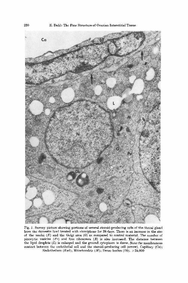
Fig. 1. Survey picture showing portions of several steroid-producing cells of the thecal gland from the domestic fowl treated with clomiphene for 28 days. There is an increase in the size of the nuclei (N) and the Golgi area (G) as compared to control material. The number of pinocytic vesicles (Pv) and free ribosomes (R) is also increased. The distance between the lipid droplets (L) is enlarged and the ground cytoplasm is dense. Note the membranous contact between the endothelial cell and the steroid-producing cell (arrow). Capillary (Ca);
Endothelium (End); Mitochondria (M); Dense bodies (Db). • 24,000
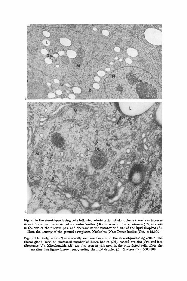
Fig. 2. In the steroid-producing cells following admistrat ion of clomiphene there is an increase in number as well as in size of the mitoehondria (M), increase of free ribosomes (R), increase in the size of the nucleus (N), and decrease in the number and size of the lipid droplets (L).
Note the density of the ground cytoplasm. Nucleolus (Nu) ; Dense bodies (Db). x 12,000
Fig. 3. The Golgi area (G) is markedly increased in size in the steroid-producing cells of the theeal gland, with an increased number of dense bodies (Db), coated vesicles (Cv), and free ribosomes (R). Mitoehondria (M) are also seen in this area in the s t imulated cells. Note the
myeline-like figure (arrow) surrounding the lipid droplet (L). Nucleus (N). X 60,000
232 E. Dahl: The Fine Structure of Ovarian Interstitial Tissue
Changes within the OrganeUes
The Steroid-Producing Cells
Mitochondria. The most pronounced ultrastructural changes were found in the mitochondria, where several alterations were demonstrated regularly (Figs. 2, 4--8). There was a definite increase in number. The size of the mitoehondrial population was significantly increased and more variable than normal (Figs. 2, 4). Gigantism of the mitochondria was regularly encountered, sometimes to an enormous degree (Figs. 5--7). Furthermore, there was an increased den i ty of the matr ix with tubular cristae, sometimes so closely packed tha t the pat tern resembled tha t of a honeycomb (Fig. 5). Sometimes the interior of the matr ix was part ly occupied by cristae, forming complex structures as a separate par t of the mito- chondrion (Fig. 6). Elongated and irregular forms, when observed, were of a larger size than in the control animals (Fig. 8). Sometimes these mitoehondria had indentations to such an extent tha t they seemed to be part ly divided, and the whole structure appeared as if one mitochondrion was divided into several smaller ones, in this way forming new mitochondria (Fig. 8). In many mitochondria, especially the large ones, small granules of the same size and morphology as ribosomes were observed (Fig. 6).
Very often the mitoehondria were lying, not only adjacent to, but in contact with the lipid droplets (Fig. 5) or even embracing them completely (Fig. 4). In others, lipid droplets of different sizes were found within the matrix of the mitochondria as a part of the mitochondrion itself (Figs. 4, 7). These forms of lipid droplets were electron-lucent and apparently empty (Figs. 4, 7). They were of the same morphology and appearance as the lipid droplets which were normally found in the cytoplasm in the basal portion of the cell. Dense material of lipid appearance was found as frequently as in the controls.
Smooth-Surfaced Endoplasmic Reticulum. The smooth endoplasmic reticulum increased significantly in size and was diffusely located in the cytoplasm in- between the mitochondria and lipid droplets (Figs. 2, 5). Contrary to findings in the controls, the smooth endoplasmic reticulum was observed in large amounts, also in the apical part of the cell. Usually, however, it was localized in the basal par t where it occupied large areas, often surrounding a single lipid droplet (Fig. 14). Generally, areas which were rich in smooth endoplasmie reticulum were poor in lipid droplets and mitochondria, compared with the normal ap- pearance.
The Golgi Apparatus. The Golgi complex increased markedly in size following administration of clomiphene (Figs. l, 3). I t occupied a substantial portion of the apical cytoplasm, often in close relation to the nucler membrane (Fig. 3). There was a definite increase in the number of parallel arrays of membranes as well as in the vesicles which formed the Golgi apparatus. A significant increase in the number of dense bodies was also observed within this region (Fig. 3). Sequesteration of these bodies was never encountered, but small blebs were occasionally found on the surrounding membrane of the dense bodies.
Mitochondria were occasionally found within the Golgi complex, often adja- cent to the dense bodies (Fig. 3), sometimes even in mutual contact with each other.
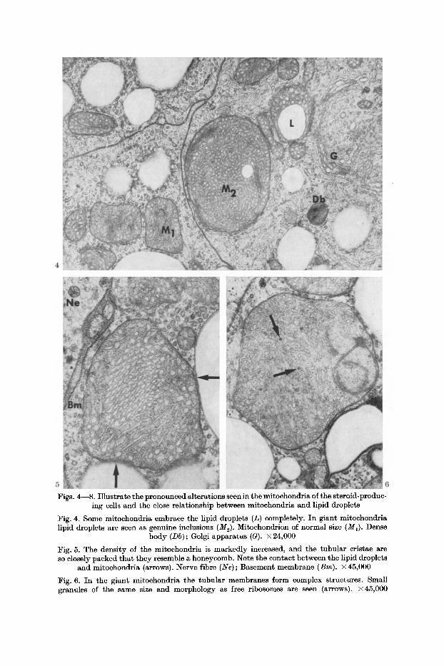
Figs. 4---8. Illustrate the pronounced alterations seen in the mitochondria of the steroid-produc- ing cells and the close relationship between mitoehondria and lipid droplets
Fig. 4. Some mitx)chondria embrace the lipid droplets (L) completely. In giant mitochondria lipid droplets are seen as genuine inclusions (M2). Mitechondrion of normal size (M1). Dense
body (Db) ; Golgi apparatus (G). • 24,000
Fig. 5. The density of the mitochondria is markedly increased, and the tubular cristae are so closely packed that they resemble a honeycomb. Note the contact between the lipid droplets
and mitochondria (arrows). Nerve fibre (Ne); Basement membrane (Bm). • 45,000
Fig. 6. In the giant mitochondria the tubular membranes form complex structures. Small granules of the same size and morphology as free ribosomes are seen (arrows). x45,000
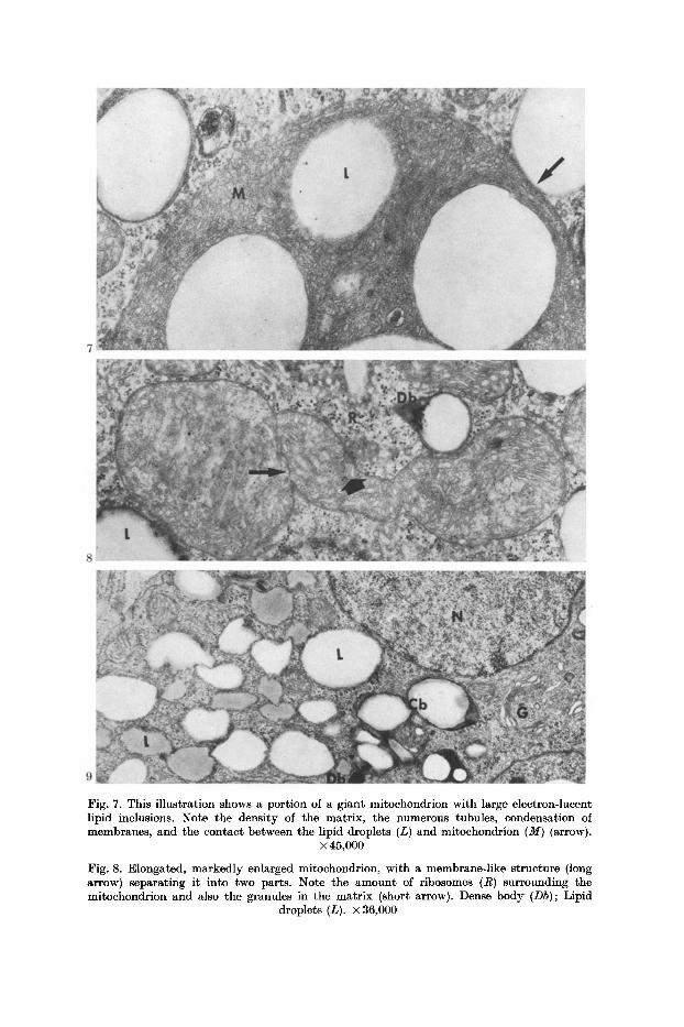
Fig. 7. This illustration shows a portion of a giant mitochondrion with large electron-lucent lipid inclusions. Note the density of the matrix, the numerous tubules, condensation of membranes, and the contact between the lipid droplets (L) and mitochondrion (M) (arrow).
• 45,000
Fig. 8. Elongated, markedly enlarged mitochondrion, with a membrane-like structure (long arrow) separating it into two parts. Note the amount of ribosomes (R) surrounding the mitochondrion and also the granules in the matrix (short arrow). Dense body (Db); Lipid
droplets (L). X 36,000
E. Dahl: The Fine Structure of Ovarian Interstitial Tissue 235
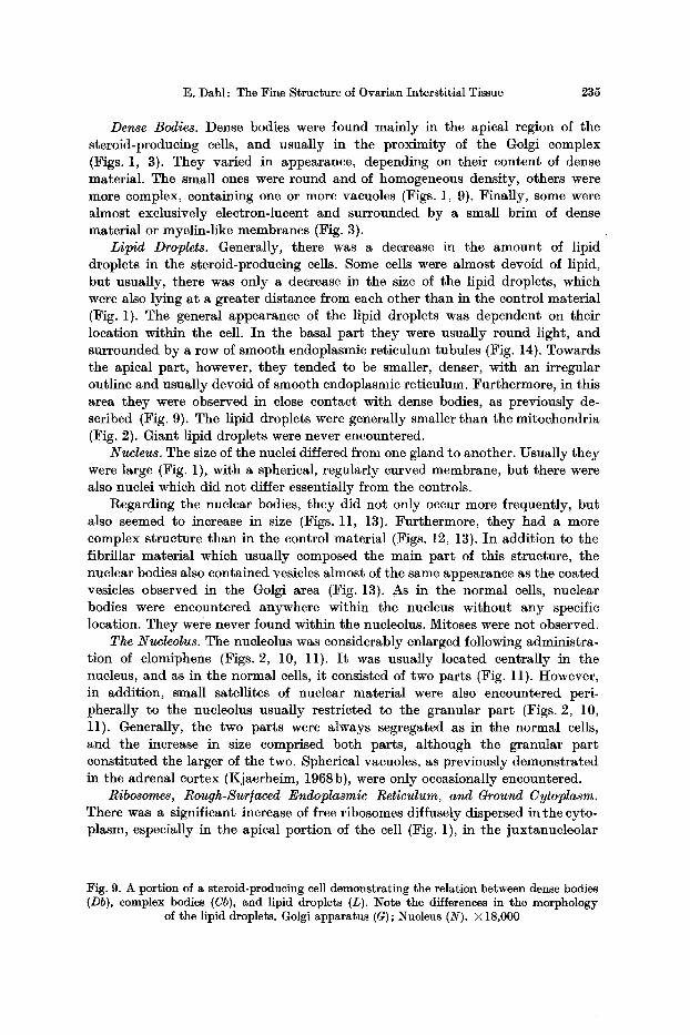
Dense Bodies. Dense bodies were found mainly in the apical region of the steroid-producing cells, and usually in the proximity of the Golgi complex (Figs. 1, 3). They varied in appearance, depending on their content of dense material. The small ones were round and of homogeneous density, others were more complex, containing one or more vacuoles (Figs. 1, 9). Finally, some were almost exclusively electron-lucent and surrounded by a small brim of dense material or myelin-like membranes (Fig. 3).
Lipid Droplets. Generally, there was a decrease in the amount of lipid droplets in the steroid-producing cells. Some cells were almost devoid of lipid, but usually, there was only a decrease in the size of the lipid droplets, which were also lying a t a greater distance from each other than in the control material (Fig. 1). The general appearance of the lipid droplets was dependent on their location within the cell. In the basal par t they were usually round light, and surrounded by a row of smooth endoplasmic reticulum tubules (Fig. 14). Towards the apical part , however, they tended to be smaller, denser, with an irregular outline and usually devoid of smooth endoplasmic reticulum. Furthermore, in this area they were observed in close contact with dense bodies, as previously de- scribed (Fig. 9). The lipid droplets were generally smaller than the mitochondria (Fig. 2). Giant lipid droplets were never encountered.
Nucleus. The size of the nuclei differed from one gland to another. Usually they were large (Fig. 1), with a spherical, regularly curved membrane, but there were also nuclei which did not differ essentially from the controls.
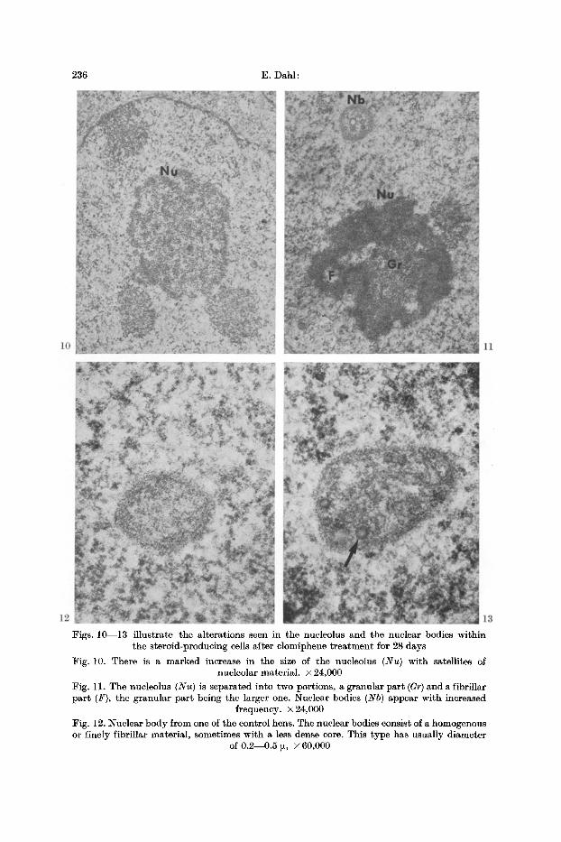
Regarding the nuclear bodies, they did not only occur more frequently, but also seemed to increase in size (Figs. 11, 13). Furthermore, they had a more complex structure than in the control material (Figs. 12, 13). In addition to the fibrillar material which usually composed the main par t of this structure, the nuclear bodies also contained vesicles almost of the same appearance as the coated vesicles observed in the Golgi area (Fig. 13). As in the normal cells, nuclear bodies were encountered anywhere within the nucleus without any specific location. They were never found within the nucleolus. Mitoses were not observed.
The Nucleolus. The nucleolus was considerably enlarged following administra- tion of clomiphene (Figs. 2, 10, 11). I t was usually located centrally in the nucleus, and as in the normal cells, it consisted of two parts (Fig. l l ) . However, in addition, small satellites of nuclear material were also encountered peri- pherally to the nucleolus usually restricted to the granular par t (Figs. 2, 10, l l ) . Generally, the two parts were always segregated as in the normal cells, and the increase in size comprised both parts, although the granular par t constituted the larger of the two. Spherical vacuoles, as previously demonstrated in the adrenal cortex (Kjaerheim, 1968b), were only occasionally encountered.
Ribosomes, Rough-Sur/aced Endoplasmic Reticulum, and Ground Cytoplasm. There was a significant increase of free ribosomes diffusely dispersed in the cyto- plasm, especially in the apical portion of the cell (Fig. 1), in the juxtanucleolar
Fig. 9. A portion of a steroid-producing cell demonstrating the relation between dense bodies (Db), complex bodies (Cb), and lipid droplets (L). Note the differences in the morphology
of the lipid droplets. Golgi apparatus (G) ; Nucleus (N). • 18,000
236 E. Dahl:
Figs. 10---13 illustrate the alterations seen in the nueleolus and the nuclear bodies within the steroid-producing cells after clomiphene treatment for 28 days
Fig. 10. There is a marked increase in the size of the nucleolus (Nu) with satellites of nucleolar material. • 24,000
Fig. 11. The nucleolus (Nu) is separated into two portions, a granular part (Gr)and a fibriltar part (F), the granular part being the larger one. Nuclear bodies (Nb) appear with increased
frequency. • 24,000
Fig. 12. Nuclear body from one of the control hens. The nuclear bodies consist of a homogenous or finely fibrillar material, sometimes with a less dense core. This type has usually diameter
of 0.2--0.5 [z, x 60,000
The Fine Structure of Ovarian Interstitial Tissue 237
region, and in areas surrounding the mitochondria. The rough endoplasmic reticulum was as scanty as in the controls and, thus, did not seem to be influenced by clomiphene.
The ground cytoplasm was homogeneous and denser than in the normal ani- mals. :Fibrillar material was only exceptionally encountered.
The Cell Surtace. No significant alteration was observed at the cell surface. Pinocy~ie vesicles (Fig. 1) were observed with the same frequency as in the control material, and there was no increase in the intercellular spaces, nor was any intercellular substance encountered.
The Enclosing Cell
There was no doubt that the administration of clomiphene had some influence on the morpohlogy of the enclosing cells, even though the alterations observed were much less evident than in the steroid-producing cells (Fig. 15). The nucleus attained a more spherical form, and the amount of ehromatin seemed to decrease. There was a transformation from rough- to smooth surfaced endoplasmic retie- ulum, an increase in the density of the mitochondria, and an accumulation of lipid droplets.
Generally, the organelles of the enclosing cells became more like those of the steroid-producing cells. The cell sometimes appeared enlarged with a prominent Golgi apparatus, but dense bodies were encountered only exceptionally.
Interstitial Tissue and Cells
Normally, the theca interna, in addition to the thecal gland, also possessed cells which seemed to be rather undifferentiated, resembling fibroeytes. However, administration of clomiphene induced significant alterations in these cells (Figs. 16, 17). Normally, they had an enlongated form with small mitochondria, occasionally small lipid droplets, and rough surfaced endoplasmic retieulum.
In the clomiphene-treated animals the cell appeared more oval, and there was an enlargement of the mitochondria which also exhibited increased density of the matrix (Figs. 16, 17). The rough surfaced endoplasmie reticulum became trans- formed to the smooth type with a direct communication between the two types (Fig. 17). Furthermore, there was also an increase in the number and size of lipid droplets (Fig. 17), proportionally to the increase in the smooth surfaced endoplasmic reticulum. Generally, there was a change in morphology towards that of the steroid-producing cells, but these cells never became identical in appearance.
Discussion
Since elomiphene had not been administrated to birds previously, it could not be predicted whether this drug would exhibit any effect on the avian ovary. However, increase in appetite, vitality, plumage, size and colour of the comb
Fig. 13. Nuclear body from a clomiphene-treated hen. There is an increase in size and in the interior there are vesicles which seem to be coated with a fuzzy material at the surface,
resembling pinocytic vesicles/coated vesicles (arrow) (cf. Fig. 12). X 60,000
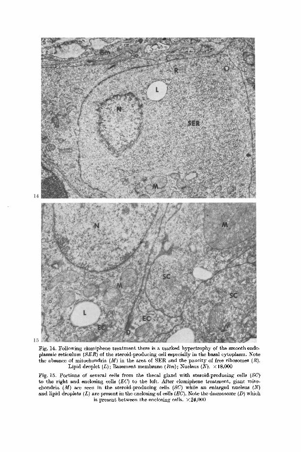
Fig. 14. Following clomiphene treatment there is a marked hypertrophy of the smooth endo. plasmic reticulum (SER) of the steroid-producing cell especially in the basal cytoplasm. Note the absence of mitochondria (M) in the area of SER and the paucity of free ribosomes (R).
Lipid droplet (L) ; Basement membrane (Bm) ; Nucleus (N). x 18,000
Fig. 15. Portions of several cells from the thecal gland with steroid-producing cells (SC) to the right and enclosing cells (EC) to the left. After clomiphene treatment, giant mito- chondria (M) are seen in the steroid-producing cells (SC) while an enlarged nucleus (N) and lipid droplets (L) are present in the enclosing of cells (EC). Note the desmosome (D) which
is present between the enclosing cells, x 24,000
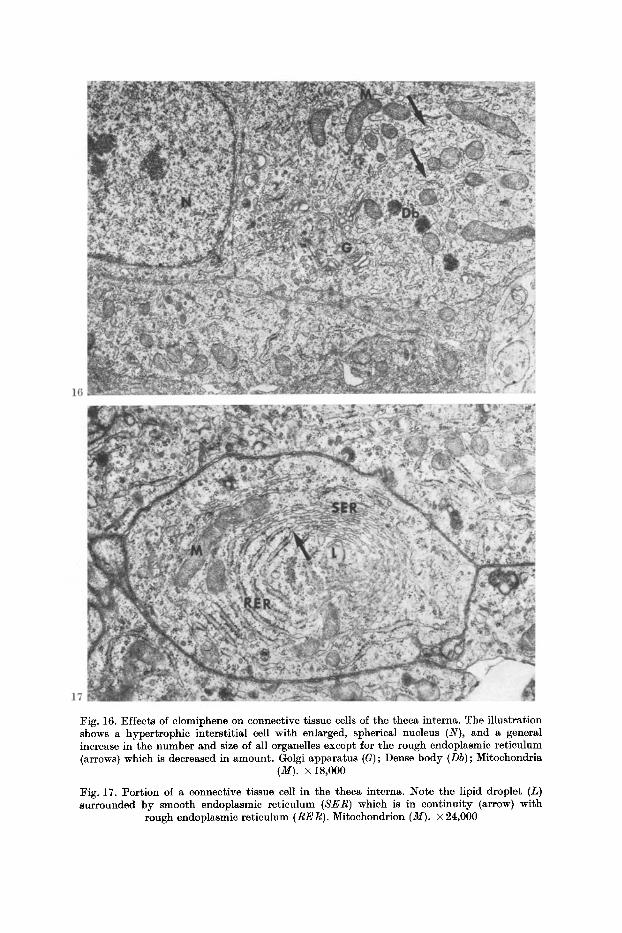
Fig. 16. Effects of clomiphene on connective tissue cells of the theca interna. The illustration shows a hypertrophic interstitial cell with enlarged, spherical nucleus (N), and a general increase in the number and size of all organelles except for the rough endoplasmic reticulum (arrows) which is decreased in amount. Golgi apparatus (G); Dense body (Db); Mitochondria
(M). • 18,000
:Fig. 17. Portion of a connective tissue cell in the theca interna. :Note the lipid droplet (L) surrounded by smooth endoplasmic reticulum (SER) which is in continuity (arrow} with
rough endoplasmic reticulum (RER). Mitochondrion (M). • 24,000
240 E. Da~:
indicated a series of general effects of elomiphene. In addition, the gross ana tomy of the ovary revealed an increase in its size and in the number of follicles. Degenerated immature follicles, as observed after administration of pregnant mare serum gonadotropin (Dahl, 1970e), were never found.
In the s tudy of changes in pathological material, homogeneous fixation of the entire organ, or major parts of it, is a prerequisite for drawing valid conclusions as to ultrastructural changes (Kjaerheim, 1969). By use of the intra-vascular alde- hyde peffusion technique for fixation of the ovary, previous investigations had shown tha t this requirement was fulfilled, compared with the immersion method (Dahl, 1970a). Using this more advanced fixation method, uniform alterations in the animals therefore increased the possibility tha t observations made were based on biology rather than fixation artifacts. In previous investigations the fine struc- ture of the normal theca interna and the thecal gland has been studied (Dahl, 1970a, b, c), thus establishing a valid ultrastruetural base line.
Changes within the OrganeUes
Steroid-Producing Cells
Mitochondria. Several prominent alterations of the mitochondria, consistent with hyperfunction, were regularly encountered. As in the gonadotropin-treated animals (Dahl, 1970e), there was an increase in their number and density. Gigantism was regularly encountered, and the mitochondrial population was more heterogeneous. However, the following changes were even more pronounced than in the hormone-treated animals.
The tubular cristae were of a more regular shape. The intramitochondrial lipid was of the electron-lucent type, and of the same morphology as the lipid droplets which are characteristic for the steroid-producing cell of the thecal gland. The dense osmiophilic accumulations accepted as lipid in hyperstimulated, exhausted mitochondria (Kjaerheim, 1968a, b; Dahl, 1970e), were not encountered more frequently than in the controls. Elongated mitochondria, which were par t ly divided or possibly in division, accounted for the increased population of the organelle, and an increased enzymatic activity of the cell. This is furthermore supported by the fact that small particles of the same morphology as ribosomes were regularly observed in the large mitochondria.
As in the controls and the gonadotropin-treated animals (Dahl, 1970e), mito- chondria were regularly found in close contact with the lipid droplets. In addition, lipid droplets of the electron-lucent type were regularly encountered within the mitochondria themselves. I t was demonstrated tha t the lipid was often embraced by a mitochondrion. The intramitochondrial lipid inclusions therefore most likely originate from the intracellular lipid droplets by way of invaginations and pseudo- inclusions which subsequently become true inclusions. I t has been presumed tha t cholesterol is t ransported to the mitochondria by way of the tubules of the smooth endoplasmic reticulum which surround the lipid droplets (Kjaerhcim, 1968 b), but as demonstrated in this investigation, the possibility exists that there can be a direct interaction between the lipid droplets and the mitochondria, probably on account of an increased demand for secretory activity of the cell.
The Fine Structure of Ovarian Interstitial Tissue 241
Smooth.Sur/aced Endoplasmie Reticulum. Although the amount of smooth endo- plasmic reticulum in the steroid-producing cells did not increase to the same extent as in the gonadotropin treated hens (DAM, 1970e), there was no doubt tha t there was a significant increase compared with the controls. Increase of smooth endo- plasmic reticulum has previously been reported in other steroid-producing cells after t rea tment with Metopiron (Kjaerheim, 1968c). There have been differences in opinion to the significance of the increase of whorls of smooth membranes (Kjaerheim, 1968c) but Meldolesi and Clementi (1967) claim that , at least in the liver, the first cellular response to pharmacological stimulation is an active for- mation of membranes. This also seems to be consistent with the results obtained after administration of ACTH (Kjaerheim, 1968b) and gonadotropins (Dahl, 1970e).
I t is therefore reasonable to conclude tha t the increase of smooth endoplasmic reticulum as seen in the steroid-producing cells in this s tudy reflects an increased function and secretory act ivi ty of the cell.
Other Cytoplasmic Structures. Consistent with the observations in the steroid- producing cells after administration of gonadotropins (Dahl, 1970e), there was a decrease in size and also in number of lipid droplets. This finding is in agreement with the observations on the adrenal cells after t rea tment with Metopiron (Luse, 1967; Mietkiewski and Malendowicz, 1965; Sehwarz and Suchowsky, 1963) and thus supports the view tha t these cells are hyperactive.
An increased activi ty of the cell is also reflected by the increase in size of the Golgi complex, an observation which was also made after t rea tment with gonado- tropins (Dahl, 1970e). Enlargement of the Golgi apparatus has previously been observed after administration of ACTH in the adrenocortieal cells (Kjaerheim, 1968b) indicating tha t the Golgi complex may be involved in increased activi ty of the cell. From the present s tudy one cannot draw any conclusions as to whether this act ivi ty is due to increased synthesis or secretion, or both these processes.
The increase in number of membrane-bounded, dense bodies within the Golgi area was not as prominent in the present s tudy as tha t observed in gonado- tropin-treated animals (Dahl, 1970e). In both studies the amount of electron- lucent lipid droplets decreased, and the dense bodies seemed to increase. Inhibit ion of the cell on the other hand, resulted in a decrease in the number of dense bodies and an increase in lipid droplets (Dahl, 1970d). These observations seem to support the view tha t the dense bodies may in some way regulate the secretory process by providing a mechanism for taking care of overproduction of secretory products (Kjaerheim, 1968c; Smith and Farquar, 1966).
Nucleus and Nucteotus. The demonstration of large, round nucleoli in the steroid-producing cells of the theeal gland is in agreement with the observations made after t rea tment with gonadotropin (Dahl, 1970e). I t is also consistent with observations made in adrenal cells after t reatment with ACTH and Metopiron (Kjaerheim, 1968b, c). The increased number of nuclear bodies is also in accord- ance with the investigations mentioned above. However, the most interesting finding in this s tudy was the fact tha t not only their frequency, but also their internal structure seemed to become more complex. Furthermore, their internal structure was almost identical with that observed after t rea tment with gonado- tropins (Dahl, 1970e). Since previous investigations have revealed tha t the fre-
242 E. DaM:
quency of nuclear bodies was dependent on the metabolic activity of the cell (e.g, Kjaerheim, 1968b; Dahl, 1970f) and tha t their morphology seems to reflect the act ivi ty of the cell (DAM, 1970e), clomiphene seems to have a stimulating effect on this organelle.
The segregation of the nucleolus is in agreement with previous observations made in gonadotropin-treated ovaries (Dahl, 1970e) and ACTH-treated adrenals (Kjaerheim, 1968b). Similar observations have also been reported after t reatment with various toxic substances (Lapis and Bernhard, 1965; Schoefl, 1964; Svoboda, Racela and Higginson, 1967).
Since possible toxic effect of clomiphene on cell morphology has never been investigated, one cannot exclude the possibility that the alterations in the nucleo- lus observed in this s tudy may be due to such effects. However, Kjaerheim (1968b) in his studies on adrenocortical cells, is of the opinion tha t the increase in size of the nucleolus, as seen in the ACTH-treated animals, is due to hyper- activity rather than toxic influence. From his observations, and also from the alterations demonstrated after gonadotropin t reatment (Dahl, 1970e), one is more apt to at tr ibute the nuclear changes seen after administration of clomiphene to hyperact ivi ty of the cell, rather than to toxic influence. This is, furthermore, supported by the fact that toxic effects have not been observed in other organ- elles of the cell. The increased amount of free ribosomes and the increased density of the ground cytoplasm, as demonstrated in this study, is also in agree- ment with the observations seen after administrations of gonadotropins (Dahl, 1970 e) and stimulation of other steroid-producing cells (Kj aerheim, 1968 b ). These observations therefore support the concept that clomiphene has a stimulating effect on the steroid-producing cells of the theca interna.
The Enclosing Cells
Apparently, clomiphene also had some influence on the morphology of the enclosing cells. The changes observed were not so pronounced as those seen in the gonadotropin-treated animals. From having the original appearance of a supporting cell, they changed somewhat towards the appearance of steroid-producing cells. However, this s tudy did not give any further clue as to whether there really exists a functional interaction between the steroid-producing cell and the en- closing cell.
Interstit ial Tissue and Cells
There was no doubt, that the alterations observed in the interstitial cells of the theca interna were in agreement with the observations seen after t rea tment with gonadotropins (DAM, 1970e). From having the appearance of a fibrocyte, these cells changed towards cells containing the morphological prerequisites for steroid synthesis, such as smooth-surfaced endoplasmic reticulum, lipid droplets, dense mitochondria with tubular cristae, and increased Golgi complex. These findings are consistent with the observations reported previously by Merker and Diaz-Encinas (1969) who, after administration of PMS to juvenile rats and rabbits, found similar changes in fibrocytes in the interna. Furthermore, one of the striking features in the present study was the continuity regularly observed be- tween the rough endoplasmic reticulum and smooth endoplasmic reticulum,
The Fine Structure of Ovarian Interstitial Tissue 243
suggesting that the latter is transformed from the former. The transformation, however, was never seen without the presence of lipid droplets. Similar obser- vations were also made in the gonadotropin-treated animals (Dahl, 1970e). I t appears as if lipid droplets are necessary for the transformation of the endo- plasmic reticulum. There may even be indications that lipid droplets are needed together with smooth endoplasmic reticulum before any synthesis of steroids in the ovary is possible.
Cyto-Physiological E//ects of Clomiphene Compared with previous studies of the normal ultrastructure of the thecal gland
(Dahl, 1970a, b, c), the present investigation has clearly demonstrated that clomiphene induce changes in the fine structure of the theca interna. The study has revealed almost identical morphological alterations in the steroid-producing cells of the thecal gland after clomiphene as observed after gonadotrophin treatment. I t has also been revealed almost identical alterations in the enclosing cells and interstitial connective tissue cells as were seen after administration of gonadotrophins. Similar change has previously been demonstrated in steroid- producing cells of the adrenal cortex after administration of ACTH (K]aerheim, 1968b).
The changes found within the different organelles with increase of smooth endo- plasmic reticulum and the Golgi complex, the changes within the nucleolus and the mitochondria, are all consistent with reports of increased steroid synthesis and secretion of steroid-producing cells. Combined and compared with previous investigations (Kjaerheim, 1968b, c; Merker and Diaz-Encinas, 1969; Dahl, 1970e) the present study has demonstrated alterations of the fine structure of the steroid-producing cells consistent with a hyperfunction of the cells. All the ultrastructural changes observed are compatible with clinical and bioassay studies reported in the literature (e.g. Spellacy and Carlson, 1968).
In conclusion, therefore, the present study has demonstrated that administar- tion of elomiphene exerts a stimulating effect on the cells of the theca interna. However, whether this is due to direct stimulation of the pituitary gland by the drug, or to the fact tha t clomiphene neutralizes the inhibitive action of estrogens on gonadotropin secretion has not been demonstrated. Therefore, further studies are still needed to elucidate the real mechanism of the action of elomiphene, where hypophysectomy will be required, and where the hypothalamie pituitary axis also has to be considered.
References Barr, A., Paulsen, C. A.: Demonstration of an antiestrogenic effect of clomiphene in human
beings. Clin. Res. 13, 129 (1965). Campenhout, J. van, Simard, R., Leduc, B.: Antiestrogenic effect of clomiphene in the
human being. Fertil. and Steril. 19, 700~70~ (1968). Dahl, E. : Studies of the fine structure of ovarian interstitial tissue. 1. A comparative
study of the fine structure of the ovarian interstitial tissue in the rat and the domestic fowl. To be published (1970a).
- - Studies of the fine structure of ovarian interstitial tissue. 2. The ultrastructure of the theeal gland of the domestic fowl. To be published (1970b).
- - Studies of the fine structure of ovarian interstitial tissue. 3. The innervation of the thecal gland of the domestic fowl. To be published (1970c).
17 Z. Zellforsch,, Bd. 109
244 E. Dahl: The Fine Structure of Ovarian Interst i t ial Tissue
Dahl, E. : Studies of the fine structure of ovarian intersti t ial tissue. 4. Effects of steroids on the thecal gland of the domestic fowl. To be published (1970d).
- - Studies of the fine structure of ovarian intersti t ial tissue. 5. Effects of gonadotropins on the thecal gland of the domestic fowl. To be published (1970e).
- - The fine structure of nuclear inclusions. J . Anat. (Lond.) 106, 255--262 (1970f). Greenblat t , R. B., Roy, S., Mahesh, V. B., with W. E. Baifield, Jungck, E. C. : Induct ion of
ovulation. Amer. J. Obstet. Gynec. 84, 90(O-912 (1962). Igarashi, M., Ibuki, Y., Kubo, H., Kamiokea, J. , Yokota, N., Ebara, Y., Matsumoto, S. : Mode
and site of action of clomiphene. Amer. J. Obstet. Gynec. 97, 120--123 (1967). Kjaerheim, A. : Studies of adrenocortical ultrastructure. 1. Aldehyde perfusion fixation of the
domest ic fowl. Acta anat . (Basel) 14, 424--453 (1969). - - Studies of adrenoeortical ultrastructure. 2. The interrenal cell of the domestic fowl as seen
after gtutaraldehyde perfusion fixation. Z. Zeltforsch. 91, 429--455 (1968a). - - Studies of adrenocortical ultrastructure. 4. Effects of ACTII on interrenal cells of the
domestic fowl. J . Microseopie 7, 715--738 (1968b). - - Studies of adrenoeortical ultrastructurc. 5. Effects of metopiron in interrenal cells of
the domestic fowl. J. Mieroscopie 7, 739--754 (1968c). Lapis, K., Bernhard, W. : The effect of Mitomycin-C on the nueleolar fine structure of K B
cells in cell culture. Cancer Res. 25, 628--645 (1965). Luse, S.: Fine s tructure of adrenal cortex. In : The adrenal cortex (A. B. Eisenstein ed.),
1st ed., p. 1--59. London: J . and A. Churchill 1967. Meldolesi, J., Clementi, F. - Le r6ticulum endoplasmique de la cellule h~patique apr~s l 'admini-
s t rat ion de medicaments. Path. et Biol. 15, 215--228 (1967). Merker, H. J., Diaz-Encinas, J . : Das elektronenmikroskopisehe Bild des Ovars j uveniler Ra t t en
und Kaninchen nach Stimulierung mit PMS und HCG. Z. Zellforsch. 94, 605~623 (1969).
Mietkiewski, K., Malendowicz, L. : Ober Ver~nderungen der Nebennierenrinde bei Ra t t en nach Behandlung mi t Metopiron. Endokrinologie 48, 241--255 (1965).
Millonig, G.: The advantages of a phosphate buffer for Os04 solutions in fixation. J . appl. Physiol. 32, 1637 (1961).
Mohla, S., Prasad, M. R. N. : Inhibi t ion of estrogen induced glycogen synthesis in the ra t by clomiphene. Steroids 11, 571--583 (1968).
Reynolds, E. S. : The use of lead citrate at high pH as an electron-opaque stain in electron microscopy. J. Cell Biol. 17, 208--212 (1963).
Roy, S., Greenblatt , R., Mahesh, V., Jungck, E. C. : Clomiphene citrate: further observations on its use in induction of ovulation in the human and on its mode of action. Fertil. and Steril. 14, 575--595 (1963).
Ryter, A., Kellenberger, E. : L'inclusion au polyester pour l 'ultramicrotomie. J . Ultrastruct . Res. 2, 200--214 (1958).
Schoefl, G. I. : The effect of Actinomyein D on the fine structure of the nucleolus. J . Ultra- struct. Res. 10, 224--243 (1964).
Sehwarz, W., Suchowsky, G . K . : Die Wirkung yon Metopiron und Amphenon B auf die Nebennierenrinde der Ratte . Virchows Arch. path. Anat. 387, 270--278 (1963).
Shearman, R. P. : Induct ion of ovulation. Aust. Ann. Med. 15, 266--280 (1966). Smith, R. E., Farquhar , M. G. : Lysosomc function in the regulation of the secretory process
in cells of the anterior pi tui tary gland. J. Cell Biol. 81, 319--347 (1966). Spellaey, W. N., Carlson, K. L. : Pi tui tary-function studies performed before t reat ing second-
ary amenorrhea with elomiphene citrate. Fertil. and Steril. 19, 690--699 (1968). Svoboda, D., Racela, A., Higginson, J . : Variations in ul trastruetural nuclear changes in
hepatocarcinogenesis. Biochem. Pharmac(~l. 16, 651--657 (1967). Thompson, R. J. , Mellinger, R. C. : The effects of clomiphene citrate in patients with pi tui tary
gonadal disorders. Amer. J. Obstet. Gynee. 92, 4 1 2 4 2 0 (1965).
Dr. Erik Dahl Odontologisk Ins t i t u t t for Anatomi Oslo 3 (Norway) Blindern
](https://img.pdfslide.us/doc/110x75/5ed7600ea5b1445fe467ceb5/ovarian-tissue-cryopreservation-and-transplantation-cases-due-to-a-reduced-ovarian.jpg)