Embed Size (px)
Citation preview
Structures of aminoacylase 3 in complexwith acetylated substratesJennifer M. Hsieha,b,1, Kirill Tsirulnikovc,1, Michael R. Sawayad, Nathaniel Magilnickc, Natalia Abuladzec, Ira Kurtzc,Jeff Abramsonb,2, and Alexander Pushkinc,2
aDepartment of Chemistry and Biochemistry, University of California, Los Angeles, CA 90095; bDepartments of Physiology and cMedicine/Nephrology,David Geffen School of Medicine at University of California, Los Angeles, CA 90095; and dHoward Hughes Medical Institute, University ofCalifornia–Department of Energy Institute for Genomics and Proteomics, Los Angeles, CA 90095
Edited* by George H. Lorimer, University of Maryland, College Park, MD, and approved August 24, 2010 (received for review June 2, 2010)
Trichloroethylene (TCE) is one of themost widespread environmen-tal contaminants, which is metabolized to N-acetyl-S-1,2-dichloro-vinyl-L-cysteine (NA-DCVC) before being excreted in the urine.Alternatively, NA-DCVC can be deacetylated by aminoacylase 3(AA3), an enzyme that is highly expressed in the kidney, liver,and brain. NA-DCVC deacetylation initiates the transformationinto toxic products that ultimately causes acute renal failure.AA3 inhibition is therefore a target of interest to prevent TCEinduced nephrotoxicity. Here we report the crystal structure ofrecombinant mouse AA3 (mAA3) in the presence of its acetatebyproduct and two substrates: Nα-acetyl-L-tyrosine and NA-DCVC.These structures, in conjunction with biochemical data, indicatedthat AA3 mediates substrate specificity through van der Waalsinteractions providing a dynamic interaction interface, whichfacilitates a diverse range of substrates.
mercapturates ∣ metalloprotein ∣ X-ray structure
Aminoacylase 3 (AA3) is a member of the aminoacylase familyof enzymes that deacylates a broad range of substrates in-
cluding bothNα-acetylated amino acids and S-cysteine conjugatesof N-acetyl-L-cysteine (mercapturic acids) (Fig. 1) (1). There arethree types of aminoacylases: (i) aminoacylase 1 (AA1) deacety-lates neutral aliphatic N-acyl-α-amino acids and mercapturicacids; (ii) aminoacylase 2 or aspartoacylase (AA2) has a strictspecificity for Nα-acetyl-L-aspartate (NAD); and (iii) aminoacy-lase 3 (AA3) preferentially deacetylates Nα-acetylated aromaticamino acids and mercapturic acids that are usually not deacety-lated by AA1 (1–6). Despite different substrate specificities, AA2and AA3 have a high degree of sequence (42% identity) andstructure homology but are both substantially different fromAA1 (∼10% of sequence identity) (2, 6–10).
AA3 is of particular interest for human health because it par-ticipates in mediating toxicity of the xenobiotic trichloroethylene(TCE). The United States produces in excess of 130,000 tons ofTCE per year (11), making it the most widespread chemical con-taminant in both soil and ground water. TCE is readily absorbedinto the body where it can enter the glutathione conjugation de-toxification pathway producing the mercapturic acid N-acetyl-S-1,2-dichlorovinyl-L-cysteine (NA-DCVC) for subsequent urin-ary excretion (12, 13). However, NA-DCVC can be deacetylatedby AA3—which is highly expressed in the renal proximal tubule,liver, and brain—to generate S-1,2-dichlorovinyl-L-cysteine(DCVC) (2) and further transformed via β-lyases or flavin mono-oxygenases into lethal products capable of causing acute renalfailure and toxicity to the liver and brain. (4, 13–22). Thus, inhibi-tion of AA3 can decrease DCVC formation and ameliorate TCEtoxicity, presenting a potential target for drug discovery.
Generating specific inhibitors for AA3 would be greatly aidedby high-resolution structural data, but structural studies of ami-noacylases have been limited to AA1 (7) and AA2 (8, 9), whichdiffer in both overall architecture and active site composition. ForAA1, the active site has two cocatalytic zinc ions residing at thedimer interface (7). In contrast, the AA2 active site is formed
within a single protein subunit comprising of a lone zinc ion atthe active site (8, 9) and bears a striking similarity to carboxypep-tidase A (CpA) (23). It is believed that both enzymes follow the“promoted-water pathway” mechanism for hydrolysis, where azinc-bound hydroxide– generated by proton abstraction via aconserved glutamate– serves as the nucleophile to attack thesubstrate-leaving group (8, 9). Based on sequence identity (42%)and homology modeling, AA3 is believed to have a similar struc-ture and mechanism for hydrolysis as AA2 (10). However, thismodel alone could not clarify the differences in substrate specifi-city between AA2 and AA3 (10). Therefore, an atomic resolutionstructure of AA3 in the presence of substrate is still required toaddress these questions and may facilitate targeted drug design.
Here we report the X-ray structure of murine AA3 (mAA3)(UniProt entry Q91XE4) in complex with the substrates Nα-acet-yl-L-tyrosine (NAY) and N-acetyl-S-1,2-dichlorovinyl-L-cysteine(NA-DCVC). This feat was accomplished by mutating the essen-tial glutamate residue (E177A) predicted to initiate the deacety-lation reaction (8–10). Comparison of the wild-type (wt) mAA3structure with the E177A-mAA3 structures in complex withsubstrate reveals key interactions involved in substrate binding.Further, a comparison with structures of AA2 highlights differ-ences that facilitate the broad substrate recognition observedfor AA3.
Fig. 1. Deacetylation reaction catalyzedbyAA3. TheAA3 substrate,N-acetyl-L-tyrosine (NAY), is hydrolyzed by AA3 to produce L-tyrosine and acetate. TheAA3 substrate, N-acetyl-S-1,2-dichlorovinyl-L-cysteine (NA-DCVC), is hydro-lyzed by AA3 to produce S-1,2-dichlorovinyl-L-cysteine (DCVC) and acetate.
Author contributions: J.M.H., K.T., I.K., J.A., and A.P. designed research; J.M.H., K.T., N.M.,and N.A. performed research; J.M.H., K.T., and M.R.S. analyzed data; and J.M.H., M.R.S.,I.K., J.A., and A.P. wrote the paper.
The authors declare no conflict of interest.
*This Direct Submission article had a prearranged editor.
Data deposition: The atomic coordinates and structure factors have been deposited in theProtein Data Bank, www.pdb.org (PDB ID codes 3NH4, 3NFZ, 3NH5, and 3NH8).1J.M.H. and K.T. contributed equally to this work.2To whom correspondence may be addressed. E-mail: [email protected] [email protected].
This article contains supporting information online at www.pnas.org/lookup/suppl/doi:10.1073/pnas.1006687107/-/DCSupplemental.
17962–17967 ∣ PNAS ∣ October 19, 2010 ∣ vol. 107 ∣ no. 42 www.pnas.org/cgi/doi/10.1073/pnas.1006687107
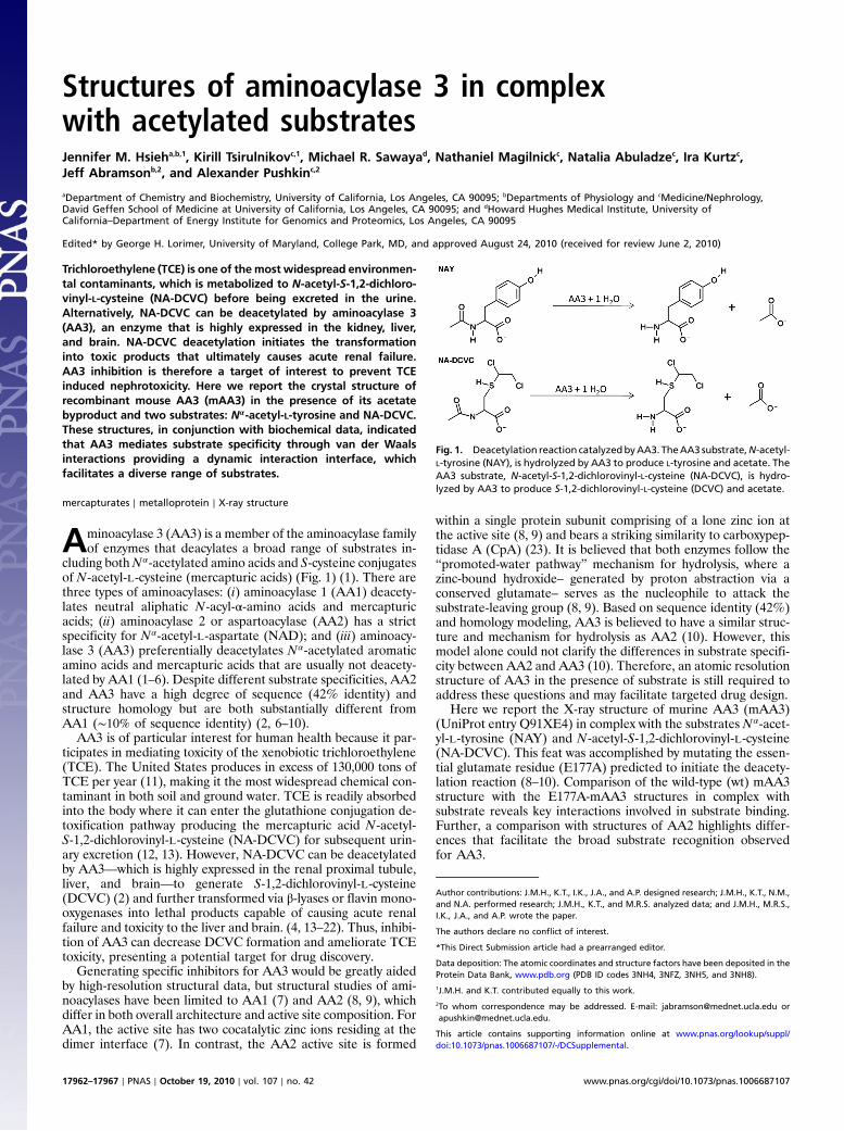
ResultsStructure of wt-mAA3. The wt-mAA3 crystals diffracted to 2.0 Å,and the data was phased by molecular replacement using rAA2(PDB entry 2GU2) (8) as a search model. The model was builtin COOT (24) and refined using PHENIX (25) with Rwork∕Rfreeof 18.4∕20.7%. The data collection parameters and refinementstatistics are presented in Table 1. All attempts to resolve thestructure of wt-mAA3 in complex with known substrates failed.The initial structure determination of wt-mAA3 revealed a simi-lar fold to AA2, and we identified a point mutation (E177A)rendering mAA3 catalytically inactive (10) but maintained thestructural integrity of active site. The creation of E177A-mAA3facilitated structure determination of mAA3 in complex withNAY (NAY-mAA3) and NA-DCVC (NA-DCVC-mAA3).
Structure Overview. A superposition of wt-mAA3 and rAA2reveals similar architecture with a calculated rmsd of 1.04 Å over298 Cα atoms. Similarly, no large conformational differences areobserved among the four mAA3 structures reported here withrmsd values ranging from 0.267 to 0.349 Å, over 302 Cα atoms.There is one molecule per asymmetric unit (Fig. 2A); however, byapplying a P62 symmetry operator the expected dimer is formed(Fig. 2B) as observed in the AA2 structures (8, 9) and is furthersubstantiated by mAA3 biochemical data (26). The monomer iscomposed of two distinct domains, which have been predicted bytrypsinolysis (26): the hydrolytic domain (residues 1–210), whichmaintains most of the essential residues for binding and hydro-lysis; and the shielding domain (residues 211–318) that covers theactive site to limit accessibility (Fig. 2).
A comparison of mAA3 with existing atomic resolution struc-tures using theDistanceALIgnment (DALI) server (27) identifiedapproximately five unique proteins (Z-score > 10) with similarstructure including AA2; however, a notable inclusion was CpA,a pancreatic exopeptidase that hydrolyzes peptide bonds ofC-terminal residues with aromatic or aliphatic side chains(sequence identity: 12.9%) (28). The two enzymes share a similarcore of secondary structure elements in the hydrolytic domainwith an overall structural alignment of 2.61 Å over 163 Cα atoms.Notably, CpA lacks the shielding domain andposses anN-terminalextension of approximately 60 residues. Nonetheless, the activesite of these two enzymes reveals striking similarities wherethe essential mAA3 catalytic residues (His21, Glu24, Arg63,Asp68, His116, and Glu177) precisely conform to the active siteresidues of CpA (His69, Glu72, Arg127, Asp142, His196, andE270) (PDB ID 1CBX) (23). These six residues align with an rmsdof 0.352 Å (Fig. S1A), suggesting that AA3 and CpA use a similarreaction scheme for hydrolysis. Substantiating this claim, weexamined the effects of the potent CpA inhibitor (L-benzylsucci-nate) (23, 29) on mAA3, which resulted in inhibition of hydrolysis(Ki ¼ 25 μM). This would suggest L-benzylsuccinate has a similar
mode of interaction for both CpA and mAA3 further corroborat-ing that AA3 uses the same reaction scheme as CpA.
mAA3 with Acetate Byproduct. The essential catalytic residues ofmAA3 (His21, Glu24, Arg63, Asp68, His116, and Glu177) areidentical to those of AA2 and CpA. A zinc ion coordinated by
Table 1. Data collection and refinement statistics
wt-mAA3 E177A-mAA3 NAY-mAA3 NA-DCVC-mAA3 Co2þ soaked wt-mAA3
Wavelength (Å) 0.9774 1.0000 1.0000 1.0000 1.2824Spacegroup P62 P62 P62 P62 P62Unit Cella, b, c (Å) 93.7, 93.7, 97.1 93.2, 93.2, 97.5 93.5, 93.5, 98.0 93.4, 93.4, 97.6 93.4, 93.4, 97.7α, β, γ (°) 90, 90, 120 90, 90, 120 90, 90, 120 90, 90, 120 90, 90, 120Resolution (Å) 50–2.00 (2.07–2.00) 50–2.10 (2.18–2.10) 50–2.15 (2.23–2.15) 50–2.80 (2.90–2.80) 50.0–2.80 (2.90–2.80)Rmerge (%) 6.4 (50.5) 9.2 (60.0) 12.2 (64.4) 15.4 (48.0) 11.0 (51.5)Completeness (%) 100.0 (100.0) 98.6 (99.3) 100.0 (99.8) 98.2 (98.0) 98.8 (99.7)Redundancy 4.9 (3.9) 6.8 (6.7) 6.7 (6.6) 4.2 (3.8) 6.7 (6.7)I∕σ 18.0 (3.0) 18.9 (2.6) 16.0 (3.4) 8.8 (2.2) 16.8 (4.0)No. of Unique Reflections 32766 (3266) 27897 (2804) 26605 (2647) 11756 (1160) 11944 (1202)Rwork∕Rfree 18.4∕20.7 19.4∕22.7 16.6∕19.7 17.5∕22.4 —rmsdBonds (Å) 0.008 0.008 0.008 0.011 —Angles (°) 1.038 1.035 1.038 1.470 —
Fig. 2. Overview of mAA3 structure. (A) Single mAA3 protomer in ribbonrepresentation with the substrate cavity and 156–164 loop indicated (lightgreen). (B) Two mAA3 protomers form the biologically observed dimer withNAY substrate present. In both A and B the hydrolytic domain is coloredgreen and the shielding domain is colored purple.
Hsieh et al. PNAS ∣ October 19, 2010 ∣ vol. 107 ∣ no. 42 ∣ 17963
BIOCH
EMISTR
Y
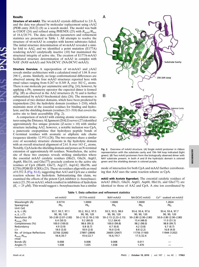
His21, Glu24, and His116 is observed in all mAA3 structures.Previous studies have demonstrated that cobalt can replacezinc in the enzyme, and it increases enzymatic activity (6, 10)—a finding supported by “soaking” crystals in a cobalt solutionrevealing a mixed occupancy for both cobalt and zinc (confirmedby anomalous diffraction) (Fig. S2). The structures of wt-mAA3and E177A-mAA3, crystallized in the presence of acetate andformate, reveal the reaction byproduct (acetate) coordinated ina bidentate fashion to the catalytic zinc. In addition, a formatemolecule is observed coordinated to Arg63, Asn70, Arg71, andTyr287 (Fig. 3A). A similar coordination pattern is observed tothe N-acetyl group and α-carboxylate oxygens for both theNA-DCVC and NAY substrates (Fig. 3 B and C), suggesting apossible catalytic role for these residues.
Mutational analysis confirmed the importance of Arg63 andTyr287 where the R63A substitution rendered the enzyme com-pletely inactive while the Y287A mutation had minimal activity(8% for NAY and 2.5% for NA-DCVC) (10). In contrast, R71Aand N70A had minimal effect on kcat or affinity for both sub-strates (10). Strikingly, the R71K mutation cripples the humanAA2 enzyme by reducing kcat by approximately 95% withoutsignificant change of Km (9), which indicates that that this residueplays quite different roles in AA2 as compared to AA3.
While both formate and acetate appear to mimic substratebinding, repeated attempts to solve the structure of wt-mAA3in complex with known substrates proved unsuccessful. A com-parative analysis with CpA highlights Glu177 as an essentialresidue for hydrolysis that when mutated to alanine rendersAA3 inactive (10). In the wt-mAA3 Glu177 is coordinated toacetate, a byprodyct of deacetylation, highlighting its criticalfunction. In the E177A-mAA3 mutant, this interaction is lost,but the overall structure and the coordination pattern to acetateand formate remains the same—a necessary requirement forstructural analysis (Fig. S3).
NAY-Complex Structure.The E177A-mAA3 mutant was crystallizedin the presence of NAY, which displaced both acetate andformate from the active site. The N-acetyl-α-amino carboxylicacid (NAACA) component of NAY—a conserved component ofAA2 and AA3 substrates—is tightly bound by seven hydrogenbonds (Fig. 3B; see also Fig. S4 A, B). Specifically, the substrate’sacetyl oxygen coordinates zinc and accepts two hydrogen bondsfromArg63; the α-carboxylic group forms a salt bridge with Arg71and a hydrogen bond with Asn70; and the amide has hydrogenbonds to the hydroxyl of Tyr287. In addition, Tyr287 has van derWaals interactions with the NAACA component of NAY aidingin positioning the substrate.
The NAACA component of NAY has extensive hydrogenbonds and van der Waals interactions, but the side-chain consti-tuent associates with AA3 primarily through van der Waals inter-actions and a lone hydrogen bond with Glu129. Eight residuesmaintain van der Waals contacts with the substrate side chain(Asn70, Arg71, Ile127, Glu129, Tyr156, Phe164, Ser165, Cys175)where five of these are conserved between human, rat, and mouseAA2 and AA3 (Asn70, Arg71, His116, Ile127, and Phe281) (6).This would indicate that these residues are likely not responsiblefor specificity between AA3 and the substrate side chain ofNAY. The electron density for the 156–164 loop is not well de-fined for all structures reported here; however, the side-chaindensity for Tyr156 and Phe164 improves in the presence ofNAY. Notably, in the presence of substrate, Phe164 rotatesapproximately 50° around the β-carbon to interact with NAY.A similar conformational rearrangement of the side chain ofthe homologous AA2 residue, Tyr164, was shown upon bindingthe intermediate analog, NPD (9). This data suggest a likely rolefor Phe164 in substrate recognition of NAY.
NA-DCVC-Complex Structure. E177A-mAA3 mutant crystals weresoaked in 2 mM NA-DCVC to resolve the complex; furtherdetails regarding crystallization and structure determinationcan be found in Materials and Methods. Although similar to
Fig. 3. mAA3 substrate binding. (A) wt-mAA3, with acetate and formatebound. (B) NAY-mAA3-E177A, with NAY bound in the active site. (C) NA-DCVC-mAA3-E177A, with NA-DCVC bound in the active site. The residuesthat form the hydrogen bonds with substrates and participate in the zincion (gray sphere) coordination are represented in green sticks, the moleculesbound in the active site are purple and the hydrogen and coordination bondsare represented by dashed black lines. The 2Fo–Fc maps (in blue) are shownfor both substrates, NAYand NA-DCVC, with a contour level of 1 and a carveradius of 1.6 and 1.8, respectively.
17964 ∣ www.pnas.org/cgi/doi/10.1073/pnas.1006687107 Hsieh et al.
the NAY structure, there is a 0.8 Å shift of the NAACA compo-nent causing a minor reorientation in the hydrogen-bondingnetwork (Fig. 3C; see also Fig. S4 C and D). Specifically, thesubstrate’s acetyl oxygen interacts with zinc and the side chainsof Glu24 and Arg63. The α-carboxylic group accepts hydrogenbonds from the side chains of Arg71 and Tyr287, but unlikeNAY-mAA3 there is no hydrogen bond with Asn70. The side-chain constituent of NA-DCVC interacts with AA3 solelythrough van der Waals interactions in a similar fashion as NAYwith the exception of Phe164. Limited electron density forPhe164 suggests it is mostly disordered and does not undergo the50° rotation observed in the NAY structure. These results suggestthat AA3 has a more dynamic active site capable of accommodat-ing a broad range of substrates.
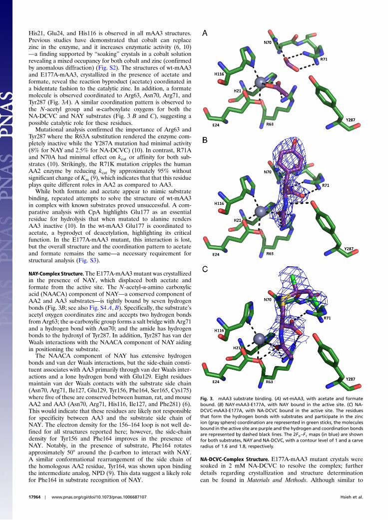
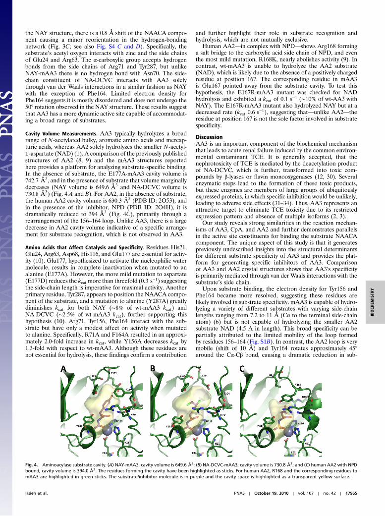
Cavity Volume Measurements. AA3 typically hydrolyzes a broadrange of N-acetylated bulky, aromatic amino acids and mercap-turic acids, whereas AA2 solely hydrolyzes the smaller N-acetyl-L-aspartate (NAD) (1). A comparison of the previously publishedstructures of AA2 (8, 9) and the mAA3 structures reportedhere provides a platform for analyzing substrate-specific binding.In the absence of substrate, the E177A-mAA3 cavity volume is742.7 Å3, and in the presence of substrate that volume marginallydecreases (NAY volume is 649.6 Å3 and NA-DCVC volume is730.8 Å3) (Fig. 4 A and B). For AA2, in the absence of substrate,the human AA2 cavity volume is 630.3 Å3 (PDB ID: 2O53), andin the presence of the inhibitor, NPD (PDB ID: 2O4H), it isdramatically reduced to 394 Å3 (Fig. 4C), primarily through arearrangement of the 156–164 loop. Unlike AA3, there is a largedecrease in AA2 cavity volume indicative of a specific arrange-ment for substrate recognition, which is not observed in AA3.
Amino Acids that Affect Catalysis and Specificity. Residues His21,Glu24, Arg63, Asp68, His116, and Glu177 are essential for activ-ity (10). Glu177, hypothesized to activate the nucleophilic watermolecule, results in complete inactivation when mutated to analanine (E177A). However, the more mild mutation to aspartate(E177D) reduces the kcat more than threefold (0.3 s−1) suggestingthe side-chain length is imperative for maximal activity. Anotherprimary residue, Tyr287, appears to position the NAACA compo-nent of the substrate, and a mutation to alanine (Y287A) greatlydiminishes kcat for both NAY (∼8% of wt-mAA3 kcat) andNA-DCVC (∼2.5% of wt-mAA3 kcat), further supporting thishypothesis (10). Arg71, Tyr156, Phe164 interact with the sub-strate but have only a modest affect on activity when mutatedto alanine. Specifically, R71A and F164A resulted in an approxi-mately 2.0-fold increase in kcat, while Y156A decreases kcat by1.3-fold with respect to wt-mAA3. Although these residues arenot essential for hydrolysis, these findings confirm a contribution
and further highlight their role in substrate recognition andhydrolysis, which are not mutually exclusive.
Human AA2—in complex with NPD—shows Arg168 forminga salt bridge to the carboxylic acid side chain of NPD, and eventhe most mild mutation, R168K, nearly abolishes activity (9). Incontrast, wt-mAA3 is unable to hydrolyze the AA2 substrate(NAD), which is likely due to the absence of a positively chargedresidue at position 167. The corresponding residue in mAA3is Glu167 pointed away from the substrate cavity. To test thishypothesis, the E167R-mAA3 mutant was checked for NADhydrolysis and exhibited a kcat of 0.1 s−1 (∼10% of wt-AA3 withNAY). The E167R-mAA3 mutant also hydrolyzed NAY but at adecreased rate (kcat 0.6 s−1), suggesting that—unlike AA2—theresidue at position 167 is not the sole factor involved in substratespecificity.
DiscussionAA3 is an important component of the biochemical mechanismthat leads to acute renal failure induced by the common environ-mental contaminant TCE. It is generally accepted, that thenephrotoxicity of TCE is mediated by the deacetylation productof NA-DCVC, which is further, transformed into toxic com-pounds by β-lyases or flavin monooxygenases (12, 30). Severalenzymatic steps lead to the formation of these toxic products,but these enzymes are members of large groups of ubiquitouslyexpressed proteins, in which specific inhibition would be unlikely,leading to adverse side effects (31–34). Thus, AA3 represents anattractive target to eliminate TCE toxicity due to its restrictedexpression pattern and absence of multiple isoforms (2, 3).
Our study reveals strong similarities in the reaction mechan-isms of AA3, CpA, and AA2 and further demonstrates parallelsin the active site constituents for binding the substrate NAACAcomponent. The unique aspect of this study is that it generatespreviously undescribed insights into the structural determinantsfor different substrate specificity of AA3 and provides the plat-form for generating specific inhibitors of AA3. Comparisonof AA3 and AA2 crystal structures shows that AA3’s specificityis primarily mediated through van der Waals interactions with thesubstrate’s side chain.
Upon substrate binding, the electron density for Tyr156 andPhe164 became more resolved, suggesting these residues arelikely involved in substrate specificity. mAA3 is capable of hydro-lyzing a variety of different substrates with varying side-chainlengths ranging from 7.2 to 11 Å (Cα to the terminal side-chainatom) (6) but is not capable of hydrolyzing the smaller AA2substrate NAD (4.5 Å in length). This broad specificity can bepartially attributed to the limited mobility of the loop formedby residues 156–164 (Fig. S1B). In contrast, the AA2 loop is verymobile (shift of 10 Å) and Tyr164 rotates approximately 45°around the Cα-Cβ bond, causing a dramatic reduction in sub-
Fig. 4. Aminoacylase substrate cavity. (A) NAY-mAA3, cavity volume is 649.6 Å3; (B) NA-DCVC-mAA3, cavity volume is 730.8 Å3; and (C) human AA2 with NPDbound, cavity volume is 394.0 Å3. The residues forming the cavity have been highlighted as sticks. For human AA2, R168 and the corresponding residues tomAA3 are highlighted in green sticks. The substrate/inhibitor molecule is in purple and the cavity space is highlighted as a transparent yellow surface.
Hsieh et al. PNAS ∣ October 19, 2010 ∣ vol. 107 ∣ no. 42 ∣ 17965
BIOCH
EMISTR
Y
strate cavity for substrate-bound human AA2 (∼395 Å3); in com-parison in mAA3, the substrate cavity with NA-DCVC and NAYis approximately 730 and 650 Å3, respectively. The differencesin loop dynamics could be attributed to the sequence divergenceobserved between the AA2 and AA3 loops (6).
One of the important differences between AA2 and AA3, con-tributing to AA2 substrate specificity, is Arg168, which stabilizesthe AA2 inhibitor analog NPD through a salt bridge. Even themild mutation of R168K nearly abolishes activity (9). mAA3has a glutamate at this position, and its mutation to an alanine didnot significantly change the enzyme kinetics (10). It is interestingthat the E167R-mAA3 mutant, contrary to wt-AA3, demon-strated limited NAD hydrolysis (kcat ∼ 10% and 20% of, respec-tively, the wt-AA3 and E167R mutant kcat with NAY). Although,these data demonstrate some similarity between AA3 and AA2,they also highlight specific differences in active site architecture.
As seen in the AA3 structures, residues His21, Glu24, andHis116 are responsible for coordinating the zinc involved in cat-alysis. Substrate binding is modulated through the substrate’sNAACA component where residues His21, Glu24, Arg63, andTyr287 interact via hydrogen bonding and van der Waals interac-tions (Fig. 3 B and C). His21, Glu24, Arg63, and Tyr288 alsoplay a similar role in AA2. For AA2, Arg71 is an essential residuefor positioning the substrate NAACA component via hydrogenbonds, and Arg71 mutation even into lysine decreases kcat by99% (9); however, it is not the case for AA3, in which theefficiency of catalysis is not significantly affected by the R71Amutation (10). Instead, AA3 uses Tyr287, located on the shieldingdomain, for positioning substrate in the active site via van derWaals interactions. Tyr287 is conserved among AA2 and AA3,and the Y287A mutant of mAA3 experiences a large drop inactivity (10), further supporting the importance of this residue.Furthermore, Asn70 that participates in positioning of thesubstrate NAACA component in AA2 (8, 9) similar to Arg71does not play a significant role in AA3 (10).
The four mAA3 structures presented here have providedinsight into the mechanism of substrate binding and begin toexplain the substrate specificity observed. The data indicatethat AA3 and AA2 use a similar mechanism for binding thesubstrate’s NAACA component, but further analysis revealedcertain structural differences that are responsible for substratespecificities. This data form the basis for generating specificinhibitors of AA3 to use for protection from TCE and similarenvironmental contaminants.
Materials and MethodsExpression and Purification of mAA3.wt-mAA3 andmutants were expressed inEscherichia coli using the pRSET vector (Invitrogen, Carlsbad, CA) containingan N-terminal Strep(II)-tag. The sequence of the construct was confirmed bybidirectional sequencing using an ABI 310 sequencer (Perkin Elmer, FosterCity, CA). E. coli cells were grown to an optical density of 0.6 and then inducedwith 1 mM isopropyl β-D-thiogalactopyranoside for 3 h. After harvesting,cells were washed with PBS and lysed in BugBuster HT Protein ExtractionReagent (Novagen, Madison, WI) supplemented with a complete proteaseinhibitor cocktail (Roche, Indianapolis, IN), centrifuged (40,000 g, 30 min),and the supernatant was loaded onto a 5 ml StrepTactin Sepharose column(GE Healthcare, Piscataway, NJ). The column was washed with 50 mM Tris-HClbuffer, pH 7.5, containing 150 mM NaCl, and eluted with 3 mM desthiobiotin(Sigma, Milwaukee) in the same buffer. Desthiobiotin was removed from the
eluate using a PD-10 desalting column (GE Healthcare). The purity of mAA3 inthe final preparation was >99% as estimated by SDS/PAGE in which proteinbands were visualized with Coomassie brilliant blue R250 (Sigma).
Active Site Mutants and Kinetic Studies. mAA3 mutants with substitutionsE167R, E177D, and C175A were made using the QuickChange site-directedmutagenesis kit from Stratagene (La Jolla, CA). Sequences of all constructswere confirmed via bidirectional sequencing using an ABI 310 sequencer(Perkin Elmer, Foster City, CA).
Activity of wt-mAA3 and mutants was determined with NAY, NA-DCVC,and NAD by measuring the deacetylated product in a fluorescence assayin the presence of 0.1 mM CoCl2 (6, 35). The calibration curves were madewith L-tyrosine, DCVC, and L-aspartate. The experiments were performedin at least triplicate. The Km and kcat values were calculated by fittingdata to the Michaelis–Menten equation using the OriginPro 7.5 software(OriginLab Corp., Northampton, MA). All values are means� S:E: of measure-ments of at least three separate experiments.
Crystal Growth and Structure Determination. Purified wt-mAA3 or E177A-mAA3 protein was maintained in a solution of 50 mM Tris, pH 7.5, and150 mM NaCl at a concentration of 10 mg∕ml. This solution was screenedfor crystallization against a number of commercially available screens usingthe mosquito crystallization robot (TTP Labtech) and the hanging drop vapordiffusion technique. The crystals were obtained at 20 °C in 2 M sodiumformate and 0.1 M sodium acetate, pH 5.0. The wt-mAA3 crystal was opti-mized further using Hampton Research’s Additive Screen where 0.1 M cesiumchloride improved the quality of the crystal diffraction. The E177A mutantwith NAY (NAY-mAA3) crystals were obtained by cocrystallizing in formate/acetate with 2 mM NAY in the drop. These three crystal types were cryopro-tected by adding 1 μl of 2 M sodium formate, 0.1 M sodium acetate, pH 5.0,and 50% glycerol to the drop. The NA-DCVC+E177A-mAA3 crystals wereobtained in 15% PEG 4000 and 0.1 M Na cacodylate, then cryoprotectedby adding 1 μl of 15% PEG 4000, 0.1 M Na cacodylate, 20% glycerol, and2 mM NA-DCVC to the drop and immediately freezing the crystals. NA-DCVCand DCVC were synthesized and characterized as previously described (10).
Datawere collected from cryocooled crystals at beamlines 5.0.1 (wt-mAA3)and 5.0.2 (E177A-mAA3, NAY-mAA3, and NA-DCVC-mAA3) of the AdvanceLight Source (Berkeley, CA). All crystals belong to the space group P62 withaverage cell dimensions of a ¼ 93.5 Å, b ¼ 93.5 Å, c ¼ 97.5 Å; exact valuesfor each can be found in Table 1. Image data were processed using theprograms DENZO and SCALEPACK (36). The structure ofwt-mAA3was phasedby molecular replacement using the program PHASER (37, 38) where thecoordinates of the rat AA2 structure (PDB accession code 2GU2) were usedas the search model (8). Model building was done using the program COOT(24) and refinement was carried out using PHENIX (25).
The NAY and NA-DCVC molecules were initially built using the programSKETCHER (a module in CCP4i) (38) and refined based on experimentaldata of the NAY+E177A-mAA3 and NA-DCVC+E177A-mAA3 structures,respectively.
Cobalt Anomalous Data Collection.wt-mAA3 crystals were grown as describedabove and then soaked in the formate and acetate crystallization solutioncontaining 6 mM CoCl2 for 1 h and then back soaked for 30 min to removeexcess Co2þ. The crystals were cryoprotected as described above. Data werecollected from cryocooled crystals at beamline 5.0.2 of the Advance LightSource (Berkeley, CA) at λ-1.2824 Å. Image data were processed using theprograms DENZO and SCALEPACK (36) and the anomalous difference Fouriermap was calculated using the CCP4 program suite (38).
ACKNOWLEDGMENTS. The authors wish to thank the Advance Light Source(P. Zwart and beamline staff at 5.0.1 and 5.0.2). This work was supportedby the National Institutes of Health Grant R01 ES012935 (A.P.) and R01GM078844 and R21 HL093278 (J.A.).
1. Anders M, Dekant W (1994) Aminoacylases. Adv Pharmacol 27:431–448.
2. Pushkin A, et al. (2004) Structural characterization, tissue distribution, and functional
expression of murine aminoacylase III. Am J Physiol-Cell Ph 286(4):C848–C856.
3. Uttamsingh V, Keller DA, Anders MW (1998) Acylase I-catalyzed deacetylation of
N-acetyl-L-cysteine and S-alkyl-N-acetyl-L-cysteines. Chem Res Toxicol 11(7):800–809.
4. Uttamsingh V, Anders MW (1999) Acylase-catalyzed deacetylation of haloalkene-
derived mercapturates. Chem Res Toxicolol 12(10):937–942.
5. Le Coq J, An HJ, Lebrilla C, Viola RE (2006) Characterization of human aspartoacylase:
The brain enzyme responsible for Canavan disease. Biochemistry 45(18):5878–5884.
6. Newman D, et al. (2007) Specificity of aminoacylase III mediated deacetylation of
mercapturic acids. Drug Metab Dispos 35(1):43–50.
7. Lindner HA, et al. (2003) Essential roles of zinc ligation and enzyme dimerization forcatalysis in the aminoacylase-1/M20 family. J Biol Chem 278(45):44496–44504.
8. Bitto E, Bingman CA, Wesenberg GE, McCoy JG, Phillips GN (2007) Structure ofaspartoacylase, the brain enzyme impaired in Canavan disease. Proc Natl Acad SciUSA 104(2):456–461.
9. Le Coq J, et al. (2008) Examination of the mechanism of human brain aspartoacylasethrough the binding of an intermediate analogue. Biochemistry 47(11):3484–3492.
10. Tsirulnikov K, et al. (2009) Mouse aminoacylase 3: A metalloenzyme activated bycobalt and nickel. Biochim Biophys Acta 1794(7):1049–1057.
11. DeRosa CT, Johnson BL, Fay M, Hansen H, Mumtaz MM (1996) Public healthimplications of hazardous waste sites: Findings, assessment and research. Food ChemToxicol 34(11–12):1131–1138.
17966 ∣ www.pnas.org/cgi/doi/10.1073/pnas.1006687107 Hsieh et al.
12. Birner G, Bernauer U, Werner M, Dekant W (1997) Biotransformation, excretion andnephrotoxicity of haloalkene-derived cysteine S-conjugates. Arch Toxicol 72:1–8.
13. Dekant W, Vamvakas S, Anders M (1994) Formation and fate of nephrotoxic andcytotoxic glutathione S-conjugates: Cysteine conjugate β-lyase pathway. Adv Pharma-col 27:114–162.
14. Silber PM, Gandolfi AJ, Brendel K (1986) Early biological indicators of S-(1,2-dichlor-ovinyl)-L-cysteine nephrotoxicity in the rabbit. Drug Chem Toxicol 9(3–4):285–303.
15. Wolfgang GHI, Gandolfi AJ, Stevens JL, Brendel K (1989) N-acetyl S-(1,2-dichlorovinyl)-L-cysteine produces a similar toxicity to S-(1,2-dichlorovinyl)-L-cysteine in rabbit renalslices—differential transport andmetabolism. Toxicol Appl Pharmacol 101(2):205–219.
16. Wallin A, Zhang GH, Jones TW, Jaken S, Stevens JL (1992) Mechanism of thenephrogenic repair response—studies on proliferation and vimentin expressionafter 35S-1,2-dichlorovinyl-L-cysteine nephrotoxicity in vivo and in cultured proximaltubule epithelial-cells. Lab Invest 66(4):474–484.
17. Kays SE, Berdanier CD, Swagler AR, Lock EA, Schnellmann RG (1993) An in-vitromodel of renal proximal tubule cell regeneration. J Pharmacol Toxicol Methods29(4):211–215.
18. Cooper A (1994) Enzymology of cysteine S-conjugate beta-lyases. Adv Pharmacol27:71–113.
19. Lash LH, Putt DA, Parker JC (2006) Metabolism and tissue distribution of orallyadministered trichloroethylene in male and female rats: Identification of glutathione-and cytochrome P-450-derived metabolites in liver, kidney, blood, and urine. J ToxicolEnv Health A 69(13):1285–1309.
20. Anders MW, Dekant W (1998) Glutathione-dependent bioactivation of haloalkenes.Annu Rev Pharmacol Toxicol 38:501–537.
21. Bull RJ (2000) Mode of action of liver tumor induction by trichloroethylene and itsmetabolites, trichloroacetate and dichroloacetate. Environ Health Persp 108(Suppl 2):241–259.
22. Barton HA, Clewell HJ, III (2000) Evaluating noncancer effects of trichloroethylene:dosimetry, mode of action, and risk assesment. Environ Health Persp 108(Suppl 2):323–334.
23. Mangani S, Carloni P, Orioli P (1992) Crystal-structure of the complex between carbox-ypeptidase A and the biproduct analog inhibitor L-benzylsuccinate at 2.0 Å resolution.J Mol Biol 223(2):573–578.
24. Emsley P, Cowtan K (2004) Coot: Model-building tools for molecular graphics. ActaCrystallogr D 60:2126–2132.
25. Adams PD, et al. (2010) PHENIX: A comprehensive Python-based system formacromolecular structure solution. Acta Crystallogr D 66:213–221.
26. Ryazantsev S, et al. (2007) Structural characterization of dimeric murine aminoacylaseIII. FEBS Lett 581(9):1898–1902.
27. Holm L, Kaariainen S, Rosenstrom P, Schenkel A (2008) Searching protein structuredatabases with DaliLite v.3. Bioinformatics 24(23):2780–2781.
28. Christianson DW, Lipscomb WN (1989) Carboxypeptidase A. Accounts Chem Res22(2):62–69.
29. Byers LD, Wolfende R (1973) Binding of by-product analog benzylsuccinic acid bycarboxypeptidase A. Biochemistry 12(11):2070–2078.
30. Dekant W, Martens G, Vamvakas S, Metzler M, Henschler D (1987) Bioactivationof tetrachloroethylene. Role of glutathione S-transferase-catalyzed conjugationversus cytochrome P-450-dependent phospholipid alkylation. Drug Metab Dispos15(5):702–709.
31. Lebrun P, Malaisse WJ, Herchuels A (1983) Impairment by aminoaxiacetate of ionicresponse to nutrients in pancreatic islets. Am J Physiol-Endoc M 245:E38–E46.
32. Guth PS, et al. (1990) Evaluation of amino-oxyacetic acid as a palliative in tinnitus. AnnOto Rhinol Laryn 99:74–79.
33. Kurozumi Y, Abe T, Yao W-B, Ubuka T (1999) Experimentyal β-alaninuria induced by(aminooxy)acetate. Acta Med Okayama 53:13–18.
34. Subramanian RK, et al. (2007) Reassessment of the mechanism by which aminoaxya-cetate (AOA) inhibits gluconeogenesis (GNG) from lactate. FASEB J 21:804.1.
35. Udenfriend S, et al. (1972) Fluorescamine—reagent for assay of amino-acids, peptides,proteins, and primary amines in picomole range. Science 178(4063):871–872.
36. Otwinowski Z, Minor W (1997) Processing of X-ray diffraction data collected inoscillation mode. Method Enzymol, eds CW Carter, Jr and RM Sweet (Academic,San Diego), 276, pp 307–326.
37. McCoy AJ, et al. (2007) Phaser crystallographic software. J Appl Crystallogr 40:658–674.38. Collaborative Computational Project N (1994) The CCP4 suite: Programs for protein
crystallography. Acta Crystallogr D 50(5):760–763.
Hsieh et al. PNAS ∣ October 19, 2010 ∣ vol. 107 ∣ no. 42 ∣ 17967
BIOCH
EMISTR
Y


![Research Article Influence of Ammonium Zirconium Carbonate ...downloads.hindawi.com/journals/jnm/2015/810464.pdf · of quaternary ammonium salt reagents [ ]. Acetylated ara-binoxylans](https://img.pdfslide.us/doc/110x75/6078011d1d99d7226372d0b8/research-article-influence-of-ammonium-zirconium-carbonate-of-quaternary-ammonium.jpg)