Embed Size (px)
Citation preview
U R I N A R Y O L I G O S A C C H A R I D E S
1360.
87, 3199. Jencks, W. P., and Gilchrist, M. (1965), J . Amer. Chem. Soc.
Kezdy, F. J., and Bender, M. L. (1962), Biochemistry I , 1097. Kiellstrom, W. A., and Bishop, S. H. (1970), Life Sci. 9 ,
Kirsanov, A. V., and Zhmurova, I. N. (1958), J . Gen. Chem.
Larsen, M., Willett, R., and Yount, R. G. (1969), Science 166,
Lueck, J. D., and Nordlie, R. C. (1970), Biochern. Biophys.
Milner, Y., and Wood, H. G. (1972), Proc. Nat. Acad. Sci.
Morton,R. K. (1954), Biochem. J . 57,595. Negi, T., Samejima, T., and Irie, M. (1972), J . Biochem. 71,29. Nielsen, M. L., Ferguson, R. R., and Coakley, W. S. (1961),
Nordlie, R. C., and Arion, W. J. (1964), J . Bioi. Chem. 239,
943.
USSR 28,2514.
1510.
Res. Commun. 39, 190.
U. S. 69,2463.
J . Amer. Chem. Soc. 83,99.
1680.
and Horne, R. N. (1968), J. Bioi. Chem. 243,1140. Nordlie, R. C., Arion, W. J., Hanson, T. L., Gilsdorf, J. R.,
Parvin, R., and Smith, R. A. (1969), Biochemistry 8, 1748. Reid, T. W., Pavlic, M., Sullivan, D. J., and Wilson, I. B.
Snyder, S. L., and Wilson, I. B. (1972), Biochemistry 11,
Sperow, J. W., and Butler, L. G. (1971), Arch. Biochem. Bio-
Sperow, J. W., Moe, 0. A., Ridlington, J. W., and Butler,
Stetten, M. R., and Burnett, F. F. (1967), Biochim. Biophys.
Stokes, H. N. (1893), Amer. Chem. J . 15, 198. Yount, R. G., Babcock, D., Ballantyne, W., and Ojala, D.
Yount, R. G., Babcock, D., Ballantyne, W., and Ojala, D.
(1969), Biochemistry 8, 3184.
3220.
phys. 146, 175.
L. G. (1973), J . Biol. Chem. 248,2062.
Acta 132, 138.
(1971a), Biochemistry 10,2484.
(1971b), Biochemistry 10,2490.
Structures and Serological Activities of Three Oligosaccharides Isolated from Urines of Nons tarved Secretors and from Secretors on Lactose Dieti
Arne Lundblad,* Peter Hallgren, Anders Rudmark, and Sigfrid Svensson
ABSTRACT: Normal food intake or lactose diet was found to induce an excretion of fucose-containing oligosaccharides in urines of ABO(H)-secretors. The urines of A- and B-secretors were found to contain the trisaccharides 3-0-(2-acetamido- ~-deoxy-cr-~-ga~actopyranosy~)-~-~-(cr-~-fucopyranosy~)-D-ga- lactose and 3-O-(cr-~-galactopyranosyl)-2-O-(cu-~-fucopyrano- sy1)-D-galactose, respectively. The urines of the O(H)-secretors
F ractionation by gel chromatography of urinary ultra- filtrates from starved secretors revealed 6-deoxyhexose (fucose) patterns characteristic of each AB0 blood group (Lundblad, 1966). Lactodifucotetraose was found to be the characteristic compound in O(H)-secretor urine (Lundblad, 1968 ; Bjorndal and Lundblad, 1970) and two blood-group- active pentasaccharides accounted for the main difference be- tween the 6-deoxyhexose patterns from A- and B-secretors (Lundblad, 1967; Bjorndal and Lundblad, 1970; Lundblad and Svensson, 1973b). After normal food intake the excretion of 6-deoxyhexose was significantly increased and gel chroma- tographic fractionation revealed the presence of additional low molecular weight 6-deoxyhexose-containing compounds (Lundblad, 1966). We now report a closer examination of
7 From the Institute of Medical Chemistry, University of Uppsala, S-751 22 Uppsala, Sweden (A. L., P. H . , and A. R.), and from the Department of Organic Chemistry, University of Stockholm, S-113 27 Stockholm, Sweden (S. S.). Receiwd MUJ, 14, 1973. Aided by grants from the Swedish Medical Research Council (B73-13X-207C, B72-40X- 2522-04), the Swedish Natural Science Research Council, and Semper fond for naringsforskning.
contained the disaccharide 2-O-a-~-fucopyranosyl-~-galactose. The three oligosaccharides represent nonreducing terminals of the A, B, and H(0) antigenic determinants which was con- firmed by inhibition studies in the appropriate precipitin sys- tems. The structures of the isolated oligosaccharides were established by sugar analysis, methylation analysis, optical rotation, and alkaline degradation studies.
these 6-deoxyhexose-containing components and demon- strate that food intake as well as lactose ingestion induces an endogenous formation of oligosaccharides characteristic of the blood group of the individual.
Materials and Methods
Urine. Nine healthy males, divided equally between blood groups A, B, and 0, gave urine specimens at 2-hr intervals in the afternoon, following a normal meal. Similar samples were collected after a 24-hr starvation. Two individuals, an A- and an O(H)-secretor, were starved for 24 hr and then given 100 g of lactose per os. Three consecutive samples of urine were then collected as before. This experiment was repeated with the A-secretor, but 100 g of sucrose was substituted for lactose.
Preservation. Bacterial growth was prevented by the addi- tion of phenylmercuric nitrate (30 ml of saturated solution/
Antisera. Human anti-A blood-grouping serum (lot A 8356-2) and human anti-B blood-grouping serum (lot B 93481) were purchased from Ortho Diagnostics.
1.).
B I O C H E M I S T R Y , V O L . 1 2 , N O . 1 7 , 1 9 7 3 3341
A-secretor starved '4 1.0
EFFLUENT VOLUMEIML)
A-secretor lactose hr: 0 -2
B-secretor starved
1.01 n
L U N D B L A D e t
OIHI-secretor lactose hr: 4 - 6
LOO 660 800 EFFLUENT VOLUME (ML)
!2 1.
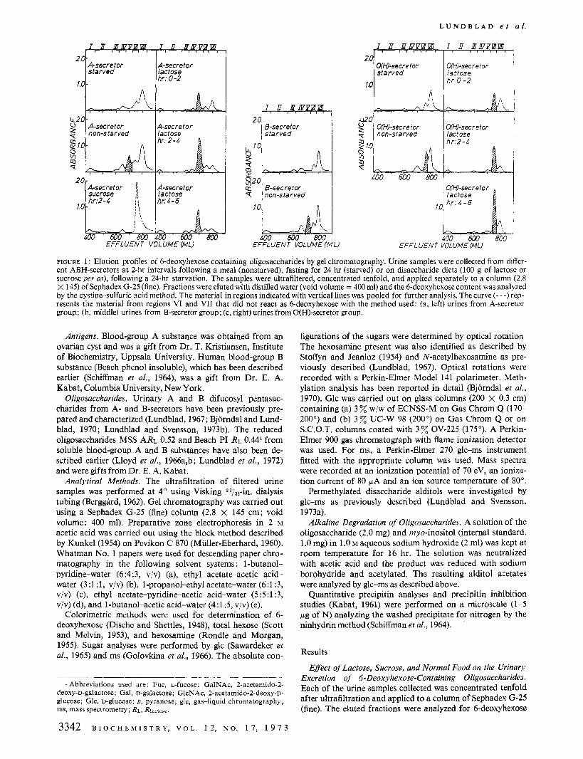
FIGURE 1 : Elution profiles of 6-deoxyhexose containing oligosaccharides by gel chromatography. Urine samples were collected from differ- ent ABH-secretors at 2-hr intervals following a meal (nonstarved), fasting for 24 hr (starved) or on disaccharide diets (100 g of lactose or sucrose per os), following a 24-hr starvation. The samples were ultrafiltered, concentrated tenfold, and applied separately to a column (2.8 X 145) of Sephadex G-25 (fine). Fractions were eluted with distilled water (void volume = 400 ml) and the 6-deoxyhexose content was analyzed by the cystine-sulfuric acid method. The material in regions indicated with vertical lines was pooled for further analysis. The curve (- - -)rep- resents the material from regions VI and VI1 that did not react as 6-deoxyhexose with the method used: (a, left) urines from A-secretor group; (b, middle) urines from B-secretor group; (c, right) urines from O(H)-secretor group.
Antigens. Blood-group A substance was obtained from an ovarian cyst and was a gift from Dr. T. Kristiansen, Institute of Biochemistry, Uppsala University. Human blood-group B substance (Beach phenol insoluble), which has been described earlier (Schiffman et al., 1964), was a gift from Dr. E. A. Kabat, Columbia University, New York.
Oligosaccharides. Urinary A and B difucosyl pentasac- charides from A- and B-secretors have been previously pre- pared and characterized (Lundblad, 1967 ; Bjorndal and Lund- blad, 1970; Lundblad and Svensson, 1973b). The reduced oligosaccharides MSS ARL 0.52 and Beach PI RL O M from soluble blood-group A and B substances have also been de- scribed earlier (Lloyd et al., 1966a,b; Lundblad et al., 1972) and were gifts from Dr. E. A. Kabat.
Analytical Methods. The ultrafiltration of filtered urine samples was performed at 4" using Visking 23/32-in. dialysis tubing (Berggird, 1962). Gel chromatography was carried out using a Sephadex G-25 (fine) column (2.8 X 145 cm; void volume: 400 ml). Preparative zone electrophoresis in 2 M acetic acid was carried out using the block method described by Kunkel(l954) on Pevikon C 870 (Muller-Eberhard, 1960). Whatman No. 1 papers were used for descending paper chro- matography in the following solvent systems : l-butanol- pyridine-water (6:4:3, v/v) (a), ethyl acetate-acetic acid- water (3:1:1, v/v) (b), 1-propanol-ethyl acetate-water (6:1:3, v/v) (c), ethyl acetate-pyridine-acetic acid-water ( 5 5 :1:3, v/v) (d), and 1-butanol-acetic acid-water (4:1:5, v/v) (e).
Colorimetric methods were used for determination of 6- deoxyhexose (Dische and Shettles, 1948), total hexose (Scott and Melvin, 1953), and hexosamine (Rondle and Morgan, 1955). Sugar analyses were performed by glc (Sawardeker et al., 1965) and ms (Golovkina et al., 1966). The absolute con-
-
1 Abbreviations used are: Fuc, L-fucose; GalNAc, 2-acetamido-2- deoxy-D-galactose; Gal, D-galactose; GlcNAc, 2-acetamido-2-deoxy-~- glucose; Glc, D-glucose; p , pyranose; glc, gas-liquid chromatography; ms, mass spectrometry; RL, Rlaotose.
3342 B I O C H E M I S T R Y , V O L . 12 , N O . 1 7 , 1 9 7 3
figurations of the sugars were determined by optical rotation The hexosamine present was also identified as described by Stoffyn and Jeanloz (1954) and N-acetylhexosamine as pre- viously described (Lundblad, 1967). Optical rotations were recorded with a Perkin-Elmer Model 141 polarimeter. Meth- ylation analysis has been reported in detail (Bjorndal et al., 1970). Glc was carried out on glass columns (200 X 0.3 cm) containing (a) 3% wjw of ECNSS-M on Gas Chrom Q (170- 200") and (b) 3% UC-W 98 (200") on Gas Chrom Q or on S.C.O.T. columns coated with 3% OV-225 (175"). A Perkin- Elmer 900 gas chromatograph with flame ionization detector was used. For ms, a Perkin-Elmer 270 glc-ms instrument fitted with the appropriate column was used. Mass spectra were recorded at an ionization potential of 70 eV, an ioniza- tion current of 80 pA and an ion source temperature of 80".
Permethylated disaccharide alditols were investigated by glc-ms as previously described (Lundblad and Svensson, 1973a).
Alkaline Degradation of Oligosaccharides. A solution of the oligosaccharide (2.0 mg) and myo-inositol (internal standard, 1.0 mg) in 1.0 M aqueous sodium hydroxide (2 ml) was kept at room temperature for 16 hr. The solution was neutralized with acetic acid and the product was reduced with sodium borohydride and acetylated. The resulting alditol acetates were analyzed by glc-ms as described above.
Quantitative precipitin analyses and precipitin inhibition studies (Kabat, 1961) were performed on a microscale (1-5 pg of N) analyzing the washed precipitate for nitrogen by the ninhydrin method (Schiffman et al., 1964).
Results
Eflect OJ Lactose, Sucrose, and Normal Food on the Urinary Excretion of 6-Deoxyhexose-Containing Oligosaccharides. Each of the urine samples collected was concentrated tenfold after ultrafiltration and applied to a column of Sephadex G-25 (fine). The eluted fractions were analyzed for 6-deoxyhexose
U R I N A R Y O L I G O S A C C H A R I D E S
~~
TABLE I : Rlactose Values of Three Urinary Oligosaccharides.
Rlactose Value in Solvent
Oligosaccharide a b c d e
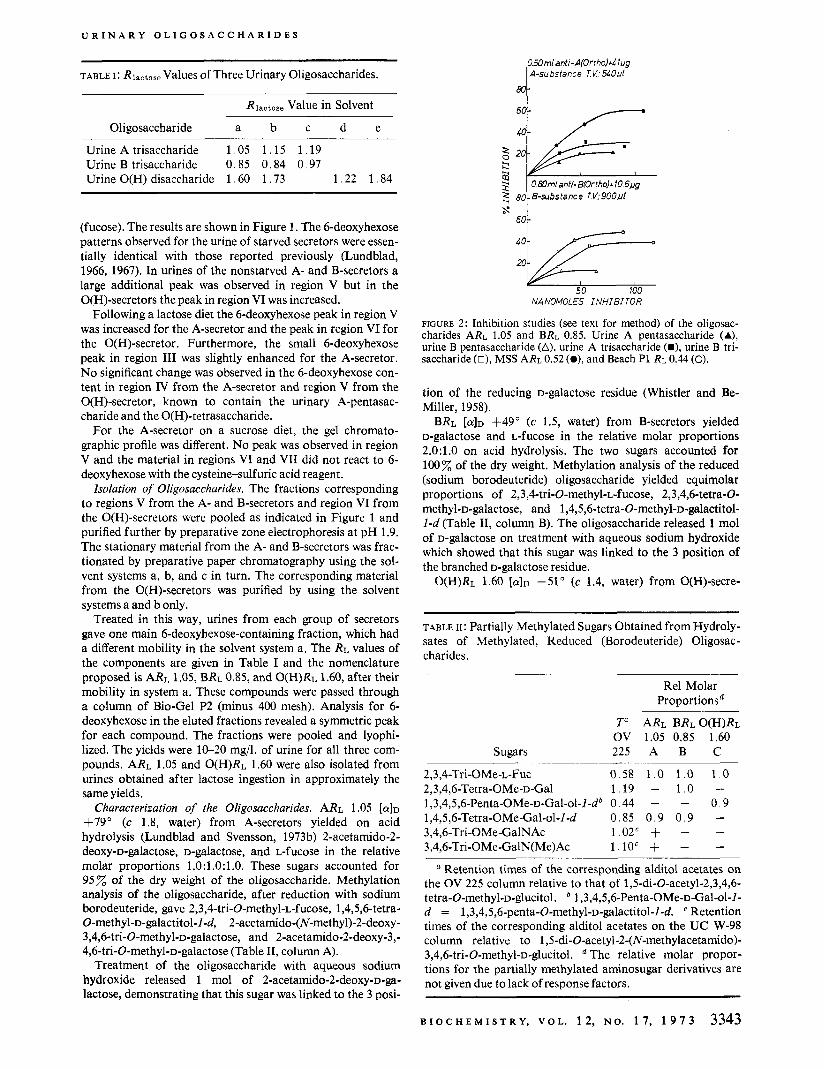
Urine A trisaccharide 1.05 1.15 1.19 Urine B trisaccharide 0.85 0.84 0.97 Urine O(H) disaccharide 1 .60 1 .73 1.22 1.84
(fucose). The results are shown in Figure 1. The 6-deoxyhexose patterns observed for the urine of starved secretors were essen- tially identical with those reported previously (Lundblad, 1966, 1967). In urines of the nonstarved A- and B-secretors a large additional peak was observed in region V but in the O(H)-secretors the peak in region VI was increased.
Following a lactose diet the 6-deoxyhexose peak in region V was increased for the A-secretor and the peak in region VI for the O(H)-secretor. Furthermore, the small 6-deoxyhexose peak in region I11 was slightly enhanced for the A-secretor. No significant change was observed in the 6-deoxyhexose con- tent in region IV from the A-secretor and region V from the O(H)-secretor, known to contain the urinary A-pentasac- charide and the O(H)-tetrasaccharide.
For the A-secretor on a sucrose diet, the gel chromato- graphic profile was different. No peak was observed in region V and the material in regions VI and VI1 did not react to 6- deoxyhexose with the cysteine-sulfuric acid reagent.
Isolation of Oligosaccharides. The fractions corresponding to regions V from the A- and B-secretors and region VI from the O(H)-secretors were pooled as indicated in Figure 1 and purified further by preparative zone electrophoresis at pH 1.9. The stationary material from the A- and B-secretors was frac- tionated by preparative paper chromatography using the sol- vent systems a, b, and c in turn. The corresponding material from the O(H)-secretors was purified by using the solvent systems a and b only.
Treated in this way, urines from each group of secretors gave one main 6-deoxyhexose-containing fraction, which had a different mobility in the solvent system a. The RL values of the components are given in Table I and the nomenclature proposed is ARL 1.05, BRL 0.85, and O(H)RL 1.60, after their mobility in system a. These compounds were passed through a column of Bio-Gel P2 (minus 400 mesh). Analysis for 6- deoxyhexose in the eluted fractions revealed a symmetric peak for each compound. The fractions were pooled and lyophi- lized. The yields were 10-20 mg/l. of urine for all three com- pounds. ARL 1.05 and O(H)RL 1.60 were also isolated from urines obtained after lactose ingestion in approximately the same yields.
Characterization of the Oligosaccharides. ARL 1.05 [ff]D
+79" (c 1.8, water) from A-secretors yielded on acid hydrolysis (Lundblad and Svensson, 1973b) 2-acetamido-2- deoxy-D-galactose, D-galactose, and L-fucose in the relative molar proportions 1.O:l.O:l.O. These sugars accounted for 95% of the dry weight of the oligosaccharide. Methylation analysis of the oligosaccharide, after reduction with sodium borodeuteride, gave 2,3,4-tri-O-methyl-~-fucose, 1,4,5,6-tetra- 0-methyl-D-galactitol-I-d, 2-acetamido-(N-methyl)-2-deoxy- 3,4,6-tri-O-methyl-~-galactose, and 2-acetamido-2-deoxy-3,- 4,6-tri-O-methyl-~-galactose (Table 11, column A).
Treatment of the oligosaccharide with aqueous sodium hydroxide released 1 mol of 2-acetamido-2-deoxy-~-ga- lactose, demonstrating that this sugar was linked to the 3 posi-
050ml anti-A(Ortho)+l lug I A-substance T V . 54Opl
OBOmlanfi-B(Ortho)+10.6~/g
60
50 100 NANOMOLES INHIBITOR
FIGURE 2: Inhibition studies (see text for method) of the oligosac- charides ARL 1.05 and BRL 0.85. Urine A pentasacchdride (A), urine B pentasaccharide (A), urine A trisaccharide (m), urine B tri- saccharide (E), MSS ARL 0.52 (e), and Beach P1 RL 0.44 (0).
tion of the reducing D-galactose residue (Whistler and Be- Miller, 1958).
BRL [ff]D 4-49" (c 1.5, water) from B-secretors yielded D-galactose and L-fucose in the relative molar proportions 2.0:l.O on acid hydrolysis. The two sugars accounted for 100% of the dry weight. Methylation analysis of the reduced (sodium borodeuteride) oligosaccharide yielded equimolar proportions of 2,3,4-tri-O-methyl-~-fucose, 2,3,4,6-tetra-O- methyl-D-galactose, and 1,4,5,6-tetra-O-methyl-~-galactitol- I-d (Table 11, column B). The oligosaccharide released 1 mol of D-galactose on treatment with aqueous sodium hydroxide which showed that this sugar was linked to the 3 position of the branched D-galactose residue.
O(H)RL 1.60 [@ID -51" (c 1.4, water) from O(H)-secre-
~ ~~~
TABLE 11: Partially Methylated Sugars Obtained from Hydroly- sates of Methylated, Reduced (Borodeuteride) Oligosac- charides.
Re1 Molar Proportionsd
Tu ARL BRLO(H)RL OV 1.05 0.85 1.60
Sugars 225 A B C
2,3,4-Tri-OMe-~-Fuc 0.58 1.0 1 . 0 1 .0
1,3,4,5,6-Penta-OMe-~-Gal-ol-I-d~ 0.44 - - 0 , 9 2,3,4,6-Tetra-OMe-~-Gal 1.19 - 1 . 0 - 1,4,5,6-Tetra-OMe-Gal-ol-l-d 0.85 0 . 9 0 .9 - 3,4,6-Tri-OMe-GalNAc 1.02c + - - 3,4,6-Tri-OMe-GalN(Me)Ac l . lOC + - -
a Retention times of the corresponding alditol acetates on the OV 225 column relative to that of 1,5-di-O-acetyl-2,3,4,6- tetra-0-methyl-D-glucitol. 1,3,4,5,6-Penta-OMe-~-Gal-ol-I- d = 1,3,4,5,6-penta-O-methyl-~-galactitol-l-d. Retention times of the corresponding alditol acetates on the UC W-98 column relative to 1,5-di-O-acetyl-2-(N-methylacetamido)- 3,4,6-tri-O-methyl-~-glucitol. The relative molar propor- tions for the partially methylated aminosugar derivatives are not given due to lack of response factors.
B I O C H E M I S T R Y , V O L . 12 , N O . 1 7 , 1 9 7 3 3343
L U N D B L A D e t a / .
- L-Fuc~ A+ oc-D-GalNAcp-(l+3.3)-O-D-Galp-(l4)-l3-D-Glc NAcp-(14-R
MSS ARL 0.52
K-L-FUC~ cc-L-Fucp 2. '4 $
cc-D-GalNAcp-(l-+3)-O-D-Galp-(l+4)-D-Glc
Urine A Denfasaccharide
CC-L-FUC~ :+ oc-D-Gal NAcp-(l+3)-D-Ga I
Urine A trisaccharide
a-L- FUCP CC- L- FUCP P '4 fl-D-Galp-II+L)-D-dlc
Urine O(H) tetrasaccharide (lactod i fucotetraose)
CC-L-FUC~ 9
u-D-G alp-(l+3)-~-D-Galp-(l4-l3-D-GlcNAcp-(l4)-R Beach P1 R ~ 0 . 4 4
O(- L-FUCD R-L-FucD
A+ ' :+' ~-D-Galp-(l+3)-O-D-Galp-(1+44)-D-Glc
Urine B pentasaccharide
CX-L-FUC~ 44
Cx-D-Galp-(1+3)-D-Ga I
Urine B trisaccharide
X-L-Fucp-(l+2)-D-Gal
Urine O(H)disaccharide (2'-fucosyl gal act ose)
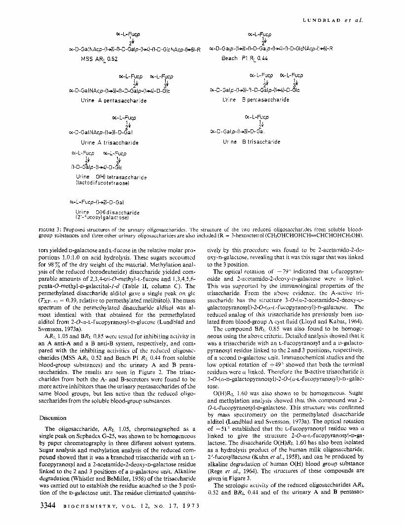
FIGURE 3 : Proposed structures of the urinaiy oligosaccharides. The structure of the two reduced oligosaccharides from soluble blood- group substances and three other urinary oligosaccharides are also included (R = 3-hexenetetrol (CH20HCHOHCH=CHCHOHCH?OH).
tors yielded D-galactose and L-fucose in the relative molar pro- portions 1 .O: l .O on acid hydrolysis. These sugars accounted for 98 of the dry weight of the material. Methylation anal- ysis of the reduced (borodeuteride) disaccharide yielded com- parable amounts of 2,3,4-tri-O-methyl-~-fucose and 1,3,4,5,6- penta-0-methyl-D-galactitol-1-d (Table 11, column C). The permethylated disaccharide alditol gave a single peak on glc (TXE--60 = 0.39, relative to permethylated melibiitol). The mass spectrum of the permethylated disaccharide alditol was al- most identical with that obtained for the permethylated alditol from 2-0-a-~-fucopyranosyl-~-glucose (Lundblad and Svensson, 1973a).
ARL 1.05 and BRL 0.85 were tested for inhibiting activity in an A anti-A and a B anti-B system, respectively, and com- pared with the inhibiting activities of the reduced oligosac- charides (MSS ARL 0.52 and Beach PI RL 0.44 from soluble blood-group substances) and the urinary A and B penta- saccharides. The results are seen in Figure 2 . The trisac- charides from both the A- and B-secretors were found to be more active inhibitors than the urinary pentasaccharides of the same blood groups, but less active than the reduced oligo- saccharides from the soluble blood-group substances.
Discussion
The oligosaccharide, ARL 1.05, chromatographed as a single peak on Sephadex G-25, was shown to be homogeneous by paper chromatography in three different solvent systems. Sugar analysis and methylation analysis of the reduced com- pound showed that it was a branched trisaccharide with an L- fucopyranosyl and a 2-acetamido-2-deoxy-~-galactose residue linked to the 2 and 3 positions of a D-galactose unit. Alkaline degradation (Whistler and BeMiller, 1958) of the trisaccharide was carried out to establish the residue attached to the 3 posi- tion of the D-galactose unit. The residue eliminated quantita-
3344 B I O C H E M I S T R Y , V O L . 1 2 , N O . 1 7 , 1 9 7 3
tively by this procedure was found to be 2-acetamido-2-de- oxy-D-galactose, revealing that it was this sugar that was linked to the 3 position.
The optical rotation of -79" indicated that L-fucopyran- oside and 2-acetamido-2-deoxy-~-galactose were a linked. This was supported by the immunological properties of the trisaccharide. From the above evidence, the A-active tri- saccharide has the structure 3-0-(a-2-acetamido-2-deoxy-~- galactopyranosyl)-2-0-(a-~-fucopyranosyl)-~-galactose. The reduced analog of this trisaccharide has previously been iso- lated from blood-group A cyst fluid (Lloyd and Kabat, 1964).
The compound BRL 0.85 was also found to be homoge- neous using the above criteria. Detailed analysis showed that it was a trisaccharide with an L-fucopyranosyl and a o-galacto- pyranosyl residue linked to the 2 and 3 positions, respectively, of a second D-galactose unit. Immunochemical studies and the low optical rotation of -49" showed that both the terminal residues were a linked. Therefore the B-active trisaccharide is 3-~-(a-~-galactopyranosyl)-2-~-(a-~-fucopyranosyl~- D - galac- tose.
O(H)RL 1.60 was also shown to be homogeneous. Sugar and methylation analysis showed that this compound was 2- 0-L-fucopyranosyl-D-galactose. This structure was confirmed by mass spectrometry on the permethylated disaccharide alditol (Lundblad and Svensson, 1973a). The optical rotation of -51 O established that the L-fucopyranosyl residue was a linked to give the structure 2-0-a-~-fucopyranosyl-~-ga- lactose. The disaccharide O(H)RL 1.60 has also been isolated as a hydrolysis product of the human milk oligosaccharide, 2'-fucosyllactose (Kuhn et al., 1958), and can be produced by alkaline degradation of human O(H) blood group substance (Rege et al., 1964). The structures of these compounds are given in Figure 3.
The serologic activity of the reduced oligosaccharides ARL 0.52 and BRL 0.44 and of the urinary A and B pentasac-
U R I N A R Y O L I G O S A C C H A R I D E S
charides was found to be in the same range as previously re- ported (Lundblad and Kabat, 1971). The presence of the second fucose is believed to prevent a firm attachment of the oligosaccharide to the antibody (Lloyd et al., 1966a,b). The length of the oligosaccharide chain may also contribute to the serological activity. Thus 3-O-cr-~-GalNAcp-3-O-P-~-Galp- D-GlcNAc is a more potent inhibitor than 3-O-cr-~-GalNAcp- D-Gal of A anti-A precipitation (Schiffman et al., 1964). The inhibitory activity of the A and B trisaccharides, on a molar basis, was found to be intermediate to the mono- and di- fucosyl oligosaccharides as expected from the above dis- cussion.
The three oligosaccharides (ARL 0.52, BRL 0.44, and 0- (H)RL 1.60), shown to be present only in the urines of AB0 (H)-secretors, represent the nonreducing terminii of the A, B, and O(H) antigenic determinants, respectively (Lloyd and Kabat, 1968; Watkins, 1966). The secretor gene is responsible for the synthesis of an a-2-~-fucosyltransferase found in human milk, submaxillary glands, and stomach mucosa (Shen et al., 1968; Grollman et al., 1969; Chester and Watkins, 1969). This enzyme transfers L-fucose to the 2 position of D-
galactose. Another enzyme with a similar or an identical spec- ificity has been demonstrated in serum of all ABO(H)- secretors, irrespective of secretor-nonsecretor status (Schenkel-Brunner et al., 1972). A possible source of this enzyme is the red blood cell or its precursors where the A, B, and O(H) determinants are synthesized independently of the secretor gene (Watkins and Morgan, 1959; Watkins, 1966). We propose that the urinary oligosaccharides are derived from stomach and intestinal mucosal cells and do not represent de- graded red blood corpuscle antigens. An interesting observa- tion is that the synthesis of these oligosaccharides appears to be induced by normal food intake or by feeding on lactose. However, a sucrose diet seems to have no effect. This finding indicates that the D-galactose derived from the hydrolysis of lactose is the immediate precursor which readily accepts the different monosaccharides to form the blood-group-specific oligosaccharides. The presence of lactose in normal food would also account for increased synthesis and urinary ex- cretion of 6-deoxyhexose compounds after normal feeding.
Acknowledgments
Miss Christina Sandberg for valuable technical assistance. The authors are indebted to Mrs. Gunilla Pettersson and
Added in proof
After submitting this paper for publication a report en- titled “Etude de la structure et de l’activite de groupe sanguin des oligosaccharides isoles de l’urine de sujets sains et patho- logiques. Mise en evidence d’une oligosaccharidourie as- sociee a diverses melituries,” by G. Strecker, B. Fournet, S. Bouguelet, T. Riazi-Farzad, and J. Montreuil, was pre- sented at Colloque International sur les Glycoconjugues (CNRS) held in Lille (France), June 20-27, 1973. This report describes the effect of galactose ingestion upon the excretion of blood group specific urinary oligosaccharides. The results obtained were analogous to those described above, but in addition small quantities of larger oligosaccharides were obtained.
References
Berggard, 1. (1962), Ark. Kerni 18,291. Bjorndal, H., Hellerqvist, C. G., Lindberg, B., and Svensson,
Bjorndal, H., and Lundblad, A . (1970), Biochim. Biophys.
Chester, M. A,, and Watkins, W. M. (1969), Biochem. Biophys.
Dische, Z., and Shettles, L. B. (1948), J . Biol. Chem. 175, 595. Golovkina, L. S., Chizhov, 0. S., and Wulfson, N. S. (1966),
Izu. Akad. Nauk. SSSR, Ser. Khim., 1915. Grollman, E. F., Kobata, A., and Ginsburg, V. (1969), J . Clin.
Invest. 48, 1489. Kabat, E. A., Ed. (1961), Kabat and Mayer’s Experimental
Immunochemistry, 2nd ed, Springfield, Ill., C. C Thomas. Kuhn, R., Baer, H. H., and Gauhe, A. (1958), Chem. Ber. 91,
364. Kunkel, H. G . (1954), Methods Biochem. Anal. I , 141. Lloyd, K. O., and Kabat, E. A. (1964), Biochem. Biophys. Res.
Lloyd, K. O., and Kabat, E. A. (1968), Proc. Nat. Acad. Sci.
Lloyd, K. O., Kabat, E. A., Layug, E. J., and Gruezo, F.
Lloyd, K. O., Kabat, E. A., and Rosenfield, R . E. (1966b),
Lundblad, A. (1966), Biochim. Biop/ij*s. Acta 130,130. Lundblad, A. (1967), Biochim. Biophys. Acta 148,151. Lundblad, A. (1968), Biochim. Biopl1J.s. Acta 165,202. Lundblad, A., Hammarstrom, S., Licerio, E., and Kabat,
Lundblad, A., and Kabat, E. A. (1971), J . In~munol. 106,
Lundblad, A., and Svensson, S. (1973a), Biochemistry 12,
Lundblad, A,, and Svensson, S. (1973b), Carbohyd. Res.
Muller-Eberhard, H. J. (1960), Scand. J . Clin. Lab. Invest. 12,
Rege, V. P., Painter, T. J., Watkins, W. M., and Morgan,
Rondle, C. J. M., and Morgan, W. T. J. (1955), Biochem. J .
Sawardeker, J. S., Sloneker, J. H., and Jeanes, A. R. (1965),
Schenkel-Brunner, M., Chester, A., and Watkins, W. M.
Schiffman, G., Kabat, E. A., and Thompson, W. (1964),
Scott, Jr., T. A,, and Melvin, E. H. (1953), Anal. Chem. 25,
Shen, L., Grollman, E. F., and Ginsburg, V. (1968), Proc.
Stoffyn, P. J., and Jeanloz, R. W. (1954), Arch. Biochem.
Watkins, W. M. (1966), Science 152,172. Watkins, W. M., and Morgan, W. T. J. (1959), Vox Sang. 4 ,
Whistler, R . L., and BeMiller, J. N. (1958), Advan. Carbohyd.
S. (1970), Angew. Chem., Int. Ed. Engl. 9,610.
Acta 201,434.
Res. Commun. 34,835.
Commun. 16,385.
U. S. 61,1470.
(1966a), Biochemistrj 5,1489.
Biochemistry 5, 1502.
E. A. (1972), Arch. Biochem. Biophys. 148,291.
1572.
306.
(in press).
33.
W. T. J. (1964), Nature (London) 203,360.
61,586.
Anal. Chem. 37,1602.
(1972), Eur. J . Biochern. 30,269.
Biochemistry 3,113.
1656.
Nat. Acad. Sci. U . S. 59,224.
Biophys. 52,373.
97.
Chem. 13,289.
B I O C H E M I S T R Y , V O L . 1 2 , N O . 1 7 , 1 9 7 3 3345