Embed Size (px)
Citation preview

Structure of Human Prostasin, a Target for the Regulationof Hypertension*□S
Received for publication, July 10, 2008, and in revised form, September 11, 2008 Published, JBC Papers in Press, October 14, 2008, DOI 10.1074/jbc.M805262200
Keith W. Rickert1, Paul Kelley, Noel J. Byrne, Ronald E. Diehl, Dawn L. Hall, Allison M. Montalvo, John C. Reid,Jennifer M. Shipman, Bradley W. Thomas, Sanjeev K. Munshi, Paul L. Darke, and Hua-Poo Su2
From the Department of Global Structural Biology, Merck Research Laboratories, West Point, Pennsylvania 19486
Prostasin (also called channel activating protease-1 (CAP1))is an extracellular serine protease implicated in the modulationof fluid and electrolyte regulation via proteolysis of the epithe-lial sodium channel. Several disease states, particularly hyper-tension, can be affected by modulation of epithelial sodiumchannel activity. Thus, understanding the biochemical functionof prostasin and developing specific agents to inhibit its activitycould have a significant impact on a widespread disease. Wereport the expression of the prostasin proenzyme in Escherichiacoli as insoluble inclusion bodies, refolding and activating viaproteolytic removal of the N-terminal propeptide. The refoldedand activated enzyme was shown to be pure and monomeric,with kinetic characteristics very similar to prostasin expressedfrom eukaryotic systems. Active prostasin was crystallized, andthe structure was determined to 1.45 A resolution. Theseapoprotein crystals were soaked with nafamostat, allowing thestructure of the inhibited acyl-enzyme intermediate structure tobe determined to 2.0 A resolution. Comparison of the inhibitedand apoprotein forms of prostasin suggest a mechanism of reg-ulation through stabilization of a loop which interferes withsubstrate recognition.
Prostasin (also known as channel activating protease-1(CAP1)) is a serine protease originally isolated from seminalfluid as a secreted protein (1). It is widely expressed inmamma-lian epithelial tissue as a 40-kDa glycosylphosphatidylinositol(GPI)-anchored protein (1–3). Prostasin is implicated in theregulation of sodium and fluid levels via proteolysis of the epi-thelial sodium channel (ENaC)3 � subunit (4–9).ENaC performs an essential function in several epithelial tis-
sues, including the colon, kidney, and lung (5, 7, 9, 10). In these
tissues sodium is primarily transported across the apical mem-brane via ENaC, and fluid transport across the membrane ishighly responsive to sodium concentration. Inappropriatefunction of ENaC leads to misregulation of blood pressure inhumans (11, 12). The same ENaC mutations which lead tohypertension in humans give rise to defects in lung fluid clear-ance in mice (11), suggesting that proper ENaC function mayalso be important in other diseases where sodium and fluidhomeostasis is severely disrupted, for example cystic fibrosisand diarrhea (13, 14).Although ENaC activity can be regulated via transcription
and translation, for example in response to the hormone aldos-terone (11), proteolysis of ENaC can lead to channel activation(9), and inhibition of this process might provide a direct andspecificmethod tomodulate ENaC activity for therapeutic ben-efit. Cleavage of an inhibitory peptide in the ENaC� subunit byprostasin has been shown to stimulate sodium transport by2–3-fold in cell-based experiments (4, 5, 7–10).Other proteasesimplicated to activate ENaC in model systems include furin,mCAP-2, mCAP-3, and TMSP-1 (transmembrane serine pro-tease 1) (5, 8–11). Some of these proteases require activation byyet other proteases (15, 16), so clarity regarding which enzymesmay represent appropriate targets for reducing ENaC activityhas yet to be achieved. Highly selective small-molecule inhibi-tors of proteases thought to modulate ENaC will aid in under-standing the roles of different proteases in channel function andmay also provide a therapeutic benefit for hypertension andother diseases, e.g. cystic fibrosis (12–14). Although there arenumerous agents for the treatment of hypertension,many indi-viduals require multiple medications to achieve adequate con-trol (17), and the condition continues to exact a high cost tosociety, and so it seems that additional modes of treatmentremain desirable.Prostasin belongs to the classical serine protease family, with
homology to trypsin, chymotrypsin, and kallikrein, and has atrypsin-like substrate specificity (1, 2, 18). In common withthese, enzyme activation of prostasin occurs via cleavage of thepro-protein to produce a light chain and a heavy chain that aredisulfide-linked. Prostasin has some features unusual in serineproteases, such as a high degree of sensitivity to monovalentand divalent cations, which may relate to the prostasin role inENaC regulation (18). However, to date there have been noreports of potent and selective small molecule inhibitors ofprostasin. High resolution structural studies have been used tosignificant advantage in the design of selective protease inhibi-tors (19–22). Availability of the three-dimensional structure of
* The costs of publication of this article were defrayed in part by the paymentof page charges. This article must therefore be hereby marked “advertise-ment” in accordance with 18 U.S.C. Section 1734 solely to indicate this fact.
□S The on-line version of this article (available at http://www.jbc.org) containssupplemental Experimental Procedures, Tables 1 and 2, and Figs. 1–3.
The atomic coordinates and structure factors (codes 3DFJ and 3DFL) have beendeposited in the Protein Data Bank, Research Collaboratory for StructuralBioinformatics, Rutgers University, New Brunswick, NJ (http://www.rcsb.org/).
1 To whom correspondence may be addressed: WP26-354, P. O. Box 4,West Point PA 19486. Tel.: 215-652-5323; Fax: 215-993-0026; E-mail:[email protected].
2 To whom correspondence may be addressed: WP14-1101, P. O. Box 4, WestPoint PA 19486. Tel.: 215-652-7347; E-mail: [email protected].
3 The abbreviations used are: ENaC, epithelial sodium channel; MS, massspectrometry; SEC, size-exclusion chromatography; CHAPS, 3-[(3-cholami-dopropyl)dimethylammonio]-1-propanesulfonic acid; Bis-Tris, 2-[bis(2-hydroxyethyl)amino]-2-(hydroxymethyl)propane-1,3-diol.
THE JOURNAL OF BIOLOGICAL CHEMISTRY VOL. 283, NO. 50, pp. 34864 –34872, December 12, 2008© 2008 by The American Society for Biochemistry and Molecular Biology, Inc. Printed in the U.S.A.
34864 JOURNAL OF BIOLOGICAL CHEMISTRY VOLUME 283 • NUMBER 50 • DECEMBER 12, 2008
by guest on July 5, 2018http://w
ww
.jbc.org/D
ownloaded from

prostasin could offer a significant advantage in the design ofprostasin inhibitors.Previous studies have shown that active prostasin can be
expressed in a recombinant baculovirus system and purified tohomogeneity, enabling more extensive biochemical studies(18). We report the expression of prostasin in Escherichia coliand the refolding, proteolytic activation, and purification toyield pure and fully active enzyme. Expression in E. coli is noveland can allow for rapid generation of site-directed mutants toallow the biochemical determination of structure-functionrelationships.We describe the high resolution crystal structureof the protease domain of this enzyme, which may provide val-uable information in the design or optimization of specificsmall-molecule inhibitors.
EXPERIMENTAL PROCEDURES
Construction of Prostasin Variants—The native sequence(accession number NM_002773) and all expression constructsare illustrated in Fig. 1. In all constructs the C-terminal glyco-sylphosphatidylinositol anchor domain and preceding linkerwere replaced with a His6 tag to aid in purification. Cysteines154 and 203 were not expected to be involved in intradomaindisulfide bonds and were mutated to serine and alanine (18). Inthe case of baculovirus variant 40, the N-linked glycosylationsite was also removed via site-directed mutagenesis. For bacu-lovirus expression constructs, the native signal sequence andpropeptide were replaced with an insect cell signal sequence(melittin, GP64, or GP67) to generate the native N terminus ofthemature protein, as the propeptide is not required for activity(18). Bacterial expression constructs contained the originalnative propeptide sequence at the N terminus but with theaddition of an enterokinase recognition sequence to allow spe-cific cleavage to generate the active enzyme. Detailed methodsfor the expression of prostasin in both baculovirus and E. coliare provided in the supplemental Experimental Procedures.Refolding and Purification of Prostasin from E. coli—Prosta-
sin variants 26 and 28 (Fig. 1) were solubilized from inclusionbodies in 8 M urea with 0.1 M Tris/HCl, pH 8.0, and 2 mM dithi-othreitol after extensive washing. Solubilized protein wasbound to nickel-nitrilotriacetic acid Superflow resin (Qiagen)and eluted with 0.3 M imidazole. The urea-solubilized materialwas then refolded in a buffer containing 1 M L-arginine, 0.1 MTris/HCl, 5 mM reduced glutathione, and 0.5 mM oxidized glu-tathione, pH 8.0, at a final concentration of 10 mg/liter. Afterdiafiltration, protein was purified via Ni(II) affinity and anionexchange chromatography. Eluted fractions were evaluated viaSDS-PAGE and mass spectrometry (MS), and fractions inwhich prostasin was detected by MS were then pooled for fur-ther processing. Prostasin zymogen was converted to the activeform by addition of enterokinase (EKMax, Invitrogen) at a finalconcentration of 2 units/ml (7.5 units/mg prostasin) in thepresence of 0.5mM reduced glutathione. The resultant cleavagereaction was maintained at 4 °C for 48 h and monitored by MSand SDS-PAGE. After completion of cleavage as determined byMS, the reaction was incubated at 4 °C overnight after the addi-tion of 1mMoxidized glutathione. Active prostasinwas purifiedfrom enterokinase and misfolded prostasin via Ni(II) affinityand anion exchange chromatography. Fractions were selected
based on activity, then pooled to give the final purified prostasinfor further characterization and crystallography. Furtherdetails of the purification are found in the supplemental Exper-imental Procedures.Enzyme Assay—Enzyme assays were performed at ambient
temperature (22–24 °C) in 0.1 M Tris/HCl, pH 9, with 0.1%CHAPS using a peptide substrate, Ac-KHYR-7-amino-4-meth-ylcoumarin (Anaspec) at 50 �M (18). Data were collected usinga BMGLabTech Fluostar Optima reader, with excitation andemission filter wavelengths of 340 and 450 nm, respectively.Measurements of Km were made by varying the concentra-tion of substrate from 10–250 �M, whereas enzyme concen-tration was held constant at 19 nM. Absolute quantitation ofproduct was determined by comparison to an independent7-amino-4-methylcoumarin standard at concentrationsfrom 2 nM to 1.7 �M.Size Exclusion Chromatography (SEC)—Analytical SEC
measurements were made using a Superdex 200 5/150 col-umn (GEHealthcare) equilibrated in 50mM potassium phos-phate, pH 6.8, and 0.3 M NaCl at a flow rate of 0.15 ml/minwith injections of 0.5–5 �g of protein. Retention times wereconverted to apparent molecular masses using mixed pro-tein standards (thyroglobulin, gamma-globulin, ovalbumin,and myoglobin) (Bio-Rad).MS—Whole-protein mass measurements were made by
binding samples (0.1–2 �g) to a reverse-phase protein trap col-umn (Michrom), where they were desalted by washing with 2%acetonitrile, 0.01% trifluoroacetic acid and eluted with a solu-tion of 64% acetonitrile, 0.01% trifluoroacetic acid into an elec-trospray mass spectrometer (LTQ, Thermo). The resultantspectrawere deconvoluted using Promass (Novatia) to yield thewhole protein mass. For free cysteine determination, sampleswere treated with 50 mM iodoacetamide at room temperaturefor 30 min before analysis. For active site determination, pros-tasin was incubated with 5 �M nafamostat mesylate (BioMolInternational) for 5 min in 50mMTris/HCl, pH 8.5, with a finalNaCl concentration of 0.2 M or less before mass spectroscopicanalysis (23).Protein Crystallization—Prostasin variant 26, comprising
human prostasin residues 45–285 with the mutations C154Sand C203A, a C-terminal thrombin-cleavage site, and His6 tag(Fig. 1B), and variant 28, comprising human prostasin residues45–289with the samemutations andC terminus, were concen-trated to a final concentration of 12–14 mg/ml as determinedby the Bradford assay (Bio-Rad) in 50 mM Tris/HCl, pH 8.5, 0.1M NaCl. Crystals of both variants were obtained by the vapordiffusion method. Initial crystals were obtained in hangingdrops containing 1 �l of protein mixed with 1 �l of reservoirsolution consisting of 100 mM Bis-Tris, pH 6.3, and 30% poly-ethylene glycol 3350. Diffraction quality crystals formed as aresult of streak seeding into hanging drops containing 1 �l ofprotein mixed with 1 �l of reservoir solution consisting of 100mM Bis-Tris, pH 5.8, and 21–26% polyethylene glycol 3350.Crystals were cryoprotected for data collection in a solutioncontaining 100 mM Bis-Tris, pH 6.5, 20% polyethylene glycol3350, 100 mM NaCl, and 20% ethylene glycol.Protein Structure Determination—In-house diffraction data
were collected with Rigaku x-ray sources using R-Axis image
Structure of Prostasin
DECEMBER 12, 2008 • VOLUME 283 • NUMBER 50 JOURNAL OF BIOLOGICAL CHEMISTRY 34865
by guest on July 5, 2018http://w
ww
.jbc.org/D
ownloaded from

plate detectors. Data were processed and scaled with HKL2000(24). Initial phases were obtained by molecular replacementwith Phaser (25) using the model of human plasma kallikrein(PDB code 2ANW) (26). The models for variant 28 apoprotein,and nafamostat-inhibited acyl-enzyme intermediate were builtand improved with iterative cycles of manual rebuilding inCoot and MIFit followed by refinement with Refmac (27, 28).There was sufficient density to build the model continuouslyfrom residues Ile-45 toGln-289, which encodes all of the native,mature protein residues. Nafamostat was introduced by soak-ing crystals of apoprotein in a buffer containing 1 mM nafamo-stat, 100 mM Bis-Tris, pH 5.8, 100 mM NaCl, and 30% polyeth-ylene glycol 3350. Structural alignments were performed usingLSQMAN (29). Figures were generated using PyMOL (30).Model geometry was verified using Procheck (31).
RESULTS AND DISCUSSION
Expression of ProstasinVariants—Initially, prostasin variantswere expressed by secretion from insect cells, as used by Ship-way et al. (18) to examine the catalytic properties of prostasin.Although the expression levels for the glycosylated prostasinvariants 801 and 35 (Fig. 1B) were acceptable (0.4–1.0mg/liter)and the purified proteins were well behaved in solution andcould be readily concentrated to 20 mg/ml, they did not formcrystals using a broad screen of conditions. Prostasin variant 40,which lacks the N-linked glycosylation site, expressed to muchlower levels in this system (�0.1 mg/liter).Expression in E. coliwas examined in two strains: BL21(DE3)
and Origami-2. In BL21(DE3), prostasin variants 26 and 28expressed at 50–100 mg/liter as insoluble inclusion bodies.Although Origami-2 cells possess an oxidizing intracellularenvironment sometimes helpful in disulfide formation, asneeded for prostasin, expression of prostasin variants 26 and 28in these cells gave mainly insoluble protein at 1–5 mg/liter.Accordingly, prostasin variants 26 and 28 were expressed inBL21(DE3) cells and refolded in vitro.Refolding of Prostasin—The primary challenge to devising a
refolding protocol for prostasin was evaluating the product forthe correctness of its fold. Although enzymatic activity wouldnormally be the preferred measure of success, prostasin wasexpressed in the inactive zymogen form to ensure generation ofa native N terminus. Enterokinase, the enzyme used to cleavethe propeptide, was active in the prostasin assay, making inter-pretation of activity assays from mixtures containing bothenzymes problematic. Although the amount of soluble proteincaptured on Ni(II) columns was of some guidance (Table 1,buffer additive conditions: none, 2Murea, and 0.4M L-arginine),the majority of the protein in these mixtures seemed to be mis-folded aggregates. The best tool in these circumstances to judgethe quality of refolded zymogen was MS after reverse-phasechromatography. Misfolded proteins are adsorbed much morestrongly to the reverse-phase surface than well folded proteins(32, 33), whereasMS ensures accurate identification of the pro-tein species eluted from the column. Purification of the solubi-lized protein on Ni(II) affinity media was performed to simplifythe mass spectrum observed after refolding, making the analy-sis more consistent and semiquantitative. Using these tools,small-scale refolding experiments were carried out to find opti-
mal conditions, detailed in Table 1. In scaled-up refolds,although the yields of soluble protein after refolding and thefirst chromatographic step were quite reasonable, the pool stillcontained substantial amounts of misfolded protein. Prostasinzymogen was heterogeneous by both anion exchange chroma-tography (supplemental Fig. 1) and analytical SEC, whichshowed amixture of monomer with an apparent size of 37 kDa,dimer at � 70 kDa, and larger species (Fig. 2B, dashed line).Cleavage of the zymogen with enterokinase was carried out
in the presence of reduced glutathione to allow for disulfideshuffling, as additional MS experiments suggested that thezymogen had not completely achieved the native disulfide con-figuration (supplemental Table 1), similar to the behaviorobserved for trypsinogen (34). Cleavage was rapid and com-pletely specific when monitored by MS, whereas SDS-PAGEcontinued to show that some of the protein was resistant tocleavage, which we hypothesized was misfolded.After cleavage, prostasin was purified on two additional col-
umns. The Ni(II) affinity column was used primarily to removeenterokinase, after which prostasin could be tracked and quan-tified via activity assay. The final anion exchange step proved tobe the most important in removing misfolded species. Activeprostasin eluted as the first species from the column at a con-siderably lower ionic strength than either the zymogen oraggregated species (Fig. 2A), and activity coincided only withthis first peak (Fig. 2A, dashed line). In contrast to the zymogen,MS of the fractions from the anion exchange column showedthat �95% of the MS-active species coincided with the firstpeak. Analytical SEC and SDS-PAGE of later fractions from theanion-exchange column showed that these contained prostasinin an inactive, aggregated state (data not shown). Althoughboth variants 26 and 28 could be refolded to yield active protein,the final yield of active protein was only 1–2% as the bulk of theprotein formed inactive misfolded species and was removed onthe final chromatographic step.Characterization of the Final Refolded Protein—Refolded
prostasin was of high purity as shown in the gel (Fig. 3). SEC isoften used to profile refolded protein, as it readily distinguisheslower-order species from the aggregates that result from mis-folding. As seen in Fig. 2B, purified refolded prostasin elutedas a single peak, corresponding to a molecular mass of 24 kDa,identical to that seen for prostasin variants 801 and 35 (supple-mental Table 2). Given that the cleaved propeptide has amolec-ular mass of 2 kDa, the difference between monomeric zymo-gen, which elutes from SEC with an apparent mass of 37 kDa(actual mass 30 kDa) and the active protein (actual mass 28kDa), is remarkable. The active protein is more compact interms of hydrodynamic radius, whichmay be related to its tend-ency to adopt a native disulfide configuration (34).A more demanding measure of the quality of the refolded pro-
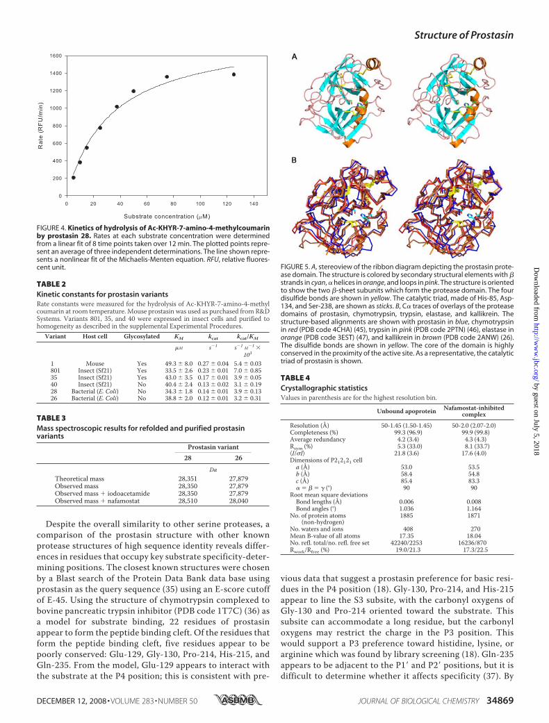
tein is a comparison of its kinetic constants with that of similarvariants folded in eukaryotic cells. Initial reaction ratesweremeas-ured across a rangeof substrate concentrations, and an example ofthe resulting data is shown in Fig. 4 alongwith the nonlinear curvefit used to obtain Km and kcat. As seen in Table 2, the kcat andkcat/Km for the refolded prostasin variants 26 and 28 are extremelysimilar to those seen for the analogous variant 40 expressed ininsect cells and only slightly lower than seen for the glycosylated
Structure of Prostasin
34866 JOURNAL OF BIOLOGICAL CHEMISTRY VOLUME 283 • NUMBER 50 • DECEMBER 12, 2008
by guest on July 5, 2018http://w
ww
.jbc.org/D
ownloaded from

variants 801 and 35. Although glycosylationmay be important forefficient expression of this protein in eukaryotes, it has no signifi-cant effect on enzymatic function in our assays.
Mass spectrometry can offerinsights into protein covalent struc-ture complementary to those seenvia other analytical measures.Refolded prostasin 26 and 28 giveclean mass spectra with only onespecies seen after deconvolutionand excellent correspondence to thepredicted mass as seen in Table 3and supplemental Fig. 2. Treatmentof either variant 26 or 28 withiodoacetamide showed no changesin the observed molecular mass(Table 3), indicating that all 8 cys-teine residues form intramoleculardisulfides. MS can also be used asan active site titrant. Treatment ofeither variant with nafamostatbefore mass measurement shows acomplete shift of the mass observedto a species at �161 Da from theoriginal mass, indicating that all ofthe protein observed viaMS is capa-ble of conversion to the expectedacyl-enzyme intermediate.The Structure of Prostasin—The
crystal structure of the proteasedomain of prostasin is structurallysimilar to other serine proteases,consisting of two � barrel-like sub-domains with four conserved disul-fide bonds (Fig. 5, Table 4). The twosubdomains are separated by a cleftthat contains the active site. In pros-tasin, the catalytic triad is formed byHis-85, Asp-134, and Ser-238 (Fig.5A), corresponding to chymotryp-sin residues His-57, Asp-102, andSer-195. The proximity of the hy-droxyl group of Ser-238 to the imid-azole ring of His-85 and the hydro-gen bond between O� of Asp-134and N� of His-85 suggest that thecatalytic mechanism observed inother serine proteases is conservedin prostasin.In comparison with other serine
proteases with known structures,the core of the domain is highly con-served, whereas the loops are lessconserved (Fig. 5B). The sequenceidentity between the proteasedomain of prostasin and those ofchymotrypsin, trypsin, elastase, andkallikrein are 37, 38, 32, and 39%,
respectively. The root mean square deviation (Å) between theC� atoms of prostasin and chymotrypsin, trypsin, elastase, andkallikrein was 1.057, 1.084, 1.060, and 0.979, respectively. The
FIGURE 1. A, map of the prostasin domain structure for the natural human sequence (top) and proteins expressed ininsect cells (middle) and bacteria (bottom) showing the signal sequence, propeptide, protease domain, and C-ter-minal region. B, alignment of prostasin variants. Residues in red represent signal sequences cleaved during expres-sion. The first variant is the full wild-type human sequence, whereas the second variant 1 is recombinant mouseprostasin as purchased from R&D Systems. Residues in blue represent the propeptide sequence either cleavedduring expression (mouse) or in vitro (variants 26 and 28). Residues in orange were removed before evaluation ofkinetic values. Residues highlighted in red represent N-linked glycosylation sites. Residues highlighted in greenrepresent introduced site-directed mutations, as described under “Experimental Procedures.”
Structure of Prostasin
DECEMBER 12, 2008 • VOLUME 283 • NUMBER 50 JOURNAL OF BIOLOGICAL CHEMISTRY 34867
by guest on July 5, 2018http://w
ww
.jbc.org/D
ownloaded from

four disulfide bonds in prostasin were conserved in the otherserine proteases. Prostasin Cys-154, which was mutated to Serto aid crystallization, is in the same position as Cys-122 in chy-motrypsin that forms a disulfide bond with Cys-1 in the N-ter-minal light chain. Analogous to Cys-1 of chymotrypsin, Cys-37is present in the N-terminal propeptide fragment of prostasin.A second cysteine, Cys-203, was mutated to Ala, also to aid
crystallization. This cysteine is not conserved among serineproteases. Prostasin has a total of 12 cysteines, so Cys-203 mayform a sixth disulfide bond with Cys-306 in the C-terminalregion. Because the C-terminal region is glycosylphosphatidyl-inositol-anchored to the cell, it is possible that formation of asixth disulfide bond may orient the active site toward the sur-face of the cell to facilitate the co-localization of the catalyticsite to its substrate.
FIGURE 2. Chromatographic traces of refolded prostasin variant 28.A, anion-exchange chromatography of prostasin after activation. The peakcorresponding to active, monomeric prostasin is marked “monomer.” Super-imposed with a dashed line are the results of activity assays of the fractionsfrom this column. B, size exclusion chromatography of the zymogen (dashedline) and activated prostasin (solid line). Molecular weights in kDa of the stand-ards used are shown on the chart at their elution positions. mAU, milliabsor-bance units.
FIGURE 3. Coomassie-stained reduced prostasin 28 examined by 14%Tris-glycine SDS-PAGE. Lane 1, molecular weight markers. Lane 2, washedand urea-solubilized inclusion bodies of prostasin. Lane 3, Ni(II)-purified, urea-solubilized prostasin proenzyme. Lanes 4 and 5, pure, active prostasin at a1.30 and 5.0 �g load. Lanes 1, 2, and 3 are from the same gel, but intermediatelanes were omitted; lanes 4 and 5 are from a different gel which was alignedby the molecular weight markers.
TABLE 1Results of refolding conditionsShown are refolding results for prostasin variant 28. Yields are expressed as total protein (determined by the Bradford assay) per liter of refold buffer either from pooledfractions after the first chromatographic step or for the final purified protein.Mass spectroscopic signal was assessed on a qualitative basis for different conditions, eachwitha nominal 1-�g protein injection. All conditions used a buffer containing 0.1 MTris/HCl, pH 8.0, 5mM reduced glutathione, and 0.5mMoxidized glutathione. Similar resultswere observed for variant 26.
ConditionBuffer additive None 2 M Urea 0.4 M L-Arginine 0.4 M L-Arginine 0.4 M L-Arginine 1.0 M L-Arginine
Protein purified prior to refold No No No Yes Yes YesProtein concentration in refold (mg/liter) 10 10 10 10 5 10
Refold results1st column yield (mg/liter) �0.1 0.1 1 1.5 2 4MS signal ND ND ND � �� ���Final yield (mg/liter) ND 0.01 0.04 0.05 0.05 0.15a
a denotes the conditions used in subsequent studies.ND, not determined.
Structure of Prostasin
34868 JOURNAL OF BIOLOGICAL CHEMISTRY VOLUME 283 • NUMBER 50 • DECEMBER 12, 2008
by guest on July 5, 2018http://w
ww
.jbc.org/D
ownloaded from
Despite the overall similarity to other serine proteases, acomparison of the prostasin structure with other knownprotease structures of high sequence identity reveals differ-ences in residues that occupy key substrate specificity-deter-mining positions. The closest known structures were chosenby a Blast search of the Protein Data Bank data base usingprostasin as the query sequence (35) using an E-score cutoffof E-45. Using the structure of chymotrypsin complexed tobovine pancreatic trypsin inhibitor (PDB code 1T7C) (36) asa model for substrate binding, 22 residues of prostasinappear to form the peptide binding cleft. Of the residues thatform the peptide binding cleft, five residues appear to bepoorly conserved: Glu-129, Gly-130, Pro-214, His-215, andGln-235. From the model, Glu-129 appears to interact withthe substrate at the P4 position; this is consistent with pre-
vious data that suggest a prostasin preference for basic resi-dues in the P4 position (18). Gly-130, Pro-214, and His-215appear to line the S3 subsite, with the carbonyl oxygens ofGly-130 and Pro-214 oriented toward the substrate. Thissubsite can accommodate a long residue, but the carbonyloxygens may restrict the charge in the P3 position. Thiswould support a P3 preference toward histidine, lysine, orarginine which was found by library screening (18). Gln-235appears to be adjacent to the P1� and P2� positions, but it isdifficult to determine whether it affects specificity (37). By
FIGURE 4. Kinetics of hydrolysis of Ac-KHYR-7-amino-4-methylcoumarinby prostasin 28. Rates at each substrate concentration were determinedfrom a linear fit of 8 time points taken over 12 min. The plotted points repre-sent an average of three independent determinations. The line shown repre-sents a nonlinear fit of the Michaelis-Menten equation. RFU, relative fluores-cent unit. FIGURE 5. A, stereoview of the ribbon diagram depicting the prostasin prote-
ase domain. The structure is colored by secondary structural elements with �strands in cyan, � helices in orange, and loops in pink. The structure is orientedto show the two �-sheet subunits which form the protease domain. The fourdisulfide bonds are shown in yellow. The catalytic triad, made of His-85, Asp-134, and Ser-238, are shown as sticks. B, C� traces of overlays of the proteasedomains of prostasin, chymotrypsin, trypsin, elastase, and kallikrein. Thestructure-based alignments are shown with prostasin in blue, chymotrypsinin red (PDB code 4CHA) (45), trypsin in pink (PDB code 2PTN) (46), elastase inorange (PDB code 3EST) (47), and kallikrein in brown (PDB code 2ANW) (26).The disulfide bonds are shown in yellow. The core of the domain is highlyconserved in the proximity of the active site. As a representative, the catalytictriad of prostasin is shown.
TABLE 2Kinetic constants for prostasin variantsRate constants were measured for the hydrolysis of Ac-KHYR-7-amino-4-methylcoumarin at room temperature. Mouse prostasin was used as purchased from R&DSystems. Variants 801, 35, and 40 were expressed in insect cells and purified tohomogeneity as described in the supplemental Experimental Procedures.
Variant Host cell Glycosylated KM kcat kcat/KM
�M s�1 s�1 M�1 �103
1 Mouse Yes 49.3 � 8.0 0.27 � 0.04 5.4 � 0.03801 Insect (Sf21) Yes 33.5 � 2.6 0.23 � 0.01 7.0 � 0.8535 Insect (Sf21) Yes 43.0 � 3.5 0.17 � 0.01 3.9 � 0.0540 Insect (Sf21) No 40.4 � 2.4 0.13 � 0.02 3.1 � 0.1928 Bacterial (E. Coli) No 34.3 � 1.8 0.14 � 0.01 3.9 � 0.1326 Bacterial (E. Coli) No 38.8 � 2.0 0.12 � 0.01 3.2 � 0.31
TABLE 3Mass spectroscopic results for refolded and purified prostasinvariants
Prostasin variant28 26
DaTheoretical mass 28,351 27,879Observed mass 28,350 27,879Observed mass � iodoacetamide 28,350 27,879Observed mass � nafamostat 28,510 28,040
TABLE 4Crystallographic statisticsValues in parenthesis are for the highest resolution bin.
Unbound apoprotein Nafamostat-inhibitedcomplex
Resolution (Å) 50-1.45 (1.50-1.45) 50-2.0 (2.07-2.0)Completeness (%) 99.3 (96.9) 99.9 (99.8)Average redundancy 4.2 (3.4) 4.3 (4.3)Rsym (%) 5.3 (33.0) 8.1 (33.7)I/�I 21.8 (3.6) 17.6 (4.0)Dimensions of P212121 cella (Å) 53.0 53.5b (Å) 58.4 54.8c (Å) 85.4 83.3� � � � � (°) 90 90
Root mean square deviationsBond lengths (Å) 0.006 0.008Bond angles (°) 1.036 1.164
No. of protein atoms(non-hydrogen)
1885 1871
No. waters and ions 408 270Mean B-value of all atoms 17.35 18.04No. refl. total/no. refl. free set 42240/2253 16236/870Rwork/Rfree (%) 19.0/21.3 17.3/22.5
Structure of Prostasin
DECEMBER 12, 2008 • VOLUME 283 • NUMBER 50 JOURNAL OF BIOLOGICAL CHEMISTRY 34869
by guest on July 5, 2018http://w
ww
.jbc.org/D
ownloaded from

using only a sequence alignment, the protease hepsin is iden-tical to prostasin in four of the five poorly conserved resi-dues, suggesting that hepsin might have a similar substratespecificity as prostasin. However, a structural comparisonshows that the loops that contain these residues are differentin lengths and would not form the same interactions withsubstrate.Adjacent to the catalytic triad in serine proteases, there is a
pocket that binds and confers specificity for the P1 position ofsubstrates. In the structure of the prostasin apoprotein, thispocket is occluded by a loop containing residues 258–262 (Fig.
6A). A visual inspection of �1200serine protease structures revealsthat blockage of the S1 site is notunique to prostasin but hasbeen found in other serine proteasessuch as prostate kallikrein (PDBcode1GVZ) (38), granzyme K (PDBcode 1MZA) (39), and �1-tryptase(PDB codes 1LTO and 2F9N) (40,41) and either Na� free or mutantforms of thrombin (PDB codes2AFQ, 1RD3, and 1TQ0) (42–44).All of these enzymes are describedas having reduced catalytic activity;in the case of thrombin, enzymeforms which show the occludedpocket are severely catalyticallyimpaired compared with non-oc-cluded enzyme forms (42–44), pre-sumably because this loop positioninterferes with substrate binding.To better understand how prostasinrecognizes substrate and to faci-litate structure-based inhibitordesign, the structure of prostasinwas solved bound to the inhibitornafamostat (23). Nafamostat mesy-late acts as a slow substrate of manyserine proteases; it reacts to form anacyl-enzyme intermediate, with acovalent bond between the catalyticserine and the resulting 4-guanidi-nobenzoic acid followed by a slowerdeacylation step (23). The structureof the acyl-enzyme intermediate ofprostasin shows that the loop shiftsto reveal the S1 site (Fig. 6A). Move-ment of the loop is tethered on theends by Trp-258 and the disulfidebonded Cys-262 (chymotrypsin res-idues Trp-215 and Cys-220). Uponinhibitor binding, the C� ofAsp-260 (chymotrypsin Ser-217)shifts by 5.4 Å. This accompanies arotation of the side chain away fromthe active site, shifting the carboxy-late oxygens by �10 Å. In the
apoprotein structure of prostasin, Asp-260 makes hydrogenbonds to a network of water molecules adjacent to the catalyticserine.For chymotrypsin-like serine proteases, the specificity for
the P1 position of the substrate is primarily determined by aresidue at the bottom of the S1 pocket (analogous to Ser-189in chymotrypsin). In prostasin the equivalent residue is Asp-232, which is conserved in proteases characteristic of thetrypsin family. This aspartate confers specificity towardlysine and arginine in the P1 position of substrates. In thestructure of the 4-guanidinobenzoate ester adduct of pros-
FIGURE 6. A, C� traces of the prostasin apoprotein and nafamostat-inhibited structures showing the shiftof the loop over the S1 substrate site. The apoprotein form is shown in dark gray with the loop shown inred. The nafamostat-inhibited structure is shown in light gray with the loop shown in purple. Asp-260 isshown in stick representation. The C� atom of Asp-260 shifts by 5.4 Å upon binding to nafamostat. The sidechain of Asp-260, which coordinates a network of hydrogen bonded water molecules near the catalyticserine, swings away from the active site upon binding. The catalytic residues are shown for reference. Thesurface of the nafamostat-bound form of prostasin is illustrated, indicating the location of the S1 site as acavity near the center. B, hydrogen bonds made by bound nafamostat. The terminal nitrogens of thecovalently attached 4-guanidinobenzoic acid form hydrogen bonds with the carboxyl oxygens of Asp-232. Additional hydrogen bonds are formed with the carbonyl oxygens of Ala-233 and Arg-267. Thecarbonyl oxygen of Asp-260 also makes an additional hydrogen bond with the guanidinium group. C, theelectron density maps from the nafamostat-inhibited acyl-enzyme intermediate crystal. Shown in green isthe difference density (Fo � Fc) map contoured at 3�, calculated in the absence of bound 4-guanidino-benzoic acid. The final electron density map (2Fo � Fc), contoured at 1�, is shown in blue. The density forthe guanidinium group is well defined, illustrating the conformational constraints resulting from thehydrogen bonds made to the protein.
Structure of Prostasin
34870 JOURNAL OF BIOLOGICAL CHEMISTRY VOLUME 283 • NUMBER 50 • DECEMBER 12, 2008
by guest on July 5, 2018http://w
ww
.jbc.org/D
ownloaded from

tasin, the terminal guanidinium group of the inhibitor formshydrogen bonds with both carboxylate oxygens of Asp-232(Fig. 6B). Additional hydrogen bonds to the inhibitor areprovided by the carbonyl oxygens of Ala-233, Asp-260, andArg-267. As shown in Fig. 6C, the electron density surround-ing nafamostat is clearly defined, suggesting that the rota-tional flexibility of the guanidinium group is constrained bymultiple hydrogen bonds. Shipway et al. (18) have exten-sively characterized prostasin substrate specificity. Theyhave found that prostasin prefers an arginine or lysine in theP1 position. The structure of the acyl-enzyme intermediatesuggests that an arginine may be preferred over lysine in theP1 position. The guanidinium moiety of nafamostat makesnumerous hydrogen bonds to prostasin, and the arginine N�may similarly hydrogen-bond to the carbonyl oxygen ofAsp-260.Shipway et al. (18) also have found that metal ions regulate
prostasin activity, and divalent cations show more potent inhi-bition. Based on the movement of the loop from comparisonsbetween the prostasin structures in this study, one may specu-late that amechanism for the previously described divalent cat-ion-mediated regulation may be to affect the energetic favor-ability for the loop conformation in which the loop moves toblock or expose the S1 pocket.The prostasin structures presented here allow a detailed
understanding of substrate specificity that was not available byjust sequence alignment. Substrates can be modeled to deter-mine key interactions that determine specificity as well as pro-viding a basis for designing inhibitors. The presence of theblocked S1 subsite in the apoprotein structure reveals a poten-tial mechanism for regulating prostasin activity. The structureof prostasin opens up the opportunity to develop specific inhib-itors and chemical probes to further understand and regulate itsrole in controlling sodium channels in normal and diseasestates.
Acknowledgments—While this manuscript was under revision anarticle describing inhibitor-bound crystals of prostasin became avail-able electronically (Tully, D. C., Vidal, A., Chatterjee, A. K.,Williams,J. A., Roberts, M. J., Petrassi, H. M., Spraggon, G., Bursulaya, B.,Pacoma, R., Shipway, A., Schumacher, A. M., Danahaym, H., andHarris, J. L. (2008) Bioorg. Med. Chem. Lett., in press) (PDB codes3E16 and 3E0P).
REFERENCES1. 1 Yu, J. X., Chao, L., and Chao, J. (1994) J. Biol. Chem. 269,
18843–188482. 2 Yu, J. X., Chao, L., and Chao, J. (1995) J. Biol. Chem. 270,
13483–134893. 3 Chen, L. M., Skinner, M. L., Kauffman, S. W., Chao, J., Chao, L., Thaler,
C. D., and Chai, K. X. (2001) J. Biol. Chem. 276, 21434–214424. 4 Tong, Z., Illek, B., Bhagwandin, V. J., Verghese, G. M., and Caughey,
G. H. (2004) Am. J. Physiol. Lung Cell. Mol. Physiol. 287, 928–9355. 5 Bruns, J. B., Carattino, M. D., Sheng, S., Maarouf, A. B., Weisz, O. A.,
Pilewski, J.M., Hughey, R. P., and Kleyman, T. R. (2007) J. Biol. Chem. 282,6153–6160
6. 6 Andreasen, D., Vuagniaux, G., Fowler-Jaeger, N., Hummler, E., andRossier, B. C. (2006) J. Am. Soc. Nephrol. 17, 968–976
7. 7 Vuagniaux, G., Vallet, V., Jaeger, N. F., Pfister, C., Bens, M., Farman, N.,Courtois-Coutry, N., Vandewalle, A., Rossier, B. C., and Hummler, E.
(2000) J. Am. Soc. Nephrol. 11, 828–8348. 8 Vuagniaux, G., Vallet, V., Jaeger, N. F., Hummler, E., and Rossier, B. C.
(2002) J. Gen. Physiol. 120, 191–2019. 9 Vallet, V., Chraibi, A., Gaeggeler, H. P., Horisberger, J. D., and Rossier,
B. C. (1997) Nature 389, 607–61010. 10 Adachi, M., Kitamura, K., Miyoshi, T., Narikiyo, T., Iwashita, K., Shi-
raishi, N., Nonoguchi, H., and Tomita, K. (2001) J. Am. Soc. Nephrol. 12,1114–1121
11. 11 Garty, H., and Palmer, L. G. (1997) Physiol. Rev. 77, 359–39612. 12 Snyder, P. M. (2005) Endocrinology 146, 5079–508513. 13 Donaldson, S. H., and Boucher, R. C. (2007) Chest 132, 1631–163614. 14 Greig, E. R., Boot-Handford, R. P., Mani, V., and Sandle, G. I. (2004)
J. Pathol. 204, 84–9215. 15 List, K., Hobson, J. P., Molinolo, A., and Bugge, T. H. (2007) J. Cell.
Physiol. 213, 237–24516. 16 Netzel-Arnett, S., Currie, B. M., Szabo, R., Lin, C. Y., Chen, L. M., Chai,
K. X., Antalis, T. M., Bugge, T. H., and List, K. (2006) J. Biol. Chem. 281,32941–32945
17. 17 Chobanian, A. V., Bakris, G. L., Black, H. R., Cushman, W. C., Green,L. A., Izzo, J. L., Jr., Jones, D.W.,Materson, B. J., Oparil, S.,Wright, J. T., Jr.,and Roccella, E. J. (2003) J. Am. Med. Assoc. 289, 2560–2572
18. 18 Shipway, A., Danahay, H.,Williams, J. A., Tully, D. C., Backes, B. J., andHarris, J. L. (2004) Biochem. Biophys. Res. Commun. 324, 953–963
19. 19 Congreve, M., Murray, C. W., and Blundell, T. L. (2005) Drug Discov.Today 10, 895–907
20. 20 Abbenante, G., and Fairlie, D. P. (2005)Med. Chem. 1, 71–10421. 21 Leung, D., Abbenante, G., and Fairlie, D. P. (2000) J. Med. Chem. 43,
305–34122. 22 Williams, S. P., Kuyper, L. F., and Pearce, K. H. (2005) Curr. Opin.
Chem. Biol. 9, 371–38023. 23 Ramjee, M. K., Henderson, I. M., McLoughlin, S. B., and Padova, A.
(2000) Thromb. Res. 98, 559–56924. 24 Otwinowski, Z., and Minor, W. (1997) Methods Enzymol. 276,
307–32625. 25 McCoy, A. J., Grosse-Kunstleve, R. W., Storoni, L. C., and Read, R. J.
(2005) Acta Crystallogr. D Biol. Crystallogr. 61, 458–46426. 26 Tang, J., Yu, C. L., Williams, S. R., Springman, E., Jeffery, D., Spren-
geler, P. A., Estevez, A., Sampang, J., Shrader, W., Spencer, J., Young,W., McGrath, M., and Katz, B. A. (2005) J. Biol. Chem. 280,41077–41089
27. 27 Emsley, P., and Cowtan, K. (2004) Acta Crystallogr. D Biol. Crystallogr.60, 2126–2132
28. 28 Murshudov, G. N., Vagin, A. A., and Dodson, E. J. (1997) Acta Crystal-logr. D Biol. Crystallogr. 53, 240–255
29. 29 Kleywegt, G. J., and Read, R. J. (1997) Structure 5, 1557–156930. 30 DeLano, W. L. (2002) The Pymol Molecular Graphics System Delano
Scientific, Palo Alto, CA31. 31 Laskowski, R. A., Moss, D. S., and Thornton, J. M. (1993) J. Mol. Biol.
231, 1049–106732. 32 Hearn, M. T.W. (2002) inHPLC of Biological Macromolecules (Gooding,
K. M., and Regnier, F. E., eds) pp. 99–246, Marcel Dekker, Inc., New York33. 33 Corran, P. H. (1989) inHPLC ofMacromolecules: A Practical Approach
(Oliver, R. W. A., ed) pp. 127–156, IRL Press at Oxford University Press,Oxford
34. 34 al-Obeidi, A. M., and Light, A. (1988) J. Biol. Chem. 263, 8642–864535. 35 Altschul, S. F., Madden, T. L., Schaffer, A. A., Zhang, J., Zhang, Z.,
Miller, W., and Lipman, D. J. (1997) Nucleic Acids Res. 25, 3389–340236. 36 Czapinska, H., Helland, R., Smalas, A. O., andOtlewski, J. (2004) J. Mol.
Biol. 344, 1005–102037. 37 Herter, S., Piper, D. E., Aaron, W., Gabriele, T., Cutler, G., Cao, P.,
Bhatt, A. S., Choe, Y., Craik, C. S.,Walker, N.,Meininger, D., Hoey, T., andAustin, R. J. (2005) Biochem. J. 390, 125–136
38. 38 Carvalho, A. L., Sanz, L., Barettino, D., Romero, A., Calvete, J. J., andRomao, M. J. (2002) J. Mol. Biol. 322, 325–337
39. 39 Hink-Schauer, C., Estebanez-Perpina, E., Wilharm, E., Fuentes-Prior,P., Klinkert, W., Bode, W., and Jenne, D. E. (2002) J. Biol. Chem. 277,50923–50933
40. 40 Rohr, K. B., Selwood, T., Marquardt, U., Huber, R., Schechter, N. M.,
Structure of Prostasin
DECEMBER 12, 2008 • VOLUME 283 • NUMBER 50 JOURNAL OF BIOLOGICAL CHEMISTRY 34871
by guest on July 5, 2018http://w
ww
.jbc.org/D
ownloaded from

Bode, W., and Than, M. E. (2006) J. Mol. Biol. 357, 195–20941. 41 Marquardt, U., Zettl, F., Huber, R., Bode, W., and Sommerhoff, C.
(2002) J. Mol. Biol. 321, 491–50242. 42 Pineda, A. O., Chen, Z. W., Caccia, S., Cantwell, A. M., Savvides, S. N.,
Waksman, G., Mathews, F. S., and Di Cera, E. (2004) J. Biol. Chem. 279,39824–39828
43. 43 Johnson, D. J., Adams, T. E., Li, W., and Huntington, J. A. (2005) Bio-
chem. J. 392, 21–2844. 44 Carter, W. J., Myles, T., Gibbs, C. S., Leung, L. L., and Huntington, J. A.
(2004) J. Biol. Chem. 279, 26387–2639445. 45 Tsukada, H., and Blow, D. M. (1985) J. Mol. Biol. 184, 703–71146. 46 Fehlhammer, H., and Bode, W. (1975) J. Mol. Biol. 98, 683–69247. 47 Meyer, E., Cole, G., Radhakrishnan, R., and Epp, O. (1988) Acta Crys-
tallogr. B 44, 26–38
Structure of Prostasin
34872 JOURNAL OF BIOLOGICAL CHEMISTRY VOLUME 283 • NUMBER 50 • DECEMBER 12, 2008
by guest on July 5, 2018http://w
ww
.jbc.org/D
ownloaded from
Munshi, Paul L. Darke and Hua-Poo SuM. Montalvo, John C. Reid, Jennifer M. Shipman, Bradley W. Thomas, Sanjeev K.
Keith W. Rickert, Paul Kelley, Noel J. Byrne, Ronald E. Diehl, Dawn L. Hall, AllisonStructure of Human Prostasin, a Target for the Regulation of Hypertension
doi: 10.1074/jbc.M805262200 originally published online October 14, 20082008, 283:34864-34872.J. Biol. Chem.
10.1074/jbc.M805262200Access the most updated version of this article at doi:
Alerts:
When a correction for this article is posted•
When this article is cited•
to choose from all of JBC's e-mail alertsClick here
Supplemental material:
http://www.jbc.org/content/suppl/2008/10/17/M805262200.DC1
http://www.jbc.org/content/283/50/34864.full.html#ref-list-1
This article cites 44 references, 14 of which can be accessed free at
by guest on July 5, 2018http://w
ww
.jbc.org/D
ownloaded from