Embed Size (px)
Citation preview
Structure±activity relationship and site of binding of polyamine derivativesat the nicotinic acetylcholine receptor
M. Gabriele Bixel1, Michael Krauss1, Ying Liu2, Maria L. Bolognesi3, Michela Rosini3, Ian S. Mellor4,Peter N. R. Usherwood4, Carlo Melchiorre3, Koji Nakanishi2 and Ferdinand Hucho1
1Institut fuÈr Biochemie, Freie UniversitaÈt Berlin, Germany; 2Department of Chemistry, Columbia University, New York, USA;3Dipartimento di Scienze Farmaceutiche, UniversitaÁ di Bologna, Italy; 4Department of Life Science, University of Nottingham, UK
Several wasp venoms contain philanthotoxins (PhTXs), natural polyamine amides, which act as noncompetitive
inhibitors (NCIs) on the nicotinic acetylcholine receptor (nAChR). Effects of varying the structure of PhTXs and
poly(methylene tetramine)s on the binding affinity have been investigated. Using the fluorescent NCI ethidium in
a displacement assay Kapp values of these compounds have been determined. We found that an increase in size of
the PhTX's hydrophobic head group significantly increased the binding affinity, while inserting positive charge
almost completely destroyed it. Elongating the PhTX polyamine chain by introducing an additional
aminomethylene group decreased the binding affinity, whereas a terminal lysine improved it. In general,
poly(methylene tetramine)s showed higher binding affinities than PhTX analogues.
The stoichiometry of PhTX binding was determined to be two PhTX molecules per receptor monomer. PhTXs
appeared to bind to a single class of nonallosterically interacting binding sites and bound PhTX was found to be
completely displaced by well-characterized luminal NCIs. To elucidate the site of PhTX binding, a photolabile,
radioactive PhTX derivative was photocross-linked to the nAChR in its closed channel conformation resulting in
labeling yields for the two a and the b, g and d subunits of 10.4, 11.1, 4.0 and 7.4%, respectively.
Based on these findings we suggest that PhTXs and poly(methylene tetramine)s enter the receptor's ionic
channel from the extracellular side. The hydrophobic head groups most likely bind to the high-affinity NCI site,
while the positively charged polyamine chains presumably interact with the negatively charged selectivity filter
located deep in the channel lumen.
Keywords: fluorescence titration; nicotinic acetylcholine receptor; noncompetitive inhibitors; photoaffinity
labeling; polyamines.
The nAChR belongs to the superfamily of ligand-gated ionchannels and is a heteropentameric transmembrane protein witha subunit stoichiometry of a2bgd [1±3]. The five receptorsubunits are arranged around a central pore, which is permeablefor cations upon agonist binding. The primary structure of eachsubunit contains four sequences M1±M4 of particular hydro-phobicity, which are long enough to traverse the plasmamembrane [4]. The M2 sequences from all subunits have beenshown to contribute structurally to the formation of the ion porethereby facing the lumen of the channel [5]. The selectivityfilter for cations is formed by several rings of negativelycharged amino acid side chains protruding into the lumen of thepore [6±8].
Two well differentiated ligand binding domains are char-acterized on the nAChR. In the extracellular region the agonistbinding sites are located mainly on both a subunits [1], moreprecisely at the a±d and a±g interfaces [9,10]. On the other
hand, several high-affinity binding sites for noncompetitiveinhibitors (NCIs), such as triphenylmethylphosphonium(TPMP1), have been found within the ion channel on the M2transmembrane domain [11,12]. Luminal NCIs are assumed toenter the open channel and to bind to different rings within theselectivity filter, thereby inhibiting the ion conductance bysterically plugging the channel pore [13].
The digger wasp Philantus triangulum, which preys onhoneybees, produces a paralyzing venom with immediatecentral and peripheral effects. As active ingredients thesevenoms were found to contain philanthotoxins (PhTXs)[14,15]. These natural polyamine amides of low molecularmass carry positive charges at physiological pH, which shouldbind to any receptor with a corresponding distribution ofanionic functionalities [16]. It is perhaps not surprisingtherefore that polyamine amides and polyamines interact withcation-selective ion channels such as the nAChR [17±20],ionotropic glutamate receptors [16] and voltage-dependentCa21-channels [21]. All PhTXs share common structuralelements. The head group is formed by a hydrophobic moiety,which is linked to a positively charged tail structure. In initialstudies Anis et al. [22] and Nakanishi et al. [23], for example,demonstrated that the binding affinity can be influenced byaltering structural elements of PhTX. Using the radiolabeledchannel blocker histrionicotoxin in a displacement test theauthors determined IC50 values of several PhTX derivatives.However, Kapp values could not be calculated from theirdata.
Eur. J. Biochem. 267, 110±120 (2000) q FEBS 2000
Correspondence to F. Hucho, Institut fuÈr Biochemie, Freie UniversitaÈt
Berlin, Thielallee 63, D-14195 Berlin, Germany. Fax: 1 49 30 8383753,
Tel.: 1 49 30 8385545, E-mail: [email protected]
Abbreviations: a-BTX, a-bungarotoxin; Kapp, apparent Kd; MALDI,
matrix-assisted laser-desorption-ionization; nAChR, nicotinic acetylcholine
receptor; NCI, noncompetitive inhibitor; PhTX, philanthotoxin; RP-HPLC,
reversed-phase HPLC; TPMP1, triphenylmethylphosphonium.
(Received 15 July 1999, revised 25 October 1999, accepted
28 October 1999)
q FEBS 2000 Nicotinic acetylcholine receptor polyamine binding site (Eur. J. Biochem. 266) 111
PhTXs are of special interest because potent inhibitors ofselectively one type of cation-selective ion channel could bedesigned from these compounds by chemically alteringelements of their structure, which determine receptor specificityand binding affinity. Furthermore, highly active photolabileanalogues could be used as powerful tools to explore inphotocross-linking studies the three-dimensional structure ofthe receptor's ligand binding domain.
The aim of the present study was to develop novel, morepotent polyamine derivatives, which selectively inhibit thenAChR, by varying the structure of several PhTX andpoly(methylene tetramine)s. We investigated the fluorescentNCI ethidium in a titration assay to determine Kapp values ofthese compounds. It was important to utilize an assay other thanthe previously histrionicotoxin displacement test, as this frogtoxin is available only in very limited amounts. To determinethe stoichiometry of PhTX binding and to further characterizethe PhTX ligand binding site, we used a radioactive photolabilePhTX derivative in direct binding assays and in photocross-linking experiments.
M A T E R I A L S A N D M E T H O D S
Materials
Carbachol, N-carbobenzyloxo-tyrosine-p-nitrophenyl ester,a,:-bis(tert-butoxycarbonyl)-lysine hydroxysuccinimide ester,glutardialdehyde, ethidium and tetracaine were obtained fromSigma (Deisenhofen, Germany). TPMP1 was from Aldrich(Steinheim, Germany). a-BTX was from Molecular Probes(Leiden, Netherlands). Na125I was purchased from AmershamBuchler (Braunschweig, Germany). Solvents were obtainedfrom Ferosa (Barcelona, Spain). PhTX derivatives wereprovided from K. Nakanishi (Dept. of Chemistry, ColumbiaUniversity, New York, USA) and described in detail elsewhere(Nakanishi et al. [23]). Poly(methylene tetramine) derivativeswere obtained from C. Melchiorre (Dept. of PharmaceuticalScience, University of Bolgna, Bologna, Italy).
Fluorescence titration
All fluorescence spectra were recorded using an AmincoBowman spectrometer series 2 (Rochester, USA). AChR-richmembranes were prepared from frozen Torpedo californicaelectric organ as described earlier [24]. For fluorescencetitration experiments aliquots of competing ligand were addedstepwise to a solution containing nAChR-rich membranes(1 mm receptor concentration), ethidium (3 mm or 6.5 mm) andcarbachol (1 mm) in 50 mm NaPi, pH 7.4. Ethidium fluores-cence was measured by employing an excitation wavelengthof 480 nm (slit widths: 4 nm/4 nm) while monitoring theemission from 540 nm to 740 nm. For chemical cross-linking nAChR-rich membranes were incubated with 3 mmethidium and 1 mm carbachol in 50 mm NaPi, pH 8.3.Glutardialdehyde was added yielding a final concentrationof 4 mm. After 24 h incubation at room temperature, thecross-linked nAChR was diluted with 50 mm NaPi, pH 7.4containing 3 mm ethidium and 1 mm carbachol to a final
receptor concentration of 1 mm and was used for fluorescencemeasurements.
Analysis of ligand binding
Dissociation constants (Kapp values) for nonfluorescent com-peting ligands were derived from analysis of their capacity todisplace the fluorescent ligand, ethidium. Fluorescence datawere plotted according to a logarithmic formula described byHerz et al. [25]:
log� f EtBr 2 f �´log21� f 2 f NCI� � nH´log�KEtBr/KNCI� 1 nH´log�EtBr�´log21�NCI�
with �NCI� � �NCI0� 2 �� f EtBr 2 f �/� f EtBr 2 f NCI��´�nAChR�
and �EtBr� � �EtBr0� 2 �� f 2 f NCI�/� f EtBr 2 f NCI��´�nAChR�where fEtBr denotes the fluorescence in absence of competingtoxins, fNCI denotes the fluorescence when bound ethidium wascompletely displaced from the nAChR by TPMP1 (100 mm),and f denotes the fluorescence observed at any givenconcentration of competing toxin during the back titration.[NCI] and [EtBr] represent free concentrations of the com-peting toxin and ethidium, respectively, and KNCI and KEtBr
their respective dissociation constants (KEtBr = 1 mm; [25]).Hill plots of the competitive dissociation of ethidium fromnAChR membranes were calculated from the loss in ethidiumfluorescence obtained by the toxins investigated
Synthesis of N3-Ph-PhTX-343-Lys
N3-Ph-PhTX-343-Lys was synthesized using a modificationof a protocol described previously [26]. N-Carbobenzyloxo-tyrosine-p-nitrophenyl ester was coupled with excess sper-mine in anhydrous tetrahydrofuran at room temperature. Theproduct was purified with silica gel eluting with CH2Cl2/MeOH/isopropylamine (4 : 4 : 1), and was reacted witha,:-bis(t-butoxycarbonyl)-lysine hydroxysuccinimide ester intetrahydrofuran. The resulted adduct was purified with silica geleluting with CH2Cl2/MeOH/isopropylamine (80 : 15 : 3). Thetwo secondary amino groups were then protected by di-t-butyldicarbonate in CH2Cl2 and the carbobenzyloxo group wasremoved by hydrogenation with palladium on activated carbon(10%) in MeOH, yielding the intermediate NH2-PhTX-343-Lys-(Boc)4, which was coupled with p-azido benzoic acid using1-(3-dimethylaminopropyl)-3-ethylcarbodiimide hydrochloride,giving N3-Ph-PhTX-343-Lys-(Boc)4. Deprotection was carriedout in trifluoruacetic acid containing 2% thiophenol at roomtemperature. N3-Ph-PhTX-343-Lys was purified by: (a) elutionfrom amberlite OH2 resin with H2O, and (b) elution fromtriethylamine-saturated silica gel with MeOH containing 0.5%and 10% isopropylamine.
Synthesis and purification of bi-125I-labeledN3-Ph-PhTX-343-Lys
N3-Ph-PhTX-343-Lys was radioactively iodinated with 125Iusing the chloramine T method [27]. The bi-125I-derivative wasisolated by reversed-phase high-performance liquid chromato-graphy (RP-HPLC) on a Vydac C18 column applying the
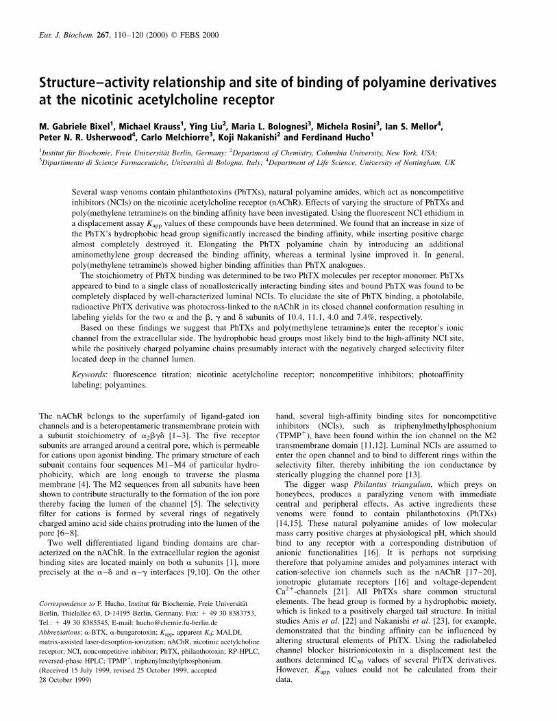
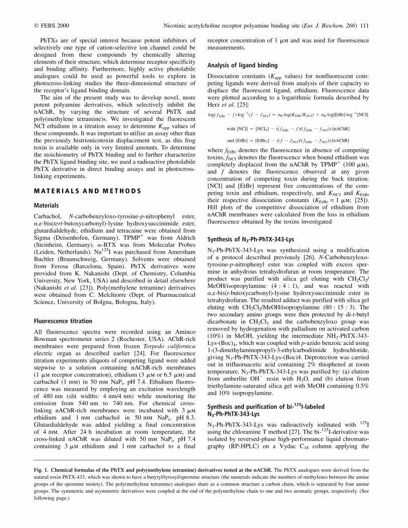
Fig. 1. Chemical formulas of the PhTX and poly(methylene tetramine) derivatives tested at the nAChR. The PhTX analogues were derived from the
natural toxin PhTX-433, which was shown to have a butyryl/tyrosyl/spermine structure (the numerals indicate the numbers of methylenes between the amine
groups of the spermine moiety). The poly(methylene tetramine) analogues share as a common structure a carbon chain, which is separated by four amine
groups. The symmetric and asymmetric derivatives were coupled at the end of the polymethylene chain to one and two aromatic groups, respectively. (See
following page.)
112 M. G. Bixel et al. (Eur. J. Biochem. 266) q FEBS 2000
q FEBS 2000 Nicotinic acetylcholine receptor polyamine binding site (Eur. J. Biochem. 266) 113
following linear gradient (1 mL´min21): solvent A (aqueoussolution containing 0.1% trifluoruacetic acid) and solvent B(acetonitril containing 0.085% trifluoruacetic acid). The UV-absorption of the HPLC fractions was determined at 280 nmand the radioactivity of each fraction was detected using ag-counter. The bi-125I-derivative was characterized bymatrix-assisted laser-desorption-ionization (MALDI)-massspectrometry.
N3-Ph-125I2-PhTX-343-Lys binding assay
Increasing concentrations of N3-Ph-125I2-PhTX-343-Lys(5000 c.p.m.´nmol21) were added to a constant amount ofnAChR-rich membranes (0.3 mg´mL21 protein, diluted in100 mm NaPi, pH 7.4; total volume per sample, 200 mL) andwere incubated for 45 min at room temperature. Bound ligandwas separated from the free ligand by ultracentrifugation in aBeckmann tabletop ultracentrifuge for 10 min at 80 000 g and4 8C. Aliquots were withdrawn prior to centrifugation todetermine total counts and duplicate aliquots of the supernatantwere removed after centrifugation to determine free ligandconcentration. Nonspecific binding was determined from boundN3-Ph-125I2-PhTX-343-Lys in the presence of 100-fold molar
excess of N3-Ph-I2-PhTX-343-Lys. a-BTX binding assays havebeen performed as described previously [28].
Photocross-linking experiments
nAChR-rich membranes were diluted in 0.1 m NaPi, pH 7.4 toa final receptor concentration of 140 nm. After addition ofcarbachol (500 mm) the samples were incubated for 30 min atroom temperature. Subsequently, TPMP1, a-BTX or unlabeledN3-Ph-PhTX-343-Lys was added and the samples wereincubated for further 30 min at room temperature. Theradioactive N3-Ph-125I2-PhTX-343-Lys (10 mm; 250 000c.p.m.´nmol21) was mixed with the sample solution followedby UV-irradiation at 254 nm for 45 s. Irradiation times longerthan 2 min irreversibly damaged the vast majority of nAChRresulting in high molecular aggregations of the receptor (datanot shown). Unbound toxin was separated from N3-Ph-125I2-PhTX-343-Lys bound to nAChR-rich membranes by centrifu-gation (15 000 g, 15 min). The pellet was dissolved andseparated by a 10% SDS/PAGE [29]. The stained gel wasdried and radioactive receptor subunits were visualized byautoradiography. Cross-linking yields were obtained by slicingthe dried gel and measuring the radioactivity of each fraction in
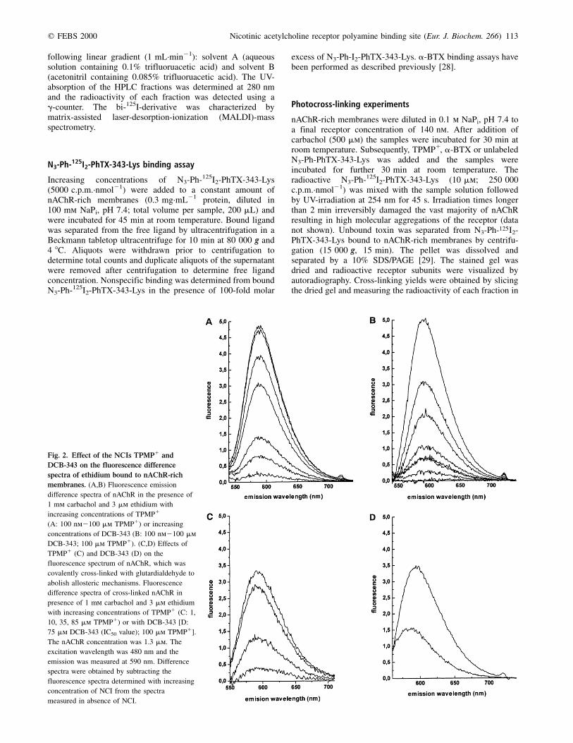
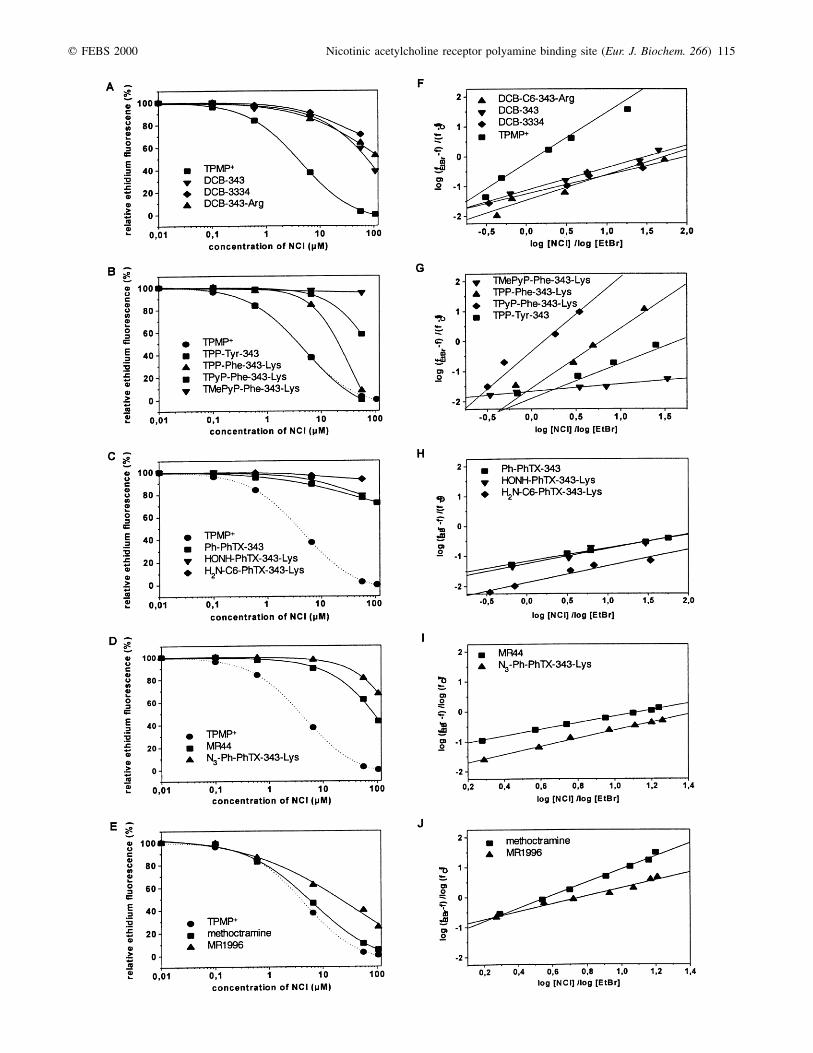
Fig. 2. Effect of the NCIs TPMP1 and
DCB-343 on the fluorescence difference
spectra of ethidium bound to nAChR-rich
membranes. (A,B) Fluorescence emission
difference spectra of nAChR in the presence of
1 mm carbachol and 3 mm ethidium with
increasing concentrations of TPMP1
(A: 100 nm2100 mm TPMP1) or increasing
concentrations of DCB-343 (B: 100 nm2100 mm
DCB-343; 100 mm TPMP1). (C,D) Effects of
TPMP1 (C) and DCB-343 (D) on the
fluorescence spectrum of nAChR, which was
covalently cross-linked with glutardialdehyde to
abolish allosteric mechanisms. Fluorescence
difference spectra of cross-linked nAChR in
presence of 1 mm carbachol and 3 mm ethidium
with increasing concentrations of TPMP1 (C: 1,
10, 35, 85 mm TPMP1) or with DCB-343 [D:
75 mm DCB-343 (IC50 value); 100 mm TPMP1].
The nAChR concentration was 1.3 mm. The
excitation wavelength was 480 nm and the
emission was measured at 590 nm. Difference
spectra were obtained by subtracting the
fluorescence spectra determined with increasing
concentration of NCI from the spectra
measured in absence of NCI.
114 M. G. Bixel et al. (Eur. J. Biochem. 266) q FEBS 2000
a g-counter. The labeling yields were calculated in percentageof mol cross-linked PhTX per mol nAChR subunit.
R E S U L T S
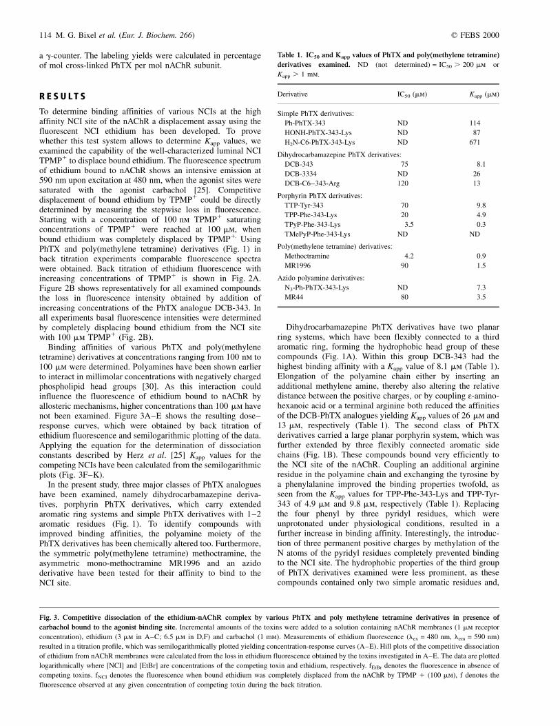
To determine binding affinities of various NCIs at the highaffinity NCI site of the nAChR a displacement assay using thefluorescent NCI ethidium has been developed. To provewhether this test system allows to determine Kapp values, weexamined the capability of the well-characterized luminal NCITPMP1 to displace bound ethidium. The fluorescence spectrumof ethidium bound to nAChR shows an intensive emission at590 nm upon excitation at 480 nm, when the agonist sites weresaturated with the agonist carbachol [25]. Competitivedisplacement of bound ethidium by TPMP1 could be directlydetermined by measuring the stepwise loss in fluorescence.Starting with a concentration of 100 nm TPMP1 saturatingconcentrations of TPMP1 were reached at 100 mm, whenbound ethidium was completely displaced by TPMP1. UsingPhTX and poly(methylene tetramine) derivatives (Fig. 1) inback titration experiments comparable fluorescence spectrawere obtained. Back titration of ethidium fluorescence withincreasing concentrations of TPMP1 is shown in Fig. 2A.Figure 2B shows representatively for all examined compoundsthe loss in fluorescence intensity obtained by addition ofincreasing concentrations of the PhTX analogue DCB-343. Inall experiments basal fluorescence intensities were determinedby completely displacing bound ethidium from the NCI sitewith 100 mm TPMP1 (Fig. 2B).
Binding affinities of various PhTX and poly(methylenetetramine) derivatives at concentrations ranging from 100 nm to100 mm were determined. Polyamines have been shown earlierto interact in millimolar concentrations with negatively chargedphospholipid head groups [30]. As this interaction couldinfluence the fluorescence of ethidium bound to nAChR byallosteric mechanisms, higher concentrations than 100 mm havenot been examined. Figure 3A±E shows the resulting dose±response curves, which were obtained by back titration ofethidium fluorescence and semilogarithmic plotting of the data.Applying the equation for the determination of dissociationconstants described by Herz et al. [25] Kapp values for thecompeting NCIs have been calculated from the semilogarithmicplots (Fig. 3F±K).
In the present study, three major classes of PhTX analogueshave been examined, namely dihydrocarbamazepine deriva-tives, porphyrin PhTX derivatives, which carry extendedaromatic ring systems and simple PhTX derivatives with 1±2aromatic residues (Fig. 1). To identify compounds withimproved binding affinities, the polyamine moiety of thePhTX derivatives has been chemically altered too. Furthermore,the symmetric poly(methylene tetramine) methoctramine, theasymmetric mono-methoctramine MR1996 and an azidoderivative have been tested for their affinity to bind to theNCI site.
Dihydrocarbamazepine PhTX derivatives have two planarring systems, which have been flexibly connected to a thirdaromatic ring, forming the hydrophobic head group of thesecompounds (Fig. 1A). Within this group DCB-343 had thehighest binding affinity with a Kapp value of 8.1 mm (Table 1).Elongation of the polyamine chain either by inserting anadditional methylene amine, thereby also altering the relativedistance between the positive charges, or by coupling :-amino-hexanoic acid or a terminal arginine both reduced the affinitiesof the DCB-PhTX analogues yielding Kapp values of 26 mm and13 mm, respectively (Table 1). The second class of PhTXderivatives carried a large planar porphyrin system, which wasfurther extended by three flexibly connected aromatic sidechains (Fig. 1B). These compounds bound very efficiently tothe NCI site of the nAChR. Coupling an additional arginineresidue in the polyamine chain and exchanging the tyrosine bya phenylalanine improved the binding properties twofold, asseen from the Kapp values for TPP-Phe-343-Lys and TPP-Tyr-343 of 4.9 mm and 9.8 mm, respectively (Table 1). Replacingthe four phenyl by three pyridyl residues, which wereunprotonated under physiological conditions, resulted in afurther increase in binding affinity. Interestingly, the introduc-tion of three permanent positive charges by methylation of theN atoms of the pyridyl residues completely prevented bindingto the NCI site. The hydrophobic properties of the third groupof PhTX derivatives examined were less prominent, as thesecompounds contained only two simple aromatic residues and,
Table 1. IC50 and Kapp values of PhTX and poly(methylene tetramine)
derivatives examined. ND (not determined) = IC50 . 200 mm or
Kapp . 1 mm.
Derivative IC50 (mm) Kapp (mm)
Simple PhTX derivatives:
Ph-PhTX-343 ND 114
HONH-PhTX-343-Lys ND 87
H2N-C6-PhTX-343-Lys ND 671
Dihydrocarbamazepine PhTX derivatives:
DCB-343 75 8�.1
DCB-3334 ND 26
DCB-C6±343-Arg 120 13
Porphyrin PhTX derivatives:
TTP-Tyr-343 70 9�.8
TPP-Phe-343-Lys 20 4�.9
TPyP-Phe-343-Lys 3�.5 0�.3
TMePyP-Phe-343-Lys ND ND
Poly(methylene tetramine) derivatives:
Methoctramine 4�.2 0�.9
MR1996 90 1�.5
Azido polyamine derivatives:
N3-Ph-PhTX-343-Lys ND 7�.3
MR44 80 3�.5
Fig. 3. Competitive dissociation of the ethidium-nAChR complex by various PhTX and poly methylene tetramine derivatives in presence of
carbachol bound to the agonist binding site. Incremental amounts of the toxins were added to a solution containing nAChR membranes (1 mm receptor
concentration), ethidium (3 mm in A±C; 6.5 mm in D,F) and carbachol (1 mm). Measurements of ethidium fluorescence (lex = 480 nm, lem = 590 nm)
resulted in a titration profile, which was semilogarithmically plotted yielding concentration-response curves (A±E). Hill plots of the competitive dissociation
of ethidium from nAChR membranes were calculated from the loss in ethidium fluorescence obtained by the toxins investigated in A±E. The data are plotted
logarithmically where [NCI] and [EtBr] are concentrations of the competing toxin and ethidium, respectively. fEtBr denotes the fluorescence in absence of
competing toxins. fNCI denotes the fluorescence when bound ethidium was completely displaced from the nAChR by TPMP 1 (100 mm), f denotes the
fluorescence observed at any given concentration of competing toxin during the back titration.
q FEBS 2000 Nicotinic acetylcholine receptor polyamine binding site (Eur. J. Biochem. 266) 115
116 M. G. Bixel et al. (Eur. J. Biochem. 266) q FEBS 2000
in addition, in one derivative the phenyl group was replaced bya positively charged amino carbonic acid (Fig. 1C). For Ph-PhTX-343, which is structurally most similar to the naturaloccurring toxin PhTX-433, a surprisingly low binding affinitywith a Kapp value of 114 mm was observed (Table 1). Increasingthe length of the polyamine chain by coupling the positivelycharged amino acid lysine and inserting a photolabile azidogroup into the aromatic ring of the phenyl residue significantlyimproved the potency (Fig. 1D), while the exchange of thismoiety by a polar uncharged residue, such as the hydroxyl-amine group generated a 10-fold less active compound(Fig. 1C; Table 1).
Methoctramine, which carries at each end of its polyaminechain an aromatic moiety (Fig. 1E), competed efficiently withethidium at the NCI site showing a Kapp value of 0.9 mm(Table 1). Removal of one of the aromatic residues generates anasymmetric poly(methylene tetramine) derivative with reducedbinding affinity. Inserting an azido group and altering thesubstituents at the aromatic ring lead to a further decrease inactivity by a factor of two (Fig. 1D; Table 1).
Fixation of nAChR with glutardialdehyde in presence ofdesensitizing amounts of carbachol `freezes' the receptor inits desensitized state [31]. Thereby, the receptor subunits arecross-linked to each other, which leads to a more rigidstructure and prevents allosteric transitions. After treatmentof nAChR with glutardialdehyde the fluorescence intensityof ethidium, when bound to the chemically fixed receptor,was reduced by 30±35%. However, the Kapp for ethidiumbound to this cross-linked receptor remained unchanged (datanot shown). Fluorescence titration using the competing NCITPMP1 (Fig. 2C) to prove the binding properties of the fixedreceptor resulted in a Kapp for TPMP1, which was comparableto that found in the unfixed form (data not shown). Addition ofDBC-343 in a concentration representing its IC50 value resultedin a 50% decrease in fluorescence intensity (Fig. 2D). Similarcompetition fluorescence spectra were obtained for the otherNCIs examined (spectra not shown). The basal fluorescencewas determined by adding 100 mm TPMP1 for completedissociation of bound ethidium from the NCI site.
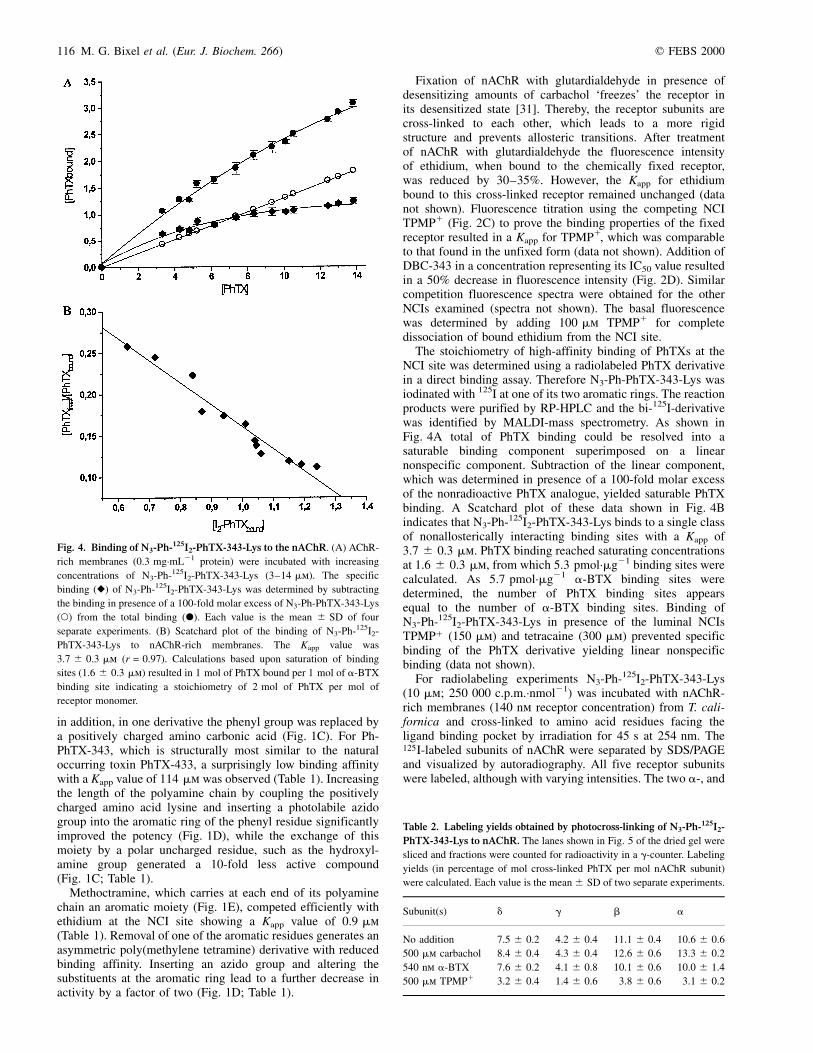
The stoichiometry of high-affinity binding of PhTXs at theNCI site was determined using a radiolabeled PhTX derivativein a direct binding assay. Therefore N3-Ph-PhTX-343-Lys wasiodinated with 125I at one of its two aromatic rings. The reactionproducts were purified by RP-HPLC and the bi-125I-derivativewas identified by MALDI-mass spectrometry. As shown inFig. 4A total of PhTX binding could be resolved into asaturable binding component superimposed on a linearnonspecific component. Subtraction of the linear component,which was determined in presence of a 100-fold molar excessof the nonradioactive PhTX analogue, yielded saturable PhTXbinding. A Scatchard plot of these data shown in Fig. 4Bindicates that N3-Ph-125I2-PhTX-343-Lys binds to a single classof nonallosterically interacting binding sites with a Kapp of3.7 ^ 0.3 mm. PhTX binding reached saturating concentrationsat 1.6 ^ 0.3 mm, from which 5.3 pmol´mg21 binding sites werecalculated. As 5.7 pmol´mg21 a-BTX binding sites weredetermined, the number of PhTX binding sites appearsequal to the number of a-BTX binding sites. Binding ofN3-Ph-125I2-PhTX-343-Lys in presence of the luminal NCIsTPMP1 (150 mm) and tetracaine (300 mm) prevented specificbinding of the PhTX derivative yielding linear nonspecificbinding (data not shown).
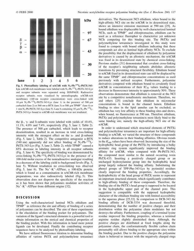
For radiolabeling experiments N3-Ph-125I2-PhTX-343-Lys(10 mm; 250 000 c.p.m.´nmol21) was incubated with nAChR-rich membranes (140 nm receptor concentration) from T. cali-fornica and cross-linked to amino acid residues facing theligand binding pocket by irradiation for 45 s at 254 nm. The125I-labeled subunits of nAChR were separated by SDS/PAGEand visualized by autoradiography. All five receptor subunitswere labeled, although with varying intensities. The two a-, and
Fig. 4. Binding of N3-Ph-125I2-PhTX-343-Lys to the nAChR. (A) AChR-
rich membranes (0.3 mg´mL21 protein) were incubated with increasing
concentrations of N3-Ph-125I2-PhTX-343-Lys (3±14 mm). The specific
binding (V) of N3-Ph-125I2-PhTX-343-Lys was determined by subtracting
the binding in presence of a 100-fold molar excess of N3-Ph-PhTX-343-Lys
(W) from the total binding (X). Each value is the mean ^ SD of four
separate experiments. (B) Scatchard plot of the binding of N3-Ph-125I2-
PhTX-343-Lys to nAChR-rich membranes. The Kapp value was
3.7 ^ 0.3 mm (r = 0.97). Calculations based upon saturation of binding
sites (1.6 ^ 0.3 mm) resulted in 1 mol of PhTX bound per 1 mol of a-BTX
binding site indicating a stoichiometry of 2 mol of PhTX per mol of
receptor monomer.
Table 2. Labeling yields obtained by photocross-linking of N3-Ph-125I2-
PhTX-343-Lys to nAChR. The lanes shown in Fig. 5 of the dried gel were
sliced and fractions were counted for radioactivity in a g-counter. Labeling
yields (in percentage of mol cross-linked PhTX per mol nAChR subunit)
were calculated. Each value is the mean ^ SD of two separate experiments.
Subunit(s) d g b a
No addition 7.5 �^ 0.2 4.2 �^ 0.4 11.1 �^ 0.4 10.6 �^ 0.6
500 mm carbachol 8.4 �^ 0.4 4.3 �^ 0.4 12.6 �^ 0.6 13.3 �^ 0.2
540 nm a-BTX 7.6 �^ 0.2 4.1 �^ 0.8 10.1 �^ 0.6 10.0 �^ 1.4
500 mm TPMP1 3.2 �^ 0.4 1.4 �^ 0.6 3.8 �^ 0.6 3.1 �^ 0.2
q FEBS 2000 Nicotinic acetylcholine receptor polyamine binding site (Eur. J. Biochem. 266) 117
the b-, g- and d-subunits were labeled with yields of 10.4%,11.1%, 4.0% and 7.4%, respectively (Fig. 5, lane 1; Table 2).The presence of 500 mm carbachol, which leads to receptordesensitization, resulted in an increase in total cross-linkingintensity with the strongest effect on the a- and b-subunit(Fig. 5, lane 2, Table 2). The competitive antagonist a-BTX(540 nm) apparently did not affect binding of N3-Ph-125I2-PhTX-343-Lys (Fig. 5, lane 3, Table 2), while TPMP1 caused a65% decrease in labeling intensity in all receptor subunits(Fig. 5, lane 4). The specificity of binding was demonstrated bycross-linking N3-Ph-125I2-PhTX-343-Lys in the presence of a100-fold molar excess of the nonradioactive analogue resultingin a decrease of the labeling yield to background levels (Fig. 5,lane 5). Without irradiation no cross-linking was observed(Fig. 5, lane 6). The Na1-K1-ATPase, a 100-kDa protein,which is found as a contamination in nAChR-rich membranepreparations, was also radioactively labeled (Fig. 5). Thisobservation does not disprove the specificity of the reactionas it has been shown that polyamines modulate activities ofNa1-K1-ATPase from different origins [32].
D I S C U S S I O N
Using the well-characterized luminal NCIs ethidium andTPMP1 as reference the site and affinity of binding of a seriesof polyamines can be described. The aim of these experimentsis the elucidation of the binding pocket for polyamines. Thevariation of the ligand's structural elements is a powerful tool toobtain information on the sterical and chemical properties ofthe binding pocket. With this information in hand more potentligands can be developed. Finally the contributing receptorsequences have to be analyzed by photoaffinity-labeling.
We have utilized fluorescence titration to determine bindingaffinities of various PhTX and poly(methylene tetramine)
derivatives. The fluorescent NCI ethidium, when bound to thehigh-affinity NCI site on the nAChR in its desensitized state,shows an intensive emission maximum at 590 nm [25]. Asbound ethidium was displaceable by well-characterized luminalNCIs, such as TPMP1 and chlorpromazine, ethidium can beused as a reference fluorophor to characterize yet unknownNCIs competing for this binding site. The PhTXs andpoly(methylene tetramine)s investigated in this study werefound to compete with bound ethidium indicating that thesecompounds act also as luminal high-affinity NCIs. To excludethe possibility that the displacement of ethidium by polyaminederivatives is caused by an allosteric mechanism, the nAChRwas fixed in its desensitized state by chemical cross-linking.Previous studies [31] demonstrated that covalent cross-linkingof the receptor's neighboring subunits abolishes allosterictransitions by preventing structural flexibility. Ethidium boundto nAChR fixed in its desensitized state can still be displaced bythe same TPMP1 and chlorpromazine concentrations as usedpreviously with unfixed receptor. Furthermore, polyaminederivatives competed with ethidium bound to covalently fixednAChR in concentrations of their IC50 values leading to adecrease in fluorescence intensity to approximately 50%. Theseobservations demonstrate that ethidium is removed at the NCIsite by a competitive nonallosteric mechanism. Therefore, weand others [25] conclude that ethidium in micromolarconcentrations is bound in the channel lumen. Ethidiumbinding to sites in the lipid annulus can be excluded, asethidium has very low affinity to these binding sites and shows,when bound, no fluorescence at 590 nm. Therefore ethidium,PhTXs and poly(methylene tetramine)s most likely bind to thesame binding site, namely the high-affinity NCI site of thenAChR.
In order to understand which structural elements of PhTXsand poly(methylene tetramine)s are important for high-affinitybinding to nAChR, we varied the structure of these compoundsto induce alterations in their binding affinities. Obvious trendsin the Kapp values have been observed: Increasing the size of thehydrophobic head group of the PhTXs by introducing a bulkyaromatic ring system significantly improved the bindingaffinity for nAChR, when compared with Ph-PhTX-343,the compound most similar to the natural occurring toxinPhTX-433. Inserting a positively charged group or anuncharged hydroxylamine group into the hydrophobic headgroup largely reduced the binding affinity. Substituting thephenyl groups of TPP-Phe-343-Lys by pyrimidyl residuesclearly improved the binding properties. Accordingly, thehydrophobicity of the head group of PhTX seems to representan important structural element, which significantly contributesto high-affinity binding of these compounds. Therefore, thebinding site of the PhTX's head group is supposed to be locatedin the hydrophobic upper part of the channel pore. Thissuggestion is compatible with the supposed hydrophobiccharacter of the ethidium binding site with limited accessibilityto the aqueous phase [25,33]. In comparison to DCB-343 thebinding affinity of DCB-3334 was decreased threefold,demonstrating that the number of positive charges and theirdistribution within the polyamine chain modulates but notdestroys the affinity. Furthermore, coupling of a terminal lysineresidue improved the binding properties, whereas a terminalarginine residue slightly diminished them. Although, thepositive charges are distributed at fixed lengths along thepolyamine chain, the conformationally flexible carbon chainpresumably still allows binding to the appropriate sites withinthe binding pocket. Due to the positive charges the polyaminechain is believed to interact with the negatively charged rings
Fig. 5. Photoaffinity labeling of nAChR with N3-Ph-125I2-PhTX-343-
Lys. nAChR-rich membranes were labeled with N3-Ph-125I2-PhTX-343-Lys
and receptor subunits were separated using SDS/PAGE. Radioactive
receptor subunits were visualized by autoradiography. nAChR-rich
membranes (140 nm receptor concentration) were cross-linked with
10 mm N3-Ph-125I2-PhTX-343-Lys (lane 1) in the presence of 500 mm
carbachol (lane 2) or 560 nm a-BTX (lane 3) or 500 mm TPMP1 (lane 4) or
1 mm N3-Ph-PhTX-343-Lys (lane 5). Lane 6 containing 10 mm N3-Ph-125I2-
PhTX-343-Lys bound to nAChR-rich membranes was not irradiated.
118 M. G. Bixel et al. (Eur. J. Biochem. 266) q FEBS 2000
located within the receptor's channel pore forming theselectivity filter [7,8]. Our observations on the binding affinitiesare in good agreement with earlier findings of Anis et al. [22]and Nakanishi et al. [23]. Additional to their data we are able topresent Kapp values for the various PhTX derivatives: constantvalues, which are independent from Kapp values of thecompeting ligand used in the test system.
In comparison to PhTX derivatives the poly(methylenetetramine)s showed higher binding affinities. Methoctra-mine, a symmetric analogue known to act as antagonist onthe muscarinic AChR [34], bound with high-affinity to theNCI site of the nAChR. Removing one of the terminalmethoxybenzyl groups results in the asymmetric MR1996,resulted in a twofold reduced binding affinity. A photo-labile poly(methylene tetramine) developed for photocross-linking experiments bound with slightly decreased affinity.Accordingly, one hydrophobic head group is sufficient forbinding to the high-affinity NCI site on the nAChR.Furthermore, the long polyamine chain of these derivativesappeared to be advantageous for binding.
To ascertain that competition of PhTX derivatives withbound ethidium takes place at the high-affinity NCI site in thechannel pore and to exclude the possibility that displacement ofbound ethidium is allosterically caused by binding of PhTXderivatives to multiple, although low-affinity binding sites inthe lipid annulus we have investigated the radiolabeled PhTXN3-Ph-125I2-PhTX-343-Lys in direct binding studies. Thestraight line obtained in the Scatchard plot indicates that thisPhTX analogue binds to a single class of nonallostericallyinteracting binding sites. The stoichiometry of PhTX bindingwas determined to be 2 PhTX molecules/receptor monomer.Furthermore the well-characterized luminal NCIs TPMP1 andtetracaine were found to completely dissociate specificallybound PhTX indicating that both PhTX binding sites arelocated in the channel lumen. Our findings demonstrate thattwo PhTX molecules bind with equal affinity with the channellumen of nAChR. It is important to mention that recently Luet al. [35] found evidence for the simultaneous binding of threepolyamines in the inner pore of inwardly rectifying K1
channels.The radioactive photolabile N3-Ph-125I2-PhTX-343-Lys was
further used in photocross-linking studies to explore thethree-dimensional structure of the PhTX binding site. DuringUV-irradiation the azidophenyl group of this compound is ableto covalently cross-link to any nearby amino acid facing theligand binding site [36]. The observation that N3-Ph-125I2-PhTX-343-Lys photolabeled all five receptor subunits stronglysuggests that the PhTX binding site is located deep within thechannel, where the PhTX derivative is in close contact to allsubunits. However, the labeling yields per subunit variedbetween 4.2% to 11.1% indicating that the hydrophobic headgroup of PhTX appeared to prefer defined sterical orientationsupon binding. When preincubating nAChR-rich membraneswith the agonist carbachol, which induces receptor desen-sitization accompanied with an increase in binding affinity forNCIs, we observed a clear enhancement in the labelingintensity. Cross-linking in presence of TPMP1 demonstratedthat 65% of the bound PhTX analogue was displaceable.Consequently, during irradiation this compound seems to cross-link to additional binding sites on the nAChR, possibly the low-affinity binding sites in the lipid annulus [37].
Electrophysiological studies have shown that spermine [19]and PhTX-343 [20] have multiple sites of action on musclenAChR, similar to those observed for ionotropic glutamatereceptors [16]. From binding studies Choi et al. [26] suggested
that PhTX analogues act on the nAChR's ionic channel in itsopen conformation. Our findings demonstrate that the photo-labile PhTX derivative binds to nAChR also in absence ofagonist with the receptor being predominantly in its restingclosed channel state. Furthermore, when nAChR was pre-incubated with carbachol, which rapidly shifted the vastmajority of nAChR to the desensitized closed channel state,the PhTX analogue was cross-linked with even higher affinity.The PhTX binding site must therefore be accessible in the openand the closed channel conformation. Results obtained byapplying the substituted cysteine accessibility method [38]showed that a number of low molecular mass reagents wereable to react in the closed channel state with a number of M2cysteine substitution mutants, including aT244C close to theintracellular end of the channel. Furthermore, using the sametechnique Wilson and Karlin [39] could recently localize thenarrowest part of the ion channel between aG240C andaT244C, a region which includes the negatively chargedcytosolic ring of the selectivity filter. In the closed state abarrier in this narrow part of the channel was detectedrestricting the accessibility for small reagents from both sidesof the ion channel [39]. Choi et al. [26] assumed that PhTX isorientated toward the cytoplasmic side of the pore. Althoughthe authors tried to label the open channel conformation,addition of carbachol as performed in their studies presumablyresulted in rapid desensitization and channel closure rather thanin channel opening. Because in the closed state moleculescannot pass the channel pore, PhTX bound from the cytosolicside would not be able to compete with ethidium for binding atthe high-affinity NCI site, which is known to be located in theextracellular part of the ion channel [13]. Based on our resultsthat PhTXs and poly(methylene tetramine)s displace ethidiumbound at the high-affinity NCI site in the channel's closedconformation we conclude that polyamine derivatives areorientated toward the extracellular side of the ion channel.Furthermore, we could demonstrate that 2 PhTX moleculesbind with equal affinity in the channel lumen. Consequently, themost likely mode of binding which leads to receptor inhibitionis that the PhTX derivatives enter from the extracellular side ofthe receptor's ionic channel. The hydrophobic head groups arebelieved to bind in the hydrophobic upper part of the channelpore to the high-affinity NCI site, while the positively chargedpolyamine chains most likely interact with the negativelycharged selectivity filter located deep within the channel pore.Experiments to prove this localization by photoaffinity labelingand microsequencing of the labeled receptor peptides areunderway. Furthermore, it would be most interesting to extendthese studies on neuronal nAChRs, however, an expressionsystem providing the amount of receptor necessary for thiswork would have first to be established.
Our results on the modification of polyamine bindingaffinities by altering their structural elements and our photo-affinity approach provide important information on themolecular structure of the binding site of polyamine derivativesat the nAChR. This model possesses optimized properties interms of charge distribution, size and hydrophobic interactionsproviding the structural data required for the rational design ofnovel, more potent polyamine derivatives selectively inhibitingthe nAChR.
A C K N O W L E D G E M E N T S
This work was supported by the European Commission, BIOMED-2
program contract BMH4-CT97-2395 (to F. H.), Fond der Chemischen
Industrie (to F. H.) and NIH grant AI 10187 (to K. N.). We would like to
q FEBS 2000 Nicotinic acetylcholine receptor polyamine binding site (Eur. J. Biochem. 266) 119
thank Prof. A. Herrmann (Institut fuÈr Biophysik, Humboldt-UniversitaÈt
Berlin) for kindly allowing us to use of Aminco Bowman spectrometer. We
also thank H. Bayer for helpful technical assistance and Dr X. Huang
(Columbia University, New York, USA) for kindly supplying porphyrin
PhTX analogues.
R E F E R E N C E S
1. Changeux, J.-P. (1990) Functional architecture and dynamics of the
nicotinic acetylcholine receptor: an allosteric ligand-gated ion
channel. Fidia Res. Found. Neurosci. Award Lect. 4, 21±168.
2. Karlin, A. & Akabas, M.H. (1995) Towards a structural basis for the
function of nicotinic acetylcholine receptors and their cousins.
Neuron 15, 1231±1244.
3. Hucho, F., Tsetlin, V. & Machold, J. (1996) The emerging three-
dimensional structure of a receptor, the nicotinic acetylcholine
receptor. Eur. J. Biochem. 239, 539±557.
4. Noda, M., Takahashi, H., Tanabe, T., Toyosato, M., Kikyotani, S.,
Furutani, Y., Hirose, T., Takashima, H., Inayama, S., Miyata, T. &
Numa, S. (1983) Structural homology of Torpedo californica
acetylcholine receptor subunits. Nature 302, 538±532.
5. Hucho, F., OberthuÈr, W. & Lotttspeich, F. (1986) The ion channel of the
nicotinic acetylcholine receptor is formed by the homologous helices
M II of the receptor subunits. FEBS Lett. 205, 137±142.
6. Galzi, J.-L., Devillers-ThieÂry, A.Q., Hussy, N., Bertrand, S., Changeux,
J.-P. & Bertrand, D. (1992) Mutations in the ion channel domain of a
neuronal nicotinic acetylcholine receptor convert ion selectivity from
cation to anionic. Nature 359, 500±505.
7. Imoto, K., Busch, C., Sakmann, B., Mishina, M., Konno, T., Nakai, J.,
Bujo, H., Mori, Y., Fukudo, K. & Numa, S. (1988) Rings of
negatively charged amino acids determine the acetylcholine receptor
channel conductance. Nature 335, 645±648.
8. Konno, T., Busch, C., Von Kitzing, E., Imoto, K., Wang, F., Nakai, J.,
Mishina, M., Numa, S. & Sakmann, B. (1991) Rings of anionic
amino acids as structural determinants of ion selectivity in the
acetylcholine receptor channel. Proc. R. Soc. Lond. B Biol. Sci. 244,
69±79.
9. Blount, P. & Merlie, J.P. (1989) Molecular basis of the two
nonequivalent ligand binding sites of the muscle nicotinic acetyl-
choline receptor. Neuron 3, 349±357.
10. Pedersen, S.E. & Cohen, J.B. (1990) d-Tubocurarin binding
sites are located at a±d and a±g subunit interfaces of the
nicotinic acetylcholine receptor. Proc. Natl Acad. Sci. USA 87,
2785±2789.
11. Giraudat, J., Dennis, M., Heidmann, T., Chang, J.Y. & Changeux, J.-P.
(1986) Structure of the high-affinity binding site for noncompetitive
blockers of the acetylcholine receptor: serine 262 of the delta
subunits is labeled by 3H-chlorpromazine. Proc. Natl Acad. Sci. USA
83, 2719±272313.
12. Arias, H.R. (1998) Binding sites of exogenous and endogenous non-
competitive inhibitors of the nicotinic acetylcholine receptor.
Biochem. Biophys. Acta 1376, 173±220.
13. Hucho, F. & Hilgenfeld, R. (1989) The selectivity filter of a ligand-
gated ion channel. The helix-M2 model of the ion channel of the
nicotinic acetylcholine receptor. FEBS Lett. 257, 17±23.
14. Adams, M.E., Carney, R.L., Enderlin, F.E., Fu, E.T., Jarema, M.A., Li,
J.P., Miller, C.A., Schooley, D.A., Shapiro, M.J. & Venema, V.J.
(1987) Structure and biological activities of three synaptic antago-
nists. Biochem. Biophys. Res. Comm. 148, 678±683.
15. Eldefrawi, A.T., Eldefrawi, M.E., Konno, K., Mansour, N.A.,
Nakanishi, K., Oltz, E. & Usherwood, P.N.R. (1988) Structure and
synthesis of a potent glutamate receptor antagonist in wasp venom.
Proc. Natl Acad. Sci. USA 85, 4910±4913.
16. Usherwood, P.N.R. & Blagbrough, I.R. (1991) Spider toxins affecting
glutamate receptors: polyamines in therapeutic neurochemistry.
Pharmacol. Therap. 52, 245±268.
17. Ragsdale, D., Gant, D.B., Anis, N.A., Eldefrawi, A.T., Eldefrawi,
M.E., Konno, K. & Miledi, R. (1989) Inhibition of rat
glutamate receptors by philanthotoxin. J. Pharmacol. Exp.
Ther. 251, 156±156.
18. Rozental, R., Scoble, G.T., Albuqueque, E.X., Idriss, M., Sherby,
S., Sattelle, D.R., Nakanishi, K., Konno, K., Eldefrawi, A.T. &
Eldefrawi, M.E. (1989) Allosteric inhibition of nicotinic
acetylcholine receptors of vertebrates and insects by philantho-
toxin. J. Pharmacol. Exp. Ther. 349, 123±130.
19. Shao, Z., Mellor, I.S., Brieley, M.J., Harris, J. & Usherwood, P.N.R.
(1998) Potentiation and inhibition of nicotinic acetylcholine receptors
by spermine in the TE671 human muscle cell clone. J. Pharmacol.
Exp. Ther. 286, 1269±1276.
20. Jayaraman, V., Usherwood, P.N.R. & Hess, G.P. (1998) Inhibition of
the nicotinic acetylcholine receptor by philanthotoxin-343; kinetic
investigations in the microsecond time region using laser-pulse
photolysis technique. Biochemistry 38, 11406±11414.
21. Scott, R.H., Sutton, K.G. & Dolphin, A.C. (1993) Interactions of
polyamines with neuronal ion channels. Trends Neurosci. 16,
153±160.
22. Anis, N., Sherby, S., Goodnow, R., Niwa, M., Konno, K.,
Kallimopoulos, T., Bukownik, R., Nakanishi, K., Usherwod, P.,
Eldefrawi, A. & Eldefrawi, M. (1995) Structure-activity relation-
ships of philanthotoxin analogs and polyamines on N-methyl-d-
aspartate and nicotinic acetylcholine receptors. J. Pharmacol.
Exp. Ther. 254, 764±773.
23. Nakanishi, K., Huang, X., Jiang, H., Liu, Y., Fang, K., Huang, D., Choi,
S.-K., Katz, E. & Eldefrawi, M. (1997) Structure-binding relation of
philanthotoxins from nicotinic acetylcholine receptor binding assay.
Bioorg. Med. Chem. 5, 1969±1988.
24. Schiebler, W., Lauffer, L. & Hucho, F. (1977) Acetylcholine receptor
enriched membranes: acetylcholine binding and excitability after
reduction in vivo. FEBS Lett. 81, 39±41.
25. Herz, J.M., Johnson, D.A. & Taylor, P. (1987) Interaction of
noncompetitive inhibitors with the acetylcholine receptor. J. Biol.
Chem. 262, 7238±7247.
26. Choi, S.-K., Kalivretenos, A.G., Usherwood, P.N.R. & Nakanishi, K.
(1995) Labeling studies of photolabile philanthotoxins with nicotinic
acetylcholine receptors: mode of interaction between toxin and
receptor. Chem. Biol. 2, 23±32.
27. Greenwood, F.C., Hinter, W.M. & Glover, J.S. (1963) The preparaion
of 131I-labelled human grosth hormone of high specific radioactivity.
Biochem. J. 89, 114±121.
28. Hartig, P.R. & Raftery, M.A. (1979) Preparation of right-side-
out, acetylcholine receptor enrichend intact vesicles form
Torpedo californica electroplaque membranes. Biochemistry 18,
1146±1150.
29. Laemmli, U.K. (1970) Cleavage of structural proteins during assembly
of head of bacteriophage T4. Nature 227, 680±685.
30. Chung, L., Kaloyanides, G., McDaniel, R., McLaughlin, A. &
McLaughlin, S. (1985) Interaction of gentamicin and spermine with
bilayer membranes containing negatively charged phospholipids.
Biochemistry 24, 442±452.
31. Watty, A., Methfessel, C. & Hucho, F. (1997) Fixation of allosteric
states of the nicotinic acetylcholine receptor by chemical cross-
linking. Proc. Natl Acad. Sci. USA 94, 8202±8707.
32. Heinrich-Hirsch, B., Ahlers, J. & Peter, H.W. (1977) Inhibition of
Na,K-ATPase from chick brain by polyamines. Enzyme 22,
235±241.
33. Herz, J.M. & Atherton, S.J. (1992) Steric factors limit access to the
noncompetitve inhibitor site of the nicotinic acetylcholine receptor.
Biophys. J. 62, 74±76.
34. Melchiorre, C. (1990) Polymethylene tetramines: a novel class of
cardioselective M2 antagonists. Med. Chem. Rev. 10, 327±349.
35. Lu, T., Nguyen, B., Zhang, X. & Yang, J. (1999) Architecture of a K1
channel inner pore revealed by stoichiometric covalent modification.
Neuron 22, 571±580.
36. Henriksen, U. & Buchardt, O. (1990) Aryl azides as photolables.
Retention of iodine during photochemical ring expansion of an
iodinated tyrosine derivative. Tetrahedron Lett. 31, 2443±2444.
37. Heidmann, T., Oswald, R.E. & Changeux, J.-P. (1983) Multiple sites of

120 M. G. Bixel et al. (Eur. J. Biochem. 266) q FEBS 2000
action for noncompetitive blockers on acetylcholine receptor rich
membrane fragments from Torpedo marmorata. Biochemistry 22,
3112±3127.
38. Akabas, M.H., Kaufmann, C., Archdeacon, P. & Karlin, A. (1994)
Identification of acetylcholine receptor channel-lining residues in the
entire M2 segment of the a subunit. Neuron 13, 919±927.
39. Wilson, G.G. & Karlin, A. (1998) The location of the gate in the
acetylcholine receptor channel. Neuron 20, 1269±1281.