Embed Size (px)
Citation preview
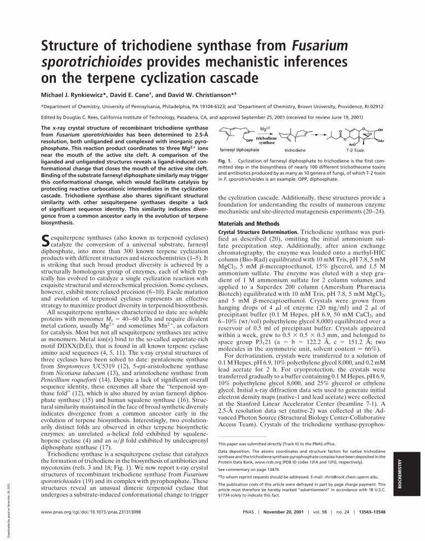
Structure of trichodiene synthase from Fusariumsporotrichioides provides mechanistic inferenceson the terpene cyclization cascadeMichael J. Rynkiewicz*, David E. Cane†, and David W. Christianson*‡
*Department of Chemistry, University of Pennsylvania, Philadelphia, PA 19104-6323; and †Department of Chemistry, Brown University, Providence, RI 02912
Edited by Douglas C. Rees, California Institute of Technology, Pasadena, CA, and approved September 25, 2001 (received for review June 19, 2001)
The x-ray crystal structure of recombinant trichodiene synthasefrom Fusarium sporotrichioides has been determined to 2.5-Åresolution, both unliganded and complexed with inorganic pyro-phosphate. This reaction product coordinates to three Mg2� ionsnear the mouth of the active site cleft. A comparison of theliganded and unliganded structures reveals a ligand-induced con-formational change that closes the mouth of the active site cleft.Binding of the substrate farnesyl diphosphate similarly may triggerthis conformational change, which would facilitate catalysis byprotecting reactive carbocationic intermediates in the cyclizationcascade. Trichodiene synthase also shares significant structuralsimilarity with other sesquiterpene synthases despite a lackof significant sequence identity. This similarity indicates diver-gence from a common ancestor early in the evolution of terpenebiosynthesis.
Sesquiterpene synthases (also known as terpenoid cyclases)catalyze the conversion of a universal substrate, farnesyl
diphosphate, into more than 300 known terpene cyclizationproducts with different structures and stereochemistries (1–5). Itis striking that such broad product diversity is achieved by astructurally homologous group of enzymes, each of which typ-ically has evolved to catalyze a single cyclization reaction withexquisite structural and stereochemical precision. Some cyclases,however, exhibit more relaxed precision (6–10). Facile mutationand evolution of terpenoid cyclases represents an effectivestrategy to maximize product diversity in terpenoid biosynthesis.
All sesquiterpene synthases characterized to date are solubleproteins with monomer Mr � 40–60 kDa and require divalentmetal cations, usually Mg2� and sometimes Mn2�, as cofactorsfor catalysis. Most but not all sesquiterpene synthases are activeas monomers. Metal ion(s) bind to the so-called aspartate-richmotif DDXX(D,E), that is found in all known terpene cyclaseamino acid sequences (4, 5, 11). The x-ray crystal structures ofthree cyclases have been solved to date: pentalenene synthasefrom Streptomyces UC5319 (12), 5-epi-aristolochene synthasefrom Nicotiana tabacum (13), and aristolochene synthase fromPenicillium roqueforti (14). Despite a lack of significant overallsequence identity, these enzymes all share the ‘‘terpenoid syn-thase fold’’ (12), which is also shared by avian farnesyl diphos-phate synthase (15) and human squalene synthase (16). Struc-tural similarity maintained in the face of broad synthetic diversityindicates divergence from a common ancestor early in theevolution of terpene biosynthesis. Interestingly, two evolution-arily distinct folds are observed in other terpene biosyntheticenzymes: an unrelated �-helical fold exhibited by squalene-hopene cyclase (4) and an ��� fold exhibited by undecaprenyldiphosphate synthase (17).
Trichodiene synthase is a sesquiterpene cyclase that catalyzesthe formation of trichodiene in the biosynthesis of antibiotics andmycotoxins (refs. 3 and 18; Fig. 1). We now report x-ray crystalstructures of recombinant trichodiene synthase from Fusariumsporotrichioides (19) and its complex with pyrophosphate. Thesestructures reveal an unusual dimeric terpenoid cyclase thatundergoes a substrate-induced conformational change to trigger
the cyclization cascade. Additionally, these structures provide afoundation for understanding the results of numerous enzymemechanistic and site-directed mutagenesis experiments (20–24).
Materials and MethodsCrystal Structure Determination. Trichodiene synthase was puri-fied as described (20), omitting the initial ammonium sul-fate precipitation step. Additionally, after anion exchangechromatography, the enzyme was loaded onto a methyl-HICcolumn (Bio-Rad) equilibrated with 10 mM Tris, pH 7.8, 5 mMMgCl2, 5 mM �-mercaptoethanol, 15% glycerol, and 1.5 Mammonium sulfate. The enzyme was eluted with a step gra-dient of 1 M ammonium sulfate for 2 column volumes andapplied to a Superdex 200 column (Amersham PharmaciaBiotech) equilibrated with 10 mM Tris, pH 7.8, 5 mM MgCl2,and 5 mM �-mercaptoethanol. Crystals were grown fromhanging drops of 4 �l of enzyme (20 mg�ml) and 2 �l ofprecipitant buffer (0.1 M Hepes, pH 6.9, 50 mM CaCl2, and6–10% (wt�vol) polyethylene glycol 8,000) equilibrated over areservoir of 0.5 ml of precipitant buffer. Crystals appearedwithin a week, grew to 0.5 � 0.5 � 0.3 mm, and belonged tospace group P3121 (a � b � 122.2 Å, c � 151.2 Å; twomolecules in the asymmetric unit, solvent content � 66%).
For derivatization, crystals were transferred to a solution of0.1 M Hepes, pH 6.9, 10% polyethylene glycol 8,000, and 0.2 mMlead acetate for 2 h. For cryoprotection, the crystals weretransferred gradually to a buffer containing 0.1 M Hepes, pH 6.9,10% polyethylene glycol 8,000, and 25% glycerol or ethyleneglycol. Initial x-ray diffraction data sets used to generate initialelectron density maps (native-1 and lead acetate) were collectedat the Stanford Linear Accelerator Center (beamline 7-1). A2.5-Å resolution data set (native-2) was collected at the Ad-vanced Photon Source (Structural Biology Center-CollaborativeAccess Team). Crystals of the trichodiene synthase-pyrophos-
This paper was submitted directly (Track II) to the PNAS office.
Data deposition: The atomic coordinates and structure factors for native trichodienesynthase and the trichodiene synthase-pyrophosphate complex have been deposited in theProtein Data Bank, www.rcsb.org (PDB ID codes 1JFA and 1JFG, respectively).
See commentary on page 13479.
‡To whom reprint requests should be addressed. E-mail: [email protected].
The publication costs of this article were defrayed in part by page charge payment. Thisarticle must therefore be hereby marked “advertisement” in accordance with 18 U.S.C.§1734 solely to indicate this fact.
Fig. 1. Cyclization of farnesyl diphosphate to trichodiene is the first com-mitted step in the biosynthesis of nearly 100 different trichothecene toxinsand antibiotics produced by as many as 10 genera of fungi, of which T-2 toxinin F. sporotrichioides is an example. OPP, diphosphate.
www.pnas.org�cgi�doi�10.1073�pnas.231313098 PNAS � November 20, 2001 � vol. 98 � no. 24 � 13543–13548
BIO
CHEM
ISTR
Y
Dow
nloa
ded
by g
uest
on
Nov
embe
r 18
, 202
1
phate complex were prepared by soaking in a solution of 0.1 MHepes, pH 6.9, 10% polyethylene glycol 8,000, 1 mM MgCl2, and2 mM sodium pyrophosphate for 2 h. Crystals were isomorphouswith those of the native enzyme, and data were collected atthe National Synchrotron Light Source (beamline X12C). Alldata were scaled and integrated by using the programs DENZOand SCALEPACK (25). Data collection statistics are reported inTable 1.
Initial phases were calculated by single isomorphous replace-ment with anomalous scattering using two lead sites per mono-mer. Heavy atom positions were refined with MLPHARE (26), anddensity modification was performed with DM (26, 27). Eachpolypeptide chain and ordered solvent molecules were fit intothe resulting electron density maps with O (28) and then refinedagainst all data with CNS (29). The C termini (R355-E374) aredisordered in both monomers, and the N terminus (M1–F4) ofmonomer B is disordered, thus these residues are excluded fromthe final model. Strict noncrystallographic symmetry constraintswere used initially and relaxed into appropriately weightedrestraints as guided by Rfree. The noncrystallographic symmetryaxis is the dyad axis of the dimer. The final model has excellentgeometry with 92 and 8% of residues adopting most favored andadditionally allowed conformations, respectively.
The structure of the trichodiene synthase-pyrophosphatecomplex was solved by difference Fourier methods using theunliganded structure as a starting point. Monomer B exhibitedclear electron density for three Mg2� ions and one pyrophos-phate molecule; monomer A did not, possibly because of anactive site partially blocked by crystal lattice interactions. Inrefinement, noncrystallographic symmetry restraints were notapplied to polypeptide segments exhibiting pyrophosphate-induced conformational changes (residues 1–29, 99–105, 229–242, and 300–335). Final refinement statistics are recorded inTable 2.
Modeling. Farnesyl diphosphate, intermediate carbocation, andtrichodiene structures were generated with MACROMODEL (30),and a Monte Carlo conformational search yielded minimumenergy conformations for each. The search was constrained toconformations that would support the reaction coordinates ofC–C bond formation previously determined for trichodienebiosynthesis. For example, the C1 and C6 atoms of farnesyldiphosphate, which form a six-membered ring in the first elec-trophilic cyclization, were constrained to a separation of 3.5 Å.The resulting structures were docked manually into the activesite by using O. Finally, 30 cycles of rigid body and 200 cycles ofconjugate gradient energy minimization with CNS optimized thestructure of each enzyme-ligand model. In energy minimiza-tions, pyrophosphate, Mg2� ions, and associated water moleculeswere fixed, and a harmonic restraint of 10 kcal�mol�1�Å�2 wasapplied to the carbon portions of the substrate or intermediate.
Nonbonded interactions in the crystal lattice were included inthe minimization, but no other constraints were applied to theprotein atoms. Changes of as much as 0.5 Å were noted in theside chain positions of D100, D101, K232, D236, Y295, and Y305after energy minimizations, but no changes in backbone posi-tions were observed.
Results and DiscussionStructure of Native Trichodiene Synthase. The trichodiene synthasestructure is formed by 17 �-helices (Fig. 2A), six of which (C, D,G, H, I, and J) define a conical and hydrophobic active site cleft.The aspartate-rich motif starting at residue 100, DDSKD, islocated at the C-terminal end of helix D (Fig. 2 A). Mutagenesisstudies demonstrate that D100 and D101 are important forcatalytic activity (21). On the opposite wall of the active sitecavity, at the C-terminal end of helix J, is the ‘‘basic motif’’starting at residue 302, DRRYR (Fig. 2 A). Mutagenesis studiesof R304, Y305, and R306 in this motif similarly indicate impor-tance for catalysis (20).
Native and recombinant trichodiene synthases are ho-modimers (19), and the dimeric quaternary structure shown inFig. 2B is the first observed for a terpenoid cyclase. Farnesyldiphosphate synthase is also a homodimer (15), but the dimerinterface differs substantially from that observed in trichodienesynthase. Moreover, active sites are oriented in a parallel fashionin the farnesyl diphosphate synthase dimer and in an antiparallelfashion in the trichodiene synthase dimer. Additionally, thedimer interface in trichodiene synthase does not overlap with thedomain-domain interface between the N-terminal domain and
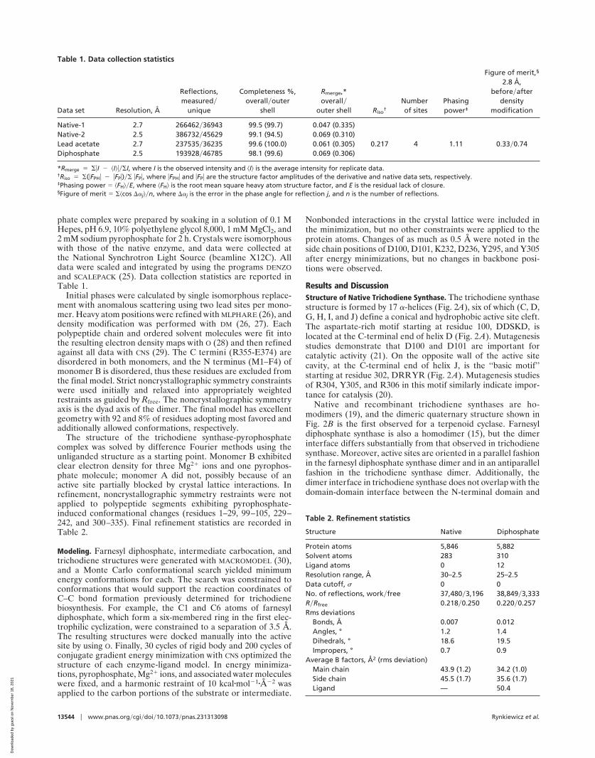
Table 1. Data collection statistics
Data set Resolution, Å
Reflections,measured�
unique
Completeness %,overall�outer
shell
Rmerge,*overall�
outer shell Riso†
Numberof sites
Phasingpower‡
Figure of merit,§
2.8 Å,before�after
densitymodification
Native-1 2.7 266462�36943 99.5 (99.7) 0.047 (0.335)Native-2 2.5 386732�45629 99.1 (94.5) 0.069 (0.310)Lead acetate 2.7 237535�36235 99.6 (100.0) 0.061 (0.305) 0.217 4 1.11 0.33�0.74Diphosphate 2.5 193928�46785 98.1 (99.6) 0.069 (0.306)
*Rmerge � ��I � �I����I, where I is the observed intensity and �I� is the average intensity for replicate data.†Riso � �(�FPH� � �FP�)�� �FP�, where �FPH� and �FP� are the structure factor amplitudes of the derivative and native data sets, respectively.‡Phasing power � �FH��E, where �FH� is the root mean square heavy atom structure factor, and E is the residual lack of closure.§Figure of merit � ��cos ��j��n, where ��j is the error in the phase angle for reflection j, and n is the number of reflections.
Table 2. Refinement statistics
Structure Native Diphosphate
Protein atoms 5,846 5,882Solvent atoms 283 310Ligand atoms 0 12Resolution range, Å 30–2.5 25–2.5Data cutoff, � 0 0No. of reflections, work�free 37,480�3,196 38,849�3,333R�Rfree 0.218�0.250 0.220�0.257Rms deviations
Bonds, Å 0.007 0.012Angles, ° 1.2 1.4Dihedrals, ° 18.6 19.5Impropers, ° 0.7 0.9
Average B factors, Å2 (rms deviation)Main chain 43.9 (1.2) 34.2 (1.0)Side chain 45.5 (1.7) 35.6 (1.7)Ligand — 50.4
13544 � www.pnas.org�cgi�doi�10.1073�pnas.231313098 Rynkiewicz et al.
Dow
nloa
ded
by g
uest
on
Nov
embe
r 18
, 202
1
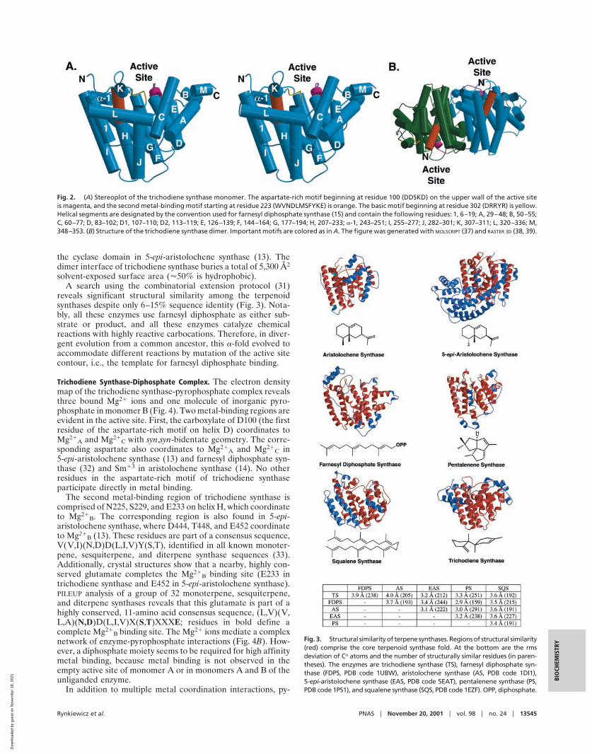
the cyclase domain in 5-epi-aristolochene synthase (13). Thedimer interface of trichodiene synthase buries a total of 5,300 Å2
solvent-exposed surface area (50% is hydrophobic).A search using the combinatorial extension protocol (31)
reveals significant structural similarity among the terpenoidsynthases despite only 6–15% sequence identity (Fig. 3). Nota-bly, all these enzymes use farnesyl diphosphate as either sub-strate or product, and all these enzymes catalyze chemicalreactions with highly reactive carbocations. Therefore, in diver-gent evolution from a common ancestor, this �-fold evolved toaccommodate different reactions by mutation of the active sitecontour, i.e., the template for farnesyl diphosphate binding.
Trichodiene Synthase-Diphosphate Complex. The electron densitymap of the trichodiene synthase-pyrophosphate complex revealsthree bound Mg2� ions and one molecule of inorganic pyro-phosphate in monomer B (Fig. 4). Two metal-binding regions areevident in the active site. First, the carboxylate of D100 (the firstresidue of the aspartate-rich motif on helix D) coordinates toMg2�
A and Mg2�C with syn,syn-bidentate geometry. The corre-
sponding aspartate also coordinates to Mg2�A and Mg2�
C in5-epi-aristolochene synthase (13) and farnesyl diphosphate syn-thase (32) and Sm�3 in aristolochene synthase (14). No otherresidues in the aspartate-rich motif of trichodiene synthaseparticipate directly in metal binding.
The second metal-binding region of trichodiene synthase iscomprised of N225, S229, and E233 on helix H, which coordinateto Mg2�
B. The corresponding region is also found in 5-epi-aristolochene synthase, where D444, T448, and E452 coordinateto Mg2�
B (13). These residues are part of a consensus sequence,V(V,I)(N,D)D(L,I,V)Y(S,T), identified in all known monoter-pene, sesquiterpene, and diterpene synthase sequences (33).Additionally, crystal structures show that a nearby, highly con-served glutamate completes the Mg2�
B binding site (E233 intrichodiene synthase and E452 in 5-epi-aristolochene synthase).PILEUP analysis of a group of 32 monoterpene, sesquiterpene,and diterpene synthases reveals that this glutamate is part of ahighly conserved, 11-amino acid consensus sequence, (L,V)(V,L,A)(N,D)D(L,I,V)X(S,T)XXXE; residues in bold define acomplete Mg2�
B binding site. The Mg2� ions mediate a complexnetwork of enzyme-pyrophosphate interactions (Fig. 4B). How-ever, a diphosphate moiety seems to be required for high affinitymetal binding, because metal binding is not observed in theempty active site of monomer A or in monomers A and B of theunliganded enzyme.
In addition to multiple metal coordination interactions, py-
Fig. 2. (A) Stereoplot of the trichodiene synthase monomer. The aspartate-rich motif beginning at residue 100 (DDSKD) on the upper wall of the active siteis magenta, and the second metal-binding motif starting at residue 223 (WVNDLMSFYKE) is orange. The basic motif beginning at residue 302 (DRRYR) is yellow.Helical segments are designated by the convention used for farnesyl diphosphate synthase (15) and contain the following residues: 1, 6–19; A, 29–48; B, 50–55;C, 60–77; D, 83–102; D1, 107–110; D2, 113–119; E, 126–139; F, 144–164; G, 177–194; H, 207–233; �-1, 243–251; I, 255–277; J, 282–301; K, 307–311; L, 320–336; M,348–353. (B) Structure of the trichodiene synthase dimer. Important motifs are colored as in A. The figure was generated with MOLSCRIPT (37) and RASTER 3D (38, 39).
Fig. 3. Structural similarity of terpene synthases. Regions of structural similarity(red) comprise the core terpenoid synthase fold. At the bottom are the rmsdeviation of C� atoms and the number of structurally similar residues (in paren-theses). The enzymes are trichodiene synthase (TS), farnesyl diphosphate syn-thase (FDPS, PDB code 1UBW), aristolochene synthase (AS, PDB code 1DI1),5-epi-aristolochene synthase (EAS, PDB code 5EAT), pentalenene synthase (PS,PDB code 1PS1), and squalene synthase (SQS, PDB code 1EZF). OPP, diphosphate.
Rynkiewicz et al. PNAS � November 20, 2001 � vol. 98 � no. 24 � 13545
BIO
CHEM
ISTR
Y
Dow
nloa
ded
by g
uest
on
Nov
embe
r 18
, 202
1
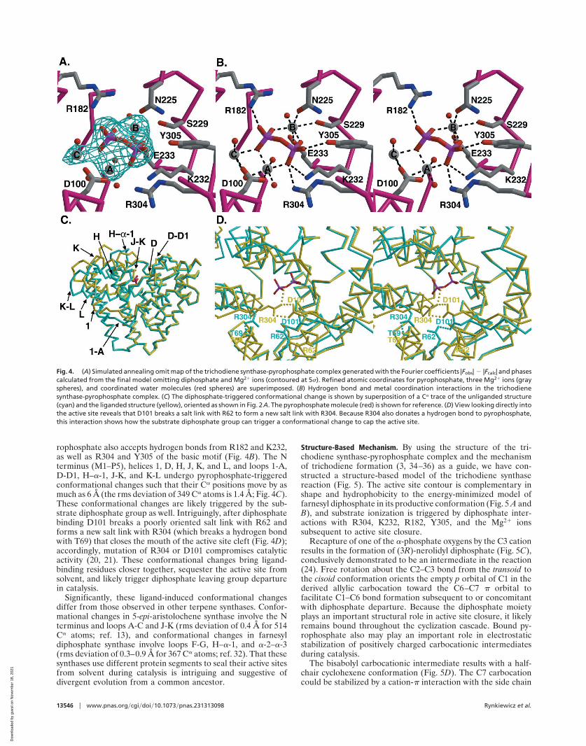
rophosphate also accepts hydrogen bonds from R182 and K232,as well as R304 and Y305 of the basic motif (Fig. 4B). The Nterminus (M1–P5), helices 1, D, H, J, K, and L, and loops 1-A,D-D1, H–�-1, J-K, and K-L undergo pyrophosphate-triggeredconformational changes such that their C� positions move by asmuch as 6 Å (the rms deviation of 349 C� atoms is 1.4 Å; Fig. 4C).These conformational changes are likely triggered by the sub-strate diphosphate group as well. Intriguingly, after diphosphatebinding D101 breaks a poorly oriented salt link with R62 andforms a new salt link with R304 (which breaks a hydrogen bondwith T69) that closes the mouth of the active site cleft (Fig. 4D);accordingly, mutation of R304 or D101 compromises catalyticactivity (20, 21). These conformational changes bring ligand-binding residues closer together, sequester the active site fromsolvent, and likely trigger diphosphate leaving group departurein catalysis.
Significantly, these ligand-induced conformational changesdiffer from those observed in other terpene synthases. Confor-mational changes in 5-epi-aristolochene synthase involve the Nterminus and loops A-C and J-K (rms deviation of 0.4 Å for 514C� atoms; ref. 13), and conformational changes in farnesyldiphosphate synthase involve loops F-G, H–�-1, and �-2–�-3(rms deviation of 0.3–0.9 Å for 367 C� atoms; ref. 32). That thesesynthases use different protein segments to seal their active sitesfrom solvent during catalysis is intriguing and suggestive ofdivergent evolution from a common ancestor.
Structure-Based Mechanism. By using the structure of the tri-chodiene synthase-pyrophosphate complex and the mechanismof trichodiene formation (3, 34–36) as a guide, we have con-structed a structure-based model of the trichodiene synthasereaction (Fig. 5). The active site contour is complementary inshape and hydrophobicity to the energy-minimized model offarnesyl diphosphate in its productive conformation (Fig. 5 A andB), and substrate ionization is triggered by diphosphate inter-actions with R304, K232, R182, Y305, and the Mg2� ionssubsequent to active site closure.
Recapture of one of the �-phosphate oxygens by the C3 cationresults in the formation of (3R)-nerolidyl diphosphate (Fig. 5C),conclusively demonstrated to be an intermediate in the reaction(24). Free rotation about the C2–C3 bond from the transoid tothe cisoid conformation orients the empty p orbital of C1 in thederived allylic carbocation toward the C6–C7 � orbital tofacilitate C1–C6 bond formation subsequent to or concomitantwith diphosphate departure. Because the diphosphate moietyplays an important structural role in active site closure, it likelyremains bound throughout the cyclization cascade. Bound py-rophosphate also may play an important role in electrostaticstabilization of positively charged carbocationic intermediatesduring catalysis.
The bisabolyl carbocationic intermediate results with a half-chair cyclohexene conformation (Fig. 5D). The C7 carbocationcould be stabilized by a cation-� interaction with the side chain
Fig. 4. (A) Simulated annealing omit map of the trichodiene synthase-pyrophosphate complex generated with the Fourier coefficients |Fobs| � |Fcalc| and phasescalculated from the final model omitting diphosphate and Mg2� ions (contoured at 5�). Refined atomic coordinates for pyrophosphate, three Mg2� ions (grayspheres), and coordinated water molecules (red spheres) are superimposed. (B) Hydrogen bond and metal coordination interactions in the trichodienesynthase-pyrophosphate complex. (C) The diphosphate-triggered conformational change is shown by superposition of a C� trace of the unliganded structure(cyan) and the liganded structure (yellow), oriented as shown in Fig. 2A. The pyrophosphate molecule (red) is shown for reference. (D) View looking directly intothe active site reveals that D101 breaks a salt link with R62 to form a new salt link with R304. Because R304 also donates a hydrogen bond to pyrophosphate,this interaction shows how the substrate diphosphate group can trigger a conformational change to cap the active site.
13546 � www.pnas.org�cgi�doi�10.1073�pnas.231313098 Rynkiewicz et al.
Dow
nloa
ded
by g
uest
on
Nov
embe
r 18
, 202
1
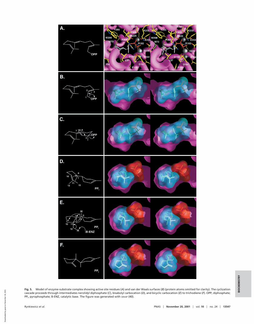
Fig. 5. Model of enzyme-substrate complex showing active site residues (A) and van der Waals surfaces (B) (protein atoms omitted for clarity). The cyclizationcascade proceeds through intermediates nerolidyl diphosphate (C), bisabolyl carbocation (D), and bicyclic carbocation (E) to trichodiene (F). OPP, diphosphate;PPi, pyrophosphate; B-ENZ, catalytic base. The figure was generated with GRASP (40).
Rynkiewicz et al. PNAS � November 20, 2001 � vol. 98 � no. 24 � 13547
BIO
CHEM
ISTR
Y
Dow
nloa
ded
by g
uest
on
Nov
embe
r 18
, 202
1
of Y93. The cyclohexene ring can rotate such that the empty porbital of C7 is oriented toward the C10–C11 � orbital. AfterC7–C11 bond formation, the resulting bicyclic secondary car-bocation at C10 could be stabilized through long range electro-static interactions with both D100 and pyrophosphate. Next,three carbocation-driven migrations occur. First, the C6–Hmigrates to the C10 carbocation through a 1,4-hydride transferfacilitated by the conformation of the intermediate (Fig. 5E).The side chain of L97 appears to provide a steric barrier topotentially competing C12 or C13 methyl migration reactions.Two 1,2-methyl migrations subsequently occur: the first from C7to the carbocation at C6 and the second from C11 to the newlygenerated carbocation at C7. This second methyl migration isspecific for C13, the migration of which may be kineticallyfavored, because it would provide anchimeric assistance to thedeparting C7 methyl group; migration of C12 to C7 appears tobe sterically hindered by T96. Finally, the C11 carbocation isquenched by proton elimination from C12, possibly mediatedeither by the carboxylate of D100 or pyrophosphate to yieldtrichodiene (Fig. 5F). Although neither aspartate nor pyrophos-phate is strongly basic, either is sufficiently reactive to abstracta proton from the extremely acidic trichodiene carbocation (pKa �10). In turn, protonation of either group could disruptintermolecular interactions with pyrophosphate, thereby trig-gering product release.
Variant trichodiene synthases with amino acid substitutions inthe diphosphate binding site exhibit altered product specificity(22–24). Changes in the diphosphate position would affect boththe proper positioning of the substrate in the active site as wellas the substrate-induced conformational change. These struc-tural changes could result in the formation of alternate productsbecause of incorrect folding of the substrate and�or prematurequenching of the reaction by solvent.
In conclusion, the x-ray crystal structures of dimeric tri-chodiene synthase and its complex with pyrophosphate allow usto visualize a putative substrate-induced conformational changeinvolving different secondary structural elements than thoseimplicated in two other sesquiterpene synthases: farnesyl diphos-phate synthase (32) and 5-epi-aristolochene synthase (13). Thetrichodiene synthase structures allow us to formulate a structure-based mechanism that rationalizes a wealth of mutagenesis andenzymological data (20–24). Future structural studies of site-specific mutants and enzyme-inhibitor complexes would allow usto test and clarify the mechanism proposed in Fig. 5.
We thank the National Synchrotron Light Source, the Advanced PhotonSource, and the Stanford Linear Accelerator Center for beamline access.We thank Drs. Todd Bowser and Hsien-Tai Chiu for helpful scientificdiscussions. This work was supported by National Institutes of HealthGrant GM56838 (to D.W.C.) and Merit Award GM30301 (to D.E.C.).
1. Cane, D. E. (1985) Acc. Chem. Res. 18, 220–226.2. Croteau, R. & Cane, D. E. (1985) in Methods in Enzymology: Steroids and
Isoprenoids (Part A), eds. Law, J. H. & Rilling, H. C. (Academic, New York)Vol. 110, pp. 383–405.
3. Cane, D. E. (1990) Chem. Rev. (Washington, D.C.) 90, 1089–1103.4. Wendt, K. U. & Schulz, G. E. (1998) Structure (London) 6, 127–133.5. Lesburg, C. A., Caruthers, J. M., Paschall, C. M. & Christianson, D. W. (1998)
Curr. Opin. Struct. Biol. 8, 695–703.6. Steele, C. L., Crock, J., Bohlmann, J. & Croteau, R. (1998) J. Biol. Chem. 273,
2078–2089.7. Crock, J., Wildung, M. & Croteau, R. (1997) Proc. Natl. Acad. Sci. USA 94,
12833–12838.8. Colby, S. M., Crock, J., Dowdle-Rizzo, B., Lemaux, P. G. & Croteau, R. (1998)
Proc. Natl. Acad. Sci. USA 95, 2216–2221.9. Mercke, P., Crock, J., Croteau, R. & Brodelius, P. E. (1999) Arch. Biochem.
Biophys. 369, 213–222.10. Mercke, P., Bengtsson, M., Bouwmeester, H. J., Posthumus, M. A. & Brodelius,
P. E. (2000) Arch. Biochem. Biophys. 381, 173–180.11. Ashby, M. N. & Edwards, P. A. (1990) J. Biol. Chem. 265, 13157–13164.12. Lesburg, C. A., Zhai, G., Cane, D. E. & Christianson, D. W. (1997) Science 277,
1820–1824.13. Starks, C. M., Back, K., Chappell, J. & Noel, J. P. (1997) Science 277,
1815–1820.14. Caruthers, J. M., Kang, I., Rynkiewicz, M. J., Cane, D. E. & Christianson, D. W.
(2000) J. Biol. Chem. 275, 25533–25539.15. Tarshis, L. C., Yan, M., Poulter, C. D. & Sacchettini, J. C. (1994) Biochemistry
33, 10871–10877.16. Pandit, J., Danley, D. E., Schulte, G. K., Mazzalupo, S., Pauly, T. A., Hayward,
C. M., Hamanaka, E. S., Thompson, J. F. & Harwood, H. J. (2000) J. Biol.Chem. 275, 30610–30617.
17. Fujihashi, M., Zhang, Y.-W., Higuchi, Y., Li, X.-Y., Koyama, T. & Miki, K.(2001) Proc. Natl. Acad. Sci. USA 98, 4337–4342. (First Published April 3, 2001;10.1073�pnas.071514398)
18. Cane, D. E., Swanson, S. & Murthy, P. P. N. (1981) J. Am. Chem. Soc. 103,2136–2138.
19. Cane, D. E., Wu, Z., Oliver, J. S. & Hohn, T. M. (1993) Arch. Biochem. Biophys.300, 416–422.
20. Cane, D. E., Shim, J. H., Xue, Q., Fitzsimons, B. C. & Hohn, T. M. (1995)Biochemistry 34, 2480–2488.
21. Cane, D. E., Xue, Q. & Fitzsimons, B. C. (1996) Biochemistry 35, 12369–12376.22. Cane, D. E., Xue, Q., Van Epp, J. E. & Tsantrizos, Y. S. (1996) J. Am. Chem.
Soc. 118, 8499–8500.23. Cane, D. E. & Xue, Q. (1996) J. Am. Chem. Soc. 118, 1563–1564.24. Cane, D. E. & Ha, H.-J. (1988) J. Am. Chem. Soc. 110, 6865–6870.25. Otwinowski, Z. & Minor, W. (1997) in Methods in Enzymology: Macromolecular
Crystallography (Part A), eds. Carter, C. W., Jr. & Sweet, R. M. (Academic, NewYork) Vol. 276, pp. 307–326.
26. Collaborative Computational Project Number 4 (1994) Acta Crystallogr. D 50,760–763.
27. Cowtan, K. (1994) Joint CCP4 and ESF-EACBM Newsletter on Protein Crys-tallography No. 31 (SERC Daresbury Laboratory, Warrington, U.K.), pp.34–38.
28. Jones, T. A., Zou, J. Y., Cowan, S. W. & Kjeldgaard, M. (1991) Acta Crystallogr.A 47, 110–119.
29. Brunger, A. T., Adams, P. D., Clore, G. M., DeLano, W. L., Gros, P.,Grosse-Kunstleve, R. W., Jiang, J.-S., Kuszewski, J., Nilges, M., Pannu, N. S.,et al. (1998) Acta Crystallogr. D 54, 905–921.
30. Mohamadi, F., Richards, N. G. J., Guida, W. C., Liskamp, R., Lipton, M.,Caufield, C., Chang, G., Hendrickson, T. & Still, W. C. (1990) J. Comput. Chem.11, 440–467.
31. Shindyalov, I. N. & Bourne, P. E. (1998) Protein Eng. 11, 739–747.32. Tarshis, L. C., Proteau, P. J., Kellogg, B. A., Sacchettini, J. C. & Poulter, C. D.
(1996) Proc. Natl. Acad. Sci. USA 93, 15018–15023.33. Cane, D. E. & Kang, I. (2000) Arch. Biochem. Biophys. 376, 354–364.34. Cane, D. E. & Yang, G. (1994) J. Org. Chem. 59, 5794–5798.35. Cane, D. E., Yang, G., Xue, Q. & Shim, J. H. (1995) Biochemistry 34,
2471–2479.36. Cane, D. E., Chiu, H.-T., Liang, P.-H. & Anderson, K. S. (1997) Biochemistry
36, 8332–8339.37. Kraulis, P. J. (1991) J. Appl. Crystallogr. 24, 946–950.38. Bacon, D. & Anderson, W. F. (1988) J. Mol. Graphics 6, 219–220.39. Merritt, E. A. & Murphy, M. E. P. (1994) Acta Crystallogr. D 50, 869–873.40. Nicholls, A., Sharp, K. A. & Honig, B. (1991) Proteins 11, 281–296.
13548 � www.pnas.org�cgi�doi�10.1073�pnas.231313098 Rynkiewicz et al.
Dow
nloa
ded
by g
uest
on
Nov
embe
r 18
, 202
1