Embed Size (px)
Citation preview
PROTEIN STRUCTURE REPORTS
Structure of the N-terminal domain of themetalloprotease PrtV from Vibrio cholerae
Aaron Edwin,1,2 Cecilia Persson,3 Maxim Mayzel,3 Sun Nyunt Wai,2,4,5
Anders €Ohman,6 B. G€oran Karlsson,3* and A. Elisabeth Sauer-Eriksson1,2*
1Department of Chemistry, Umea University, Umea SE-901 87, Sweden2Umea Centre for Microbial Research (UCMR), Umea University, Umea SE-901 87, Sweden3Swedish NMR Centre at the University of Gothenburg, Gothenburg SE-40530, Sweden4Department of Molecular Biology, Umea University, Umea SE-901 87, Sweden5The Laboratory for Molecular Infection Medicine Sweden (MIMS), Umea University, Umea SE-901 87, Sweden6Department of Pharmacology and Clinical Neuroscience, Umea University, Umea SE-901 85, Sweden
Received 10 August 2015; Accepted 25 September 2015
DOI: 10.1002/pro.2815Published online 5 October 2015 proteinscience.org
Abstract: The metalloprotease PrtV from Vibrio cholerae serves an important function for the ability
of bacteria to invade the mammalian host cell. The protein belongs to the family of M6 proteases,with a characteristic zinc ion in the catalytic active site. PrtV constitutes a 918 amino acids (102
kDa) multidomain pre-pro-protein that undergoes several N- and C-terminal modifications to form
a catalytically active protease. We report here the NMR structure of the PrtV N-terminal domain(residues 23–103) that contains two short a-helices in a coiled coil motif. The helices are held
together by a cluster of hydrophobic residues. Approximately 30 residues at the C-terminal end,
which were predicted to form a third helical structure, are disordered. These residues are highlyconserved within the genus Vibrio, which suggests that they might be functionally important.
Keywords: Vibrio cholera; metalloproteases; PrtV; N-terminal domain; NMR
Introduction
Cholera is an infection in the small intestine caused
by the motile Gram-negative bacterium Vibrio chol-
erae.1 During an infection, V. cholerae pathogenicity
genes code for virulence factors that are directly or
indirectly involved in the virulence of the bacteria.
These factors include proteases that attack the tar-
get cells by breaking down tissue barriers and cellu-
lar matrix components.2–4 One of these proteases is
the secreted metalloprotease PrtV that exhibits a
very potent cytotoxic effect.5,6
PrtV belongs to the M6 peptidase family and is
natively expressed as a 102 kDa full-length pre-pro-
protein (918 amino acids). In addition to the signal
sequence, it consists of four domain types: the N-
This is an open access article under the terms of the CreativeCommons Attribution-NonCommercial-NoDerivs License, whichpermits use and distribution in any medium, provided the origi-nal work is properly cited, the use is non-commercial and nomodifications or adaptations are made.
Aaron Edwin, Cecilia Persson, and Maxim Mayzel share equalauthorship
Grant sponsors: Kempe Foundation, Swedstruct, WallenbergFoundation Swedish Research Council; Grant number: K2013-67X-13001-15-3.
*Correspondence to: E. Sauer-Eriksson, Department ofChemistry, Umea University, SE-90187 Umea, Sweden. E-mail:[email protected] or G Karlsson, Swedish NMRCentre at the University of Gothenburg, SE-40530 Gothenburg,Sweden. E-mail: [email protected]
2076 PROTEIN SCIENCE 2015 VOL 24:2076—2080 VC 2015 The Authors Protein Science published by Wiley Periodicals, Inc. on behalf of The Protein Society
terminal domain (residues 23–103), the catalytic
active M6 domain (residues 106–749), two polycystic
kidney disease domains, PKD1 (residues 755–838)
and the PKD2 (residues 839–918).5,6 M6 constitutes
the catalytic metalloprotease domain with the charac-
teristic HexxHxxgxxD Zn21-binding motif.7 PKD
domains are found in a variety of eukaryotic and pro-
karyotic proteins and consist of relatively short
domains of 80–90 amino acids; they are usually found
in the extracellular parts of proteins where they are
involved in protein–protein or protein–carbohydrate
interactions. It has been suggested that calcium bind-
ing at the PKD1 domain controls domain linker flexi-
bility, and plays a regulatory role in the auto-
proteolytic activity of the 81 kDa pro-protein.8
Sequence analysis shows that the N-terminal domain
is present in many proteases from gammaproteobacte-
ria; however, its specific function is unidentified.
Atomic resolution structures of the N-terminal
domain are needed to elucidate how the N-terminal
domain contributes to PrtV function. In this study, we
present the NMR structure of the N-terminal domain.
Results
NMR spectroscopy of the N-terminal domainThe solution structure of the N-terminal domain was
solved at the Swedish NMR Centre (www.nmr.gu.se).
The 1H–15N heteronuclear single quantum correlation
(HSQC) NMR spectrum of the PrtV 83 amino-acid N-
terminal domain exhibited characteristics of a par-
tially folded protein. The spectrum was well dispersed,
but exhibited a signal clustering characteristic of a dis-
ordered protein. The total number of observed signals
was fewer than expected from the sequence, either due
to overlapping or broadening beyond detection. Simi-
larly, peak shapes showed a distribution from narrow
to broad, and intensities varied considerably.
NMR-derived structure of the N-terminal
domain
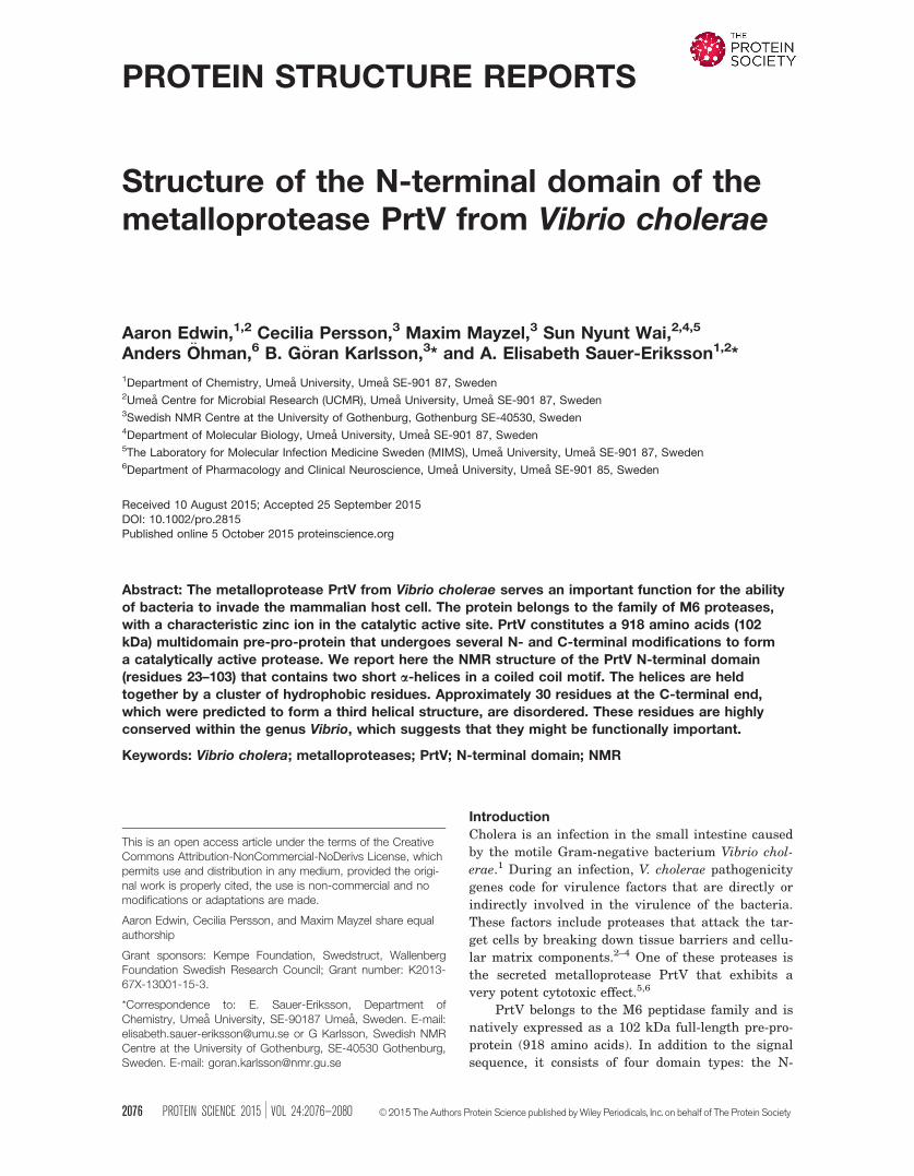
Structure statistics for the 20 lowest energy con-
formers are summarized in Table I. The first 8 resi-
dues from the N-terminal end and the last 32
residues from the C-terminal end of the domain
exhibited random-coil chemical shifts with non-
uniform intensities suggesting that these regions
are disordered. Remaining residues (residues 31–70)
exhibited highly dispersed chemical shifts with uni-
form intensities that indicated a folded core struc-
ture [Fig. 1(A)]. A ribbon structure, representative
of the final ensemble of NMR-derived structures of
the N-terminal domain is shown in Figure 1(B). The
main chain structure contains two alpha helices: a1
(residues Val31-Gly45) and a2 (residues Asp53-
Ser70). The first helix a1 is bent and the first turn
has a 310-helical geometry. The two helices are con-
nected by a well-defined loop region comprising resi-
dues Gln46-Ser52. The C-terminal part of a1
(residues Leu37-Gly45) packs against the C-terminal
part of a2 at an angle of about 908. This is in accord-
ance with the general rule for packing interactions
between a helices, that is, the ridges on one helix fit
into the grooves of the other, and vice versa. In this
area, the two helices are held together by hydropho-
bic interactions between conserved amino acids [Fig.
2(A,B)]. A space-filling representation of the N-
terminal domain reveals a cluster of hydrophobic
and basic residues at the N- and C-terminal ends of
the folded structure, whereas the loop region of the
motif is more negatively charged [Fig. 1(C)].
Discussion
The secreted metalloprotease PrtV is a potent viru-
lence protein of V. cholerae causing immediate cyto-
toxic effects during infection.6 The function of its N-
terminal domain is unknown, but its sequence is
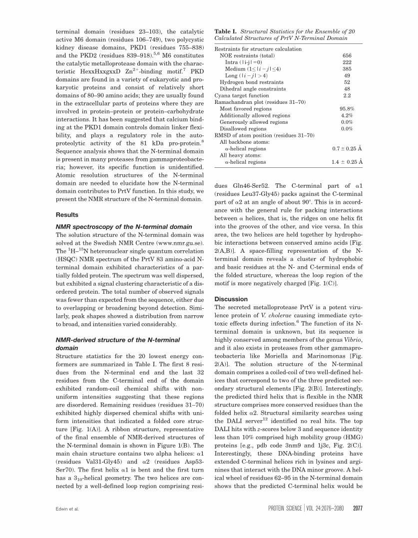
highly conserved among members of the genus Vibrio,
and it also exists in proteases from other gammapro-
teobacteria like Moriella and Marinomonas [Fig.
2(A)]. The solution structure of the N-terminal
domain comprises a coiled-coil of two well-defined hel-
ices that correspond to two of the three predicted sec-
ondary structural elements [Fig. 2(B)]. Interestingly,
the predicted third helix that is flexible in the NMR
structure comprises more conserved residues than the
folded helix a2. Structural similarity searches using
the DALI server12 identified no real hits. The top
DALI hits with z-scores below 3 and sequence identity
less than 10% comprised high mobility group (HMG)
proteins [e.g., pdb code 3nm9 and 1j3c, Fig. 2(C)].
Interestingly, these DNA-binding proteins have
extended C-terminal helices rich in lysines and argi-
nines that interact with the DNA minor groove. A hel-
ical wheel of residues 62–95 in the N-terminal domain
shows that the predicted C-terminal helix would be
Table I. Structural Statistics for the Ensemble of 20Calculated Structures of PrtV N-Terminal Domain
Restraints for structure calculationNOE restraints (total) 656
Intra (|i-j|50) 222Medium (1�|i 2 j|�4) 385Long (|i 2 j|>4Þ 49
Hydrogen bond restraints 52Dihedral angle constraints 48
Cyana target function 2.2Ramachandran plot (residues 31–70)
Most favored regions 95.8%Additionally allowed regions 4.2%Generously allowed regions 0.0%Disallowed regions 0.0%
RMSD of atom position (residues 31–70)All backbone atoms:
a-helical regions 0.7 6 0.25 AAll heavy atoms:
a-helical regions 1.4 6 0.25 A
Edwin et al. PROTEIN SCIENCE VOL 24:2076—2080 2077
amphiphilic with aligned lysine residues forming a
highly positively charged side [Fig. 2(D)]. This led us
to test the DNA-binding ability of the N-terminal
domain; however, the domain does not seem to bind
unspecifically to the minor groove DNA [Fig. 2(E)].
Possibly the conserved and positively charged lysine
residues provide a region involved in membrane asso-
ciation (e.g., anchored to the membrane via lysine
snorkeling)—this hypothesis needs to be tested.
Methods
Expression and purification of N-terminal
domainThe cloning, expression, and purification of the PrtV
N domain construct (residues 23–103) is described
elsewhere.13 In brief, the N-terminal domain was
cloned into the pET1a-TrxA vector14 and overex-
pressed in E. coli Bl21 (DE3) pLysS (Novagen) cells.
The protein was purified by affinity chromatography
on a Ni-NTA agarose column (Qiagen), cation
exchange chromatography on a MonoS 5/5 column
(GE Healthcare), followed by a Superdex 200-16/60
size exclusion column (GE Healthcare). The protein is
very soluble and can be concentrated to>100 mg/mL.
Pure fractions of the PrtV N domain were pooled and
concentrated to 20 mg/mL in 20 mM Tris pH 7.5 and
100 mM NaCl. From 1 L media �10 mg of protein was
obtained.
Electrophoretic mobility shift assayDNA–protein interaction was examined by a gel retar-
dation assay using purified N-terminal PrtV protein.
As substrates for DNA binding, 400 bp PCR products
containing the promoter regions of vca0107 and
vc0017 genes of V. cholerae wild type strain A1552
were used. A known H-NS binding promoter and puri-
fied H-NS protein (4 and 12 mM) were used as the posi-
tive control in the electrophoretic mobility shift assay.
The reaction buffer containing 25 mM 4-(2-hydrox-
yethyl)-1-piperazineethanesulfonic acid (HEPES) (pH
7.3), 0.1 mM ethylenediaminetetraacetic acid (EDTA),
5 mM dithiothreitol (DTT), 10% glycerol, 50 mM KCl,
and 0.1 mg of poly(dIdC) non-specific competitor DNA
was used for the assay. Purified PrtV protein (4 and
12 mM) or H-NS protein (4 and 12 mM) were incubated
with 100 ng of PCR products for 20 min at 208C. Sam-
ples were loaded onto a 6% Tris-glycine gel and DNA
was visualized by ethidium bromide staining.
Isotope labeling
Expression of PrtV N-terminal domain for NMR anal-
ysis was done in 1 L of M9 minimal media supple-
mented with 15NH4Cl (0.5 g/L media) as the sole
nitrogen source, 13C-glucose (1 g/L media) as the sole
carbon source, and trace elements as specified.13
Labeled PrtV N-terminal domain expression was
induced and protein purified as described for the unla-
beled protein.
Structure calculation
Samples of the recombinant PrtV N-terminal domain
(0.7 mM) were prepared in 90%/10% H2O/D2O or
Figure 1. NMR-derived structure of the PrtV N-terminal
domain. A: The 20 lowest-energy conformers of the N
domain with their backbones (residues 31–70) superimposed.
The first 8 N-terminal residues and the last 32 C-terminal res-
idues are not folded in the structure. B: Ribbon representa-
tion of the folded N-terminal domain structure closest to the
average structure. The structure is shown in two views
rotated by 908. The pink helix includes residues 31–45 (a1)
and the blue residues 53–70 (a2). C: Accessibility and elec-
trostatic potential of surfaces of the structures are presented
in (B). The loop side is more negatively charged, whereas the
N- and C-terminal side surfaces are hydrophobic/positively
charged. An interactive view is available in the electronic
version of the article.
This figure also includes an iMolecules 3D interactive version
that can be accessed via the link at the bottom of this figure’s
caption.
2078 PROTEINSCIENCE.ORG NMR Structure of the N-Terminal Domain of PrtV from Vibrio cholera
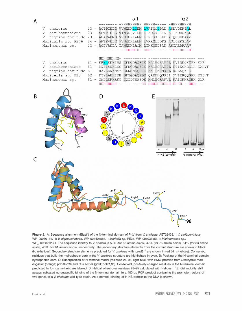
Figure 2. A: Sequence alignment (Blast9) of the N-terminal domain of PrtV from V. cholerae. AET29433.1; V. caribbenthicus,
WP_009601447.1; V. nigripulchritudo, WP_004405586.1; Moritella sp. PE36, WP_006031551.1; Marinomonas sp.,
WP_009832723.1. The sequence identity to V. cholera is 59% (for 83 amino acids), 47% (for 76 amino acids), 54% (for 83 amino
acids), 43% (for 81 amino acids), respectively. The secondary structure elements from the current structure are shown in black
(H, a-helices). Secondary structure elements predicted for V. cholerae with jpred310 are shown in red (H, a-helices). Conserved
residues that build the hydrophobic core in the V. cholerae structure are highlighted in cyan. B: Packing of the N-terminal domain
hydrophobic core. C: Superposition of N-terminal model (residues 28–98, light-blue) with HMG proteins from Drosophila mela-
nogaster (orange; pdb:3nm9) and Sus scrofa (gold; pdb:1j3c). Conserved, positively charged residues in the N-terminal domain
predicted to form an a-helix are labeled. D: Helical wheel over residues 78–95 calculated with Heliqust.11 E: Gel mobility shift
assays indicated no unspecific binding of the N-terminal domain to a 400 bp PCR product containing the promoter regions of
two genes of a V. cholerae wild type strain. As a control, binding of H-NS protein to the DNA is shown.
Edwin et al. PROTEIN SCIENCE VOL 24:2076—2080 2079
100% D2O with 20 mM sodium phosphate (pH 7.0).
Sequential assignment was obtained using incremen-
tal non-uniform sampling combined with targeted
acquisition on a 600 MHz Varian Inova NMR spec-
trometer with a cold probe, essentially as earlier
described.15 Side chain assignment was obtained
from 3D H(CCO)NH and (H)C(CCO)NH experiments.
Structure information was obtained from 3D 15N-
NOESY-HSQC, 3D 13C-NOESY-HSQC, and 2D
NOESY experiments obtained at 800 MHz and 900
MHz Avance III HD spectrometers equipped with
5 mm TCI cryoprobes. 15N-NOE relaxation data were
obtained as previously described. All NMR experi-
ments were performed at 108C. NMR data were proc-
essed with NMRPipe16 and analyzed using the CCPN
package.17
Structures of PrtV N-terminal domain were cal-
culated with CYANA.18 NOE distance restraints
combined with dihedral restraints and hydrogen
bond distance constraints in the alpha helices
obtained from the CSI-method19 were used to calcu-
late ensemble model structures. The 20 lowest
energy structures were selected and analyzed.
Molecular graphics were produced using
CCP4mg.20 The NMR-derived ensemble structures
of the N-terminal domain were deposited in the Pro-
tein Data Bank, with accession code 5abk. The
assigned chemical shifts have been deposited within
the BMRB database (accession number 25745).
Acknowledgments
Authors thank Drs. Karina Persson, Uwe H. Sauer,
Bernt Eric Uhlin, and Tobias Hainzl for valuable
ideas and discussions, and Monica Persson for tech-
nical assistance. The work was performed at the
Umea Centre for Microbial Research (UCMR).
References
1. Harris JB, LaRocque RC, Qadri F, Ryan ET,
Calderwood SB (2012) Cholera. Lancet 379:2466–2476.2. Miyoshi S, Shinoda S (2000) Microbial metalloproteases
and pathogenesis. Microbes Infect 2:91–98.3. Hase CC, Finkelstein RA (1993) Bacterial extracellular
zinc-containing metalloproteases. Microbiol Rev 57:
823–837.4. Shinoda S, Miyoshi S (2011) Proteases produced by
vibrios. Biocontrol Sci 16:1–11.5. Vaitkevicius K, Lindmark B, Ou G, Song T, Toma C,
Iwanaga M, Zhu J, Andersson A, Hammarstrom ML,
Tuck S, Wai SN (2006) A Vibrio cholerae protease
needed for killing of Caenorhabditis elegans has a role
in protection from natural predator grazing. Proc NatlAcad Sci USA 103:9280–9285.
6. Vaitkevicius K, Rompikuntal PK, Lindmark B,Vaitkevicius R, Song T, Wai SN (2008) The metallopro-tease PrtV from Vibrio cholerae. FEBS J 275:3167–3177.
7. Kurisu G, Kinoshita T, Sugimoto A, Nagara A, Kai Y,Kasai N, Harada S (1997) Structure of the zinc endo-protease from Streptomyces caespitosus. J Biochem121:304–308.
8. Edwin A, Rompikuntal P, Bjorn E, Stier G, Wai SN,Sauer-Eriksson AE (2013) Calcium binding by thePKD1 domain regulates interdomain flexibility inVibrio cholerae metalloprotease PrtV. FEBS Open Biol3:263–270.
9. Altschul SF, Gish W, Miller W, Myers EW, Lipman DJ(1990) Basic local alignment search tool. J Mol Biol215:403–410.
10. Cole C, Barber JD, Barton GJ (2008) The Jpred 3 sec-ondary structure prediction server. Nucleic Acids Res36:W197–W201.
11. Gautier R, Douguet D, Antonny B, Drin G (2008)HELIQUEST: a web server to screen sequences withspecific alpha-helical properties. Bioinformatics 24:2101–2102.
12. Holm L, Kaariainen S, Rosenstrom P, Schenkel A(2008) Searching protein structure databases withDaliLite v.3. Bioinformatics 24:2780–2781.
13. Edwin A, Grundstrom C, Wai SN, Ohman A, Stier G,Sauer-Eriksson AE (2014) Domain isolation, expres-sion, purification and proteolytic activity of the metal-loprotease PrtV from Vibrio cholerae. Protein ExpressPurif 96:39–47.
14. Bogomolovas J, Simon B, Sattler M, Stier G (2009)Screening of fusion partners for high yield expressionand purification of bioactive viscotoxins. ProteinExpress Purif 64:16–23.
15. Isaksson L, Mayzel M, Saline M, Pedersen A,Rosenlow J, Brutscher B, Karlsson BG, Orekhov VY(2013) Highly efficient NMR assignment of intrinsicallydisordered proteins: application to B- and T cell recep-tor domains. PloS One 8:e62947.
16. Delaglio F, Grzesiek S, Vuister GW, Zhu G, Pfeifer J,Bax A (1995) NMRPipe: a multidimensional spectralprocessing system based on UNIX pipes. J BiomolNMR 6:277–293.
17. Vranken WF, Boucher W, Stevens TJ, Fogh RH, PajonA, Llinas M, Ulrich EL, Markley JL, Ionides J, LaueED (2005) The CCPN data model for NMR spectros-copy: development of a software pipeline. Proteins 59:687–696.
18. Guntert P, Mumenthaler C, Wuthrich K (1997) Torsionangle dynamics for NMR structure calculation with thenew program DYANA. J Mol Biol 273:283–298.
19. Wishart DS, Sykes BD (1994) The 13C chemical-shiftindex: a simple method for the identification of proteinsecondary structure using 13C chemical-shift data.J Biomol NMR 4:171–180.
20. McNicholas S, Potterton E, Wilson KS, Noble ME(2011) Presenting your structures: the CCP4mgmolecular-graphics software. Acta Cryst D67:386–394.
2080 PROTEINSCIENCE.ORG NMR Structure of the N-Terminal Domain of PrtV from Vibrio cholera