Embed Size (px)
Citation preview

Structure of the human Parkin ligase domainin an autoinhibited state
Tobias Wauer and David Komander*
Division of Protein and Nucleic Acid Chemistry, Medical ResearchCouncil Laboratory of Molecular Biology, Cambridge, UK
Mutations in the protein Parkin are associated with
Parkinson’s disease (PD), the second most common
neurodegenerative disease in men. Parkin is an E3
ubiquitin (Ub) ligase of the structurally uncharacterized
RING-in-between-RING(IBR)-RING (RBR) family, which, in
an HECT-like fashion, forms a catalytic thioester intermedi-
ate with Ub. We here report the crystal structure of human
Parkin spanning the Unique Parkin domain (UPD, also
annotated as RING0) and RBR domains, revealing a tightly
packed structure with unanticipated domain interfaces. The
UPD adopts a novel elongated Zn-binding fold, while RING2
resembles an IBR domain. Two key interactions keep Parkin
in an autoinhibited conformation. A linker that connects the
IBR with the RING2 over a 50-A distance blocks the con-
served E2BUb binding site of RING1. RING2 forms a hydro-
phobic interface with the UPD, burying the catalytic Cys431,
which is part of a conserved catalytic triad. Opening of intra-
domain interfaces activates Parkin, and enables Ub-based
suicide probes to modify Cys431. The structure further
reveals a putative phospho-peptide docking site in the
UPD, and explains many PD-causing mutations.
The EMBO Journal advance online publication, 31 May 2013;
doi:10.1038/emboj.2013.125Subject Categories: proteins; molecular biology of disease;structural biologyKeywords: E3 ligase; neurodegenerative disease; Parkin;
ubiquitin; X-ray crystallography
Introduction
Parkinson’s disease (PD) is a neurodegenerative disorder
characterized by loss of dopaminergic neurons from the
substantia nigra, and appearance of a-synuclein aggregates
known as Lewy bodies (Goedert et al, 2013). PD occurs
sporadically in 1–2% of people above 65 years of age, but
can also arise earlier, most commonly due to genetic predis-
position of individuals (Corti et al, 2011). This familial form
of autosomal recessive juvenile Parkinsonism (AR-JP) results
from mutations in the protein kinase PINK1 (Valente et al,
2004), the adaptor DJ-1 (Bonifati et al, 2003), or, in about
50% of cases, in the E3 ubiquitin (Ub) ligase Parkin (Kitada
et al, 1998). Heterozygous Parkin mutations have been
identified in patients with sporadic PD (Sun et al, 2006;
Wang et al, 2008; reviewed in Corti et al, 2011).
Protein ubiquitination is an important post-translational
modification that regulates most aspects of cell biology
(Komander and Rape, 2012). Attachment of Ub or more
frequently of polyUb chains to proteins regulates turnover,
localization, complex assembly or activity of the substrate.
Ubiquitination is facilitated by a three-step enzymatic
cascade involving E1 Ub activating, E2 Ub conjugating
enzymes, and E3 Ub ligases (Hershko and Ciechanover,
1998). Two mechanistically distinct classes of E3 ligases
have been described. RING E3 ligases facilitate transfer of
Ub directly from the E2 catalytic Cys to the substrate, while in
HECT E3 ligases, an intermediate thioester with the E3 ligase
is formed prior to substrate modification (Dye and Schulman,
2007). Recently, a hybrid mechanism involving RING-
mediated formation of a ligase thioester was reported for
RING-in-between-RING(IBR)-RING (RBR) E3 ligases (Wenzel
et al, 2011). In RBR E3 ligases, a RING domain (RING1)
mediates transfer of Ub from the E2 to a catalytic Cys of the
RING2 domain, from which Ub is subsequently transferred to
a substrate (Wenzel and Klevit, 2012). RBR E3 ligases are
found in all eukaryotes, and 13 RBR E3 ligases exist in
humans (Eisenhaber et al, 2007; Marın, 2009; Wenzel and
Klevit, 2012). Prominent members of this family are HOIP and
HOIL-1 that form the linear Ub chain assembly complex
(LUBAC) (Kirisako et al, 2006), Ariadne-1 that is important
for neuronal differentiation in Drosophila (Aguilera et al,
2000) and Parkin.
Genetic and cell biological work in the last decade have
uncovered essential roles of Parkin and PINK1 in mitochondrial
quality control (Corti et al, 2011; Narendra et al, 2012). PINK1
senses damaged mitochondria, and recruits and activates
Parkin to ubiquitinate mitochondrial outer membrane
proteins including mitofusins (Poole et al, 2010; Tanaka et al,
2010; Ziviani et al, 2010; Chen and Dorn, 2013; Sarraf et al,
2013) and Miro (Wang et al, 2011). This leads to the selective
autophagy of damaged mitochondria termed mitophagy. Much
evidence suggests that defects in this pathway may cause PD
(Youle and Narendra, 2011). Parkin also impacts other cellular
pathways, including TNFa signalling (Muller-Rischart et al,
2013) and Wnt/b-catenin signalling (Rawal et al, 2009), and
Parkin was also found to be a tumour suppressor (Poulogiannis
et al, 2010; Veeriah et al, 2010).
With these important biological roles, it is not surprising
that Parkin E3 ligase activity is under tight regulation
(Walden and Martinez-Torres, 2012), and it is clear from
many studies that Parkin requires activation (Xiong et al,
2009; Geisler et al, 2010; Matsuda et al, 2010; Narendra et al,
2010; Vives-Bauza et al, 2010; Chaugule et al, 2011; Chew
et al, 2011). Parkin regulation is in part facilitated by its
domain structure (Figure 1A). A C-terminal RBR domain
comprises an E2-binding RING1 domain, an IBR domain
and a RING2 domain that contains the catalytic Cys residue
(Cys431) for E2-mediated Ub charging. N-terminal to the RBR
*Corresponding author. Protein and Nucleic Acid Chemistry, MRCLaboratory of Molecular Biology, Francis Crick Avenue, CambridgeBiomedical Campus, Cambridge, Cambridgeshire CB2 0QH, UK.Tel.: þ44 (0)1223 267160; E-mail: [email protected]
Received: 18 March 2013; accepted: 7 May 2013
The EMBO Journal (2013), 1–14
www.embojournal.org
EMBO
THE
EMBOJOURNAL
THE
EMBOJOURNAL
1&2013 European Molecular Biology Organization The EMBO Journal

is a further Zn-binding fold termed Unique Parkin domain
(UPD) or RING0 (Hristova et al, 2009) that interacts with
PINK1 (Xiong et al, 2009). Finally, a Ub-like (Ubl) domain at
the very N-terminus binds the RBR in cis, and inhibits
Parkin activity (Chaugule et al, 2011). PINK1 was reported
to phosphorylate the Ubl of Parkin to release this inhibition
(Kondapalli et al, 2012). However, the role of phosphoryla-
tion in PINK1/Parkin function is more complicated, as PINK1
does not always phosphorylate Parkin (Vives-Bauza et al,
2010), PINK1 requires autophosphorylation to recruit Parkin
(Okatsu et al, 2012), and PINK1 phosphorylates substrates to
be recognized by Parkin (Chen and Dorn, 2013).
Catalyticcentre
A
DC
UPD
RING1
IBRRING2
465 (C)142(N)
Linker helix
E
142
227
CysCys
CysCys
CysCys
HisCys
Zn
Zn
228
Cys Cys
CysHis
CysCys
CysCys
327
Zn
Zn
383Zn
Cys
Cys
His Cys
CysCys
CysCys
328Zn
UP
D
RING1
IBR
RING2
RING2IBRRING1UPDUBL
B
1 465
137
142227
228
327
383
328
Figure 1 Structure of Parkin. (A) Domain structure of Parkin. A yellow asterisk (*) indicates the catalytic Cys431 in RING2. (B) Structure of theParkin UPD-RBR domain. Individual domains are coloured blue (UPD), cyan (RING1), purple (IBR) and orange (RING2). The linker helix isshown in red and the Zn atoms as yellow spheres. Putative catalytic residues in RING2 are labelled. Dotted lines indicate disordered stretches.Terminal residue numbers are indicated. A cartoon based on (A) depicts domain interactions. (C) Structure and topology of the UPD. Residuescoordinating Zn atoms are shown. (D) Structure and topology of the extended RING1. (E) Structure of the crystallized IBR domain with eightsuperposed models from the previously described NMR ensemble (pdb-id 2jmo, Beasley et al, 2007). The unresolved loop in Parkin IBR is mostflexible also in NMR analysis. The topology is shown below, the region in grey is disordered in the structure.
Structure of the Parkin UPD-RBRT Wauer and D Komander
2 The EMBO Journal &2013 European Molecular Biology Organization

The contribution of individual Parkin domains in ubiquiti-
nation activity is however still unclear since a construct
spanning IBR and RING2 but lacking the E2-interacting
RING1 domain shows high in vitro activity, which depends
on Cys431 in RING2 (Chew et al, 2011). While charging of the
RING2 with Ub could be shown for HHARI (Wenzel et al,
2011) and HOIP (Smit et al, 2012; Stieglitz et al, 2012), the
thioester reaction intermediate could not be observed in an
in vitro reaction for the Parkin RBR (Wenzel et al, 2011).
Recent data generated in an elegant cell-free system
recapitulating activated PINK1 on depolarized
mitochondria, showed that catalytic activity of PINK1 can
lead to formation of charged Parkin in an E2-dependent
manner (Lazarou et al, 2013). Surprisingly, Parkin C431S,
which forms a stabilised Ub-loaded Parkin intermediate in
this system, is not localized to mitochondria, but self-
associates into an oligomeric complex in the cytosol
(Lazarou et al, 2013).
Loss-of-function mutations in Parkin are implicated in
AR-JP, and missense mutations have been an important and
intensely used resource to gain insight into Parkin function
(Sriram et al, 2005; Wang et al, 2005; Hampe et al, 2006;
Matsuda et al, 2006, 2010; Wong et al, 2007; Schlehe et al,
2008). Many mutations affect Parkin stability but a surprising
number of soluble variants appear to retain ligase activity
(Sriram et al, 2005; Hampe et al, 2006; Matsuda et al, 2006).
Molecular insights into Parkin structure would elucidate the
mechanism of Parkin and of RBR E3 ligases, and could
explain the connection between Parkin and AR-JP. However,
while mechanisms of HECT and RING E3 ligases have been
revealed in atomic detail (Dye and Schulman, 2007; Lima and
Schulman, 2012), the RBR family of E3 ligases has resisted
structural analysis.
We here report the structure of the catalytic fragment of
Parkin spanning its UPD and RBR domains. A surprising
domain disposition reveals unanticipated interfaces between
UPD and RING2, which bury the catalytic Cys431 at the
interface. Moreover, the IBR and RING2 domains are sepa-
rated by an extended linker that occludes the canonical
E2-binding site in RING1. Our structure and biochemical
studies suggest that significant conformational changes are
required for Parkin activation and shed light on the dysfunc-
tion of many AR-JP-associated mutations of Parkin.
Results and discussion
Crystallization and structure of Parkin UPD-RBR
RBR domains are challenging to express in bacteria due to
their multiple Zn-binding folds. We were inspired by recent
work on HOIP in which inclusion of a C-terminal Zn-binding
region improved protein stability (Smit et al, 2012; Stieglitz
et al, 2012). Parkin does not contain a C-terminal Zn-binding
domain, but an N-terminal UPD Zn-binding fold that was
predicted to resemble RING domains (Hristova et al, 2009;
Figure 1A). Hristova et al further use limited proteolysis to
identify a stable Parkin fragment corresponding to amino
acids (aa) 145 to the C-terminal residue 465. We generated
sufficient yields of soluble protein from human Parkin con-
struct spanning aa 137–465, which was codon-optimized for
expression in E. coli. Purified protein was crystallized and
the structure was determined to 2.25 A resolution using
the anomalous signal of the eight coordinated zinc atoms
from a single anomalous dispersion experiment (Table I;
Supplementary Figure 1).
The structure of human Parkin UPD-RBR revealed four
domains with unanticipated inter-domain interactions
(Figure 1B). Central to the structure is the UPD (aa 142–
227), a previously undescribed elongated Zn-binding fold in
which two central anti-parallel b-strands arrange two Zn-
coordinating loops. That the UPD is a Zn-binding fold was
known, and annotation of this domain as RING0 was based
on modelling it as a cross-brace structure (Hristova et al,
2009). We found that the UPD does not show a cross-brace
topology. The two Zn atoms in the UPD are coordinated by
the first and fourth, and second and third zinc-binding loops
(Figure 1C). Since the UPD does not resemble a RING, and
since there are no similar structures in the protein data bank,
or similar sequences in the human genome, we refer to this
domain as the UPD as in earlier work (Hampe et al, 2006).
The UPD serves as an interacting platform for the subse-
quent RING1 domain that binds to one tip of the UPD, and
also for the RING2 domain that forms an extensive hydro-
phobic interface with the side of the UPD (see below).
The RING1 domain of Parkin (aa 228–327) is a variation
of the canonical RING fold (Figure 1D). It contains two
Zn-binding sites in cross-brace topology, and all features
required for E2 binding (see below). In addition, it contains
an internal solvent exposed b-hairpin insertion located after
the RING domain helix (see below). Three C-terminal helices
constitute a platform for IBR interaction (Figure 1B and D).
The IBR domain (aa 328–383) is least well defined in the
electron density, most likely due to high domain mobility.
While the position of the Zn atoms is clear from the electron
density, one Zn-coordinating Cys residue in the first
Zn-binding site is not resolved (Figure 1E). This is consistent
with NMR analysis of this domain (Beasley et al, 2007),
which matches the observed electron density (backbone
RMSD 2.0 A) throughout the structured regions, but
suggests high flexibility in this Zn-binding loop (Figure 1E).
The NMR analysis indicated an extended unstructured
Table I Data collection, phasing and refinement statistics
Parkin native Parkin Zn peak
Data collectionSpace group H32 H32
Cell dimensionsa, b, c (A) 168.42, 168.42, 97.18 168.79, 168.79, 97.42
a, b, g (deg) 90, 90, 120 90, 90, 120Zn peak
Wavelength 1.00000 1.28310Resolution (A) 58.33–2.25 (2.37–2.25) 84.39–3.50 (3.69–3.50)Rsym or Rmerge 0.089 (0.642) 0.227 (0.560)I/sI 10.3 (2.3) 27.5 (16.7)Completeness (%) 98.0 (99.1) 100 (100)Redundancy 5.0 (5.0) 47.8 (48.7)
RefinementResolution (A) 48.6–2.25No. of reflections 24 502/1246Rwork/Rfree 0.190/0.218
No. of atomsProtein 2282Ligand/ion 29Water 87
B factorsProtein 46.1Water 39.3
R.m.s. deviationsBond lengths (A) 0.006Bond angles (deg) 0.970
Structure of the Parkin UPD-RBRT Wauer and D Komander
3&2013 European Molecular Biology Organization The EMBO Journal

N-terminus (Beasley et al, 2007), which in the crystal
structure corresponds to the RING1 helices that provide the
binding site for the IBR (Figure 1B).
Intriguingly, the RING2 domain is located B49 A away
from the IBR domain, on the other side of the molecule
(Figure 1B). A flexible linker connects these domains, and
reaches across RING1, where aa 391–403 forms a short helix
(see below). The intermittent residues (aa 383–390 and 404–
412) span the gaps over the IBR–RING1 and RING1–RING2
interfaces, respectively, and are disordered. The disordered
loop connecting the linker helix and RING2 (aa 404–412) has
the potential to reach two RING2 molecules in neighbouring
asymmetric units of the crystal (Supplementary Figure 1C).
While in one conformation, the RING2 forms a tight hydro-
phobic interface with the UPD, the second conformation of
RING2 would loosely attach to RING1 and IBR domains,
forming few polar contacts. Mutations in the UPD or in
RING2 to affect the hydrophobic interface render Parkin
significantly less soluble, and hence conformation 1 was
refined as the asymmetric unit.
Insights into Parkin mechanism from the RING2
structure
The C-terminal RING2 domain shows a fold that does not
resemble RING domains but rather the Parkin IBR domain
(Figure 2A). Two b-sheets of two antiparallel strands each
coordinate two Zn atoms, not in a cross-brace, but in a
linear fashion. While the first Zn atom is coordinated by
two b-hairpin loops, the second Zn is coordinated by a
sequence of three Cys and one His residue, forming a
‘Zn-knuckle’ also observed in the IBR domain, with the
differences that in RING2, the last two Zn-coordinating
residues are part of a short helix (aa 455–461). The similarity
to the IBR fold was confirmed by DALI analysis, which listed
the HOIP IBR domain (pdb-id 2ct7 (unpublished), DALI score
2.8, backbone RMSD 0.7 A) and the Parkin IBR NMR struc-
ture (pdb-id 2jmo, (Beasley et al, 2007), DALI score 1.8,
backbone RMSD with crystal structure 0.4 A) as the most
similar structures (Figure 2B).
The most important feature of this domain is the catalytic
Cys residue, Cys431, which is charged with Ub in RBR-
mediated ubiquitination (Lazarou et al, 2013). Within the
structure of RING2, Cys431 is in close proximity to His433
which itself interacted with Glu444, resembling a catalytic
triad. All three residues are evolutionarily conserved
(Supplementary Figure 2A), surface exposed and although
His433 is not in hydrogen bonding distance with Cys431, only a
small rotation would allow it generate a Cys431 thiolate.
Moreover, analysis of conserved surface residues on the
RING2 surface indicated a shallow hydrophobic groove extend-
ing from Cys431 (Figure 2C). Whether this is involved in binding
to the Ub C-terminus requires further structural analysis.
Parkin terminates with a C-terminal Trp462-Phe-
Asp-Val465 motif, which is the centrepiece of an extensive
(686 A2) hydrophobic interface with the UPD (Figure 2D). In
particular, Phe463 is buried in a hydrophobic pocket on the
UPD formed by Phe146, Pro180, Trp183, Phe208 and Phe210.
In addition to these hydrophobic contacts, eight hydrogen
bonds between backbone and side-chain residues are formed.
Ser145 of the UPD contacts His461 in the Zn-coordinating
helix and the backbone of Phe463 in RING2, Tyr143 contacts
Asp464, and Lys161 contacts Asp460 (Figure 2D).
Importantly, Cys431 is also located at the interface, buried
from solvent, and inaccessible for transthiolation. Backbone
hydrogen bonds between UPD residue Ser181 and Gly430/
Cys431 contribute to a tight interface of the Cys431 containing
loop (Figure 2D). Hence, the structure suggests that Cys431
cannot be charged with Ub in this conformation. This would
be consistent with data by Chew et al, (2011) who reported
that a Parkin RBR fragment (aa 237–465) was significantly
more active as compared to a Parkin fragment including the
UPD, however, the fragment used (aa 152–465) would lack a
central b-sheet and its stability could have been compro-
mised.
Probing the Parkin catalytic triad
The identified putative catalytic triad in Parkin required
further analysis, as similar catalytic triads have not been
observed in, for example, HECT E3 ligases. GST-tagged Parkin
with UPD (aa 137–465) showed weak autoubiquitination
in vitro with E1 and UBE2L3 (Figure 2E), consistent with
an autoinhibition of the RING2 domain. In contrast, a UPD-
lacking RBR construct (aa 216–465) was readily modified,
and most of the input Parkin protein shifted to higher
molecular weight bands. This confirmed that the UPD was
indeed autoinhibitory. Moreover, mutation of the putative
catalytic Cys (C431A) rendered the RBR construct inactive,
and Ala mutations of the catalytic triad residues His433 and
Glu444 significantly reduced autoubiquitination, suggesting
that the Cys431-mediated Ub transfer is compromised.
We set out to test the reactivity of the catalytic Cys residue
of Parkin in a more direct manner. The catalytic triad in
Parkin RING2 resembles that of Cys-based deubiquitinases
(DUBs), in which a polarized catalytic His lowers the pKa of
the catalytic Cys (Komander et al, 2009). In DUBs, this has
been exploited by the development of Ub-based suicide
inhibitors, in which the C-terminal Gly76 of Ub has been
replaced with an electrophilic group such as a vinyl-sulphone
(VS) or vinylmethyl ester (VME), that covalently modify the
active site Cys of DUBs (Borodovsky et al, 2002). Ub-based
suicide inhibitors have been useful reagents to identify and
structurally characterize DUBs, but so far were considered as
DUB specific.
Due to the presence of a similar catalytic triad, we tested
whether Ub-based suicide probes would modify Parkin, and
found that the crystallized fragment of Parkin did not react
with Ub-based suicide inhibitors (Supplementary Figure 2B),
while the same Parkin fragment including an N-terminal GST
tag reacted weakly with Ub-VS (Figure 2F). Importantly, a
Parkin RBR fragment lacking the UPD was significantly more
reactive with Ub-VS compared to the UPD-RBR fragment
(Figure 2F). This modification was dependent on Cys431,
His433 and Glu444 (Figure 2F), suggesting that His433 and
Glu444 indeed modulate the reactivity of Cys431 and consti-
tute a catalytic triad.
Conservation of a RING2 catalytic triad in other RBR E3
ligases
We wondered whether a Cys-His-Glu catalytic triad was a
unique feature of Parkin, or whether it was conserved
amongst other RBR enzymes. A sequence alignment of
RING2 domains from all human RBR domains shows high
diversity at the sequence level (Figure 3A) as noted pre-
viously (Wenzel and Klevit, 2012). The first Zn-binding site
Structure of the Parkin UPD-RBRT Wauer and D Komander
4 The EMBO Journal &2013 European Molecular Biology Organization

is clearly conserved, and due to the linear arrangement of
Zn-coordinating motifs, this also preserves the position of the
catalytic Cys, which is located between the first and
second Zn-binding motifs. A further downstream Cys-
Xaa-Xaa-Cys motif is conserved in 12 out of 13 RBRs,
but a final Zn-binding motif is not obvious. It is unclear
+
Zn
Cys431A
RING2413
D
465(C-terminus)
His433
Glu444
CTyr143
Ser145
Phe146
Trp183
Leu162
Lys161
Phe210
Pro180
Phe463
Trp462
Cys431
His461
Asp464Asp460
Cys
Cys
Cys His
CysCys
CysCys
413Zn
465
Glu His Cys
Cys431
His433
Glu444
Ub access channel ?
BParkin RING2
E
No E3
RBR wt
RBR C43
1A
RBR H43
3ARBR E
444A
E2+ – + +++
UPD-RBR w
t
UPD-RBR w
t
98-
38-
14-
3-Anti-Parkin
F
UPD-RBR w
t
UbVS+ + – + – + – + – +–
RBR wt
RBR C43
1ARBR H
433A
RBR E44
4A
RBR~UbRBR
98-
38-
14-
3-Coomassie
SuperpositionHOIP IBRParkin IBR
Figure 2 Insights into RBR mechanism from RING2 structure. (A) RING2 structure and topology. The RING2 fold is most similar to the ParkinIBR domain (compare Figure 1E). (B) Comparison of RING2 (left) to the most similar structures in the pdb, the IBR domain of Parkin itself(pdb-id 2jmo, Beasley et al, 2007, second from left and Figure 1E) and the IBR domain of HOIP (pdb-id 2ct7, unpublished, third from left). Theright image shows a superposition of all structures. (C) Sequence conservation in Parkin based on Supplementary Figure 2, mapped onto thesurface of RING2 (purple—conserved, cyan—not conserved), suggests a potential Ub access channel. (D) Parkin RING2 interface with UPD.Key residues are shown and dotted lines indicate hydrogen bonds. (E) Parkin activity assay using GST-tagged Parkin 137–465 (UPD-RBR) or216–465 (RBR) in the presence of E1, Ub, MgATP and with or without the E2 UBE2L3 as indicated. Western blotting for anti-Parkin reveals thatwhile the UPD-containing fragment does not autoubiquitinate strongly, most of the Parkin RBR fragment shifts to higher molecular weightbands, and unmodified Parkin disappears. This is dependent on the three residues of the catalytic triad, mutation of which renders Parkininactive (C431A) or less active (H433A, E444A). (F) Modification of Parkin variants (as in E) with the Ub-based suicide probe Ub-vinyl-sulphone (UbVS). A Coomassie-stained SDS–PAGE gel is shown, and bands for Parkin RBR and Ub-modified RBR (RBRBUb) are indicated.
Structure of the Parkin UPD-RBRT Wauer and D Komander
5&2013 European Molecular Biology Organization The EMBO Journal

whether other RING2 domains also bind two Zn atoms, and
the NMR structure of the HHARI RING2 domain (Capili et al,
2004) differs significantly from Parkin RING2 (backbone
RMSD 7.6 A, Figure 3B). HHARI RING2 binds only one Zn
atom, despite a downstream sequence containing three un-
paired Cys and one His, resembling Parkin (Figure 3A and B).
Also, Cys357 in HHARI is not exposed, and it is unclear
whether it can be charged with Ub in this conformation
(Figure 3B).
Interestingly, 8 out of 13 RBR E3 ligases, including HHARI
and HOIP, contain a His two residues downstream of the
catalytic Cys, and 6 of these have a negatively charged residue
at the position corresponding to Parkin Glu444 (Figure 3A).
Remarkably, the highly catalytically active minimal RBR-LDD
region of HOIP (Smit et al, 2012; Stieglitz et al, 2012) was
modified by Ub-VME suggesting the presence of a reactive Cys
(Figure 3C), and HOIP was no longer modified when the
catalytic Cys885 was mutated to Ala. In contrast to Parkin,
mutation of HOIP His887 or Gln896 to Ala did not alter its
ability to be modified by Ub-VME, indicating that these resi-
dues were not essential to increase the reactivity of Cys885 with
suicide probes. However, activity assays with HOIP mutants
revealed that HOIP C885A and H887A were inactive, while
Q896A had no effect. This suggests that while His887 is not
A
B
***** ***
TTT TT TTT . Parkin/413–465 420 430 440 450 460
Parkin/413–465 C C G C C C C TK V VEK M C KT P PR H P .NG . MH K PQPQ RLEW WN GCE.WNRVC.MGDHWFDV....HHARI/339–391 C C G C C C C TK V IEK M C AN E PK H T .DG . NH V RNQN KAEF WV LGP.WEPHG...SAWYNCNR..Triad1/292–344 C C G C C C C TK I IEK M C AH D PK N C .NG . NH Q S..K KHDF WM LGD.WKTHG...SEYYECSRYKHOIP/866–918 C C G C C C C GI F YAL F C EN D PK K S ARG . MH H T..Q RHQF SG YNA.FYAKN..KCPEPNCRV..RNF19A/296–348 C C G C C C C IK A IIK M C DD P PR A Y MND S NH T A..V GCEF WL MKE.ISDLH.....YLSPSGCTRNF19B/282–333 C C G C C C C IK A IIK M C DD P PR S Y MND S NH T A..V GCEF WL MKE.ISDLH.....YLSPSGC.ANKIB1/514–566 C C G C C C C SK S IQK M C TN P AN K P .NE . NH Q A..K KYDF WI LEE.WKKHSSSTGGYYRCT...Cullin-9/2231–2283 C C G C C C C SK A IEK M C LI R PS Q P .NE . LH T A..K NHGF WR LKS.WKPN...HKDYYNCSAMVRNF144A/180–232 C C G C C C C IK V IER M C AP R PK K Y .DE . AQ M K..N KHAF WY LES.LDDDF..LLIHYDKGPC.RNF144B/188–240 C C G C C C C IK V IER M C AP Q PV R Y .NE . AQ M K..N KHTF WY LQN.LDNDI..FLRHYDKGPC.RNF14/398–450 C C G C C C C SK T IEK M C KN S PC G P .LD . NK T T..G MQYF WI MGS.LSRAN..PYKHFNDPG..RNF216/670–722 C C G C C C C IR T LIK M C AR K HK G G .SE . NR S R... GAQM YL RVS.INGYD.HFCQHPRSPGA.HOIL-1/442–497 C C G C C C C AM I VQK I T GE R PQ Q V .KD . DW R T..V HTEI WV KGPRWGPGG.PGDTSGGCRCRV
β1 β2 β3 1α4β
Cys431
Parkin RING2
413
465
His433Glu444
HHARI RING2
Cys357His359
Glu370(Cys)
(Cys)(Cys)
(His)
Superposition
HOIP
wt
Ub-VME+–
HOIP
C88
5A
HOIP
H88
7A
HOIP
Q89
6A
38 -
14 -
3 -
+– +– +–+
HOIP~Ub
HOIP
C
No E3
HOIP
wt
HOIP
C88
5AHO
IP H
887A
HOIP
Q89
6A
98 -
38 -
14 -
D
38 -
49 -
Anti-ubiquitin (FK2)
Coomassie
Anti-HOIP
Figure 3 RING2 domain comparison. (A) Sequence alignment of RING2 domains from all human RBR enzymes. Numbering and secondarystructure according to Parkin. The position of Zn-binding residues is indicated by an * above the sequence, and catalytic Cys as well as putativecatalytic His and Glu are indicated by arrows. (B) Comparison of Parkin RING2 (left) with HHARI RING2 (pdb-id 1wd2, Capili et al, 2004,middle). The right image shows a superposition. (C) Reactivity of HOIP RBR-LDD (aa 699–1072, Smit et al, 2012) with UbVME. A Coomassie-stained SDS–PAGE gel is shown, and HOIP as well as the modified HOIP-Ub complex are indicated. (D) Activity assay for HOIP mutants.Western blotting for anti-Ub (FK2, top) and for HOIP (bottom) show that HOIP assembled free Ub chains and did not autoubiquitinate. Whileubiquitination activity depends on His887, probe reactivity was independent of this catalytic residue.
Structure of the Parkin UPD-RBRT Wauer and D Komander
6 The EMBO Journal &2013 European Molecular Biology Organization

essential for the reactivity of HOIP towards Ub-based suicide
probes, this residue is important for HOIP Ub transfer. For
HHARI, a stable extended RBR domain construct (aa 183–557)
that was inactive in ubiquitination reactions was not modified
by Ub-based suicide probes (data not shown).
Hence, Ub-based suicide inhibitors can be used to modify
some RBR E3 ligases that contain a reactive, low-pKa Cys
residue. This finding may benefit future studies of this E3
ligase family. However, RBR E3 ligases have not been
identified to be among the targets of Ub-based inhibitors
when used in cell lysates (Borodovsky, 2002) suggesting
that RBR E3 ligases are perhaps kept in an inactive form in
cells. Consistent with this notion, HOIP (Smit et al, 2012),
and, as we show here, Parkin are both autoinhibited.
The autoinhibited RING1 domain of Parkin
The model of RBR function involves a Ub-loading step that is
facilitated by RING1-mediated transfer of Ub from an acti-
vated E2 to the RING2 domain (Wenzel and Klevit, 2012). The
molecular requirements for RING–E2 interactions have by
now been studied for several E3–E2 combinations, such as
the c-cbl–UBE2L3 interaction (Zheng et al, 2000). It was
however only last year that the first glimpses of
RING domains bound to Ub-loaded E2 were reported
(Dou et al, 2012; Plechanovova et al, 2012; Pruneda et al,
2012). These studies revealed how a RING domain restricts
movement of Ub when attached to the E2, thus activating the
E2 and facilitating aminolysis.
Comparison of the RING1 domain of Parkin with RING
domains of c-cbl (RMSD 2.3 A, Zheng et al, 2000) or BIRC7
(RMSD 1.9 A, Dou et al, 2012) confirmed its canonical
features (Figure 4A). The E2 binding site in the first Zn-
binding loop, and the second Zn-binding site that interacts
with the Ile36-patch of Ub are conserved.
However, it became immediately apparent that in our
structure of the Parkin UPD-RBR, RING1 would not be able
to bind a charged E2. Superposition of Parkin with RING-E2
complex structures (Figure 4B–D) revealed significant steric
clashes of E2 with the Parkin linker helix that connects IBR
and RING2 (red in Figure 4B and E), as this binds RING1
across its E2 and Ub binding sites. The linker helix inserts
tightly into the groove between Zn-binding sites on the
BIRC7-UBE2D2~Ub
BIRC7 RING
UBE2D2
Ub
Tyr391
Arg392
Val393
Asp394
Glu395
Arg396
Ala397
Ala398
Glu399Gln400
Ala401
Arg402
Trp403
Glu404
A
B C
E
SuperpositionBIRC7c-cblParkin RING1
D
c-cbl RING
UBE2L3
Linkerhelix
RING1
c–cbl–UBE2L3Parkin
UBE2L3
Superposition
Linkerhelix
Superposition
Parkin
UBE2D2
Ub
Figure 4 RING1–E2 interactions. (A) Comparison of Parkin RING1 with RING domains of c-cbl (pdb-id 1fbv, Zheng et al, 2000) and BIRC7(pdb-id 4auq, Dou et al, 2012). Zn atoms are shown as yellow spheres. (B) Parkin RING1 in context of the RBR, with linker helix bound.(C) Left—structure of c-cbl RING domain bound to UBE2L3, right—superposition of Parkin RING1 and c-cbl RING. The linker helix is shownunder a semitransparent surface. (D) Representation as in (C) for BIRC7 bound to UBE2D2BUb. The Parkin linker helix blocks E2 and Ubinteractions. (E) Detail of linker helix:RING1 interactions. Transparent side chains were disordered in the crystal structure and were included intheir favoured rotamer to indicate the amphipathic nature of the helix.
Structure of the Parkin UPD-RBRT Wauer and D Komander
7&2013 European Molecular Biology Organization The EMBO Journal
RING1 domain, facilitated by Ala residues on the linker helix
(Ala397, Ala398, Ala401) as well as Val393 and Trp403 just N-
and C-terminal to the helix (Figure 4E). These hydrophobic
residues interact with apolar groups on RING1, including
Ile236, Ile239, Val250, Val258 and Ala291. In contrast, the
solvent exposed side of the helix contains almost exclusively
large, charged residues (Arg392, Glu395, Arg396, Glu399,
Arg402, Glu404) (Figure 4C–E). The location and charged
nature of the new surface created by linker helix binding
abrogates the ability of RING1 to form canonical interactions
with a Ub-charged E2. Residues N-terminal to the linker helix
(aa 391–396) would obstruct crucial E2 contacts with resi-
dues in the first Zn-binding loop of RING1 (Figure 4C and D).
The linker helix itself blocks the RING1 surface that engages
with the Ile36 patch of Ub (Figure 4D). Hence, our structure
suggests that the linker helix between IBR and RING2 inhibits
E2 and E2BUb interaction with RING1. This is consistent
with biochemical findings in which we could not observe an
interaction of Parkin UPD-RBR with Ub-charged UBE2L3 on
analytical gel filtration, while other RBR domains (HHARI,
RNF144) formed a complex with E2BUb complex under
identical conditions (data not shown). It is also interesting
to note that Wenzel et al (2011) could not observe E2-
dependent charging of a Parkin RBR construct. In the light
of our structure, this suggests that even in absence of the
UPD, the linker helix may still impair RING1–E2 interactions.
The IBR domain and E2-independent ubiquitination
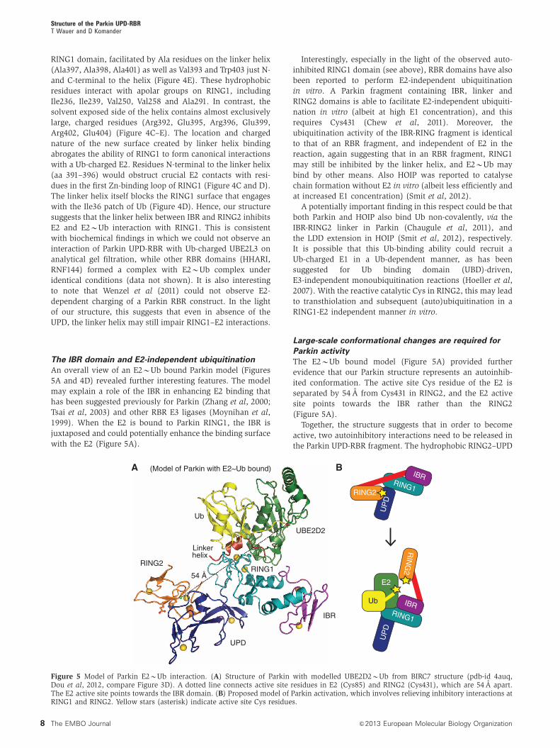
An overall view of an E2BUb bound Parkin model (Figures
5A and 4D) revealed further interesting features. The model
may explain a role of the IBR in enhancing E2 binding that
has been suggested previously for Parkin (Zhang et al, 2000;
Tsai et al, 2003) and other RBR E3 ligases (Moynihan et al,
1999). When the E2 is bound to Parkin RING1, the IBR is
juxtaposed and could potentially enhance the binding surface
with the E2 (Figure 5A).
Interestingly, especially in the light of the observed auto-
inhibited RING1 domain (see above), RBR domains have also
been reported to perform E2-independent ubiquitination
in vitro. A Parkin fragment containing IBR, linker and
RING2 domains is able to facilitate E2-independent ubiquiti-
nation in vitro (albeit at high E1 concentration), and this
requires Cys431 (Chew et al, 2011). Moreover, the
ubiquitination activity of the IBR-RING fragment is identical
to that of an RBR fragment, and independent of E2 in the
reaction, again suggesting that in an RBR fragment, RING1
may still be inhibited by the linker helix, and E2BUb may
bind by other means. Also HOIP was reported to catalyse
chain formation without E2 in vitro (albeit less efficiently and
at increased E1 concentration) (Smit et al, 2012).
A potentially important finding in this respect could be that
both Parkin and HOIP also bind Ub non-covalently, via the
IBR-RING2 linker in Parkin (Chaugule et al, 2011), and
the LDD extension in HOIP (Smit et al, 2012), respectively.
It is possible that this Ub-binding ability could recruit a
Ub-charged E1 in a Ub-dependent manner, as has been
suggested for Ub binding domain (UBD)-driven,
E3-independent monoubiquitination reactions (Hoeller et al,
2007). With the reactive catalytic Cys in RING2, this may lead
to transthiolation and subsequent (auto)ubiquitination in a
RING1-E2 independent manner in vitro.
Large-scale conformational changes are required for
Parkin activity
The E2BUb bound model (Figure 5A) provided further
evidence that our Parkin structure represents an autoinhib-
ited conformation. The active site Cys residue of the E2 is
separated by 54 A from Cys431 in RING2, and the E2 active
site points towards the IBR rather than the RING2
(Figure 5A).
Together, the structure suggests that in order to become
active, two autoinhibitory interactions need to be released in
the Parkin UPD-RBR fragment. The hydrophobic RING2–UPD
RING2
UPD
IBR
RING1
UBE2D2
Ub
Linkerhelix
54 Å
UP
D
RING1
IBR
RING2
UP
D
RING1
RIN
G2
E2
E2
Ub IBR
BA (Model of Parkin with E2~Ub bound)
Figure 5 Model of Parkin E2BUb interaction. (A) Structure of Parkin with modelled UBE2D2BUb from BIRC7 structure (pdb-id 4auq,Dou et al, 2012, compare Figure 3D). A dotted line connects active site residues in E2 (Cys85) and RING2 (Cys431), which are 54 A apart.The E2 active site points towards the IBR domain. (B) Proposed model of Parkin activation, which involves relieving inhibitory interactions atRING1 and RING2. Yellow stars (asterisk) indicate active site Cys residues.
Structure of the Parkin UPD-RBRT Wauer and D Komander
8 The EMBO Journal &2013 European Molecular Biology Organization

interface needs to open to enable access to Cys431, and the
linker helix must be released from RING1 to enable E2BUb
binding. This suggests that Parkin activation requires ‘open-
ing’ of interdomain contacts within the UPD-RBR itself
(Figure 5B).
Parkin mutations give insight into Parkin mechanism
Parkin is mutated in B50% of patients suffering from AR-JP,
as well as in a subgroup of patients that display sporadic
forms of PD. A large number of mutations generate deletions,
truncations and exon duplications, all of which lead to a loss
of function of Parkin (Corti et al, 2011; Walden and Martinez-
Torres, 2012). More interesting for studies on ligase function,
however, are missense mutations. We collated the informat-
ion available for Parkin mutations from the Leiden Open
Variation Database (LOVD; http://grenada.lumc.nl/LOVD2/
TPI/home.php?select_db=PARK2), the Parkinson Disease
mutation database (PDmutDB; (http://www.molgen.ua.ac.
be/PDmutDB/default.cfm?MT=0&ML=0&Page=Home)
and (Corti et al, 2011), and mapped mutations situated in
the crystallized construct onto our structure. Fifty-seven
annotated point mutations lie within our crystallized
construct (Supplementary Table I; Figure 6A).
We grouped the reported mutations by predicted effects
on Parkin folding/stability, catalytic mechanism, interface
formation, and mutations for which the effect cannot be
easily predicted from our structure. As expected, most
Parkin mutations were predicted to affect protein folding
and stability, such as in Zn-coordinating residues, which
are usually crucial for correct folding of Zn-binding
domains. Eleven of the thirty-two Zn-coordinating residues
are found mutated in patient samples (Figure 6B). In two
cases (R256C, R334C), mutation introduces additional Cys or
His residues next to a Zn-coordinating residue, and these
likely compete for Zn binding and disrupt the fold. In addi-
tion, amino-acid changes in the core of individual domains
could be predicted to disrupt domain folding, likely destabi-
lizing Parkin (Figure 6B). Several of the Zn-coordinating and
core mutants have been characterized in vitro, and all
showed decreased solubility (Gu et al, 2003; Sriram et al,
2005; Wang et al, 2005; Hampe et al, 2006) (Supplementary
Table I).
Mutations that could lead to mechanistic insights into
Parkin function are those that affect the catalytic mechanism.
In RING1, T240M/T240R in the first Zn-binding loop and
D243N alter the conserved E2 interface, and consistently,
Parkin T240R does not interact with UBE2L3 (Shimura et al,
2000). Some mutations in RING2 affect the catalytic triad
(C431F and intriguingly also E444Q). Interestingly, Parkin
C431F is less soluble, heavily ubiquitinated, but unable
to ubiquitinate substrates (Sriram et al, 2005). This was
interpreted as Parkin autoubiquitination, although it may be
B
UPD:C166Y (Zn2)C212G/Y (Zn1)H215Q (Zn1)P153R (Zn1 loop)T173M (Zn2 loop)
RING2:C418R (Zn1)C441R (Zn1)P437L (near Zn1)
IBR:R334C (Zn1)T351P (core)R366T/Q (interface)
RING1 (Zn):C238Q (Zn1)C253Y/W (Zn2)R256C (Zn2)H257R (Zn2)C289G (Zn2)
RING1 (core):T240R/M (Zn1 loop, E2)V258M (core)C268R (core helical)R275N/W/T (core helical)I298S/L (core)
A
C431F
RING2(C)
M458LE444Q M192L/V
S193I
S145NG430D
G429D
A230T
K161N
C
R234Q
A398T
R402C/H
A401D(Trp403)
A230T
D E
E2+++ – +++ ++
wt wt K161N
S145N
M19
2AF4
63A
R234Q
A398T
98-
38-anti-Parkin
(No
E3)
Figure 6 Mapping PD mutations onto Parkin structure. (A) Parkin is shown in grey with yellow Zn atoms. Residues affected by pointmutations are shown in ball-and-stick representation with orange carbon atoms. (B) As in (A), but only mutations of Zn-coordinating residues(orange) and core residues (yellow) predicted to affect folding of individual domains are shown and labelled. (C) Mutations of catalytic siteresidues (red carbon atoms) and of residues involved in the UPD: RING2 interface (blue carbon atoms). A230T mutation of RING1 may createan additional RING1: RING2 interface. (D) Residues in the linker helix and RING1-linker helix binding site. R402C or R402H may create an extraZn-coordinating residue that competes for binding to RING1 Zn2. See Supplementary Figure 3 for further mutations. (E) Activity assay forParkin mutants in the presence of E1, Ub, MgATP and with or without the E2 UBE2L3 as indicated (compare Figure 2E). Mutants M192A, F463A(at RING2: UPD interface) and A398T (in linker helix) were more active in autoubiquitination as compared to wt Parkin UPD-RBR. Mostmutants are impaired in solubility (perhaps indicating Parkin ‘opening’) and have to be expressed with a GST tag to achieve soluble protein.For protein input, compare Supplementary Figure 3.
Structure of the Parkin UPD-RBRT Wauer and D Komander
9&2013 European Molecular Biology Organization The EMBO Journal

possible that other E3 ligases in mammalian cells modify the
solubility-impaired protein.
Parkin mutations affect intrinsic domain interfaces
Of particular interest are mutations in the various domain
interfaces, since we speculate that opening of the interfaces
might regulate Parkin activity (Figure 5B). Indeed, two hot-
spots of mutations are apparent, one in the UPD–RING2
domain interface (Figure 6C), and another in the linker
helix interface with the RING1 domain (Figure 6D).
As discussed above, Ser145 is a central residue at the
UPD–RING2 interface, and mutation to Asn would likely
alter the interface, as would mutation of Met458 to Leu.
Similarly, the two Gly residues upstream of the catalytic
Cys431 are found mutated to Asp potentially disrupting the
interface with the UPD. Two further interesting mutations are
S193I and M192L/V. These UPD residues are not directly at
the interface, but stabilize the UPD loop that interacts with
the Gly429-Gly430-Cys431 loop (Figure 6C).
Even more striking are mutations in the linker helix–RING1
interface (Figure 6D). Two fully conserved Ala residues,
Ala398 and Ala401, are mutated in patients and the structure
suggests that larger residues cannot be accommodated.
It is intriguing that several patient mutations are found at
the UPD–RING2, and in the linker helix-RING1 interfaces.
Such mutations could stabilize the contact locking Parkin into
an autoinhibited state, or disrupt the interface, leading to
‘opening’ of the structure, which we predict to be required for
activation (Figure 5B). This could result in activation-inde-
pendent autoubiquitination, and Parkin turnover.
To test this, several Parkin mutants were generated in the
UPD-RBR fragment (Figure 6E; Supplementary Figure 3A).
It was immediately apparent that most mutants were im-
paired in their solubility when the GST tag was cleaved,
suggesting that Parkin mutants indeed expose hydrophobic
regions as would be predicted for ‘open’ domain interfaces.
Accordingly, activity assays were performed with GST-tagged
Parkin, and while some mutants (K161N, R234Q) showed
similar autoubiquitination to wild-type Parkin, interface mu-
tants M192A and F463A on either side of the UPD–RING2
interface, and linker helix mutation A398T showed increased
autoubiquitination activity (Figure 6E), which in cells may
lead to increased turnover.
In addition to the above, several not so easy to explain
mutations reside in the helical extension of the RING1
domain that may affect the interface with the IBR domain
(Supplementary Figure 3B). A hotspot of three mutations in
the unique b-hairpin extension of RING1 is fully solvent
exposed (Supplementary Figure 3B) and so is His200 at
the tip of the UPD (Supplementary Figure 3C). It is unclear
how these mutations disrupt Parkin function, but perhaps
they affect formation of Parkin complexes.
A putative phospho-peptide binding site in the UPD
Two patient mutations in the UPD, K161N and K211N, have
been studied extensively, and were found to be soluble,
expressed as well as wild-type Parkin (Sriram et al, 2005;
Hampe et al, 2006) and active in autoubiquitination both
in vitro and in cells (Hampe et al, 2006; Chew et al, 2011).
However, these mutants are disabled in complex formation
(Van Humbeeck et al, 2008), and do not localize to
mitochondria (Matsuda et al, 2010), suggesting that they are
functionally impaired.
We noticed that these residues are adjacent to each other
on the backside of the UPD, solvent exposed (Figure 7A;
Supplementary Figure 3B) and K161N had no impact
on autoubiquitination activity (Figure 6E). Interestingly,
together with nearby Arg163, they constitute a highly posi-
tively charged surface area that forms a pocket occupied by a
sulphate ion from the crystallization condition (Figure 7A
and B). There are several examples in which sulphates in
crystal structures can be indicative of phosphate binding sites
in proteins (e.g., Biondi et al, 2002). It is hence a possibility
that Lys161, Arg163 and Lys211 are involved in binding to
phosphorylated proteins.
This is interesting due to the role of the PINK1 kinase in
Parkin recruitment and regulation. PINK1 binds to the Parkin
UPD (Xiong et al, 2009) and requires autophosphorylation at
two sites to recruit Parkin to mitochondria (Okatsu et al,
2012). Moreover, PINK1 mediated phosphorylation targets
BA
K211N
K161N
(Arg163)
Sulphate
Positive charge Negative chargeApolar
Figure 7 A putative phospho-peptide binding site in the UPD. (A) Structural detail of the backside of the UPD (in relation to Figure 1B, also seeSupplementary Figure 3B), showing residues Lys161 and Lys211 that are mutated in PD (orange) and Arg163 (in grey). These residuescoordinate a sulphate ion from the crystallization mother liquor (shown in ball-and-stick, with green sulphur and red oxygen). (B) Electrostaticsurface potential of the UPD (generated with PyMol) showing the positively charged pocket in the UPD that is occupied by a sulphate ion.
Structure of the Parkin UPD-RBRT Wauer and D Komander
10 The EMBO Journal &2013 European Molecular Biology Organization

proteins to Parkin (Chen & Dorn 2013), and this could be
mediated by the putative phosphopeptide binding site. It will
be interesting to study whether Lys161/Lys211 are involved in
binding phosphorylated proteins.
Multi-step activation of Parkin
Based on our structure, we describe a two-fold autoinhibition
of Parkin, where the linker helix blocks E2 access to RING1,
and the UPD blocks access to Cys431 in RING2. It appears that
for full Parkin activation, both interactions have to be released,
and that the structure of Parkin has to open (Figure 5B).
Walden and colleagues recently revealed a further mechan-
ism of autoinhibition, in which the N-terminal Ubl of Parkin
interacts in cis with a region corresponding to the IBR-RING2
linker (Chaugule et al, 2011). Interestingly, the reported
region that binds the Ubl includes the linker helix, which
we here show binds and inhibits RING1. However, the
solvent-accessible side of the linker helix contains
numerous charged residues (Figure 4E), making this an
unlikely interaction surface.
In the light of our structure, this still does not explain why a
construct without the Ubl shows activity (Chaugule et al, 2011),
which is in fact contradictory to several previous reports
showing that a Parkin fragment lacking the Ubl is inactive
(Corti et al, 2003; Cha et al, 2005; Henn et al, 2007). Moreover,
we show here that at least autoubiquitination activity is
comparably weak (Figures 2E and 6E), even in the presence
of a GST tag that often acts as a Ub acceptor in autoubiquitina-
tion assays (Yang et al, 2005). In vitro GST-RING mediated
autoubiquitination reactions or chain assembly by HOIP
(Figure 3D) are significantly more processive.
The Ub ligase activity of Parkin has been intensely studied;
however, the results from such studies are hard to interpret
due to the various autoinhibitory mechanisms, and differ-
ences in performing the assays. Indeed, it is often not clear
whether the (auto)ubiquitination observed is due to RING1-
mediated discharging of an E2 onto the catalytic Cys431, or
directly onto another nearby Lys or Cys residue. This can
be partly addressed by using UBE2L3, which discharges
exclusively onto Cys (Wenzel et al, 2011), and such
autoubiquitination is Cys431 dependent (Figure 2E).
However, many experiments in the literature have been per-
formed using UBE2D (UbcH5), and since UBE2D discharges onto
both Cys and Lys residues, autoubiquitination and perceived
activity could be Cys431 independent.
As mentioned above, it is also not clear whether RING1 is
involved in the discharge of the E2, as RING1-independent
ubiquitination has been demonstrated (Chew et al, 2011).
The mentioned Ubl-binding region mapped by Chaugule et al
(2011) also binds Ub, and this might directly attract a
Ub-charged E2 enzyme to perform RING1-independent
ubiquitination (see above).
Hence, it seems essential to understand whether Parkin-
mediated (auto)ubiquitination is RING1 and RING2 depen-
dent. Our structural findings in combination with cell-free
Parkin activation assays (Lazarou et al, 2013) should allow
separation of the reported Parkin activities mediated by
RING1, IBR and RING2.
Conclusions
The E3 ligase Parkin is under intense investigation due to its
involvement in PD and important roles in mitochondrial
maintenance (Corti et al, 2011; Narendra et al, 2012).
The here reported structure of a Parkin fragment
comprising UPD and RBR domains explains many
biochemical findings, but also reveals several unexpected
features, and highlights the extensive regulation of Parkin,
since several mechanisms of autoinhibition seem to be in
place to keep Parkin inactive.
It is clear that our structure of Parkin has to be devoid of
catalytic activity, yet the fact that our tagged Parkin fragments
(which lack Ubl-mediated autoinhibition) show (weak) cata-
lytic activity, and the notion that large conformational
changes are required for activity, indicates a much more
dynamic interplay between Parkin domains. It is clear that
our structure is just a first step to understanding Parkin
activation, and future work should focus on generating active
protein, and activated structures of Parkin.
A large fraction of the loss-of-function Parkin mutations
found in PD patients destabilize the protein by affecting
folding of the complicated Zn-bound architecture. More
interesting are those mutations for which loss-of-function
cannot be immediately attributed to decreased Parkin stabi-
lity, and we find several of such instances, most notably in a
to-be-confirmed docking site for phospho-peptides in the
UPD. An interesting case is provided further by mutations
that would affect inter-domain interactions. Our model of
Parkin activation and biochemical data indicate that domain-
opening mutations might be ‘gain-of-function’ with respect to
ligase activity. Since Parkin autoubiquitinates, this may lead
to increased turnover, and lower steady state protein levels.
Another explanation would be that aberrant opening of the
Parkin interfaces, that is, in the absence of an activating
binding partner or bound substrate, would simply expose
hydrophobic surfaces that lead to Parkin aggregation. Indeed,
our preliminary experiments suggest that ‘open’ Parkin is
highly insoluble when expressed in bacteria.
The key question that remains is how Parkin is activated,
for instance by PINK1 once it arrives at mitochondria, as
Parkin activators must release a variety of safety belts that
keep Parkin inactive. Moreover, the active form of Parkin may
be oligomeric, as recently reported (Van Humbeeck et al,
2008; Lazarou et al, 2013), and we would expect this to be
distinct from the autoinhibited form reported here. Under-
standing oligomeric status and composition of the complexes
containing active Parkin should help to further understand
the cellular roles of Parkin in mitophagy, and its patho-
physiological roles in neurodegenerative disease.
Materials and methods
Molecular biologycDNA for full-length human Parkin was obtained by gene synthesis(Genscript) and codon-optimized for expression in E. coli. TheParkin UPD-RBR, aa 137–465 was cloned into pOPINK (Berrowet al, 2007), which contains an N-terminal 3C-protease-cleavableGST tag, using the Infusion HD Kit (Clontech). The followingprimers were used: Parkin 137–465 fwd 50-AAGTTCTGTTTCAGGGCCCGGCTGGTCGTTCGATCTACAACAGCTTCTATGTGTAC-30 andParkin 137–465 rev 50-ATGGTCTAGAAAGCTTTACACATCAAACCAGTGGTCACCCATACACACAC-30. The RBR and the C-terminal regionof HOIP (residues 699–1072) (Smit et al, 2012; Stieglitz et al, 2012)were cloned into pOPINK with the Infusion HD Kit (Clontech). HOIPand Parkin mutants were generated in pOPINK by site-directedmutagenesis using the Quikchange method with KOD HotStartDNA polymerase according to the manufacturer’s protocol. Allconstructs were confirmed by DNA sequencing.
Structure of the Parkin UPD-RBRT Wauer and D Komander
11&2013 European Molecular Biology Organization The EMBO Journal

Protein expression and purificationParkin UPD-RBR was expressed in Rosetta2 pLacI cells, which weregrown to an OD600 of 1.0 at 371C. In all cases 200 mM zinc chloridewas added upon induction with 50 mM IPTG, and cells were grownat 161C for 12 h. The cells were lysed by sonication in 270 mMsucrose, 10 mM glycerol 2-phosphate disodium, 50 mM NaF, 14 mMb-mercaptoethanol, 50 mM Tris (pH 8.0). Lysate was cleared bycentrifugation (45 000 g, 30 min, 41C), applied to GlutathioneSepharose 4B beads (GE Healthcare, 0.5 ml/l of culture), andincubated under agitation for 1 h at 41C. Beads were washed withhigh salt buffer (500 mM NaCl, 10 mM DTT, 25 mM Tris (pH 8.5))and buffer A (200 mM NaCl, 10 mM DTT, 25 mM Tris (pH 8.5)).GST-tagged Parkin constructs used for biochemical analysis waseluted by incubating the beads with buffer A containing 30 mMglutathione for 0.5 h at 41C. Untagged Parkin used for crystallizationwas cleaved on the beads by incubating with GST-3C protease for12h at 41C. After diluting the buffer to 75mM NaCl, 10 mM DTT,25mM Tris (pH 8.5), the protein was applied to anion exchange(RESOURCE Q, GE Life Sciences) and eluted with a linear gradientof 75–600mM NaCl in 10 mM DTT, 25 mM Tris (pH 8.5). Proteincontaining fractions were pooled, concentrated and applied to gelfiltration (Superdex 75, GE Life Sciences) in Buffer A. The proteineluted as a single peak at the size expected for a monomer, and wasconcentrated using a 3-kDa MWCO spin concentrator (VWR) and flashfrozen in liquid nitrogen.
HOIP 699-1072 was expressed and purified as described forParkin by cleaving the protein from Glutathione Sepharose 4Bbeads and applying the eluted protein to gel filtration.
CrystallizationHuman Parkin UPD-RBR was crystallized at a concentration ofB4 mg/ml using vapour diffusion. Initial screening in nano-litrevolume gave two hits in similar conditions that were optimized toobtain diffraction quality crystals. Final crystals were grown frommixing 0.5ml protein with 0.5ml mother liquor (1.6 M lithiumsulphate, 10 mM magnesium chloride, 50 mM MES (pH 5.4)).Prior to vitrification in a nitrogen cryostream, crystals were brieflysoaked in mother liquor containing 15% glycerol.
Data collection and structure determinationDiffraction data were collected the Diamond Light Source (Harwell,UK), beamline I-04. Crystals belonged to space group H32 contain-ing one molecule per asymmetric unit, and diffracted to a maximumresolution of 2.25 A (at 100 K, l¼ 1.0000 A). For phasing, a highlyredundant data set to 3.5 A was collected at the Zn peak wavelength(at 100 K, l¼ 1.28310 A), which allowed phasing by single anom-alous dispersion due to the eight bound Zn atoms, using the SHELXpackage (Zheng et al, 2000; Rodrıguez et al, 2009). Automatedmodel building in ArpWarp (Langer et al, 2008; Dou et al, 2012)was followed by rounds of manual building in coot (Emsley et al,2010; Dou et al, 2012) and refinement in Phenix (Adams et al, 2011).Final statistics are shown in Table I. There are no Ramachandranoutliers in the structure.
Modification with Ub-based suicide probesUbiquitin suicide probes UbC2Cl, UbC3Cl and UbC3Br were gener-ated as described previously (Borodovsky et al, 2002; Akutsu et al,2011). In brief, 200 ml Ub-thioester was mixed with 40 mg 2-bromoethylamine hydrochloride solved in 200 ml phosphate-buffersaline (PBS, pH 4.8) and the reaction was initiated by adding 80ml2 M NaOH. After 15 min on ice, the probes were dialysed against200 mM NaCl, 25 mM Tris pH 8.5 using Slide-A-Lyzer DialysisCassettes (Thermo Scientific). Ub-VS and Ub-VME were obtainedfrom Boston Biochem, dissolved in 0.08% TFA.
For time-course experiments in Supplementary Figure 2B, 10 ml ofprotein (0.4 mg/ml) in buffer A was mixed with 4 ml probe (0.83 mg/ml). The reaction was incubated at room temperature and stoppedat the indicated time points by adding 4� LDS sample buffer(Invitrogen). In all, 7ml of sample was resolved on NuPAGE 4–12% Bis-Tris gels (Invitrogen).
The reaction of HOIP mutants with UbVME was performed bymixing 0.28 mg/ml of protein with 0.2 mg/ml of the probe inreaction buffer (50 mM Tris (pH 8.5), 200 mM NaCl, 10 mM DTT).The reaction took place for 1 h at 371C and was quenched by adding4� LDS sample buffer (Invitrogen). In all, 7 ml of sample wasloaded on NuPAGE 4–12% Bis-Tris gels (Invitrogen).
Parkin mutants were reacted with UbVS by mixing 0.14 mg/mlprotein with 0.1 mg/ml probe, in Parkin reaction buffer (50 mM Tris(pH 7.4), 200 mM NaCl, 10 mM DTT). The assay was performed at371C and quenched after 30 min by adding 4� LDS sample buffer(Invitrogen). In all, 4ml of sample was applied on NuPAGE 4–12%Bis-Tris gels (Invitrogen). All gels were stained with Instant BlueSafeStain (Expedeon).
Ubiquitination assayUbiquitination assays were performed by mixing 100 nM E1, 6mMUBE2L3 (UBCH7), 4mM GST-Parkin or HOIP, 0.25 mg/mlUbiquitin and 10 mM ATP in ubiquitination buffer (40 mM Tris pH7.5, 10 mM MgCl2, 0.6 mM DTT). The reaction took place for 1 h at371C and was quenched by adding 4� LDS sample buffer(Invitrogen). The sample was further diluted 1:30 and resolved onNuPAGE 4–12% Bis-Tris gels (Invitrogen). After western blotting,membranes were incubated with anti-Parkin (anti-PRK8, fromAbcam), anti-Ubiquitin (FK2, from Millipore) or anti-HOIP(Sigma), and analysed by enhanced chemiluminescence (ECLPrime, Roche).
Accession numbersCoordinates and structure factors have been deposited with theprotein data bank, accession code 4bm9.
Note added in proofOur data is consistent with recent structural work on rat Parkin,which additionally resolves the position of the Ubl domain (Trempeet al, 2013).
Supplementary dataSupplementary data are available at The EMBO Journal Online(http://www.embojournal.org).
Acknowledgements
We are grateful to Francesco Melandri and Boston Biochem forproviding Ub-VS and Ub-VME probes. Moreover we would like tothank members of the Komander lab for reagents and helpfuldiscussions, and Paul Elliott for help with crystallization and datacollection. Crystallographic data were collected at the DiamondLight Source, beamline I-04. This work was supported by theMedical Research Council (U105192732), the European ResearchCouncil (309756), the Lister Institute for Preventive Medicine, andthe EMBO Young Investigator Programme.
Author contributions: TW and DK designed and performedexperiments, analysed the data and wrote the manuscript.
Conflict of interest
The authors declare that they have no conflict of interest.
ReferencesAdams PD, Afonine PV, Bunkoczi G, Chen VB, Echols N,
Headd JJ, Hung L-W, Jain S, Kapral GJ, GrosseKunstleve RW, McCoy AJ, Moriarty NW, Oeffner RD,Read RJ, Richardson DC, Richardson JS, Terwilliger TC,Zwart PH (2011) The Phenix software for automateddetermination of macromolecular structures. Methods 55:94–106
Aguilera M, Oliveros M, Martınez-Padron M, Barbas JA, Ferrus A(2000) Ariadne-1: a vital Drosophila gene is required indevelopment and defines a new conserved family of ring-fingerproteins. Genetics 155: 1231–1244
Akutsu M, Ye Y, Virdee S, Chin JW, Komander D (2011) Molecularbasis for ubiquitin and ISG15 cross-reactivity in viral ovariantumor domains. Proc Natl Acad Sci 108: 2228–2233
Structure of the Parkin UPD-RBRT Wauer and D Komander
12 The EMBO Journal &2013 European Molecular Biology Organization

Beasley SA, Hristova VA, Shaw GS (2007) Structure of the Parkinin-between-ring domain provides insights for E3-ligase dysfunc-tion in autosomal recessive Parkinson’s disease. Proc Natl AcadSci USA 104: 3095–3100
Berrow NS, Alderton D, Sainbury S, Nettkeship J, Assenberg R,Rahman N, Stuart DI, Owens RJ (2007) A versatile ligation-independent cloning method suitable for high-throughput expres-sion screening applications. Nucleic acids research 35, e45
Biondi RM, Komander D, Thomas CC, Lizcano JM, Deak M, AlessiDR, van Aalten DM (2002) High resolution crystal structure of thehuman PDK1 catalytic domain defines the regulatory phospho-peptide docking site. EMBO J 21: 4219–4248
Bonifati V, Rizzu P, van Baren MJ, Schaap O, Breedveld GJ,Krieger E, Dekker MCJ, Squitieri F, Ibanez P, Joosse M,van Dongen JW, Vanacore N, van Swieten JC, Brice A, Meco G,van Duijn CM, Oostra BA, Heutink P (2003) Mutations in the DJ-1gene associated with autosomal recessive early-onset parkinson-ism. Science 299: 256–259
Borodovsky A, Ovaa H, Kolli N, Gan-Erdene T, Wilkinson KD,Ploegh HL, Kessler BM (2002) Chemistry-based functional pro-teomics reveals novel members of the deubiquitinating enzymefamily. Chem Biol 9: 1149–1159
Capili AD, Edghill EL, Wu K, Borden KLB (2004) Structure of theC-terminal RING finger from a RING-IBR-RING/TRIAD motifreveals a novel zinc-binding domain distinct from a RING.J Mol Biol 340: 1117–1129
Cha G-H, Kim S, Park J, Lee E, Kim M, Lee SB, Kim J-M, Chung J,Cho KS (2005) Parkin negatively regulates JNK pathway in thedopaminergic neurons of Drosophila. Proc Natl Acad Sci USA 102:10345–10350
Chaugule VK, Burchell L, Barber KR, Sidhu A, Leslie SJ, Shaw GS,Walden H (2011) Autoregulation of Parkin activity through itsubiquitin-like domain. EMBO J 30: 2853–2867
Chen Y, Dorn GW (2013) PINK1-phosphorylated mitofusin 2 is aParkin receptor for culling damaged mitochondria. Science 340:471–475
Chew KCM, Matsuda N, Saisho K, Lim GGY, Chai C, Tan H-M,Tanaka K, Lim KL (2011) Parkin mediates apparent E2-indepen-dent monoubiquitination in vitro and contains an intrinsic activ-ity that catalyzes polyubiquitination. PLoS ONE 6: e19720
Corti O, Hampe C, Koutnikova H, Darios F, Jacquier S, Prigent A,Robinson J-C, Pradier L, Ruberg M, Mirande M, Hirsch E, RooneyT, Fournier A, Brice A (2003) The p38 subunit of the aminoacyl-tRNA synthetase complex is a Parkin substrate: linking proteinbiosynthesis and neurodegeneration. Hum Mol Genet 12: 1427–1437
Corti O, Lesage S, Brice A (2011) What genetics tells us about thecauses and mechanisms of Parkinson’s disease. Physiol Rev 91:1161–1218
Dou H, Buetow L, Sibbet GJ, Cameron K, Huang DT (2012) BIRC7-E2 ubiquitin conjugate structure reveals the mechanism of ubi-quitin transfer by a RING dimer. Nat Struct Mol Biol 19: 876–883
Dye BT, Schulman BA (2007) Structural mechanisms underlyingposttranslational modification by ubiquitin-like proteins. AnnuRev Biophys Biomol Struct 36: 131–150
Eisenhaber B, Chumak N, Eisenhaber F, Hauser M-T (2007) The ringbetween ring fingers (RBR) protein family. Genome Biol 8: 209
Emsley P, Lohkamp B, Scott WG, Cowtan K (2010) Features anddevelopment of Coot. Acta Crystallogr D Biol Crystallogr 66:486–501
Geisler S, Holmstrom KM, Skujat D, Fiesel FC, Rothfuss OC,Kahle PJ, Springer W (2010) PINK1/Parkin-mediated mitophagy isdependent on VDAC1 and p62/SQSTM1. Nat Cell Biol 12: 119–131
Goedert M, Spillantini MG, Del Tredici K, Braak H (2013) 100 yearsof Lewy pathology. Nat Rev Neurol 9: 13–24
Gu W-J, Corti O, Araujo F, Hampe C, Jacquier S, Lucking CB, AbbasN, Duyckaerts C, Rooney T, Pradier L, Ruberg M, Brice A (2003)The C289G and C418R missense mutations cause rapid seques-tration of human Parkin into insoluble aggregates. Neurobiol Dis14: 357–364
Hampe C, Ardila-Osorio H, Fournier M, Brice A, Corti O (2006)Biochemical analysis of Parkinson’s disease-causing variants ofParkin, an E3 ubiquitin-protein ligase with monoubiquitylationcapacity. Hum Mol Genet 15: 2059–2075
Henn IH, Bouman L, Schlehe JS, Schlierf A, Schramm JE, WegenerE, Nakaso K, Culmsee C, Berninger B, Krappmann D, Tatzelt J,Winklhofer KF (2007) Parkin mediates neuroprotection through
activation of IkappaB kinase/nuclear factor-kappaB signaling.J. Neurosci 27: 1868–1878
Hershko A, Ciechanover A (1998) The ubiquitin system. Annu RevBiochem 67: 425–479
Hoeller D, Hecker C-M, Wagner S, Rogov V, Dotsch V, Dikic I (2007)E3-independent monoubiquitination of ubiquitin-bindingproteins. Mol Cell 26: 891–898
Hristova VA, Beasley SA, Rylett RJ, Shaw GS (2009) Identification ofa novel Zn2þ -binding domain in the autosomal recessivejuvenile Parkinson-related E3 ligase parkin. J Biol Chem 284:14978–14986
Kirisako T, Kamei K, Murata S, Kato M, Fukumoto H, Kanie M, SanoS, Tokunaga F, Tanaka K, Iwai K (2006) A ubiquitin ligasecomplex assembles linear polyubiquitin chains. EMBO J 25:4877–4887
Kitada T, Asakawa S, Hattori N, Matsumine H, Yamamura Y,Minoshima S, Yokochi M, Mizuno Y, Shimizu N (1998)Mutations in the parkin gene cause autosomal recessive juvenileparkinsonism. Nature 392: 605–608
Komander D, Rape M (2012) The ubiquitin code. Annu Rev Biochem81: 203–229
Komander D, Clague MJ, Urbe S (2009) Breaking the chains:structure and function of the deubiquitinases. Nat Rev Mol CellBiol 10: 550–563
Kondapalli C, Kazlauskaite A, Zhang N, Woodroof HI,Campbell DG, Gourlay R, Burchell L, Walden H, Macartney TJ,Deak M, Knebel A, Alessi DR, Muqit MMK (2012) PINK1 isactivated by mitochondrial membrane potential depolarizationand stimulates Parkin E3 ligase activity by phosphorylatingSerine 65. Open Biol 2: 120080
Langer G, Cohen SX, Lamzin VS, Perrakis A (2008) Automatedmacromolecular model building for X-ray crystallography usingARP/wARP version 7. Nat Protoc 3: 1171–1179
Lazarou M, Narendra DP, Jin SM, Tekle E, Banerjee S, Youle RJ(2013) PINK1 drives Parkin self-association and HECT-like E3activity upstream of mitochondrial binding. J Cell Biol 200:163–172
Lima CD, Schulman BA (2012) Structural biology: a protein engage-ment RING. Nature 489: 43–44
Marın I (2009) RBR ubiquitin ligases: diversification andstreamlining in animal lineages. J Mol Evol 69: 54–64
Matsuda N, Kitami T, Suzuki T, Mizuno Y, Hattori N, Tanaka K(2006) Diverse effects of pathogenic mutations of Parkin thatcatalyze multiple monoubiquitylation in vitro. J Biol Chem 281:3204–3209
Matsuda N, Sato S, Shiba K, Okatsu K, Saisho K, Gautier CA, Sou Y-S, Saiki S, Kawajiri S, Sato F, Kimura M, Komatsu M, Hattori N,Tanaka K (2010) PINK1 stabilized by mitochondrial depolariza-tion recruits Parkin to damaged mitochondria and activates latentParkin for mitophagy. J Cell Biol 189: 211–221
Moynihan TP, Ardley HC, Nuber U, Rose SA, Jones PF, MarkhamAF, Scheffner M, Robinson PA (1999) The ubiquitin-conjugatingenzymes UbcH7 and UbcH8 interact with RING finger/IBRmotif-containing domains of HHARI and H7-AP1. J Biol Chem274: 30963–30968
Muller-Rischart AK, Pilsl A, Beaudette P, Patra M, Hadian K,Funke M, Peis R, Deinlein A, Schweimer C, Kuhn PH,Linchtenthaler SF, Motori E, Hrelia S, Wurst W, Trumbach D,Langer T, Krappmann D, Dittmar G, Tatzelt J, Winklhofer KF(2013) The E3 ligase parkin maintains mitochondrial integrityby increasing linear ubiquitination of NEMO. Mol Cell 149:908–921
Narendra D, Walker JE, Youle R (2012) Mitochondrial qualitycontrol mediated by PINK1 and Parkin: links to parkinsonism.Cold Spring Harb Perspect Biol 4: pii: a011338
Narendra DP, Jin SM, Tanaka A, Suen D-F, Gautier CA, Shen J,Cookson MR, Youle RJ (2010) PINK1 is selectively stabilizedon impaired mitochondria to activate Parkin. PLoS Biol 8:e1000298
Okatsu K, Oka T, Iguchi M, Imamura K, Kosako H, Tani N,Kimura M, Go E, Koyano F, Funayama M, Shiba-Fukushima K,Sato S, Shimizu H, Fukunaga Y, Taniguchi H, Komatsu M,Hattori N, Mihara K, Tanaka K, Matsuda N (2012) PINK1autophosphorylation upon membrane potential dissipation isessential for Parkin recruitment to damaged mitochondria. NatCommun 3: 1016
Structure of the Parkin UPD-RBRT Wauer and D Komander
13&2013 European Molecular Biology Organization The EMBO Journal

Plechanovova A, Jaffray EG, Tatham MH, Naismith JH, Hay RT(2012) Structure of a RING E3 ligase and ubiquitin-loaded E2primed for catalysis. Nature 489: 115–120
Poole AC, Thomas RE, Yu S, Vincow ES, Pallanck L (2010) Themitochondrial fusion-promoting factor mitofusin is a substrate ofthe PINK1/parkin pathway. PLoS ONE 5: e10054
Poulogiannis G, McIntyre RE, Dimitriadi M, Apps JR, Wilson CH,Ichimura K, Luo F, Cantley LC, Wyllie AH, Adams DJ, Arends MJ(2010) PARK2 deletions occur frequently in sporadic colorectalcancer and accelerate adenoma development in Apc mutant mice.Proc Natl Acad Sci 107: 15145–15150
Pruneda JN, Littlefield PJ, Soss SE, Nordquist KA, Chazin WJ,Brzovic PS, Klevit RE (2012) Structure of an E3:E2BUb complexreveals an allosteric mechanism shared among RING/U-boxligases. Mol Cell 47: 933–942
Rawal N, Corti O, Sacchetti P, Ardilla-Osorio H, Sehat B, Brice A,Arenas E (2009) Parkin protects dopaminergic neurons fromexcessive Wnt/beta-catenin signaling. Biochem Biophys ResCommun 388: 473–478
Rodrıguez D, Grosse C, Himmel S, Gonzalez C, de Ilarduya I,Becker S, Sheldrick G, Uson I (2009) Crystallographic ab initioprotein structure solution below atomic resolution. Nat Methods6: 651–653
Sarraf SA, Raman M, Guarani-Pereira V, Sowa ME, Huttlin EL, GygiSP, Harper JW (2013) Landscape of the PARKIN-dependentubiquitylome in response to mitochondrial depolarization.Nature 496: 372–376
Schlehe JS, Lutz AK, Pilsl A, Lammermann K, Grgur K, Henn IH,Tatzelt J, Winklhofer KF (2008) Aberrant folding of pathogenicParkin mutants: aggregation versus degradation. J Biol Chem 283:13771–13779
Shimura H, Hattori N, Kubo SI, Mizuno Y, Asakawa S, Minoshima S,Shimizu N, Iwai K, Chiba T, Tanaka K, Suzuki T (2000) FamilialParkinson disease gene product, parkin, is a ubiquitin-proteinligase. Nat Genet 25: 302–305
Smit JJ, Monteferrario D, Noordermeer SM, van Dijk WJ, van derReijden BA, Sixma TK (2012) The E3 ligase HOIP specifies linearubiquitin chain assembly through its RING-IBR-RING domain andthe unique LDD extension. EMBO J 31: 3833–3844
Sriram SR, Li X, Ko HS, Chung KKK, Wong E, Lim KL,Dawson VL, Dawson TM (2005) Familial-associated mutationsdifferentially disrupt the solubility, localization, bindingand ubiquitination properties of parkin. Hum Mol Genet 14:2571–2586
Stieglitz B, Morris-Davies AC, Koliopoulos MG, Christodoulou E,Rittinger K (2012) LUBAC synthesizes linear ubiquitin chains viaa thioester intermediate. EMBO Rep 13: 840–846
Sun M, Latourelle JC, Wooten GF, Lew MF, Klein C, Shill HA, GolbeLI, Mark MH, Racette BA, Perlmutter JS, Parsian A, Guttman M,Nicholson G, Xu G, Wilk JB, Saint-Hilaire MH, DeStefano AL,Prakash R, Williamson S, Suchowersky O et al. (2006) Influenceof heterozygosity for parkin mutation on onset age infamilial Parkinson disease: the GenePD study. Arch Neurol 63:826–832
Tanaka A, Cleland MM, Xu S, Narendra DP, Suen D-F, Karbowski M,Youle RJ (2010) Proteasome and p97 mediate mitophagy anddegradation of mitofusins induced by Parkin. J Cell Biol 191:1367–1380
Trempe J-F, Sauve V, Grenier K, Seirafi M, Tang MY, Menade M,Al-Abdul-Wahid S, Krett J, Wong K, Kozlov G, Nagar B, Fon EA,Geherng K (2013) Structure of Parkin Reveals Mechanisms forUbiquitin Ligase Activation. Science. (advance online publication9 May 2013; doi:10.1126/science.1237908)
Tsai YC, Fishman PS, Thakor NV, Oyler GA (2003) Parkin facilitatesthe elimination of expanded polyglutamine proteins andleads to preservation of proteasome function. J Biol Chem 278:22044–22055
Valente EM, Abou-Sleiman PM, Caputo V, Muqit MMK, Harvey K,Gispert S, Ali Z, Del Turco D, Bentivoglio AR, Healy DG,Albanese A, Nussbaum R, Gonzalez-Maldonado R, Deller T,Salvi S, Cortelli P, Gilks WP, Latchman DS, Harvey RJ,
Dallapiccola B et al. (2004) Hereditary early-onset Parkinson’sdisease caused by mutations in PINK1. Science 304: 1158–1160
Van Humbeeck C, Waelkens E, Corti O, Brice A, Vandenberghe W(2008) Parkin occurs in a stable, non-covalent, approximately110-kDa complex in brain. Eur J Neurosci 27: 284–293
Veeriah S, Taylor BS, Meng S, Fang F, Yilmaz E, Vivanco I,Janakiraman M, Schultz N, Hanrahan AJ, Pao W, Ladanyi M,Sander C, Heguy A, Holland EC, Paty PB, Mischel PS, Liau L,Cloughesy TF, Mellinghoff IK, Solit DB et al. (2010) Somaticmutations of the Parkinson’s disease-associated gene PARK2 inglioblastoma and other human malignancies. Nat Genet 42:77–82
Vives-Bauza C, Zhou C, Huang Y, Cui M, de Vries RLA, Kim J, MayJ, Tocilescu MA, Liu W, Ko HS, Magrane J, Moore DJ, Dawson VL,Grailhe R, Dawson TM, Li C, Tieu K, Przedborski S (2010) PINK1-dependent recruitment of Parkin to mitochondria in mitophagy.Proc Natl Acad Sci 107: 378–383
Walden H, Martinez-Torres RJ (2012) Regulation of Parkin E3ubiquitin ligase activity. Cell Mol Life Sci 69: 3053–3067
Wang C, Tan JMM, Ho MWL, Zaiden N, Wong SH, Chew CLC,Eng PW, Lim TM, Dawson TM, Lim KL (2005) Alterations inthe solubility and intracellular localization of parkin by severalfamilial Parkinson’s disease-linked point mutations. J Neurochem93: 422–431
Wang X, Winter D, Ashrafi G, Schlehe J, Wong YL, Selkoe D, Rice S,Steen J, LaVoie MJ, Schwarz TL (2011) PINK1 and Parkin targetMiro for phosphorylation and degradation to arrest mitochondrialmotility. Cell 147: 893–906
Wang Y, Clark LN, Louis ED, Mejia-Santana H, Harris J, Cote LJ,Waters C, Andrews H, Ford B, Frucht S, Fahn S, Ottman R,Rabinowitz D, Marder K (2008) Risk of Parkinson disease incarriers of parkin mutations: estimation using the kin-cohortmethod. Arch Neurol 65: 467–474
Wenzel DM, Klevit RE (2012) Following Ariadne’s thread: a newperspective on RBR ubiquitin ligases. BMC Biol 10: 24
Wenzel DM, Lissounov A, Brzovic PS, Klevit RE (2011) UBCH7reactivity profile reveals parkin and HHARI to be RING/HECThybrids. Nature 474: 105–108
Wong ESP, Tan JMM, Wang C, Zhang Z, Tay S-P, Zaiden N,Ko HS, Dawson VL, Dawson TM, Lim KL (2007) Relativesensitivity of parkin and other cysteine-containing enzymesto stress-induced solubility alterations. J Biol Chem 282:12310–12318
Xiong H, Wang D, Chen L, Choo YS, Ma H, Tang C, Xia K, Jiang W,Ronai Z, Zhuang X, Zhang Z (2009) Parkin, PINK1, and DJ-1 forma ubiquitin E3 ligase complex promoting unfolded proteindegradation. J Clin Invest 119: 650–660
Yang Y, Lorick KL, Jensen JP, Weissman AM (2005) Expressionand evaluation of RING finger proteins. Methods Enzymol 398:103–112
Youle RJ, Narendra DP (2011) Mechanisms of mitophagy. Nat RevMol Cell Biol 12: 9–14
Zhang Y, Gao J, Chung KK, Huang H, Dawson VL, Dawson TM(2000) Parkin functions as an E2-dependent ubiquitin- proteinligase and promotes the degradation of the synaptic vesicle-associated protein, CDCrel-1. Proc Natl Acad Sci USA 97:13354–13359
Zheng N, Wang P, Jeffrey PD, Pavletich NP (2000) Structure of ac-Cbl-UbcH7 complex: RING domain function in ubiquitin-pro-tein ligases. Cell 102: 533–539
Ziviani E, Tao RN, Whitworth AJ (2010) Drosophila parkin requiresPINK1 for mitochondrial translocation and ubiquitinates mitofu-sin. Proc Natl Acad Sci 107: 5018–5023
The EMBO Journal is published by NaturePublishing Group on behalf of the European
Molecular Biology Organization. This article is licensedunder a Creative Commons Attribution 3.0 UnportedLicence. To view a copy of this licence visit http://creativecommons.org/licenses/by/3.0/.
Structure of the Parkin UPD-RBRT Wauer and D Komander
14 The EMBO Journal &2013 European Molecular Biology Organization

![Multitasking guardian of mitochondrial quality: Parkin …...the autoubiquitination activity of Parkin [41]. Involvement of Parkin in mitochondrial processes As an E3 ligase, Parkin](https://img.pdfslide.us/doc/110x75/60ff3ba3c386cc67f77a5535/multitasking-guardian-of-mitochondrial-quality-parkin-the-autoubiquitination.jpg)
![PINK1 import regulation; a fine system to convey ......mitochondria, and the E3 ligase activity of Parkin is acti-vated by binding to phospho-ubiquitin [20, 21]. PINK1 also phosphorylates](https://img.pdfslide.us/doc/110x75/60ff3ba3c386cc67f77a5536/pink1-import-regulation-a-fine-system-to-convey-mitochondria-and-the-e3.jpg)